SCIENTIFIC OPINION ADOPTED: 18 May 2017 doi: 10.2903/j.efsa.2017.4862 Guidance on allergenicity assessment of genetically modified plants EFSA Panel on Genetically Modified Organisms (GMO), Hanspeter Naegeli, Andrew Nicholas Birch, Josep Casacuberta, Adinda De Schrijver, Mikolaj Antoni Gralak, Philippe Guerche, Huw Jones, Barbara Manachini, Antoine Mess ean, Elsa Ebbesen Nielsen, Fabien Nogu e, Christophe Robaglia, Nils Rostoks, Jeremy Sweet, Christoph Tebbe, Francesco Visioli, Jean-Michel Wal, Philippe Eigenmann, Michelle Epstein, Karin Hoffmann-Sommergruber, Frits Koning, Martinus Lovik, Clare Mills, Francisco Javier Moreno, Henk van Loveren, Regina Selb and Antonio Fernandez Dumont Abstract This document provides supplementary guidance on specific topics for the allergenicity risk assessment of genetically modified plants. In particular, it supplements general recommendations outlined in previous EFSA GMO Panel guidelines and Implementing Regulation (EU) No 503/2013. The topics addressed are non-IgE-mediated adverse immune reactions to foods, in vitro protein digestibility tests and endogenous allergenicity. New scientific and regulatory developments regarding these three topics are described in this document. Considerations on the practical implementation of those developments in the risk assessment of genetically modified plants are discussed and recommended, where appropriate. © 2017 European Food Safety Authority. EFSA Journal published by John Wiley and Sons Ltd on behalf of European Food Safety Authority. Keywords: guidance, allergenicity assessment, newly expressed proteins, endogenous allergenicity, GMO Requestor: EFSA Question number: EFSA-Q-2014-00547 Correspondence: [email protected] EFSA Journal 2017;15(6):4862 www.efsa.europa.eu/efsajournal

Welcome message from author
This document is posted to help you gain knowledge. Please leave a comment to let me know what you think about it! Share it to your friends and learn new things together.
Transcript
SCIENTIFIC OPINION
ADOPTED: 18 May 2017
doi: 10.2903/j.efsa.2017.4862
Guidance on allergenicity assessment of geneticallymodified plants
EFSA Panel on Genetically Modified Organisms (GMO),Hanspeter Naegeli, Andrew Nicholas Birch, Josep Casacuberta, Adinda De Schrijver,
Mikolaj Antoni Gralak, Philippe Guerche, Huw Jones, Barbara Manachini, Antoine Mess�ean,Elsa Ebbesen Nielsen, Fabien Nogu�e, Christophe Robaglia, Nils Rostoks, Jeremy Sweet,
Christoph Tebbe, Francesco Visioli, Jean-Michel Wal, Philippe Eigenmann, Michelle Epstein,Karin Hoffmann-Sommergruber, Frits Koning, Martinus Lovik, Clare Mills,
Francisco Javier Moreno, Henk van Loveren, Regina Selb and Antonio Fernandez Dumont
Abstract
This document provides supplementary guidance on specific topics for the allergenicity risk assessmentof genetically modified plants. In particular, it supplements general recommendations outlined inprevious EFSA GMO Panel guidelines and Implementing Regulation (EU) No 503/2013. The topicsaddressed are non-IgE-mediated adverse immune reactions to foods, in vitro protein digestibility testsand endogenous allergenicity. New scientific and regulatory developments regarding these three topicsare described in this document. Considerations on the practical implementation of those developmentsin the risk assessment of genetically modified plants are discussed and recommended, whereappropriate.
© 2017 European Food Safety Authority. EFSA Journal published by John Wiley and Sons Ltd on behalfof European Food Safety Authority.
Keywords: guidance, allergenicity assessment, newly expressed proteins, endogenous allergenicity,GMO
Requestor: EFSA
Question number: EFSA-Q-2014-00547
Correspondence: [email protected]
EFSA Journal 2017;15(6):4862www.efsa.europa.eu/efsajournal

Panel members: Andrew Nicholas Birch, Josep Casacuberta, Adinda De Schrijver, Mikolaj AntoniGralak, Philippe Guerche, Huw Jones, Barbara Manachini, Antoine Mess�ean, Hanspeter Naegeli, ElsaEbbesen Nielsen, Fabien Nogu�e, Christophe Robaglia, Nils Rostoks, Jeremy Sweet, Christoph Tebbe,Francesco Visioli and Jean-Michel Wal.
Acknowledgements: The Panel wishes to thank the hearing expert John Mclaughlin, and the EFSAstaff members Claudia Paoletti and Elisabeth Waigmann for the support provided to this scientificopinion.
Suggested citation: EFSA GMO Panel (EFSA Panel on Genetically Modified Organisms), Naegeli H,Birch AN, Casacuberta J, De Schrijver A, Gralak MA, Guerche P, Jones H, Manachini B, Mess�ean A,Nielsen EE, Nogu�e F, Robaglia C, Rostoks N, Sweet J, Tebbe C, Visioli F, Wal J-M, Eigenmann P, Epstein M,Hoffmann-Sommergruber K, Koning F, Lovik M, Mills C, Moreno FJ, van Loveren H, Selb R and FernandezDumont A, 2017. Guidance on allergenicity assessment of genetically modified plants. EFSA Journal2017;15(5):4862, 49 pp. https://doi.org/10.2903/j.efsa.2017.4862
ISSN: 1831-4732
© 2017 European Food Safety Authority. EFSA Journal published by John Wiley and Sons Ltd on behalfof European Food Safety Authority.
This is an open access article under the terms of the Creative Commons Attribution-NoDerivs License,which permits use and distribution in any medium, provided the original work is properly cited and nomodifications or adaptations are made.
The EFSA Journal is a publication of the European FoodSafety Authority, an agency of the European Union.
Guidance on allergenicity assessment of GM plants
www.efsa.europa.eu/efsajournal 2 EFSA Journal 2017;15(6):4862

Summary
Following a request from the European Food Safety Authority (EFSA) to the Panel on GeneticallyModified Organisms (GMO Panel), a Working Group was established to develop supplementaryguidance for the allergenicity assessment of genetically modified (GM) plants.
This EFSA GMO Panel document provides supplementary guidance for the risk assessment of GMplants and derived food and feed, submitted within the framework of Regulation (EC) No 1829/2003.It supplements the EFSA GMO Panel guidance document on risk assessment of food and feed from GMplants published in 2011 and Implementing Regulation EU (No) 503/2013. The purpose of thisdocument is to provide detailed guidance to assist the applicant in the preparation and presentation ofan application according to such Regulation.
In particular, this document addresses three main topics: (i) non-IgE-mediated adverse immunereactions to food; (ii) in vitro protein digestibility tests; and (iii) endogenous allergenicity.
New scientific and regulatory developments on these three topics are described. Considerationsregarding the practical implementation of those developments in the risk assessment of GM plants arediscussed and, where appropriate recommendations made to supplement previous guidancedocuments.
Briefly, for non-IgE-mediated adverse immune reactions to food detailed risk assessmentconsiderations are provided to determine the safety profile of the protein or peptide under assessmentwith regard to its potential to cause celiac disease. This assessment will include available informationon the source of the transgene and on the protein itself as well as on data from in silico and in vitrotesting, as and when appropriate.
For in vitro protein digestibility tests, the EFSA GMO Panel considers that additional investigationsare needed before any additional recommendation in the form of guidance for applicants can beprovided. To this end, an interim phase is considered necessary to evaluate the revisions to the in vitrogastrointestinal digestion test, proposed by EFSA, which are presented in an Annex to this document.
For assessing endogenous allergenicity of GM plants and to support the practical implementation ofmandatory requirements in Implementing Regulation EU (No) 503/2013, this guidance documentprovides further information on: (i) relevant crops subjected to such analysis; (ii) relevant allergensthat should be quantified; (iii) methodology to be used for quantification; and (iv) principles to befollowed for data interpretation and risk assessment considerations.
During the development of this document, EFSA involved stakeholders and the general public atdifferent stages, strengthening new means of engagement in its scientific process.
Guidance on allergenicity assessment of GM plants
www.efsa.europa.eu/efsajournal 3 EFSA Journal 2017;15(6):4862

Table of contents
Abstract.................................................................................................................................................. 1Summary................................................................................................................................................ 31. Introduction................................................................................................................................... 51.1. Background as provided by EFSA..................................................................................................... 51.2. Terms of Reference as provided by EFSA ......................................................................................... 51.3. Objectives...................................................................................................................................... 51.4. Scope............................................................................................................................................ 51.5. Transition period ............................................................................................................................ 62. Allergenicity assessment ................................................................................................................. 62.1. Non-IgE-mediated adverse immune reactions to foods...................................................................... 72.1.1. Celiac disease ................................................................................................................................ 72.1.2. Risk assessment considerations ....................................................................................................... 82.2. In vitro protein digestibility tests...................................................................................................... 112.2.1. Risk assessment considerations ....................................................................................................... 112.3. Endogenous allergenicity................................................................................................................. 122.3.1. Relevant crops for analysis.............................................................................................................. 132.3.2. Relevant allergens for quantification ................................................................................................ 132.3.3. Methodology for quantification ........................................................................................................ 142.3.4. Data interpretation and risk assessment........................................................................................... 14Documentation provided to EFSA ............................................................................................................. 15References.............................................................................................................................................. 15Abbreviations .......................................................................................................................................... 16Annex A – Non-IgE-mediated adverse immune reactions to foods ............................................................... 18Annex B – In vitro protein digestibility tests............................................................................................... 29Annex C – Endogenous allergenicity.......................................................................................................... 46
Guidance on allergenicity assessment of GM plants
www.efsa.europa.eu/efsajournal 4 EFSA Journal 2017;15(6):4862
1. Introduction
1.1. Background as provided by EFSA
Allergenicity assessment of genetically modified (GM) plants is performed following therecommendations laid down in the EFSA Guidance Document (2011). These recommendations aremainly based on considerations from the EFSA GMO Panel (2010) Scientific Opinion on allergenicityassessment of GM plants and microorganisms, and derived food and feed.
In 2012, the European Food Safety Authority (EFSA) launched a procurement call entitled:‘Literature reviews on: (i) non-IgE-mediated adverse immune reactions to foods, and (ii) in vitrodigestibility tests for allergenicity assessment’. The aim of the project was to obtain relevantinformation related to these two topics to be used as background information for further discussionwithin the EFSA Panel on Genetically Modified Organisms (GMO Panel). The review on non-IgE-mediated adverse immune reactions to food identified relevant methodology (i.e. in silico and in vitro)that could be applied in the allergenicity assessment process (Mills et al., 2013a). The review dealingwith in vitro digestibility testing for allergenicity assessment highlighted the need for betterstandardisation and harmonisation of the conditions used (e.g. pHs, enzyme:substrate ratios, controls)when performing in vitro digestibility studies (Mills et al., 2013b).
In addition, the new Implementing Regulation (EU) No 503/20131 (IR503/2013) on applications forauthorisation of GM food and feed has been in place since December 2013. This most recentregulation includes certain allergens (as defined in OECD Consensus documents) in the compositionalanalysis, and consequently, the requirement for quantitative measurement of individual allergens. Thedevelopment of supplementary guidelines on this topic would be useful to assist both applicants andrisk assessors in the practical implementation of this requirement.
Therefore, the EFSA GMO Panel was of the opinion that supplementary guidelines on allergenicityassessment are needed to incorporate new developments in the area into the risk assessment process.
1.2. Terms of Reference as provided by EFSA
The tasks of the Working Group of the GMO Panel are (i) to develop supplementary guidelines forthe allergenicity assessment of GM plants; (ii) to participate in a workshop with stakeholders organisedby EFSA; (iii) to consult the public on the draft Scientific Opinion; and (iv) to review and revise thedraft Scientific Opinion accordingly.
1.3. Objectives
This guidance document is designed to assist applicants in the preparation and presentation of awell-structured application to demonstrate the safety of the GM plant under assessment, with respectto the allergenicity risks. Recommendations are also provided for the correct interpretation of the datain the risk assessment process.
EFSA will continue to review the state-of-the-art in science and in the light of experience gainedfrom the evaluation of GM plant applications, updating the guidance document, as and whenappropriate.
1.4. Scope
This document provides supplementary guidance for the risk assessment of GM plants and derivedfood and feed, submitted within the framework of Regulation (EC) No 1829/20032. It supplements theGuidance Document on risk assessment of food and feed from GM plants (EFSA GMO Panel, 2011) andIR503/2013.
The supplementary Guidance Document addresses three main topics: (i) non-IgE-mediated adverseimmune reactions to food; (ii) in vitro protein digestibility tests; and (iii) endogenous allergenicity.
1 Commission Implementing Regulation (EU) No 503/2013 of 3 April 2013 on applications for authorisation of geneticallymodified food and feed in accordance with Regulation (EC) No 1829/2003 of the European Parliament and of the Council andamending Commission Regulations (EC) No 641/2004 and (EC) No 1981/2006. Official Journal of the European Union L157, p.1–48.
2 Regulation (EC) No 1829/2003 of the European Parliament and of the Council of 22 September 2003 on genetically modifiedfood and feed. OJ L 268, 18.10.2003, 1–23.
Guidance on allergenicity assessment of GM plants
www.efsa.europa.eu/efsajournal 5 EFSA Journal 2017;15(6):4862
The purpose of this document is to provide detailed guidance to assist the applicant in the preparationand presentation of an application, according to Articles 5(8) and 17(8) of Regulation (EC) No 1829/2003. Specific guidance on the submission of an application will be revised by EFSA, accordingly.
1.5. Transition period
The transition period to allow for the implementation of the EFSA recommendations – the precisetime point from the time of adoption of this document when the requirements laid down in thisguidance document will be fully applicable to newly submitted applications – is as follows.
For non-IgE-mediated adverse immune reactions to food, a 6-month transition period is consideredappropriate. While a period of 3 months would have been in line with the indicative timelines for theapplicant to submit updated bioinformatic analysis (EFSA, 2014), an overall 6-month period isconsidered adequate because a new algorithm, even if simple, will be needed.
For endogenous allergenicity, if plant material needs to be generated for testing, a 24-monthtransition period is considered appropriate (in line with indicative timelines for the applicant to submitsimilar requests, EFSA 2014). If plant material does not need to be generated, a 12-month period isconsidered appropriate for the applicants to fully align the strategy for the selection and measurementof relevant allergens as recommended in this EFSA guidance document. It is noted that assessment ofendogenous allergenicity is currently required in IR503/2013 and that the present document providesadditional considerations regarding the practical implementation of such a requirement. Based on theexperience gained reviewing applications submitted under IR503/2013, many relevant allergens havealready been selected and measured by applicants.
For the remaining topic on in vitro protein digestibility testing, the EFSA GMO Panel considers thatadditional investigation is needed before any further recommendation in the form of guidance forapplicants can be provided. To this end, an interim phase is considered necessary to evaluate therevisions proposed by EFSA to the in vitro gastrointestinal digestion test, which is presented inAnnex B. During this interim phase, the laboratory(ies) involved will further detail and apply the refineddigestion test methodology proposed by the EFSA GMO Panel. After this period, EFSA will evaluatewhether the test adds value and, if so, what further steps are needed for its final implementation inthe form of guidance for applicants, which will focus only on in vitro protein digestibility testing. Duringsuch interim phase and until the evaluation of the new approach is completed, the EFSA GMOPanel will continue to follow the weight-of-evidence approach for allergenicity assessment as describedby the EFSA (EFSA GMO Panel, 2011) and Codex Alimentarius (Codex Alimentarius, 2003, 2009).
2. Allergenicity assessment
Food allergies represent an important public health problem affecting approximately 2–4% of theadult population and up to 8–9% of children (medically diagnosed), the prevalence rate for self-reported food allergy being several times higher (EFSA NDA Panel, 2014). Essentially, the only way toavoid triggering reactions in individuals who are already allergic is avoidance of the relevant food(s).Nevertheless, it has been previously noted that in everyday life strict avoidance of specific foods isdifficult to achieve (Crevel et al., 2008) and not completely effective in preventing allergic reactions(Madsen et al., 2010; Fernandez et al., 2013).
Concerning the potential allergenicity of (novel) proteins, there is in practice no possibility to ensurefull certainty, as to the absence of allergenic risk. There is no single test or parameter that, on its own,can provide sufficient evidence to predict the allergenicity of a protein or peptide. This is because ourunderstanding of what causes a protein or a peptide to become allergenic in susceptible individuals isincomplete. Furthermore, the development of allergic disease depends not only on the allergen butalso on genetic predisposition of the individual and other environmental factors. Nevertheless, a highdegree of confidence in genetically modified organisms (GMOs) safety can be reached using a weight-of-evidence approach (EFSA, 2006; Codex Alimentarius 2009; EFSA GMO Panel, 2011). Importantly,this approach must be based on the best and most up-to-date scientific knowledge andmethodologies. The field of molecular biology is rapidly developing and consequently regulations andguidance documents need to be updated, as and when appropriate, to take scientific advances onboard and to reduce remaining uncertainty after a weight-of-evidence evaluation.
To this end, and following the outcome of an EFSA procurement regarding literature reviews on(i) non-IgE-mediated adverse immune reactions to food and (ii) in vitro digestibility tests forallergenicity assessment of the newly expressed protein, the EFSA GMO Panel identified new scientific
Guidance on allergenicity assessment of GM plants
www.efsa.europa.eu/efsajournal 6 EFSA Journal 2017;15(6):4862

information to be considered in the allergenicity assessment of GMOs. In addition, the more recentIR503/2013 on applications for authorisation of GM food and feed included the mandatorymeasurement of certain allergens in the compositional analysis of GM plants. The development of asupplementary guidance document on the topic of allergenicity was considered necessary to assistboth applicants as well as risk assessors.
Within this context, in 2014 a Working Group of the EFSA GMO Panel was established to developa supplementary guidance document on allergenicity assessment of GMOs focusing on three topics:(i) non-IgE-mediated adverse immune reactions to food; (ii) in vitro protein digestibility testing; and(iii) endogenous allergenicity in the recipient plant. A stakeholder meeting to provide early input to theguidance development was held in Brussels in June 2015.3 To further secure timely feedback to theEFSA GMO Panel Working Group during the guidance development, a ‘Focus group’ was established.4
Additional engagement with stakeholders took place during a public consultation,5 and a subsequentEFSA info session to further address comments received.6
2.1. Non-IgE-mediated adverse immune reactions to foods
Non-IgE-mediated adverse immune reactions to antigenic food components comprise a large groupof diseases, mostly occurring during childhood. Of these, the best characterised diseases include foodprotein-induced enterocolitis (FPIES), as well as eosinophilic diseases of the gastrointestinal tract,where food products play a role in the pathogenesis (eosinophilic oesophagitis, proctocolitis) (seeAnnex A). However, the exact pathogenic mechanisms of these diseases are insufficiently understood,and the diagnosis mostly relies on positive food challenges. Thus, insights about the food componentsinvolved on the molecular level and knowledge on clearly recognised immune mechanisms for thesediseases are currently lacking.
In contrast, celiac disease (CD) is a well characterised non-IgE-mediated adverse immune reactionto food, and the food proteins involved were described (Koning et al., 2015; van Bergen et al., 2015;Annex A). Here, gluten has been identified as the environmental trigger initiating the immune reaction.The involvement of the immune system in the disease is well established, as pro-inflammatory T cellsspecific for gluten fragments bound to the disease-predisposing HLA-DQ2 or HLA-DQ8 molecules aretypically present in the inflamed intestine of patients. This T-cell response also leads to the productionof IgA autoantibodies specific for tissue transglutaminase. CD diagnosis is based on the examination ofcharacteristic histopathological changes in small intestinal biopsies in combination with serological testswith positive IgA against tissue transglutaminase being the most reliable.
Consequently, at the present time, assessment of newly expressed proteins with regard to non-IgE-mediated adverse immune reactions should focus only on CD. For other non-IgE-mediated adverseimmune reactions to foods than CD, additional knowledge on the pathogenic mechanisms is necessarybefore they can be considered into the allergenicity assessment.
2.1.1. Celiac disease
CD is a disease of the small intestine characterised by flattening of the intestinal mucosa, resultingin a variety of clinical symptoms including malabsorption, failure to thrive, diarrhoea and stomachache. A detailed description of the pathogenesis of the disease can be found in Annex A.
Briefly, the disease is caused by an uncontrolled intestinal immune response to gluten proteins inwheat (Triticum spp), gluten-like hordeins in barley (Hordeum vulgare) and secalins in rye(Secale cereale) (Green and Cellier, 2007). Oat (Avena sativa) is generally considered safe for patients(Garsed and Scott, 2007), although exceptions were reported (Lundin et al., 2003; Comino et al.,2015). The only available treatment is a lifelong gluten-free diet implying the exclusion of all foodproducts that contain wheat, barley and rye or gluten and gluten-like proteins from these cereals. CDaffects approximately 1% of the world population (Abadie et al., 2011).
3 http://www.efsa.europa.eu/en/supporting/pub/899e4 http://www.efsa.europa.eu/sites/default/files/assets/shp_dg_guidance_document_allergenicity.pdf5 http://www.efsa.europa.eu/en/consultations/call/1607266 http://www.efsa.europa.eu/en/events/event/161123
Guidance on allergenicity assessment of GM plants
www.efsa.europa.eu/efsajournal 7 EFSA Journal 2017;15(6):4862

2.1.2. Risk assessment considerations
A large number of methods and tests can be used to investigate the potential (detrimental)properties of proteins and peptides under assessment with regard to CD, but in practice it will not benecessary to apply this full array to safeguard CD patients.
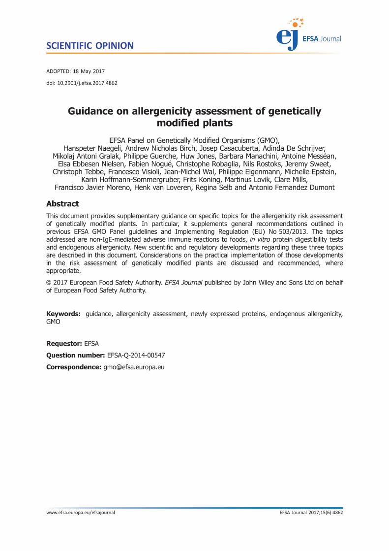
Rather, an integrated, stepwise, case-by-case approach should be used in the assessment of thenewly expressed protein(s) in relation to its(their) potential to cause CD. This overall strategy is in linewith the general principles followed for the allergenicity assessment of newly expressed protein(s) asdefined by EFSA (EFSA GMO Panel, 2011) and Codex Alimentarius (2009). In this context and briefly,the first step in the assessment should consider the available information on the source of the proteinand on the human exposure to the protein itself. This knowledge on the protein can be used tocalibrate the risk assessment strategy to follow on a case-by-case basis. If the available knowledge onthe protein under assessment is insufficient to support its safety, additional considerations on itsproperties are necessary to investigate the potential to cause CD. To this end, in silico approaches canbe employed, starting with searches for sequence identity (e.g. searches with known CD peptidesequences and motif searches). In a second step, if concerns from the sequence identity search arepresent, in silico peptide modelling can be applied. When potentially CD relevant sequences whichcannot be disregarded by in silico testing are identified, in a third step other in vitro tests such as HLA-DQ-peptide binding assays and/or testing with T-cell clones derived from patients with CD (Figures 1and 2) can be performed to determine the safety profile of the protein/peptide under assessment.Further details on the approach to follow can be found below.
A) Step 1 – knowledge on the protein/searches for sequence identity
Knowledge on the protein
Knowledge on the protein should include detailed information on the source of the transgene asregard its use as food and solid documented information that the protein itself is consumed byindividuals with CD without causing the disease. An exposure assessment might be necessary tosupport this consideration.
If the knowledge on the protein is unavailable or insufficient to support its safety, additionalconsiderations are needed as described below (see also Figure 1).
Searches for sequence identity
a) Searches involving a perfect sequence match with known CD peptide sequences
It is well established that prolamins and closely related proteins (Shewry et al., 2003) harbour thesequences that cause CD (Tye-Din et al., 2010). Therefore, an initial consideration is to determine ifthe protein of interest belongs to these families of proteins. Thus, identity searches with known CDpeptide sequences (see Annex A-1) should be performed. If this search results in a perfect match witha peptide sequence known to cause CD, a hazard has been identified.
If this is not the case and there is insufficient knowledge on the protein under assessment, furtherinvestigations to identify/dismiss proteins/peptides that could potentially cause CD should be provided(Figures 1 and 2).
b) Searches involving a partial sequence match with known CD peptide sequences
T cells respond to antigenic peptides bound to an HLA-molecule. In the case of CD, it concernsgluten-derived peptides bound to either HLA-DQ2 or -DQ8. However, it is well known that such T cellscan also respond to peptides in which one or more amino acids are replaced. This is the basis, forexample, for T-cell cross-reactivity towards gluten peptides and homologous, but not identical,peptides derived from barley and rye. For this reason, antigenicity of a protein or fragments thereof forpatients with CD cannot be excluded only on the basis of a lack of a perfect match with glutensequences.
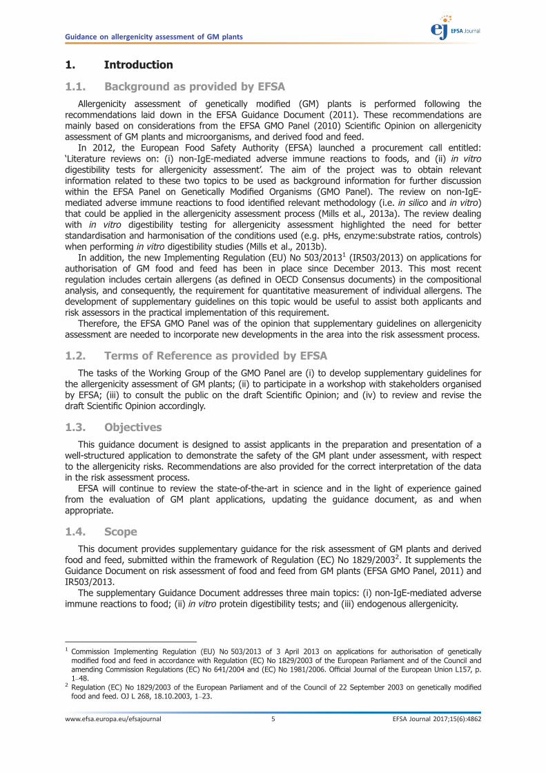
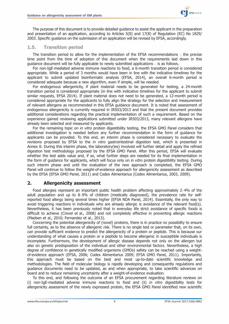
– Searches for sequence identity with the Q/E-X1-P-X2 motif (Figure 3):
Examination of the list of epitopes currently identified (see Annex A-1) reveals that a characteristicQ-X1-P-X2 motif is present in the large majority of HLA-DQ2 epitopes. This motif is a target for theenzyme tissue transglutaminase 2 (TG2) which yields E-X1-P-X2 (X1 = L, Q, F, S or E; X2 = Y, F, A, V orQ; for further details see Figure 3 and Annex A).
Guidance on allergenicity assessment of GM plants
www.efsa.europa.eu/efsajournal 8 EFSA Journal 2017;15(6):4862

Additional considerations on the position and nature of adjacent amino acid sequences to theQ/E-X1-P-x2 motif should also be taken into account when performing the assessment (examplesare provided in Annex A-2). If concerns are raised, additional tests will be required (Figures 1 and 2).
– Searches for sequence identity without the Q/E-x1-P-x2 motif:
Few known CD peptide sequences do not contain the Q/E-X1-P-X2 motif. Therefore, an identitysearch with known CD peptide sequences, which do not contain a Q/E-X1-P-X2 motif, should beperformed allowing one or more amino acid mismatches. The position and nature of the mismatchedand identical amino acids determine if the peptide sequence has the potential to be a T-cell stimulatoryepitope (additional considerations and examples are provided in Annex A-2). If concerns are raised,additional tests will be required (Figures 1 and 2). Known CD peptide sequences are listed in Table A.1as well as in publicly available celiac peptide databases (see Annex A-1).
Following these searches for sequence identity, four outcomes are possible:
• if the Q/E-X1-P-X2 motif is not present and no concerns are raised during the identity searchwith specific known CD peptide sequences, the probability of a T-cell epitope is unlikely;
• if the Q/E-X1-P-X2 motif is not present but concerns are raised during the identity search withspecific known CD peptide sequences, further investigation is required;
• if the Q/E-X1-P-X2 motif is present and considerations on adjacent sequences can be used toeliminate concerns, the probability of a T-cell epitope is unlikely;
• if the Q/E-X1-P-X2 motif is present and considerations on adjacent sequences cannot be usedto eliminate concerns, a potential T-cell epitope is detected and further investigation isrequired.
Further details on the search for sequence identity are listed in Annex A-2.
B) Step 2 – HLA-DQ-peptide modelling
Several HLA-DQ2-gliadin and HLA-DQ8-gliadin structures are publicly available. These structures canbe used to model a peptide of interest into HLA-DQ2 or HLA-DQ8. This HLA-DQ-peptide modelling canthen allow for a comparison, which can indicate the likelihood that such a peptide will bind to HLA-DQ2or HLA-DQ8 and will provide insights into the position and orientation of the T-cell receptor contactresidues in the HLA-DQ-bound peptide.
Two outcomes are possible:
• if no relevant HLA-DQ binding and/or similarity with the available HLA-DQ-gliadin structures arepredicted, the probability of a T-cell epitope is unlikely;
• if HLA-DQ binding and a high degree of similarity with the available HLA-DQ-gliadin structuresare predicted, this indicates potential cross-reactivity of the investigated peptide. Consequently,the potential capacity of the peptide to trigger CD should be determined by additional in vitroapproach(es) described below.
Further details on this step are listed in Annex A-3.
C) Step 3 – In vitro approaches
Proposals of in vitro approaches that can be used to further investigate the potential of the newlyexpressed protein to cause CD are found below.
HLA-DQ peptide binding assays:
For peptides to evoke T-cell responses, they must bind to HLA-molecules. HLA-DQ2- and HLA-DQ8-specific peptide binding assays were developed and can be exploited to determine the likelihood thatpeptides under investigation might be immunogenic.
Two outcomes are possible:
• if high affinity binding is detected (see Annex A-4), further testing is required;• if low or no affinity binding is detected, the probability that the peptide is immunogenic is low.
Therefore, no further testing is required.
For further details, please see Annex A-4 for an overview of publications that have reportedHLA-DQ-peptide binding assays.
Guidance on allergenicity assessment of GM plants
www.efsa.europa.eu/efsajournal 9 EFSA Journal 2017;15(6):4862

T-cell testing:
Recognition of gluten peptides by CD4+ T cells from one or more CD patients has been aprerequisite for defining toxic CD peptides (Sollid et al., 2012).
Such T cells were isolated in a number of laboratories where the necessary expertise andappropriate infrastructure are available. These T cells were used to provide conclusive evidence on thecapacity of a specific peptide sequence to stimulate CD-causative T-cell responses. If a T-cell responsewith the protein/peptide under assessment is observed, then a hazard has been identified.
For further details, please see Annex A-5 for an overview of publications that have reported T cellsspecific for HLA-DQ-gluten complexes.
In vitro protein digestibility:
Due to the proline-rich nature, gluten proteins are highly resistant to proteolytic degradation. Thisresults in relatively long peptides that harbour one or more T-cell stimulatory epitopes. Further detailsare listed in Annex A-6 and in the chapter on in vitro protein digestibility testing (Annex B).
Search for sequence iden�ty
T-cell tes�ng
In vitro diges�bility*
HLA-DQ binding assays
modelling
Step 1
Stepwise approach for risk assessment
Knowledge on the protein (exposure,
source, etc.)
* For details, please see chapter on in vitro diges�bility.
If concerns are raised
If concerns are raised Step 2 Step 3
If insufficient
Figure 1: Stepwise approach for risk assessment
Guidance on allergenicity assessment of GM plants
www.efsa.europa.eu/efsajournal 10 EFSA Journal 2017;15(6):4862
2.2. In vitro protein digestibility tests
2.2.1. Risk assessment considerations
In vitro digestibility tests can provide useful data on the susceptibility of a protein to digestionwhich can reflect its digestibility in the human gastrointestinal tract and subsequently provide
Figure 2: Search for sequence identity
Figure 3: Q/E-X1-P-X2 motif: possible combinations for the Q/E-X1-P-X2 motif found in the largemajority of identified immunogenic gluten-derived epitopes. It was noted that, whileposition 1 is always either glutamic acid (E) or glutamine (Q) and position 3 always consistsof a proline (P), also positions 2 (X1) and 4 (X2) are restricted to certain amino acids
Guidance on allergenicity assessment of GM plants
www.efsa.europa.eu/efsajournal 11 EFSA Journal 2017;15(6):4862
information on its immunogenicity. There is evidence that gastrointestinal digestion can affect theimmunogenicity of dietary proteins related to both IgE and non-IgE-mediated adverse reactions tofoods (see Annex B). Therefore, in vitro protein digestion can be used as an additional piece ofinformation in the weight-of-evidence approach followed for the allergenicity assessment of newlyexpressed proteins, because no single test is fully predictive of the allergenic potential of a protein(Codex Alimentarius, 2003, 2009; EFSA GMO Panel, 2011).
The pepsin resistance test is the most commonly used digestion test for this assessment, in linewith international guidelines (Codex Alimentarius 2003, 2009), the EFSA Guidance Document (2011)and Implementing Regulation (EU) No 503/2013 (IR503/2013). EFSA previously highlighted thelimitations of the classical pepsin resistance test for allergenicity risk assessment and recommendedthat resistance to digestion of (novel) proteins should be evaluated using other in vitro digestibilitymethods designed to more closely simulate the conditions of the human digestion process (EFSA GMOPanel, 2010, 2011).
In Annex B of the present document and based on state-of-the-art in science, the EFSA GMOPanel proposes a refined in vitro digestion test that extends the conditions currently used in theclassical pepsin resistance test in order to better reflect the range of conditions found in vivo. Thiselaborated test includes additional conditions more representative of the gastric environment withregard to pH and pepsin levels, together with an intestinal digestion phase. In addition, moreinformative read-outs of the test are laid out which define the extent to which either the intact proteinor resistant fragments remain after in vitro digestion.
The EFSA GMO Panel considers that additional investigation is needed before any additionalrecommendation in the form of guidance for applicants can be provided on the proposed in vitroprotein digestibility tests.
To this end, an interim phase (~ 2 years duration) is considered necessary to evaluate the proposedrevisions to the in vitro gastrointestinal digestion test (see Annex B). During this interim phase, thelaboratory(ies) involved, working with EFSA, will further detail and apply the refined digestion testmethodology. After this period, EFSA will assess whether the test adds value to the allergenicity riskassessment and, if so, what further steps are needed for its final implementation in the form ofguidance for applicants. An outline proposal for such an interim phase is provided in Annex B-5.
During the interim phase, and until the evaluation of the new approach is completed, the EFSAGMO Panel will continue to follow the weight-of-evidence approach for allergenicity assessment asdescribed by EFSA (EFSA GMO Panel, 2011) and Codex Alimentarius (Codex Alimentarius, 2003, 2009).
2.3. Endogenous allergenicity
According to the EFSA Guidance Document (EFSA GMO Panel, 2011) and in line with principles ofCodex Alimentarius (2003, 2009), endogenous allergenicity assessment of GM plants is a relevantelement to be considered. The purpose of the assessment of endogenous allergenicity is to investigatethat no unintended effect of the genetic modification changes the levels of endogenous allergens in amanner that would adversely impact on human and animal health (EFSA GMO Panel, 2011; K€onig et al.,2004; Metcalfe et al., 1996; Thomas et al., 2008). This follows the same principles as those used tosupport the measurement of any other compound/endpoint in the compositional analysis of GM plants.
EFSA (EFSA GMO Panel, 2011) and Codex Alimentarius (2003, 2009) foresee the assessment ofendogenous allergenicity only when the plant receiving the new gene(s) is recognised to be allergenic.In these cases, any potential change in the overall allergenicity of the GM plant compared with that ofits non-GM comparator(s) should be analysed. Historically, this analysis was performed using sera fromallergic individuals, but limitations of this assay for routine risk assessment purposes have beenpreviously described (Fernandez et al., 2013; Selb et al., 2017).
EFSA has previously recommended the inclusion of relevant endogenous allergens in thecomparative compositional analysis, implying the quantitative measurement of individual allergens(EFSA GMO Panel, 2010, 2011). This recommendation became a mandatory requirement whenImplementing Regulation (EU) No 503/2013 (IR503/2013) on applications for authorisation of GM foodand feed came into force. To assist applicants and risk assessors in its practical implementation, thissupplementary guidance document provides further information on: (i) relevant crops for analysis; (ii)relevant allergens for quantification; (iii) methodology for quantification; and (iv) principles to befollowed for data interpretation and risk assessment considerations.
The EFSA GMO Panel Guidance Documents are continuously updated to consider new scientific andregulatory developments in the field. To this end, further revisions and updates to this supplementary
Guidance on allergenicity assessment of GM plants
www.efsa.europa.eu/efsajournal 12 EFSA Journal 2017;15(6):4862
EFSA Guidance Document might be needed once experience has been gained with the assessment ofendogenous allergenicity and the new strategies/tools being deployed to support them.
2.3.1. Relevant crops for analysis
According to IR503/2013 and in line with EFSA Guidance Document (EFSA GMO Panel, 2011), anassessment of endogenous allergenicity should be performed on a case-by-case basis. When therecipient plant is recognised to be allergenic, the applicant should test any potential change in theallergenicity of the GM food or feed by comparison of the allergen repertoire with that of itsappropriate comparator(s).
To date, EFSA has performed endogenous allergenicity risk assessments based on experimentaldata for foods recognised to be common food allergens and of public health importance as listed inAnnex II of the European Regulation on food information to consumers.7,8 In this context, soybean isrecognised to be a common allergenic food and EFSA GMO Panel Scientific Opinions on GM soybeanapplications, which included an endogenous allergenicity assessment, were published previously(Annex C-1). To date, EFSA has not received any application involving a common allergenic food otherthan soybean. For crops not recognised as being commonly allergenic, specific experimental data onendogenous allergenicity are not requested by EFSA. In these cases, the assessment is carried outconsidering potential effects of the genetic modification on the general composition and molecularcharacteristics of the GM plant. However, this does not preclude EFSA requesting experimental data onendogenous allergenicity for these, if considered necessary, e.g. if the allergenic status of these foodschanges. In addition, other plant-derived foods not currently listed in Annex II of such EuropeanRegulation (e.g. fruits), which might be genetically engineered in the future, should be subjected tosuch assessments if considered necessary. For such considerations, risk assessors, risk managers,health professionals and stakeholders can provide valuable input.
2.3.2. Relevant allergens for quantification
Soybean
Soybean is recognised as a common allergenic food by European Regulation,7,8 and suggested asone of the eight foods accounting for approximately 90% of food allergies (FDA, 2004; OECD, 2012).
The quantitative measurement of soybean allergens as part of the compositional analysis is now amandatory requirement in the IR503/2013 where reference to allergens in OECD consensus documentis provided.
In the OECD consensus document on soybean, proteins termed as ‘potential allergens’ aredescribed in Table 20, Section III-C (OECD, 2012). Nevertheless, in accordance with Article 5(2) and 5(3) of the IR503/2013: (i) EFSA may accept derogations from specific requirements, if they aredemonstrated not to be scientifically necessary for food/feed safety assessment or technically notpossible to perform; and/or (ii) EFSA may request data not foreseen in OECD consensus documentsanytime, if considered necessary based on new scientific findings.
In line with IR503/2013, the OECD allergen list should be taken as the starting point for thecollection of ‘potential allergens’ (Figure 4). In addition, this list should be complemented withsearches in the scientific and medical literature, and in various updated databases (EFSA GMO Panel,2010; Appendix 3.13, Table I for a list of relevant databases). It is noted that on the one hand theOECD list of allergens might not be complete at a given point in time, e.g. it might be out dated and/ormiss relevant (more recent) entries. On the other hand, not all ‘potential allergens’ listed in this OECDconsensus document can currently be measured due to technical reasons (e.g. amino acid sequencenot available) and/or their clinical relevance might not have been demonstrated. Once acomprehensive search of ‘potential allergens’ in the literature and databases is conducted, the relevantallergens selected for quantification and subsequent comparative analysis should be justified. As acomplementary and/or alternative approach, a systematic review could be performed, aiming to
7 EC, 2003. Directive 2003/89/EC of the European Parliament and of the Council of 10 November 2003 amending Directive2000/13/EC as regards indication of the ingredients present in foodstuffs.
8 EC, 2011. Regulation (EU) No 1169/2011 of the European Parliament and of the Council of 25 October 2011 on the provisionof food information to consumers, amending Regulations (EC) No 1924/2006 and (EC) No 1925/2006 of the EuropeanParliament and of the Council, and repealing Commission Directive 87/250/EEC, Council Directive 90/496/EEC, CommissionDirective 1999/10/EC, Directive 2000/13/EC of the European Parliament and of the Council, Commission Directives 2002/67/ECand 2008/5/EC and Commission Regulation (EC) No 608/2004.
Guidance on allergenicity assessment of GM plants
www.efsa.europa.eu/efsajournal 13 EFSA Journal 2017;15(6):4862

identify clinically relevant allergens. A scientific rational, explaining why an allergen is not consideredrelevant should be provided. A representation of these considerations can be found in Figure 4.
A possible approach how to identify proteins relevant for the endogenous allergenicity assessmentof soybean is described in Annex C-2.
Other GM plants
For foods other than soybean which are recognised allergenic (risk assessors, risk managers, healthprofessionals and stakeholders can provide valuable input), a similar approach/strategy for theidentification of relevant allergens as the one followed for soybean (see Annex C-2) should be applied,whenever considered necessary. To date, EFSA has not received any application involving a commonallergenic food other than soybean.
2.3.3. Methodology for quantification
Either enzyme-linked immunosorbent assay (ELISA) or mass spectrometry (MS) approaches areappropriate methods for the quantification of endogenous allergens, both allowing the specificdetection and quantification of single known allergens. Further considerations on these methodologiesare described in Annex C-3.
2.3.4. Data interpretation and risk assessment
According to IR503/2013, conclusions of the allergenicity assessment should indicate ‘whether thegenetically modified food or feed is likely to be more allergenic than its conventional counterpart’.The starting point in the assessment is the identification of any potential change in the allergenicity ofthe GM food or feed by comparison of the allergen repertoire of the GM plant and its conventionalcounterpart, taking into account natural variability.
Allergens included in the compositional analysis should be measured and analysed according to theprinciples of the comparative assessment performed for any other compositional compounds (seesection 1.3.2 of IR503/2013). To this end, the starting point of the assessment should be theidentification of statistically significant differences between the GM plant and its conventionalcounterpart. A further evaluation should investigate whether or not the differences observed fall withinor outside the range of natural variation estimated from the reference varieties included in the fieldtrial, i.e. the equivalence test (IR503/2013). In case the levels of a specific allergen in a GM plantincreases significantly from the levels observed in the appropriate comparator(s) and falls outside theestimated range of natural variation, the biological relevance in relation to human and animal healthshould be assessed.
Additional considerations and/or experimental data might be needed on a case-by-case basis. Asfor other compounds included in the compositional analysis, the nature of these additionalconsiderations and/or experimental data depend on the number and magnitude of the changesidentified, as well as on the clinical/safety relevance of the specific allergen(s)/compound(s) involved.
Ultimately, when a potential increase in allergenicity due to the genetic modification cannot beexcluded, the GM food or feed should be further characterised in the light of its anticipated intake, asrequested by IR503/2013. Occupational allergy should also be considered with respect to inhalation orcontact with potential allergens. In all cases, an exposure assessment should focus on the Europeanpopulation aiming at identifying particular groups at high risk, which might be affected by a specificchange of the allergen content.
Possible approaches for data interpretation and risk assessment of soybean endogenousallergenicity are summarised in Annex C-4.
Guidance on allergenicity assessment of GM plants
www.efsa.europa.eu/efsajournal 14 EFSA Journal 2017;15(6):4862

Documentation provided to EFSA
1) Proposal for a self-task mandate of the EFSA GMO Panel to establish a Working Group todevelop supplementary guidelines for the allergenicity assessment of GM plants toincorporate new developments. May 2014. Submitted by the Chair of the ESA GMO Panel.
2) Acceptance of the self-task mandate of the EFSA GMO Panel to establish a Working Groupto develop supplementary guidelines for the allergenicity assessment of GM plants toincorporate new developments. July 2014. Submitted by EFSA Executive Director.
ReferencesAbadie V, Sollid LM, Barreiro LB and Jabri B, 2011. Integration of genetic and immunological insights into a model
of celiac disease pathogenesis. Annual Review of Immunology, 29, 493–525.van Bergen J, Mulder CJ, Mearin ML and Koning F, 2015. Local communication among mucosal immune cells in
patients with celiac disease. Gastroenterol, 148, 1187–1194.Codex Alimentarius, 2003. Foods derived from modern biotechnology. Codex Alimentarius Commission, Joint FAO/
WHO Food Standards Programme, Rome.Codex Alimentarius, 2009. Foods derived from modern biotechnology. Codex Alimentarius Commission, Joint FAO/
WHO Food Standards Programme, Rome.Comino I, de Lourdes Moreno M and Sousa C, 2015. Role of oats in celiac disease. World Journal of
Gastroenterology, 21, 11825–11831.Crevel RWR, Ballmer-Weber BK, Holzhauser T, Hourihane JO’B, Knulst AC, Mackie AR, Timmermans F and Taylor
SL, 2008. Thresholds for food allergens and their value to different stakeholders. Allergy 63:597–609.EFSA (European Food Safety Authority), 2006. Guidance document for the risk assessment of geneticallymodified
plants and derived food and feed by the Scientific Panel on Genetically Modified Organisms (GMO) - includingdraft document updated in 2008 (reference EFSA-Q-2003-005A). EFSA Journal 2006;4(4):99, 105 pp. https://doi.org/10.2903/j.efsa.2006.99
EFSA (European Food Safety Authority), 2014. Indicative timelines for submitting additional or supplementaryinformation to EFSA during the risk assessment process of regulated products. EFSA Journal 2014;12(1):3553,37 pp. https://doi.org/10.2903/j.efsa.2014.3553
Figure 4: Endogenous allergenicity workflow for the selection of relevant allergens in soybean
Guidance on allergenicity assessment of GM plants
www.efsa.europa.eu/efsajournal 15 EFSA Journal 2017;15(6):4862
EFSA GMO Panel (EFSA Panel on Genetically Modified Organisms), 2010. Scientific Opinion on the assessment ofallergenicity of GM plants and microorganisms and derived food and feed. EFSA Journal 2010;8(7):1700, 168 pp.https://doi.org/10.2903/j.efsa.2010.1700
EFSA GMO Panel (EFSA Panel on Genetically Modified Organisms), 2011. Scientific Opinion on Guidance for riskassessment of food and feed from genetically modified plants. EFSA Journal 2011;9(5): 2150, 37 pp. https://doi.org/10.2903/j.efsa.2011.2150
EFSA NDA Panel (EFSA Panel on Dietetic Products, Nutrition and Allergies), 2014. Scientific Opinion on theevaluation of allergenic foods and food ingredients for labelling purposes. EFSA Journal 2014;12(11):3894, 286pp. https://doi.org/10.2903/j.efsa.2014.3894
FDA, 2004. Food allergen labeling and consumer protection act (FALCPA). Congressional record v. 150. Availableonline: http://www.fda.gov/downloads/Food/LabelingNutrition/FoodAllergensLabeling/GuidanceComplianceREgulatoryInformation/UCM179394.pdf
Fernandez A, Mills EN, Lovik M, Spoek A, Germini A, Mikalsen A and Wal JM, 2013. Endogenous allergens andcompositional analysis in the allergenicity assessment of genetically modified plants. Food and ChemicalToxicology, 62, 1–6.
Garsed K and Scott BB, 2007. Can oats be taken in a gluten-free diet? A systematic review. Scandinavian Journalof Gastroenterology, 42, 171–178.
Green PH and Cellier C, 2007. Celiac disease. New England Journal of Medicine, 357, 1731–1743.K€onig A, Cockburn A, Crevel RWR, Debruyne E, Grafstroem R, Hammerling U, Kimber I, Knudsen I, Kuiper HA,
Peijnenburg AACM, Penninks AH, Poulsen M, Schauzu M and Wal JM, 2004. Assessment of the safety of foodsderived from genetically modified (GM) crops. Food and Chemical Toxicology, 42, 1047–1088.
Koning F, Thomas R, Rossjohn J and Toes RE, 2015. Coeliac disease and rheumatoid arthritis: similar mechanisms,different antigens. Nature Reviews Rheumatology, 11, 450–461.
Lundin KEA, Nilsen EM, Scott HG, Løberg EM, Gjøen A, Bratlie J, Skar V, Mendez E, Løvik A and Kett K, 2003. Oatsinduced villous atrophy in coeliac disease. Gut, 52, 1649–1652.
Madsen CB, Crevel RWR, Chan C-H, Dubois AEJ, DunnGalvin A, Flokstra-de Blok BMJ, Gowland MH, Hattersley S,Hourihane JO’B, Nørhede P, Pfaff S, Rowe G, Schnadt S and Vlieg-Boerstra BJ, 2010. Food allergy: stakeholderperspectives on acceptable risk. Regulatory Toxicology and Pharmacology 57, 256–265.
Metcalfe DD, Astwood JD, Townsend R, Sampson HS, Taylor SL and Fuchs RL, 1996. Assessment of the allergenicpotential of foods derived from genetically engineered crop plants. Critical Reviews in Food Science andNutrition, 36, S165–S186.
Mills ENC, Marsh JT, Boyle R, Hoffmann-Sommergruber K, DuPont D, Bartara J, Bakalis S, McLaughlin J andShewry PR; The University of Manchester, 2013a. Literature review: ‘non-IgE-mediated immune adversereactions to foods’. EFSA supporting publication 2013:EN-527, 40 pp.
Mills ENC, Marsh JT, Johnson PE, Boyle R, Hoffmann-Sommergruber K, DuPont D, Bartras J, Bakalis S, McLaughlinJ and Shewry PR; The University of Manchester, 2013b. Literature review: ‘in vitro digestibility tests forallergenicity assessment’. EFSA supporting publication 2013:EN-529, 52 pp.
OECD, 2012. Revised consensus document on compositional considerations for new 452 varieties of soybean[Glycine max (L.) Merr.]: key food and feed nutrients, 453 antinutrients, toxicants and allergens. Series on theSafety of Novel Foods and 454 Feeds No. 25.
Selb R, Wal JM, Moreno FJ, Lovik M, Mills C, Hoffmann-Sommergruber K and Fernandez A, 2017. Assessment ofendogenous allergenicity of genetically modified plants exemplified by soybean - where do we stand? Food andChemical Toxicology, 101, 139–148.
Shewry PR, Halford NG and Lafiandra D, 2003. Genetics of wheat gluten proteins. Advances in Genetics, 49, 111–184.Sollid LM, Qiao S, Anderson RP, Gianfrani C and Koning F, 2012. Nomenclature and listing of celiac disease relevant
gluten T-cell-epitopes restricted by HLA-DQ molecules. Immunogenetics, 64, 455–460.Thomas K, Herouet-Guicheney C, Ladics G, McClain S, MacIntosh S, Privalle L and Woolhiser M, 2008. Current and
future methods for evaluating the allergenic potential of proteins: international workshop report 23–25 October2007. Food and Chemical Toxicology, 46, 3219–3225.
Tye-Din JA, Stewart JA, Dromey JA, Beissbarth T, van Heel DA, Tatham A, Henderson K, Mannering SI, GianfraniC, Jewell DP, Hill AV, McCluskey J, Rossjohn J and Anderson RP, 2010. Comprehensive, quantitative mapping ofT cell epitopes in gluten in celiac disease. Science Translational Medicine, 2, 41–51.
Abbreviations
CD celiac diseaseDBPCFC double-blind placebo-controlled food challengeELISA enzyme-linked immunosorbent assayFPIES food-protein induced enterocolitisGANA N-carbobenzoxy-diglycyl-L-arginyl-2-naphthylamide hydrochlorideGGPNA c-glutamyl-p-nitroanilideGM genetically modified
Guidance on allergenicity assessment of GM plants
www.efsa.europa.eu/efsajournal 16 EFSA Journal 2017;15(6):4862

GMO genetically modified organismHMW high molecular weightIgA immunoglobulin AIgE immunoglobulin ELMW low molecular weightMHC major histocompatibility complexMS mass spectrometryMS-MS tandem mass spectrometryOECD Organisation for Economic Co-operation and DevelopmentPPI proton pump inhibitorSDS-PAGE sodium dodecyl sulfate-polyacrylamide gel electrophoresisTG2 transglutaminase 2
Guidance on allergenicity assessment of GM plants
www.efsa.europa.eu/efsajournal 17 EFSA Journal 2017;15(6):4862

Annex A – Non-IgE-mediated adverse immune reactions to foodsBackground
Celiac disease (CD) has a strong genetic component. It is associated with particular immuneresponse genes, i.e. those responsible for the class II major histocompatibility complex (MHC)molecules, called HLA in humans. Most CD patients express particular HLA-DQ-molecules. HLA-DQmolecules are dimers of an alpha-(DQA1) and a beta-(DQB1) chain. Like all HLA-molecules, HLA-DQmolecules bind short peptides and present these to T cells of the immune system. While T cells ignoreHLA-bound peptides derived from harmless (‘self’) proteins, HLA-bound peptides derived frompathogens are specifically detected and this recognition leads to the generation of a protective T-cellresponse and eradication of the pathogen. The large majority of CD patients (approximately 95%)express HLA-DQ2.5 (DQA1*05:01, DQB1*02:01) (Sollid et al., 2012) while the remainder are usuallyHLA-DQ8 positive (DQA1*03, DQB1*03:02). The few patients that express neither DQ2.5 nor DQ8,often express HLA-DQ molecules that contain only one of the DQ2.5-chains, e.g. DQ2.2 (DQA1*02:01,DQB1*02:01) or DQ7.5 (DQA1*05, DQB1*03:01) (Karell et al., 2003). In affected people, but not inhealthy individuals, pro-inflammatory gluten-specific CD4+ T cells are present in the lamina propria ofthe affected duodenum. Importantly, these CD4+ T cells recognise gluten peptides only whenpresented by the disease associated HLA-DQ molecules (Lundin et al., 1993, 1994; Tye-Din et al.,2010; Vader et al., 2002b; van de Wal et al., 1998b). In essence, in patients with CD the immunesystem displays an aberrant response: the harmless gluten proteins in the food are recognised ashazardous, leading to a pro-inflammatory response as long as gluten is consumed. Elimination ofgluten from the diet constitutes an effective treatment because the T-cell stimulatory gluten peptidesare no longer present. Unfortunately, once a gluten-specific T-cell response has developed, this leadsto immunological memory and every subsequent exposure to gluten will reactivate the gluten-reactiveT cells and consequently lead to inflammation. A lifelong gluten-free diet is thus required.
Gluten is the cohesive mass that remains when starch has been removed from wheat dough(Shewry et al., 1992). Gluten consists of gliadin and glutenin subcomponents. The gliadins aresubdivided into a-, c- and x-gliadins, and dozens of variants of each type are typically present in asingle wheat variety. The glutenins are subdivided into high molecular weight (HMW) and lowmolecular weight (LMW) subunits. The most commonly used wheat varieties are bread wheats(Triticum aestivum), which are hexaploid species, and pasta wheats (Triticum durum), which aretetraploid species. Thus, in any single wheat variety up to a hundred different gluten proteins arelisted, many of which are highly similar and only differ by a few amino acids from each other.
T-cell epitopes derived from the a-, c- and x-gliadins as well as from the HMW- and LMW-gluteninswere reported (Arentz-Hansen et al., 2000; Shan et al., 2002; Sj€ostr€om et al., 1998; Vader et al.,2002b; van de Wal et al., 1998b, 1999). In addition, T-cell epitopes in both hordeins and secalins wereidentified that are highly homologous or identical to those found in wheat (Tye-Din et al., 2010; Vaderet al., 2003). The gluten-like avenins of oat are more distinct; however, avenin-specific as well ascross-reactive T-cell responses were described (Arentz-Hansen et al., 2004; Vader et al., 2003).
High affinity binding of peptides to either HLA-DQ2.5 or -DQ8 depends on the presence of one ormore negatively charged amino acids. As gluten proteins are virtually devoid of negatively chargedamino acids, native gluten-derived peptides bind poorly to these HLA-DQ molecules. Due to the activityof the enzyme tissue transglutaminase 2 (TG2) in the gastrointestinal tract, the required negativecharge(s) are introduced when this enzyme converts glutamine residues within gluten peptides intonegatively charged glutamic acid (Molberg et al., 1998; Vader et al., 2002a; van de Wal et al., 1998a).These deamidated gluten peptides then bind with increased affinity to HLA-DQ2.5 or -DQ8, and thisstronger binding enhances or causes immunogenicity (van de Wal et al., 1998; Arentz-Hansen et al.,2000; Henderson et al., 2007; Kim et al., 2004; Moustakas et al., 2000; Quarsten et al., 1999).
The specificity of TG2 for particular target sequences in gluten proteins plays a crucial role in thegeneration of a relatively large number of gluten-derived peptides that bind to HLA-DQ2.5. Glutamineand proline are abundantly present in gluten proteins, together they comprise over 50% of the aminoacids in gluten. Therefore, Q-X-P and Q-P sequences (where Q is glutamine; P is proline; X is anyamino acid except P) are often found in gluten proteins and TG2 typically deamidates glutamineresidues in Q-X-P sequences, but not in QP sequences (Vader et al., 2002a). Therefore, immunogenicgluten-derived peptides are typically found in the proline rich-regions of gluten proteins and usuallycontain a Q-X-P motif. ‘Classic’ examples of such peptides are the immunodominant T-cell epitopespresent in the N-terminal part of the a-gliadins: PFPQPQLPY and PQPQLPYPQ. In fact, these sequences
Guidance on allergenicity assessment of GM plants
www.efsa.europa.eu/efsajournal 18 EFSA Journal 2017;15(6):4862
have a 7-amino acid overlap and in both sequences only one Q-residue is a target for TG2, the Q inthe QLPY sequence which allows the introduction of a negative charge at either position 6 or position4, respectively. In both instances, this generates a gluten peptide that binds with high affinity to HLA-DQ2.5. The available crystal structures of HLA-DQ2.5-gliadin and the bound cognate T-cell receptordemonstrate that the negatively charged glutamic acid serves as an anchor residue for peptide bindingto HLA-DQ2.5 and does not contact the T-cell receptor (Petersen et al., 2014). Additionally, the proline-rich nature of gluten renders these proteins resistant to degradation by enzymes in the gastrointestinaltract. Relatively long gluten fragments are, therefore, present in the small intestine. This likelycontributes to the immunogenic nature of these peptides (Shan et al., 2002). Thus, at least threefactors contribute to the immunogenicity of gluten: (a) resistance to proteolytic degradation, (b)specific recognition by TG2, and (c) peptide binding properties of HLA-DQ2.5 and HLA-DQ8.
The glutamic acid introduced by TG2 is usually in position 4 (p4) or p6 in HLA-DQ2.5 restrictedepitopes and at position p1 and/or p9 in HLA-DQ8 restricted epitopes (Sollid et al., 2012). As aconsequence, HLA-DQ2.5-restricted gluten epitopes carry a proline at either p6 or p8. This positioningof proline residues is less strict in the case of the DQ8 epitopes. In all cases, the glutamic acid residuesserve as anchors important for binding of the peptides to either HLA-DQ2.5 or -DQ8.
It is important to note that, while polyclonal T-cell responses to multiple T-cell epitopes are usuallydetected in CD patients, responses to the DQ2.5-glia-a1, DQ2.5-glia-a2 epitopes and homologuesthereof in the x-gliadins, hordeins and secalins are dominant in DQ2.5-positive patients (Arentz-Hansen et al., 2000; Tollefsen et al., 2006; Tye-Din et al., 2010). In DQ8-positive patients, responsesto the DQ8-glia-a1 epitope are most frequently found (Tollefsen et al., 2006; van de Wal et al.,1998b).
The following criteria were used to define CD reactive epitopes in Sollid et al. (2012):
• reactivity against the epitope must have been defined by at least one specific T-cell clone;• the HLA-restriction element involved must have been unequivocally defined;• the 9-amino acid core of the epitope must have been defined either by an analysis with
truncated peptides and/or HLA-binding with lysine scan of the epitope, or a comparableapproach. In a lysine scan, all amino acids in the sequence of interest are individually replacedby a lysine and the impact of these single amino acids substitutions on HLA-binding isdetermined, information which usually reveals which amino acids in the sequence are requiredfor binding to HLA.
Because CD is caused by an immune response to a foreign protein and all symptoms disappearupon withdrawal of gluten from the diet, the condition should not be regarded as a true autoimmunedisease. Autoantibodies specific for tissue transglutaminase appear to be secondary to the T-cell drivenimmune response to gluten and disappear if gluten is eliminated from the diet (Rossjohn and Koning,2016).
Other conditions linked to wheat or gluten, summarised as ‘non-celiac gluten sensitivity’, are notpart of this document because there are no known definite underlying pathomechanisms (Aziz et al.,2015). In the last years, the diagnosis of non-celiac gluten sensitivity has emerged and refers toclinically diagnosed gluten-related symptoms without criteria for gluten allergy or celiac disease (Czaja-Bulsa, 2015). The unambiguous diagnosis of this condition is hampered by several facts. Symptomsare pleomorphic and often vague and limited in time. They mostly concern the gastrointestinal tract,but can also involve among many others: mood disorders, chronic recurrent headache, chronic fatigueor muscle disorders. Nevertheless, avoidance of gluten-containing foods leading to symptom relief, andre-exposure provoking relapse are suggestive of a gluten-related origin of the disease. It can, however,not be excluded that the symptoms are due to other components present in wheat and relatedcereals. The cause of this condition remains undefined. Immune mechanisms have not been identifiedand the character of the symptoms mostly suggests a gluten-intolerance (difficult digestion). Non-celiac gluten sensitivity seems more prominent in individuals with irritable bowel disease, and in thosesuffering of FODMAP intolerance (Biesiekierski et al., 2013).
The current chapter of the guidance document dealing with non-IgE-mediated food sensitivity is atthe current stage of knowledge not addressing non-celiac gluten sensitivity. This disease is currentlyonly marginally understood, in particular with regard to its mechanism, and is increasingly (mostly self)diagnosed. Future studies might unravel the exact mechanism of the disease and thus indicatewhether additional risk assessment considerations will be needed – aspects of which we currently haveno evidence/sufficient knowledge.
Guidance on allergenicity assessment of GM plants
www.efsa.europa.eu/efsajournal 19 EFSA Journal 2017;15(6):4862

To sum up, CD, a condition in which immunological mechanisms were extensively studied, can beconsidered as a pathognomonic, gluten-related non-IgE mediated food sensitivity. CD is within the fieldof this guidance document and has been extensively addressed accordingly.
Annex A-1. T-cell epitopes known in celiac disease
Several listings/databases of the known T-cell stimulatory sequences identified in gluten, hordeins,secalins and avenins are available including the https://propepper.net database and the http://www.allergenonline.org/ database. Furthermore, an overview of the best characterised epitopes along witha unified nomenclature is presented in Sollid et al. (2012). Table A.1 lists the epitopes described in thelatter manuscript. This compilation lists both the HLA-DQ2- and HLA-DQ8-restricted epitopes andincludes the known immunodominant epitopes present in the a- and x-gliadins as well as the lesscommonly recognised epitopes in the c-gliadins and the LMW- and HMW-glutenins. The list contains aconvenient overview of the most important and well defined epitopes. It is, nevertheless, important tonote that due to the extreme variability of the gluten and gluten-like proteins in barley and rye,(single) amino acid variants of these epitopes do exist, some of which may also exhibit T-cellstimulatory activity. Also, it cannot be fully excluded that additional gluten epitopes may be identifiedin the future. However, this is unlikely because large numbers of patients have already beenextensively tested for gluten reactivity, on the basis of gluten peptide libraries (Tye-Din et al., 2010).Thus, any protein containing one or more sequences that display high sequence identity to the epitopesequences present in this list will likely have the capacity to trigger gluten-specific T cells.
The development of a comprehensive database that is publicly available, curated regularly andappropriately built and designed for risk assessment purposes is an important aspect to be furtherinvestigated.
References provided in background section of Annex A and Annex A-1:
Arentz-Hansen H, K€orner R, Molberg, Quarsten H, Vader W, Kooy YM, Lundin KEA, Koning F, Roepstorff P, SollidLM and McAdam SN, 2000. The intestinal T cell response to a-gliadin in adult celiac disease is focused on asingle deamidated glutamine targeted by tissue transglutaminase. The Journal of Experimental Medicine 191,603–612.
Arentz-Hansen H, Fleckenstein B, Molberg, Scott H, Koning F, Jung G, Roepstorff P, Lundin KE and Sollid LM, 2004.The molecular basis for oat intolerance in patients with celiac disease. Plos Med, 1, e1.
Aziz I, Hadjivassilou M and Sanders DS, 2015. The Spectrum of Noncoeliac Gluten Sensitivity. Nature Reviews.Gastroenterology and Hepatology, 12, 516–526.
Biesiekierski JR, Peters S, Newnham ED, Rosella O, Muir JG and Gibson PR, 2013. No effects of gluten in patientswith self-reported non-celiac gluten sensitivity after dietary reduction of fermentable, poorly absorbed, short-chain carbohydrates. Gastroenterology, 145, 320–328.
Czaja-Bulsa G, 2015. Non coeliac gluten sensitivity. A new disease with gluten intolerance. Clinical Nutrition, 34,189–194.
Ellis HJ, Pollock EL, Engel W, Fraser JS, Rosen-Bonson S, Wieser H and Ciclitira PJ, 2003. Investigation of theputative immunodominant T cell epitopes in coeliac disease. Gut, 52, 212–217.
Henderson KN, Tye-Din JA, Reid HH, Chen Z, Borg NA, Beissbarth T, Tatham A, Mannering SI, Purcell AW, DudekNL, van Heel DA, McCluskey J, Rossjohn J and Anderson RP, 2007. A structural and immunological basis for therole of human leukocyte antigen DQ8 in celiac disease. Immunity, 27, 23–34.
Karell K, Louka AS, Moodie SJ, Ascher H, Clot F, Greco L, Ciclitira PJ, Sollid LM and Partanen J, 2003. HLA types inceliac disease patients not carrying the DQA1*05-DQB1*02 (DQ2) heterodimer: results from the EuropeanGenetics Cluster on Celiac Disease. Human Immunology, 64, 469–477.
Kim CY, Quarsten H, Bergseng E, Khosla C and Sollid LM, 2004. Structural basis for HLA-DQ2-mediatedpresentation of gluten epitopes in celiac disease. Proceedings of the National Academy of Sciences of theUnited States of America, 101, 4175–4179.
Lundin KEA, Scott H, Fausa O, Thorsby E and Sollid LM, 1994. T cells from the small intestinal mucosa of a DR4,DQ7/DR4, DQ8 celiac disease patient preferentially recognize gliadin when presented by DQ8. HumanImmunology, 41, 285–291
Lundin KEA, Scott H, Hansen T, Paulsen G, Halstensen TS, Fausa O, Thorsby E and Sollid LM, 1993. Gliadin-specific, HLA-DQ(a*0501,b1*0201) restricted T cells isolated from the small intestinal mucosa of celiac diseasepatients. Journal of Experimental Medicine, 178, 187–196.
Mitea C, Salentijn EM, van Veelen P, Goryunova SV, van der Meer IM, van den Broeck HC, Mujico JR, Monserrat V,Gilissen LJ, Drijfhout JW, Dekking L, Koning F and Smulders MJ, 2010. A universal approach to eliminateantigenic properties of alpha-gliadin peptides in celiac disease. PLoS One, 5, e15637.
Molberg, McAdam SN, K€orner R, Quarsten H, Kristiansen C, Madsen L, Fugger L, Scott H, Roepstorff P, LundinKEA, Sj€ostr€om H and Sollid LM, 1998. Tissue transglutaminase selectively modifies gliadin peptides that arerecognized by gut-derived T cells. Nature Medicine, 4, 713–717.
Guidance on allergenicity assessment of GM plants
www.efsa.europa.eu/efsajournal 20 EFSA Journal 2017;15(6):4862
Moustakas AK, van de WY, Routsias J, Kooy YM, Van VP, Drijfhout JW, Koning F and Papadopoulos GK, 2000.Structure of celiac disease-associated HLA-DQ8 and non-associated HLA-DQ9 alleles in complex with twodisease-specific epitopes. International of Immunology, 12, 1157–1166.
Petersen J, Montserrat V, Mujico JR, Loh KL, Beringer DX, van Lummel M, Thompson A, Mearin ML, Schweizer J,Kooy-Winkelaar Y, van Bergen J, Drijfhout JW, Kan WT, La Gruta NL, Anderson RP, Reid HH, Koning F andRossjohn J, 2014. T-cell receptor recognition of HLA-DQ2-gliadin complexes associated with celiac disease.Nature Structural and Molecular Biology, 21, 480–488.
Quarsten H, Molberg, Fugger L, McAdam SN and Sollid LM, 1999. HLA binding and T cell recognition of a tissuetransglutaminase-modified gliadin epitope. European Journal of Immunology, 29, 2506–2514.
Rossjohn J and Koning F, 2016. A biased view toward celiac disease. Mucosal Immunology, 9, 583–586.Shan L, Molberg, Parrot I, Hausch F, Filiz F, Gray GM, Sollid LM and Khosla C, 2002. Structural basis for gluten
intolerance in celiac sprue. Science, 297, 2275–2279.Shewry PR, Tatham AS and Kasarda DD, 1992. Cereal proteins and coeliac disease. In Marsh MN (ed.) Coeliac
disease. Blackwell Scientific Publications, Oxford.Sj€ostr€om H, Lundin KEA, Molberg, K€orner R, McAdam SN, Anthonsen D, Quarsten H, Noren O, Roepstorff P,
Thorsby E and Sollid LM, 1998. Identification of a gliadin T-cell epitope in coeliac disease: general importanceof gliadin deamidation for intestinal T-cell recognition. Scandinavian Journal of Immunology, 48, 111–115.
Sollid LM, Qiao S, Anderson RP, Gianfrani C and Koning F, 2012. Nomenclature and listing of celiac disease relevantgluten T-cell-epitopes restricted by HLA-DQ molecules. Immunogenetics, 64, 455–460.
Tollefsen S, Arentz-Hansen H, Fleckenstein B, Molberg, Raki M, Kwok WW, Jung G, Lundin KE and Sollid LM, 2006.HLA-DQ2 and -DQ8 signatures of gluten T cell epitopes in celiac disease. Journal of Clinical Investigation, 116,2226–2236.
Tye-Din JA, Stewart JA, Dromey JA, Beissbarth T, van Heel DA, Tatham A, Henderson K, Mannering SI, GianfraniC, Jewell DP, Hill AV, McCluskey J, Rossjohn J and Anderson RP, 2010. Comprehensive, quantitative mapping ofT cell epitopes in gluten in celiac disease. Science Translational Medicine, 21, 41–51.
Vader LW, de Ru A, van Der WY, Kooy YM, Benckhuijsen W, Mearin ML, Drijfhout JW, van Veelen P and Koning F,2002a. Specificity of tissue transglutaminase explains cereal toxicity in celiac disease. Journal of ExperimentalMedicine, 195, 643–649.
Vader W, Kooy Y, van Veelen P, de Ru A, Harris D, Benckhuijsen W, Pena S, Mearin L, Drijfhout JW and Koning F,2002b. The gluten response in children with celiac disease is directed toward multiple gliadin and gluteninpeptides. Gastroenterology, 122, 1729–1737.
Vader LW, Stepniak DT, Bunnik EM, Kooy YM, de Haan W, Drijfhout JW, van Veelen PA and Koning F, 2003.Characterization of cereal toxicity for celiac disease patients based on protein homology in grains.Gastroenterology, 125, 1105–1113.
van de Wal Y, Kooy Y, van Veelen P, Pena S, Mearin L, Papadopoulos G and Koning F, 1998a. Selective deamidationby tissue transglutaminase strongly enhances gliadin-specific T cell reactivity. Journal of Immunology 161,1585–1588.
van de Wal Y, Kooy YM, van Veelen PA, Pena SA, Mearin LM, Molberg, Lundin KEA, Sollid LM, Mutis T,Benckhuijsen WE, Drijfhout JW and Koning F, 1998b. Small intestinal T cells of celiac disease patients recognizea natural pepsin fragment of gliadin. Proceedings of the National Academy of Sciences USA, 95, 10050–10054.
van de Wal Y, Kooy YM, van Veelen P, Vader W, August SA, Drijfhout JW, Pena SA and Koning F, 1999. Glutenin isinvolved in the gluten-driven mucosal T cell response. European Journal of Immunology, 29, 3133–3139.
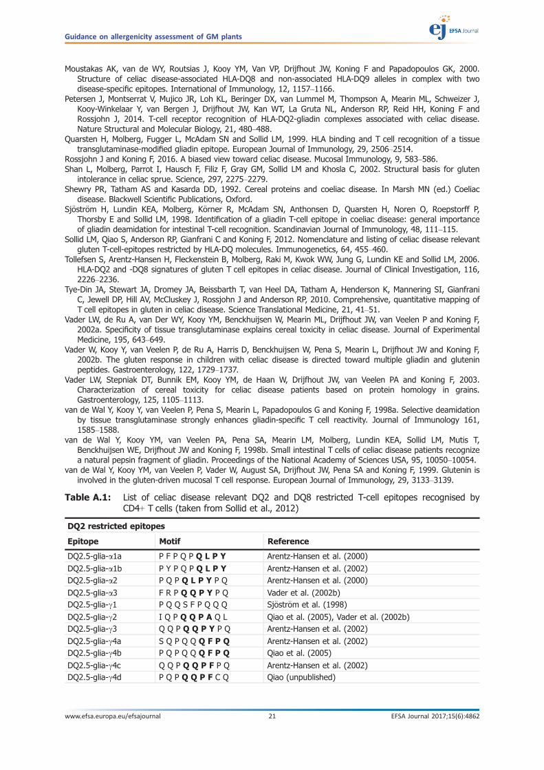
Table A.1: List of celiac disease relevant DQ2 and DQ8 restricted T-cell epitopes recognised byCD4+ T cells (taken from Sollid et al., 2012)
DQ2 restricted epitopes
Epitope Motif Reference
DQ2.5-glia-a1a P F P Q P Q L P Y Arentz-Hansen et al. (2000)
DQ2.5-glia-a1b P Y P Q P Q L P Y Arentz-Hansen et al. (2002)DQ2.5-glia-a2 P Q P Q L P Y P Q Arentz-Hansen et al. (2000)
DQ2.5-glia-a3 F R P Q Q P Y P Q Vader et al. (2002b)DQ2.5-glia-c1 P Q Q S F P Q Q Q Sj€ostr€om et al. (1998)
DQ2.5-glia-c2 I Q P Q Q P A Q L Qiao et al. (2005), Vader et al. (2002b)DQ2.5-glia-c3 Q Q P Q Q P Y P Q Arentz-Hansen et al. (2002)
DQ2.5-glia-c4a S Q P Q Q Q F P Q Arentz-Hansen et al. (2002)DQ2.5-glia-c4b P Q P Q Q Q F P Q Qiao et al. (2005)
DQ2.5-glia-c4c Q Q P Q Q P F P Q Arentz-Hansen et al. (2002)DQ2.5-glia-c4d P Q P Q Q P F C Q Qiao (unpublished)
Guidance on allergenicity assessment of GM plants
www.efsa.europa.eu/efsajournal 21 EFSA Journal 2017;15(6):4862

Annex A-2 Considerations for sequence identity searches
Closer inspection of the Q/E-X1-P-X2 sequences demonstrates that only a limited number of aminoacids are present at the X-positions: L, Q, F, S or E at position X1 and Y, F, A, V or Q at position X2(Figure 3). Thus, a search motif that incorporates these amino acids should identify peptide sequenceswith the potential to bind to HLA-DQ2.5 and stimulate gluten-specific T cells. Additional considerationson position and nature of adjacent amino acid sequences to the Q/E-X1-P-X2 motif should also betaken into account. For example, positively charged amino acids in general diminish the likelihood ofDQ-binding and T-cell recognition, in particular positive charge at positions p1, p4, p6, p7 and p9 aredetrimental. It is noted that P-P is not identified in T-cell epitopes. In contrast, P-X-P in addition toQ/E-X1-P-X2 is associated with the most immunogenic epitopes.
As discussed in the document, any 9 amino acid-residue peptide which shows identity to a knownT-cell epitope might be able to induce an immune response in CD patients. However, the necessarynumber of amino acids identical in a peptide to trigger a response is challenging to define, becausethe ability to bind to CD-specific MHC molecules and the interaction with T cells is highly dependent onthe nature and position of certain amino acids. Therefore, a definite size cut-off in respect to identityto a known epitope indicating potential hazardous peptides, for which further assessment would beneeded, is demanding. Single amino acids substitutions were described to abolish T-cell reactivity inepitopes of a-2 gliadin (Ellis et al., 2003, Mitea et al., 2010). On the other hand, a major alpha gliadinpeptide of wheat (glia-a1b, Table A.1) shares five out of nine amino acids with peptides in oat (DQ2.5-ave-1(b)), which are highly suspected to induce immune responses in some celiac disease patients(Vader et al., 2003, Arentz-Hansen et al., 2004). In this case, however, the motif Q/E-X1-P-X2 ispresent in both sequences.
Considering the search for sequence identity without the motif, based on current knowledge andgiven that the vast majority of sequences that do not contain the motif Q/E-X1-P-X2 are DQ8 epitopes,it is reasonable to consider allowing three amino acid mismatches. It is highly unlikely that more thanthree amino acid mismatches in sequences lacking the motif Q/E-X1-P-X2 would result in a safetyconcern. For DQ8 epitopes, the three amino acids mismatch consideration excludes a potentialexchange of glutamines (Q) at position 1 and/or position 9 by glutamic acid (E) where E is known to
DQ2 restricted epitopes
Epitope Motif Reference
DQ2.5-glia-c5 Q Q P F P Q Q P Q Arentz-Hansen et al. (2002)DQ2.5-glia-x1 P F P Q P Q Q P F Tye-Din et al. (2010)
DQ2.5-glia-x2 P Q P Q Q P F P W Tye-Din et al. (2010)DQ2.2-glut-L1 P F S Q Q Q Q P V Vader et al. (2002b)
DQ2.5-glut-L2 F S Q Q Q Q S P F Stepniak et al. (2005), Vader et al. (2002b)DQ2.5-hor-1 P F P Q P Q Q P F Tye-Din et al. (2010), Vader et al. (2003)
DQ2.5-hor-2 P Q P Q Q P F P Q Vader et al. (2003)DQ2.5-sec-1 P F P Q P Q Q P F Tye-Din et al. (2010), Vader et al. (2003)
DQ2.5-sec-2 P Q P Q Q P F P Q Vader et al. (2003)DQ2.5-ave-1 P Y P E Q Q E P F Arentz-Hansen et al. (2004), Vader et al. (2003)
DQ2.5-ave-1b P Y P E Q Q Q P F Arentz-Hansen et al. (2004), Vader et al. (2003)
DQ8 restricted epitopes
Epitope Motif Reference
DQ8-glia-a1 Q G S F Q P S Q Q van de Wal et al. (1998b)
DQ8-glia-c1a Q Q P Q Q P F P Q Tollefsen et al. (2006)DQ8-glia-c1b Q Q P Q Q P Y P Q Tollefsen et al. (2006)
DQ8-glut-H1 Q G Y Y P T S P Q van de Wal et al. (1999)
The single letter code for amino acids is used. A characteristic Q-X1-P-X2 motif is present in the large majority of HLA-DQ2epitopes (in bold). This sequence is a target sequence for TG2, which yields E-X1-P-X2. Due to the introduction of the negativelycharged amino acid glutamate, the peptides become high affinity binders for HLA-DQ2. glia-a = a-gliadin; glia-c = c-gliadin;hor = hordein; sec = secalin; ave = avenin; glut-L = LWM-glutenin, glut-H = HMW-glutenin.
Guidance on allergenicity assessment of GM plants
www.efsa.europa.eu/efsajournal 22 EFSA Journal 2017;15(6):4862
enhance binding to HLA-DQ8. It is noted that these considerations might require adaptations based onexperience gained in the assessment and on additional knowledge acquired.
Few examples illustrating in practical terms such considerations are provided below.Two immunodominant T-cell epitopes are present in the a-gliadins with partially overlapping
sequences, sequence 1 PFPQPQLPY and sequence 2 PQPQLPYPQ. In these cases, the sequence QLPYand thus the Q-X1-P-X2 motif is present.
– Example 1, the motif is present.
The protein of interest contains the following sequence ALPLTQLPASR that we called sequence 3.The comparison of this sequence with sequences 1 (PFPQPQLPY) and 2 (PQPQLPYPQ) is the following.
Comparison of sequences 3 (ALPLTQLPASR) and 1 (PFPQPQLPY):
At position 1 (p1), the P is replaced by A, both being uncharged small amino acids.At p2, F is replaced by L, both uncharged large amino acids.At p3, an identical amino acid is present.At p4, Q is replaced by L, both uncharged large amino acids.At p5, P is replaced by T, different properties but both small amino acids.At p6, an identical amino acid is present.At p7, an identical amino acid is present.At p8, an identical amino acid is present.At P9, Y is replaced by A. Here, a large amino acid is replaced by a small amino acid but thisdoes not prohibit binding to HLA-DQ2 (Petersen et al., 2016).Overall: there is the potential for cross-reactivity of the 9-amino acid sequence ALPLTQLPA,
additional investigations are needed.
Comparison of sequence 3 (ALPLTQLPASR) but considering first P as p1 and sequence 2(PQPQLPYPQ):
At p1, an identical amino acid is present.At p2, Q is replaced by L, both uncharged large amino acids.At p3, P is replaced by T, different properties but both small amino acids.At p4, an identical amino acid is present.At p5, an identical amino acid is present.At p6, an identical amino acid is present.At P7, Y is replaced by A. Here, a large amino acid is replaced by a small amino acid and atthis position this is known to eliminate T-cell reactivity (Petersen et al., 2016).At p8, P is replaced by S and this is known to eliminate T-cell reactivity (Mitea et al., 2012).At p9, Q is replaced by R, a positively charged amino acid and this is known to have a negativeeffect of binding to HLA-DQ2.Overall: there are three significant differences, two of which are independently known toeliminate T-cell reactivity. Altogether, it is highly unlikely that the nine amino acid sequencePLTQLPASR will bind to HLA-DQ2 and stimulate T cells. No additional investigations for this nineamino acid sequence are needed.
– Example 2, the motif is present.
The protein of interest contains the following sequence KARGVQSPAEI that we called sequence 4.The comparison of this sequence with sequences 1 (PFPQPQLPY) and 2 (PQPQLPYPQ) is the following.
Comparison of sequences 4 (KARGVQSPAEI) and 1 (PFPQPQLPY):
At p1, the P is replaced by K, a positively charged amino acid and this is known to have anegative effect of binding to HLA-DQ2.At p2, F is replaced by A. Here a large amino acid is replaced by a small amino acid.At p3, P is replaced by R, a positively charged amino acid.At p4, Q is replaced by G. Here a large amino acid is replaced by a small amino acid.At p5, P is replaced by V.At p6, an identical amino acid is present.At p7, L is replaced by S. Here, a large amino acid is replaced with a small amino acid that hasdifferent properties.At p8, an identical amino acid is present.
Guidance on allergenicity assessment of GM plants
www.efsa.europa.eu/efsajournal 23 EFSA Journal 2017;15(6):4862

At P9, Y is replaced by A. Here, a large amino acid is replaced by a small amino acid but thisdoes not prohibit binding to HLA-DQ2 (Petersen et al., 2016).Overall: there are six significant differences. Altogether, it is highly unlikely that this sequencewill bind to HLA-DQ2 and stimulate T cells. No additional investigations are needed.
Comparison of sequences 4 (KARGVQSPAEI) but considering R as p1, and sequence 2 (PQPQLPYPQ):
At p1, the P is replaced by R, a positively charged amino acid and this is known to have anegative effect of binding to HLA-DQ2.At p2, Q is replaced by G. Here, a large amino acid is replaced by a small amino acid.At p3, P is replaced by V.At p4, an identical amino acid is present.At p5, L is replaced by S. Here, a large amino acid is replaced by a small amino acid that hasdifferent properties.At p6, an identical amino acid is present.At P7, Y is replaced by A. Here, a large amino acid is replaced by a small amino acid and atthis position this is known to eliminate T-cell reactivity (Petersen et al., 2016).At p8, P is replaced by E, a negatively charged amino acid.At p9, Q is replaced by I, both large non-charged amino acids.Overall: there are five significant differences, one of which is known to eliminate T-cellreactivity by its own. Altogether, it is highly unlikely that this sequence will bind to HLA-DQ2and stimulate T cells. No additional investigations are needed.
– Example 3, the motif is not present.
The protein of interest contains the following sequence: EGSIQAGQQ. This sequence has someidentity to the HLA-DQ8 epitope QGSFQPSQQ where four mismatches (including an exchange of a Qby an E at position 1) can be identified.
Comparison of sequence EGSIQAGQQ with sequence QGSFQPSQQ:
At p1, Q has been replaced by E, as previously mentioned; this change is known to enhancebinding to HLA-DQ8.At p2, an identical amino acid is present. This is an important T-cell receptor contact residue(Broughton et al., 2012).At p3, an identical amino acid is present. This is an important T-cell receptor contact residue(Broughton et al., 2012).At p4, F is replaced by I, both large uncharged amino acids.At p5, an identical amino acid is present. This is an important T-cell receptor contact residue(Broughton et al., 2012).At p6, P is replaced by A, both small uncharged amino acids.At p7, S is replaced by G, both small amino acids but with different properties.At p8, an identical amino acid is present. This is an important T-cell receptor contact residue(Broughton et al., 2012).At p9, an identical amino acid is present. This is an HLA-DQ8 anchor residue.Overall: despite the four amino acid mismatch critical amino acids for HLA-binding and T-cellrecognition are conserved (including a replacement at p1 by E). There is the potential forcross-reactivity, additional investigations are needed.
Annex A-3. Considerations for HLA-DQ peptide modelling
Recent studies have determined the T-cell receptor repertoire used by CD4+ T cells specific forimmunodominant gluten epitopes and crystal structures of such T-cell receptors bound to the HLA-DQ-gluten complexes were determined. As a consequence, detailed knowledge is available indicatingwhich amino acids in the gluten peptides are responsible for the high-affinity binding to HLA-DQ andwhich amino acids mediate the specific interaction with the T-cell receptor. Several publicationsdescribing the binding of CD peptides to HLA-DQ molecules are available. The coordinates of all thesestructures are available through public databases (Broughton et al., 2012; Henderson et al., 2007; Kimet al., 2004; Petersen et al., 2014; Petersen et al., 2015, 2016; Rossjohn and Koning, 2016). As anexample, the x-ray coordinates of HLA-DQ8 bound to a gliadin peptide were deposited in the ProteinData Bank (www.rcsb.org) with the accession number 2NNA while the coordinates and structurefactors for the HLA-DQ2-gliadin complexes have been deposited in the Protein Data Bank under the
Guidance on allergenicity assessment of GM plants
www.efsa.europa.eu/efsajournal 24 EFSA Journal 2017;15(6):4862
following accession codes: S2 TCR–DQ2.5-glia-a1a, 4OZI; S16 TCR–HLA-DQ2a2, 4OZH; D2 TCR–HLA-DQ2a2, 4OZG; JR5.1 TCR–HLA-DQ2a2, 4OZF. This extensive information allows modelling studies inwhich the gluten peptide can be replaced by any peptide sequence of choice (Moustakis et al., 2000;Wiesner et al., 2008; van Heemst et al., 2015). It can be anticipated that, if the selected peptide islikely to bind to HLA-DQ in a way that it resembles the known structure of gluten peptides bound toHLA-DQ, it might have the capacity to stimulate gluten-specific T cells (Figure A.1, Wiesner et al.,2008). Thus, molecular modelling can be employed to aid the determination of potential T-cellstimulatory properties of peptide sequences.
Considering the great relevance of this HLA-DQ peptide modelling step, the future development ofa publicly available software tool specifically designed for risk assessment purposes is desirable.
A non-exhaustive list of selected publications reporting on HLA-DQ-modelling assays:
Broughton SE, Petersen J, Theodossis A, Scally SW, Loh KL, Thompson A, van Bergen J, Kooy-Winkelaar Y,Henderson KN, Beddoe T, Tye-Din JA, Mannering SI, Purcell AW, McCluskey J, Anderson RP, Koning F, Reid Hand Rossjohn J, 2012. Biased T cell receptor usage directed against human leukocyte antigen DQ8-restrictedgliadin peptides is associated with Celiac Disease. Immunity, 37, 611–621.
Henderson KN, Tye-Din JA, Reid HH, Chen Z, Borg NA, Beissbarth T, Tatham A, Mannering SI, Purcell AW, DudekNL, van Heel DA, McCluskey J, Rossjohn J and Anderson RP, 2007. A structural and immunological basis for therole of human leukocyte antigen DQ8 in celiac disease. Immunity, 27, 23–34.
Kim CY, Quarsten H, Bergseng E, Khosla C and Sollid LM, 2004. Structural basis for HLA-DQ2-mediatedpresentation of gluten epitopes in celiac disease. Proceedings of the National Academy of Sciences of theUnited States of America, 101, 4175–4179.
Moustakas AK, van de WY, Routsias J, Kooy YM, Van VP, Drijfhout JW, Koning F and Papadopoulos GK, 2000.Structure of celiac disease-associated HLA-DQ8 and non-associated HLA-DQ9 alleles in complex with twodisease-specific epitopes. International Immunology, 12, 1157–1166.
Petersen J, Montserrat V, Mujico JR, Loh KL, Beringer DX, van Lummel M, Thompson A, Mearin ML, Schweizer J,Kooy-Winkelaar Y, van Bergen J, Drijfhout JW, Kan WT, La Gruta NL, Anderson RP, Reid HH, Koning F andRossjohn J, 2014. T-cell receptor recognition of HLA-DQ2-gliadin complexes associated with celiac disease.Nature Structural and Molecular Biology, 21, 480–488.
Petersen J, Kooy-Winkelaar Y, Loh KL, Tran M, van Bergen J, Koning F, Jamie Rossjohn J and Reid HH, 2016.Diverse T cell receptor gene usage in HLA-DQ8-associated celiac disease converges into a consensus bindingsolution. Structure, 24, 1643–1657.
Petersen J, van Bergen J, Loh K, Kooy-Winkelaar Y, Beringer DX, Thompson A, Bakker SF, Mulder CJ, Ladell K,McLaren JE, Price DA, Rossjohn J, Reid HH and Koning F, 2015. Determinants of gliadin-specific T cell selectionin celiac disease. Journal of Immunology, 194, 6112–6122.
Rossjohn J and Koning F, 2016. A biased view toward celiac disease. Mucosal Immunology, 9, 583–586.van Heemst J, Jansen DT, Polydorides S, Moustakas AK, Bax M, Feitsma AL, Bontrop-Elferink DG, Baarse M, van
der Woude D, Wolbink GJ, Rispens T, Koning F, de Vries RR, Papadopoulos GK, Archontis G, Huizinga TW andToes RE, 2015. Crossreactivity to vinculin and microbes provides a molecular basis for HLA-based protectionagainst rheumatoid arthritis. Nature Communications, 6, 6681.
Wiesner M, Stepniak D, de Ru AH, Moustakis AK, Drijfhout JW, Papadopoulos GK, van Veelen PA and Koning F,2008. Dominance of an alternative CLIP-sequence in the celiac disease associated HLA-DQ2 molecule.Immunogenetics, 60, 551–555.
Guidance on allergenicity assessment of GM plants
www.efsa.europa.eu/efsajournal 25 EFSA Journal 2017;15(6):4862
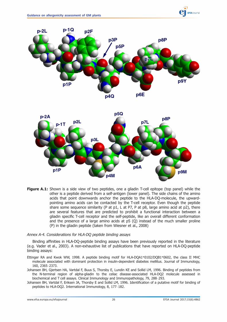
Annex A-4. Considerations for HLA-DQ peptide binding assays
Binding affinities in HLA-DQ-peptide binding assays have been previously reported in the literature(e.g. Vader et al., 2003). A non-exhaustive list of publications that have reported on HLA-DQ-peptidebinding assays:
Ettinger RA and Kwok WW, 1998. A peptide binding motif for HLA-DQA1*0102/DQB1*0602, the class II MHCmolecule associated with dominant protection in insulin-dependent diabetes mellitus. Journal of Immunology,160, 2365–2373.
Johansen BH, Gjertsen HA, Vartdal F, Buus S, Thorsby E, Lundin KE and Sollid LM, 1996. Binding of peptides fromthe N-terminal region of alpha-gliadin to the celiac disease-associated HLA-DQ2 molecule assessed inbiochemical and T cell assays. Clinical Immunology and Immunopathology, 79, 288–293.
Johansen BH, Vartdal F, Eriksen JA, Thorsby E and Sollid LM, 1996. Identification of a putative motif for binding ofpeptides to HLA-DQ2. International Immunology, 8, 177–182.
Figure A.1: Shown is a side view of two peptides, one a gliadin T-cell epitope (top panel) while theother is a peptide derived from a self-antigen (lower panel). The side chains of the aminoacids that point downwards anchor the peptide to the HLA-DQ-molecule, the upward-pointing amino acids can be contacted by the T-cell receptor. Even though the peptideshare some sequence similarity (P at p1, L at P7, P at p8, large amino acid at p2), thereare several features that are predicted to prohibit a functional interaction between agliadin specific T-cell receptor and the self-peptide, like an overall different conformationand the presence of a large amino acids at p5 (Q) instead of the much smaller proline(P) in the gliadin peptide (taken from Wiesner et al., 2008)
Guidance on allergenicity assessment of GM plants
www.efsa.europa.eu/efsajournal 26 EFSA Journal 2017;15(6):4862

Johansen BH, Jensen T, Thorpe CJ, Vartdal F, Thorsby E and Sollid LM, 1996. Both alpha and beta chainpolymorphisms determine the specificity of the disease-associated HLA-DQ2 molecules, with beta chainresidues being most influential. Immunogenetics, 45, 142–150.
Kwok WW, Nepom GT and Raymond FC, 1995. HLA-DQ polymorphisms are highly selective for peptide bindinginteractions. Journal of Immunology, 155, 2468–2476.
Nepom BS, Nepom GT, Coleman M and Kwok WW, 1996. Critical contribution of beta chain residue 57 in peptidebinding ability of both HLA-DR and -DQ molecules. Proceedings of the National Academy of Sciences of theUnited States of America, 93, 7202–7206.
Stepniak D, Wiesner M, de Ru AH, Moustakas AK, Drijfhout JW, Papadopoulos GK, van Veelen PA and Koning F,2008. Large-scale characterization of natural ligands explains the unique gluten-binding properties of HLA-DQ2.Journal of Immunology, 180, 3268–3278.
Straumfors A, Johansen BH, Vartdal F, Sollid LM, Thorsby E and Buus S, 1998. A peptide-binding assay for thedisease-associated HLA-DQ8 molecule. Scandinavian Journal of Immunology, 47, 561–567.
Terreaux C, Walk T, van de Wal Y, Koning F, Jung G and Fleckenstein B, 1998. Increased HLA-DQ2-affinity of asynthetic gliadin peptide by acid-induced deamidation of glutamine residues. Bioorganic and MedicinalChemistry Letters, 8, 2039–2044.
Vader W, Stepniak D, Kooy Y, Mearin L, Thompson A, van Rood JJ, Spaenij L and Koning F, 2003. The HLA-DQ2gene dose effect in celiac disease is directly related to the magnitude and breadth of gluten-specific T cellresponses. Proceedings of the National Academy of Sciences of the United States of America, 100, 12390–12395.
van de Wal Y, Kooy YM, Drijfhout JW, Amons R, Papadopoulos GK and Koning F, 1997. Unique peptide bindingcharacteristics of the disease-associated DQ(alpha 1*0501, beta 1*0201) vs the non-disease-associated DQ(alpha 1*0201, beta 1*0202) molecule. Immunogenetics, 46, 484–492.
van de Wal Y, Kooy YM, Drijfhout JW, Amons R and Koning F, 1996. Peptide binding characteristics of the coeliacdisease-associated DQ(alpha1*0501, beta1*0201) molecule. Immunogenetics, 44, 246–253.
van de Wal Y, Amons R and Koning F, 2000. Characterization of HLA-DQ-specific peptide-binding motifs. Methodsin Molecular Medicine, 41, 97–103.
Vartdal F, Johansen BH, Friede T, Thorpe CJ, Stevanovi�c S, Eriksen JE, Sletten K, Thorsby E, Rammensee HG andSollid LM, 1996. The peptide binding motif of the disease associated HLA-DQ (alpha 1* 0501, beta 1* 0201)molecule. European Journal of Immunology, 26, 2764–2772.
Wiesner M, Stepniak D, de Ru AH, Moustakis AK, Drijfhout JW, Papadopoulos GK, van Veelen PA and Koning F,2008. Dominance of an alternative CLIP sequence in the celiac disease associated HLA-DQ2 molecule.Immunogenetics, 60, 551–555.
Annex A-5. Non-exhaustive list of publications that have reported on gluten-specific T cells
Anderson RP, Degano P, Godkin AJ, Jewell DP and Hill AV, 2000. In vivo antigen challenge in celiac diseaseidentifies a single transglutaminase-modified peptide as the dominant A-gliadin T-cell epitope. Nature Medicine,6, 337–342
Arentz-Hansen H, Fleckenstein B, Molberg, Scott H, Koning F, Jung G, Roepstorff P, Lundin KE and Sollid LM, 2004.The molecular basis for oat intolerance in patients with celiac disease. Plos Medicine, 1, e1.
Arentz-Hansen H, K€orner R, Molberg, Quarsten H, Vader W, Kooy YM, Lundin KEA, Koning F, Roepstorff P, SollidLM and McAdam SN, 2000. The intestinal T cell response to a-gliadin in adult celiac disease is focused on asingle deamidated glutamine targeted by tissue transglutaminase. Journal of Experimental Medicine, 191, 603–612.
Molberg, McAdam SN, K€orner R, Quarsten H, Kristiansen C, Madsen L, Fugger L, Scott H, Roepstorff P, LundinKEA, Sj€ostr€om H and Sollid LM, 1998. Tissue transglutaminase selectively modifies gliadin peptides that arerecognized by gut-derived T cells. Nature Medicine, 4, 713–717.
Moustakas AK, van de WY, Routsias J, Kooy YM, Van VP, Drijfhout JW, Koning F and Papadopoulos GK, 2000.Structure of celiac disease-associated HLA-DQ8 and non-associated HLA-DQ9 alleles in complex with twodisease-specific epitopes. International Immunology, 12, 1157–1166.
Petersen J, Montserrat V, Mujico JR, Loh KL, Beringer DX, van Lummel M, Thompson A, Mearin ML, Schweizer J,Kooy-Winkelaar Y, van Bergen J, Drijfhout JW, Kan WT, La Gruta NL, Anderson RP, Reid HH, Koning F andRossjohn J, 2014. T-cell receptor recognition of HLA-DQ2-gliadin complexes associated with celiac disease.Nature Structural and Molecular Biology, 21, 480–488.
Quarsten H, Molberg, Fugger L, McAdam SN and Sollid LM, 1999. HLA binding and T cell recognition of a tissuetransglutaminase-modified gliadin epitope. European Journal of Immunology, 29, 2506–2514.
Shan L, Molberg, Parrot I, Hausch F, Filiz F, Gray GM, Sollid LM and Khosla C, 2002. Structural basis for glutenintolerance in celiac sprue. Science, 297, 2275–2279.
Sj€ostr€om H, Lundin KEA, Molberg, K”rner R, McAdam SN, Anthonsen D, Quarsten H, Noren O, Roepstorff P,Thorsby E and Sollid LM, 1998. Identification of a gliadin T-cell epitope in coeliac disease: general importanceof gliadin deamidation for intestinal T-cell recognition. Scandinavian Journal of Immunology, 48, 111–115.
Guidance on allergenicity assessment of GM plants
www.efsa.europa.eu/efsajournal 27 EFSA Journal 2017;15(6):4862
Tollefsen S, Arentz-Hansen H, Fleckenstein B, Molberg, Raki M, Kwok WW, Jung G, Lundin KE and Sollid LM, 2006.HLA-DQ2 and -DQ8 signatures of gluten T cell epitopes in celiac disease. The Journal of Clinical Investigation,116, 2226–2236.
Tye-Din JA, Stewart JA, Dromey JA, Beissbarth T, van Heel DA, Tatham A, Henderson K, Mannering SI, GianfraniC, Jewell DP, Hill AV, McCluskey J, Rossjohn J and Anderson RP, 2010. Comprehensive, quantitative mapping ofT cell epitopes in gluten in celiac disease. Science Translational Medicine, 2, 41–51.
Vader LW, de Ru A, van Der WY, Kooy YM, Benckhuijsen W, Mearin ML, Drijfhout JW, van Veelen P and Koning F,2002a. Specificity of tissue transglutaminase explains cereal toxicity in celiac disease. Journal of ExperimentalMedicine, 195, 643–649.
Vader LW, Stepniak DT, Bunnik EM, Kooy YM, de Haan W, Drijfhout JW, van Veelen PA and Koning F, 2003.Characterization of cereal toxicity for celiac disease patients based on protein homology in grains.Gastroenterology, 125, 1105–1113.
Vader W, Kooy Y, van Veelen P, de Ru A, Harris D, Benckhuijsen W, Pena S, Mearin L, Drijfhout JW and Koning F,2002b. The gluten response in children with celiac disease is directed toward multiple gliadin and gluteninpeptides. Gastroenterology, 122, 1729–1737.
van de Wal Y, Kooy Y, van Veelen P, Pena S, Mearin L, Papadopoulos G and Koning F, 1998a. Selective deamidationby tissue transglutaminase strongly enhances gliadin-specific T cell reactivity. The Journal of Immunology, 161,1585–1588.
van de Wal Y, Kooy YM, van Veelen P, Vader W, August SA, Drijfhout JW, Pena SA and Koning F, 1999. Glutenin isinvolved in the gluten-driven mucosal T cell response. European Journal of Immunology, 29, 3133–3139.
van de Wal Y, Kooy YM, van Veelen PA, Pena SA, Mearin LM, Molberg, Lundin KEA, Sollid LM, Mutis T,Benckhuijsen WE, Drijfhout JW and Koning F, 1998b. Small intestinal T cells of celiac disease patients recognisea natural pepsin fragment of gliadin. Proceedings of the Nationall Academy of Sciences United States ofAmerica, 95, 10050–10054.
Annex A-6. Considerations for in vitro digestibility tests
As specified above and because a 9-amino acid core is almost invariably required for efficientpeptide binding to HLA-DQ, proteins that are easily degraded into fragments shorter than 9 aminoacids are unlikely to harbour T-cell stimulatory epitopes. Thus, determination of proteolytic resistanceof proteins can aid in the identification of proteins with potential harmful potential. For more details,please see chapter on in vitro protein digestibility in this document (Annex B).
Guidance on allergenicity assessment of GM plants
www.efsa.europa.eu/efsajournal 28 EFSA Journal 2017;15(6):4862
Annex B – In vitro protein digestibility tests
As specified in Section 2.2 of this document, an interim phase (~2 years duration) is proposed toassess the utility of an enhanced in vitro digestion test, with a review undertaken in the final year.During this phase, the laboratory(ies) involved will detail and apply the refined digestion testmethodology. After this period, EFSA will discuss whether the test adds value and, if so, what furthersteps are needed for its final implementation.
Annex B-1. Background
In vitro digestibility tests can provide useful data on the susceptibility of a protein to digestionwhich may reflect its digestibility in the human gastrointestinal system and subsequent presentation tothe host’s immune system (Foster et al., 2013). There is evidence that gastrointestinal digestion canaffect the immunogenicity of dietary proteins in relation to both IgE and non-IgE-mediated adversereactions to foods as discussed below. However, notably, the ability of digestion-resistant dietaryproteins or derived fragments to initiate diseases, such as IgE-mediated allergies or celiac disease(CD), also depends on the predisposition of the host. For CD, expression of certain HLA genotypes is awell described risk factor for developing the condition (See Section 2.1). However, while a geneticpredisposition towards atopy is also thought to play a role in the development of IgE-mediatedallergies to a variety of environmental agents including foods, specific genetic and other risk factorsinvolved in the development of specific IgE have yet to be defined. This makes the risk assessmentprocess less certain for IgE-mediated allergies compared to CD.
IgE-mediated adverse reactions to foods: The events leading to the breakdown of oral tolerance,allergic sensitisation and development of food allergies in susceptible individuals are poorlyunderstood. It is likely that multiple pathways could ultimately lead to a failure to develop or loss oforal tolerance (Chinthrajah et al., 2016). Notwithstanding the role of route of exposure in sensitisation,understanding how proteins are presented to the gastrointestinal mucosal immune system mayprovide insight into the mechanisms controlling the balance between tolerisation and sensitisation.Furthermore, oral exposure is also central to non-IgE assessments where gastrointestinal digestion hasbeen demonstrated to be important in delivery of immunologically active fragments to gastrointestinalmucosal sites. Impaired gastric digestion of food allergens has been associated with both developmentof IgE responses to foods (sensitisation) and modulating the severity of IgE-mediated reactions tofoods (elicitation). A recent study of patients undergoing gastric-bypass surgery, a procedure known toreduce post-prandial gastric acidity and where the bulk of food ingested reaches the small intestinewithout prior gastric digestion, resulted in a significant increase in sensitisation to food (Shakeri-Leidenmuhler et al., 2015). Impaired digestion of cod fish proteins may also be a risk factor for severereactions in fish allergic individuals (Untersmayr et al., 2007). Studies on animal models have shownthat suppression of gastric acid secretion using widely prescribed antacid medication increasespropensity to sensitisation to proteins from cod fish (Untersmayr et al., 2003), celery (Untersmayret al., 2008), hazelnut (Scholl et al., 2005) and egg (Diesner et al., 2008). Furthermore, an associationbetween the use of antiulcer drugs and the induction of IgE-mediated allergy to a variety of foodssuch as milk, potato, celery, carrots, apple, orange, wheat, and rye flour has been reported(Untersmayr et al., 2005). Such studies suggest that elevated gastric pH, and the resulting reduction inpeptic digestion, may enhance the potential of foods to cause allergies. Uptake of allergens into thecirculation may play an important role in eliciting allergic reactions (Strait et al., 2011) but data inhumans are sparse and conflicting as to the role of allergen uptake in triggering reactions, althoughuptake appears much greater in allergic than healthy individuals, as indicated in studies of wheatallergy (Brockow et al., 2015; Matsuo et al., 2005).
Non-IgE-mediated adverse reactions to foods: Studies seeking to define the structural basis of thetoxicity of gluten in CD have made extensive use of in vitro gastrointestinal digestion, including theaction of brush border proteases, to generate physiologically relevant fragments of gluten. Thesestudies notably identified a 33mer peptide that is especially resistant to digestion and which is likely topersist in the brush border with a half-life estimated to be ~20 h. This peptide is also an excellentsubstrate for tissue transglutaminase TG2 and was a potent stimulator of T cells from celiac patients(Shan et al., 2002).
Such data support the premise that immunologically active fragments of food proteins persisting inthe gastrointestinal lumen can play a role in driving immune-mediated adverse reactions to foods.Persistent, soluble intact proteins and fragments are more likely to be sampled by the gastrointestinal
Guidance on allergenicity assessment of GM plants
www.efsa.europa.eu/efsajournal 29 EFSA Journal 2017;15(6):4862
epithelium and are hence exposed to cells of the immune system as potentially immunogenicfragments, with particulates being sampled by M cells within the Peyer’s patches. In animal models,there is evidence that uptake of particulate peanut protein bodies by M cells may promote sensitisationby virtue of their size, with soluble material being taken up by the same route (Chambers et al., 2004).Although the exact mechanism(s) of antigen uptake in the intestine are still to be clearly elucidated(Pabst and Mowat, 2012), it is generally accepted that most food allergens should retain sufficientstructural integrity as they pass through the human gastrointestinal tract to sensitise or elicit anallergic response (Metcalfe et al., 1996). After uptake by antigen presenting cells, peptides generatedthrough endosomal proteolysis bind to MHC class II molecules (Rudensky et al., 1991; Chicz et al.,1993). Peptides shorter than 9 amino acid residues are probably unable to bind to MHC class IImolecules and to activate T cells because there is a minimum length requirement – the so called‘peptide binding register’ (Mohan and Unanue, 2012). Thus, peptides shorter than 9 amino acidresidues are highly unlikely to stimulate an immune response. However, unlike celiac disease, IgE-mediated allergies involve development of humoral (i.e. antibody) responses which requires activationof both B cells and T cells. The B-cell receptors involved in B-cell activation have a requirement formultivalent antigen and it has emerged that the way in which B cells encounter antigen in vivodepends on its properties, such as size (Harwood et al., 2010). As a consequence, larger peptide sizesare required for development of IgE responses. Thus, digestion of the allergen b-lactoglobulin intofragments of less than 3,000 Da abolished the proteins immunogenicity (Bogh et al., 2013), whilepeptides of less than 1,500 Da resulting from pepsinolysis of peanut Ara h 1, had markedly reducedimmunogenicity, which was completely lost when the digest was fractionated (Bogh et al., 2012). Withregard to elicitation of reactions, synthetic antigens have been used to investigate the mechanism ofdegranulation (Handlogten et al., 2013). These studies have shown that a minimum of two distinctepitopes is required to trigger degranulation, with the epitopes separated by a maximum of 6.4 nm onthe synthetic antigen. On this basis peptides of at least 3–5 kDa would be required to cross-link IgEbound to the surface of effector cells, such as mast cells and basophils. However, peptides as small as791 Da and 1413 Da resulting from the pepsinolysis of the avocado pear allergen Prs s 1, have beenfound to inhibit IgE binding to crude avocado extract in ELISA-inhibition assays and to elicit reactionsin vivo using skin testing, respectively (Diaz-Perales et al., 2003). Such peptides are unlikely to carrymultiple IgE epitopes, suggesting that peptide aggregation may play a role in triggering degranulation.
To either sensitise or trigger an allergic reaction in an already sensitised individual, a food proteinneeds to be ‘bioaccessible’ by the hosts’ immune system. ‘Bioaccessibility’ describes the ability of achemical entity (such as a protein) to be released from food during the digestive process, which canconsequently interact with and/or be absorbed by the gastrointestinal epithelium (Holst andWilliamson, 2008). The EFSA Guidance Document for risk assessment of food and feed fromgenetically modified plants published in 2011 states that ‘the impact of the possible interactionbetween the protein and other components of the matrix as well as the effects of the processingshould be taken into account in in vitro digestibility tests’ (EFSA GMO Panel, 2011). In vitro digestibilitytests have been applied to investigate the effects of digestion on the food matrix, and how processingconditions (including thermal treatment) may affect susceptibility to simulated gastrointestinalproteolysis (e.g. Minekus et al., 2014, Smith et al., 2015). However, given the diversity of foodmatrices and food processing procedures, our knowledge of their effects on susceptibility of proteins todigestion is limited. As a consequence, the effects of processing and of the food matrix on thesusceptibility of a particular protein to digestion are difficult to predict. Because there is no effectiveanimal model for food allergy, studies are often limited to investigating the impact of the food matrixon elicitation of allergic reactions in allergic human subjects while data on allergic sensitisation is verylimited regarding this aspect (Ballmer-Weber et al., 2002; Bartnikas et al., 2012; Brenna et al., 2000;Grimshaw et al., 2003; Mackie et al., 2012; Netting et al., 2013; Worm et al., 2009).
Annex B-2. Types of in vitro digestibility tests
The pepsin resistance test, which is embedded in EFSA Guidance Document (EFSA GMO Panel,2011) and Codex Alimentarius (2003, 2009), is currently used for allergenicity risk assessment as anadditional piece of information within the weight-of-evidence approach, because no single test is fullypredictive of the allergenic potential of a protein. The classical pepsin resistance test has severallimitations (EFSA GMO Panel, 2010) including:
Guidance on allergenicity assessment of GM plants
www.efsa.europa.eu/efsajournal 30 EFSA Journal 2017;15(6):4862

• the pH value usually employed in the assay is extremely acidic. Because pepsin activity is pHdependent, the pattern of proteolysis may not reflect the one likely to be found in vivo otherthan at the end of gastric emptying or in the fasted state;
• pepsin is added in a gross excess to the protein substrate, affecting the kinetics of thedigestion;
• the correlation with allergenicity of proteins has been questioned. Studies comparing thedigestibility of allergens with that of non-allergenic dietary proteins showed that food allergenswere not always inherently more stable to pepsin digestion than non-allergenic proteins (Fuet al., 2002; Thomas et al., 2004; Herman et al., 2007).
While this test may contribute to understanding the biochemical properties of newly expressedproteins (information on the physicochemical stability of a protein, such as the rigidity of thepolypeptide backbone at low pH, determine susceptibility to pepsinolysis), it does not allow tocharacterise how these proteins behave under the overall physiological conditions encountered in thedigestive tract. Furthermore, the test does not reflect changes in the digestive process that take placeacross the life course (R�emond et al., 2015). These limitations were previously highlighted by EFSA(EFSA GMO Panel, 2010). In addition, the EFSA Guidance Document for risk assessment of food andfeed from genetically modified plants (EFSA GMO Panel, 2011) indicates that ‘the digestibility of thenewly expressed proteins in specific segments of the population such as infants and individuals withimpaired digestive functions may be assessed employing in vitro digestibility tests using differentconditions’. Given these considerations, it is proposed to embed the classical pepsin resistance testwithin a suite of tests aimed to characterise how a newly expressed protein may behave during thedigestive process (see Annex B-5).
Gastrointestinal digestion assays usually aim to simulate ‘normal’ digestive function with regard todigestive enzymes and acid secretion (Macierzanka et al., 2009; Moreno, 2007; Minekus 2014).However, alterations in the digestive milieu are often observed in patients with various gastrointestinalconditions (Kay and Jorgensen, 1994), in young infants with underdeveloped digestive system(Armand et al., 1996) and the elderly with a weakened digestive function (Hosking et al., 1975). In anin vitro study assessing the impact of digestion on celery allergenicity in an aged population, decreasedgastric proteolysis was identified (Untersmayr et al., 2008). Simulated infant digestion models (Dupontet al., 2010; M�enard et al., 2014) were used to study the gastrointestinal tolerance of casein and a-lactalbumin and to optimise the milk processing and formula production. Experiments using simulatedgastric fluid where the pH was increased were used to assess the stability of allergens in fish, milk andhazelnut in patients taking antacids where intragastric pH is increased (Scholl et al., 2005; Untersmayret al., 2005; Untersmayr et al., 2007). Another study developed a gastrointestinal model simulating thephysicochemical conditions of the elderly’s gastrointestinal tract which was applied to investigate thefate of bovine whey proteins (Levi and Lesmes, 2014). Finally, the COST Infogest network hasproposed a standardised batch gastrointestinal digestion method based on physiologically relevantconditions that could be applied for various endpoints (Minekus et al., 2014).
In vitro models simulating the physiological conditions of gastrointestinal digestion (in either healthyor diseased individuals) by sequential addition of digestive enzymes, biosurfactants and fluids havebeen designed to understand the degradation of proteins and other constituents during digestion.Some in vitro models have also included biosurfactants, such as phosphatidylcholine, which can befound at low levels in gastric juice and which is also a component of bile (Moreno et al., 2005a;Mandalari et al., 2009a). Biosurfactants can have complex effects on the phase behaviour of lipid richfoods, because emulsification is an important aspect of lipid digestion (Macierzanka et al., 2009), butthey may also have effects on digestion of proteins associated with lipids, and there is some evidence,although equivocal, that bile salts may affect the activity of pancreatic proteases. For instance, theactivity of trypsin and chymotrypsin is increased in the presence of bile salts (Robic et al., 2011).
The time course of simulated digestion tests can be based on the residence time of food in thestomach. This is dependent on the type of meal ingested. For example, liquid and solid meals displaydifferent gastric emptying rates after ingestion (R�emond et al., 2015). The half-time (t1/2), whichindicates when 50% of an ingested meal is emptied, ranges from 10 to 60 min for liquid meals,whereas t1/2 values reported for solid foods ranges from 50 to 115 min. Other factors, such as othermeal components, meal volume, caloric content, ratio between liquid and solid in the meal or the typeof dietary fibres also have an influence on the gastric emptying rate.
Digestibility studies using gastrointestinal conditions can provide useful data regarding persistencyof newly expressed proteins and/or of digestion derived immunologically active fragments in the
Guidance on allergenicity assessment of GM plants
www.efsa.europa.eu/efsajournal 31 EFSA Journal 2017;15(6):4862
gastrointestinal lumen which may pose a risk of causing an immune-mediated adverse reaction in asusceptible individual. The presence of digestion resistant fragments only provides an indication ofexposure of the gastrointestinal mucosal surface and is not on its own predictive of allergenicity,because this property is a function of the way in which the fragments interact with the individual.Consequently, resistance to gastrointestinal digestion of a newly expressed protein should beconsidered as part of the assessment for its potential to cause allergic reactions via the oral route.However, it should also be noted that other non-oral routes of exposure such as respiratory orcutaneous have to be considered (EFSA GMO Panel, 2010).
There is no internationally accepted model/protocol available to perform gastrointestinal in vitrodigestibility tests for purified proteins although this has been developed for whole foods (Minekus,2014). This is a consensus model applied to several foods and based on available in vivo physiologicaldata resulting from the COST Infogest network. This batch in vitro digestion assay includes an oralphase, as well as subsequent gastric and intestinal phases. Interlaboratory trials were performed atthe European level to assess digestion of skim milk powder (Egger et al., 2016). Such in vitrodigestibility methods will require adaptation to make them useful for analysis of purified proteins,taking into account the need for further standardisation and validation.
There are only two validated (ring-trialled) studies of digestibility tests for purified proteins whichwere published up to date:
• Thomas et al. (2004). Modification of the seminal paper of Astwood et al., (1996) by loweringthe pepsin:protein ratio and looking at the effect of very acidic pH values (1.2 and 2.0). Invitro pepsinolysis of 10 proteins (allergens and non-allergens) at pH 1.2 and 2.0 wereevaluated by nine laboratories. The authors observed that pH did not have an influence on thetime of digestion of protein large-fragments but the detection by gel electrophoresis of lowmolecular weight proteolytic fragments was less consistent because the different fixation andstaining methods used;
• Mandalari et al. (2009b). These authors evaluated the in vitro gastrointestinal digestibility ofb-casein and b-lactoglobulin by using a low-protease assay with and without the addition ofphospholipids (based on Moreno et al., 2005a,b and Mandalari et al., 2009a) and the high-protease assay (based on Astwood et al., 1996 and Fu et al., 2002). Twelve laboratories testedthe method without the addition of phospholipids and five labs studied the effect of theaddition of surfactants. This study demonstrated that the low-protease assay was robust andreproducible although further validation should be undertaken using a more extensive panel ofproteins. In addition, this interlaboratory trial showed that the largest factor governingirreproducibility was the sampling and electrophoresis methods used to analyse digestionproducts.
Annex B-3. In vivo pH range and fluids compositions determined in thegastrointestinal tract
B-3.1. pH conditions in the gastrointestinal tract
There is an increasing body of data on the in vivo fluctuations in pH along the gastrointestinal tractcoming from telemetry studies using single use capsules which house pH and temperature sensors anda wireless transceiver capable of reporting pH measurements and temperature values with highprecision. Nevertheless, much of the data on infants comes from intubation studies where there areconcerns that the placement of probes in the stomach, may have resulted in an underestimation ofintragastric pH and an over-estimation of the buffering effect of food in pre-term infants (Omari andDavidson, 2003). Since the presence of food affects luminal conditions, both by virtue of its ownbuffering capacity and the stimulatory effect food has on gastric and pancreatic secretions, conditionsare summarised below for both the fasted and the fed state.
B-3.1.1. Stomach
Fasted state: Telemetry studies have shown wide fluctuations in intragastric pH in adults, rangingfrom pH 1 to 8 with median values of 1.4–4.6 and an overall mean pH of 2.7 (�0.8) (n = 20) (Kozioleket al., 2015). Other studies also showed fasting intragastric pH to be ~ 2.0 in a group of 12 subjects(Banerjee et al., 2010), or ranges of 1.5–3 with seven subjects (Ono et al., 2009) and 1.0–2.5 (n = 66)(Evans et al., 1988). These study groups were dominated by male subjects so no findings were
Guidance on allergenicity assessment of GM plants
www.efsa.europa.eu/efsajournal 32 EFSA Journal 2017;15(6):4862

available to show any gender variations. Concerning infants, a recent meta-analysis of data from bothpre- and full-term infants indicated the mean intragastric pH in the fasted infant was 2.8 (�0.9)(n = 102) (Kamstrup et al., 2017).
Fed state: After consuming a meal, the intragastric pH increases due to the buffering capacity ofthe food. Thus, intragastric pH in adults is generally around 5.5 and slowly drops to around 1.5 at theend of gastric emptying (Michalek et al., 2011). In one group of healthy full-term newborns (n = 25),the pH increased to ~ 6.4 after feeding and gradually decreased to ~pH 3.5 (Mason, 1962). A secondgroup of 15 healthy preterm infants found that the intragastric pH rose to around 7 after feedingdropping to 1.5–2.0 at the end of gastric emptying (Omari and Davidson, 2003). A meta-analysis of anumber of studies on the changes in intragastric pH of pre- and full-term infants shows the samepatterns as adults with a higher pH (~ 6.5) immediately after feeding, which reduces over time tobetween pH 2.0–4.5 depending on the study (Bourlieu et al., 2014). A second meta-analysis of datacollected in infants (pre- and full-term) showed that the mean intragastric pH in the fed infant is 6.4(�0.6) (n = 102) (Kamstrup et al., 2017).
Drug interventions with proton pump inhibitors (PPI), such as omeprazole or lansoprazole, are usedto treat peptic ulcers and gastroesophageal reflux disease in adults and infants (Omari et al., 2007).Their use results in an increase in fasting gastric pH to 6.0 although the length of treatment requireddepends on the dose and form in which the drugs are given (Ono et al., 2009; Banerjee et al., 2010).Similar effects are observed in infants (Faure et al., 2001). The fed-state intragastric pH of individualstaking PPIs remains around 6.0 and does reduce but only to around 4.5 at the end of gastric emptying(n = 10 and n = 7) (Ono et al., 2009; Banerjee et al., 2010).
B-3.1.2. Small intestine
An early study showed duodenal pH to range from 5.0 to 7.2 with a median value of 6.4 which roseto 7.3 in the distal small intestine (n = 39) (Fallingborg et al., 1989). In a more recent study, fastedmeasurements of pH in the proximal segment of the small bowel gave values of 5.9–6.3 with a meanvalue of 6.0 (� 0.2) while in the distal intestine the pH was higher at around 7.4–7.8 with a meanvalue of 7.7 (� 0.15) (n = 20) (Koziolek et al., 2015). In another study, duodenal pH in the fed statewas found to be 5.2–6.1 (mean of 5.6, n = 17) and did not seem to alter greatly in either the fastedstate or in patients with dyspepsia (Bratten and Jones, 2009). The pH in the distal ileum has also beenfound to be ~ 8.1 in both the fed and fasted state. In another study, the mean pH in the proximalsmall intestine measured in 55 normal subjects was 6.63 (� 0.53), whereas in the terminal ileum was7.49 (�0.46) (n = 58) (Evans et al., 1988).
There is very little data available on the pH of the intestine in infants (either pre- or full-term) withonly two studies reported. These data indicate that the intestinal pH of infants is 6.6–7.0 and onlydrops slightly to about 6.4 after feeding (Kamstrup et al., 2017).
B.3.2. Composition and concentration of digestive enzymes in gastric andintestinal fluids
The current state-of-the-art reveals that gastrointestinal digestion of proteins is a multistageprocess, starting with pepsin cleavage in stomach and proceeding through to the small intestine withdigestion taking place through the action of a mixture of pancreatic endoproteases (such as trypsin,chymotrypsin and elastase) together with amino- and carboxy-peptidases. Finally, at the intestinalepithelial surface further digestion takes place through the action of brush border aminopeptidases(Akimov and Bezuglov, 2012). Collectively, these enzymes reduce proteins down to free amino acidsand the simple di- and tripeptides which can be taken up by transporters on epithelial cell surfaces.
Data on the composition of gastric and intestinal fluids are generally obtained from analysis ofaspirates taken through invasive intubation procedures and consequently data from healthy individualsare sparse. Furthermore, analysis of fluid composition has generally been undertaken to identifydisease biomarkers. Thus, in contrast to gastric and intestinal pH which is directly measured, the levelsof digestive enzymes are normally reported either in activity levels (directly measured in thegastrointestinal chyme but using different and non-standardised activity assays, as well as differentdefinitions of enzyme units), or in enzyme outputs (flow rates). Interpretation of such data, which isobtained from fluids generally collected from individuals either in the fasted state or after treatment tostimulate pancreatic secretion, is further complicated by the fact the caloric content, nutrientcomposition, and physical properties of the ingested meal are all known to be factors that can strongly
Guidance on allergenicity assessment of GM plants
www.efsa.europa.eu/efsajournal 33 EFSA Journal 2017;15(6):4862
influences pancreatic enzyme secretory responses (Keller and Layer, 2005). Therefore, the comparisonof data from different studies is challenging and makes it difficult to identify well-defined levels andactivities of digestive enzymes to be included in in vitro test systems.
B.3.2.1. Gastric juice
Median values for the pepsin content of gastric aspirates in a study of 20 fasted healthy individualsranged from 0.11 to 0.22 mg/mL depending on sampling time (Kalantzi et al., 2006). After eating, thelevels increased from 0.26 to 0.58 mg/mL (Kalantzi et al., 2006). In another study, pepsin activity wasdetermined using haemoglobin as a standard and found to be 349.1 (�14.5) lg/mL using porcinepepsin A (2,100 U/mg) as a standard (n =343) (Metheny et al., 1997). One drawback of the laterstudy is that the patient population was heterogeneous (aged 15–95 years; approximately 54% were> 60 years of age). Around 50% were receiving H2-receptor antagonists and eight omeprazole, a PPI.Post-prandial measurement indicated that there was 87 U/mL pepsin in preterm infants (n = 28)(Armand et al., 1996) as compared to levels of 942–1,333 U/mL in adults (n = 6), levels depending ondiet (Armand et al., 1995). In both the Armand and Metheny studies, pepsin activity was measured ingastric aspirates and using haemoglobin as a substrate. The much lower levels of pepsin, coupled withhigher intragastric pH mean that gastric digestion in infants is very limited (Bourlieu et al., 2014).
The other major gastric enzyme is human gastric lipase. Levels of both gastric lipase and pepsinincrease in infancy as the gastrointestinal tract matures, reaching those of adults by the age of12 months (Bourlieu et al., 2014). In some instances, gastric fluid can contain bile and often containssalivary proteins and salivary mucins. The gastric mucus layer contains of surfactant, believed toenhance its barrier properties with the dominant lipid being phosphatidyl choline and phosphatidylethanolamine (Schmitz and Renooij, 1990). As a consequence, phospholipids find their way into thelumen, with contents of dipalmitoyl phosphatidyl choline ranging between 49 and 95 lg/mL in a groupof 68 children (Chang et al., 2006). In another study, the concentration levels of total phospholipidsfound in the gastric juice of healthy adults ranged from 60.8 to 206.4 lg/mL (n = 15) (Wenner et al.,2000).
B.3.2.2. Pancreatic juice
Pancreatic secretions are highly complex mixtures of proteins, including a wide range ofendoproteases and carboxy- and aminopeptidases, as recent proteomic profiling experiments havedemonstrated. For example, proteomic profiling of gastroduodenal fluid collected during endoscopyallowed 71–89 proteins to be identified in six subjects demonstrated the presence of trypsin,enteropeptidase, chymotrypsin and elastase although many of the isoforms were not identified (Pauloet al., 2010). The gastrointestinal fluid also showed the presence of salivary proteins, such as the basicproline-rich salivary protein, together with a variety of mucins.
Analysis of pancreatic juice from healthy individuals sampled during endoscopic retrogradecholangiopancreatography has been undertaken in a limited number of individuals. One study in threefemale subjects identified 90 proteins with a high degree of confidence using MS–MS analysis (Doyleet al., 2012). Digestive enzymes identified that degrade proteins include carboxypeptidase A1 and A2,chymotrypsin C and chymotrypsinogen B1, elastase 2A, 3A, 3B, trypsin 1, peptide prolyl isomerase,together with other digestive enzymes such as a-amylase 1A, 2A, 2B, carboxyl ester lipase, lipase andco-lipase and phospholipase A2. The same proteins were identified in another proteomic profiling studyof pancreatic juice from individuals with chronic pancreatitis (n = 9) and a similar number of controlsubjects, with the addition of aminopeptidase N and a Xaa-Pro dipeptidase. Semiquantitative analysisof the protein profiles showed that digestive enzymes were downregulated two- to -threefold inpatients with pancreatitis (Paulo et al., 2012).
It has classically been defined that trypsin and chymotrypsin are the most abundant humanpancreatic digestive enzymes making up about 19% and 9%, respectively, of total pancreatic juiceprotein (Whitcomb and Lowe, 2007). One of the first studies showed the levels of trypsin andchymotrypsin drop along the length of the intestine (Borgstrom et al., 1957), presumably as aconsequence of their being inactivated or digested. Determination of trypsin in intestinal fluid hasshown there to be 577 (� 44) mg/L immunoreactive trypsin (n = 8) with an enzymatic activity of11.13 (� 0.12) kU/L of activity (as measured using hydrolysis of N-benzoyl-L-arginine ethyl ester(Rinderknecht et al., 1978a). A mean post-prandial trypsin activity of 150 U/mL of duodenal juice wasdetermined in 6 healthy adults (DiMagno et al., 1977). In another study, trypsin was determined inaspirates using N-benzoyl-L-arginine-p-nitroanilide as a substrate and was determined to be 143
Guidance on allergenicity assessment of GM plants
www.efsa.europa.eu/efsajournal 34 EFSA Journal 2017;15(6):4862

(� 6.7) lg/mL (n = 399) using bovine pancreatic trypsin as a standard (11,000 U/mg) (Metheny et al.,1997).
Analysis of enzyme activities in pancreatic juice of normal subjects found levels of trypsin of24.4 mg/g (with a range of 7.7–60.5, n = 20) while chymotrypsin was found at 4.8 mg/g protein(range of 1–19.4, n = 27) (Rinderknecht et al., 1978a). These authors titrated the enzymes againstbovine standards using N-carbobenzoxy-diglycyl-L-arginyl-2-naphthylamide hydrochloride (GANA) forthe trypsin activity and c-glutamyl-p-nitroanilide (GGPNA) for chymotrypsin although it appeared thehuman enzyme was four times less active with this substrate compared to the bovine enzyme whenanother substrate was used (Rinderknecht et al., 1978b). Yet another study measured chymotrypsinactivity in duodenal aspirates of healthy individuals and found on average 120.5 international units/mlof chymotrypsin but no clear indication is given as to the substrate used (Gaia et al., 1984). There isalso data on the analysis of pancreatic tissue from human fetal, newborn and adult origin whichsuggest that adult pancreas contains 100 mU/mg protein trypsin while chymotrypsin was around6 mU/mg protein; however, the methods and reporting units are not clearly described and enzymaticassays may lack specificity (Track et al., 1975). Analysis of intestinal contents in a fed state suggestedthat trypsin and chymotrypsin levels vary greatly between 50 and 850 lg/mL (Track et al., 1975).While enzyme activities were measured no indication is given as to how the mass of enzyme in theintestinal contents was calculated in this study.
Phosphatidylcholine is the main phospholipid found in the lumen of the proximal small intestine,with an estimated concentration of 3 mM (Carey and Hernell, 1992). In another study, the totalphospholipid concentration determined in the proximal jejunum of healthy adults (n = 6) variedbetween 0.1 and 3.9 mM, which resulted from the mixture of endogenous secreted phospholipids andphospholipids contained in the dietary meal given to the volunteers (Persson et al., 2006). Aspreviously described, phospholipids have also been shown to affect the rate of protein digestion in thegastric and small intestinal environments (Macierzanka et al., 2009).
B.3.3. Concentration of bile salts in gastric and intestinal fluids
Conjugated bile salts affect the digestion of proteins by pancreatic proteases such as trypsin andchymotrypsin, having an effect at concentrations as low as 2 mM, a level below the critical micelleconcentration, although it was more pronounced at 3.5 mM. Thus, bile salts should be included inmodel intestinal fluid. While low concentrations of bile salts (0.2 � 0.5 mM) can be found in thehuman fasting gastric fluid (16 out of 36 individuals; Lindahl et al., 1997), concentrations of bile saltsin the intestinal lumen are variable but usually high, estimated in the medium millimolar range(Mart�ınez-Augustin and S�anchez de Medina, 2008). In agreement with this, the typical concentration ofbile salts found in healthy adults in the fed state is 10 mM in total fluid (n = 20) (Kalantzi et al., 2006);in another study, the overall bile salt concentration found in the proximal jejunum of healthy adults(n = 6) in response to a dietary meal varied between 0.5 and 8.6 mM depending on the sampling time(Persson et al., 2006). The mean bile salt concentration for the fasted and fed state determined in pre-and full-term infants (n = 214) was calculated to be 4.7 � 1.4 mM and 1.0 � 0.3 mM, respectively(Kampstrum et al., 2017).
Annex B-4. Principles of in vitro protein digestibility tests for riskassessment
B.4.1. Test conditions
B.4.1.1. Material for testing
With regard to the test protein to be used for the revised/refined in vitro digestibility assays, it isconsidered important that the test protein comes from the parts of the plant commonly considered asedible, at present mostly the seeds/grain, because this material will be consumed.
Therefore, testing of the newly expressed proteins purified from the edible raw plant material is amore realistic scenario, if sufficient quality and quantity of the protein is available.
Should strong evidence be provided that the protein cannot be purified from GM plant material, theuse of recombinant protein may be justified. Such recombinant protein should be produced in amanner which allows it to display the same structural, biochemical and functional properties, e.g. post-translational modifications (glycosylation, phosphorylation, hydroxylation, carboxylation, disulfide bondformation) as the protein expressed in a plant tissue. An expression system closely matching the
Guidance on allergenicity assessment of GM plants
www.efsa.europa.eu/efsajournal 35 EFSA Journal 2017;15(6):4862

natural host should increase the structural, biochemical and functional equivalence of the recombinantprotein to the newly expressed plant protein (Hoffman-Sommergruber et al., 2008).
B.4.1.2. Digestion conditions
Digestion conditions should be selected based on the range of conditions found in vivo and whichencompass the needs of special groups and those receiving medication, such as antacids. The currentdocument provides a set of conditions to reflect the different situations experienced in vivo (Annex B-3).Different concentrations for proteolytic enzymes and/or biosurfactants have also been described inin vitro digestion models (Dupont et al., 2010; Mandalari et al., 2009b; Minekus et al., 2014, Thomaset al., 2004) and summarised in Annexes B-3.2 and B-3.3.
An elaboration of the current classical pepsin resistance test would build on both in vivo data andcurrent in vitro test methodology thus maintaining a link to previous test results and building on bestpractice. Different test conditions combining low and high pepsin concentrations with low and high pHvalues could potentially be used to represent the range of conditions found in vivo (Figure B.1). Forsimulated gastric digestion, one set of conditions that should be considered for inclusion is the currentpepsin test, which represents the low pH conditions observed in the fasted state or at the end ofgastric emptying with an excess of pepsin. Alternative gastric digestion conditions could be includedrepresenting the fed state, and could be followed by an intestinal in vitro digestion step shoulddigestion resistant fragments be observed.
B.4.2. Data interpretation
Criteria such as length, persistence and abundance of peptides derived from in vitro gastrointestinaldigestion of newly expressed proteins will play a key role in identifying potential hazards within theweight-of-evidence approach for the allergenicity assessment. The kinetic parameter of ‘half-life’ couldprovide means of defining ‘transient’ and ‘persistent’ peptides generated by a given set of digestionconditions. This has been used in other assessments of the allergenic risk of novel proteins, such asthe ice structuring protein (Baderschneider et al., 2002). However, further evidence to support theapplication of such a parameter needs to be obtained. Figure B.2 displays different example scenariosand possible subsequent data interpretation:
• if a protein digest is composed of peptides < 9 amino acid residues in length, the allergenicpotential can be considered to be low;
• if a protein digest is composed of peptides ≥ 9 amino acids residues in length but transient,the allergenic potential can be assumed to be low;
• if digestion fragments of ≥ 9 amino acids or longer are identified and are persistent, furtherconsideration is required. In such a case, the abundance of the stable peptides ≥ 9 aminoacids in length within the whole mixture of digestion products should be considered in the riskassessment process. If the abundance of the stable peptides ≥ 9 amino acids in length isconsidered to be significant, further assessment may be requested on a case-by-case basis.This may include information on the potential of such digestion resistant fragments to interactwith the immune system. Although additional tests including in vitro cell based assays orin vivo tests on animal models have not been validated so far for regulatory purposes, theymay be considered useful to provide additional information (EFSA GMO Panel, 2010, 2011). Fornon-IgE-mediated allergic reactions (celiac disease), specific HLA-DQ-peptide modelling andbinding assays, as well as T-cell testing are proposed in this document (see section 2.1describing Non-IgE-mediated adverse immune reactions to foods). For IgE-mediated allergicreactions, IgE reactivity and formation of immune complexes may depend on the proximity andnumber of IgE epitopes (Huby et al., 2000; Gieras et al., 2016). Therefore, in contrast to T-cellepitopes, the minimum size of peptides which might act as B-cell receptor epitopes and causeIgE cross-linking is less clear, and will require the presence of multiple epitopes (at least two)which can only be accommodated in peptides greater than 9 amino acids in length (Harwoodet al., 2010; Handlogten et al., 2013).
Finally, an appropriate exposure assessment, on a case-by-case basis, is envisaged to allow a morereliable and accurate allergenicity assessment.
Guidance on allergenicity assessment of GM plants
www.efsa.europa.eu/efsajournal 36 EFSA Journal 2017;15(6):4862

Annex B-5. Interim phase for refining the conditions of in vitro digestiontestsGeneral considerations:
During the interim phase, two aspects of the in vitro digestion test should be considered:
1) Test conditions, the interplay between pH, enzyme concentration and duration of thedigestion are key aspects to be considered taking into account data on in vivo conditions.
2) End-points of digestion and objective measurements of the extent of digestion should bedeveloped which can enable better comparison of test results. This includes (at least)semiquantitative assessment of the abundance of peptide fragments in a digest and adefinition of transient and persistent digestion fragments, using concepts such as half-life.The identification of persistent peptide fragments which are ≥ 9 amino acids in length iscritical, because these may indicate that further assessment is required.
Specific and key considerations:
A) Digestion model conditions: Because the physiological process of digestion is inherently dynamic,in vitro batch models of digestion inevitably restrict the range of conditions that exist in vivo. To betterreflect the range of conditions found in vivo, without overly increasing the complexity of the test, it isproposed that a minimum of two gastrointestinal test conditions should be used to reflect the rangefound in vivo. The classical pepsin resistance test should form one part of this testing scenario, havingthe added value of enabling comparison with past data collected on resistance of newly expressedproteins to digestion. (a) pH conditions: The pH employed in the in vitro digestibility tests range from1.2 to 4.2 with many using pH conditions of 1.2 or 2.0, the former based on the simulated gastric fluidconditions detailed in the US Pharmacopeia (1995) and used for drug dissolution tests. However,intragastric pH conditions found in vivo tend only to go down to around pH 2 towards the end ofgastric emptying, and are generally much higher because of the buffering capacity of many foods(Kalantzi et al., 2006) (Annex B-3.1). (b) Pepsin:protein ratio: Many reports only describe it on a (w:w)basis and do not take differences in enzyme activity into account. However, the two available ring-trialled studies of digestibility tests for purified proteins used a ratio of 10 U (Thomas et al., 2004) and0.165 U (Mandalari et al., 2009b) of pepsin per lg of tested protein, respectively. (c) Durations ofdigestion: Different durations of digestion based on digestion of whole foods have been proposed(Minekus et al., 2014). Test material is typically exposed to gastric digestion for 60 min followed byintestinal digestion for 30–60 min with corresponding intermediate sampling time points (Mandalariet al., 2009b; Dupont et al., 2010). Adaptation and integration of these approaches may provide theconditions for a refined in vitro digestion test. These adaptations should take in vivo physiologicalconditions, described in Annex B-3, into account.
Although a flexible framework for performing the digestion tests is considered in line with thecurrent considerations, the following recommendations should ensure consistency between thedifferent laboratories undertaking the tests:
i) The source, purity and specific activities of digestive enzymes (e.g. pepsin and intestinalendoproteases) should be determined using standardised protocols. Individual intestinalenzymes (i.e. trypsin and chymotrypsin) should be prioritised over the use of pancreatin. Theuse of individual duodenal enzymes instead of pancreatin should provide a more consistent,accurate and detailed analysis of protein hydrolysis. In the case of using pancreatin, proteolytic,lipolytic and amylolytic activity of the extract should be determined and the amount ofpancreatin added should be based on the trypsin activity. Finally and under specificcircumstances, the use of other intestinal lumen proteases such as elastase andcarboxypeptidases and additional brush border enzymes as aminopeptidases could also beconsidered. Further considerations in such respect should take place with the laboratory(ies)involved in the interim phase.
ii) Enzyme concentrations and activity used in digestion tests should be specified and could bebased on approaches previously used in interlaboratory studies performed with purifiedproteins (Thomas et al., 2004; Mandalari et al., 2009b) taking into consideration data onphysiological levels reported in human studies (Annex B-3).
iii) Different gastric pH values should be specified which reflect those found in vivo andencompass those found in infants, adults, elderly or people with impaired digestive functions.
Guidance on allergenicity assessment of GM plants
www.efsa.europa.eu/efsajournal 37 EFSA Journal 2017;15(6):4862

iv) The addition of biosurfactants (bile salts and phospholipids) at physiologically relevant levelsshould be considered. The use of individual and well-defined lipids and bile salt standardsinstead of extracts could provide more reproducible and standardised conditions unless thehigh quality of the extracts can be guaranteed and assured the non-variability of theircomposition among different lots.
It is proposed that, at a minimum, two gastrointestinal digestion test conditions should beconsidered. These conditions should encompass the most extensive (such as the current classicalpepsin resistance test conditions) and the less extensive digestion conditions, reflective of those foundin fed state in adults and children.
1) Low pH/high [pepsin], these conditions should include those used in the classical pepsinresistance test (Thomas et al., 2004). In the case that persistent peptides ≥ 9 amino acidsresidues in length are detected at the end of the digestion phase, a subsequent intestinaldigestion should be performed (Figure B.1). Based on different physiological conditionsdescribed in Annex B-3, these set of conditions could simulate an adult in fasted state. Aspreviously mentioned, the interplay between pH, enzyme concentration and duration of thedigestion is a key aspect to be considered, together with the inclusion of gastricbiosurfactants.
2) High pH/low [pepsin] followed by intestinal conditions, if persistent peptides ≥ 9 amino acidsresidues in length are detected at the end of the digestion phase. In the gastric phasephysiological conditions reported in in vivo studies and to be considered are the following: apH of 5.5 and a pepsin concentration of 1,000 U/mL of gastric juice (when usinghaemoglobin as a substrate – see Annex B-3) (Figure B.1). These conditions would reflectthose found in the fed state. Again, an important aspect to consider further is the interplaybetween pH, enzyme concentration and duration of the digestion, together with theinclusion of biosurfactants.
In relation to the intestinal phase, a pH of 6.5 could be considered as a physiologically relevantvalue which has been reported in the proximal intestinal tract. The evidence available regarding theconcentration and activity of intestinal enzymes observed in vivo is much weaker than for pepsinconcentrations in gastric fluid. For trypsin, it is proposed that a reference value of 145 lg/mL ofduodenal juice, equating to 1,595 U/mL, identified in vivo (when using N-benzoyl-L-arginine-p-nitroanilide as a substrate and bovine pancreatic trypsin with 11,000 U/mg as a standard – see AnnexB-3). In relation to chymotrypsin, the data are even more sparse. One approach to defining a levelwould be to have a ratio of trypsin:chymotrypsin by mass which relates to the ratio observed in vivo of2:1 (w/w). In line with previous deliberations, the inclusion of intestinal biosurfactants should beconsidered. Finally and given the complexity of pancreatic secretions, the use of other endo- andexopeptidases should also be considered.
Replication of the in vitro digestion experiments should also be implemented to obtain more reliableand statistically significant results.
B) Control proteins: Digestion studies should be performed including control proteins todemonstrate the effectiveness of the digestion system employed and allow benchmarking of differentdigestion models. The control proteins are not to provide an indication of allergenicity, but ratherreflect the different susceptibilities of proteins to gastrointestinal digestion.
For proteins to act as appropriate controls in the in vitro digestion tests, they must be:
1) either commercially available, and/or purified in reasonable quantities using publishedmethods and made available for use by the community (e.g. either as quality control (QC)or reference materials);
2) well characterised with regard to their primary sequence, post-translational modifications (ifany) and physicochemical state (e.g. size, oligomerisation, pI, hydrophobicity, ligand binding/prosthetic group);
3) previously subjected to in vitro digestion tests allowing their susceptibility to digestion to beclassified as either highly resistant, moderately resistant or labile to the action of digestiveenzymes.
One protein which meets these criteria, has been extensively tested in digestion tests and wasfound to be extremely resistant to gastric digestion is bovine b-lactoglobulin (Reddy et al., 1988;
Guidance on allergenicity assessment of GM plants
www.efsa.europa.eu/efsajournal 38 EFSA Journal 2017;15(6):4862
Schmidt et al., 1995; Astwood et al., 1996; Yagami et al., 2000; Fu et al., 2002; Takagi et al., 2003;Thomas et al., 2004; Sanz et al., 2007; Herman et al., 2007; Lucas et al., 2008; Ofori-Anti et al., 2008;Macierzanka et al., 2009; Mandalari et al., 2009a,b; Misra et al., 2009; Dupont et al., 2010; Zhenget al., 2010; Bogh et al., 2013). b-Lactoglobulin has also been reported to be relatively stable undergastrointestinal digestion conditions (Mandalari et al., 2009a,b; Dupont et al., 2010; Borgh et al.,2013). On the other hand, phosphofructokinase and/or sucrose synthetase could also berecommended as control proteins because both enzymes have been described to be rapidly digestedunder simulated gastric (Astwood et al., 1996; Fu et al., 2002) and intestinal conditions (Fu et al.,2002). A representative gluten protein could also be used as control protein.
C) Digestion end-points and read-out considerations: The terms ‘persistent’ and ‘transient’ are usedfor classification of proteins and peptides with different kinetic behaviour and for fragments in relationto their rate of formation as well as their rate of further degradation. Kinetic studies are founded onfollowing the time course of a reaction or process, and hence, there is a necessity for the digestiontests to take the form of time-course experiments. Consequently, sampling should be undertaken atvarious time points during the gastric and intestinal digestion steps, to allow the evolution of peptidefragments to be monitored. Sampling time points should be selected which are appropriate and willallow transient and persistent peptides to be distinguished based on kinetic parameters.
Standardised methodology for monitoring protein digestion needs to be used which is suitable forprofiling of both large resistant fragments and lower molecular weight peptides of ~ 1,000 Da (theaverage mass of a 9 residue peptide fragment). Techniques should also allow at least semiquantitativeprofiling of residual intact protein and digestion products. Traditionally, protein digestibility has beenmeasured using SDS-PAGE. However, while providing valuable data especially for intact proteins andlarge resistant fragments, this technique is essentially qualitative in nature, can provide inconsistentresults between laboratories and is not an appropriate technique to carry out reliable quantification ofpeptide fragments. Tandem mass spectrometry, even with caveats with regard to peptide ionisationefficiency, is a more effective tool to carry out a comprehensive peptide mapping of digesta andidentify stable digestion fragments ≥ 9 amino acids in length. Since at present no single methodologycan readily characterise the digestion of both proteins and peptides effectively, a combination of thebest available methodology, such as SDS-PAGE and mass spectrometry, should be used. Thesetechniques can provide at least a semi-quantitative output of digestion following disappearance of theintact protein and appearance of digestion resistant fragments.
Replicate analysis should be undertaken using two biological replicates (i.e. digestion tests) beinganalysed in duplicate, which is considered the minimum. Best laboratory practice should be addressedfor any methodology such as use of standards, use of appropriate protein stains able to provide abroad dynamic range and stain many proteins, molecular weight markers (SDS-PAGE) or internalpeptide standards spanning a range of masses (mass spectrometry).
Special attention should be paid to the pretreatment of the digesta before analysis (e.g. analysesneed to take account of maintaining the integrity of disulfide bonds during sample handling andanalysis to remain as close as possible to physiological conditions). Hence, the use of reductants orderivatisation of the sulfhydryl groups of cysteine residues should be avoided if possible. It isnecessary to not only take the presence of covalent linkages (such as disulfide bridges) into account,but also non-covalent interactions, which may lead to smaller peptides assembling to complexes ofhigher molecular weight and size.
D) Classification of digestion resistant proteins/peptides: A consensus definition of transient andpersistent proteins and peptides is required that can apply to the different in vitro digestion testconditions. Control proteins accepted to be highly resistant, moderately resistant and highly digestiblecould be used to support development of such a definition. To do that, an approach based on theobjective measurement of the extent of digestion could be explored. To this end, the half-life of theintact protein and resulting peptide fragments could be determined and used to establish definitions oftransient and resistant peptides (Shan et al., 2002; Baderschneider et al., 2002, Herman et al., 2007;Ofori-Anti et al., 2008; Macierzanka et al., 2009; Defernez et al., 2010; Yao et al., 2013; Smith et al.,2015).
References
Akimov M and Bezuglov V, 2012. Methods of protein digestive stability assay - state of the art. New Advances inthe Basic and Clinical Gastroenterology, Prof. Tomasz Brzozowski (ed.), InTech.
Guidance on allergenicity assessment of GM plants
www.efsa.europa.eu/efsajournal 39 EFSA Journal 2017;15(6):4862

Armand M, Hamosh M, DiPalma JS, Gallagher J, Benjamin SB, Philpott JR, Lairon D and Hamosh P, 1995. Dietaryfat modulates gastric lipase activity in healthy humans. The American Journal of Clinical Nutrition, 62, 74–80.
Armand M, Hamosh M, Mehta NR, Angelus PA, Philpott JR, Henderson TR, Dwyer NK, Lairon D and Hamosh P,1996. Effect of human milk or formula on gastric function and fat digestion in the premature infant. PediatrResearch, 40, 429–437.
Astwood JD, Leach JN and Fuchs RL, 1996. Stability of food allergens to digestion in vitro. Nature Biotechnology,14, 1269–1273.
Baderschneider B, Crevel RW, Earl LK, Lalljie A, Sanders DJ and Sanders IJ, 2002. Sequence analysis andresistance to pepsin hydrolysis as part of an assessment of the potential allergenicity of ice structuring proteintype II HPLC 12. Food and Chemical Toxicology, 40, 965–978.
Ballmer-Weber BK, Hoffmann A, W€uthrich B, L€uttkopf D, Pompei C, Wangorsch A, K€astner M and Vieths S, 2002.Influence of food processing on the allergenicity of celery: DBPCFC with celery spice and cooked celery inpatients with celery allergy. Allergy, 57, 228–235.
Banerjee R, Reddy DN, Guda NM, Kalpala R, Mahurkar S, Darisetty S and Rao GV, 2010. Oral bufferedesomeprazole is superior to i.v. pantoprazole for rapid rise of intragastric pH: a wireless pH metry analysis.Journal of Gastroenterology Hepatology, 25, 43–47.
Bartnikas LM, Sheehan WJ, Hoffman EB, Permaul P, Dioun AF, Friedlander J, Baxi SN, Schneider LC andPhipatanakul W, 2012. Predicting food challenge outcomes for baked milk: role of specific IgE and skin pricktesting. The Annals of Allergy, Asthma, and Immunology, 109, 309–313.
Blanquet S, Marol-Bonnin S, Beyssac E, Pompon D, Renaud M and Alric M, 2001. The ‘biodrug’ concept: aninnovative approach to therapy. Trends Biotechnology, 19, 393–400.
Bogh KL, Barkholt V, Rigby NM, Mills EN and Madsen CB, 2012. Digested Ara h 1 loses sensitizing capacity whenseparated into fractions. Journal of Agricultural and Food Chemistry, 60, 2934–2942.
Bogh KL, Barkholt V and Madsen CB, 2013. The sensitising capacity of intact b-lactoglobulin is reduced by co-administration with digested b-lactoglobulin. International Archives of Allergy and Immunology, 161, 21–36.
Borgstrom B, Dahlqvist A, Lundh G and Sjovall J, 1957. Studies of intestinal digestion and absorption in thehuman. Journal of Clinical Investigation, 36, 1521–1536.
Bourlieu C, Menard O, Bouzerzour K, Mandalari G, Macierzanka A, Mackie AR and Dupont D, 2014. Specificity ofinfant digestive conditions: some clues for developing relevant in vitro models. Critical Reviews in Food Scienceand Nutrition, 54, 1427–1457.
Bratten J and Jones MP, 2009. Prolonged recording of duodenal acid exposure in patients with functionaldyspepsia and controls using a radiotelemetry pH monitoring system. Journal of Clinical Gastroenterology, 43,527–533.
Brenna O, Pompei C, Ortolani C, Pravettoni V, Farioli L and Pastorello EA, 2000. Technological processes todecrease the allergenicity of peach juice and nectar. J Agric Food Chem 48, 493–497.
Brockow K, Kneissl D, Valentini L, Zelger O, Grosber M, Kugler C, Werich M, Darsow U, Matsuo H, Morita E andRing J, 2015. Using a gluten oral food challenge protocol to improve diagnosis of wheat-dependent exercise-induced anaphylaxis. J Allergy Clin Immunol 135, 977–984.
Carey MC and Hernell O, 1992. Digestion and absorption of fat. Semin Gastrointest Dis 3, 189–208.Chambers SJ, Wickham MS, Regoli M, Bertelli E, Gunning PA and Nicoletti C, 2004. Rapid in vivo transport of
proteins from digested allergen across pre-sensitized gut. Biochem Biophys Res Commun 325, 1258–1263.Chang AB, Hills YC, Cox NC, Cleghorn GJ, Valery PC, Lewindon PJ, Ee LC, Withers GD, Beem C and Hills BA, 2006.
‘Free’ surfactant in gastric aspirates and bronchoalveolar lavage in children with and without refluxoesophagitis. Intern Med J 36, 226–230.
Chicz RM, Urban RG, Gorga JC, Vignali DAA, Lane WS and Strominger JL, 1993. Specificity and promiscuity amongnaturally processed peptides bound to HLA-DR alleles. J Exp Med 178, 27–47.
Chinthrajah RS, Hernandez JD, Boyd SD, Galli SJ and Nadeau KC, 2016. Molecular and cellular mechanisms of foodallergy and food tolerance. J Allergy Clin Immunol 137, 984–997.
Codex Alimentarius, 2003. Foods Derived from Modern Biotechnology. Codex Alimentarius Commission, Joint FAO/WHO Food Standards Programme, Rome.
Codex Alimentarius, 2009. Foods Derived from Modern Biotechnology. Codex Alimentarius Commission, Joint FAO/WHO Food Standards Programme, Rome.
Defernez M, Mandalari G and Mills ENC, 2010. Quantitative assessment of multi-laboratory reproducibility of SDS-PAGE assays: Digestion pattern of beta-casein and beta-lactoglobulin under simulated conditions.Electrophoresis 31, 2838–2848.
D�ıaz-Perales A, Blanco C, S�anchez-Monge R, Varela J, Carrillo T and Salcedo G, 2003. Analysis of avocado allergen(Prs a 1) IgE-binding peptides generated by simulated gastric fluid digestion. J Allergy Clin Immunol 112,1002–1007.
Diesner SC, Knittelfelder R, Krishnamurthy D, Pali-Scholl I, Gajdzik L, Jensen-Jarolim E and Untersmayr E, 2008.Dose-dependent food allergy induction against ovalbumin under acid-suppression: a murine food allergymodel. Immunol Lett 121, 45–51.
DiMagno EP, Malagelada JR, Go VL and Moertel CG, 1977. Fate of orally ingested enzymes in pancreaticinsufficiency. Comparison of two dosage schedules. N Engl J Med 296, 1318–1322.
Guidance on allergenicity assessment of GM plants
www.efsa.europa.eu/efsajournal 40 EFSA Journal 2017;15(6):4862
Doyle CJ, Yancey K, Pitt HA, Wang M, Bemis K, Yip-Schneider MT, Sherman ST, Lillemoe KD, Goggins MD andSchmidt CM, 2012. The proteome of normal pancreatic juice. Pancreas 41, 186–194.
Dupont D, Mandalari G, Moll�e D, Jardin J, Rolet-R�ep�ecaud O, Duboz G, L�eonil J, Mills ENC and Mackie AR, 2010.Food processing increases casein resistance to simulated infant digestion. Mol Nutr Food Res 54, 1677–1689.
Egger L, M�enard O, Delgado-Andrade C, Alvito P, Assunc�~ao R, Balance S, Barber�a R, Brodkorb A, Cattenoz T,Clemente A, Comi I, Dupont D, Garcia-Llatas G, Lagarda MJ, Le Feunteun S, JanssenDuijghuijsen L, KarakayaS, Lesmes U, Mackie AR, Martins C, Meynier A, Miralles B, Murray BS, Pihlanto A, Picariello G, Santos CN,Simsek S, Recio I, Rigby N, Rioux LE, Stoffers H, Tavares A, Tavares L, Turgeon S, Ulleberg EK, Vegarud GE,Verg�eres G and Portmann R, 2016. The harmonized INFOGEST in vitro digestion method: From knowledge toaction. Food Res Int 88, 217–225.
Evans DF, Pye G, Bramley R, Clark AG, Dyson TJ and Hardcastle JD, 1988. Measurement of gastrointestinal pHprofiles in normal ambulant human subjects. Gut 29, 1035–1041.
Fallingborg J, Christensen LA, Ingeman-Nielsen M, Jacobsen BA, Abildgaard K and Rasmussen HH, 1989. pH-profile and regional transit times of the normal gut measured by a radiotelemetry device. Aliment PharmacolTher 3, 605–613.
Faure C, Michaud L, Shaghaghi EK, Popon M, Turck D, Navarro J and Jacqz-Aigrain E, 2001. Intravenousomeprazole in children: pharmacokinetics and effect on 24-hour intragastric pH. J Pediatr Gastroenterol Nutr33, 144–148.
Foster ES, Kimber I and Dearman RJ, 2013. Relationship between protein digestibility and allergenicity:comparisons of pepsin and cathepsin. Toxicology 308, 30–38.
Fu T-J, Abbott UR and Hatzos C, 2002. Digestibility of food allergens and nonallergenic proteins in simulatedgastric fluid and simulated intestinal fluid. A comparative study. J Agric Food Chem 50, 7154–7160.
Gaia E, Andriulli A, Tappero G, Piantino P, Jayme A, Rocca G and Musorrofiti A, 1984. Influence of sex and bodysize on pancreatic bicarbonate and enzyme output. J Clin Gastroenterol 6, 429–433.
Gieras A, Linhart B, Roux KH, Dutta M, Khodoun M, Zafred D, Cabauatan CR, Lupinek C, Weber M, Focke-Tejkl M,Keller W, Finkelman FD and Valenta R, 2016. IgE epitope proximity determines immune complex shape andeffector cell activation capacity. J Allergy Clin Immunol 137, 1557–1565.
Grimshaw KE, King RM, Nordlee JA, Hefle SL, Warner JO and Hourihane JO, 2003. Presentation of allergen indifferent food preparations affects the nature of the allergic reaction–a case series. Clin Exp Allergy 33, 1581–1585.
Handlogten MW, Kiziltepe T, Serezani AP, Kaplan MH and Bilgicer B, 2013. Inhibition of weak-affinity epitope-IgEinteractions prevents mast cell degranulation. Nat Chem Biol 9, 789–795.
Herman RA, Woolhiser MM, Ladics GS, Korjagin VA, Schafer BW, Storer NP, Green SB and Kan L, 2007. Stability ofa set of allergens and non-allergens in simulated gastric fluid. Int J Food Sci Nutr 58, 125–141.
Hoffmann-Sommergruber K, Mills ENC and Vieths S, 2008. Coordinated and standardized production, purificationand characterization of natural and recombinant food allergens to establish a food allergen library. Mol NutrFood Res 52, S159–S165.
Holst B and Williamson G, 2008. Nutrients and phytochemicals: from bioavailability to bioefficacy beyondantioxidants. Curr Opin Biotechnol 19, 73–82.
Hosking DJ, Moody F, Stewart IM and Atkinson M, 1975. Vagal impairment of gastric secretion in diabeticautonomic neuropathy. Br Med J 2, 588–590.
Huby RD, Dearman RJ and Kimber I, 2000. Why are some proteins allergens? Toxicol Sci 55, 235–246.Kalantzi L, Goumas K, Kalioras V, Abrahamsson B, Dressman JB and Reppas C, 2006. Characterization of the
human upper gastrointestinal contents under conditions simulating bioavailability/bioequivalence studies.Pharm Res 23, 165–176.
Kamstrup D, Berthelsen R, Sassene PJ, Selen A and Mullertz A, 2017. In vitro model simulating gastro-intestinaldigestion in the pediatric population (neonates and young infants). AAPS PharmSciTech. 18, 317–329.
Kay L and Jorgensen T. Epidemiology of upper dyspepsia in a random population (1994) Prevalence, incidence,natural history, and risk factors. Scand J Gastroenterol 29, 2–6.
Keller J and Layer P, 2005. Human pancreatic exocrine response to nutrients in health and disease. Gut 54, 1–28.Koziolek M, Grimm M, Becker D, Iordanov V, Zou H, Shimizu J, Wanke C, Garbacz G and Weitschies W, 2015.
Investigation of pH and temperature profiles in the gi tract of fasted human subjects using the Intellicap(�)system. J Pharm Sci 104, 2855–2863.
Levi CS and Lesmes U, 2014. Bi-compartmental elderly or adult dynamic digestion models applied to interrogateprotein digestibility. Food Funct 5, 2402–2409.
Lindahl A, Ungell AL, Knutson L and Lennern€as H, 1997. Characterization of fluids from the stomach and proximaljejunum in men and women. Pharm Res 14, 497–502.
Lucas JS, Cochrane SA, Warner JO and Hourihane JO, 2008. The effect of digestion and pH on the allergenicity ofkiwifruit proteins. Pediatr Allergy Immunol 19, 392–398.
Macierzanka A, Sancho AI, Mills ENC, Rigby NM and Mackie AR, 2009. Emulsification alters simulatedgastrointestinal proteolysis of b-casein and b-lactoglobulin. Soft Matter 5, 538–550.
Guidance on allergenicity assessment of GM plants
www.efsa.europa.eu/efsajournal 41 EFSA Journal 2017;15(6):4862

Mackie A, Knulst A, Le TM, Bures P, Salt L, Mills EN, Malcolm P, Andreou A and Ballmer-Weber BK, 2012. High fatfood increases gastric residence and thus thresholds for objective symptoms in allergic patients. Mol Nutr FoodRes 56, 1708–1714.
Mandalari G, Mackie AR, Rigby NM, Wickham MSJ and Mills ENC, 2009a. Physiological phosphatidylcholine protectsbovine b-lactoglobulin from simulated gastrointestinal proteolysis. Mol Nutr Food Res 53, S131–S139.
Mandalari G, Adel-Patient K, Barkholt V, Baro C, Bennett L, Bublin M, Gaier S, Graser G, Ladics GS, MierzejewskaD, Vassilopoulou E, Vissers YM, Zuidmeer L, Rigby NM, Salt LJ, Defernez M, Mulholland F, Mackie AR, WickhamMS and Mills ENC, 2009b. In vitro digestibility of b-casein and b -lactoglobulin under simulated human gastricand duodenal conditions: a multi-laboratory evaluation. Regul Toxicol Pharmacol 55, 372–381.
Mart�ınez-Augustin O and S�anchez de Medina F, 2008. Intestinal bile acid physiology and pathophysiology. World JGastroenterol 14, 5630–5640.
Mason S, 1962. Some aspects of gastric function in the newborn. Arch Dis Child 37, 387–391.Matsuo H, Morimoto K, Akaki T, Kaneko S, Kusatake K, Kuroda T, Niihara H, Hide M and Morita, 2005. Exercise
and aspirin increase levels of circulating gliadin peptides in patients with wheat-dependent exercise-inducedanaphylaxis. Clin Exp Allergy 35, 461–466.
M�enard O, Cattenoz T, Guillemin H, Souchon I, Deglaire A, Dupont D and Picque D, 2014. Validation of a newin vitro dynamic system to simulate infant digestion. Food Chem 145, 1039–1045.
Metheny NA, Stewart BJ, Smith L, Yan H, Diebold M and Clouse RE, 1997. pH and concentrations of pepsin andtrypsin in feeding tube aspirates as predictors of tube placement. J Parenter Enteral Nutr 21, 279–285.
Mercuri A, Passalacqua A, Wickham MS, Faulks RM, Craig DQ and Barker SA, 2011. The effect of composition andgastric conditions on the self-emulsification process of ibuprofen-loaded self-emulsifying drug delivery systems:a microscopic and dynamic gastric model study. Pharm Res 28, 1540–1551.
Metcalfe DD, Astwood JD, Townsend R, Sampson HS, Taylor SL and Fuchs RL, 1996. Assessment of the allergenicpotential of foods derived from genetically engineered crop plants. Crit Rev Food Sci Nutr 36, S165–S186.
Michalek W, Semler JR and Kuo B, 2011. Impact of acid suppression on upper gastrointestinal pH and motility. DigDis Sci 56, 1735–1742.
Minekus M, Alminger M, Alvito P, Ballance S, Bohn T, Bourlieu C, Carri�ere F, Boutrou R, Corredig M, Dupont D,Dufour C, Egger L, Golding M, Karakaya S, Kirkhus B, Le Feunteun S, Lesmes U, Macierzanka A, Mackie A,Marze S, McClements DJ, M�enard O, Recio I, Santos CN, Singh RP, Vegarud GE, Wickham MS, Weitschies Wand Brodkorb A, 2014. A standardised static in vitro digestion method suitable for food - an internationalconsensus. Food Funct 5, 1113–1124.
Misra A, Prasad R, Das M and Dwivedi PD, 2009. Probing novel allergenic proteins of commonly consumedlegumes. Immunopharmacol Immunotoxicol 31, 186–194.
Mohan JF and Unanue ER, 2012. Unconventional recognition of peptides by T cells and the implications forautoimmunity. Nat Rev Immunol 12, 721–728.
Moreno FJ, Mackie AR and Mills ENC, 2005a. Phospholipid interactions protect the milk allergen alpha-lactalbuminfrom proteolysis during in vitro digestion. J Agric Food Chem 53, 9810–9816.
Moreno FJ, Mellon FA, Wickham MS, Bottrill AR and Mills ENC, 2005b. Stability of the major allergen Brazil nut 2Salbumin (Ber e 1) to physiologically relevant in vitro gastrointestinal digestion. FEBS J 272, 341–352.
Moreno FJ, 2007. Gastrointestinal digestion of food allergens: effect on their allergenicity. Biomed Pharmacother61, 50–60.
Netting M, Makrides M, Gold M, Quinn P and Penttila I, 2013. Heated allergens and induction of tolerance in foodallergic children. Nutrients 5, 2028–2046.
Ofori-Anti AO, Ariyarathna H, Chen L, Lee HL, Pramod SN and Goodman RE, 2008. Establishing objective detectionlimits for the pepsin digestion assay used in the assessment of genetically modified foods. Regul ToxicolPharmacol 52, 94–103.
Omari TI and Davidson GP, 2003. Multipoint measurement of intragastric pH in healthy preterm infants. Arch DisChild Fetal Neonatal 88, F517–F520.
Omari T, Davidson G, Bondarov P, Naucler E, Nilsson C and Lundborg P, 2007. Pharmacokinetics and acid-suppressive effects of esomeprazole in infants 1–24 months old with symptoms of gastroesophageal refluxdisease. J Pediatr Gastroenterol Nutr 45, 530–537.
Ono S, Kato M, Ono Y, Imai A, Yoshida T, Shimizu Y and Asaka M, 2009. Immediate acid-suppressing effects ofranitidine hydrochloride and rabeprazole sodium following initial administration and reintroduction: Arandomized, cross-over study using wireless pH monitoring capsules. J Gastroenterol Hepatol 24, 639–645.
Pabst O and Mowat AM, 2012. Oral tolerance to food protein. Mucosal Immunol 5, 232–239.Paulo JA, Lee LS, Wu B, Repas K, Banks PA, Conwell DL and Steen H, 2010. Proteomic analysis of endoscopically
(endoscopic pancreatic function test) collected gastroduodenal fluid using in-gel tryptic digestion followed byLC-MS/MS. Proteomics Clin Appl 4, 715–725.
Paulo JA, Kadiyala V, Lee LS, Banks PA, Conwell DL and Steen H, 2012. Proteomic analysis (GeLC-MS/MS) of ePFT-collected pancreatic fluid in chronic pancreatitis. J Proteome Res 11, 1897–1912.
Persson EM, Nilsson RG, Hansson GI, L€ofgren LJ, Lib€ack F, Knutson L, Abrahamsson B and Lennern€as H, 2006. Aclinical single-pass perfusion investigation of the dynamic in vivo secretory response to a dietary meal inhuman proximal small intestine. Pharm Res 23, 742–751.
Guidance on allergenicity assessment of GM plants
www.efsa.europa.eu/efsajournal 42 EFSA Journal 2017;15(6):4862
Reddy IM, Kella NKD and Kinsella JE, 1988. Structural and conformational basis of the resistance of beta-lactoglobulin to peptic and chymotrypsin digestion. J Agric Food Chem 36, 737–741.
R�emond D, Shahar DR, Gille D, Pinto P, Kachal J, Peyron MA, Dos Santos CN, Walther B, Bordoni A, Dupont D,Tom�as-Cobos L and Verg�eres G, 2015. Understanding the gastrointestinal tract of the elderly to develop dietarysolutions that prevent malnutrition. Oncotarget 6, 13858–13898.
Rinderknecht H, Nagaraja MR and Adham NF, 1978a. Effect of bile acids and pH on the release of enteropeptidasein man. Am J Dig Dis 23, 332–336.
Rinderknecht H, Nagaraja MR and Adham NF, 1978b. Enteropeptidase levels in duodenal juice of normal subjectsand patients with gastrointestinal disease. Am J Dig Dis 23, 327–331.
Robic S, Linscott KB, Aseem M, Humphreys EA and McCartha SR, 2011. Bile acids as modulators of enzymeactivity and stability. Protein J 30, 539–545.
Rudensky AY, Preston-Hurlburt P, Hong S-C, Barlow A and Janeway CA Jr, 1991. Sequence analysis of peptidesbound to MHC class II molecules. Nature 353, 622–627.
Sanz ML, Corzo-Martinez M, Rastall RA, Olano A and Moreno FJ, 2007. Characterization and in vitro digestibility ofbovine b-lactoglobulin glycated with galactooligosaccharides. J Agric Food Chem 55, 7916–7925.
Schmidt DG, Meijer RJ, Slangen CJ and van Beresteijn EC, 1995. Raising the pH of the pepsin-catalysed hydrolysisof bovine whey proteins increases the antigenicity of the hydrolysates. Clin Exp Allergy 25, 1007–1017.
Scholl L, Untersmayr E, Bakos N, Roth-Walter F, Gleiss A, Boltz-Nitulescu G, Scheiner O and Jensen-Jarolim E,2005. Antiulcer drugs promote oral sensitization and hypersensitivity to hazelnut allergens in BALB/c mice andhumans. Am J Clin Nutr 81, 154–160.
Shakeri-Leidenmuhler S, Lukschal A, Schultz C, Bohdjalian A, Langer F, Birsan T, Diesner SC, Greisenegger EK,Scheiner O, Kopp T, Jensen-Jarolim E, Prager G and Untersmayr E, 2015. Surgical Elimination of the GastricDigestion by Roux-en-Y Gastric Bypass Impacts on Food Sensitisation-a Pilot Study. Obes Surg 25, 2268–2275.
Shan L, Molberg O, Parrot I, Hausch F, Filiz F, Gray GM, Sollid LM and Khosla C, 2002. Structural basis for glutenintolerance in celiac sprue. Science 297, 2275–2279.
Smith F, Pan X, Bellido V, Toole GA, Gates FK, Wickham MS, Shewry PR, Bakalis S, Padfield P and Mills ENC, 2015.Digestibility of gluten proteins is reduced by baking and enhanced by starch digestion. Mol Nutr Food Res 59,2034–2043.
Schmitz MG and Renooij W, 1990. Phospholipids from rat, human, and canine gastric mucosa. Composition andmetabolism of molecular classes of phosphatidylcholine. Gastroenterol 99, 1292–1296.
Strait RT, Mahler A, Hogan S, Khodoun M, Shibuya A and Finkelman FD, 2011. Ingested allergens must beabsorbed systemically to induce systemic anaphylaxis. J Allergy Clin Immunol 127, 982–989.
Takagi K, Teshima R, Okunuki H and Sawada J, 2003. Comparative study of in vitro digestibility of food proteinsand effect of preheating on the digestion. Biol Pharm Bull 26, 969–973.
Thomas K, Aalbers M, Bannon GA, Bartels M, Dearman RJ, Esdaile DJ, Fu TJ, Glatt CM, Hadfield N, Hatzos C, HefleSL, Heylings JR, Goodman RE, Henry B, Herouet C, Holsapple M, Ladics GS, Landry TD, MacIntosh SC, RiceEA, Privalle LS, Steiner HY, Teshima R, Van Ree R, Woolhiser M and Zawodny J, 2004. A multi-laboratoryevaluation of a common in vitro pepsin digestion assay protocol used in assessing the safety of novel proteins.Regulatory Toxicology and Pharmacology, 39, 87–98.
Track NS, Creutzfeldt C and Bokermann M, 1975. Enzymatic, functional and ultrastructural development of theexocrine pancreas–II. The human pancreas. Comparative Biochemistry and Physiology A Comp Physiol, 51, 95–100.
Untersmayr E, Scholl I, Swoboda I, Beil WJ, Forster-Waldl E, Walter F, Riemer A, Kraml G, Kinaciyan T, Spitzauer S,Boltz-Nitulescu G, Scheiner O and Jensen-Jarolim E, 2003. Antacid medication inhibits digestion of dietaryproteins and causes food allergy: A fish allergy model in Balb/c mice. The Journal of Allergy and ClinicalImmunology, 112, 616–623.
Untersmayr E, Bakos N, Scholl I, Kundi M, Roth-Walter F, Szalai K, Riemer AB, Ankersmit HJ, Scheiner O, Boltz-Nitulescu G and Jensen-Jarolim E, 2005. Anti-ulcer drugs promote IgE formation toward dietary antigens inadult patients. FASEB Journal, 19, 656–658.
Untersmayr E, Vestergaard H, Malling HJ, Jensen LB, Platzer MH, Boltz-Nitulescu G, Scheiner O, Skov PS, Jensen-Jarolim E and Poulsen LK, 2007. Incomplete digestion of codfish represents a risk factor for anaphylaxis inpatients with allergy. The Journal of Allergy and Clinical Immunology, 119, 711–717.
Untersmayr E, Diesner SC, Bramswig KH, Knittelfelder R, Bakos N, Gundacker C, Lukschal A, Wallmann J, Szalai K,Pali-Scholl I, Boltz-Nitulescu G, Scheiner O, Duschl A and Jensen-Jarolim E, 2008. Characterization of intrinsicand extrinsic risk factors for celery allergy in immunosenescence. Mechanisms of Ageing and Development,129, 120–128.
USP, 1995. Simulated gastric fluid and simulated intestinal fluid. In: The United States Pharmacopeia 23, TheNational Formulary 18. Rockville, MD: United States Pharmacopeial Convention, Inc.; 1995. 2053 pp.
Wenner J, Gunnarsson T, Graffner H and Lindell G, 2000. Influence of smoking and Helicobacter pylori on gastricphospholipids. Digestive Diseases and Sciences, 45, 1648–1652.
Whitcomb DC and Lowe ME, 2007. Human pancreatic digestive enzymes. Digestive Diseases and Sciences, 52, 1–17.
Guidance on allergenicity assessment of GM plants
www.efsa.europa.eu/efsajournal 43 EFSA Journal 2017;15(6):4862

Worm M, Hompes S, Fiedler EM, Illner AK, Zuberbier T and Vieths S, 2009. Impact of native, heat-processed andencapsulated hazelnuts on the allergic response in hazelnut-allergic patients. Clinical and Experimental Allergy,39, 159–166.
Yagami T, Haishima Y, Nakamura A, Osuna H and Ikezawa Z, 2000. Digestibility of allergens extracted from naturalrubber latex and vegetable foods. The Journal of Allergy and clinical Immunology, 106, 752–762.
Yao X, Bunt C, Cornish J, Quek SY and Wen J, 2013. Improved RP-HPLC method for determination of bovinelactoferrin and its proteolytic degradation in simulated gastrointestinal fluids. Biomedical Chromatography, 27,197–202.
Zheng C, Liu Y, Zhou Q and Di X, 2010. Capillary electrophoresis with noncovalently bilayer-coated capillaries forstability study of allergenic proteins in simulated gastrointestinal fluids. Journal of Chromatography B, AnalyticalTechnologies in the Biomedical and Life Sciences, 878, 2933–2936.
Figure B.1: Illustrative examples of in vitro gastrointestinal test conditions and proposedgastrointestinal conditions for the interim phase
Guidance on allergenicity assessment of GM plants
www.efsa.europa.eu/efsajournal 44 EFSA Journal 2017;15(6):4862
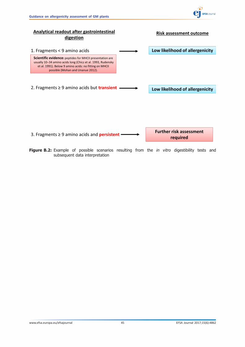
Figure B.2: Example of possible scenarios resulting from the in vitro digestibility tests andsubsequent data interpretation
Guidance on allergenicity assessment of GM plants
www.efsa.europa.eu/efsajournal 45 EFSA Journal 2017;15(6):4862

Annex C – Endogenous allergenicityAnnex C-1. History of endogenous allergenicity assessments
Scientific Opinions on soybean including an assessment of endogenous allergenicity were publishedby the EFSA GMO panel (e.g. EFSA, 2007, 2011, 2015, 2016, 2017). Historically, the assessment wascarried out using sera from allergic individuals, e.g. using 2D gel electrophoresis (immunoblot) incombination with spot quantification. More recently, however, novel absolute quantification methodswere developed and applicants employed those in the latest applications submitted to EFSA. To date andconsidering the EFSA GMO Panel Scientific Opinions published, no significant changes in the allergenrepertoire raising concern was observed in the GM plants assessed. The EFSA GMO Panel concluded thatthere was no evidence that the genetic modification changed the overall allergenicity of the GM plantwhen compared with its conventional counterpart. EFSA is committed to incorporate latest scientificdevelopments in its risk assessment process and approaches for endogenous allergenicity evaluationshould be revisited accordingly in the light of the latest novelties in this area.
Annex C-2. Possible approach for selection of soybean allergens to be analysed
The OECD list of ‘potential allergens’ (Table 20 of OECD, 2012) which is based on allergens listed inthe WHO/IUIS database (2011 www.allergen.org) and a review (L’Hocine and Boyne 2007), was takenas a basis, and further complemented with other allergen databases including the updated WHO/IUIS,the FARRP allergen database of the University of Nebraska-Lincoln (www.allergenonline.org) andAllergome (http://allergome.org/). Sequence information was additionally reviewed using the NCBI andUniprot databases. Other allergen databases, listed in the EFSA 2010 scientific opinion (EFSA GMOPanel, 2010), were also considered, but could not be screened in detail because they did not provideadditional information or were no longer available.
Using these sources, data on individual ‘potential allergens’ and on the allergic individuals testedwere retrieved. Furthermore, information was collected connecting clinical reactivity to total soypreparations with data on specific allergens, as well as possible clinical reactions to single purifiedallergens (either isolated from the plant or produced as recombinant allergens). To date, clinical datafor soy allergy were obtained by double-blind placebo-controlled food challenge (DBPCFC) using wholesoybean protein extract or soy flour (Ballmer-Weber et al., 2007), while only limited clinical researchstudies, mostly restricted to component resolved diagnostics (Tuano and Davis, 2015), are available forsingle proteins. The available information retrieved on the soybean ‘potential allergens’ are listed inTable C.1 (available in Supporting Information; please see also Selb et al., 2017).
It is challenging to unequivocally connect clinical data obtained on soy extracts by DBPCFC withreactivity to single proteins. In this particular example, taking the information collected for Table C.1 asstarting point and following the strategy proposed above, defined criteria were applied for a possibleselection of relevant allergens. WHO/IUIS defines criteria to include a protein in the allergen list. Theseare: (i) a minimum of five sera from allergic individuals (allergic to the allergen source in question)have to contain IgE which binds to the protein in question, or (ii) at least 5% of the allergic individuals’sera tested react with the allergen in question by IgE binding. It is noteworthy that WHO/IUISdatabase was the main contributor for listing allergens in the OECD document on soybean. WHO/IUISis also mentioned as a key reference for listing allergens in the EFSA NDA Panel opinion on foodallergens (See section 8 of EFSA NDA Panel opinion 2014). WHO/IUIS committee meetings decidingthe inclusion of new allergen in the list are held in the frame of EAACI and AAAAI international allergymeetings. Notably, because an application must be filed to include a protein into the WHO/IUISdatabase, not all proteins clearly fulfilling the requirements are included in the current database. In ourexample approach following the current WHO/IUIS criteria, the following allergens listed in Table C.1are also part of the WHO/IUIS database and should be measured accordingly: Gly m 1, Gly m 2, Gly m 3,Gly m 4, Gly m 5, Gly m 6, Gly m 7 and Gly m 8. Out of these, Gly m 2 can currently not be measuredsince the sequence is unknown. The following proteins potentially fulfil the primary WHO/IUIS criteriaof required tested allergic individuals, but did not undergo an expert peer-review by the WHO/IUIScommittee: Gly m Bd 28 K, Gly m Bd 30 K, soybean lectin, lipoxygenase, Kunitz trypsin inhibitor andGly m 50 KD. Out of these proteins, for Gly m Bd 28 K, Gly m Bd 30 K and Kunitz trypsin inhibitorconsiderable peer-reviewed literature is available and endogenous allergenicity measurement waspreviously suggested by other scientists (Ladics et al., 2014). In this context, international recognitionand/or clinical relevance should be the main aspects to consider for the selection of proteins that willrequire an endogenous allergenicity assessment.
Guidance on allergenicity assessment of GM plants
www.efsa.europa.eu/efsajournal 46 EFSA Journal 2017;15(6):4862

In contrast, the evidence for the involvement of soybean lectin and lipoxygenase in soy allergy ismore scarce (evidence from peer-reviewed scientific literature is limited/non-existent), and additionalevidence from clinical studies would be useful. Gly m 50KD is currently not measurable since thesequence is unknown. The other listed proteins, Gly m 39KD, P22-25, Gly m CPI, Gly m EAP and the‘unknown possible allergen’ currently do not fulfil the primary WHO/IUIS criteria. Measurement ofthese would therefore not be performed at the current time. However, more evidence might becomeavailable in the future. It should be noted that the above stated considerations (i) might be incompleteand (ii) do not prevent a revision of the scientific progresses in science at any time in the future, whichmight considerably change the list of potential allergens suitable for assessment as well as thenecessity to rank their relevance for the safety assessment.
Even though food allergy to soybean is known in animals (Suto et al., 2015; Kang et al., 2014),importantly, limited data are available on relevant individual soybean allergens in animals. However,certain proteins known to cause soybean allergy in humans (e.g. Gly m 5, Gly m 6) also causedreactions in calves, piglets, dogs and other animals (Taliercio et la., 2014; Lalles and Peltre, 1996;Dreau et al., 1994).
Annex C-3. Methodology
For ELISAs, individual allergens should be quantified using purified monoclonal or polyclonalantibodies raised against each purified allergen molecule. The measurement should be performedtogether with calibrated standards to ensure adequate information on allergen quantities in a sample.Protocols for allergen quantification by quantitative ELISA were developed previously (Geng et al.,2015, 2017; Chen et al., 2014; Hei et al., 2012; Liu et al., 2012, 2013).
Mass spectrometry (MS) approaches can also be used to specifically detect and quantify singleallergens. MS protocols for the assessment of endogenous allergenicity of soybean were developed forsome potential allergen molecules (Kuppannan et al., 2014; Stevenson et al., 2012; Houston et al.,2011).
The standardisation and harmonisation of these analytical methods among applicants would bebeneficial to enhance measurement comparability. This would support the future establishment of anallergen database, including data on the natural variability, which would provide useful additionalinformation to improve the robustness of the safety assessment.
Annex C-4. Data interpretation and risk assessment considerations
According to IR503/2013, after comparison to a conventional counterpart within the comparativeanalysis, natural variability is currently considered the main tool to identify significant and potentialrelevant changes in allergen content. The European Commission clarified this issue further.9
Currently, the experimental field design for comparative compositional analysis requires theinclusion of at least six non-GM reference commercial varieties – the selection of which should bejustified according to defined criteria (see section 1.3.2 of IR503/2013). These varieties are used toestimate the overall natural variability to which consumers are routinely exposed. The application ofsuch an approach allows an objective comparative evaluation independently of the absolute content ofendogenous allergens ensuring a high degree of protection for consumers.
It is recognised that the natural variability of any given endogenous allergen content estimatedfrom the reference commercial varieties, even if appropriately selected, may not capture the full rangeof its variability, which is currently unknown. Further efforts in such respect would be beneficial, asdescribed in Annex C-3.
Finally, specific considerations associated with a particular allergenic risk have to be taken intoaccount. Among others (see also Selb et al., 2017), the following considerations are of principlerelevance:
• exposure to a certain allergen should be taken into account as a last step in the riskassessment process with particular interest in understanding levels of allergens in foodsderived from soybean varieties consumed in Europe by humans and animals at a given time;
• efforts to reduce the uncertainty could be defined according to the single allergen in questionfor which enhanced allergen content was encountered and for which there is a potential
9 Letter from European Commission to EuropaBio (Ref. SANCO/El/SP/mb sanco.ddg2.e.l(2014)l 140685) where it was clarifiedthat ‘allergens included in the compositional analysis should be treated as any other compound meaning that allergens in thereference varieties included in the assessment should also be analysed (so that an equivalence test as well as a different testcan be performed)’.
Guidance on allergenicity assessment of GM plants
www.efsa.europa.eu/efsajournal 47 EFSA Journal 2017;15(6):4862

increase in allergenicity to be communicated to risk managers. The relevance of any givenincrease might be evaluated with the help of clinical scientist, taking into account: (a) themagnitude of the increase, (b) the potency of the increased potential allergen in question, (c)the overall percentage of the allergen in the non-GM crop and the consequent possible impactof an increase, (d) the route how the allergen is encountered by the allergic individual, (e) thefrequency of the potential allergen in question in various products;
• depending on the level of uncertainties identified and, in the case of need, the allergenicity ofa GM plant can be compared with that of its appropriate comparator by DBPCFC. This wouldimply performing a clinical study investigating the reactivity of selected allergic individuals tothe varieties under assessment. Therefore, well-characterised allergic individuals reacting withthe allergen in question would have to be challenged;
• as a future consideration/perspective, the probability of elicitation of an allergic reaction couldbe further investigated with the help of dose–distribution curves (Ballmer-Weber et al., 2015)obtained by DBPCFC to single allergens (Kinaciyan et al., 2016) and with reference values (e.g.threshold of elicitation) more precisely evaluated.
ReferencesBallmer-Weber BK, Holzhauser T, Scibilia J, Mittag D, Zisa G, Ortolani C, Oesterballe M, Poulsen LK, Vieths S and
Bindslev-Jensen C, 2007. Clinical characteristics of soybean allergy in Europe: a double-blind, placebo-controlled food challenge study. The Journal of Allergy and Clinical Immunology, 119, 1489–1496.
Ballmer-Weber BK, Fernandez-Rivas M, Beyer K, Defernez M, Sperrin M, Mackie AR, Salt LJ, Hourihane JO, AseroR, Belohlavkova S, Kowalski M, de Blay F, Papadopoulos NG, Clausen M, Knulst AC, Roberts G, Popov T,Sprikkelman AB, Dubakiene R, Vieths S, van Ree R, Crevel R and Mills EN, 2015. How much is too much?Threshold dose distributions for 5 food allergens. The Journal of Allergy and Clinical Immunology, 135, 964–971.
Chen J, Wang J, Song P and Ma X, 2014. Determination of glycinin in soybean and soybean products using asandwich enzyme-linked immunosorbent assay. Food Chemistry, 162, 27–33.
Dreau D, Lalles JP, Philouze-Rome V, Toullec R and Salmon H, 1994. Local and systemic immune responses tosoybean protein ingestion in early-weaned pigs. Journal of Animal Sciences, 72, 2090–2098.
EC, 2013. Commission Implementing Regulation (EU) No. 503/2013 of 3 April 2013 on applications forauthorisation of genetically modified food and feed in accordance with Regulation (EC) No. 1829/2003 of theEuropean Parliament and of the Council and amending Commission Regulations (EC) No. 641/2004 and (EC)No. 1981/2006. Official Journal of European Union L157, 1–48.
EFSA GMO Panel (EFSA Panel on Genetically Modified Organisms, 2010. Scientific Opinion on the assessment ofallergenicity of GM plants and microorganisms and derived food and feed. EFSA Journal 2010; 8(7):1700, 168pp. https://doi.org/10.2903/j.efsa.2010.1700
EFSA GMO Panel (EFSA Panel on Genetically Modified Organisms), 2007. Opinion of the Scientific Panel ongenetically modified organisms (GMO) on an application (Reference EFSA-GMO-NL-2005-18) for the placing onthe market of the glufosinate tolerant soybean A2704-12, for food and feed uses, import and processing underRegulation (EC) No 1829/2003 from Bayer CropScience. https://doi.org/10.2903/j.efsa.2007.524.
EFSA GMO Panel (EFSA Panel on Genetically Modified Organisms), 2011. Scientific Opinion on application (EFSA-GMO-UK-2007-43) for the placing on the market of herbicide tolerant genetically modified soybean 356043 forfood and feed uses, import and processing under Regulation (EC) No 1829/2003 from Pioneer. EFSA Journal2011;9(7):2310, 40 pp. https://doi.org/10.2903/j.efsa.2011.2310
EFSA GMO Panel (EFSA Panel on Genetically Modified Organisms), 2015. Scientific Opinion on application (EFSA-GMO-NL-2012-108) for the placing on the market of the herbicide-tolerant genetically modified soybean MON87708 9 MON 89788 for food and feed uses, import and processing under Regulation (EC) No 1829/2003from. EFSA Journal 2015;13(6):4136, 26 pp. https://doi.org/10.2903/j.efsa.2015.4136
EFSA GMO Panel (EFSA Panel on Genetically Modified Organisms), 2017. Naegeli H,Birch AN, Casacuberta J, D eSchrijver A, Gralak MA, Jones H, Manachini B, Messean A, Nielsen EE,Nogue F, Robaglia C, Rostoks N, Sweet J,Tebbe C, Visioli F, Wal J-M,Alvarez F, Ardizzone M, Liu Y,Neri FM and Ramon M, 2017. Scientific opinion on anapplication by Dow AgroSciences LLC (EFSA-GMO-NL-2012-106) for the placing on the market of geneticallymodified herbicide-tolerant soybean DAS-44406-6 for food and feed uses, im port and processing underRegulation (EC) No 1829/2003. EFSA Journal 2017;15(3):4738, 33 pp. https://doi.org/10.2903/j.efsa.2017.4738
EFSA GMO Panel (EFSA Panel on Genetically Modified Organisms), 2016. Naegeli H,Birch AN, Casacuberta J, DeSchr ijver A, Gralak MA, Guerche P, Jones H, Manachini B, Messean A,Nielsen EE, Nogue F, Robaglia C,Rostoks N, Sweet J, Tebbe C, Visioli F, Wal J-M, Diveki Z, Fernandez-Dumont A, Gennaro A, Lanzoni A, NeriFM and Paraskevopoulos K, 2016. Scientific Opinion on an application by Dow AgroSciences (EFSA-GMO-NL-2013-116) for placing on the market of genetically modified insect-resistant soybean DAS-81419-2 for food andfeed uses, import and processing under Regulation (EC) No 1829/2003. EFSA Journal 2016;14(12):4642, 23pp. https://doi.org/10.2903/j.efsa.2016.4642
Guidance on allergenicity assessment of GM plants
www.efsa.europa.eu/efsajournal 48 EFSA Journal 2017;15(6):4862
EFSA NDA Panel (EFSA Panel on Dietetic Products, Nutrition and Allergies), 2014. Scientific Opinion on theevaluation of allergenic foods and food ingredients for labelling purposes. EFSA Journal 2014;12(11):3894, 286pp. https://doi.org/10.2903/j.efsa.2014.3894
Geng T, Liu K, Frazier R, Shi L, Bell E, Glenn K and Ward JM, 2015. Development of a Sandwich ELISA forQuantification of Gly m 4, a Soybean Allergen. Journal of Agricultural and Food Chemistry, 63, 4947–4953.
Geng T, Stoj�sin D, Liu K, Schaalje B, Postin C, Ward J, Wang Y, Liu ZL, Li B and Glenn K, 2017. Natural Variabilityof Allergen Levels in Conventional Soybeans: Assessing Variation across North and South America from FiveProduction Years. Journal of Agricultural and Food Chemistry, 65, 463–472.
Hei W, Li Z, Ma X and He P, 2012. Determination of beta-conglycinin in soybean and soybean products using asandwich enzyme-linked immunosorbent assay. Analytica Chimica Acta, 734, 62e68.
Houston NL, Lee D, Stevenson SE, Ladics GS, Bannon GA, McClain S, Privalle L, Stagg N, Herouet-Guicheney C,MacIntosh SD and Thelen JJ, 2011. Quantitation of soybean allergens using tandem mass spectrometry.Journal of Proteome Research, 10, 763–773.
Kang MH, Kim HJ, Jang HJ and Park HM, 2014. Sensitization rates of causative allergens for dogs with atopicdermatitis: detection of canine allergen-specific IgE. Journal of Veterinary Science, 15, 545–550.
Kinaciyan T, Nagl B, Faustmann S, Kopp S, Wolkersdorfer M and Bohle B, 2016. Recombinant Mal d 1 facilitatessublingual challenge tests of birch pollen-allergic patients with apple allergy. Allergy, 71, 272–274.
Kuppannan K, Julka S, Karnoup A, Dielman D and Schafer B, 2014. 2DLC-UV/MS assay for the simultaneousquantification of intact soybean allergens Gly m 4 and hydrophobic protein from soybean (HPS). The Journal ofAgricultural and Food Chemistry, 62, 4884–4892.
Ladics GS, Budziszewski GJ, Herman RA, Herouet-Guicheney C, Joshi S, Lipscomb EA, McClain S and Ward JM,2014. Measurement of endogenous allergens in genetically modified soybeans–short communication.Regulatory Toxicology and Pharmacology, 70, 75–79.
Lalles JP and Peltre G, 1996. Biochemical features of grain legume allergens in humans and animals. NutritionReviews, 54, 101–107.
L’Hocine L and Boye JI, 2007. Allergenicity of soybean: new developments in identification of allergenic proteins,cross-reactivities and hypoallergenization technologies. Critical Reviews in Food Science and Nutrition, 47, 127–143.
Liu B, Teng D, Wang X, Yang Y and Wang J, 2012. Expression of the soybean allergenic protein P34 in Escherichiacoli and its indirect ELISA detection method. Applied Microbiology and Biotechnology, 94, 1337–1345.
Liu B, Teng D, Wang X and Wang J, 2013. Detection of the soybean allergenic protein Gly m Bd 28K by an indirectenzyme-linked immunosorbent assay. Journal of Agricultural and Food Chemistry, 61, 822–828.
OECD, 2012. Revised consensus document on compositional considerations for new varieties of soybean [Glycinemax (L.) Merr.]: key food and feed nutrients, antinutrients, toxicants and allergens. Series on the Safety ofNovel Foods and Feeds No. 25.
Selb R, Wal JM, Moreno FJ, Lovik M, Mills C, Hoffmann-Sommergruber K and Fernandez A, 2017. Assessment ofendogenous allergenicity of genetically modified plants exemplified by soybean - Where do we stand? Foodand Chemical Toxicology, 101, 139–148.
Suto A, Suto Y, Onahara N, Tomizawa Y, Yamamoto-Sugawara Y, Okayama T and Masuda K, 2015. Food allergensinducing a lymphocyte-mediated immunological reaction in canine atopic-like dermatitis. Journal of VeterinaryMedical Science, 77, 251–254.
Stevenson SE, Woods CA, Hong B, Kong X, Thelen JJ and Ladics GS, 2012. Environmental effects on allergenlevels in commercially grown non-genetically modified soybeans: assessing variation across North America.Frontiers in Plant Science, 3, 1–13.
Taliercio E, Loveless TM, Turano MJ and Kim SW, 2014. Identification of epitopes of the beta subunit of soybeanbeta-conglycinin that are antigenic in pigs, dogs, rabbits and fish. Journal of the Science of Food andAgriculture, 94, 2289–2294.
Tuano KS and Davis CM, 2015. Utility of component-resolved diagnostics in food allergy. Current Allergy andAsthma Reports, 15, 32.
Guidance on allergenicity assessment of GM plants
www.efsa.europa.eu/efsajournal 49 EFSA Journal 2017;15(6):4862
Related Documents


![Allergenicity Study of Genetically Modified Herbicide ... · Bioinformatics analysis for allergenicity assessment of proteins is performed via allergen databases [23]. Bioinformatics,](https://static.cupdf.com/doc/110x72/5f203060a0eec836ba085823/allergenicity-study-of-genetically-modified-herbicide-bioinformatics-analysis.jpg)