Chemistry & Biology, Vol. 10, 1255–1266, December, 2003, 2003 Elsevier Science Ltd. All rights reserved. DOI 10.1016/j.chembiol.2003.11.010 GSK-3-Selective Inhibitors Derived from Tyrian Purple Indirubins but not 1-methyl-BIO reduced -catenin phosphoryla- tion on a GSK-3-specific site in cellular models. BIO but not 1-methyl-BIO closely mimicked Wnt signaling Laurent Meijer, 1,2, * Alexios-Leandros Skaltsounis, 4 Prokopios Magiatis, 4 Panagiotis Polychronopoulos, 4 Marie Knockaert, 1 Maryse Leost, 1 Xiaozhou P. Ryan, 2 in Xenopus embryos. 6-bromoindirubins thus provide a new scaffold for the development of selective and Claudia Alin Vonica, 3 Ali Brivanlou, 3 Rana Dajani, 6 Claudia Crovace, 5 Cataldo Tarricone, 5 potent pharmacological inhibitors of GSK-3. Andrea Musacchio, 5 S. Mark Roe, 6 Laurence Pearl, 6 and Paul Greengard 2 Introduction 1 C.N.R.S. Cell Cycle Group In mammals, different genes encode two closely related Station Biologique, B.P. 74 glycogen synthase kinase-3 (GSK-3) enzymes, GSK-3 29682 Roscoff cedex and GSK-3. The discovery of GSK-3 functions in Wnt Bretagne signal-transducing pathways, insulin action, apoptosis, France and circadian rhythm, the deciphering of unique regula- 2 Laboratory of Molecular & Cellular Neuroscience tion mechanisms of GSK-3 (inhibitory phosphorylation 3 Laboratory of Molecular Embryology on Ser9, priming of substrate by phosphorylation by The Rockefeller University another kinase), the determination of the crystal struc- 1230 York Avenue ture of human GSK-3, and the clear implication of New York, New York 10021 GSK-3 in various human diseases (diabetes, Alzheimer’s 4 Division of Pharmacognosy and Natural disease, cancers) have all recently contributed to a re- Products Chemistry newed interest in this kinase (for reviews see [1–5]. Department of Pharmacy Growing evidence supports the view that GSK-3 acti- University of Athens vation [6, 7] and nuclear translocation [8] are a prerequi- Panepistimiopolis Zografou site for neuronal apoptosis. The data stem from cell GR-15771 Athens culture experiments in which apoptosis induced by vari- Greece ous agents can be prevented in several ways, leading 5 Structural Biology Unit to GSK-3 inactivation: (1) overexpression of FRAT1, a Department of Experimental Oncology negative regulator of GSK-3, confers resistance of PC12 European Institute of Oncology cells to apoptosis [9, 10]; (2) pharmacological inhibitors Via Ripamonti 435 of GSK-3 prevent apoptosis in PC12 cells [10–12], hu- 20141 Milano man SH-SY5Y neuroblastoma cells [13, 14], and a Hun- Italy tington disease cell model [15]; (3) activation of Wnt 6 Centre for Structural Biology signaling (and therefore GSK-3 inhibition) prevents Institute of Cancer Research c-Myc-induced apoptosis in Rat-1 cells [16]; (4) expres- Chester Beatty Laboratories sion of a kinase-deficient GSK-3 reduces cell death 237 Fulham Road induced by Akt inhibition [9]; (5) FGF (which activates London SW3 6JB Akt and thus inhibits GSK-3) reduces cell death induced United Kingdom by glutamate [17], while overexpression of GSK-3 in- creases cell death; and (6) inhibition of GSK-3 by Ser9 phosphorylation by PKA is associated with neuronal Summary survival [18]. In addition to these in vitro data, two animal models reinforce the link between GSK-3 activation and Gastropod mollusks have been used for over 2500 apoptosis: (1) conditional transgenic mice overexpress- years to produce the “Tyrian purple” dye made famous ing GSK-3 in brain during adulthood show signs of neu- by the Phoenicians. This dye is constituted of mixed ronal stress and apoptosis [19], and (2) expression of bromine-substituted indigo and indirubin isomers. shaggy, the Drosophila homolog of GSK-3, enhances Among these, the new natural product 6-bromoindiru- the neurodegeneration induced by expression of human bin and its synthetic, cell-permeable derivative, 6-bro- Tau in flies, while expression of a loss-of-function mu- moindirubin-3-oxime (BIO), display remarkable selec- tant of shaggy prevents this neurodegeneration [20]. tive inhibition of glycogen synthase kinase-3 (GSK-3). Only a very limited number of pharmacological inhibi- Cocrystal structure of GSK-3/BIO and CDK5/p25/ tors of GSK-3 are available [21], lithium being the most indirubin-3-oxime were resolved, providing a detailed frequently used [22] despite its effects being in the 10–20 view of indirubins’ interactions within the ATP binding mM range and its demonstrated effect on inositol phos- pocket of these kinases. BIO but not 1-methyl-BIO, its phatases [23]. Recently, we noted that several pharma- kinase inactive analog, also inhibited the phosphoryla- cological inhibitors of cyclin-dependent kinases (CDKs) tion on Tyr276/216, a GSK-3/ activation site. BIO were in fact quite potent on GSK-3 [24, 25]. Among these is a family of bis-indoles known as indirubins, initially identified as CDK inhibitors [26, 27]. Indirubins derive *Correspondence: [email protected]

Welcome message from author
This document is posted to help you gain knowledge. Please leave a comment to let me know what you think about it! Share it to your friends and learn new things together.
Transcript

Chemistry & Biology, Vol. 10, 1255–1266, December, 2003, 2003 Elsevier Science Ltd. All rights reserved. DOI 10.1016/j .chembiol .2003.11.010
GSK-3-Selective InhibitorsDerived from Tyrian Purple Indirubins
but not 1-methyl-BIO reduced �-catenin phosphoryla-tion on a GSK-3-specific site in cellular models. BIObut not 1-methyl-BIO closely mimicked Wnt signaling
Laurent Meijer,1,2,* Alexios-Leandros Skaltsounis,4
Prokopios Magiatis,4
Panagiotis Polychronopoulos,4 Marie Knockaert,1
Maryse Leost,1 Xiaozhou P. Ryan,2 in Xenopus embryos. 6-bromoindirubins thus providea new scaffold for the development of selective andClaudia Alin Vonica,3 Ali Brivanlou,3 Rana Dajani,6
Claudia Crovace,5 Cataldo Tarricone,5 potent pharmacological inhibitors of GSK-3.Andrea Musacchio,5 S. Mark Roe,6
Laurence Pearl,6 and Paul Greengard2
Introduction1C.N.R.S.Cell Cycle Group
In mammals, different genes encode two closely relatedStation Biologique, B.P. 74glycogen synthase kinase-3 (GSK-3) enzymes, GSK-3�29682 Roscoff cedexand GSK-3�. The discovery of GSK-3 functions in WntBretagnesignal-transducing pathways, insulin action, apoptosis,Franceand circadian rhythm, the deciphering of unique regula-2 Laboratory of Molecular & Cellular Neurosciencetion mechanisms of GSK-3 (inhibitory phosphorylation3 Laboratory of Molecular Embryologyon Ser9, priming of substrate by phosphorylation byThe Rockefeller Universityanother kinase), the determination of the crystal struc-1230 York Avenueture of human GSK-3�, and the clear implication ofNew York, New York 10021GSK-3 in various human diseases (diabetes, Alzheimer’s4 Division of Pharmacognosy and Naturaldisease, cancers) have all recently contributed to a re-Products Chemistrynewed interest in this kinase (for reviews see [1–5].Department of PharmacyGrowing evidence supports the view that GSK-3� acti-University of Athensvation [6, 7] and nuclear translocation [8] are a prerequi-Panepistimiopolis Zografousite for neuronal apoptosis. The data stem from cellGR-15771 Athensculture experiments in which apoptosis induced by vari-Greeceous agents can be prevented in several ways, leading5 Structural Biology Unitto GSK-3 inactivation: (1) overexpression of FRAT1, aDepartment of Experimental Oncologynegative regulator of GSK-3, confers resistance of PC12European Institute of Oncologycells to apoptosis [9, 10]; (2) pharmacological inhibitorsVia Ripamonti 435of GSK-3 prevent apoptosis in PC12 cells [10–12], hu-20141 Milanoman SH-SY5Y neuroblastoma cells [13, 14], and a Hun-Italytington disease cell model [15]; (3) activation of Wnt6 Centre for Structural Biologysignaling (and therefore GSK-3 inhibition) preventsInstitute of Cancer Researchc-Myc-induced apoptosis in Rat-1 cells [16]; (4) expres-Chester Beatty Laboratoriession of a kinase-deficient GSK-3� reduces cell death237 Fulham Roadinduced by Akt inhibition [9]; (5) FGF (which activatesLondon SW3 6JBAkt and thus inhibits GSK-3) reduces cell death inducedUnited Kingdomby glutamate [17], while overexpression of GSK-3� in-creases cell death; and (6) inhibition of GSK-3 by Ser9phosphorylation by PKA is associated with neuronal
Summary survival [18]. In addition to these in vitro data, two animalmodels reinforce the link between GSK-3 activation and
Gastropod mollusks have been used for over 2500 apoptosis: (1) conditional transgenic mice overexpress-years to produce the “Tyrian purple” dye made famous ing GSK-3 in brain during adulthood show signs of neu-by the Phoenicians. This dye is constituted of mixed ronal stress and apoptosis [19], and (2) expression ofbromine-substituted indigo and indirubin isomers. shaggy, the Drosophila homolog of GSK-3�, enhancesAmong these, the new natural product 6-bromoindiru- the neurodegeneration induced by expression of humanbin and its synthetic, cell-permeable derivative, 6-bro- Tau in flies, while expression of a loss-of-function mu-moindirubin-3�-oxime (BIO), display remarkable selec- tant of shaggy prevents this neurodegeneration [20].tive inhibition of glycogen synthase kinase-3 (GSK-3). Only a very limited number of pharmacological inhibi-Cocrystal structure of GSK-3�/BIO and CDK5/p25/ tors of GSK-3 are available [21], lithium being the mostindirubin-3�-oxime were resolved, providing a detailed frequently used [22] despite its effects being in the 10–20view of indirubins’ interactions within the ATP binding mM range and its demonstrated effect on inositol phos-pocket of these kinases. BIO but not 1-methyl-BIO, its phatases [23]. Recently, we noted that several pharma-kinase inactive analog, also inhibited the phosphoryla- cological inhibitors of cyclin-dependent kinases (CDKs)tion on Tyr276/216, a GSK-3�/� activation site. BIO were in fact quite potent on GSK-3 [24, 25]. Among these
is a family of bis-indoles known as indirubins, initiallyidentified as CDK inhibitors [26, 27]. Indirubins derive*Correspondence: [email protected]

Chemistry & Biology1256
from the spontaneous, nonenzymatic dimerization of exposed to sunlight to generate the expected Tyrianisatin and indoxyl, two colorless precursors found either purple dye (Figure 1). The mollusks were then frozenfree or conjugated to carbohydrates, which are found and lyophilized prior to extraction by dichloromethane.in over 200 species of indigo-producing plants [28, 29]. Organic phase soluble indigoids were resolved by silicaAlthough they constitute minor side products, indirubins gel chromatography. Indigo derivatives were discarded,contribute by their red color to the unique blue-purplish and indirubins (0.000025%) were further separated bycolor of natural indigo that distinguishes it from synthetic MPLC and identified by 1D and 2D NMR and mass spec-indigo. Indirubins, along with indigo, are also produced trometry as indirubin (1), 6�-bromoindirubin (2), 6-bro-by various bacterial strains (reviewed in [30, 31]) and by moindirubin (3), and 6,6�-dibromoindirubin (4) (Figure 1).a number of gastropod mollusks [32]. Moreover, they 6-bromoindirubin (3) has never been isolated from anycan be found in urine from diseased and healthy patients Muricidae or Thaididae gastropod or from any other([33] and references therein). Indirubin constitutes the natural source [32]. Each of these compounds was syn-main active ingredient of a traditional Chinese medicinal thesized, along with its corresponding 3�-oxime deriva-recipe, Danggui Longhui Wan, used to treat various dis- tive (5–8), as this modification enhances solubility, ki-eases including chronic myelocytic leukemia [34, 35]. nase inhibition, and cell permeability [24, 26]. 1-methylIndirubin and indigo have been recently discovered as derivatives (9–11) were also synthesized, as they werepotent ligands of the aryl hydrocarbon receptor (AhR), expected, from previous CDK2/indirubin cocrystal struc-also known as the “dioxin receptor” [33] (M.K. et al., tures [26, 27], to provide kinase inactive control com-submitted). pounds. Finally, 3�-oxime in compound (7) was also re-
Gastropods of the Muricidae and Thaididae families placed by 3�-methoxime (12) or 3�-acetoxime (13). Wehave been used in various parts of the world as the next evaluated the effects of these compounds on puri-source of a vivid purplish-red dye known as “Tyrian fied GSK-3�/�, CDK1/cyclin B, and CDK5/p25 (Tablepurple” or “royal blue” around the Mediterranean sea 1A). As expected, indirubin (1) was active on GSK-3�/�[32]. The highly prized, beautiful Tyrian purple, obtained and on CDKs (10-fold less). Although a 6�-bromo substi-at great expense essentially from the two species Murex tution (2) led to reduced kinase inhibition, the 6-bromobrandaris and Hexaplex trunculus, was the object of an substitution (3) greatly enhanced the selectivity forimportant trade in Crete (5th century BC), Phoenicia, GSK-3 over CDKs. Addition of a 3�-oxime substitutionGreece, and later in Rome. The color was considered a
(5–8) led to an overall increase in kinase inhibitory effectsprivilege of the emperors Nero and Caligula. In Britain
and increased solubility, although the selectivity forand Ireland, the dog whelk (Nucella lapillus) was also
GSK-3 was slightly reduced. The 6-bromoindirubinsused for many years as a source of purple dye. Othersubstituted on 3� by methoxime (12) or acetoxime (13)dye-producing mollusks include Purpura pansa (Mex-were also quite potent and GSK-3 selective (but lessico, Pacific Ocean), Purpura patula (Mexico, Atlanticsoluble, which precluded their use in cells). As expected,Ocean), and Rapana venosa (Japan). The hypobranchialmethylation on position N1 inactivated the indirubinsgland of these gastropods contains colorless precur-(9–11) as kinase inhibitors.sors, indoxyl-sulfate, its mercaptan derivative, and their
bromo derivatives, which release various indoxyls uponmaceration (in fact hydrolysis by purpurase). When ex- 6-Bromoindirubins Are Selectiveposed to light and oxygen, these indols dimerize in a Inhibitors of GSK-3mixture of indigo and indirubin, mainly 6,6�-dibromo- We next tested BIO (7) and methoxime and acetoximeindigo. Paul Friedlander was the first to determine the (12, 13) on a series of 20 purified protein kinases assayedcomposition of the dye from Murex brandaris, in 1909, in the presence of 15 �M ATP. The results of thesefrom 1.4 g of dye isolated from 12,000 mollusks [36]. assays confirm the strong selectivity of 6-bromo-substi-The famous “Tekhelet” described in the Bible was also tuted indirubins for GSK-3�/� (Table 1B). An acetoximeidentified as a mixture of indigoids specifically obtained or methoxime substitution on position N3� further con-from Hexaplex trunculus [32, 37]. tributes to the GSK-3 selectivity. As an alternative ap-
In an effort to identify new kinase inhibitors with in- proach to evaluate the selectivity of BIO, we immobilizedcreased potency and selectivity, we investigated the indirubin-3�-oxime on Affigel beads through a polyethyl-natural indirubins produced by the Mediterranean mol- ene linker attached at the 3� position (Figures 2A andlusk Hexaplex trunculus. Beside the dominant bromo- 2B). A porcine brain extract was then run on these beadsindigos, we identified various bromo-substituted indiru- in the absence or presence of free BIO, and the boundbins, and one of them, 6-bromoindirubin, which was proteins were resolved by SDS-PAGE followed by silverisolated for the first time as a natural product, turned out staining and anti-GSK-3 Western blotting (Figures 2Cto be a potent GSK-3 inhibitor. Its synthetic derivative, and 2D). Although a large number of proteins were found6-bromoindirubin-3�-oxime (BIO), and related analogs to bind to indirubin beads, the presence of free BIOconstitute unique cell-permeable and selective inhibi- did not lead to major changes in the overall patterntors of GSK-3. We here provide the first cocrystal struc- of indirubin binding proteins. However, it completelytures of GSK-3 and CDK5 with an inhibitor.
prevented the binding of GSK-3�/�.
ResultsCocrystal Structure of BIO with GSK-3�We next attempted to soak BIO into a GSK-3� crystal,Purification, Identification, and Kinase Inhibitorythe structure of which had recently been describedProperties of Shellfish Indirubins[38, 39]. A cocrystal structure was obtained with 2.8 ASixty kilograms of Hexaplex trunculus were collected,
and the animals were extracted from the shells and resolution (Figure 3). The overall structure of GSK-3�/
Tyrian Purple-Derived Indirubins Inhibit GSK-31257
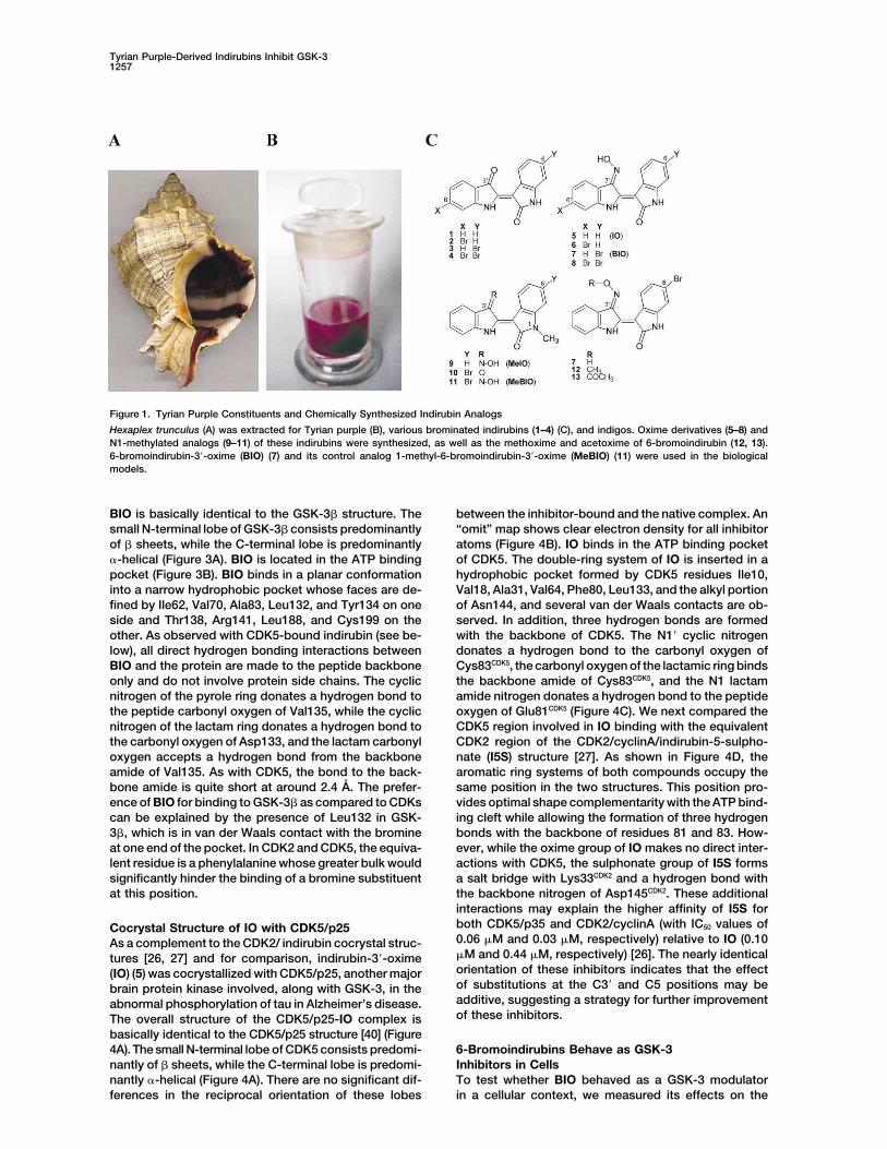
Figure 1. Tyrian Purple Constituents and Chemically Synthesized Indirubin Analogs
Hexaplex trunculus (A) was extracted for Tyrian purple (B), various brominated indirubins (1–4) (C), and indigos. Oxime derivatives (5–8) andN1-methylated analogs (9–11) of these indirubins were synthesized, as well as the methoxime and acetoxime of 6-bromoindirubin (12, 13).6-bromoindirubin-3�-oxime (BIO) (7) and its control analog 1-methyl-6-bromoindirubin-3�-oxime (MeBIO) (11) were used in the biologicalmodels.
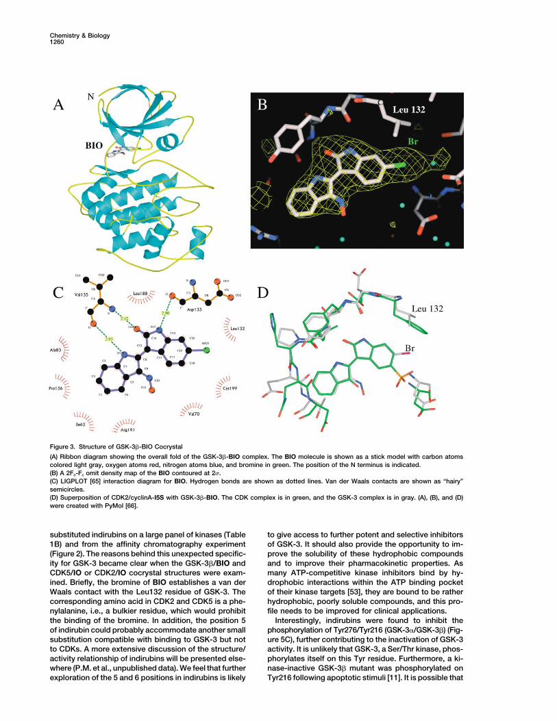
BIO is basically identical to the GSK-3� structure. The between the inhibitor-bound and the native complex. An“omit” map shows clear electron density for all inhibitorsmall N-terminal lobe of GSK-3� consists predominantly
of � sheets, while the C-terminal lobe is predominantly atoms (Figure 4B). IO binds in the ATP binding pocketof CDK5. The double-ring system of IO is inserted in a�-helical (Figure 3A). BIO is located in the ATP binding
pocket (Figure 3B). BIO binds in a planar conformation hydrophobic pocket formed by CDK5 residues Ile10,Val18, Ala31, Val64, Phe80, Leu133, and the alkyl portioninto a narrow hydrophobic pocket whose faces are de-
fined by Ile62, Val70, Ala83, Leu132, and Tyr134 on one of Asn144, and several van der Waals contacts are ob-served. In addition, three hydrogen bonds are formedside and Thr138, Arg141, Leu188, and Cys199 on the
other. As observed with CDK5-bound indirubin (see be- with the backbone of CDK5. The N1� cyclic nitrogendonates a hydrogen bond to the carbonyl oxygen oflow), all direct hydrogen bonding interactions between
BIO and the protein are made to the peptide backbone Cys83CDK5, the carbonyl oxygen of the lactamic ring bindsthe backbone amide of Cys83CDK5, and the N1 lactamonly and do not involve protein side chains. The cyclic
nitrogen of the pyrole ring donates a hydrogen bond to amide nitrogen donates a hydrogen bond to the peptideoxygen of Glu81CDK5 (Figure 4C). We next compared thethe peptide carbonyl oxygen of Val135, while the cyclic
nitrogen of the lactam ring donates a hydrogen bond to CDK5 region involved in IO binding with the equivalentCDK2 region of the CDK2/cyclinA/indirubin-5-sulpho-the carbonyl oxygen of Asp133, and the lactam carbonyl
oxygen accepts a hydrogen bond from the backbone nate (I5S) structure [27]. As shown in Figure 4D, thearomatic ring systems of both compounds occupy theamide of Val135. As with CDK5, the bond to the back-
bone amide is quite short at around 2.4 A. The prefer- same position in the two structures. This position pro-vides optimal shape complementarity with the ATP bind-ence of BIO for binding to GSK-3� as compared to CDKs
can be explained by the presence of Leu132 in GSK- ing cleft while allowing the formation of three hydrogenbonds with the backbone of residues 81 and 83. How-3�, which is in van der Waals contact with the bromine
at one end of the pocket. In CDK2 and CDK5, the equiva- ever, while the oxime group of IO makes no direct inter-actions with CDK5, the sulphonate group of I5S formslent residue is a phenylalanine whose greater bulk would
significantly hinder the binding of a bromine substituent a salt bridge with Lys33CDK2 and a hydrogen bond withthe backbone nitrogen of Asp145CDK2. These additionalat this position.interactions may explain the higher affinity of I5S forboth CDK5/p35 and CDK2/cyclinA (with IC50 values ofCocrystal Structure of IO with CDK5/p250.06 �M and 0.03 �M, respectively) relative to IO (0.10As a complement to the CDK2/ indirubin cocrystal struc-�M and 0.44 �M, respectively) [26]. The nearly identicaltures [26, 27] and for comparison, indirubin-3�-oximeorientation of these inhibitors indicates that the effect(IO) (5) was cocrystallized with CDK5/p25, another majorof substitutions at the C3� and C5 positions may bebrain protein kinase involved, along with GSK-3, in theadditive, suggesting a strategy for further improvementabnormal phosphorylation of tau in Alzheimer’s disease.of these inhibitors.The overall structure of the CDK5/p25-IO complex is
basically identical to the CDK5/p25 structure [40] (Figure4A). The small N-terminal lobe of CDK5 consists predomi- 6-Bromoindirubins Behave as GSK-3
Inhibitors in Cellsnantly of � sheets, while the C-terminal lobe is predomi-nantly �-helical (Figure 4A). There are no significant dif- To test whether BIO behaved as a GSK-3 modulator
in a cellular context, we measured its effects on theferences in the reciprocal orientation of these lobes

Chemistry & Biology1258
Table 1. Selectivity of Tyrian Purple-Derived Indirubins
A. Effect of Indirubins on CDK1/Cyclin B, CDK5/p25, and GSK-3 Kinase Activitiesa
No. Compound GSK-3 �/� CDK1/Cyclin B CDK5/p25
1 indirubin 1.00 10.00 10.002 6�-bromoindirubin 22.00 �100.00 �100.003 6-bromoindirubin 0.045 90.00 53.004 6,6�-dibromoindirubin 4.50 100.00 �100.005 indirubin-3�-oxime (IO) 0.022 0.18 0.106 6�-bromoindirubin-3�-oxime 0.34 3.00 1.207 6-bromoindirubin-3�-oxime (BIO) 0.005 0.32 0.0838 6,6�-dibromoindirubin-3�-oxime 0.12 17.00 1.309 1-methyl-indirubin-3�-oxime (MeIO) �100.00 73.00 �100.0010 1-methyl-6-bromoindirubin �100.00 80.00 �100.0011 1-methyl-6-bromoindirubin-3�-oxime (MeBIO) 44.00 55.00 �100.0012 6-bromoindirubin-3�-methoxime 0.03 3.40 2.2013 6-bromoindirubin-3�-acetoxime 0.01 63.00 2.40
B. Selectivity of 6-Bromoindirubinsb
Protein Kinases 5 (IO)c 7 (BIO) 12 13
GSK-3�/� 0.022 0.005 0.030 0.010CDK1/cyclin B 0.18 0.32 3.40 63CDK2/cyclin A 0.44 0.30 15 4.3CDK4/cyclin D1 3.33 10 �10 �10CDK5/p35 0.10 0.08 2.20 2.4erk1 �100 �10 �10 �10erk2 �100 �10 �10 �10MAPKK �100 10 �10 �10protein kinase C � 27 12 �10 �10protein kinase C �1 4 �10 �10 �10protein kinase C �2 20 �10 �10 �10protein kinase C � 8.40 �10 �10 �10protein kinase C � �100 �10 �10 �10protein kinase C � 20 �10 �10 �10protein kinase C 52 �10 �10 �10protein kinase C �100 �10 �10 �10cAMP-dependent PK 6.3 �10 �10 �10cGMP-dependent PK 9 �10 �10 �10casein kinase 2 12 �10 9 �10insulin receptor Tyr kinase 11 �10 �10 �10
a A series of indirubin analogs were tested at various concentrations in three kinase assays, as described in Experimental Procedures. IC50
values were calculated from the dose-response curves and are reported in �M.b Kinases were assayed in the presence of various concentrations of each compound. IC50 values, determined from the dose-response curves,are expressed in �M.c Data from [26].
phosphorylation of specific GSK-3 substrates. First, we compared to BIO, and therefore to its binding to alterna-tive targets.made use of the monoclonal ABC antibody, a dephos-
pho-specific antibody that crossreacts with �-catenin Indirubins have recently been described to interactwith AhR [33]. To rule out the possibility that BIO-when it is not phosphorylated by GSK-3 (on either Ser37
or Thr41) [41]. This antibody thus provides a convenient induced �-catenin dephosphorylation/stabilization wasdue to an indirect, AhR-dependent effect, we made usepositive signal on Western blots when GSK-3 is inhib-
ited. We also made use of a general anti-�-catenin anti- of cell lines deficient either in AhR or ARNT (a cotran-scriptional factor of AhR) [43]. BIO triggered a robustbody, as inhibition of �-catenin phosphorylation leads
to its stabilization [42]. COS-1 cells were exposed for �-catenin stabilization in both cell lines (Figure 5B). Fur-thermore, although MeBIO is a potent AhR agonist (M.K.24 hr to various indirubins or to LiCl, and their levels of
unphosphorylated �-catenin, total �-catenin, and total et al., submitted), MeBIO is inactive on GSK-3� (Table1A) and on cellular �-catenin phosphorylation/stabiliza-GSK-3 were examined by Western blotting (Figure 5A).
BIO and LiCl treatment, but neither the kinase-inactive tion (Figure 5). Altogether, these data demonstrate thatBIO acts by a direct effect on GSK-3 rather than throughMeBIO nor IO, led to a major increase of the unphos-
phorylated �-catenin level. This was also accompanied an indirect, AhR-dependent pathway.GSK-3� and GSK-3� activity are enhanced by theby a modest increase in total �-catenin level, as ex-
pected from the dephosphorylation-dependent increased phosphorylation of a specific tyrosine residue (Tyr276and Tyr216, respectively) [44, 45]. The tyrosine kinasehalf-life of �-catenin. BIO thus acted as a true GSK-3
inhibitor in this cell line. The apparent lack of effect of responsible for this phosphorylation in mammals is yetunknown, but there is some evidence that it indirectlyIO in this cell line may be due to its reduced specificity

Tyrian Purple-Derived Indirubins Inhibit GSK-31259
Figure 2. BIO Is a Selective GSK-3 Inhibitor In Vitro
Indirubin was immobilized on Affigel through a linker (A). Indirubin beads (B) and control ethanolamine beads were exposed to a porcine brainlysate in the presence (�) or absence (�) of 20 �M BIO. Following stringent washing, the bound proteins were analyzed by SDS-PAGE followedby silver staining (C) or Western blotting with anti-GSK-3 antibody (D).
depends on GSK-3 activity [11, 46]. We therefore investi- plants treated with LiCl (Figure 6H, lane 3) or BIO (lane4) shows induction of both genes. This effect was resis-gated the effect of indirubins on this key tyrosine phos-
phorylation. SH-SY5Y cells were exposed to IO, MeIO, tant to injection of RNA encoding the Wnt inhibitor axin(Figure 6H, lanes 9 and 10), which requires GSK-3 forBIO, and MeBIO for 12 hr, and the phosphorylation of
Tyr276 (GSK-3�) and Tyr216 (GSK-3�) was estimated its activity, but not to a dominant-negative Tcf molecule(DN Xtcf-3) [50] (lanes 6 and 7) which blocks the pathwayby Western blotting with phospho-specific antibodies.
As little as 1 �M IO and BIO dramatically reduced the downstream of GSK-3 (Figure 6A). Another outcome ofectopic Wnt pathway activation in Xenopus is the indi-level of tyrosine phosphorylation of both GSK-3 isoforms
(Figure 5C). This was accompanied, as expected, by an rect induction of neural tissue in animal cap explants[51]. We compared such explants from embryos treatedincreased level in total GSK-3�, total �-catenin, and
dephospho-�-catenin. None of these changes were ob- with LiCl (Figure 6I, lanes 4 and 5), BIO (lanes 6 and 7),or injected with RNA for the neuralizing factor nogginserved with MeIO and MeBIO.(lane 3) by RT-PCR for various neural and anterior mark-ers. BIO had the strongest effect on anterior markers6-Bromoindirubins Behave as GSK-3like the neural gene otx2 and the cement gland markerInhibitors In Vivoxag1 even at the lowest concentration tested (5 �M). WeWe then investigated the in vivo effect of BIO in a well-conclude that BIO is an effective and specific inhibitorknown developmental system. GSK-3 is a componentof GSK-3 activity in vivo, with higher specific activityof the Wnt signal transduction pathway, where its phos-than LiCl.phorylation of �-catenin inhibits signaling in the absence
of Wnt ligand (Figure 6A) [42]. In Xenopus laevis em-bryos, maternal Wnt activity is necessary for dorsal axis Discussionformation, while inhibition of zygotic Wnt activity is re-quired for head formation [47]. We compared the pheno- Indirubins have been known for over a century as a
contaminant of the plant-derived indigo dye. They cantypes of embryos treated with BIO or LiCl (Figures 6B–6G). When applied during early cleavage stage, LiCl be obtained from plants, mollusks, bacteria, and mam-
malian urine. A few years ago, we discovered their po-leads to a hyper dorso-anteriorization at the expense oftrunk and tail (anteriorized phenotype, Figure 6D). BIO tent action as protein kinase inhibitors, first on CDKs
[26] and later on GSK-3 (24). In this article, we reportphenocopies the LiCl effect in a dose-dependent man-ner (Figures 6E and 6F), while MeBIO remains without that a minor modification (substitution on position 6)
leads to enhanced selectivity for GSK-3 over CDKs.effect (Figure 6C). The Wnt pathway can ectopically in-duce dorsal genes, like the direct Wnt target siamois Compared to the nonsubstituted indirubins [26, 52], the
bromine substitution appears to impart increased global[48], and the Spemann organizer marker and siamoistarget chordin [49]. RT-PCR analysis of animal cap ex- selectivity as seen from the lack of effects of 6-bromo-
Chemistry & Biology1260
Figure 3. Structure of GSK-3�-BIO Cocrystal
(A) Ribbon diagram showing the overall fold of the GSK-3�-BIO complex. The BIO molecule is shown as a stick model with carbon atomscolored light gray, oxygen atoms red, nitrogen atoms blue, and bromine in green. The position of the N terminus is indicated.(B) A 2Fo-Fc omit density map of the BIO contoured at 2 .(C) LIGPLOT [65] interaction diagram for BIO. Hydrogen bonds are shown as dotted lines. Van der Waals contacts are shown as “hairy”semicircles.(D) Superposition of CDK2/cyclinA-I5S with GSK-3�-BIO. The CDK complex is in green, and the GSK-3 complex is in gray. (A), (B), and (D)were created with PyMol [66].
substituted indirubins on a large panel of kinases (Table to give access to further potent and selective inhibitorsof GSK-3. It should also provide the opportunity to im-1B) and from the affinity chromatography experiment
(Figure 2). The reasons behind this unexpected specific- prove the solubility of these hydrophobic compoundsand to improve their pharmacokinetic properties. Asity for GSK-3 became clear when the GSK-3�/BIO and
CDK5/IO or CDK2/IO cocrystal structures were exam- many ATP-competitive kinase inhibitors bind by hy-drophobic interactions within the ATP binding pocketined. Briefly, the bromine of BIO establishes a van der
Waals contact with the Leu132 residue of GSK-3. The of their kinase targets [53], they are bound to be ratherhydrophobic, poorly soluble compounds, and this pro-corresponding amino acid in CDK2 and CDK5 is a phe-
nylalanine, i.e., a bulkier residue, which would prohibit file needs to be improved for clinical applications.Interestingly, indirubins were found to inhibit thethe binding of the bromine. In addition, the position 5
of indirubin could probably accommodate another small phosphorylation of Tyr276/Tyr216 (GSK-3�/GSK-3�) (Fig-ure 5C), further contributing to the inactivation of GSK-3substitution compatible with binding to GSK-3 but not
to CDKs. A more extensive discussion of the structure/ activity. It is unlikely that GSK-3, a Ser/Thr kinase, phos-phorylates itself on this Tyr residue. Furthermore, a ki-activity relationship of indirubins will be presented else-
where (P.M. et al., unpublished data). We feel that further nase-inactive GSK-3� mutant was phosphorylated onTyr216 following apoptotic stimuli [11]. It is possible thatexploration of the 5 and 6 positions in indirubins is likely

Tyrian Purple-Derived Indirubins Inhibit GSK-31261
Figure 4. Structure of CDK5/p25-IO Cocrystal
(A) Ribbon diagram showing the overall fold of the CDK5/p25-IO complex. The IO molecule is shown in a ball-and-stick representation withcarbon atoms colored light gray, oxygen atoms red, and nitrogen atoms blue. The position of the N and C termini are indicated. The figurewas created with the program RIBBONS [67].(B) A 2Fo-Fc omit electron density map for IO contoured at one time the root mean square deviation (1 ) of the map.(C) Schematic diagram of the interaction of IO with CDK5/p25. Hydrogen bonds are shown as dotted lines. Van der Waals contacts are shownas “hairy” semicircles. The diagram was created with the program LIGPLOT [65].(D) Superposition of CDK5/p25-IO and CDK2/cyclinA-I5S obtained considering the kinase residues adjacent to the inhibitors. IO is coloredas in (A), while atoms in CDK2/cyclinA-I5S are colored green.
Figure 5. BIO Is a Selective GSK-3 Inhibitorin Cell Cultures
(A) BIO inhibits the phosphorylation of�-catenin on GSK-3-specific sites. Cos-1cells were untreated (mock) or exposed to5 �M IO, BIO, MeBIO, or 20 mM LiCl for 24 hr.Proteins were then separated by SDS-PAGEfollowed by Western blotting with antibodiesdirected (top to bottom) against �-catenin,dephospho-�-catenin, GSK-3, and actin (load-ing control). A nonspecific band detected withthe dephospho-�-catenin was used as an ad-ditional loading control.(B) Independence from AhR. Cell lines defi-cient either for AhR or ARNT were exposedto 10 �M BIO and 50 �M MeBIO for 24 hr.Western blots were then made with antibod-
ies directed against �-catenin and actin (loading control).(C) Indirubins inhibit the tyrosine phosphorylation of GSK-3. SH-SY5Y cells were untreated (mock) or exposed to 1 �M IO, MeIO, BIO, andMeBIO for 12 hr. Proteins were then separated by SDS-PAGE followed by Western blotting with antibodies directed (top to bottom) againsttotal GSK-3�, phospho Tyr276 (GSK-3�)/Tyr216 (GSK-3�), dephospho-�-catenin, and total �-catenin.

Chemistry & Biology1262
Figure 6. BIO Activates the Maternal Wnt Signaling Pathway in Xenopus laevis Embryos
(A) Diagram of the Wnt pathway in Xenopus. The site of action and mechanism for Wnt pathway activation are indicated. Green arrows indicatepositive effects, and red capped lines indicate negative ones.(B) Untreated embryo, tadpole stage.(C–G) Activation of the maternal Wnt pathway. Embryos were treated with the specified reagents before stage 8, and were allowed to developto tadpole stage. (C), embryos treated with 50 �M of the inactive MeBIO are unchanged; (D) embryos treated with 0.3 M LiCl are anteriorized.(E)–(G) demonstrate the dose-dependent effect of BIO. The intensity of the anteriorized phenotype increases with BIO concentration (50 �M,15 �M, and 5 �M, respectively).(H) BIO activates ectopically the dorsal genes siamois and chordin, and epistasis analysis of their induction is consistent with in vivo inhibitionof GSK-3. RT-PCR of animal cap explants for the direct Wnt target gene siamois and the siamois target chordin with odc as loading control.Li� (0.3 M) and BIO (50 �M) induce both siamois and chordin (lanes 3 and 4), which are absent in explants from untreated embryos (lane 2).The GSK-3-independent inhibitor of the Wnt pathway DN-Xtcf-3 completely blocks the effect of Li� (lane 6) and partially blocks the effect ofBIO (lane 7). The GSK-3-dependent inhibitor axin fails to block either Li� (lane 9, compare to lane 3) or BIO (lane 10, compare to lane 4).(I) BIO is a potent anterior and neural tissue inducer in animal cap explants. RT-PCR of animal cap explants for general neural (nrp1), anteriorneural (otx2), anterior tissue (cement gland marker xag1), midbrain (en2), and posterior neural (xhoxb9) markers. Lane 1 is a control embryo,stage 16. Cap explants from untreated embryos do not express anterior or neural markers (lane 2). Animal cap explants from embryos treatedwith BIO (lanes 6 and 7) express anterior neural markers more effectively than explants from embryos treated with LiCl (lanes 4 and 5) orinjected with RNA for the neural inducer noggin (lane 3). odc is a loading control marker.
the presence of indirubin in the ATP binding pocket of simply by introducing a methyl substitution on N1. Thesecompounds could be useful as controls but also asGSK-3 prohibits access to the tyrosine kinase responsi-
ble for Tyr216/276 phosphorylation. This steric hin- molecular sinks to neutralize nonkinase, indirubin bind-ing proteins such as AhR. Unfortunately, the 1-methyl-drance may thus lead to a secondary inhibitory effect
on GSK-3. Another possibility is that the tyrosine kinase ated indirubins are rather active AhR agonists, and thisshould be taken into account. So far, we have beenphosphorylating Tyr276/Tyr216 is downstream of GSK3�/
GSK-3� in a feedback pathway, such that inhibition of unable to generate indirubin analogs that are active onthe kinases but totally inactive on AhR (M.K. et al., sub-GSK-3 by indirubins prevents the activation of the GSK-3
kinase that acts upon these tyrosine residues. mitted).There is a strong need for pharmacological inhibitorsFrom the diverse available kinase/indirubin cocrystal
structures, we were able to generate indirubins (such as to probe the multiple functions of GSK-3 in cell physiol-ogy. Only a limited number of such inhibitors are avail-MeIO and MeBIO) that are unable to inhibit the kinases,

Tyrian Purple-Derived Indirubins Inhibit GSK-31263
1H-13C NMR experiments were performed using standard Brukerable (reviewed in [21, 54, 55]), and their selectivity ismicroprograms. CI-MS spectra were determined on a Finnigan GCQusually uncharacterized. BIO, MeBIO, and related indi-Plus ion-trap mass spectrometer using CH4 as the CI ionizationrubins thus provide potent, micromolar pharmacologicalreagent. Medium pressure liquid chromatography (MPLC) was per-
tools, as well as relevant inactive controls, to investigate formed with a Buchi model 688 apparatus on columns containingthe cellular functions of GSK-3. However, in addition Si gel 60 Merck (20–40 �m). Thin layer chromatography (TLC) was
performed on plates coated with Si gel 60 F254 Merck, 0.25 mm. Allto these indirubins the parallel use of other reported,the compounds gave satisfactory combustion analyses (C, H, N,structurally unrelated, GSK-3 inhibitors would help towithin � 0.4% of calculated values).probe the involvement of GSK-3 in cellular processes.Biological MaterialIn this context, the combined use of kenpaullone [54]The marine mollusk Hexaplex trunculus L. was collected in shallow
and lithium has been recently recommended [52]. waters in the Saronikos gulf near the island of Salamina (Greece).Pharmacological inhibitors of GSK-3 are being devel- Voucher specimens are deposited in the collection of the Goulandris
Natural History Museum.oped as potential drugs against diabetes, neurodegen-Extraction and Isolation of Indirubinserative diseases, cancer, inflammation, and infectiousThe mollusks (60 kg), after removal of the shells, were exposed todiseases. We are currently interested in the implicationsunlight for 6 hr, lyophilized, and extracted with CH2Cl2 (3 � 15 literof GSK-3 in neurodegenerative disorders, which are as-for 48 hr). The CH2Cl2 extract (162 g) was subjected to vacuum liquid
sociated with neuronal cell death, such as Alzheimer’s chromatography on silica gel 60H with increasing polarity mixturesdisease (see Introduction; reviewed in [56]). Indeed, the of cyclohexane/CH2Cl2 (from 100:0 to 0:100) to afford 45 fractions
of 500 ml. Fractions 34–45 were rechromatographed with vacuumtwo main diagnostic features of Alzheimer’s disease,liquid chromatography on silica gel 60H with increasing polarity�-amyloid toxicity/agregation and tau hyperphosphory-mixtures of cyclohexane/EtOAc (from 95:5 to 0:100) to afford 40lation/aggregation, are both directly linked to GSK-3fractions of 300 ml. Fractions 36–39 were submitted to medium(reviewed in [4, 56, 57]). �-amyloid aggregates inducepressure liquid chromatography (MPLC) using a cyclohexane/EtOAc
the activation of GSK-3 and apoptotic cell death, and gradient (from 95:5 to 85:15) to give indirubin (1) (3.5 mg), 6�-bro-this is prevented by lithium ([58–60] and references moindirubin (2) (5.5 mg), 6-bromoindirubin (3) (2.8 mg), and 6,6�-
dibromoindirubin (4) (3 mg).therein). Very recently, a role for GSK-3� in the produc-General Synthetic Procedurestion of �-amyloid peptides was demonstrated [61]. Inhi-The synthesis of indirubin precursors and indirubins is described inbition of GSK-3 by lithium, kenpaullone, or RNAi resulteddetail elsewhere (P.M. et al., unpublished data). Indirubin (1) wasin a dramatic decrease in �-amyloid production [61].prepared from isatin and 3-acetoxyindol. 6�-bromoindirubin (2) was
Aggregation of the microtubule binding protein tau is prepared from isatin and 3-acetoxy-6-bromoindol. 6-bromoindirubindirectly linked to hyperphosphorylation by two main en- (3) was prepared from 6-bromoisatin and 3-acetoxyindol. 6,6�-Dibro-
moindirubin (4) was prepared from 6-bromoisatin and 3-acetoxy-zymes, CDK5/p25 and GSK-3. GSK-3 is one of the en-6-bromoindol. 6-bromo-1-methylindirubin (10) was prepared fromzymes responsible for the appearance of Alzheimer’s6-bromo-1-methylisatin and 3-acetoxyindol. Indirubin-3�-oxime (5)disease-specific epitopes in tau [4, 62, 63]. Altogether,(IO), 6�-bromoindirubin-3�-oxime (6), 6-bromoindirubin-3�-oxime (7)these data support the validity of evaluating pharmaco-(BIO), 6,6�-dibromoindirubin-3�-oxime (8), 1-methylindirubin-3�-oxime
logical inhibitors of GSK-3 for the treatment of Alzhei- (9) (MeIO),and 1-methyl-6-bromoindirubin-3�-oxime (11) (MeBIO) weremer’s disease. The cocrystal structure of GSK-3� and prepared from the corresponding indirubins (1, 2, 3, 4, 1-methylindi-
rubin, 10) and hydroxylamine hydrochloride.an inhibitor presented here provides tools for molecularmodeling of new inhibitors.
Kinase AssaysThe preparation and assay of protein kinases is described in theSignificanceSupplemental Data (http://www.chembiol.com/cgi/content/full/10/12/1255/DC1).
The bis-indole indirubin has been known for over acentury as a contaminant of the plant-derived indigo Affinity Chromatography on Immobilized Indirubindye. It also constitutes the active ingredient of a tradi- The immobilization of indirubin on a matrix is described in the Sup-
plemental Data (see URL above).tional Chinese medicinal recipe used to treat chronicPreparation of Extractsmyelocytic leukemia. We here show that 6-brominatedPork brains were obtained from a local slaughterhouse and directlyindirubins extracted from another natural source, thehomogenized and processed for affinity chromatography or storedMollusk Hexaplex trunculus, provide an unexpectedat �80�C prior to use. Tissues were weighed, homogenized, and
scaffold to generate potent kinase inhibitors selective sonicated in homogenization buffer (2 ml per g of material). Homoge-for GSK-3 as compared to CDKs. We also provide nates were centrifuged for 10 min at 14,000 � g at 4�C. The superna-
tant was recovered, assayed for protein content (BIO-Rad assay),cocrystal structures of indirubins in complex withand immediately loaded batchwise on the affinity matrix.GSK-3 and CDK5. These 6-brominated indirubins mayAffinity Chromatography of Interacting Proteinsbe useful as pharmacological tools in cell biology. Fur-Just before use, 20 �l of packed indirubin beads were washed withthermore, they could be used to investigate the effects1 ml of bead buffer (50 mM Tris [pH 7.4], 5 mM NaF, 250 mM NaCl,
of GSK-3 modulation on the long-term outcome of 5 mM EDTA, 5 mM EGTA, 0.1% Nonidet P-40, 10 �g leupeptin/ml,neurodegenerative diseases such as Alzheimer’s dis- 10 �g aprotinin/ml, 10 �g soybean trypsin inhibitor/ml, and 100 �M
benzamidine) and resuspended in 600 �l of this buffer. The tissueease and other diseases such as diabetes.extract supernatant (4 mg total protein) was then added in the pres-ence or absence of 20 �M BIO; the tubes were rotated at 4�C forExperimental Procedures30 min. After a brief spin at 10,000 � g and removal of the superna-tant, the beads were washed four times with bead buffer beforeNatural Products Extraction and Chemistry
General Chemistry Experimental Procedures addition of 50 �l of 2� Laemmli sample buffer.Electrophoresis and Western BlottingNMR spectra were recorded on Bruker DRX 400 and Bruker AC 200
spectrometers (1H [400 and 200 MHz] and 13C [50 MHz]); chemical Following heat denaturation for 3 min, the proteins bound to theindirubin matrix were separated by 10% SDS-PAGE (0.7 mm thickshifts are expressed in ppm downfield from TMS. The 1H-1H and the

Chemistry & Biology1264
gels) followed by immunoblotting analysis or silver staining. Silver 2. Grimes, C.A., and Jope, R.S. (2001). The multifaceted roles ofglycogen synthase kinase 3 in cellular signaling. Prog. Neuro-staining was performed according to a “home recipe” (fixative: 250
�l 37% formaldehyde in 250 ml 50% methanol; rinsing with milliQ biol. 65, 391–426.3. Eldar-Finkelman, H. (2002). Glycogen synthase kinase 3: anwater containing 35 �M DTT, followed by 0.1% AgNO3 in milliQ
water (w/v); developer: 12 g Na2CO3 in 400 ml milliQ water containing emerging therapeutic target. Trends Mol. Med. 8, 126–132.4. Kaytor, M.D., and Orr, H.T. (2002). The GSK3� signaling cascade200 �l 37% formaldehyde). For immunoblotting, proteins were trans-
ferred to 0.1 �m nitrocellulose filters (Schleicher and Schuell). These and neurodegenerative disease. Curr. Opin. Neurobiol. 12,275–278.were blocked with 5% lowfat milk in Tris-Buffered Saline-Tween-
20 (TBST), incubated with anti-GSK-3�/� (mouse monoclonal anti- 5. Doble, B.W., and Woodgett, J.R. (2003). GSK-3: tricks of theGSK-3�/� antibody [KAM-ST002C], from StressGen Biotechnolog- trade for a multi-tasking kinase. J. Cell Sci. 116, 1175–1186.ies Corp.; 1:1000; 1 hr), and analyzed by Enhanced Chemilumines- 6. Ding, Y., and Dale, T. (2002). Wnt signal transduction: kinasecence (ECL, Amersham). cogs in a nano-machine. Trends Biochem. Sci. 27, 327–329.
7. Li, X., Bijur, G.N., and Jope, R.S. (2002). Glycogen synthaseEffects of Indirubins on Level and Phosphorylation kinase-3beta, mood stabilizers, and neuroprotection. Bipolarof �-Catenin and GSK-3 in Cell Lines Disord. 4, 137–144.COS1, Hepa (wild-type, CEM/LM AhR deficient and ELB1 ARNT 8. Bijur, G.N., and Jope, R.S. (2001). Proapoptotic stimuli inducedeficient), or SH-SY5Y cells were grown in 6 cm culture dishes in nuclear accumulation of glycogen synthase kinase-3 beta. J.Dulbecco’s Modified Medium (DMEM) containing 10% fetal bovine Biol. Chem. 276, 37436–37442.serum (Invitrogen). For treatment, IO (5 �M), BIO (5 or 10 �M), MeBIO 9. Crowder, R.J., and Freeman, R.S. (2000). Glycogen synthase(5 or 50 �M), LiCl (20 or 40 mM), or mock solution (DMSO, 0.5% kinase-3 beta activity is critical for neuronal death caused byfinal concentration) was added to medium when cell density reached inhibiting phosphatidylinositol 3-kinase or Akt but not for death�70% confluence. After 12 (SH-SY5Y) or 24 hr, the cells, while caused by nerve growth factor withdrawal. J. Biol. Chem. 275,still in plate, were lysed with lysis buffer (1% SDS, 1 mM sodium 34266–34271.orthovanadate, 10 mM Tris [pH 7.4]). The lysate was passed several 10. Culbert, A.A., Brown, M.J., Frame, S., Hagen, T., Cross, D.A.,times through a 26G needle, centrifuged at 10,000 � g for 5 min, Bax, B., and Reith, A.D. (2001). GSK-3 inhibition by adenoviraland adjusted to equal protein concentration. About 8 �g of each FRAT1 overexpression is neuroprotective and induces Tau de-sample was loaded for immunoblotting. Enhanced chemilumines- phosphorylation and beta-catenin stabilisation without eleva-cence (PerkinElmer) was used for detection. The following primary tion of glycogen synthase activity. FEBS Lett. 507, 288–294.antibodies were used: mouse anti-�-catenin CT (Upstate Biotech- 11. Bhat, R.V., Shanley, J., Correll, M.P., Fieles, W.E., Keith, R.A.,nolgies, Clone 7D8, recognizes total �-catenin), mouse anti-phos- Scott, C.W., and Lee, C.M. (2000). Regulation and localizationpho-�-catenin (Upstate Biotechnologies, Clone 8E7, recognizes of tyrosine216 phosphorylation of glycogen synthase kinase-dephosphorylated �-catenin) [41], mouse anti-GSK-3 � (BD Trans- 3beta in cellular and animal models of neuronal degeneration.duction Laboratories, 610201), mouse anti-GSK-3 phosphoTyr216 Proc. Natl. Acad. Sci. USA 97, 11074–11079.(Transduction Laboratories), rabbit anti-AhR (Aryl hydrocarbon re- 12. Cross, D.A., Culbert, A.A., Chalmers, K.A., Facci, L., Skaper,ceptor) (BIOMOL Research Laboratories, SA-210), and rabbit anti- S.D., and Reith, A.D. (2001). Selective small-molecule inhibitorsactin (Sigma, A5060). of glycogen synthase kinase-3 activity protect primary neurones
from death. J. Neurochem. 77, 94–102.Effects of Indirubins on Xenopus Embryos 13. Bijur, G.N., De Sarno, P., and Jope, R.S. (2000). Glycogen syn-Handling of Embryos thase kinase-3beta facilitates staurosporine- and heat shock-Xenopus laevis embryos obtained by in vitro fertilization were cul- induced apoptosis. Protection by lithium. J. Biol. Chem. 275,tured in 0.1� MMR and staged [64]. For lithium treatment, embryos 7583–7590.were placed in 0.3 M LiCl solution for 10 min at the 16-cell stage. 14. Song, L., De Sarno, P., and Jope, R.S. (2002). Central role ofFor experiments with BIO, the reagent was added to the indicated glycogen synthase kinase-3beta in endoplasmic reticulumfinal concentrations at the 4-cell stage and washed away at stage stress-induced caspase-3 activation. J. Biol. Chem. 277, 44701–8. Embryos were subsequently allowed to develop to tadpole stage. 44708.RNA injections were performed at 2- to 4-cell stage with the indi- 15. Carmichael, J., Sugars, K.L., Bao, Y.P., and Rubinsztein, D.C.cated amounts of in vitro transcribed RNA in a 10 nl volume. Animal (2002). Glycogen synthase kinase-3beta inhibitors prevent cel-caps were dissected at blastula (stage 9) and cultured to neurula lular polyglutamine toxicity caused by the Huntington’s disease(stage 18) for RT-PCR analysis. mutation. J. Biol. Chem. 277, 33791–33798.In Vitro Transcription and RT-PCR 16. You, Z., Saims, D., Chen, S., Zhang, Z., Guttridge, D.C., Guan,The sequences of oligonucleotides used for RT-PCR are provided in K.L., MacDougald, O.A., Brown, A.M., Evan, G., Kitajewski, J.,the Supplemental Data (available at Chemistry & Biology’s website). et al. (2002). Wnt signaling promotes oncogenic transforma-
tion by inhibiting c-Myc-induced apoptosis. J. Cell Biol. 157,Acknowledgments 429–440.
17. Hashimoto, M., Sagara, Y., Langford, D., Everall, I.P., Mallory,We thank Dr. J. Wang for the CDK5 and p25 clones and Drs. Xu Wu M., Everson, A., Digicaylioglu, M., and Masliah, E. (2002). Fibro-and Dr. Nathanael Gray for the indirubin resin. The photograph of blast growth factor 1 regulates signaling via the glycogen syn-H. trunculus is courtesy of Herve Bordas and Giorgio Griffon and that thase kinase-3beta pathway. Implications for neuroprotection.of Tyrian purple by Stephane Bach. This research was supported by J. Biol. Chem. 277, 32985–32991.the “Association pour la Recherche sur le Cancer” (ARC 5343 & 18. Li, M., Wang, X., Meintzer, M.K., Laessig, T., Birnbaum, M.J.,ARC5732) (L.M.) and the Ministere de la Recherche/INSERM/CNRS and Heidenreich, K.A. (2000). Cyclic AMP promotes neuronal“Molecules et Cibles Therapeutiques” Programme. L.M.’s sabbati- survival by phosphorylation of glycogen synthase kinase 3beta.cal leave in Dr. P. Greengard’s laboratory was supported by the Mol. Cell. Biol. 20, 9356–9363.Rockefeller University and the CNRS. 19. Lucas, J.J., Hernandez, F., Gomez-Ramos, P., Moran, M.A.,
Hen, R., and Avila, J. (2001). Decreased nuclear beta-catenin,Received: August 6, 2003 tau hyperphosphorylation and neurodegeneration in GSK-3betaRevised: October 3, 2003 conditional transgenic mice. EMBO J. 20, 27–39.Accepted: October 6, 2003 20. Jackson, G.R., Wiedau-Pazos, M., Sang, T.-K., Wagle, N.,Published: December 19, 2003 Brown, C.A., Massachi, S., and Geschwind, D.H. (2002). Human
wild-type tau interacts with wingless pathway components andproduces neurofibrillary pathology in Drosophila. Neuron 34,References509–519.
21. Martinez, A., Castro, A., Dorronsoro, I., and Alonso, M. (2002).1. Cohen, P., and Frame, S. (2001). The renaissance of GSK3. Nat.Rev. Mol. Cell Biol. 2, 769–776. Glycogen synthase kinase 3 (GSK-3) inhibitors as new promising

Tyrian Purple-Derived Indirubins Inhibit GSK-31265
drugs for diabetes, neurodegeneration, cancer, and inflamma- signaling pathways in higher vertebrates. Curr. Opin. Genet.Dev. 11, 547–553.tion. Med. Res. Rev. 22, 373–384.
22. Davies, S.P., Reddy, H., Caivano, M., and Cohen, P. (2000). 43. Elbi, C., Misteli, T., and Hager, G.L. (2002). Recruitment of dioxinreceptor to active transcription sites. Mol. Biol. Cell 13, 2001–Specificity and mechanism of action of some commonly used
protein kinase inhibitors. Biochem. J. 351, 95–105. 2015.44. Hughes, K., Nikolakaki, E., Plyte, S.E., Totty, N.F., and Woodgett,23. Patel, S., Yenush, L., Rodriguez, P.L., Serrano, R., and Blundell,
T.L. (2002). Crystal structure of an enzyme displaying both inosi- J.R. (1993). Modulation of the glycogen synthase kinase-3 familyby tyrosine phosphorylation. EMBO J. 12, 803–808.tol-polyphosphate-1-phosphatase and 3�-phosphoadenosine-
5�-phosphate phosphatase activities: a novel target of lithium 45. Dajani, R., Fraser, E., Roe, S.M., Yeo, M., Good, V.M., Thomp-son, V., Dale, T.C., and Pearl, L.H. (2003). Structural basis fortherapy. J. Mol. Biol. 315, 677–685.
24. Leclerc, S., Garnier, M., Hoessel, R., Marko, D., Bibb, J.A., Sny- recruitment of glycogen synthase kinase 3beta to the axin-APCscaffold complex. EMBO J. 22, 494–501.der, G.L., Greengard, P., Biernat, J., Wu, Y.Z., Mandelkow, E.-M.,
et al. (2001). Indirubins inhibit glycogen synthase kinase-3� and 46. Shaw, M., Cohen, P., and Alessi, D.R. (1997). Further evidencethat the inhibition of glycogen synthase kinase-3beta by IGF-1CDK5/p25, two kinases involved in abnormal tau phosphoryla-
tion in Alzheimer’s disease–A property common to most CDK is mediated by PDK1/PKB-induced phosphorylation of Ser-9and not by dephosphorylation of Tyr-216. FEBS Lett. 416,inhibitors? J. Biol. Chem. 276, 251–260.
25. Knockaert, M., Viking, K., Schmitt, S., Leost, M., Mottram, J., 307–311.47. Glinka, A., Wu, W., Onichtchouk, D., Blumenstock, C., andKunick, C., and Meijer, L. (2002). Intracellular targets of paul-
lones: identification by affinity chromatography using immobi- Niehrs, C. (1997). Head induction by simultaneous repressionof Bmp and Wnt signalling in Xenopus. Nature 389, 517–519.lized inhibitor. J. Biol. Chem. 277, 25493–25501.
26. Hoessel, R., Leclerc, S., Endicott, J., Noble, M., Lawrie, A., Tun- 48. Carnac, G., Kodjabachian, L., Gurdon, J.B., and Lemaire, P.(1996). The homeobox gene Siamois is a target of the Wntnah, P., Leost, M., Damiens, E., Marie, D., Marko, D., et al. (1999).
Indirubin, the active constituent of a Chinese antileukaemia dorsalisation pathway and triggers organiser activity in the ab-sence of mesoderm. Development 122, 3055–3065.medicine, inhibits cyclin-dependent kinases. Nat. Cell Biol. 1,
60–67. 49. Kessler, D.S. (1997). Siamois is required for formation of Spe-mann’s organizer. Proc. Natl. Acad. Sci. USA 94, 13017–13022.27. Davies, T.G., Tunnah, P., Meijer, L., Marko, D., Eisenbrand, G.,
Endicott, J.A., and Noble, M.E.M. (2001). Inhibitor binding to 50. Zeng, L., Fagotto, F., Zhang, T., Hsu, W., Vasicek, T.J., Perry,W.L., 3rd, Lee, J.J., Tilghman, S.M., Gumbiner, B.M., and Cos-active and inactive CDK2. The crystal structure of a CDK2-cyclin
A/indirubin-5-sulphonate. Structure 9, 389–397. tantini, F. (1997). The mouse Fused locus encodes Axin, aninhibitor of the Wnt signaling pathway that regulates embryonic28. Balfour-Paul, J. (1998). Indigo. (London:British Museum Press).
29. Maugard, T., Enaud, E., Choisy, P., and Legoy, M.D. (2001). axis formation. Cell 90, 181–192.51. Baker, J.C., Beddington, R.S., and Harland, R.M. (1999). WntIdentification of an indigo precursor from leaves of Isatis tinct-
oria (Woad). Phytochemistry 58, 897–904. signaling in Xenopus embryos inhibits bmp4 expression andactivates neural development. Genes Dev. 13, 3149–3159.30. MacNeil, I.A., Tiong, C.L., Minor, C., August, P.R., Grossman,
T.H., Loiacono, K.A., Lynch, B.A., Phillips, T., Narula, S., Sunda- 52. Bain, J., McLauchlan, H., Elliott, M., and Cohen, P. (2003). Thespecificities of protein kinase inhibitors: an update. Biochem.ramoorthi, R., et al. (2001). Expression and isolation of antimi-
crobial small molecules from soil DNA libraries. J. Mol. Micro- J. 371, 199–204.53. Williams, D.H., and Mitchell, T. (2002). Latest developments inbiol. Biotechnol. 3, 301–308.
31. Gillam, E.M.J., Notley, L.M., Cai, H., DeVoss, J.J., and Guenge- crystallography and structure-based design of protein kinase in-hibitors as drug candidates. Curr. Opin. Pharmacol. 2, 567–573.rich, F.P. (2000). Oxidation of indole by cytochrome P450 en-
zymes. Biochemistry 39, 13817–13824. 54. Leost, M., Schultz, C., Link, A., Wu, Y.-Z., Biernat, J., Mandel-kow, E.-M., Bibb, J.A., Snyder, G.L., Greengard, P., Zaharevitz,32. Cooksey, C.J. (2001). Tyrian purple: 6,6’-dibromoindigo and re-
lated compounds. Mol. 6, 736–769. D.W., et al. (2000). Paullones are potent inhibitors of glycogensynthase kinase-3beta and cyclin-dependent kinase 5/p25. Eur.33. Adachi, J., Mori, Y., Matsui, S., Takigami, H., Fujino, J., Kita-
gawa, H., Miller, C.A., 3rd, Kato, T., Saeki, K., and Matsuda, T. J. Biochem. 267, 5983–5994.55. Dorronsoro, I., Castrob, A., and Martinez, A. (2002). Inhibitors(2001). Indirubin and indigo are potent aryl hydrocarbon recep-
tor ligands present in human urine. J. Biol. Chem. 276, 31475– of glycogen synthase kinase-3: future therapy for unmet medicalneeds? Expert Opin. Ther. Patents 12, 1–10.31478.
34. Tang, W., and Eisenbrand, G. (1992). Chinese Drugs of Plant 56. Bhat, R.V., and Budd, S.L. (2002). GSK3beta signalling: castinga wide net in Alzheimer’s disease. Neurosignals 11, 251–261.Origin: Chemistry, Pharmacology, and Use in Traditional and
Modern Medicine (Heidelberg, Germany: Springer-Verlag). 57. Gozes, I. (2002). Tau as a drug target in Alzheimer’s disease. J.Mol. Neurosci. 19, 337–338.35. Xiao, Z., Hao, Y., Liu, B., and Qian, L. (2002). Indirubin and
meisoindigo in the treatment of chronic myelogenous leukemia 58. Hoshi, M., Sato, M., Matsumoto, S., Noguchi, A., Yasutake, K.,Yoshida, N., and Sato, K. (2003). Spherical aggregates of {beta}-in China. Leuk. Lymphoma 43, 1763–1768.
36. Friedlander, P. (1909). Uber den Farbstoff des antiken Purpurs amyloid (amylospheroid) show high neurotoxicity and activatetau protein kinase I/glycogen synthase kinase-3{beta}. Proc.aus Murex brandaris. Angew. Chem. 22, 2492–2494.
37. Fouquet, H., and Bielig, H.J. (1971). Biological precursors and Natl. Acad. Sci. USA 100, 6370–6375.59. De Ferrari, G.V., Chacon, M.A., Barria, M.I., Garrido, J.L., Godoy,genesis of tyrian-purple. Angew. Chem. Int. Ed. Engl. 10,
816–817. J.A., Olivares, G., Reyes, A.E., Alvarez, A., Bronfman, M., andInestrosa, N.C. (2003). Activation of Wnt signaling rescues neu-38. Dajani, R., Fraser, E., Roe, S.M., Young, N., Good, V., Dale, T.C.,
and Pearl, L.H. (2001). Crystal structure of glycogen synthase rodegeneration and behavioral impairments induced by beta-amyloid fibrils. Mol. Psychiatry 8, 195–208.kinase 3�: structural basis for phosphate-primed substrate
specificity and autoinhibition. Cell 105, 721–732. 60. Inestrosa, N., De Ferrari, G.V., Garrido, J.L., Alvarez, A., Olivares,G.H., Barria, M.I., Bronfman, M., and Chacon, M.A. (2002). Wnt39. ter Haar, E., Coll, J.T., Austen, D.A., Hsiao, H.M., Swenson,
L., and Jain, J. (2001). Structure of GSK3� reveals a primed signaling involvement in beta-amyloid-dependent neurodegen-eration. Neurochem. Int. 41, 341–344.phosphorylation mechanism. Nat. Struct. Biol. 8, 593–596.
40. Tarricone, C., Dhavan, R., Peng, J., Areces, L.B., Tsai, L.H., and 61. Phiel, C.J., Wilson, C.A., Lee, V.M., and Klein, P.S. (2003). GSK-3alpha regulates production of Alzheimer’s disease amyloid-Musacchio, A. (2001). Structure and regulation of the CDK5-
p25nck5a complex. Mol. Cell 8, 657–669. beta peptides. Nature 423, 435–439.62. Larner, A.J. (2002). Alzheimer’s disease: targets for drug devel-41. van Noort, M., Meeldijk, J., van der Zee, R., Destree, O., and
Clevers, H. (2002). Wnt signaling controls the phosphorylation opment. Mini Rev. Med. Chem. 2, 1–9.63. Noble, W., Olm, V., Takata, K., Casey, E., Mary, O., Meyerson,status of beta-catenin. J. Biol. Chem. 277, 17901–17905.
42. Huelsken, J., and Birchmeier, W. (2001). New aspects of Wnt J., Gaynor, K., LaFrancois, J., Wang, L., Kondo, T., et al. (2003).
Chemistry & Biology1266
Cdk5 is a key factor in tau aggregation and tangle formation invivo. Neuron 38, 555–565.
64. Nieuwkoop, P.D., and Faber, J. (1967). Normal Table of Xenopuslaevis (Amsterdam: North Holland Publishing Co.)
65. Wallace, A.C., Laskowski, R.A., and Thornton, J.M. (1995). LIG-PLOT: a program to generate schematic diagrams of protein-ligand interactions. Protein Eng. 8, 127–134.
66. DeLano, W.L. (2002). The PyMOL Molecular Graphics System.DeLano Scientific, San Carlos, CA.
67. Carson, M. (1991). Ribbons 2.0. J. Appl. Crystallogr. 24, 958–961.68. Bhat, R., Xue, Y., Berg, S., Hellberg, S., Ormo, M., Nilsson, Y.,
Radesater, A.C., Jerning, E., Markgren, P.O., Borgegard, T., etal. (2003). Structural insights and biological effects of glycogensynthase kinase 3-specific inhibitor AR-A014418. J. Biol. Chem.278, 45937–45945.
69. Bertrand, J.A., Thieffine, S., Vulpetti, A., Cristiani, C., Valsasina,B., Knapp, S., Kalisz, H.M., and Flocco, M. (2003). Structuralcharacterization of the GSK-3� active site using selective andnon-selective ATP-mimetic inhibitors. J. Mol. Biol. 333, 393–407.
Note Added in Proof
While this work was in press, the crystal structure of GSK-3� incomplex with several inhibitors was reported: the thiazole AR-A014418 [68] and staurosporine, IO, alsterpaullone, and the anilino-maleimide I-5 [69].
Related Documents


