GSK-3 Activity Is Critical for the Orientation of the Cortical Microtubules and the Dorsoventral Axis Determination in Zebrafish Embryos Ming Shao, Yushuang Lin, Zhongzhen Liu, Ying Zhang, Lifeng Wang, Changbin Liu, Hongwei Zhang* Key Laboratory of Experimental Teratology of the Ministry of Education, Key Laboratory of Animal Cells and Developmental Biology of Shandong Province, Life Science College, Shandong University, Jinan, China Abstract The formation of dorsal-ventral (D–V) axis is the earliest event that breaks the radial symmetry and determines the bilateral body plan of a vertebrate embryo, however, the maternal control of this process is not fully understood. Here, we discovered a new dorsalizing window of acute lithium treatment, which covers only less than 10 minutes after fertilization. Lithium treatment in this window was not able to reverse the ventralized phenotype in tokkeabi (tkk) mutant embryos, and its dorsalizing activity on wild-type embryos was inhibited by nocodazole co-treatment. These evidences indicate that the underlying mechanism is independent of a direct activation of Wnt/b-catenin signaling, but depends on the upstream level of the microtubule mediated dorsal determinant transport. In order to identify the target of lithium in this newly discovered sensitive window, GSK-3 inhibitor IX as well as the IMPase inhibitor L690, 330 treatments were performed. We found that only GSK-3 inhibitor IX treatment mimicked the lithium treatment in the dorsalizing activity. Further study showed that the parallel pattern of cortical microtubules in the vegetal pole region and the directed migration of the Wnt8a mRNA were randomized by either lithium or GSK-3 inhibitor IX treatment. These results thus revealed an early and critical role of GSK-3 activity that regulates the orientation of the cortical microtubules and the directed transport of the dorsal determinants in zebrafish embryos. Citation: Shao M, Lin Y, Liu Z, Zhang Y, Wang L, et al. (2012) GSK-3 Activity Is Critical for the Orientation of the Cortical Microtubules and the Dorsoventral Axis Determination in Zebrafish Embryos. PLoS ONE 7(5): e36655. doi:10.1371/journal.pone.0036655 Editor: Ramani Ramchandran, Medical College of Wisconsin, United States of America Received August 27, 2011; Accepted April 4, 2012; Published May 4, 2012 Copyright: ß 2012 Shao et al. This is an open-access article distributed under the terms of the Creative Commons Attribution License, which permits unrestricted use, distribution, and reproduction in any medium, provided the original author and source are credited. Funding: This work was supported by the Independent Innovation Foundation of Shandong University, IIFSDU (11200070614069) and China Postdoctoral Science Foundation (10000070311136), the National Natural Science Foundation of China (30570967, 30671072), and the 973 Major Science Programs (2007CB947100, 2007CB815800) from the Ministry of Science and Technology of China. The funders had no role in study design, data collection and analysis, decision to publish, or preparation of the manuscript. Competing Interests: The authors have declared that no competing interests exist. * E-mail: [email protected] Introduction Dorsal-ventral axis formation is one of the earliest and vital developmental processes that determine the bilateral body plan of all vertebrate embryos. The dorsal organizer plays an important role in this process, and the molecular mechanisms of its induction have been elucidated before [1–7]. However, the upstream maternal control of the dorsal-ventral axis determination is still poorly understood for the moment. In Xenopus and zebrafish, the dorsal-ventral axis is determined shortly after fertilization. In Xenopus, fertilization triggers a ‘‘cortical rotation’’, during which the egg cortex rotates with respect to the sperm entry point. Some proteins together with small granules and organelles move from the vegetal pole region to the perspective dorsal side by polarizedly aligned parallel microtubule arrays [8–10]. Although cortical rotation was not observed in zebrafish embryos [11], parallel microtubule arrays are also present at the vegetal pole about 20 minutes after fertilization (mpf) [12,13]. Depolymerizing this microtubule arrays by UV, cold or nocodazole treatment leads to absence of the dorsal organizer and a ventralized phenotype [12,14]. Vegetal yolk ablation before the first cleavage efficiently causes severely ventralized phenotype [15,16]. These studies strongly indicate that some ‘‘dorsal determinants’’ (DDs) exist in the vegetal pole region of the zebrafish zygote. This hypothesis was further evidenced in a recent study, which identified the maternal- supplied Wnt8a mRNA as one of these determinants [17]. Wnt8a transcripts initially located in the vegetal pole after fertilization and were asymmetrically transported to one side of the yolk cortex in a microtubule dependent manner during the first several cell divisions [17]. The DDs are believed to trigger the Wnt/b-catenin signaling and cause the stabilization of b-catenin in the perspective dorsal region. The accumulated cytosolic b-catenin was observed to enter dorsal cell nuclei at about 128-cell stage in zebrafish embryos [18,19]. The ichabod mutant harbors a mutation significantly reducing the expression level and nuclear localization of zebrafish b-catenin 2, which leads to the loss of organizer gene expression and severely ventralized phenotype [20,21]. This ventralized phenotype can also be achieved by overexpressing Tob1, which can bind b-catenin and prevent the formation of b-catenin/LEF1 complex [22]. Nuclear b-catenin is missing in ventralized embryos caused by blocking the transport of the DDs, like the case in the tokkeabi (tkk) mutant, and early nocodazole or cold treated embryos [12,23]. Activating Wnt/b-catenin signaling by overexpressing its components like Wnt3, Wnt8, CA-b-catenin, GBP, Dishevelled, dn-Axin1 or dn-GSK3b results in expansion or ectopic formation PLoS ONE | www.plosone.org 1 May 2012 | Volume 7 | Issue 5 | e36655

Welcome message from author
This document is posted to help you gain knowledge. Please leave a comment to let me know what you think about it! Share it to your friends and learn new things together.
Transcript
GSK-3 Activity Is Critical for the Orientation of theCortical Microtubules and the Dorsoventral AxisDetermination in Zebrafish EmbryosMing Shao, Yushuang Lin, Zhongzhen Liu, Ying Zhang, Lifeng Wang, Changbin Liu, Hongwei Zhang*
Key Laboratory of Experimental Teratology of the Ministry of Education, Key Laboratory of Animal Cells and Developmental Biology of Shandong Province, Life Science
College, Shandong University, Jinan, China
Abstract
The formation of dorsal-ventral (D–V) axis is the earliest event that breaks the radial symmetry and determines the bilateralbody plan of a vertebrate embryo, however, the maternal control of this process is not fully understood. Here, wediscovered a new dorsalizing window of acute lithium treatment, which covers only less than 10 minutes after fertilization.Lithium treatment in this window was not able to reverse the ventralized phenotype in tokkeabi (tkk) mutant embryos, andits dorsalizing activity on wild-type embryos was inhibited by nocodazole co-treatment. These evidences indicate that theunderlying mechanism is independent of a direct activation of Wnt/b-catenin signaling, but depends on the upstream levelof the microtubule mediated dorsal determinant transport. In order to identify the target of lithium in this newly discoveredsensitive window, GSK-3 inhibitor IX as well as the IMPase inhibitor L690, 330 treatments were performed. We found thatonly GSK-3 inhibitor IX treatment mimicked the lithium treatment in the dorsalizing activity. Further study showed that theparallel pattern of cortical microtubules in the vegetal pole region and the directed migration of the Wnt8a mRNA wererandomized by either lithium or GSK-3 inhibitor IX treatment. These results thus revealed an early and critical role of GSK-3activity that regulates the orientation of the cortical microtubules and the directed transport of the dorsal determinants inzebrafish embryos.
Citation: Shao M, Lin Y, Liu Z, Zhang Y, Wang L, et al. (2012) GSK-3 Activity Is Critical for the Orientation of the Cortical Microtubules and the Dorsoventral AxisDetermination in Zebrafish Embryos. PLoS ONE 7(5): e36655. doi:10.1371/journal.pone.0036655
Editor: Ramani Ramchandran, Medical College of Wisconsin, United States of America
Received August 27, 2011; Accepted April 4, 2012; Published May 4, 2012
Copyright: � 2012 Shao et al. This is an open-access article distributed under the terms of the Creative Commons Attribution License, which permitsunrestricted use, distribution, and reproduction in any medium, provided the original author and source are credited.
Funding: This work was supported by the Independent Innovation Foundation of Shandong University, IIFSDU (11200070614069) and China PostdoctoralScience Foundation (10000070311136), the National Natural Science Foundation of China (30570967, 30671072), and the 973 Major Science Programs(2007CB947100, 2007CB815800) from the Ministry of Science and Technology of China. The funders had no role in study design, data collection and analysis,decision to publish, or preparation of the manuscript.
Competing Interests: The authors have declared that no competing interests exist.
* E-mail: [email protected]
Introduction
Dorsal-ventral axis formation is one of the earliest and vital
developmental processes that determine the bilateral body plan of
all vertebrate embryos. The dorsal organizer plays an important
role in this process, and the molecular mechanisms of its induction
have been elucidated before [1–7]. However, the upstream
maternal control of the dorsal-ventral axis determination is still
poorly understood for the moment. In Xenopus and zebrafish, the
dorsal-ventral axis is determined shortly after fertilization. In
Xenopus, fertilization triggers a ‘‘cortical rotation’’, during which
the egg cortex rotates with respect to the sperm entry point. Some
proteins together with small granules and organelles move from
the vegetal pole region to the perspective dorsal side by polarizedly
aligned parallel microtubule arrays [8–10]. Although cortical
rotation was not observed in zebrafish embryos [11], parallel
microtubule arrays are also present at the vegetal pole about
20 minutes after fertilization (mpf) [12,13]. Depolymerizing this
microtubule arrays by UV, cold or nocodazole treatment leads to
absence of the dorsal organizer and a ventralized phenotype
[12,14]. Vegetal yolk ablation before the first cleavage efficiently
causes severely ventralized phenotype [15,16]. These studies
strongly indicate that some ‘‘dorsal determinants’’ (DDs) exist in
the vegetal pole region of the zebrafish zygote. This hypothesis was
further evidenced in a recent study, which identified the maternal-
supplied Wnt8a mRNA as one of these determinants [17]. Wnt8a
transcripts initially located in the vegetal pole after fertilization and
were asymmetrically transported to one side of the yolk cortex in a
microtubule dependent manner during the first several cell
divisions [17].
The DDs are believed to trigger the Wnt/b-catenin signaling
and cause the stabilization of b-catenin in the perspective dorsal
region. The accumulated cytosolic b-catenin was observed to enter
dorsal cell nuclei at about 128-cell stage in zebrafish embryos
[18,19]. The ichabod mutant harbors a mutation significantly
reducing the expression level and nuclear localization of zebrafish
b-catenin 2, which leads to the loss of organizer gene expression
and severely ventralized phenotype [20,21]. This ventralized
phenotype can also be achieved by overexpressing Tob1, which
can bind b-catenin and prevent the formation of b-catenin/LEF1
complex [22]. Nuclear b-catenin is missing in ventralized embryos
caused by blocking the transport of the DDs, like the case in the
tokkeabi (tkk) mutant, and early nocodazole or cold treated embryos
[12,23]. Activating Wnt/b-catenin signaling by overexpressing its
components like Wnt3, Wnt8, CA-b-catenin, GBP, Dishevelled,
dn-Axin1 or dn-GSK3b results in expansion or ectopic formation
PLoS ONE | www.plosone.org 1 May 2012 | Volume 7 | Issue 5 | e36655
of the dorsal organizer, and can rescue or reverse the ventralized
phenotype in tkk mutant embryos [23]. These studies put Wnt/b-
catenin downstream of the DDs transport. Although the DDs
model was established on solid evidence, the regulation of the DDs
transport still needs further study.
Lithium salt, known as an anti-psychotic drug, is widely used to
control the pathology of the bipolar disorder. The most accepted
targets of lithium ion are GSK-3 and the phosphatidylinositol
monophosphatase (IMPase) [24,25]. GSK-3 is a component in
Wnt signaling, which is inhibited after the canonical Wnt
activation. Lithium can noncompetitively inhibit GSK-3 activity,
probably by competing with Mg2+ for binding site in this enzyme
[26–28]. Owing to this, lithium treatment can mimic the Wnt/b-
catenin signaling activation by dephosporylating and stabilizing b-
catenin, the direct substrate of GSK-3. And this is widely accepted
to interpret the reason why lithium treatment at late cleavage stage
causes dorsalization of vertebrate embryos [28]. As GSK-3
participates other metabolic processes and signaling transductions
like insulin/insulin-like growth factor signaling, neurotrophic
factor signaling and the phosphorylation of microtubule associated
proteins [24], it can also regulate many other processes
independent of Wnt signaling. IMPase is a key enzyme mediating
inositol recycling in the IP3-DAG-Ca2+ signaling. Inhibiting this
enzyme by lithium causes inositol depletion and eventual
shutdown of the IP3-DAG-Ca2+ signaling, which is believed as
the main mechanism for lithium’s pharmacological effects on
bipolar disorder [25].
It has been reported that acute lithium treatment at late
cleavage stage can cause dorsalization of the zebrafish embryo via
activating Wnt/b-catenin signaling. Previous studies only observed
one sensitive window of lithium treatment [29]. Here in this study,
an earlier sensitive window of lithium treatment was discovered,
and this sensitive window is limited in an extremely short period,
and lasts for only less than 10 min after fertilization. Although the
target of lithium treatment in this window is still GSK-3, the
mechanism is completely different from the 32-cell-stage lithium
treatment, and depends on microtubule assembly. Further study
revealed that the parallel alignment of the vegetal microtubule
arrays in response to fertilization and the polarized migration of
Wnt8a transcripts were randomized by GSK-3 inhibitors. Thus
our study revealed for the first time that Wnt/b-catenin
independent GSK-3 activity is required to regulate the orientation
of microtubule arrays and the dorsal determinants transport, and
also provided new insight to the different phases of the maternal
control during zebrafish dorsal-ventral axis formation.
Results
1. Dorsalizing activity of acute lithium treatment exists intwo separate windows
Stachel et al. reported the dorsalizing activity of lithium
treatment on zebrafish embryos and showed only one sensitive
window from 32-cell stage to sphere stage, before which existed an
unresponsive window with an earliest data obtained at 2-cell stage
[29]. Here in this study, another sensitive window (SW1 in
Figure 1D) was discovered, which was observed just after
fertilization with a very short duration of about 10 minutes or
less. The zebrafish embryos were synchronizely collected and were
treated with 0.3 M lithium chloride (LiCl) solution for 8 min at
specific developmental stages, and the phenotype was analyzed at
12.5 hours post-fertilization (hpf). The results showed that 85.6%
of the embryos treated just after fertilization exhibited a radially
dorsalized phenotype similar to the phenotype caused by 32-cell-
stage lithium treatment [29]. These embryos were radially
symmetric and showed a long elliptical shape at the end of
gastrulation. The hypoblast cells streamed upwards from the
circumference and accumulated at the animal pole (Figure 1A, C
serves as a control). The percentage of these radially dorsalized
embryos decreased significantly (lowered from 85.6% to 11.3%)
when the lithium treatment was carried out 10 minutes later
(Figure 1D). After the first cell division (45 mpf), zebrafish embryos
gradually turned more and more sensitive to lithium treatment,
with increasing percentage of partially dorsalized plus radially
dorsalized embryos and decreasing percentage of the normal ones
(Figure 1A, B, C, D). At the 32-cell stage (,105 mpf), the
dorsalizing activity of lithium treatment was comparable with that
of 0 mpf lithium treatment (85.2% of radially dorsalized embryos,
11.4% of mild dorsalized embryos and 4.4% of normal). The
dorsalizing activity of lithium lasts from 32-cell stage until the late
blastula stage (Figure 1D), after which lithium treatment mainly
caused anterior head truncation instead of dorsalization (data not
shown). According to the curves in Figure 1D, we could define
three different windows in zebrafish early development: sensitive
window 1 (SW1, 0 mpf to 10 mpf), unresponsive window 1 (UW1,
10mpf to 32-cell stage), and sensitive window 2 (SW2, 32-cell stage
to midblastula stage).
The osmotic stress of 0.3 M LiCl solution is about 53 fold
higher than egg water. To exclude the possibility that any physical
factor is responsible for the dorsalizing activity of lithium
treatment, we used 0.3 M NaCl solution as control. The result
showed that 8-minute treatment of 0.3 M NaCl at either 0-mpf or
32-cell stage had no effect on zebrafish embryogenesis, while most
of the 0-mpf and 32-cell stage lithium treated embryos exhibited
radially dorsalized phenotype (Figure 1E). These results indicate
that the dorsalizing activity of lithium treatment in both windows
is not dependent on physical factors like the osmotic stress, but on
lithium-ion targeting biochemical processes.
2. Lithium treatment at 0 mpf causes the overall b-catenin nuclear localization and the expansion oforganizer gene expression
The dorsal axis specification of zebrafish embryos is dependent
on maternal Wnt signaling. Dorsal determinants (DDs) activate
Wnt/b-catenin signaling in the prospective dorsal margin and
stabilize the b-catenin protein. The stabilized b-catenin protein
enters the nuclei of the dorsal yolk syncytial layer and the dorsal
marginal cells and triggers the expression of downstream target
genes like bozozok, goosecoid, squint, etc. [30–32]. Lithium treatment
at 32–64 cell stage has been proved to enlarge the region where b-
catenin enters the nuclei, and accordingly causes the expansion of
dorsally expressed genes at the expense of ventral markers [19,29].
To test if 0-mpf lithium treatment has such an effect, we stained
the embryos at blastula stage using a b-catenin antibody. The
result showed that the nuclear b-catenin appeared in the
blastomeres located in all directions of mid-blastula embryos after
0-mpf lithium treatment (Figure 2B). In contrast, the nuclear b-
catenin can only be observed in the dorsal marginal zone of NaCl
treated embryos (Figure 2A). Next, we tested if the expression of
the downstream organizer gene goosecoid (gsc) at 50% epiboly was
altered by 0-mpf lithium treatment. We found that 0-mpf lithium
treatment was able to expand the gsc expression region. More than
half (53.8%, n = 26) of the embryos presented a circular expression
pattern of this gene (Figure 2D), which is consistent with the
radially dorsalized phenotype and the wide spread nuclear b-
catenin. However, when lithium treatment was carried out at the 2
cell stage, no embryo presented the circular gsc expression and
most (92.0%, n = 25) looked rather normal (Figure 2C and E).
These results indicated that the 0-mpf lithium treatment can also
GSK-3 Functions in Early DV Axis Determination
PLoS ONE | www.plosone.org 2 May 2012 | Volume 7 | Issue 5 | e36655

Figure 1. The dorsalizing activity of lithium treatment during zebrafish early development. (A) A severely dorsalized embryo (radialized).(B) A mildly dorsalized embryo. (C) A normal embryo. (D) Diagram demonstrating the dynamics of the dorsalizing capability of acute lithiumtreatment (0.3 M LiCl for 8 min). The abscissa axis designates the time at which lithium treatment began. The ordinate axis designates the percentageof three kinds of embryos with different degrees of dorsalization at 12.5 hpf. SW1: Sensitive Window 1; SW2: Sensitive Window 2; UW: UnresponsiveWindow. The data were obtained in three or more separate experiments, and the number of the embryos used for each data set is more than 100. (E)The dorsalizing effect of the lithium treatment is not caused by osmotic stress by comparing with NaCl treatment at the same salt concentration andtreatment time. Embryos in A, B, C and E was at 12.5 hpf, and lateral viewed. The bar in A represents 500 mm.doi:10.1371/journal.pone.0036655.g001
GSK-3 Functions in Early DV Axis Determination
PLoS ONE | www.plosone.org 3 May 2012 | Volume 7 | Issue 5 | e36655

cause widespread Wnt/b-catenin signaling activation in the
blastula stage.
3. The 0-mpf lithium treatment perturbs a microtubule-mediated mechanism upstream of the Wnt/b-cateninsignaling
It has been reported that the 32-cell-stage lithium treatment can
directly inhibit GSK-3 and stabilize b-catenin, thus activating the
canonical Wnt signaling, and this mechanism is responsible for the
dorsalizing activity in the SW2 [28]. The lithium treatment in the
SW1 can also cause the expansion of b-catenin nuclear
localization and widespread organizer gene expression. Are the
dorsalizing activities of lithium treatment in these two separate
sensitive windows via the same mechanism, i.e. by directly
activating Wnt/b-catenin pathway? To answer this question, we
should first find a mutant strain with ventralized phenotype, and
the mutant gene should function upstream of the maternal Wnt/b-
catenin signaling. Tkk is such a maternal mutant in which the
function of Kinesin binding protein Syntabulin is lost, so that the
transport of the DDs from the vegetal pole to the perspective
dorsal region was inhibited, and the embryo exhibits ventralized
phenotype [13]. 32-cell-stage lithium treatment can rescue or even
reverse the ventralized phenotype of the tkk embryos (personal
communication from Dr. Hibi), so if 0-mpf lithium treatment
functions via the same mechanism, the ventralized phenotype
should also be reversed.
To test this hypothesis, we carried out 0-mpf and 32-cell-stage
lithium treatment on both tkk mutant embryos and wild type
embryos. The results showed that 0-mpf lithium treatment cannot
rescue or reverse the ventralized phenotype, but in sharp contrast,
it synergistically aggravates the ventralized phenotype. Tkk females
were crossed with young AB males, and this cross often generated
embryos with low percentage of ventralization. To show the
synergistic effect, we used several batches of such embryos with
low penetrance from this cross. In these batches, only 7.3% of
embryos presented severely ventralized phenotype (V4), and
13.4% with moderately ventralized phenotype (V2-V3,
Figure 3A, the classification of phenotypes is according to
Kishimoto et al. [33], with modifications). When these embryos
were subjected to 0-mpf lithium treatment, the percentage of
severe ventralized embryos rose to 17.1%, and the partial
ventralized embryos increased to 36.6%. As expected, 32-cell-
stage lithium treatment can reverse the ventralized phenotype of
tkk embryos: no ventralized embryos were observed in this group
and more than 64.6% showed dorsalized phenotype (C2 C5,
Figure 3A). To further test this phenomenon, tkk or wild-type (WT)
embryos were analysed by in situ hybridization at 50% epiboly
stage using the probe of gsc. As expected, the gsc expression domain
was reduced significantly in tkk embryos compared to the wild-
type, with 12.0% (n = 25) no expression. But when 0-mpf lithium
treatment was applied to the tkk mutant embryos, the expression of
gsc was even much weaker than untreated tkk embryos and the gsc
negative embryos rose significantly to 43.5% (n = 23) (Figure 3B,
C, D). By measuring the central angle of the gsc expressing
crescent, we found that for wild-type embryos, 0-mpf lithium
treatment greatly increased the average central angle from 76.7u to
245.0u. But for tkk mutant embryos, the same lithium treatment
caused a significant decrease in the angle (from 36.2u to 18.3u)(Figure 3E and F). These experiments demonstrated that Wnt/b-
catenin signaling activation cannot explain the dorsalizing activity
of 0-mpf lithium exposure. In addition, the results also indicate a
possible connectedness between the mechanism of 0-mpf lithium
treatment and Syntabulin associated processes.
4. Comparison between 0-mpf and 32-cell-stage lithiumtreatment on dorsal-ventral gene expression
The dorsalizing activity of 0-mpf and 32-cell-stage lithium
treatment is by way of different mechanisms, which may be
reflected by differences in dorsal-ventral gene expression, although
no phenotypic differences could be distinguished. To test this
possibility, we reexamined by the in situ hybridization 0-mpf and
32-cell-stage lithium treated wild-type and tkk embryos at 50%-
epiboly, using gsc and eve1 as dorsal-ventral markers. As expected,
several differences were discovered. 0-mpf lithium treatment
usually caused a scattered expression of gsc, with distinct gsc
negative cells in between, and the circular area of gsc expression
was much thicker with respect to the untreated or 32-cell-stage
lithium treated embryos (Figure 4A, B, C). eve1 expression was
reduced, but not absent in 0-mpf lithium treated embryos, while
for 32-cell-stage lithium treated embryos, the expression of eve1
frequently disappeared (Figure 4D, E, F). These results indicated
that 0-mpf lithium treatment is less potent to induce dorsal gene
expression or to inhibit ventral gene expression, which is in
support of the possibility that 0-mpf lithium treatment altered the
distribution of DDs rather than directly activated the Wnt/b-
catenin signaling.
For tkk mutant embryos, 0-mpf lithium treatment caused a
decrease or disappearance of the gsc expression and the
enhancement of eve1 expression (Figure 4G, H, J, K). But the
32-cell-stage lithium treatment can enlarge gsc expression region,
and some embryos displayed a circular gsc expression and
disappeared eve1 expression (Figure 4I, L), although the frequency
was much lower than the treated wild-type embryos. In addition,
the width of gsc expression region was significantly thinner than the
32-cell-stage lithium treated wild-type embryos (Figure 4C, I),
which is in accordance with the lower percentage of the radially
Figure 2. 0-mpf lithium treatment activates Wnt/b-cateninsignaling at mid-blastula stage and expands the organizerregion. (A and B) Confocal immunofluoresence image of of b-cateninin a 0-mpf NaCl treated embryo (A) and a 0-mpf lithium treated embryo(B) To ensure that the entire marginal zone is investigated, each embryowas scanned for two focal planes near the marginal zone of twohemispheres. The embryos were at sphere stage. Nuclear b-catenin wasmarked with white asterisks. (C–E) Expression pattern of organizer genegsc in untreated (C), 0-mpf lithium treated (D), and 2-cell-stage lithiumtreated (E) embryos. All the embryos were animal pole view, dorsal up ifit can be distinguished.doi:10.1371/journal.pone.0036655.g002
GSK-3 Functions in Early DV Axis Determination
PLoS ONE | www.plosone.org 4 May 2012 | Volume 7 | Issue 5 | e36655

Figure 3. 0-mpf lithium treatment exacerbates the ventralized phenotype of tokkeabi mutant embryos. (A) Phenotypic analysis oflithium treated tkk embryos. We adopted the Dorsoventral Index previously described [33], but some of the categories were combined in order tosimplify the statistics, as stated below: (Aa) V4: a representative radially ventralized embryo; (Ab) V2-V3: a moderately ventralized embryo withdistinguishable D–V axis but no eyes; (Ac) C1-Normal-V1: embryos with eyes (regardless of the size) and relatively normal D–V axis; (Ad) C2–C4: Apartially dorsalized embryo with shortened anterioposterior length; (Ae) C5: A radially dorsalized embryo. (B–D) the expression of gsc in wild-type (B),tkk mutant (C), and 0-mpf lithium treated tkk mutant embryos (D). (E) The central angle of gsc expression showing a significant decrease in 0-mpflithium treated embryos with respect to wild-type untreated, 0-mpf lithium treated wild-type and tkk untreated embryos. (F) The measurement of thecentral angle of gsc expression. The error bars in (E) designate the standard deviation of each data set. ** means that the p value is lower than 0.001according to the Student’s t test. Embryo numbers were designated for each column in (A) and (E).doi:10.1371/journal.pone.0036655.g003
GSK-3 Functions in Early DV Axis Determination
PLoS ONE | www.plosone.org 5 May 2012 | Volume 7 | Issue 5 | e36655

dorsalized phenotype for 32-cell-stage lithium-treated tkk mutant
embryos. These observations further support that the dorsalizing
activities of the 0-mpf and the 32-cell-stage lithium treatment are
via different mechanisms. They also suggest that tkk mutant
embryos are less sensitive to the 32-cell-stage lithium treatment
than the wild-type.
5. The dorsalizing activity of 0-mpf lithium treatment isdependent on microtubule assembly
Vegetal cortical microtubules align parallelly for the directed
DDs transport after fertilization. Disrupting the microtubule
assembly by nocodazole treatment stopped the polarized migra-
tion of Wnt8a mRNA and caused ventralization of zebrafish
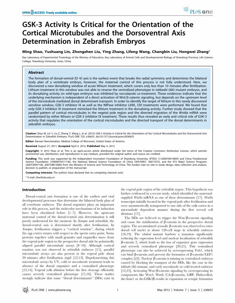
embryos [12,17]. To verify the relationship between 0-mpf lithium
treatment and cortical microtubule assembly, we tested if
nocodazole treatment can reverse the dorsalized phenotype caused
by the 0-mpf lithium treatment. The results showed that compared
to 0-mpf NaCl treated embryos (Figure 5A, E), all the 0-mpf
lithium treated embryos (26/26) showed a typical dorsalized shape
with a significantly elongated animal-vegetal axis at 12 hpf
(Figure 5C). However, when 0.1 mM nocodazole was added to the
0.35 M LiCl solution and treated at 0-mpf, all the 12 hpf embryos
showed a much round shape, 43.6% embryos (n = 39) with more
cells accumulated near the blastopore (Figure 5D), which is similar
to the typical ventralized phenotype caused by NaCl-nocodazole
co-treatment (27.9% ventralized, n = 43) (Figure 5B). At 22hpf, the
0-mpf lithium treated embryos showed dorsalized phenotype
(radially or with curved or trunked tail) (Figure 5G), while for the
LiCl-nocodazole co-treated embryos, ventralized phenotype with
no head and enlarged yolk extension dominated the group
(Figure 5H), very similar to NaCl-nocodazole co-treated embryos
(Figure 5F). The changes in the phenotype after adding
nocodazole demonstrated that depolymerizing microtubules can
block the dorsalizing effect of 0-mpf lithium treatment (statistics
shown in Figure 5I), which strongly indicated that the dorsalizing
activity of the 0-mpf lithium treatment requires successful assembly
of vegetal cortical microtubules.
6. The dorsalizing activity of 0-mpf lithium treatment isnot due to the stabilization of microtubules
Xenopus embryos can be dorsalized by D2O treatment in the first
cell cycle [34], the mechanism of which is stabilizing the
microtubules, resulting in the expanded distribution of DDs. In
this study, we showed that depolymerizing the microtubules by
nocodazole can reverse the dorsalized phenotype caused by 0-mpf
lithium exposure, so a question arises as to whether lithium’s effect
on dorsal-ventral axis formation is a consequence of microtubule
stabilization. Therefore we treated the wild-type embryos at 0-mpf
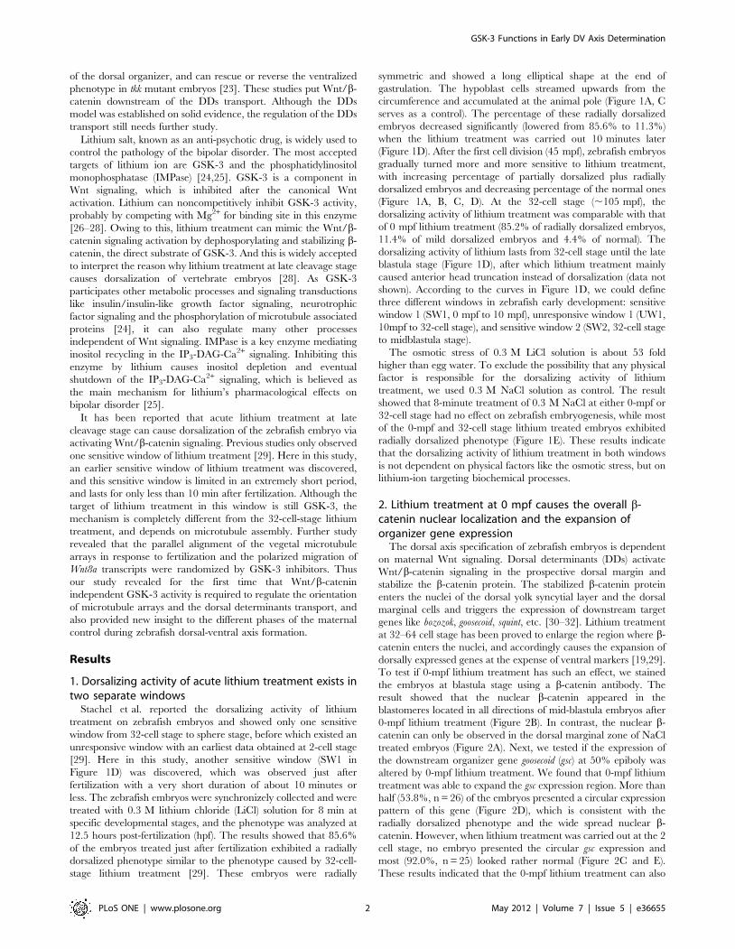
with paclitaxel, a proved microtubule stabilizer. Interestingly, and
unexpectedly, no dorsalized embryos were obtained after 0-mpf
7.5 mg/ml paclitaxel exposure, and on the contrary, 10.0%
(n = 40) treated embryos showed a typical ventralized phenotype
with no notochord observed at 12 hpf (Figure 6B, A as an
untreated control). Other treated embryos exhibited a relatively
normal dorsal-ventral axis but with mild defective convergence-
extension, small head and malformed somites (Figure 6D, C as an
untreated control, E is a ventralized embryo). We also tested the
effect of paclitaxel exposure on tkk mutant embryos, and
Figure 4. The comparison of dorsal and ventral gene expression between 0-mpf lithium treatment and 32-cell-stage lithiumtreatment. Representative embryos from indicated groups stained by gsc probe (A–C, and G–I) or eve1 probe (D–F and J–L) at 50% epiboly. All theembryos are animal pole view and with dorsal side upward if it can be distinguished.doi:10.1371/journal.pone.0036655.g004
GSK-3 Functions in Early DV Axis Determination
PLoS ONE | www.plosone.org 6 May 2012 | Volume 7 | Issue 5 | e36655
accordingly did not observe any rescuing effect of the ventralized
phenotype (Figure 6F). These results suggested that stabilized
microtubules are not sufficient to mediate the dorsalizing activity
of 0-mpf lithium treatment.
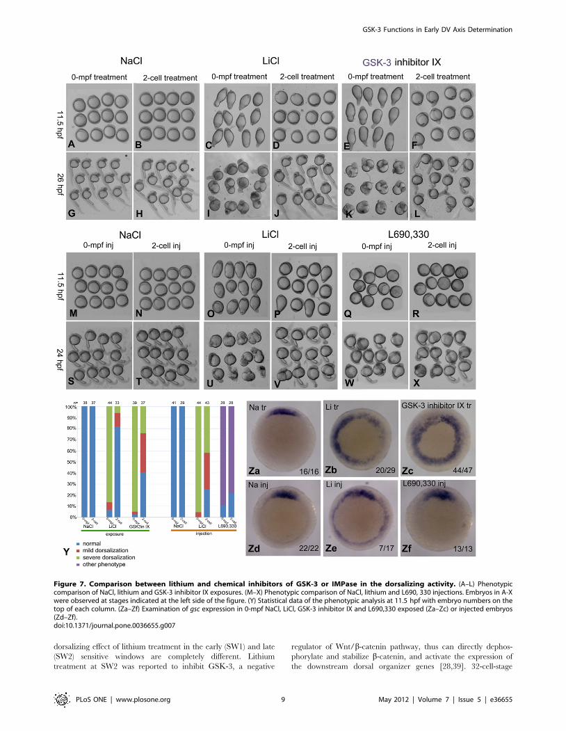
7. The dorsalizing effect of 0-mpf lithium exposure isowing to the inhibition of GSK-3 activity
Lithium inhibits GSK-3 and IMPase, so another important
question is to distinguish which enzyme serves as the real target.
To address this question, we further used much more specific
chemical inhibitors including GSK-3 inhibitor IX and IMPase
inhibitor L690, 330, to analyze which one can mimic the lithium
treatment in the dynamic dorsalizing activity. Just like lithium
treatment, nearly all the 0-mpf GSK-3 inhibitor IX treated
embryos exhibited a long elliptical shape at 11.5 hpf, and the large
cell aggregation at the animal pole was also observed at 26 hpf.
The dorsalizing activity of 2-cell-stage GSK-3 inhibitor IX
treatment became much weaker than the 0-mpf treatment, which
is also very similar to that of lithium exposure (Figure 7A, B, C, D,
E, F, G, H, I, J, K, L, Y). We then injected L690, 330 or LiCl at 0
mpf zebrafish embryos and compared the resulting phenotypes.
Injection of L690, 330 resulted in embryos with a typical
convergence-extension (CE) defect rather than the dorsalized
phenotype. These embryos appeared spherical at 11.5 hpf and
showed distinct dorsal-ventral axis at 24 hpf, although the length
of the dorsal axis was much shorter. In sharp contrast, 0-mpf LiCl
injected embryos only exhibited dorsalized phenotype. When the
injection was performed at 2-cell stage, the dorsalizing effect
lithium was reduced, however we failed to find such a
phenomenon in L690, 330 injection experiment (Figure 7M, N,
O, P, Q, R, S, T, U, V, W, X, Y). To confirm the phenotypic data
at the molecular level, we performed in situ hybridization
experiment on 50% epiboly embryos using the probe of gsc. As
expected, most of the 0-mpf GSK-3 inhibitor IX and 0-mpf
lithium treated embryos showed radial or expanded expression of
gsc. However, all of the L690, 330 treated embryos showed a
relatively normal expression pattern (Figure 7Za–Zf). These data
demonstrated that the 0-mpf treatment of GSK-3 inhibitor IX but
not L690, 330 can mimic lithium in the dorsalizing activity, which
strongly suggested that GSK-3 activity is required for the dorsal-
ventral specification shortly after fertilization.
Figure 5. 0-mpf nocodazole treatment reverses the dorsalizing effect of the 0-mpf lithium treatment. The 0-mpf embryos were treatedwith 0.35 M LiCl solution in the absence or presence of 0.1 mM nocodazole for 5 min, and then observed at 12 hpf and 22 hpf. 0 35 M NaCl treatmentserved as control. (A and E) 0-mpf NaCl treated embryos at 12 hpf (A) and 22 hpf (E). (B and F) 0-mpf NaCl and nocodazole co-treated embryos at12 hpf (B) and 22 hpf (F). (C and G) 0-mpf lithium treated embryos at 12 hpf (C) and 22 hpf (G). (D and H) 0-mpf lithium and nocodazole co-treatedembryos at 12 hpf (D) and 22 hpf (H). (I) Statistical data were obtained at 12 hpf for the experiment with embryo numbers on the top of each column.doi:10.1371/journal.pone.0036655.g005
GSK-3 Functions in Early DV Axis Determination
PLoS ONE | www.plosone.org 7 May 2012 | Volume 7 | Issue 5 | e36655
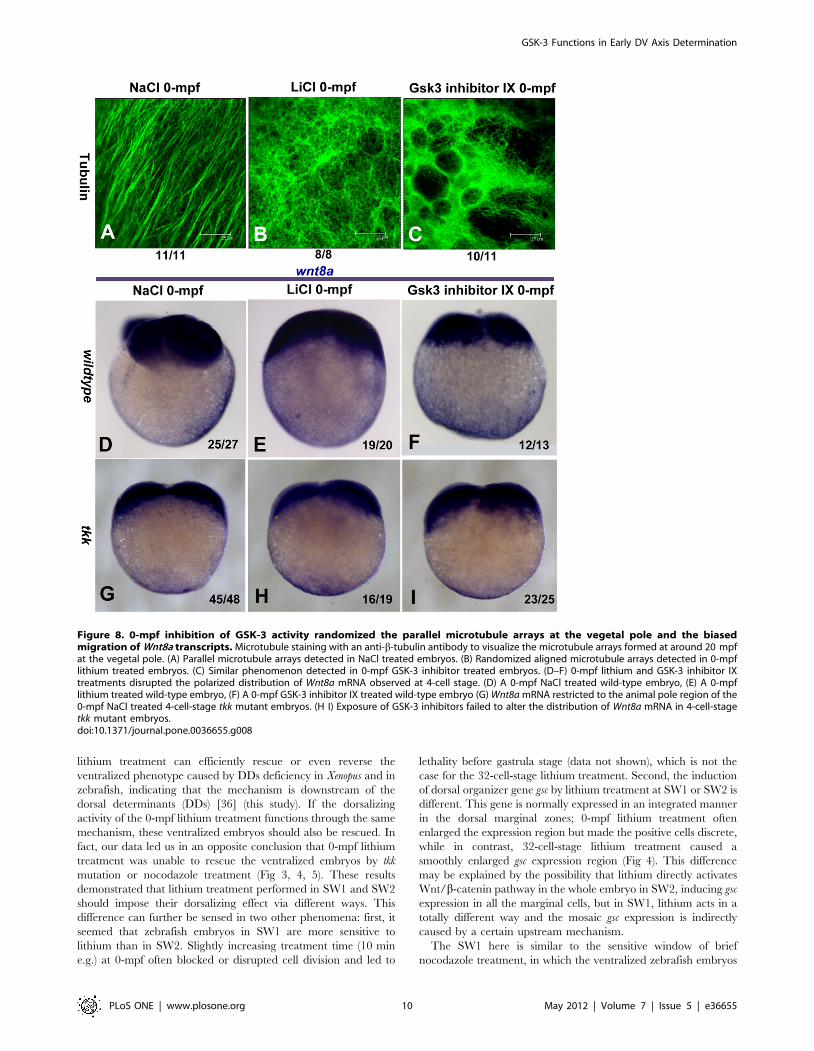
8. 0-mpf GSK-3 inhibition disrupts the parallel pattern ofcortical microtubules and the polarized migration ofWnt8a transcripts
It was reported that destroying the assembly of the microtubules
by chemical or physical factors can ventralize vertebrate embryos
[12,14]. Blocking the microtubule dependent transport of DDs can
also generate ventralized phenotype [21]. However, 0-mpf lithium
treatment acts in an opposite way to generate the dorsalized
phenotype, so it is impossible that 0-mpf lithium treatment can
disrupt the assembly of cortical microtubules or inhibit the DDs
transport. In fact, 0-mpf lithium treatment may affect the transport
and broaden the distribution of DDs by influencing the
remodeling of cortical microtubules after fertilization. To test this
possibility, we stained and visualized the vegetal microtubule
arrays formed ,20 mpf by confocal microscopy. As reported,
well-formed parallelly distributed microtubules in the vegetal pole
region were detected in NaCl treated embryos (Figure 8A,), but in
sharp contrast, the orientation of microtubules in 0-mpf lithium
and GSK-3 inhibitor IX treated embryos were randomized and
organized like an irregularly weaved net, and the microtubule
bundles appeared much thinner with respect to control (Figure 8B,
C). We further tested if the polarized migration of a recently
identified DD, Wnt8a mRNA, was affected by GSK-3 inhibition.
In 0-mpf NaCl treated 4-cell stage embryos, almost all the
embryos (92.6%, n = 27) tested showed a biased distribution of
Wnt8a in the yolk cortex (Figure 8D), but after inhibiting GSK-3 at
0-mpf by lithium or GSK-3 inhibitor IX, this asymmetric pattern
disappeared, instead, the distribution of Wnt8a transcripts became
much more smearing (Figure 8E and F). We also examined the
expression of Wnt8a in 4-cell stage tkk mutant embryos, and found
that in most cases (93.8%, n = 48), Wnt8a transcripts were
restricted to the vegetal pole with indistinguishable biased
distribution, and 0-mpf lithium treatment or GSK-3 inhibitor IX
treatment did not alter their distribution in this mutant (Figure 8G,
H, I), which was consistent with the previous data that 0-mpf
lithium treatment failed to reverse the ventralized phenotype of tkk
mutant. These observations suggest that GSK-3 activity is critical
for the parallel alignment of the microtubule arrays after
fertilization, and the dorsalizing activity of 0-mpf GSK-3
inhibition is probably the consequence of randomized microtubule
arrays, which lead the DDs to a much broader area of the
perspective marginal zone.
Discussion
1. Lithium can dorsalize zebrafish embryos in twocompletely different ways
In this work, we have characterized another lithium-sensitive
window to cause dorsalization of the zebrafish embryos. Lithium
treatment carried out at the late cleavage stage was previously
described for its capability to induce dorsalization in Xenopus as
well as in zebrafish [29,35–38]. However, the mechanisms of the
Figure 6. Paclitaxel treatment on wild-type and tkk mutant embryos. (A) Dorsal view of an untreated 12 hpf embryo. (B) Dorsal view of a 0-mpf paclitaxel treated embryo with ventralized phenotype. (C) An embryo with normal phenotype. (D) An embryo with CE defect like phenotype,showing a smaller head, shorter anterior-posterior axis and malformed somites. (E) A ventralized embryo. Embryos in (C-E) were observed at 24 hpf.The statistical data based on the 24 hpf observation were shown in (F) with embryo numbers shown on the top of each column.doi:10.1371/journal.pone.0036655.g006
GSK-3 Functions in Early DV Axis Determination
PLoS ONE | www.plosone.org 8 May 2012 | Volume 7 | Issue 5 | e36655
dorsalizing effect of lithium treatment in the early (SW1) and late
(SW2) sensitive windows are completely different. Lithium
treatment at SW2 was reported to inhibit GSK-3, a negative
regulator of Wnt/b-catenin pathway, thus can directly dephos-
phorylate and stabilize b-catenin, and activate the expression of
the downstream dorsal organizer genes [28,39]. 32-cell-stage
Figure 7. Comparison between lithium and chemical inhibitors of GSK-3 or IMPase in the dorsalizing activity. (A–L) Phenotypiccomparison of NaCl, lithium and GSK-3 inhibitor IX exposures. (M–X) Phenotypic comparison of NaCl, lithium and L690, 330 injections. Embryos in A-Xwere observed at stages indicated at the left side of the figure. (Y) Statistical data of the phenotypic analysis at 11.5 hpf with embryo numbers on thetop of each column. (Za–Zf) Examination of gsc expression in 0-mpf NaCl, LiCl, GSK-3 inhibitor IX and L690,330 exposed (Za–Zc) or injected embryos(Zd–Zf).doi:10.1371/journal.pone.0036655.g007
GSK-3 Functions in Early DV Axis Determination
PLoS ONE | www.plosone.org 9 May 2012 | Volume 7 | Issue 5 | e36655
lithium treatment can efficiently rescue or even reverse the
ventralized phenotype caused by DDs deficiency in Xenopus and in
zebrafish, indicating that the mechanism is downstream of the
dorsal determinants (DDs) [36] (this study). If the dorsalizing
activity of the 0-mpf lithium treatment functions through the same
mechanism, these ventralized embryos should also be rescued. In
fact, our data led us in an opposite conclusion that 0-mpf lithium
treatment was unable to rescue the ventralized embryos by tkk
mutation or nocodazole treatment (Fig 3, 4, 5). These results
demonstrated that lithium treatment performed in SW1 and SW2
should impose their dorsalizing effect via different ways. This
difference can further be sensed in two other phenomena: first, it
seemed that zebrafish embryos in SW1 are more sensitive to
lithium than in SW2. Slightly increasing treatment time (10 min
e.g.) at 0-mpf often blocked or disrupted cell division and led to
lethality before gastrula stage (data not shown), which is not the
case for the 32-cell-stage lithium treatment. Second, the induction
of dorsal organizer gene gsc by lithium treatment at SW1 or SW2 is
different. This gene is normally expressed in an integrated manner
in the dorsal marginal zones; 0-mpf lithium treatment often
enlarged the expression region but made the positive cells discrete,
while in contrast, 32-cell-stage lithium treatment caused a
smoothly enlarged gsc expression region (Fig 4). This difference
may be explained by the possibility that lithium directly activates
Wnt/b-catenin pathway in the whole embryo in SW2, inducing gsc
expression in all the marginal cells, but in SW1, lithium acts in a
totally different way and the mosaic gsc expression is indirectly
caused by a certain upstream mechanism.
The SW1 here is similar to the sensitive window of brief
nocodazole treatment, in which the ventralized zebrafish embryos
Figure 8. 0-mpf inhibition of GSK-3 activity randomized the parallel microtubule arrays at the vegetal pole and the biasedmigration of Wnt8a transcripts. Microtubule staining with an anti-b-tubulin antibody to visualize the microtubule arrays formed at around 20 mpfat the vegetal pole. (A) Parallel microtubule arrays detected in NaCl treated embryos. (B) Randomized aligned microtubule arrays detected in 0-mpflithium treated embryos. (C) Similar phenomenon detected in 0-mpf GSK-3 inhibitor treated embryos. (D–F) 0-mpf lithium and GSK-3 inhibitor IXtreatments disrupted the polarized distribution of Wnt8a mRNA observed at 4-cell stage. (D) A 0-mpf NaCl treated wild-type embryo, (E) A 0-mpflithium treated wild-type embryo, (F) A 0-mpf GSK-3 inhibitor IX treated wild-type embryo (G) Wnt8a mRNA restricted to the animal pole region of the0-mpf NaCl treated 4-cell-stage tkk mutant embryos. (H I) Exposure of GSK-3 inhibitors failed to alter the distribution of Wnt8a mRNA in 4-cell-stagetkk mutant embryos.doi:10.1371/journal.pone.0036655.g008
GSK-3 Functions in Early DV Axis Determination
PLoS ONE | www.plosone.org 10 May 2012 | Volume 7 | Issue 5 | e36655

can be generated when nocodazole treatment is performed before
10 mpf, but the embryos were seldom ventralized when treated at
15 mpf [12]. As nocodazole treatment inhibits the microtubule
polymerization, the similarity between SW1 and ventralizing
window of nocodazole treatment may suggest that lithium also
imposes its effect on the microtubule organization, but in a very
different way. In this study, we presented three lines of evidence in
support of this hypothesis. First, in the presence of nocodazole, the
dorsalizing activity of the 0-mpf lithium treatment was completely
lost (Figure 5), and the embryos only showed the ventralized
phenotype, i.e. nocodazole treatment can override the lithium
treatment, suggesting that the dorsalizing activity of 0-mpf lithium
treatment requires the normal polymerization of microtubules.
Second, 0-mpf lithium treatment was unable to dorsalize the
maternal mutant tokkaebi (tkk) zebrafish embryos which harbors a
mutation in the syntabulin gene encoding a protein required for the
cargo transport along microtubule arrays [13]. Third, and most
directly, we showed that the parallel microtubule array formed
,20 mpf in the vegetal pole region was randomized by the 0-mpf
lithium treatment, which raised a possibility that after 0-mpf
lithium treatment, the transport of DDs is not directed to the
perspective dorsal region but to the circumference of the margin,
and this was further confirmed by marking a newly identified
dorsal determinant, Wnt8a mRNA. These results thus put the
mechanism of the 0-mpf lithium treatment at the upstream level to
the microtubule dependent transport of DDs.
2. Wnt/b-catenin independent GSK-3 activity is requiredfor the dorsal-ventral axis formation
GSK-3 and inositol monophosphatase (IMPase) are proved
targets of lithium [25]. Our study using specific chemical inhibitors
demonstrates GSK-3 as the real target of 0-mpf lithium treatment.
GSK-3 plays an important role in Wnt/b-catenin signaling.
Figure 9. Model of dorsal-ventral axis formation in zebrafish. The zebrafish DV axis specification can be divided to four phases based on thedynamic alteration of the dorsalizing activity of lithium treatment. SW1 designates 0–10 mpf in which lithium treatment can cause dorsalization of theembryos. In this phase, fertilization initiates a GSK-3 dependent mechanism regulating the orientation but not stablization of vegetal microtubuleswhich is critical for the dorsalward transport of DDs like Wnt8a mRNA. UW1 designates the first unresponsive window of lithium treatment, from10 mpf to the 32-cell stage, in which, especially in the early period, lithium treatment fails to efficiently cause dorsalization. In this period, dorsaldeterminants are transported from the vegetal pole to the perspective dorsal side. The transduction of Wnt/b-catenin pathway is probably blockedby some unknown mechanism in SW1 and UW1. SW2 designates the period from the 32-cell stage to the mid-blastula stage. In this period, dorsallylocated DDs are able to inhibit GSK-3, causing the stabilization and nuclear localization of b-catenin, and lead to the expression of dorsal organizergenes. In UW2, lithium treatment loses its ability to dorsalize zebrafish embryos and the organizer gene expression is translated gradually by cellmovement to morphologically distinguished dorsal-ventral axis. Arrow head at the lower-left corner indicates the shield.doi:10.1371/journal.pone.0036655.g009
GSK-3 Functions in Early DV Axis Determination
PLoS ONE | www.plosone.org 11 May 2012 | Volume 7 | Issue 5 | e36655

However, our data indicate that directly activating Wnt/b-catenin
pathway cannot be the cause of the dorsalizing activity of lithium
treatment in SW1. In fact, GSK-3 inhibitors can randomize the
alignment of vegetal cortical microtubule arrays and disturb the
biased transport of the Wnt8a transcripts initiated by fertilization.
Based on these observations, the dorsalizing effect of 0-mpf GSK-3
inhibition can be properly interpreted. In normal embryos, the
active GSK-3 shortly after fertilization may facilitate the formation
of parallelly aligned microtubule arrays, which is essential for the
polarized transport of Wnt8a mRNA. But the polymerized
microtubules failed to form parallel bundles after GSK-3
inhibition, instead, randomized and much thinner microtubule
filament formed after lithium or GSK-3 inhibitor IX treatment.
These net-like microtubule arrays can still transport DDs like
Wnt8a transcripts, but might lead them to migrate in all the
directions across the yolk cortex, so that many ventral lateral
marginal cells receive sufficient dorsal-determining signals to
change their fate. This deduction was supported by the fact that gsc
positive cells often discretely distributed around the margin in 0-
mpf lithium or GSK-3 inhibitor IX treated embryos. Syntabulin
was thought as a linker between DDs and Kinesin motors during
the microtubule dependent transport, so lacking this protein can
reduce the amount of DDs transported to the dorsal margin
(Wnt8a transcripts still located at the vegetal pole in 4-cell stage tkk
embryos shown in Figure 8G), and causes ventralized phenotype.
It can be imagined that the reduced DDs successfully transported
in tkk embryos will be further diluted after their randomized
migration caused by GSK-3 inhibition, and this dilution will make
a sub-threshold supply of DDs for more marginal cells resulting in
a more frequent appearance of ventralized embryos.
Data in this study also raised a question of whether GSK-3
regulates any microtubule-related protein independent of the
Wnt/b-catenin signaling shortly after fertilization. It has been
established that the microtubule associated protein Tau and
MAP1B are substrate of GSK-3b (reviewed by [24]). Although no
literature deals with the role that these proteins plays in the early
D–V axis formation, it has been well studied that Tau is essential
for microtubule stabilization in neuronal axons. Hyperphosphor-
ylation and intracellular fibrillar formation of tau protein deter its
ability to bind to and stabilize microtubules, and is a pathology
found in Alzheimer’s disease [40,41]. Lithium is able to reduce the
amount of phosphorylated Tau in cell culture [39], so it is
conceivable that lithium may activate too much Tau by inhibiting
the activity of GSK-3 after fertilization and randomize the vegetal
paralleled microtubule arrays. This hypothesis, however, is far
from being solidified. First, whether Tau owns a maternal
expression is not verified. Second, our data showed that only
stabilizing microtubules by paclitaxel treatment was not sufficient
to dorsalize zebrafish embryos like GSK-3 inhibitors. Beside these
doubts, another interesting aspect is that GSK-3 seemed to
function oppositely in tkk mutant embryos for the unexpected
ventralizing effect of lithium treatment. This phenomenon might
indicate that the GSK-3 regulated process is extremely sensitive to
slight disturbance of the cargo transport system, and the
Syntabulin protein might functionally interact with GSK-3 or its
substrate. Thus, further investigation is required to identify GSK-3
substrates responsible for the short-lived microtubule remodeling
process. Functional analysis of these unknown molecules might
shed light on the mystery of the instantaneous SW1 and the
anomalous behavior of lithium treatment on tkk embryos.
3. The unresponsive window (UW) of lithium treatmentduring the early cleavage stage
It is very interesting that lithium had almost no effect on
zebrafish embryos when treatment was performed during the first
1–2 cell cycles [29] (this study). Wnt signaling is not activated until
midblastula stage, as revealed by TopdGFP transgenic zebrafish
[42]. Our data also suggest that Wnt/b-catenin signaling cannot
be efficiently activated by lithium treatment from fertilization to
the late cleavage stage. There might be two possibilities for the
existence of the unresponsive window: one is that the extra b-
catenin stabilized by lithium treatment is degraded by an unknown
negative feedback loop in the time span between the treatment
and mid-blastula stage when Wnt signaling begins to activate, and
the second possibility is that lithium treatment in this UW cannot
rescue b-catenin from GSK-3. For the second possibility, we
hypothesize that before the 32-cell stage, GSK-3 and its substrate
b-catenin might be separated, and importantly at the same time,
the average concentration of b-catenin is controlled below the
activation threshold of the downstream cascade of Wnt signaling.
GSK-3 is shown to be sequestered into the multi vesicular
endosomes (MVB) in response to Wnt activation, and this
phenomenon is required for the secondary axis induction in
Xenopus embryos by Wnt ligands overexpression [43]. According to
this, it is conceivable that GSK-3 and its substrate b-catenin might
be segregated from each other by structures like MVB in the SW1
and UW, and GSK-3 is possible to be released gradually to the
cytosol when cleavage continues under an unknown mechanism.
According to this assumption, the existence of the unresponsive
window can be properly interpreted: before the 32-cell stage, as
very limited b-catenin can meet GSK-3, the lithium treatment in
this stage can only increase a very small amount of b-catenin,
which is not sufficient to activate organizer genes around the
margin at mid-blastula stage. However, after 32-cell stage, as most
b-catenin can contact the freed GSK-3 in the lateral-ventral
regions, lithium treatment can stabilize large amount of b-catenin
which may break the threshold to activate downstream target
genes. To test these two models, more work is needed to verify the
fluctuation of b-catenin before the 32-cell stage in control and
lithium treated embryos, and to test if the maternal mutant with
MVB formation defect is associated with the D-V axis formation
problem in the zebrafish embryos, and if GSK-3 localizes in MVB
before the late cleavage stage and is released to cytosol after the
32-cell stage.
4. Distinct phases of the dorsal-ventral axis specificationrevealed by lithium treatment, and remaining questions
Based on the data in this study, we could divide the D V axis
formation process into four phases: 1) 0-mpf to 10-mpf, identical to
the SW1, in this phase, fertilization induces a GSK-3 dependent
mechanism that determines the orientation of vegetal microtubule
arrays; 2) 10-mpf to 32-cell stage, corresponding to the UW1, in
this phase, the dorsal determinants move directionally to the
perspective dorsal marginal zone along paralleled microtubule
arrays. 3) 32-cell stage to mid-blastula stage (SW2). Dorsal
determinants begin to inhibit GSK-3 in the dorsal marginal zone,
leading to b-catenin stabilization and nuclear localization, which
further activate the early marker of the organizer; 4) Mid-blastula
to early gastrula stage (UW2), in this phase, lithium treatment
tends to induce posteriorization of neural system but not
dorsalization. The dorsal-ventral axis specification is accomplished
at the molecular level, and the asymmetric expression of dorsal-
ventral genes is gradually translated to the morphologically distinct
dorsal organizer structure–the shield (Figure 9).
GSK-3 Functions in Early DV Axis Determination
PLoS ONE | www.plosone.org 12 May 2012 | Volume 7 | Issue 5 | e36655
So far, we know very little about what happens in the first two
stages. Specifically, we know little about the signaling cascade that
initiates and directs the transport of dorsal determinants; and we
have not perceived the reason why GSK-3 inhibition cannot
efficiently activate Wnt/b-catenin signaling in SW1 and UW1.
More genetic and functional work is needed to identify and
analyze maternal mutants with dorsal-ventral axis defect, which
may shed light on these questions, and help to understand this
earliest, vital, and intricate patterning process.
Materials and Methods
1. Ethic statementAll embryos were handled according to relevant national and
international guidelines. The study was approved by the Com-
mittee on the Ethics of Animal Experiments of Shandong
University (Permit number: ECAESDUSM 2009035).
2. Fish StrainsWild-type and the mutant tokkeabi (tkk) fish were used. The
maternal mutant tkk embryos were generated by crossing wild-type
male with tkk homogeneous female. The penetrance was evaluated
by observing the phenotype at 24 hpf. The tokkeabi mutant strain is
a gift from Dr. Hibi.
3. Egg collection, lithium, nocodazole, paclitaxel andGSK-3 inhibitor IX exposure
To ensure synchronic development of the zebrafish embryos,
eggs were collected immediately when the female spawned. The
fish mated in a chamber made up of three parts, an outer tank, an
inner tank with narrow slits that allow the eggs to fall through to
the outer tank, and a plastic sheet set in the middle of the inner
tank to separate the male and female fish before mating. We kept a
pair of fish separated in this chamber overnight and prepared an
extra outer tank with egg water before the experiment. On the
morning of the next day, the plastic sheet was removed and the
fish began to mate. Once the female began to spawn eggs, the
male and female were quickly separated by the plastic sheet and
the inner tank was immediately transferred to the prepared extra
outer tank. The synchronic eggs were collected immediately from
the original outer tank and subjected to treatment or injection at
indicated stages. Another batch of synchronic eggs can be
collected by removing the plastic sheet again when convenient
and repeating the procedure described above. In order to get
sufficient synchronic eggs, the male and female fish were raised
separately for more than a week before the experiment, and the
female normally releases 20–40 eggs at one time during the
mating. The data was obtained by several treatments with
different batches of synchronic eggs. The collected embryos were
treated with 0.3 M LiCl solution (diluted in E3 buffer) for 8 min at
indicated stages. The nocodazole (Sigma, M1404-2MG) were
dissolved in DMSO at a concentration of 5 mg/ml, and dilute in
E3 buffer or mixed with the 0.35 M LiCl solution at a final
concentration of 0.1 mg/ml. The 0-mpf treatment of LiCl,
nocodazole or LiCl/nocodazole mixture was carried out imme-
diately when the female fish spawned. As the embryos were
extremely sensitive to nocodazole, so the treated time was reduced
to 5 min, which is sufficient for 0.35 M LiCl to induce
dorsalization. Paclitaxel (Sigma, T7191-5MG) was dissolved in
DMSO at 1 mg/ml, then diluted to 7.5 mg/ml in an E3 buffer
based solution containing 0.5 mg/ml Pronase (Roche, 11 459 643
001), and the embryos were treated in this solution for 8 min.
GSK-3 inhibitor IX (Santa Cruz, sc-202634) was dissolved in
DMSO at 10 mg/ml as a stock solution, and the embryos were
exposed in a working solution of 10 mg/ml containing 0.5 mg/ml
Pronase in E3 buffer for 8 min. The treated embryos were rinsed
in E3 buffer for three times and incubated at 28.5uC until
observation and fixation.
4. MicroinjectionLiCl and NaCl were diluted in deionized water at a
concentration of 0.15 M respectively, and was injected 2 nl to
the 0–5 mpf and 2-cell-stage embryos. 2.5 mM L690, 330 (Santa
Cruz, sc-202685A) aqueous solution was injected 2 nl for each
embryo at the same stage to those used in LiCl and NaCl
injections (the concentration of L690, 330 was used according to
previous studies [44,45]). The microinjection was performed using
the MPPI-3 Pressure Injector.
5. Whole mount in situ hybridizationSequences of goosecoid (gsc), eve1 and Wnt8a were cloned in
pGEM-Teasy vector. Anti-sense RNA probes were synthesized
using the digoxigenin-UTP (DIG) in vitro transcription kit (Roche
Applied Science, Indianapolis, IN, USA). Whole-mount in situ
hybridization was conducted according to the zebrafish book [46].
6. Antibodies, immunofluorescence and confocalmicroscopy
The b-catenin localization was visualized by using an antibody
from Abcam (ab6302), and the whole mount immunofluorecence
was performed in a routine way as described [47], and the focal
plane was selected near the margin; The microtubule was stained
by an anti-b-tubulin antibody (Chemicon KMX-1), and the
protocol were previously described [13,48]. The focal planes were
selected near the vegetal pole. The images were taken under a 10x
objective of a Leica TCS SP2 confocal microscope.
Acknowledgments
We are grateful to Dr. Hibi Masahiko, Bioscience and Biotechnology
Center, Nagoya University, Japan, for presenting tokkeabi mutant strain,
and helpful discussions. We also thank Dr. Shi De-li for critical reading of
the manuscript.
Author Contributions
Conceived and designed the experiments: MS HZ. Performed the
experiments: MS YL ZL YZ LW. Analyzed the data: MS YL CL HZ.
Wrote the paper: MS HZ.
References
1. Langdon YG, Mullins MC (2010) Maternal and Zygotic Control of Zebrafish
Dorsoventral Axial Patterning. Annu Rev Genet.
2. De Robertis EM (2006) Spemann’s organizer and self-regulation in amphibian
embryos. Nat Rev Mol Cell Biol 7: 296–302.
3. Hibi M, Hirano T, Dawid IB (2002) Organizer formation and function. Results
Probl Cell Differ 40: 48–71.
4. Harland R, Gerhart J (1997) Formation and function of Spemann’s organizer.
Annu Rev Cell Dev Biol 13: 611–667.
5. De Robertis EM (2009) Spemann’s organizer and the self-regulation of
embryonic fields. Mech Dev 126: 925–941.
6. Schier AF, Talbot WS (2005) Molecular genetics of axis formation in zebrafish.
Annu Rev Genet 39: 561–613.
7. Driever W (1995) Axis formation in zebrafish. Curr Opin Genet Dev 5:
610–618.
8. Hainski AM, Moody SA (1992) Xenopus maternal RNAs from a dorsal animal
blastomere induce a secondary axis in host embryos. Development 116:
347–355.
GSK-3 Functions in Early DV Axis Determination
PLoS ONE | www.plosone.org 13 May 2012 | Volume 7 | Issue 5 | e36655

9. Weaver C, Farr GH 3rd, Pan W, Rowning BA, Wang J, et al. (2003) GBP binds
kinesin light chain and translocates during cortical rotation in Xenopus eggs.Development 130: 5425–5436.
10. Miller JR, Rowning BA, Larabell CA, Yang-Snyder JA, Bates RL, et al. (1999)
Establishment of the dorsal-ventral axis in Xenopus embryos coincides with thedorsal enrichment of dishevelled that is dependent on cortical rotation. J Cell
Biol 146: 427–437.11. Kimmel CB, Ballard WW, Kimmel SR, Ullmann B, Schilling TF (1995) Stages
of embryonic development of the zebrafish. Dev Dyn 203: 253–310.
12. Jesuthasan S, Stahle U (1997) Dynamic microtubules and specification of thezebrafish embryonic axis. Curr Biol 7: 31–42.
13. Nojima H, Rothhamel S, Shimizu T, Kim CH, Yonemura S, et al. (2010)Syntabulin, a motor protein linker, controls dorsal determination. Development
137: 923–933.14. Strahle U, Jesuthasan S (1993) Ultraviolet irradiation impairs epiboly in
zebrafish embryos: evidence for a microtubule-dependent mechanism of epiboly.
Development 119: 909–919.15. Ober EA, Schulte-Merker S (1999) Signals from the yolk cell induce mesoderm,
neuroectoderm, the trunk organizer, and the notochord in zebrafish. Dev Biol215: 167–181.
16. Mizuno T, Yamaha E, Kuroiwa A, Takeda H (1999) Removal of vegetal yolk
causes dorsal deficencies and impairs dorsal-inducing ability of the yolk cell inzebrafish. Mech Dev 81: 51–63.
17. Lu FI, Thisse C, Thisse B (2011) Identification and mechanism of regulation ofthe zebrafish dorsal determinant. Proc Natl Acad Sci U S A 108: 15876–15880.
18. Dougan ST, Warga RM, Kane DA, Schier AF, Talbot WS (2003) The role ofthe zebrafish nodal-related genes squint and cyclops in patterning of
mesendoderm. Development 130: 1837–1851.
19. Schneider S, Steinbeisser H, Warga RM, Hausen P (1996) Beta-catenintranslocation into nuclei demarcates the dorsalizing centers in frog and fish
embryos. Mech Dev 57: 191–198.20. Bellipanni G, Varga M, Maegawa S, Imai Y, Kelly C, et al. (2006) Essential and
opposing roles of zebrafish beta-catenins in the formation of dorsal axial
structures and neurectoderm. Development 133: 1299–1309.21. Kelly C, Chin AJ, Leatherman JL, Kozlowski DJ, Weinberg ES (2000)
Maternally controlled (beta)-catenin-mediated signaling is required for organizerformation in the zebrafish. Development 127: 3899–3911.
22. Xiong B, Rui Y, Zhang M, Shi K, Jia S, et al. (2006) Tob1 controls dorsaldevelopment of zebrafish embryos by antagonizing maternal beta-catenin
transcriptional activity. Dev Cell 11: 225–238.
23. Nojima H, Shimizu T, Kim CH, Yabe T, Bae YK, et al. (2004) Geneticevidence for involvement of maternally derived Wnt canonical signaling in
dorsal determination in zebrafish. Mech Dev 121: 371–386.24. Gould TD, Manji HK (2005) Glycogen synthase kinase-3: a putative molecular
target for lithium mimetic drugs. Neuropsychopharmacology 30: 1223–1237.
25. Shaldubina A, Agam G, Belmaker RH (2001) The mechanism of lithium action:state of the art, ten years later. Prog Neuropsychopharmacol Biol Psychiatry 25:
855–866.26. Gurvich N, Klein PS (2002) Lithium and valproic acid: parallels and contrasts in
diverse signaling contexts. Pharmacol Ther 96: 45–66.27. Ryves WJ, Harwood AJ (2001) Lithium inhibits glycogen synthase kinase-3 by
competition for magnesium. Biochem Biophys Res Commun 280: 720–725.
28. Klein PS, Melton DA (1996) A molecular mechanism for the effect of lithium ondevelopment. Proc Natl Acad Sci U S A 93: 8455–8459.
29. Stachel SE, Grunwald DJ, Myers PZ (1993) Lithium perturbation and goosecoid
expression identify a dorsal specification pathway in the pregastrula zebrafish.Development 117: 1261–1274.
30. Fekany K, Yamanaka Y, Leung T, Sirotkin HI, Topczewski J, et al. (1999) The
zebrafish bozozok locus encodes Dharma, a homeodomain protein essential forinduction of gastrula organizer and dorsoanterior embryonic structures.
Development 126: 1427–1438.31. Sirotkin HI, Dougan ST, Schier AF, Talbot WS (2000) bozozok and squint act
in parallel to specify dorsal mesoderm and anterior neuroectoderm in zebrafish.
Development 127: 2583–2592.32. Joore J, Fasciana C, Speksnijder JE, Kruijer W, Destree OH, et al. (1996)
Regulation of the zebrafish goosecoid promoter by mesoderm inducing factorsand Xwnt1. Mech Dev 55: 3–18.
33. Kishimoto Y, Lee KH, Zon L, Hammerschmidt M, Schulte-Merker S (1997)The molecular nature of zebrafish swirl: BMP2 function is essential during early
dorsoventral patterning. Development 124: 4457–4466.
34. Scharf SR, Rowning B, Wu M, Gerhart JC (1989) Hyperdorsoanterior embryosfrom Xenopus eggs treated with D2O. Dev Biol 134: 175–188.
35. Kao KR, Elinson RP (1988) The entire mesodermal mantle behaves asSpemann’s organizer in dorsoanterior enhanced Xenopus laevis embryos. Dev
Biol 127: 64–77.
36. Kao KR, Masui Y, Elinson RP (1986) Lithium-induced respecification ofpattern in Xenopus laevis embryos. Nature 322: 371–373.
37. Kao KR, Elinson RP (1989) Dorsalization of mesoderm induction by lithium.Dev Biol 132: 81–90.
38. Aanstad P, Whitaker M (1999) Predictability of dorso-ventral asymmetry in thecleavage stage zebrafish embryo: an analysis using lithium sensitivity as a dorso-
ventral marker. Mech Dev 88: 33–41.
39. Stambolic V, Ruel L, Woodgett JR (1996) Lithium inhibits glycogen synthasekinase-3 activity and mimics wingless signalling in intact cells. Curr Biol 6:
1664–1668.40. Buee L, Bussiere T, Buee-Scherrer V, Delacourte A, Hof PR (2000) Tau protein
isoforms, phosphorylation and role in neurodegenerative disorders. Brain Res
Brain Res Rev 33: 95–130.41. Johnson GV, Hartigan JA (1999) Tau protein in normal and Alzheimer’s disease
brain: an update. J Alzheimers Dis 1: 329–351.42. Dorsky RI, Sheldahl LC, Moon RT (2002) A transgenic Lef1/beta-catenin-
dependent reporter is expressed in spatially restricted domains throughoutzebrafish development. Dev Biol 241: 229–237.
43. Taelman VF, Dobrowolski R, Plouhinec JL, Fuentealba LC, Vorwald PP, et al.
(2010) Wnt signaling requires sequestration of glycogen synthase kinase 3 insidemultivesicular endosomes. Cell 143: 1136–1148.
44. Sconzo G, Cascino D, Amore G, Geraci F, Giudice G (1998) Effect of theIMPase inhibitor L690,330 on sea urchin development. Cell Biol Int 22: 91–94.
45. Westfall TA, Hjertos B, Slusarski DC (2003) Requirement for intracellular
calcium modulation in zebrafish dorsal-ventral patterning. Dev Biol 259:380–391.
46. Westerfield M (1993) The zebrafish book: a guide for the laboratory use ofzebrafish (Brachydanio rerio). Eugene, OR: M. Westerfield. 1 v. (unpaged) p.
47. Umbhauer M, Djiane A, Goisset C, Penzo-Mendez A, Riou JF, et al. (2000) TheC-terminal cytoplasmic Lys-thr-X-X-X-Trp motif in frizzled receptors mediates
Wnt/beta-catenin signalling. EMBO J 19: 4944–4954.
48. Inoue D, Wittbrodt J (2011) One for all – a highly efficient and versatile methodfor fluorescent immunostaining in fish embryos. PLoS One 6: e19713.
GSK-3 Functions in Early DV Axis Determination
PLoS ONE | www.plosone.org 14 May 2012 | Volume 7 | Issue 5 | e36655
Related Documents