Growth hormone responsive neural precursor cells reside within the adult mammalian brain Daniel G. Blackmore 1 , Brent A. Reynolds 1,2 , Mohammad G. Golmohammadi 1,3 , Beatrice Large 1 , Roberto M. Aguilar 4 , Luis Haro 5 , Michael J. Waters 6 & Rodney L. Rietze 1,7 1 Queensland Brain Institute, The University of Queensland, Brisbane Queensland, 4072, Australia, 2 Department Neurosurgery, University of Florida, McKnight Brain Institute, Gainesville, Florida 32611, 3 School of Medicine, Ardabil University of Medical Sciences, Ardabil, Iran, 4 Reeve-Irvine Research Centre, University of California at Irvine, California, United States, 5 Division of Biology, University of Texas at San Antonio, 6900N. Loop 1604 West San Antonio, TX 78249, 6 Institute for Molecular Bioscience, The University of Queensland, Brisbane Queensland, 4072, Australia, 7 Pfizer Neusentis, Portway Building, Granta Park, Great Abington, Cambridge, CB21 6GS. The detection of growth hormone (GH) and its receptor in germinal regions of the mammalian brain prompted our investigation of GH and its role in the regulation of endogenous neural precursor cell activity. Here we report that the addition of exogenous GH significantly increased the expansion rate in long-term neurosphere cultures derived from wild-type mice, while neurospheres derived from GH null mice exhibited a reduced expansion rate. We also detected a doubling in the frequency of large (i.e. stem cell-derived) colonies for up to 120 days following a 7-day intracerebroventricular infusion of GH suggesting the activation of endogenous stem cells. Moreover, gamma irradiation induced the ablation of normally quiescent stem cells in GH-infused mice, resulting in a decline in olfactory bulb neurogenesis. These results suggest that GH activates populations of resident stem and progenitor cells, and therefore may represent a novel therapeutic target for age-related neurodegeneration and associated cognitive decline. I t is now clear that the adult mammalian brain contains populations of endogenous neural stem 1,2 and progenitor cells (together termed precursor cells) that have the ability to replace lost populations of cells under normal conditions 3 and can become activated after injury 4,5 . Stem cells are best defined by their ability to proliferate, self-renew over an extended period of time, and generate a large number of differentiated progeny 6,7 . While this functional definition is accurate, it unfortunately restricts investigators to a retrospective analysis. This degree of uncertainty has made the identification of neural stem cells (NSCs) a controversial area of research since their discovery 1 . Coincident with the investigation of NSC biology using functional assays, was the pioneering work of Alvarez-Buylla and colleagues 8,9 which elegantly described the cytoarchitecture and cellular hierarchy of the adult subventricular zone (SVZ) of the lateral ventricle; one of two locations within the adult mammalian brain known to contain NSCs and their progeny. As recently reviewed by Kreigstein and Alvarez-Buylla 10 , NSCs in this region (termed Type B cells) proliferate to produce transient amplifying cells (Type C cells) that in turn generate migratory neuroblasts (Type A cells). It is these Type A cells that ultimately repopulate lost populations of interneurons in the olfactory bulb (OB) via the rostral migratory stream (RMS). There is now a growing list of NSC markers that have been reported to localize with Type B cells such as glial fibrillary acidic protein (GFAP) 11,12 , Nestin 13 , CD133 14 and platelet derived growth factor receptor alpha (PDGFRa) 15,16 . Unfortunately, as these markers are not found exclusively on NSCs, investigators continue to use a multifaceted approach, combining the use of these markers with functional studies to more confidently identify neural stem and progenitor cells in vivo 17 . Indeed, previous studies have used the neurosphere assay to assess stem and progenitor cell numbers and have shown that both type B and C cells have the ability to form neurospheres 19,20 making it difficult to discriminate between the two populations. Fortunately, an in vivo culture technique, the neural colony forming cell assay (N-CFCA), has recently been developed whereby colony size enables the discrimination between NSC- and progenitor-derived colonies 21 . Studies employing this new tech- nique have reinforced the hypothesis that Type C cells do not appear to possess the extensive self-renewal capabilities typically observed in populations of NSCs 22–25 . SUBJECT AREAS: CELL SIGNALLING CELLULAR NEUROSCIENCE STEM CELLS PLASTICITY Received 22 June 2011 Accepted 16 January 2012 Published 7 February 2012 Correspondence and requests for materials should be addressed to R.L.R. (rodney.rietze@ pfizer.com) or D.G.B. ([email protected]. au). SCIENTIFIC REPORTS | 2 : 250 | DOI: 10.1038/srep00250 1

Welcome message from author
This document is posted to help you gain knowledge. Please leave a comment to let me know what you think about it! Share it to your friends and learn new things together.
Transcript
Growth hormone responsive neuralprecursor cells reside within the adultmammalian brainDaniel G. Blackmore1, Brent A. Reynolds1,2, Mohammad G. Golmohammadi1,3, Beatrice Large1,Roberto M. Aguilar4, Luis Haro5, Michael J. Waters6 & Rodney L. Rietze1,7
1Queensland Brain Institute, The University of Queensland, Brisbane Queensland, 4072, Australia, 2Department Neurosurgery,University of Florida, McKnight Brain Institute, Gainesville, Florida 32611, 3School of Medicine, Ardabil University of MedicalSciences, Ardabil, Iran, 4Reeve-Irvine Research Centre, University of California at Irvine, California, United States, 5Division ofBiology, University of Texas at San Antonio, 6900N. Loop 1604 West San Antonio, TX 78249, 6Institute for Molecular Bioscience,The University of Queensland, Brisbane Queensland, 4072, Australia, 7Pfizer Neusentis, Portway Building, Granta Park, GreatAbington, Cambridge, CB21 6GS.
The detection of growth hormone (GH) and its receptor in germinal regions of the mammalian brainprompted our investigation of GH and its role in the regulation of endogenous neural precursor cell activity.Here we report that the addition of exogenous GH significantly increased the expansion rate in long-termneurosphere cultures derived from wild-type mice, while neurospheres derived from GH null mice exhibiteda reduced expansion rate. We also detected a doubling in the frequency of large (i.e. stem cell-derived)colonies for up to 120 days following a 7-day intracerebroventricular infusion of GH suggesting theactivation of endogenous stem cells. Moreover, gamma irradiation induced the ablation of normallyquiescent stem cells in GH-infused mice, resulting in a decline in olfactory bulb neurogenesis. These resultssuggest that GH activates populations of resident stem and progenitor cells, and therefore may represent anovel therapeutic target for age-related neurodegeneration and associated cognitive decline.
It is now clear that the adult mammalian brain contains populations of endogenous neural stem1,2 andprogenitor cells (together termed precursor cells) that have the ability to replace lost populations of cellsunder normal conditions3 and can become activated after injury4,5. Stem cells are best defined by their ability to
proliferate, self-renew over an extended period of time, and generate a large number of differentiated progeny6,7.While this functional definition is accurate, it unfortunately restricts investigators to a retrospective analysis. Thisdegree of uncertainty has made the identification of neural stem cells (NSCs) a controversial area of research sincetheir discovery1. Coincident with the investigation of NSC biology using functional assays, was the pioneeringwork of Alvarez-Buylla and colleagues8,9 which elegantly described the cytoarchitecture and cellular hierarchy ofthe adult subventricular zone (SVZ) of the lateral ventricle; one of two locations within the adult mammalianbrain known to contain NSCs and their progeny. As recently reviewed by Kreigstein and Alvarez-Buylla10, NSCsin this region (termed Type B cells) proliferate to produce transient amplifying cells (Type C cells) that in turngenerate migratory neuroblasts (Type A cells). It is these Type A cells that ultimately repopulate lost populationsof interneurons in the olfactory bulb (OB) via the rostral migratory stream (RMS).
There is now a growing list of NSC markers that have been reported to localize with Type B cells such as glialfibrillary acidic protein (GFAP)11,12, Nestin13, CD13314 and platelet derived growth factor receptor alpha(PDGFRa)15,16. Unfortunately, as these markers are not found exclusively on NSCs, investigators continue touse a multifaceted approach, combining the use of these markers with functional studies to more confidentlyidentify neural stem and progenitor cells in vivo17. Indeed, previous studies have used the neurosphere assay toassess stem and progenitor cell numbers and have shown that both type B and C cells have the ability to formneurospheres19,20 making it difficult to discriminate between the two populations. Fortunately, an in vivo culturetechnique, the neural colony forming cell assay (N-CFCA), has recently been developed whereby colony sizeenables the discrimination between NSC- and progenitor-derived colonies21. Studies employing this new tech-nique have reinforced the hypothesis that Type C cells do not appear to possess the extensive self-renewalcapabilities typically observed in populations of NSCs22–25.
SUBJECT AREAS:CELL SIGNALLING
CELLULAR NEUROSCIENCE
STEM CELLS
PLASTICITY
Received22 June 2011
Accepted16 January 2012
Published7 February 2012
Correspondence andrequests for materials
should be addressed toR.L.R. ([email protected]) or D.G.B.
SCIENTIFIC REPORTS | 2 : 250 | DOI: 10.1038/srep00250 1
In addition to phenotypic identification, understanding how NSCsand progenitor populations are activated is of considerable import-ance. While best known for its role in regulating somatic growth andmetabolic processes, there is substantial evidence to suggest thatgrowth hormone (GH) plays an important role in the developmentand repair of the mammalian central nervous system26–29. For ex-ample, growth hormone receptor GHR null (GHR2/2)30 andSuppressor of Cytokine Signaling-2 knockout mice, which repre-sent loss and gain of GHR function respectively, display altered brainsize, cortical architecture, and neuron and glial cell number31,32.Moreover, GH administration improves cognitive deficits in GH-deficient rodents33,34, and acts as a neuroprotective agent in agedanimals35.
Prior work describing the widespread expression of GH and GHRin the perinatal and adult rat brain36–39, the ability of GH to cross theblood brain barrier40,41, and its ability to act as a neuroprotectiveagent when infused directly into the ventricles of rodents afterstroke27 suggests GH acts directly on neural cells. Building on thesefindings, more recent in vitro studies have demonstrated that theaddition of exogenous GH increases the frequency of both rodent-42
and human-derived neurospheres43. Moreover, neurospheres gener-ated from GHR2/2 mice appear smaller, contain fewer proliferatingcells, and exhibit reduced self-renewal42. These results clearly dem-onstrate that GH is acting on neural precursors, but do not directlyaddress whether a functional GHR is found on NSCs. Although, thedetection of GH and GHR-immunoreactive (GHR1ve) cells in ger-minal regions of the adult brain highly enriched in precursor cells32,38
and our recent observation of an absence of exercise-dependentenhancement of NSC number in the SVZ of adult GHR2/2 animals24
further suggests the GH/GHR pathway plays a direct role in activ-ating endogenous NSCs. Accordingly, we sought to directly invest-igate whether a functional GHR is present on resident neural stemand progenitor cells in the adult mouse brain and determine whetherit is able to regulate the activity of these cells.
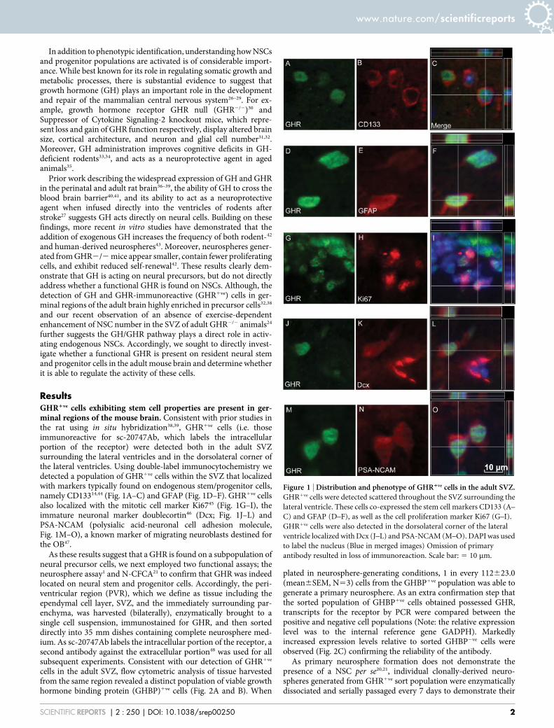
ResultsGHR1ve cells exhibiting stem cell properties are present in ger-minal regions of the mouse brain. Consistent with prior studies inthe rat using in situ hybridization38,39, GHR1ve cells (i.e. thoseimmunoreactive for sc-20747Ab, which labels the intracellularportion of the receptor) were detected both in the adult SVZsurrounding the lateral ventricles and in the dorsolateral corner ofthe lateral ventricles. Using double-label immunocytochemistry wedetected a population of GHR1ve cells within the SVZ that localizedwith markers typically found on endogenous stem/progenitor cells,namely CD13314,44 (Fig. 1A–C) and GFAP (Fig. 1D–F). GHR1ve cellsalso localized with the mitotic cell marker Ki6745 (Fig. 1G–I), theimmature neuronal marker doublecortin46 (Dcx; Fig. 1J–L) andPSA-NCAM (polysialic acid-neuronal cell adhesion molecule,Fig. 1M–O), a known marker of migrating neuroblasts destined forthe OB47.
As these results suggest that a GHR is found on a subpopulation ofneural precursor cells, we next employed two functional assays; theneurosphere assay1 and N-CFCA21 to confirm that GHR was indeedlocated on neural stem and progenitor cells. Accordingly, the peri-ventricular region (PVR), which we define as tissue including theependymal cell layer, SVZ, and the immediately surrounding par-enchyma, was harvested (bilaterally), enzymatically brought to asingle cell suspension, immunostained for GHR, and then sorteddirectly into 35 mm dishes containing complete neurosphere med-ium. As sc-20747Ab labels the intracellular portion of the receptor, asecond antibody against the extracellular portion48 was used for allsubsequent experiments. Consistent with our detection of GHR1ve
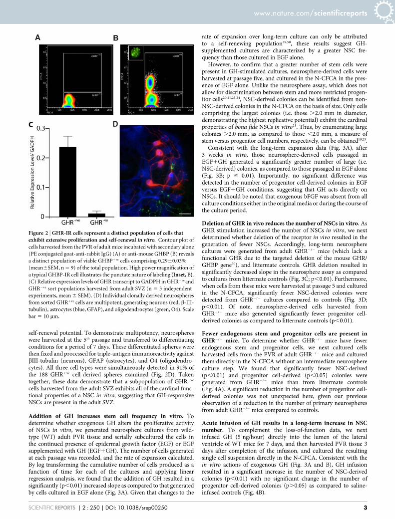
cells in the adult SVZ, flow cytometric analysis of tissue harvestedfrom the same region revealed a distinct population of viable growthhormone binding protein (GHBP)1ve cells (Fig. 2A and B). When
plated in neurosphere-generating conditions, 1 in every 112623.0(mean6SEM, N53) cells from the GHBP1ve population was able togenerate a primary neurosphere. As an extra confirmation step thatthe sorted population of GHBP1ve cells obtained possessed GHR,transcripts for the receptor by PCR were compared between thepositive and negative cell populations (Note: the relative expressionlevel was to the internal reference gene GADPH). Markedlyincreased expression levels relative to sorted GHBP2ve cells wereobserved (Fig. 2C) confirming the reliability of the antibody.
As primary neurosphere formation does not demonstrate thepresence of a NSC per se20,21, individual clonally-derived neuro-spheres generated from GHR1ve sort population were enzymaticallydissociated and serially passaged every 7 days to demonstrate their
Figure 1 | Distribution and phenotype of GHR1ve cells in the adult SVZ.GHR1ve cells were detected scattered throughout the SVZ surrounding the
lateral ventricle. These cells co-expressed the stem cell markers CD133 (A–
C) and GFAP (D–F), as well as the cell proliferation marker Ki67 (G–I).
GHR1ve cells were also detected in the dorsolateral corner of the lateral
ventricle localized with Dcx (J–L) and PSA-NCAM (M–O). DAPI was used
to label the nucleus (Blue in merged images) Omission of primary
antibody resulted in loss of immunoreaction. Scale bar: 5 10 mm.
www.nature.com/scientificreports
SCIENTIFIC REPORTS | 2 : 250 | DOI: 10.1038/srep00250 2
self-renewal potential. To demonstrate multipotency, neurosphereswere harvested at the 5th passage and transferred to differentiatingconditions for a period of 7 days. These differentiated spheres werethen fixed and processed for triple-antigen immunoreactivity againstbIII-tubulin (neurons), GFAP (astrocytes), and O4 (oligodendro-cytes). All three cell types were simultaneously detected in 91% ofthe 188 GHR1ve cell-derived spheres examined (Fig. 2D). Takentogether, these data demonstrate that a subpopulation of GHR1ve
cells harvested from the adult SVZ exhibits all of the cardinal func-tional properties of a NSC in vitro, suggesting that GH-responsiveNSCs are present in the adult SVZ.
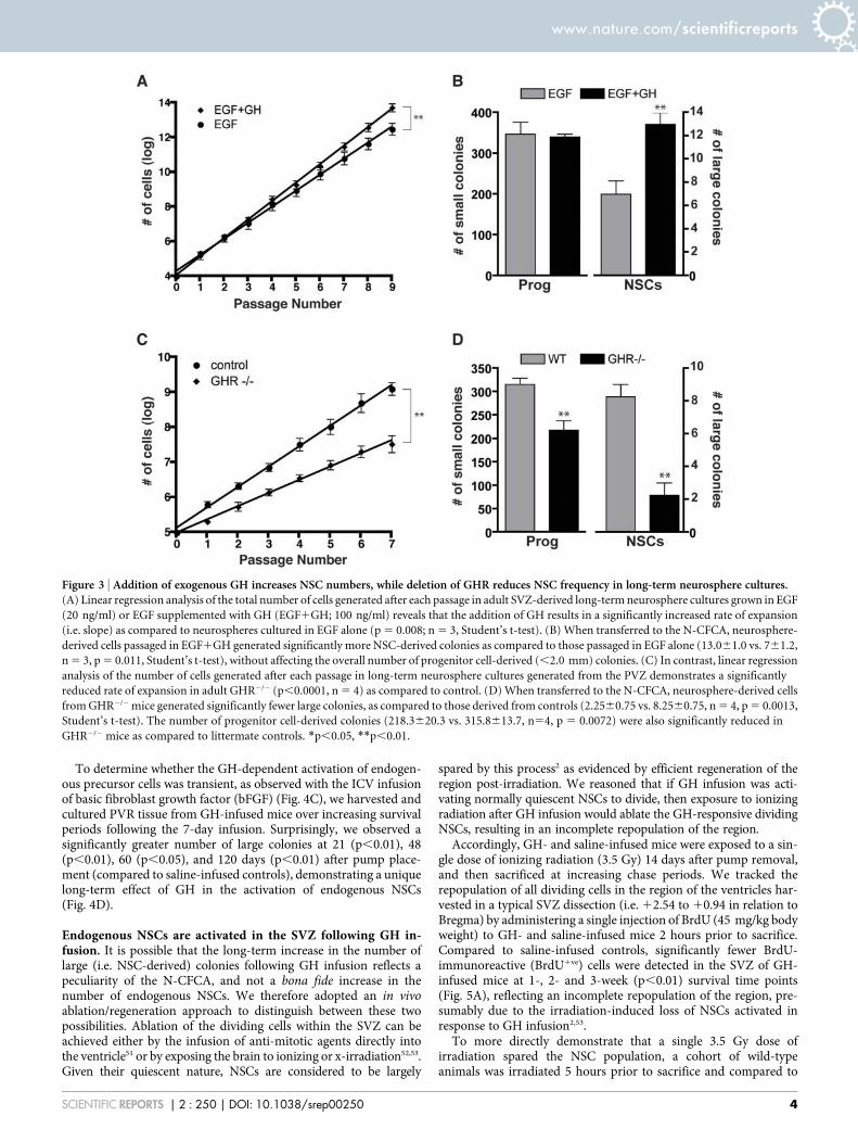
Addition of GH increases stem cell frequency in vitro. Todetermine whether exogenous GH alters the proliferative activityof NSCs in vitro, we generated neurosphere cultures from wild-type (WT) adult PVR tissue and serially subcultured the cells inthe continued presence of epidermal growth factor (EGF) or EGFsupplemented with GH (EGF1GH). The number of cells generatedat each passage was recorded, and the rate of expansion calculated.By log transforming the cumulative number of cells produced as afunction of time for each of the cultures and applying linearregression analysis, we found that the addition of GH resulted in asignificantly (p,0.01) increased slope as compared to that generatedby cells cultured in EGF alone (Fig. 3A). Given that changes to the
rate of expansion over long-term culture can only be attributedto a self-renewing population49,50, these results suggest GH-supplemented cultures are characterized by a greater NSC fre-quency than those cultured in EGF alone.
However, to confirm that a greater number of stem cells werepresent in GH-stimulated cultures, neurosphere-derived cells wereharvested at passage five, and cultured in the N-CFCA in the pres-ence of EGF alone. Unlike the neurosphere assay, which does notallow for discrimination between stem and more restricted progen-itor cells20,21,23,24, NSC-derived colonies can be identified from non-NSC-derived colonies in the N-CFCA on the basis of size. Only cellscomprising the largest colonies (i.e. those .2.0 mm in diameter,demonstrating the highest replicative potential) exhibit the cardinalproperties of bona fide NSCs in vitro21. Thus, by enumerating largecolonies .2.0 mm, as compared to those ,2.0 mm, a measure ofstem versus progenitor cell numbers, respectively, can be obtained19,25.
Consistent with the long-term expansion data (Fig. 3A), after3 weeks in vitro, those neurosphere-derived cells passaged inEGF1GH generated a significantly greater number of large (i.e.NSC-derived) colonies, as compared to those passaged in EGF alone(Fig. 3B; p # 0.01). Importantly, no significant difference wasdetected in the number of progenitor cell-derived colonies in EGFversus EGF1GH conditions, suggesting that GH acts directly onNSCs. It should be noted that exogenous bFGF was absent from allculture conditions either in the original media or during the course ofthe culture period.
Deletion of GHR in vivo reduces the number of NSCs in vitro. AsGHR stimulation increased the number of NSCs in vitro, we nextdetermined whether deletion of the receptor in vivo resulted in thegeneration of fewer NSCs. Accordingly, long-term neurospherecultures were generated from adult GHR2/2 mice (which lack afunctional GHR due to the targeted deletion of the mouse GHR/GHBP gene30), and littermate controls. GHR deletion resulted insignificantly decreased slope in the neurosphere assay as comparedto cultures from littermate controls (Fig. 3C; p,0.01). Furthermore,when cells from these mice were harvested at passage 5 and culturedin the N-CFCA, significantly fewer NSC-derived colonies weredetected from GHR2/2 cultures compared to controls (Fig. 3D;p,0.01). Of note, neurosphere-derived cells harvested fromGHR2/2 mice also generated significantly fewer progenitor cell-derived colonies as compared to littermate controls (p,0.01).
Fewer endogenous stem and progenitor cells are present inGHR2/2 mice. To determine whether GHR2/2 mice have fewerendogenous stem and progenitor cells, we next cultured cellsharvested cells from the PVR of adult GHR2/2 mice and culturedthem directly in the N-CFCA without an intermediate neurosphereculture step. We found that significantly fewer NSC-derived(p,0.01) and progenitor cell-derived (p,0.05) colonies weregenerated from GHR2/2 mice than from littermate controls(Fig. 4A). A significant reduction in the number of progenitor cell-derived colonies was not unexpected here, given our previousobservation of a reduction in the number of primary neurospheresfrom adult GHR2/2 mice compared to controls.
Acute infusion of GH results in a long-term increase in NSCnumber. To complement the loss-of-function data, we nextinfused GH (5 ng/hour) directly into the lumen of the lateralventricle of WT mice for 7 days, and then harvested PVR tissue 3days after completion of the infusion, and cultured the resultingsingle cell suspension directly in the N-CFCA. Consistent with thein vitro actions of exogenous GH (Fig. 3A and B), GH infusionresulted in a significant increase in the number of NSC-derivedcolonies (p,0.01) with no significant change in the number ofprogenitor cell-derived colonies (p.0.05) as compared to saline-infused controls (Fig. 4B).
Figure 2 | GHR-IR cells represent a distinct population of cells thatexhibit extensive proliferation and self-renewal in vitro. Contour plot of
cells harvested from the PVR of adult mice incubated with secondary alone
(PE conjugated goat-anti-rabbit IgG) (A) or anti-mouse GHBP (B) reveals
a distinct population of viable GHBP1ve cells comprising 0.2960.03%
(mean6SEM, n 5 9) of the total population. High power magnification of
a typical GHBP-IR cell illustrates the punctate nature of labeling (Inset, B).
(C) Relative expression levels of GHR transcript to GADPH in GHR1ve and
GHR2ve sort populations harvested from adult SVZ (n 5 3 independent
experiments, mean 6 SEM). (D) Individual clonally derived neurospheres
from sorted GHR1ve cells are multipotent, generating neurons (red, b-III-
tubulin), astrocytes (blue, GFAP), and oligodendrocytes (green, O4). Scale
bar 5 10 mm.
www.nature.com/scientificreports
SCIENTIFIC REPORTS | 2 : 250 | DOI: 10.1038/srep00250 3
To determine whether the GH-dependent activation of endogen-ous precursor cells was transient, as observed with the ICV infusionof basic fibroblast growth factor (bFGF) (Fig. 4C), we harvested andcultured PVR tissue from GH-infused mice over increasing survivalperiods following the 7-day infusion. Surprisingly, we observed asignificantly greater number of large colonies at 21 (p,0.01), 48(p,0.01), 60 (p,0.05), and 120 days (p,0.01) after pump place-ment (compared to saline-infused controls), demonstrating a uniquelong-term effect of GH in the activation of endogenous NSCs(Fig. 4D).
Endogenous NSCs are activated in the SVZ following GH in-fusion. It is possible that the long-term increase in the number oflarge (i.e. NSC-derived) colonies following GH infusion reflects apeculiarity of the N-CFCA, and not a bona fide increase in thenumber of endogenous NSCs. We therefore adopted an in vivoablation/regeneration approach to distinguish between these twopossibilities. Ablation of the dividing cells within the SVZ can beachieved either by the infusion of anti-mitotic agents directly intothe ventricle51 or by exposing the brain to ionizing or x-irradiation52,53.Given their quiescent nature, NSCs are considered to be largely
spared by this process2 as evidenced by efficient regeneration of theregion post-irradiation. We reasoned that if GH infusion was acti-vating normally quiescent NSCs to divide, then exposure to ionizingradiation after GH infusion would ablate the GH-responsive dividingNSCs, resulting in an incomplete repopulation of the region.
Accordingly, GH- and saline-infused mice were exposed to a sin-gle dose of ionizing radiation (3.5 Gy) 14 days after pump removal,and then sacrificed at increasing chase periods. We tracked therepopulation of all dividing cells in the region of the ventricles har-vested in a typical SVZ dissection (i.e. 12.54 to 10.94 in relation toBregma) by administering a single injection of BrdU (45 mg/kg bodyweight) to GH- and saline-infused mice 2 hours prior to sacrifice.Compared to saline-infused controls, significantly fewer BrdU-immunoreactive (BrdU1ve) cells were detected in the SVZ of GH-infused mice at 1-, 2- and 3-week (p,0.01) survival time points(Fig. 5A), reflecting an incomplete repopulation of the region, pre-sumably due to the irradiation-induced loss of NSCs activated inresponse to GH infusion2,53.
To more directly demonstrate that a single 3.5 Gy dose ofirradiation spared the NSC population, a cohort of wild-typeanimals was irradiated 5 hours prior to sacrifice and compared to
Figure 3 | Addition of exogenous GH increases NSC numbers, while deletion of GHR reduces NSC frequency in long-term neurosphere cultures.(A) Linear regression analysis of the total number of cells generated after each passage in adult SVZ-derived long-term neurosphere cultures grown in EGF
(20 ng/ml) or EGF supplemented with GH (EGF1GH; 100 ng/ml) reveals that the addition of GH results in a significantly increased rate of expansion
(i.e. slope) as compared to neurospheres cultured in EGF alone (p 5 0.008; n 5 3, Student’s t-test). (B) When transferred to the N-CFCA, neurosphere-
derived cells passaged in EGF1GH generated significantly more NSC-derived colonies as compared to those passaged in EGF alone (13.061.0 vs. 761.2,
n 5 3, p 5 0.011, Student’s t-test), without affecting the overall number of progenitor cell-derived (,2.0 mm) colonies. (C) In contrast, linear regression
analysis of the number of cells generated after each passage in long-term neurosphere cultures generated from the PVZ demonstrates a significantly
reduced rate of expansion in adult GHR2/2 (p,0.0001, n 5 4) as compared to control. (D) When transferred to the N-CFCA, neurosphere-derived cells
from GHR2/2 mice generated significantly fewer large colonies, as compared to those derived from controls (2.2560.75 vs. 8.2560.75, n 5 4, p 5 0.0013,
Student’s t-test). The number of progenitor cell-derived colonies (218.3620.3 vs. 315.8613.7, n54, p 5 0.0072) were also significantly reduced in
GHR2/2 mice as compared to littermate controls. *p,0.05, **p,0.01.
www.nature.com/scientificreports
SCIENTIFIC REPORTS | 2 : 250 | DOI: 10.1038/srep00250 4

non-irradiated animals. When PVR tissue was harvested from thesemice and cultured directly in the N-CFCA, we observed a significant(p,0.01) decrease in the number of progenitor cell-derived colonies(Fig. 5B). However, consistent with the sparing of a relatively qui-escent population of NSCs, we failed to observe a change in thenumber of NSC-derived colonies between control and irradiatedanimals (Fig. 5B; 127.3622.9 vs. 129.365.5, respectively).
Therefore, to complement our in vivo finding of an incompleterepopulation of BrdU1ve cells in the SVZ of GH-infused mice(Fig. 5A), we next repeated the ablation/repopulation approach usingthe reappearance of neurosphere-forming cells as an additional invitro readout of SVZ repopulation. We included two additionalcohorts of mice to provide support for our hypothesis that theincomplete repopulation effect was due to the GH-induced activa-tion (and subsequent radiation-induced ablation) of endogenousNSCs. One cohort received a 7-day ICV infusion of EGF (20 ng/ml;a treatment we previously demonstrated stimulates endogenous pro-genitor cell proliferation, leaving NSC number unchanged54) 14 daysprior to irradiation to demonstrate that ablation of mitotically-activeprogenitor cells does not have the same detrimental effect on SVZrepopulation. A second cohort of mice received an additional dose ofirradiation (3.5 Gy) 2 days prior to normal irradiation. These ‘‘dou-ble-dose’’ mice were employed based on the observation by
Morshead and colleagues that normally quiescent NSCs (which wereactivated in response to the initial ablation of dividing cells) could beablated by subjecting the mice to a second ‘‘kill’’ 2 days following theinitial ablation2. Having demonstrated (Fig. 5A) that a single dose ofionizing radiation was sufficient to ablate dividing cells, largely spar-ing and then activating quiescent NSCs (as evidenced by the prev-iously reported efficient repopulation of the region2,51 and our workhere, see Fig. 5B), we reasoned that the ‘‘double-dose’’ approach ofNSC ablation would mimic GH-infused animals, where a substantialproportion of NSCs were GH-activated prior to irradiation.
As expected from previous reports52,53 the number of neurosphere-forming cells in saline-infused mice was reduced to approximately60% of normal (i.e. the number of cells present in non-irradiated(T0) animals), 24 hours following irradiation, rebounding to supra-control levels in the period following ablation (Fig. 5C). Interestingly,although EGF-infused mice generated significantly more neuro-spheres immediately prior to irradiation (T0, p,0.01), and displayeda more rapid SVZ repopulation post-irradiation compared to con-trols (1 vs. 3 weeks, respectively), no significant difference in thenumber of neurospheres generated by 6 weeks post-irradiation wasobserved in the EGF-infused mice compared to the controls, reflect-ing complete repopulation of the region. In contrast, the repopu-lation of the SVZ in both ‘‘double-dose’’ and GH-infused mice was
Figure 4 | Fewer NSCs are detected in the PVR of adult GHR2/2 mice, while an acute ICV infusion of GH into wild-type mice results in a long-termincrease in endogenous NSCs. (A) PVR cells harvested from adult GHR2/2 mice generate significantly fewer NSC-derived colonies (15.062.9 vs.
30.662.1, n 5 4, p 5 0.0066) and progenitor cell-derived colonies, as compared to littermate controls (158.365.5 vs. 266.3625.7, n 5 4, p 5 0.017).
(B) Cells harvested from the PVR of GH-infused mice 10 days from the onset of infusion generate a significantly greater number of stem cell-derived
colonies compared to vehicle-infused control mice (17.163.0 vs. 7.161.4, n 5 9, p 5 0.004), with no associated change in the number of progenitor cell-
derived colonies (311.6649.5 vs. 228.8613.8, n 5 9, p 5 0.127). (C) Ten days after bFGF infusion, the number of progenitor (253.967.2 vs. 467637.9, n
5 5, p 5 0.002) and NSC-derived colonies (5.560.33 vs. 17.861.3, n 5 5, p 5 0.0001) was significantly increased relative to saline-infused controls. After
21 days of the bFGF infusion, however, the number of NSC-derived (4.660.8, p 5 0.643, n54) and progenitor cell-derived (247617.1, n 5 4, p 5 0.060)
colonies returned to levels similar to that of saline-infused controls. (D) The number of NSC-derived colonies remained significantly increased at 21 days
(12.860.7, n 5 6, p 5 0.009), 48 days (14.161.4, n 5 6, p 5 0.003), 60 days (13.161.8, n 5 6, p 5 0.021) and 120 days (13.660.7, n 5 6, p 5 0.004)
following the onset of GH infusion as compared to vehicle-infused controls (7.161.4, n 5 9, p 5 0.0038). *p,0.05, **p,0.01, Student’s t-test.
www.nature.com/scientificreports
SCIENTIFIC REPORTS | 2 : 250 | DOI: 10.1038/srep00250 5
severely compromised, with sphere-forming cells remaining signifi-cantly reduced in these cohorts, as compared to saline controls 3weeks post-ablation (p,0.01) and pre-irradiation levels (p,0.01).Indeed, repopulation of GH-infused mice remained static between 3and 6 weeks post-irradiation.
GH-induced increase in SVZ NSCs augments adult neurogenesis.Considering the known contribution of SVZ stem and progenitorcells to OB neurogenesis, we next investigated how GH infusionaffected the frequency of GH-responsive cell types in the SVZ, andwhether the GH-dependent increase in SVZ NSCs increased OBneurogenesis. Accordingly, brains were removed from GH-infusedmice 10 and 21 days after the onset of a 7-day ICV infusion, sectioned(14 mm), and processed for double-label immunocytochemisty todetect GHR1ve cells from the onset of the lateral ventricles to thelevel of the anterior commissure (i.e. 11.42 to 0.14 mm rostral toBregma). We failed to detect any change in the overall frequency ofGHR1ve cells in the PVR surrounding the lateral ventricles or thedorsolateral corner of the ventricle at either 10- (1282656) or 21-day(1084617) survival times, compared to naı̈ve mice (1098647).However, the frequencies of four subsets of GHR1ve cells (thoseexpressing putative NSC antigens) did increase transiently at 10days, returning to basal levels by 21 days. These included GHR1ve/Ki671ve (7266 to 11166, p 5 0.01, n 5 3), GHR1ve/Nestin1ve (6265to 10067, p,0.01, n 5 3), and GHR1ve/CD1331ve (184615 to21969, p,0.05, n 5 3) cells in the PVR surrounding the ventricle,and GHR1ve/PSA-NCAM1ve (474612 to 594640, p,0.05, n 5 3)cells in the dorsolateral corner of the ventricle.
Although these results suggest that GH infusion increases thefrequency of migrating neuroblasts; to more accurately determinewhether the GH infusion alters the number of new neurons beinggenerated in the OB, and whether this is a NSC-related effect, twofinal experiments were performed. In the first experiment, GH-infused mice were given a single i.p. injection of BrdU (45 mg/kgbody weight) 7 days after GH pump placement and sacrificed after a28-day chase period, providing sufficient time for newly generatedcells to migrate to the OB and differentiate into neurons. The numberof newly generated cells reaching the OB (i.e. BrdU1ve) and newlydifferentiated neurons (i.e. BrdU1ve/NeuN1ve) was determined byexamining tissue sections through the entire OB. While GH didnot alter the overall number of migrating cells reaching the OB(BrdU, Fig. 6A), it did significantly increase the number of newneurons (BrdU/NeuN, p,0.01, n 5 3).
In the second experiment to determine whether radiation ablationof GH-activated NSCs would also result in a significant decline inOB neurogenesis, a cohort of mice received a 7-day ICV infusion ofGH, followed by a single dose (3.5 Gy) of irradiation 7 days later.Twelve days after the end of the infusion, these mice were adminis-tered one i.p. injection of BrdU every 2 hours until five injectionswere given. Multiple injections of BrdU were employed to ensure ameaningful number could be detected following the irradiation-induced ablation of the majority of dividing cells. Twenty-eight
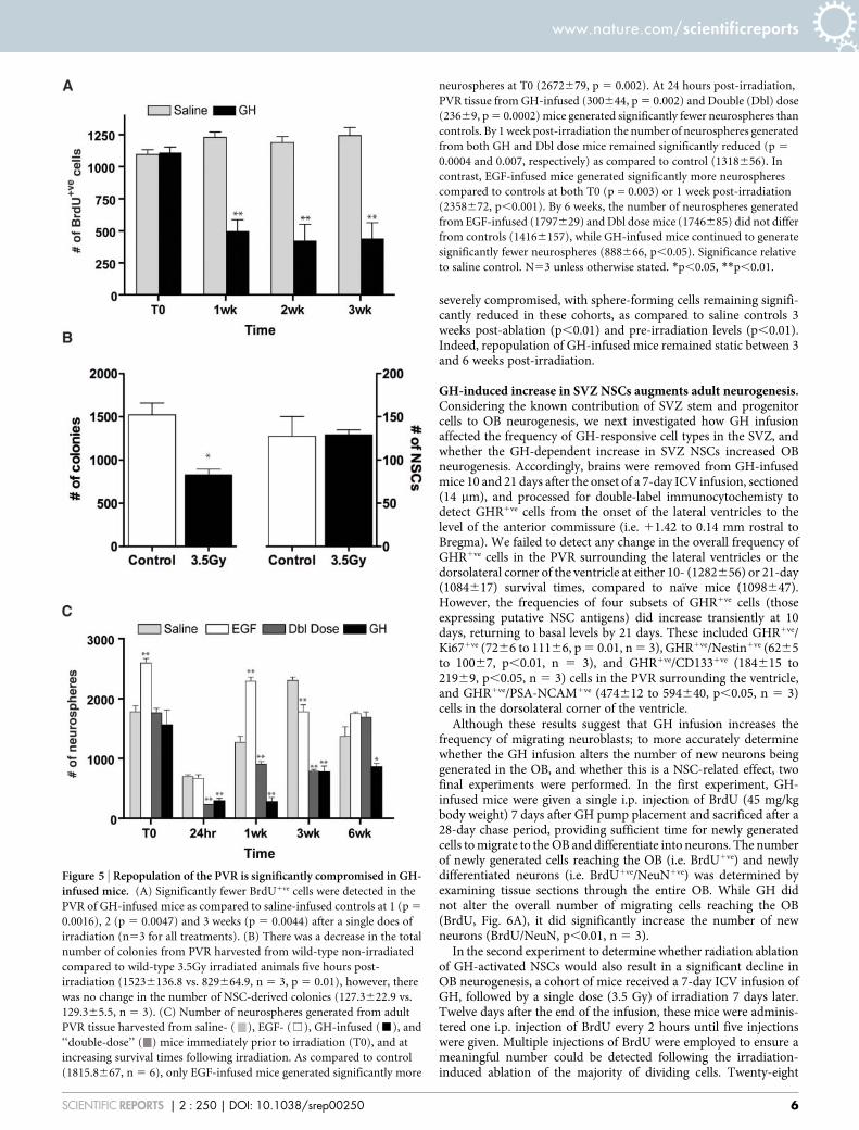
Figure 5 | Repopulation of the PVR is significantly compromised in GH-infused mice. (A) Significantly fewer BrdU1ve cells were detected in the
PVR of GH-infused mice as compared to saline-infused controls at 1 (p 5
0.0016), 2 (p 5 0.0047) and 3 weeks (p 5 0.0044) after a single does of
irradiation (n53 for all treatments). (B) There was a decrease in the total
number of colonies from PVR harvested from wild-type non-irradiated
compared to wild-type 3.5Gy irradiated animals five hours post-
irradiation (15236136.8 vs. 829664.9, n 5 3, p 5 0.01), however, there
was no change in the number of NSC-derived colonies (127.3622.9 vs.
129.365.5, n 5 3). (C) Number of neurospheres generated from adult
PVR tissue harvested from saline- ( ), EGF- (%), GH-infused (&), and
‘‘double-dose’’ ( ) mice immediately prior to irradiation (T0), and at
increasing survival times following irradiation. As compared to control
(1815.8667, n 5 6), only EGF-infused mice generated significantly more
neurospheres at T0 (2672679, p 5 0.002). At 24 hours post-irradiation,
PVR tissue from GH-infused (300644, p 5 0.002) and Double (Dbl) dose
(23669, p 5 0.0002) mice generated significantly fewer neurospheres than
controls. By 1 week post-irradiation the number of neurospheres generated
from both GH and Dbl dose mice remained significantly reduced (p 5
0.0004 and 0.007, respectively) as compared to control (1318656). In
contrast, EGF-infused mice generated significantly more neurospheres
compared to controls at both T0 (p = 0.003) or 1 week post-irradiation
(2358672, p,0.001). By 6 weeks, the number of neurospheres generated
from EGF-infused (1797629) and Dbl dose mice (1746685) did not differ
from controls (14166157), while GH-infused mice continued to generate
significantly fewer neurospheres (888666, p,0.05). Significance relative
to saline control. N53 unless otherwise stated. *p,0.05, **p,0.01.
www.nature.com/scientificreports
SCIENTIFIC REPORTS | 2 : 250 | DOI: 10.1038/srep00250 6
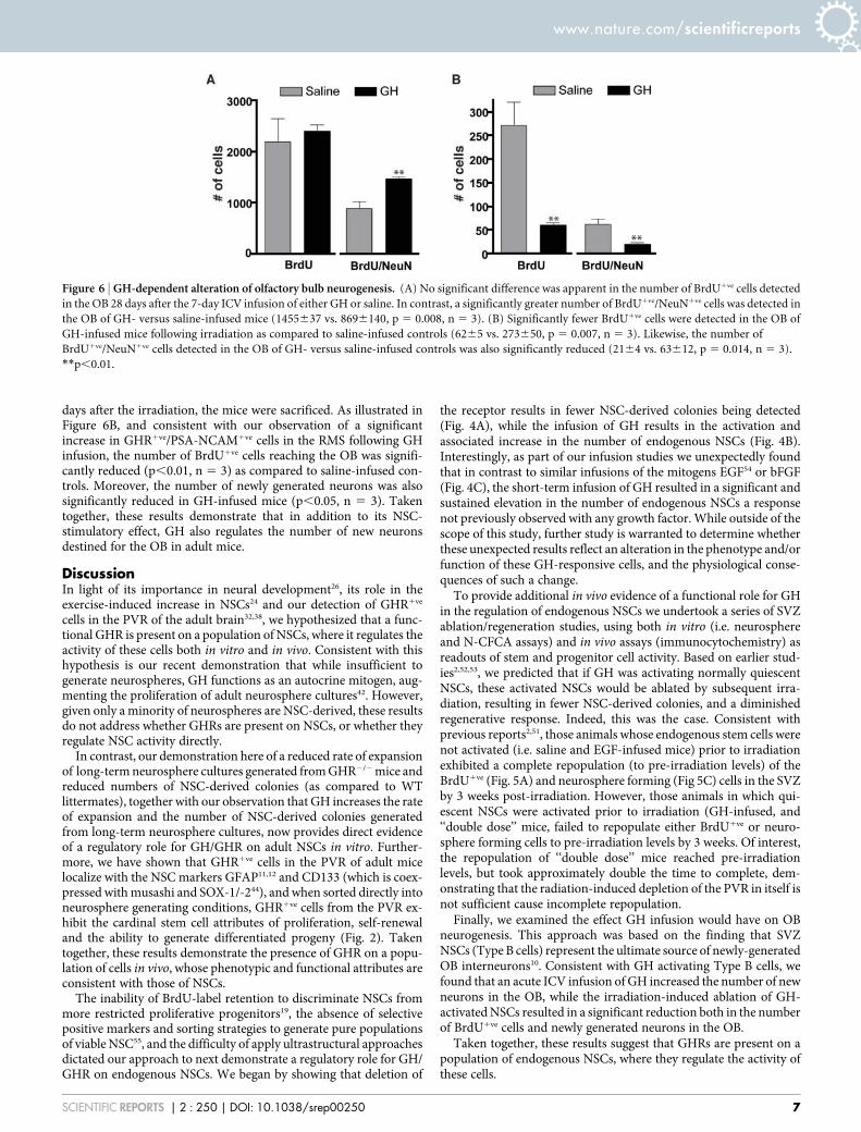
days after the irradiation, the mice were sacrificed. As illustrated inFigure 6B, and consistent with our observation of a significantincrease in GHR1ve/PSA-NCAM1ve cells in the RMS following GHinfusion, the number of BrdU1ve cells reaching the OB was signifi-cantly reduced (p,0.01, n 5 3) as compared to saline-infused con-trols. Moreover, the number of newly generated neurons was alsosignificantly reduced in GH-infused mice (p,0.05, n 5 3). Takentogether, these results demonstrate that in addition to its NSC-stimulatory effect, GH also regulates the number of new neuronsdestined for the OB in adult mice.
DiscussionIn light of its importance in neural development26, its role in theexercise-induced increase in NSCs24 and our detection of GHR1ve
cells in the PVR of the adult brain32,38, we hypothesized that a func-tional GHR is present on a population of NSCs, where it regulates theactivity of these cells both in vitro and in vivo. Consistent with thishypothesis is our recent demonstration that while insufficient togenerate neurospheres, GH functions as an autocrine mitogen, aug-menting the proliferation of adult neurosphere cultures42. However,given only a minority of neurospheres are NSC-derived, these resultsdo not address whether GHRs are present on NSCs, or whether theyregulate NSC activity directly.
In contrast, our demonstration here of a reduced rate of expansionof long-term neurosphere cultures generated from GHR2/2 mice andreduced numbers of NSC-derived colonies (as compared to WTlittermates), together with our observation that GH increases the rateof expansion and the number of NSC-derived colonies generatedfrom long-term neurosphere cultures, now provides direct evidenceof a regulatory role for GH/GHR on adult NSCs in vitro. Further-more, we have shown that GHR1ve cells in the PVR of adult micelocalize with the NSC markers GFAP11,12 and CD133 (which is coex-pressed with musashi and SOX-1/-244), and when sorted directly intoneurosphere generating conditions, GHR1ve cells from the PVR ex-hibit the cardinal stem cell attributes of proliferation, self-renewaland the ability to generate differentiated progeny (Fig. 2). Takentogether, these results demonstrate the presence of GHR on a popu-lation of cells in vivo, whose phenotypic and functional attributes areconsistent with those of NSCs.
The inability of BrdU-label retention to discriminate NSCs frommore restricted proliferative progenitors19, the absence of selectivepositive markers and sorting strategies to generate pure populationsof viable NSC55, and the difficulty of apply ultrastructural approachesdictated our approach to next demonstrate a regulatory role for GH/GHR on endogenous NSCs. We began by showing that deletion of
the receptor results in fewer NSC-derived colonies being detected(Fig. 4A), while the infusion of GH results in the activation andassociated increase in the number of endogenous NSCs (Fig. 4B).Interestingly, as part of our infusion studies we unexpectedly foundthat in contrast to similar infusions of the mitogens EGF54 or bFGF(Fig. 4C), the short-term infusion of GH resulted in a significant andsustained elevation in the number of endogenous NSCs a responsenot previously observed with any growth factor. While outside of thescope of this study, further study is warranted to determine whetherthese unexpected results reflect an alteration in the phenotype and/orfunction of these GH-responsive cells, and the physiological conse-quences of such a change.
To provide additional in vivo evidence of a functional role for GHin the regulation of endogenous NSCs we undertook a series of SVZablation/regeneration studies, using both in vitro (i.e. neurosphereand N-CFCA assays) and in vivo assays (immunocytochemistry) asreadouts of stem and progenitor cell activity. Based on earlier stud-ies2,52,53, we predicted that if GH was activating normally quiescentNSCs, these activated NSCs would be ablated by subsequent irra-diation, resulting in fewer NSC-derived colonies, and a diminishedregenerative response. Indeed, this was the case. Consistent withprevious reports2,51, those animals whose endogenous stem cells werenot activated (i.e. saline and EGF-infused mice) prior to irradiationexhibited a complete repopulation (to pre-irradiation levels) of theBrdU1ve (Fig. 5A) and neurosphere forming (Fig 5C) cells in the SVZby 3 weeks post-irradiation. However, those animals in which qui-escent NSCs were activated prior to irradiation (GH-infused, and‘‘double dose’’ mice, failed to repopulate either BrdU1ve or neuro-sphere forming cells to pre-irradiation levels by 3 weeks. Of interest,the repopulation of ‘‘double dose’’ mice reached pre-irradiationlevels, but took approximately double the time to complete, dem-onstrating that the radiation-induced depletion of the PVR in itself isnot sufficient cause incomplete repopulation.
Finally, we examined the effect GH infusion would have on OBneurogenesis. This approach was based on the finding that SVZNSCs (Type B cells) represent the ultimate source of newly-generatedOB interneurons10. Consistent with GH activating Type B cells, wefound that an acute ICV infusion of GH increased the number of newneurons in the OB, while the irradiation-induced ablation of GH-activated NSCs resulted in a significant reduction both in the numberof BrdU1ve cells and newly generated neurons in the OB.
Taken together, these results suggest that GHRs are present on apopulation of endogenous NSCs, where they regulate the activity ofthese cells.
Figure 6 | GH-dependent alteration of olfactory bulb neurogenesis. (A) No significant difference was apparent in the number of BrdU1ve cells detected
in the OB 28 days after the 7-day ICV infusion of either GH or saline. In contrast, a significantly greater number of BrdU1ve/NeuN1ve cells was detected in
the OB of GH- versus saline-infused mice (1455637 vs. 8696140, p 5 0.008, n 5 3). (B) Significantly fewer BrdU1ve cells were detected in the OB of
GH-infused mice following irradiation as compared to saline-infused controls (6265 vs. 273650, p 5 0.007, n 5 3). Likewise, the number of
BrdU1ve/NeuN1ve cells detected in the OB of GH- versus saline-infused controls was also significantly reduced (2164 vs. 63612, p 5 0.014, n 5 3).
**p,0.01.
www.nature.com/scientificreports
SCIENTIFIC REPORTS | 2 : 250 | DOI: 10.1038/srep00250 7

It has become apparent that stem cell numbers in a variety oftissues decline as part of the aging process, and are unable to main-tain tissue homeostasis at youthful levels24,56. It is plausible that activ-ating a cohort of NSCs by GH infusion could lead to a long-termtemporal extension of functional tissue homeostasis. This could be apowerful therapeutic in countering age-related decline in regenerat-ive capacity and cognitive function, as this progressive loss also cor-relates with the age-dependent decline in GH secretion in rodentsand humans33. Independent of its potential therapeutic value, invest-igation into the mechanism by which NSC numbers are altered willundoubtedly increase our understanding of how the brain respondsto its environment.
MethodsTissue processing. Animals were treated in accordance with the Australian Code ofPractice for the Care and Use of Animals for Scientific Purposes, and all experimentswere approved by The University of Queensland Animal Ethics Committee. Adult(6–8 weeks old) male and female C57BL/6J mice were deeply anesthetized withsodium pentobarbitone (260 mg/kg), and then transcardially perfused with ice-cold0.9% saline followed by 4.0% paraformaldehyde in 0.1 M phosphate buffer (pH 7.4).Brains were harvested, post-fixed, and cryoprotected, as previously described57. Serialfrontal sections (14 mm) were cut with a MICROM cryostat, mounted on SuperFrostPlus slides (SuperFrost Plus), dried at room temperature (RT) and stored at 220uCuntil processed. Every second section was collected from the rostral tip of the OB toBregma (12.58 mm) for examination of the OB, and from the onset of the lateralventricle (11.42 mm) to the joining of the anterior commissure (10.14 mm) forexamination of the lateral ventricles.
In all cases, tissue sections were initially rinsed in PBS (335 minutes) beforeincubation in blocking solution (5% fetal bovine serum (JRH Biosciences, USA) plus5% normal goat serum (Sigma-Aldrich, USA) in 0.1 M PBS 1 0.2% Triton (Sigma-Aldrich)) for 60 minutes at RT.
Tissue sections were processed for the detection of bromodeoxyuridine (BrdU) ordual-label BrdU/anti-neuronal nuclei (NeuN, 1560; Chemicon, USA, MAB377.GHR1ve cells were detected on fixed tissue using anti-GHR (15300; Santa Cruz, USA,SC-20747 which labels an intracellular portion of the GHR). Note: specificity of theanti-GHR antibody was verified by an absence of immunoreactivity on tissue sectionsharvested from GHR2/2 mice. For all GHR dual-label immunocytochemistry, sec-tions were incubated with anti-GHR (SC-20747) and anti-Ki67 (1560; Novacastra,UK, NCL-L-Ki67-MM1), anti-glial fibrillary acidic protein (GFAP, 15300;Chemicon, MAB360), anti-NeuN (15100; Chemicon, MB377), anti-Nestin (15500;Abcam, AB6142), anti-PDGFRa (15300, Abcam, AB69506) anti-PSA-NCAM(15200; Chemicon, MAB5324), and anti-CD133 (15100; Abcam, AB27699), dilutedin blocking solution and incubated overnight at 4uC. Sections were washed threetimes with PBS and then incubated for 60 minutes (RT) with the appropriate sec-ondary antibodies (15300; Alexa Fluor goat anti-rabbit-488 for anti-GHR and151000; Alexa Fluor goat anti-mouse-568 for remaining antibodies; MolecularProbes, USA) plus 49,6-diamidino-2-phenylindole (DAPI, 151000, Sigma-Aldrich).Sections were then washed three times with PBS and once in dH20, before beingcoverslipped with fluorescent mounting medium (DakoCytomation, USA). Imageswere captured using a Zeiss Axioimager Z1 with Apotome and Axiocam MRmsoftware (Zeiss, Germany).
Intracerebroventricular (ICV) infusions. Twelve hours prior to surgery, osmoticmini pumps (Alzet, #1007D; 7 day infusion at 0.5 ml/hour) were loaded with GH (10mg/ml, recombinant rat, GroPep Australia), bFGF (20 mg/ml, human recombinant,Stem Cell Technologies, Canada), EGF (40 mg/ml, Stem Cell Technologies) or vehiclesolution (0.9% sterile physiological saline, Sigma-Aldrich), and attached to theinfusion cannula. Although the physiological concentration of GH in the cerebralspinal fluid of mice is unknown, a previous study27 carried out a single ICV infusion inrats of 20 mg/ml, thus we reasoned that a conservative dose of 5 ng/hour of GH shouldmimic endogenous levels in the mouse. Details of the infusion protocol are given inBlackmore et al. 2009. A separate cohort of GH- and vehicle-infused mice received asingle 150 ml intraperitoneal (i.p.) injection of BrdU (9 mg/ml, 45 mg/kg bodyweight), dissolved in 0.07 N NaOH in 0.9% NaCl; Sigma-Aldrich) 2 hours prior tothe completion of the infusion period, and were subsequently sacrificed 28 days later.
Irradiation. Mice were restrained within a plastic chamber and placed in a lead-shielded container leaving only the head exposed for irradiation. Single or ‘‘double-dose’’ irradiation was induced by exposure to a Co60 source in a Gamma Cell 200irradiator until a 3.5 Gy dose had been given. The PVR was harvested from GH-, EGF-and vehicle-infused animals at 24 hours, 1-, 3- or 6-weeks post-irradiation andcultured in the neurosphere assay (described below). In addition, a second cohort ofGH- and vehicle-infused mice received a single injection of BrdU (as above) 2 hoursprior to sacrifice, after which the brain was removed, sectioned, and processed for thedetection of BrdU1ve cells. For OB experiments, owing to the irradiation-induced lossof dividing cells, cohorts of GH- and vehicle-infused mice received a total of fiveBrdU injections (as above) over a period of 10 hours, 14 days post-infusion. These
mice were irradiated 48-hours after the last BrdU injection and sacrificed 28 dayspost-irradiation.
Primary neurosphere cultures. Adult (6–8 week old) mice (C57Bl/6J or GHR2/2 onthis background) were sacrificed by cervical dislocation, their brains removedimmediately, and transferred to Petri dishes containing HEPES-buffered minimumessential medium (HEM). Full details of the protocol are given in Blackmore et al.2009.
Neurosphere passaging and differentiation markers. Adult neurospheres werecollected by centrifugation (5 minutes at 100 g rcf), after which they wereresuspended and incubated in 0.1% trypsin-EDTA for 4 minutes before being washedwith trypsin inhibitor in neurosphere media (NS media). Adult neurosphere cultureswere passaged every 7 days in vitro (DIV) in EGF (20 ng/ml) containing completemedium either with or without rat GH (100 ng/ml). For differentiation, neurosphereswere plated at time of passage onto poly-L-ornithine (Sigma-Aldrich) coated glasscoverslips in NeuroCultTM NSC Basal Medium plus Proliferation Supplements (StemCell Technologies) without 1% fetal calf serum for 7 days. The differentiatedneurospheres were then fixed with 4% paraformaldehyde in 0.1 M PBS at RT for 15minutes, rinsed three times with PBS, and incubated with anti-O4 (Clone 81, IgM,Boehringer Mannheim, Germany) diluted 1550 with PBS/10% normal goat serum for2 hours at 37uC. Cells were then rinsed three times with PBS and incubated withFITC-conjugated goat-anti-mouse IgM (15200, Molecular Probes) for 30 minutes at37uC. Cells were again rinsed in PBS (335 minutes) before incubation with anti-bIII-tubulin (mouse monoclonal IgG, 151500, Chemicon) 1 anti-GFAP (rabbitpolyclonal, 15500, DakoCytomation) diluted in PBS/0.3% Triton/10% normal goatserum for 120 minutes at 37uC. Again, cells were rinsed in PBS (335 minutes) andthen incubated with goat-anti-mouse-CY3 and goat-anti-rabbit-AMCA (both 15200,Molecular Probes) diluted in MTPBS/10% NGS at 37uC for 30 minutes. Finally, cellswere rinsed (335 minutes in PBS) and mounted using Fluorsave (DakoCytomation).
Neural colony forming cell assay. Primary tissue was dissociated and the number ofviable cells determined as per the primary neurosphere cultures (see above).Neurosphere-derived cells were cultured in 35 mm cell culture dishes with a 2 grid(Nunc, USA) at a density recommended for the mouse NeuroCultTM Neural ColonyForming Cell Assay (Stem Cell Technologies). Cells were incubated for 21 DIV in 5%CO2 with appropriate growth factors (either EGF alone or EGF 1GH) being addedevery 7 days. After 21 DIV, the total number of colonies and the diameter of eachcolony was determined using an eyepiece graticule on an inverted Leica lightmicroscope with phase contrast.
Flow cytometry. Single cell suspensions were generated from tissue and sorted forsurface growth hormone binding protein (GHBP) expression using an anti-GHBPantibody (BETO 8041)48. Murine GHBP consists of a ligand-binding domain that isidentical to the extracellular portion of the GHR, together with a short C-terminalsequence coded by an alternate exon. For immunostaining, cell suspensions wereincubated in NS basal medium with anti-GHBP48 (15100) for 15 minutes at 4uC. Cellsuspensions were rinsed via centrifugation (7 minutes at 100 g rcf), and thenincubated in blocking solution containing Alexa Fluor 488 conjugated goat-anti-rabbit IgG antibody (151000; Molecular Probes) for 30 minutes at RT. The cellsuspension(s) were then rinsed in PBS 1 propidium iodide (100 mg/ml; MolecularProbes) before cell sorting. Cells were sorted using a FACS Vantage SE DiVA (Becton,Dickinson, USA) with data analysis performed using FlowJo 6 (TreeStar Inc., USA)software. Cell to event ratios were determined for each population by seeding 500events into individual wells of a 96-well plate, then visually scoring cells ,1 hour afterplating. All cells were collected directly into 35 mm dishes containing NSA1EGF(and GH where appropriate) and incubated at 37uC in a 5% CO2 incubator. Images offixed GHR-IR cells were captured using an AMNIS Image Stream Analyzer (AMNIS,USA).
qRT-PCR. RNA was extracted using TRIZOLH (Invitrogen, USA) and cDNA wasmade using Superscript IIIH (Invitrogen). TaqMAN qRT PCR was undertaken withspecific GHR primers for the cytoplasmic domain provided from Applied BiosystemsTaqManH Gene Expression Assays (ID #Mm00439093-m1). cDNA was diluted 155and samples were normalized to 18S rRNA.
Statistical Analysis. Factorial design analysis of variance (ANOVA) or Student’s two-tailed unpaired t-tests were used to analyze data as appropriate. Significant ANOVAvalues were followed by post hoc comparisons of individual means using the Tukeymultiple comparisons test where appropriate. All values are expressed as mean 6
SEM unless otherwise indicated with significance for all comparisons p,0.05.
1. Reynolds, B. A. & Weiss, S. Generation of neurons and astrocytes from isolatedcells of the adult mammalian central nervous system. Science 255, 1707–1710(1992).
2. Morshead, C. M. et al. Neural stem cells in the adult mammalian forebrain: arelatively quiescent subpopulation of subependymal cells. Neuron 13, 1071–1082(1994).
3. Ming, G. L. & Song, H. Adult neurogenesis in the mammalian central nervoussystem. Annu Rev Neurosci 28, 223–250 (2005).
www.nature.com/scientificreports
SCIENTIFIC REPORTS | 2 : 250 | DOI: 10.1038/srep00250 8

4. Emsley, J. G., Mitchell, B. D., Kempermann, G. & Macklis, J. D. Adultneurogenesis and repair of the adult CNS with neural progenitors, precursors, andstem cells. Progress in neurobiology 75, 321–341 (2005).
5. Thored, P. et al. Persistent production of neurons from adult brain stem cellsduring recovery after stroke. Stem Cells 24, 739–747 (2006).
6. Potten, C. S. & Loeffler, M. Stem cells: attributes, cycles, spirals, pitfalls anduncertainties. Lessons for and from the crypt. Development 110, 1001–1020(1990).
7. Till, J. E. & Mc, C. E. A direct measurement of the radiation sensitivity of normalmouse bone marrow cells. Radiation research 14, 213–222 (1961).
8. Doetsch, F., Garcia-Verdugo, J. M. & Alvarez-Buylla, A. Cellular composition andthree-dimensional organization of the subventricular germinal zone in the adultmammalian brain. J Neurosci 17, 5046–5061 (1997).
9. Doetsch, F., Caille, I., Lim, D. A., Garcia-Verdugo, J. M. & Alvarez-Buylla, A.Subventricular zone astrocytes are neural stem cells in the adult mammalianbrain. Cell 97, 703–716 (1999).
10. Kriegstein, A. & Alvarez-Buylla, A. The glial nature of embryonic and adult neuralstem cells. Annual review of neuroscience 32, 149–184 (2009).
11. Merkle, F. T., Tramontin, A. D., Garcia-Verdugo, J. M. & Alvarez-Buylla, A.Radial glia give rise to adult neural stem cells in the subventricular zone. Proc NatlAcad Sci U S A 101, 17528–17532 (2004).
12. Garcia, A. D., Doan, N. B., Imura, T., Bush, T. G. & Sofroniew, M. V. GFAP-expressing progenitors are the principal source of constitutive neurogenesis inadult mouse forebrain. Nat Neurosci 7, 1233–1241 (2004).
13. Mignone, J. L., Kukekov, V., Chiang, A. S., Steindler, D. & Enikolopov, G. Neuralstem and progenitor cells in nestin-GFP transgenic mice. J Comp Neurol 469,311–324 (2004).
14. Coskun, V. et al. CD1331 neural stem cells in the ependyma of mammalianpostnatal forebrain. Proc Natl Acad Sci U S A 105, 1026–1031 (2008).
15. Jackson, E. L. et al. PDGFR alpha-positive B cells are neural stem cells in the adultSVZ that form glioma-like growths in response to increased PDGF signaling.Neuron 51, 187–199 (2006).
16. Chojnacki, A., Mak, G. & Weiss, S. PDGFRalpha expression distinguishes GFAP-expressing neural stem cells from PDGF-responsive neural precursors in the adultperiventricular area. J Neurosci 31, 9503–9512 (2011).
17. Blackmore, D. G. & Rietze, R. L. in Heart Development and Regeneration Vol. 2(eds R. P. Harvey & N. Rosenthal) Ch. 13.1, 857–877 (Academic Press, 2010).
18. Blau, H. M., Brazelton, T. R. & Weimann, J. M. The evolving concept of a stem cell:entity or function? Cell 105, 829–841 (2001).
19. Golmohammadi, M. G. et al. Comparative analysis of the frequency anddistribution of stem and progenitor cells in the adult mouse brain. Stem Cells 26,979–987 (2008).
20. Reynolds, B. A. & Rietze, R. L. Neural stem cells and neurospheres--re-evaluatingthe relationship. Nat Methods 2, 333–336 (2005).
21. Louis, S. A. et al. Enumeration of neural stem and progenitor cells in the neuralcolony forming cell assay. Stem Cells 26, 988–996 (2008).
22. Pluchino, S. et al. Persistent inflammation alters the function of the endogenousbrain stem cell compartment. Brain : a journal of neurology 131, 2564–2578(2008).
23. Azari, H., Louis, S. A., Sharififar, S., Vedam-Mai, V. & Reynolds, B. A. Neural-colony forming cell assay: an assay to discriminate bona fide neural stem cellsfrom neural progenitor cells. J Vis Exp, (2011).
24. Blackmore, D. G., Golmohammadi, M. G., Large, B., Waters, M. J. & Rietze, R. L.Exercise increases neural stem cell number in a growth hormone-dependentmanner, augmenting the regenerative response in aged mice. Stem Cells 27, 2044–2052 (2009).
25. Young, K. M., Fogarty, M., Kessaris, N. & Richardson, W. D. Subventricular zonestem cells are heterogeneous with respect to their embryonic origins andneurogenic fates in the adult olfactory bulb. J Neurosci 27, 8286–8296 (2007).
26. Ajo, R., Cacicedo, L., Navarro, C. & Sanchez-Franco, F. Growth hormone actionon proliferation and differentiation of cerebral cortical cells from fetal rat.Endocrinology 144, 1086–1097 (2003).
27. Scheepens, A. et al. Growth hormone as a neuronal rescue factor during recoveryfrom CNS injury. Neuroscience 104, 677–687 (2001).
28. Noguchi, T. Effects of growth hormone on cerebral development: morphologicalstudies. Horm Res 45, 5–17 (1996).
29. Morisawa, K., Sugisaki, T., Kanamatsu, T., Aoki, T. & Noguchi, T. Factorscontributing to cerebral hypomyelination in the growth hormone-deficient littlemouse. Neurochem Res 14, 173–177 (1989).
30. Zhou, Y. et al. A mammalian model for Laron syndrome produced by targeteddisruption of the mouse growth hormone receptor/binding protein gene (theLaron mouse). Proc Natl Acad Sci U S A 94, 13215–13220 (1997).
31. Ransome, M. I., Goldshmit, Y., Bartlett, P. F., Waters, M. J. & Turnley, A. M.Comparative analysis of CNS populations in knockout mice with altered growthhormone responsiveness. Eur J Neurosci 19, 2069–2079 (2004).
32. Turnley, A. M., Faux, C. H., Rietze, R. L., Coonan, J. R. & Bartlett, P. F. Suppressorof cytokine signaling 2 regulates neuronal differentiation by inhibiting growthhormone signaling. Nat Neurosci 5, 1155–1162 (2002).
33. Nieves-Martinez, E. et al. Early-onset GH deficiency results in spatial memoryimpairment in mid-life and is prevented by GH supplementation. J Endocrinol204, 31–36 (2010).
34. Ramsey, M. M., Weiner, J. L., Moore, T. P., Carter, C. S. & Sonntag, W. E. Growthhormone treatment attenuates age-related changes in hippocampal short-termplasticity and spatial learning. Neuroscience 129, 119–127 (2004).
35. Azcoitia, I. et al. Growth hormone prevents neuronal loss in the aged rathippocampus. Neurobiology of aging 26, 697–703 (2005).
36. Hojvat, S. et al. Growth hormone (GH), thyroid-stimulating hormone (TSH), andluteinizing hormone (LH)-like peptides in the rodent brain: non-parallelontogenetic development with pituitary counterparts. Brain Res 256, 427–434(1982).
37. Gossard, F., Dihl, F., Pelletier, G., Dubois, P. M. & Morel, G. In situ hybridizationto rat brain and pituitary gland of growth hormone cDNA. Neurosci Lett 79,251–256 (1987).
38. Lobie, P. E. et al. Localization and ontogeny of growth hormone receptor geneexpression in the central nervous system. Brain Res Dev Brain Res 74, 225–233(1993).
39. Zhai, Q., Lai, Z., Roos, P. & Nyberg, F. Characterization of growth hormonebinding sites in rat brain. Acta Paediatr Suppl 406, 92–95 (1994).
40. Nyberg, F. Growth hormone in the brain: characteristics of specific brain targetsfor the hormone and their functional significance. Front Neuroendocrinol 21,330–348 (2000).
41. Pan, W. et al. Permeation of growth hormone across the blood-brain barrier.Endocrinology 146, 4898–4904 (2005).
42. McLenachan, S., Lum, M. G., Waters, M. J. & Turnley, A. M. Growth hormonepromotes proliferation of adult neurosphere cultures. Growth Horm IGF Res 19,212–218 (2009).
43. Pathipati, P. et al. Growth hormone and prolactin regulate human neural stem cellregenerative activity. Neuroscience 190, 409–427 (2011).
44. Corti, S. et al. Isolation and characterization of murine neural stem/progenitorcells based on Prominin-1 expression. Exp Neurol 205, 547–562 (2007).
45. Kee, N., Sivalingam, S., Boonstra, R. & Wojtowicz, J. M. The utility of Ki-67 andBrdU as proliferative markers of adult neurogenesis. J Neurosci Methods 115,97–105 (2002).
46. Brown, J. P. et al. Transient expression of doublecortin during adult neurogenesis.The Journal of comparative neurology 467, 1–10 (2003).
47. Gascon, E., Vutskits, L., Jenny, B., Durbec, P. & Kiss, J. Z. PSA-NCAM inpostnatally generated immature neurons of the olfactory bulb: a crucial role inregulating p75 expression and cell survival. Development 134, 1181–1190 (2007).
48. Aguilar, R. M. et al. MAP dendrimer elicits antibodies for detecting rat and mouseGH-binding proteins. J Pept Sci 15, 78–88 (2009).
49. Piccirillo, S. G. et al. Bone morphogenetic proteins inhibit the tumorigenicpotential of human brain tumour-initiating cells. Nature 444, 761–765 (2006).
50. Deleyrolle, L. P. et al. Determination of somatic and cancer stem cell self-renewingsymmetric division rate using sphere assays. PLoS One 6, e15844 (2011).
51. Doetsch, F., Garcia-Verdugo, J. M. & Alvarez-Buylla, A. Regeneration of agerminal layer in the adult mammalian brain. Proc Natl Acad Sci U S A 96,11619–11624 (1999).
52. Marshall, G. P., 2nd, Scott, E. W., Zheng, T., Laywell, E. D. & Steindler, D. A.Ionizing radiation enhances the engraftment of transplanted in vitro-derivedmultipotent astrocytic stem cells. Stem Cells 23, 1276–1285 (2005).
53. Tada, E., Yang, C., Gobbel, G. T., Lamborn, K. R. & Fike, J. R. Long-termimpairment of subependymal repopulation following damage by ionizingirradiation. Exp Neurol 160, 66–77 (1999).
54. Craig, C. G. et al. In vivo growth factor expansion of endogenous subependymalneural precursor cell populations in the adult mouse brain. J Neurosci 16,2649–2658 (1996).
55. Kornblum, H. I. & Geschwind, D. H. Molecular markers in CNS stem cellresearch: hitting a moving target. Nature reviews. Neuroscience 2, 843–846 (2001).
56. Rando, T. A. Stem cells, ageing and the quest for immortality. Nature 441,1080–1086 (2006).
57. Rietze, R., Poulin, P. & Weiss, S. Mitotically active cells that generate neurons andastrocytes are present in multiple regions of the adult mouse hippocampus.J Comp Neurol 424, 397–408 (2000).
AcknowledgementsThis study was supported by the National Health and Medical Research Council (NHMRC;Australia) project grants 301134 and 511200 and a Pfizer Australia Senior ResearchFellowship awarded to RLR, as well as by a NHMRC project grant (401668) and anAustralian Research Council Discovery (DP0985145) to MJW. We thank Geoff Osborneand Virginia Nink for their assistance with the flow cytometry, Rowan Tweedale and AshleyCooper for their assistance in the preparation of this manuscript, and the staff at theUniversity of Queensland Biological Resources for maintaining the animals used in thisstudy.
Author contributionsLH and RA contributed study material, while BL and MG contributed to the collection andassembly of data. All of the remaining authors contributed to experimental design,collection and assembly of data, data analysis and interpretation. Composition of themanuscript was performed by DB, MW and RR.
www.nature.com/scientificreports
SCIENTIFIC REPORTS | 2 : 250 | DOI: 10.1038/srep00250 9
Additional informationCompeting financial interests: The authors declare no competing financial interests.
License: This work is licensed under a Creative CommonsAttribution-NonCommercial-NoDerivative Works 3.0 Unported License. To view a copy
of this license, visit http://creativecommons.org/licenses/by-nc-nd/3.0/
How to cite this article: Blackmore, D.G. et al. Growth hormone responsive neuralprecursor cells reside within the adult mammalian brain. Sci. Rep. 2, 250; DOI:10.1038/srep00250 (2012).
www.nature.com/scientificreports
SCIENTIFIC REPORTS | 2 : 250 | DOI: 10.1038/srep00250 10
Related Documents


