CONTENTS CONTENTS INTRODUCTION EARLY EMBRYONIC DEVELOPMENT DEVELOPMENT OF FACE DEVELOPMENT OF BRANCHIAL ARCHES DEVELOPMENTAL ANOMALIES AND CRANIOFACIAL ANOMALIES OF GENETIC ORIGIN GROWTH OF FACE ANATOMY OF FACE PROSTHODONTIC CONSIDERATIONS SUMMARY AND CONCLUSION REFERENCES INDIAN DENTAL ACADEMY Leader in continuing Dental Education www.indiandentalacademy.com

Welcome message from author
This document is posted to help you gain knowledge. Please leave a comment to let me know what you think about it! Share it to your friends and learn new things together.
Transcript

CONTENTSCONTENTS INTRODUCTION EARLY EMBRYONIC DEVELOPMENT DEVELOPMENT OF FACE DEVELOPMENT OF BRANCHIAL ARCHES DEVELOPMENTAL ANOMALIES AND
CRANIOFACIAL ANOMALIES OF GENETIC ORIGIN GROWTH OF FACE ANATOMY OF FACE PROSTHODONTIC CONSIDERATIONS SUMMARY AND CONCLUSION REFERENCES
INDIAN DENTAL ACADEMYLeader in continuing Dental Education
www.indiandentalacademy.com

“A KNOWLEDGE OF DEVELOPMENT IS A PRECIOUS KEY TO A GRASP OF THE ANATOMICAL FINISHED PRODUCTS. ALTHOUGH THIS PRINCIPLE HOLDS FOR ALL PARTS OF THE BODY ,IT IS PROBABLY TRUE TO SAY THAT IT IS MOST VALID FOR THE HEAD AND NECK.”
www.indiandentalacademy.com

INTRODUCTIONINTRODUCTION
An individual spends 9 months/ 38 weeks/266 days or nearly 383040minutes of his life in his mother’s womb. Human development is a continuous process and does not stop at birth.
Anatomical structures are more diverse in the mouth than in any other region.
www.indiandentalacademy.com

The development of the dentofacial complex is dependent primarily upon the following:
1. Genetic endowment.
2. Environmental factors.
3. Functional forces.
www.indiandentalacademy.com

DEFINITION OF DEFINITION OF DEVELOPEMENTDEVELOPEMENT
•Development refers to all the naturally occuring unidirectional changes in the life of an individual from its existence as a single cell to its elobaration as a multifunctional unit terminating in death. Thus it encompasses the normal sequential events between fertilization and death.
www.indiandentalacademy.com

EARLY EMBRYONIC DEVELOPMENTEARLY EMBRYONIC DEVELOPMENT
a) Preimplantation period (first 7 days).
b) Embryonic period (next 7 weeks).
c) Fetal period (next 7 months).
www.indiandentalacademy.com

PREIMPLANTAION PERIODPREIMPLANTAION PERIOD
www.indiandentalacademy.com

www.indiandentalacademy.com

PERIOD OF EMBRYOPERIOD OF EMBRYO
It’s divided into 3 periods they are:
1) PRESOMITE(8-21days)
2) SOMITE(21-31days)
3) POSTSOMITE(32-56days)
www.indiandentalacademy.com

FORMATION OF GERM LAYERSFORMATION OF GERM LAYERS
There are three germ layers in the human embryo:
•Ectoderm
•Endoderm
•Mesoderm
www.indiandentalacademy.com

www.indiandentalacademy.com

www.indiandentalacademy.com

Primitive streak
www.indiandentalacademy.com

Primitive streak showing migration of the cells(mesodermal)
www.indiandentalacademy.com

www.indiandentalacademy.com

Fetal periodFetal period
Last 7 months of fetal life are devoted to very rapid growth and repositioning of body components, with little further organogenesis or tissue differentiation.
- By 3rd month sex of fetus is known- 4 months human face is seen - Last 2 months of fetal life fat is deposited
subcutaneously.
www.indiandentalacademy.com

FORMATION OF PHARYNGEAL ARCHES
Pharyngeal archeswww.indiandentalacademy.com

DEVELOPMENT OF BRANCHIAL DEVELOPMENT OF BRANCHIAL ARCHESARCHES
During the 4th week of i.u. the mesoderm of foregut region becomes segmented to form a series of five distinct bilateral mesenchymal swellings the branchial arches.
The neural crest tissue surrounds the mesodermal core.
Skeletal and connective tissue muscle myoblast
www.indiandentalacademy.com

www.indiandentalacademy.com

• Cartilage component :
Adapt to form Bony, Cartilagenous or Ligamentous structures
• Muscle component :
Give rise to special visceral muscles composed of straited muscle fibers.
• Vascular component :
Provides necessary blood supply
Branchial arches
www.indiandentalacademy.com

• Enter mesoderm of branchial arches
• Initiate muscle development in the mesoderm
• Nerve component
Branchial arches
www.indiandentalacademy.com

1st BRANCHIAL ARCH
(Mandibular arch)
Branchial arches
• Precursor of both the jaws:
Maxilla + Mandible
• Initially gives rise to a large mandibular prominence.
• Gives rise to a small maxillary prominence which extends cranioventrally
Maxillawww.indiandentalacademy.com

www.indiandentalacademy.com

COMPONENTS OF 1st ARCH
Cartilage : MECKEL’S CARTILAGE
--Arises 41st – 45th Day I.U
--It provides a template for subsequent development of the mandible.
Branchial arches
www.indiandentalacademy.com

Derivatives of Meckel’s Cartilage
-- Mental Ossicle (Endochondral Oss.)
-- Head and neck of Malleus.
-- Short crus of the Incus.
-- Ant. Ligament of the Malleus.
-- Sphenomandibular Ligament
Branchial arches
www.indiandentalacademy.com
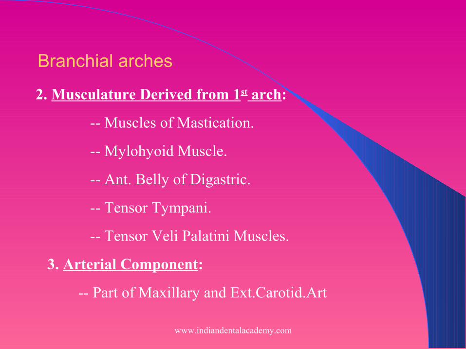
2. Musculature Derived from 1st arch:
-- Muscles of Mastication.
-- Mylohyoid Muscle.
-- Ant. Belly of Digastric.
-- Tensor Tympani.
-- Tensor Veli Palatini Muscles.
3. Arterial Component:
-- Part of Maxillary and Ext.Carotid.Art
Branchial arches
www.indiandentalacademy.com

4. Nerve components:
-- Mandibular division of Trigeminal.N
( Vth Cranial Nerve)
-- Sensory component supplies
: Mandible and covering mucosa.
: Mandibular teeth including Gingiva.
: Mucosa of ant. 2/3 of Tongue.
: Floor of the mouth.
: Skin of the lower third of Face
Branchial arches
www.indiandentalacademy.com

22ndnd BRANCHIAL ARCH BRANCHIAL ARCH (Hyoid Arch)
•Components: 1. Cartilage : Reichert’s Cartilage
(45th – 48th I.U)
-- Greater part of the third ear ossicle.
-- Stapes .
-- Styloid process of the temporal bone.
-- Stylohyoid ligament.
-- Lesser horn and
-- Cranial part - Body of Hyoid.
-- Segments of the facial canal.
Branchial arches
www.indiandentalacademy.com

2. Muscles :
-- Stapedius
-- Stylohyoid
-- Post. Belly of Digastric.
-- Muscles of facial expression.
-- Levator Veli Palatini.
Branchial arches
www.indiandentalacademy.com

3. Nerve :
-- Facial / VII Cranial nerve.
-- Special sensory component
- Chorda tympani nerve
(Ant 2/3rd of Tongue)
4. Artery :
-- Stapedial artery
- Transient i.e. disappears during fetal life.
Branchial arches
www.indiandentalacademy.com

www.indiandentalacademy.com

•Pharyngeal arch I •Cranial nerve - Maxillary and mandibular division of trigeminal nerve (cranial nerve V) •Artery - Maxillary (terminal branch) •Muscles - Muscles of mastication (ie, temporalis, masseter, pterygoids), mylohyoid, anterior belly of digastric, tensor tympani, tensor veli palatini •Skeleton - Maxillary cartilage (incus, alisphenoid), mandibular or Meckel cartilage (malleus), arch dermal mesenchyme (maxilla, zygomatic, squamous portion of temporal bone, mandible)
A summary of the derivatives of the first and second pharyngeal (ie, branchial) arches is as follows:
www.indiandentalacademy.com

•Pharyngeal arch II (hyoid) •Facial nerve - Cranial nerve VII •Artery - Stapedial •Muscles - Muscles of facial expression (ie, orbicularis oculi, orbicularis oris, risorius, buccinator, platysma, auricularis, frontalis), stapedius muscle, posterior belly of digastric, stylohyoid muscle •Skeleton - Stapes, styloid process, stylohyoid ligament, lesser cornu of hyoid, upper part of the body of the hyoid bone
www.indiandentalacademy.com

Anomalies associated with branchial arches
•Deficient development of the branchial arches result in syndromes according to the arch involved.
•First arch anomalies
- Agnathia
- Microstomia
- Treacher Collins syndrome (mandibular dysostosis)
- Pierre Robin syndrome (micrognathia+cleft palate)
Branchial arches
www.indiandentalacademy.com

Pharyngeal pouches and branchial Pharyngeal pouches and branchial groovesgrooves
Primitive pharynx project a series of pouches internally between the branchial arches called pharyngeal pouches.
Intervening between the branchial arches externally are branchial grooves
www.indiandentalacademy.com

First branchial groove deepens to form the external acoustic meatus and the membrane in the depth of groove forms the tympanic membrane.
3rd and 4th branchial groove may form cervical sinus.
Failure to obliterate completely these grooves may result in branchial fistula or sinus.
www.indiandentalacademy.com

1st pharyngeal pouch Ventral portion obliterated by the developing
tongue. Dorsal portion deepens as tubotympanic recess to
form auditory tube.2nd pharyngeal pouch Forms tonsillar fossa and palatine tonsil.3rd pharyngeal pouch Forms thymus and inferior parathyroid gland.4th pharyngeal pouch Forms superior parathyroid gland.5th pharyngeal pouch Forms the ultimobrachial bodyCommon anomaly is DiGeorge Syndromewww.indiandentalacademy.com

DEVELOPMENT OF FACEDEVELOPMENT OF FACE
Development of face depends upon the inductive activities of organizing centres
Procencephalic Rhombencephalic
Induces the inner ear apparatus and upper third of face
Induces the middle and external ear apparatus and the middle and lower third of face
www.indiandentalacademy.com

www.indiandentalacademy.com

Oral development in embryo is demarcated extremely early in life by the appearance of the prechordal plate
with
Endodermal Thickening forms
Oropharyngeal Membrane
www.indiandentalacademy.com

www.indiandentalacademy.com

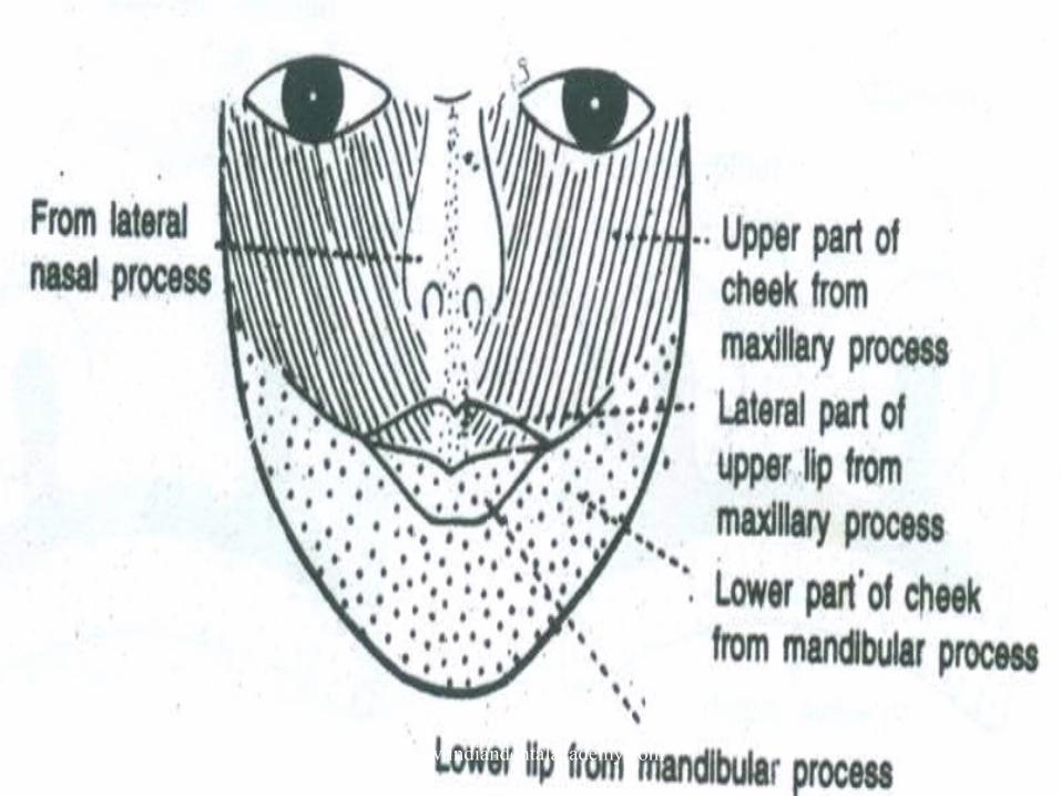
Face develops from 5 prominences that surround the stomatodeum
- Frontonasal- Paired maxillary processes.- Paired mandibular processes.
www.indiandentalacademy.com

Frontonasal prominence formed by proliferation of mesenchyme ventral to the forebrain. It forms
- Lateral optic diverticula eyes - Forehead (between the eyes)- Nasal placodes
www.indiandentalacademy.com

Mesenchyme proliferates around the placodes producing medial and lateral nasal prominences
Lateral nasal prominence separated from maxillary process by nasolacrimal groove
www.indiandentalacademy.com

www.indiandentalacademy.com

Fusion of medial nasal prominences and the maxillary and lateral nasal prominences requires disintegration of nasal fin.
Failure of normal disintegration of nasal fin cause cleft of upper lip and anterior palate.
www.indiandentalacademy.com

Midline merging of medial nasal processMidline merging of medial nasal process
Forms:- Philtrum of upper lip.- Tip of the nose.- Primary palate.
Merging of medial nasal and maxillary Merging of medial nasal and maxillary processprocess
Continuity of the upper jaw and lip.Causes separation of nasal pits from stomodeum
www.indiandentalacademy.com

www.indiandentalacademy.com

Merging of mandibular process in midlineMerging of mandibular process in midline
Forms lower jaws and lips
www.indiandentalacademy.com
www.indiandentalacademy.com

DEVELOPMENT OF NOSEDEVELOPMENT OF NOSE
The nose is a complex of contributions from:
- Frontal prominence The bridge.
- Medial nasal prominence Median ridge and tip
- Lateral nasal prominence The alae
- The cartilage of nasal capsule the septum and nasal conchae
www.indiandentalacademy.com

www.indiandentalacademy.com

A summary of the derivatives of the prominences is as follows: •Frontonasal prominence - Forehead and the dorsum apex of the nose •Lateral nasal prominences - Sides (alae) of the nose •Medial nasal prominences - Nasal septum •Maxillary prominences - Upper cheek region and most of the upper lip •Mandibular prominences - Chin, lower lip, and lower cheek regions •Mesenchyme in the facial prominences - Fleshy derivatives and various bones
www.indiandentalacademy.com

Paranasal SinusesParanasal Sinuses
Paranasal sinuses develop during late fetal life the remainder develops after birth.
They form as outgrowths or diverticula of the walls of the nasal cavities and become air filled extensions of the nasal cavities in the adjacent bone.– Frontal – Ethmoidal– Maxillary – Sphenoidal
www.indiandentalacademy.com

www.indiandentalacademy.com

Time of development of Time of development of paranasal sinusesparanasal sinuses
•Maxillary sinus: Develops at 10
weeks.
•Sphenoidal sinus. At 4 months i.u.
•Ethmoidal sinus. At 4 months i.u.
•Frontal sinus. 3 to 4 months I.U.
www.indiandentalacademy.com

Expand in nasal fossae by growth of mucous membrane sacs- primary pneumatization
Enlarged by secondary pneumatization
Retain communication with nasal fossae through ostia
www.indiandentalacademy.com

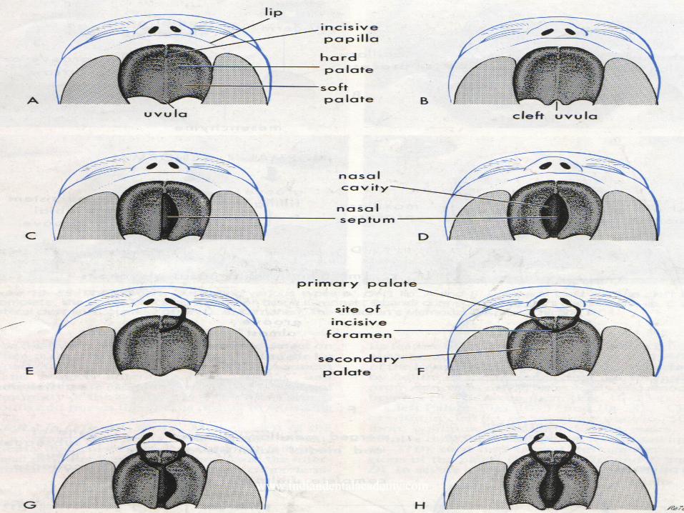
DEVELOPMENT OF PALATEDEVELOPMENT OF PALATE
Palatogenesis begins towards the end of 5 th week and is completed by about 12th week.
The palate develops from two primordia.– Primary palate– Secondary palate
www.indiandentalacademy.com

www.indiandentalacademy.com

www.indiandentalacademy.com

DEVELOPMENT OF THE EARDEVELOPMENT OF THE EAR
Ear consists of 3 anatomical parts:Internal earMiddle earExternal ear
www.indiandentalacademy.com

External ear:External acoustic meatus:
– Develops by deepening of the dorsal end of the 1st pharyngeal groove
Pinna or Auricle:– Six mesenchymal hillocks –
Auricular hillocks develop from the 1st and 2nd pharyngeal arch.
www.indiandentalacademy.com
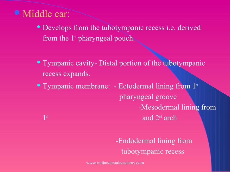
Middle ear: Develops from the tubotympanic recess i.e. derived
from the 1st pharyngeal pouch.
Tympanic cavity- Distal portion of the tubotympanic recess expands.
Tympanic membrane: - Ectodermal lining from 1st pharyngeal groove
-Mesodermal lining from 1st and 2nd arch
-Endodermal lining from tubotympanic recess
www.indiandentalacademy.com

Ear ossicles:1st bone to attain ultimate size.
Maleus and Incus develop from the 1st arch.
Stapes develop from 2nd arch.
Ossification begins in the 16th week and
continues up to the 25th week.
www.indiandentalacademy.com

Internal Ear:
•Otic Placode
•Otic Pit
•Otic Vesicle
•Primordium for Membrane labyrinth
www.indiandentalacademy.com
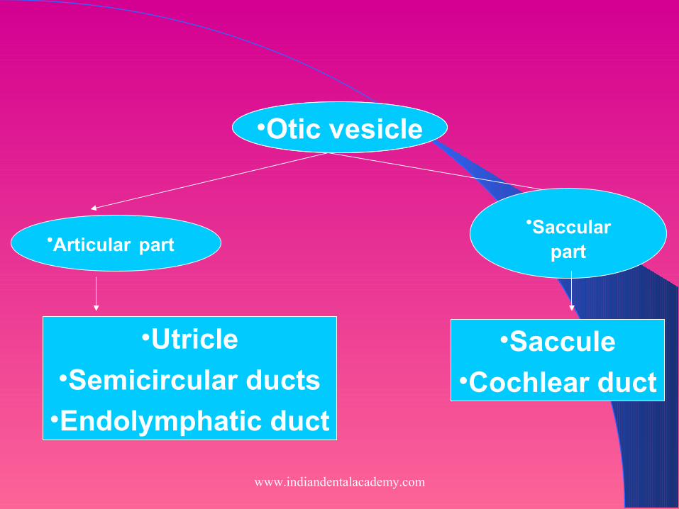
•Otic vesicle
•Saccular part
•Utricle•Semicircular ducts
•Endolymphatic duct
•Saccule•Cochlear duct
•Otic vesicle
•Articular part
www.indiandentalacademy.com

DEVELOPMENT OF EYEDEVELOPMENT OF EYE
Eyes develop from three sources:
Neuroectoderm of the forebrain retina,optic n.
Surface ectoderm of the headlens
Mesoderm between these layers eye muscle
and vascular tissues
1st indication of eye formation is optic sulcus which is
formed in the 4th week.www.indiandentalacademy.com

•Optic sulcus
•Optic vesicle
•Lens placode
•Lens pit •Lens vesicle
•Optic cup
•Retina•Contact surface ectoderm
www.indiandentalacademy.com

www.indiandentalacademy.com

ANATOMY OF FACEANATOMY OF FACE
SKIN Vascular Rich in sebaceous and sweat glands Laxity facilitates spread of edema Elastic and thick due to insertion of muscles.
Wounds tend to gape
www.indiandentalacademy.com

Muscles of the Face
Develop from 2nd branchial arch and supplied by facial nerve
Grouped under1. Ms. of scalp2. Ms. of the auricle3. Ms. of the eyelid4. Ms. of nose5. Ms. around mouth6. Ms. of the neck
www.indiandentalacademy.com

Common facial expressions and muscles producing them-
1. Smiling and laughing- zygomaticus major
2. Sadness- levator labii superioris, levator anguli oris
3. Grief- depressor anguli oris
4. Anger- dilator naris, depressor septii
5. Frowning- corrugator supercilli, procerus
www.indiandentalacademy.com

Nerve supply of faceNerve supply of face
www.indiandentalacademy.com

Arteries of faceArteries of faceVeins of faceVeins of face
www.indiandentalacademy.com
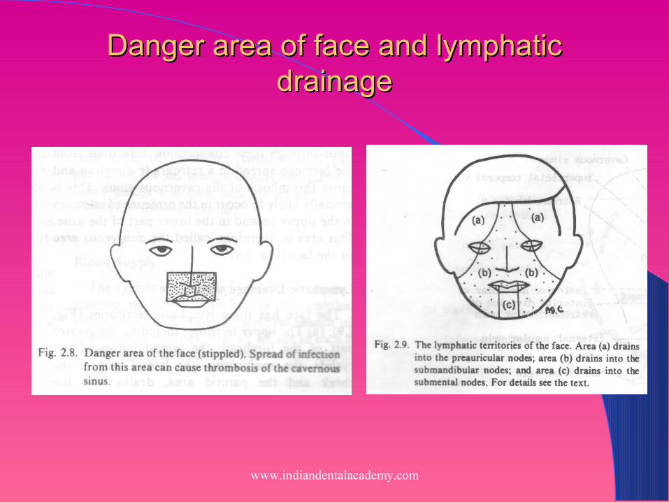
Danger area of face and lymphatic Danger area of face and lymphatic drainagedrainage
www.indiandentalacademy.com

Growth of FaceGrowth of Face Growth usually refers to increase in size or
number (Todd). Development is progress towards maturity (Todd). Growth is largely an anatomic whereas
development is physiologic. Differential growth can be described by - Scammons curve- Cephalocaudal gradient
www.indiandentalacademy.com

THEORIES OF GROWTHTHEORIES OF GROWTH
1. GENETIC THEORY
2. SUTURAL THEORY
3. CARTILAGENOUS THEORY
4. FUNCTIONAL MATRIX THEORY
5. VAN LIMBORGH’S THEORY
6. ENLOWS EXPANDING MASS PRINCIPLE
7. ENLOWS COUNTERPART PRINCIPLE
www.indiandentalacademy.com

Factors Affecting GrowthFactors Affecting Growth
1. Hereditary 2. Nutrition3. Illness4. Race5. Socioeconomic factors6. Family size7. Climatic and seasonal affects8. Psychological disturbances9. Exercise
www.indiandentalacademy.com

Growth patterns in the Dentofacial complexGrowth patterns in the Dentofacial complex
Growth of nasomaxillary complex.
1. Passive displacement.
2. Growth at sutures.
3. Surface remodelling.
www.indiandentalacademy.com

Growth in mandible is seen as a series of bone remodelling process by the deposition and resorptive processes.
GROWTH IN MANDIBLEGROWTH IN MANDIBLE
www.indiandentalacademy.com

Anomalies of Development Anomalies of Development Defects of facial development are the result
of various etiological factors.The study of these anomalies constitute
teratology.– Defective brain development– Acephaly– Acrania– Acalvaria– Holoprosencephaly
www.indiandentalacademy.com

Otomandibular Syndrome
- Agnathia
- Synotia
- Micrognathia
- Mandibulofacial dysostosis
www.indiandentalacademy.com

Failure of facial prominences to merge results in developmental clefts.
1. Oblique facial cleft.2. Microstomia.3. Macrostomia.
www.indiandentalacademy.com

Cleft lip and Palate Cleft lip results from the failure of the maxillary prominence
to unite with the mesial nasal prominences. Unilateral or bilateral.
www.indiandentalacademy.com

Cleft palateUnilateral and bilateral cleft palate can be
classified into – Cleft of the anterior palate– Clefts of the posterior palate– Clefts of anterior and posterior palate.
www.indiandentalacademy.com
www.indiandentalacademy.com

Anomalies of Genetic OriginAnomalies of Genetic Origin
AchondroplasiaCleidocranial dysostosisProgeriaDown’s syndromeMandibulofacial dysostosis
www.indiandentalacademy.com

www.indiandentalacademy.com

Maxillofacial prosthodontics focuses on optimizing the rudimentary functions of speech and swallowing. Although the primary goal is the restoration of these functions but patient esthetics and mastication is important and also has an psychological effect on the patient.
www.indiandentalacademy.com

Nasal prosthesis Nasal prosthesis
Orbital prosthesis Orbital prosthesis
Ear prosthesis Ear prosthesis
www.indiandentalacademy.com

ObturatorObturator
An obturator prosthesis is required for patients who undergo resection of the maxilla for various reasons like neoplasms
Types: Surgical obturator. Interim obturator. Definitive obturator.
www.indiandentalacademy.com

Various factors to be considered during complete denture construction
Lip support.Lip thickness.Tone of facial tissues.
www.indiandentalacademy.com

Summary & ConclusionSummary & Conclusion
Development of the vertebrate face is a dynamic multiple-step process, which starts with the formation of neural crest cells in the developing brain and their subsequent migration to form, together with mesodermal cells, the facial primordia. Signaling interactions coordinate the outgrowth of the facial primordia from buds of undifferentiated mesenchyme into the intricate series of bones and cartilage structures that, together with muscle and other tissues, form the adult face. www.indiandentalacademy.com

ReferencesReferences
Keith L. Moore “The developing human”. 4th edition.
T.W. Saddler “Langman’s Medical Embryology”. 5th edition.
G.H. Sperber “Craniofacial embryology”. 4th edition.
Moore, Persuad “Color atlast of clinical embryology”. 2nd edition.
W.R. Proffit “Contemporary orthodontics”. 3rd edition.
S.I. Balaji “Orthodontics the art and science”. 2nd edition.
www.indiandentalacademy.com

A.R. Tencate “Oral histology”. 5th edition. Shafer “A textbook of Oral pathology”. 4th edition. B.D. Chaurasia “Human anatomy Head and neck”.
3rd edition. Snell “Clinical anatomy for medical students”. Zarb Bolender “Prosthodontic treatment for
edentulous patients”. 12th edition. Keith F. Thomas “ Prosthetic Rehabilitation”.
www.indiandentalacademy.com
THANK YOU
www.indiandentalacademy.com
Related Documents