Georgia Southern University Digital Commons@Georgia Southern Legacy ETDs Summer 2001 Group VIII Spiroplasma of Costa Rica Kimberly M. Stewart Follow this and additional works at: https://digitalcommons.georgiasouthern.edu/etd_legacy Part of the Biochemistry, Biophysics, and Structural Biology Commons, and the Biology Commons Recommended Citation Stewart, Kimberly M., "Group VIII Spiroplasma of Costa Rica" (2001). Legacy ETDs. 403. https://digitalcommons.georgiasouthern.edu/etd_legacy/403 This thesis (open access) is brought to you for free and open access by Digital Commons@Georgia Southern. It has been accepted for inclusion in Legacy ETDs by an authorized administrator of Digital Commons@Georgia Southern. For more information, please contact [email protected].

Welcome message from author
This document is posted to help you gain knowledge. Please leave a comment to let me know what you think about it! Share it to your friends and learn new things together.
Transcript
Georgia Southern University
Digital Commons@Georgia Southern
Legacy ETDs
Summer 2001
Group VIII Spiroplasma of Costa Rica Kimberly M. Stewart
Follow this and additional works at: https://digitalcommons.georgiasouthern.edu/etd_legacy
Part of the Biochemistry, Biophysics, and Structural Biology Commons, and the Biology Commons
Recommended Citation Stewart, Kimberly M., "Group VIII Spiroplasma of Costa Rica" (2001). Legacy ETDs. 403. https://digitalcommons.georgiasouthern.edu/etd_legacy/403
This thesis (open access) is brought to you for free and open access by Digital Commons@Georgia Southern. It has been accepted for inclusion in Legacy ETDs by an authorized administrator of Digital Commons@Georgia Southern. For more information, please contact [email protected].

J* V
Is Georgia Southern University §
js Zach 3. Henderson library m
\ e
Xl ^
GROUP VIII SPIROPLASMA OF COSTA RICA
A Thesis
Presented to
the College of Graduate Studies of
Georgia Southern University
In Partial Fulfillment
of the Requirements for the Degree
Master of Science
In the Department of Biology
by
Kimberly M. Stewart
July 2001
June 28, 2001
To the Graduate School:
This thesis entitled, "Group VIII Spiroplasma of Costa Rica," and written by
Kimberly M. Stewart is presented to the College of Graduate Studies of Georgia Southern
University. I recommend that it be accepted in partial fulfillment of the requirements of
the Master of Science Degree in Biology.
Frank E. French, Supervising Committee Chair
We have reviewed this thesis and recommend its acceptance:
Laura B. Regassa, Committee Member
Accepted for the College of Graduate Studies
G. Lane Van Tassell Dean, College of Graduate Studies
ACKNOWLEDGEMENTS
First, I would like to thank my major professor and chairman of my advisory
committee. Dr. Frank E. French, for the support, advice, and encouragement he has
offered throughout my course of study. He has exhibited extreme patience and
perseverance throughout my many "crises" (no matter how trivial) over the past two
years and for that I am forever grateful. I would also like to thank Lorraine French for
the delicious treats and dinners that she has provided for our lab.
I want to thank my committee members Dr. Dan Hagan, Dr. William Irby, and
Dr. Laura Regassa for their support, advice, and critical review of my thesis. I would
especially like to thank Dr. Regassa for her patience and assistance as she has helped me
venture into the molecular realm.
I would like to express my appreciation to Dr. Sturgis McKeever for offering
advice and providing me with many entertaining stories throughout my graduate studies.
Additionally, I would like to offer thanks to the following people who have helped me
during my graduate studies at GSU: Lisa Morris, Jill Ehling, Dr. Lome Wolfe, Dr. J.B.
Claibome, Dr. Jonathan Copeland, Virginia Bennett, Dr. John Averett, Dr. Lance A.
Durden, Dr. Steve Vives, Dr. Quentin Fang, and all of the Parasitology Journal Club, and
Graduate Student Organization members.
iii

Thanks to the following graduate and undergraduate students for their friendship,
help, and support: Raquel Williams, Tonya Mixson, Denise Bonilla Steinlein, Chris
Gorham, Derek McCoy, Stephanie Schopmeyer, April Murphy, Kelsie Fitzgerald,
Christina Harris, Heather Merten, Jenny Young, Nichole Hair, and Dexter Sowell. A
special thanks to Tonya who has offered unlimited encouragement and support with my
research, teaching, and personal endeavors, and to Raquel who has been my "right-hand"
in the lab as well as a dear friend.
I thank David Abbott for his friendship and support throughout both my
undergraduate and graduate endeavors. Since Dr. Wolfe introduced us in 1996 he has
been one of my dearest and closest friends and 1 feel very lucky to have had the
opportunity to get to know Nancy as well. I look forward to many great adventures
ahead.
Thanks to my mom and dad for having faith in my abilities and offering support
no matter what the situation. Thanks to Matt who has endured much grief throughout the
past two years as a result of my high stress level. I appreciate his patience and
understanding more than he will ever know.
I thank the National Geographic Society for funding to facilitate the initial tabanid
collections and spiroplasma isolations for this project. Additionally, I thank the Allen E.
Paulson College of Science and Technology and Graduate Student Professional
Development Fund for their support of this research project. Thanks to Dr. Robert F.
Whitcomb and Dr. Joseph G. Tully for producing and providing the antisera for the five
temperate North American Spiroplasma.
iv
Curriculum Vitae Kimberly Michelle Stewart
Personal:
Business Address: Department of Biology, Georgia Southern University, Statesboro, Georgia 30460-8042
Business Phone:
(912) 681-5901 E-mail:
Education:
1994-1998: B.S. in Biology, Georgia Southern University, Statesboro, Georgia Major: Biology
1999-2001: M.S. in Biology, Georgia Southern University, Statesboro, Georgia Major: Biology
Employment History: Summer 2000:
Cotton Scout, High Cotton Consulting, Statesboro, Georgia Spring 2000:
Teaching Assistant, Department of Biology, Georgia Southern University, Statesboro, Georgia
1999-2000: Graduate Student Assistant, Department of Biology, Georgia Southern University, Statesboro, Georgia
1999-2000:
House Director, Kappa Delta Sorority, Statesboro, Georgia 1998-1999:
Veterinary Assistant, Metter Veterinary Clinic, Metter, Georgia 1997-1998:
Student Assistant, Payroll Department, Georgia Southern University, Statesboro, Georgia
Memberships: 2000-present:
Graduate Student Organization 1999-present:
Parasitology Journal Club 1999-present:
Georgia Entomological Society 1999-present:
Entomological Society of America 1998-present:
Kappa Delta Alumnae Organization, Delta Lambda Chapter 1999-2001:
Chapter Advisory Board, Kappa Delta Alumnae Organization, Delta Lambda Chapter
1981-present: Gail Hursey School of Dance
1996-1998: Kappa Delta Sorority, Delta Lambda Chapter
Leadership Positions: 2001:
Associate Host, Biting Fly Workshop, Alpine, Texas 2001-present:
Biology Representative, Research/Travel Grant Review Committee 2000-present:
Senator, Graduate Student Organization
2000-2001: Membership Advisor, Chapter Advisory Board, Kappa Delta Sorority,
Delta Lambda Chapter 1999-2000:
House Director, Kappa Delta Sorority, Delta Lambda Chapter 1997-1998:
Coach Chairman, Kappa Delta Sorority, Delta Lambda Chapter
Honors: 1997-1998:
Beta Beta Beta Biological Honor Society Spring 1996:
Dean's List 1994-1998:
Gamma Beta Phi Honor Society 1994-1998:
HOPE Scholarship
VI
Grants, Awards: 2001:
Biology Department Travel Funds, Department of Biology, Georgia Southern University; $250
2001:
Winner of Spring 2001 Sigma Xi Research Competition. Georgia Southern University Chapter of Sigma Xi, Statesboro, Georgia. 24 April 2001; $50.
2001:
Competitive Travel Grant, Department of Graduate Studies, Graduate Student Professional Development Fund, Georgia Southern University; $500
2001:
1st Runner-up in Poster Competition at 5th Joint Meeting of the Georgia
Entomological Society (65th Annual Meeting) and South Carolina Entomological Society (47th Annual Meeting). 5 April 2001. Gainesville, Georgia; $70
2000:
Competitive Research Grant, Department of Graduate Studies, Graduate Student Professional Development Fund, Georgia Southern University; $450
2000:
Academic Excellence Award, College of Science and Technology, Georgia Southern University; $250
Field, Research Experience:
2000-2001: St. Catherine's Island, Georgia -Relocated and Monitored sea turtle nests
1999-present: Department of Biology, Georgia Southern University, Statesboro, Georgia -Serological and Molecular Evaluation of Spiroplasma Bacteria Isolated from
Costa Rican Tabanid Flies -Assistant with undergraduate research projects
1999: Highlands, North Carolina -Assistant in Field Entomology Class -Collected and processed tabanids -Prepared field cultures of tabanid fly spiroplasma
1999:
Costa Rica -Collected and processed tabanids -Prepared field cultures of tabanid fly spiroplasma
1996: Department of Biology, Georgia Southern University, Statesboro, Georgia
-Seed size variation in Ipomoea coccinea and I. quamoclit
vii
Diving Certifications:
1997-present: Nitrox
1997-present: Open Water Diver
Publications: Stewart, K.M. and F.E. French. 2001. Serological Evaluation of Costa Rican Tabanid Fly Bacteria. Abstract. Georgia J. of Science, 59:54.
Presentations:
"Group VIII Spiroplasma of Costa Rica." 5 July 2001. Exit Seminar, Georgia Southern University, Statesboro, Georgia.
"Sero logical Profiling and Molecular Characterization of Spiroplasma Bacteria Isolated From Costa Rican Tabanids." 2 June 2001. Joint Meeting of the North American Dipterists' Society and Biting Fly Workshop, Alpine, Texas.
"Sero logical Profiling and Molecular Characterization of Spiroplasma Bacteria Isolated From Costa Rican Tabanids." 24 April 2001. Sigma Xi Research Competition, Georgia Southern University Chapter of Sigma Xi, Statesboro, Georgia.
"Serological and Molecular Evaluation of Costa Rican Tabanid Spiroplasma Bacteria." 5 April 2001. 5th Joint Meeting of the Georgia Entomological Society (65th Annual Meeting) and South Carolina Entomological Society (47th Annual Meeting), Gainesville, GA.
"Serological Evaluation of Costa Rican Tabanid Fly Spiroplasma Bacteria." 24
March 2001. Georgia Academy of Science, Brunswick, GA.
"Serological and Molecular Evaluation of Costa Rican Tabanid Spiroplasma." 22 March 2001. Parasitology Journal Club at Georgia Southern University.
Critical Review of: Williamson, et al. 1999. Spiroplasma poulsonii sp. nov., a new species associated with male lethality on Drosophila willistoni, a neotropical species of fruit fly. Int. J. Syst. Bacteriology, 49:611-618. Parasitology Journal Club at Georgia Southern University. Fall 2000.
"Systematics of Microscopic Corkscrews" Spring 2000. Research Methods Seminar at Georgia Southern University.
Critical Review of: Jacobs, et al. 2000. Inhibition of Immature Ctenocephalides felis felis (Siphonaptera: Pulicidae) Development in the Immediate Environment of Cats Treated with Imidacloprid. J. Med. Entomology, 37:228-230. Parasitology Journal Club at Georgia Southern University. Spring 2000.
viii
"Spiroplasmas." Fall 1999. Parasitology Journal Club at Georgia Southern University.
"Seed Size Variation in Ipomoea coccinea and /. QuamoclitFall 1996.
Undergraduate Research Symposium, Department of Biology, Georgia Southern University.
Conferences, Workshops Attended: 2001:
Joint Meeting of the North American Dipterists' Society and Biting Fly
Workshop, Alpine, Texas
2001:
Clarke Mosquito Control Workshop, Statesboro, Georgia
2001: 5th Joint Meeting of the Georgia Entomological Society (65th Annual Meeting) and South Carolina Entomological Society (47th Annual Meeting), Gainesville, Georgia
2001: Georgia Academy of Science Annual Meeting, Brunswick, Georgia
2000: Southeastern and South Carolina Branches of the American Society for Microbiology, Statesboro, Georgia
1999: Entomological Society of America, Annual National Meeting, Atlanta, Georgia
1998: Biting Fly Workshop, Black Water Falls State Park, West Virginia
ix
ABSTRACT
GROUP VIII SPIROPLASMA OF COSTA RICA
July 2001
Kimberly M. Stewart
BS IN BIOLOGY GEORGIA SOUTHERN UNIVERSITY
MS IN BIOLOGY GEORGIA SOUTHERN UNIVERSITY
Directed by: Professor Frank E. French
Spiroplasma bacteria isolates are classified or identified through a series of
serological tests that normally consists of screening, one-way deformation tests, cloning,
antisera production, and reciprocal deformation tests. Serological tests on the
spiroplasmas are followed by molecular analysis. The standard molecular analysis used
for spiroplasmas has been 16S rRNA sequencing. The primary goal of my research was
to evaluate sero logically Group VIII spiroplasmas isolated from tropical Costa Rican
tabanids (Diptera: Tabanidae) and to compare them to the temperate North American
Group VIII spiroplasmas. A secondary goal was to evaluate both the temperate and
tropical Group VIII strains by sequencing the 16S-23S rRNA intergenic spacer region.
Spiroplasma cultures were obtained from Costa Rican tabanids and serologically
screened. This screening procedure placed ten of the 72 cultures (GSU 5367, 5401, 5404,
5408, 5429, 5431, 5436, 5437, BARC 4898, and BARC 4899) in Group VIII. Further
serological procedures including one-way deformation tests, serocloning, dilution
cloning, antisera production, and reciprocal deformation tests indicated that the isolates
are serologically related to five serovars previously reported for temperate North
American Spiroplasma. Five of the strains (GSU 5401, 5404, 5408, 5436, and BARC
4899) are serologically related to TAAS-1 and GSU 5367 is closely related to BARC
2649 of the southern United States. Four of the strains, GSU 5429, 5431, 5437, and
BARC 4898, form a cluster of intermediate strains, linking TAAS-1 and BARC 1357.
The frequency of the serovar TAAS-1 (5/10) in the Costa Rican sample was notably
higher than the frequency of 7.7% in Tabanus lineola of temperate North America.
Since 16S rRNA analyses were found to be too conservative in Group VIII
spiroplasmas, attempts were made to sequence the 16S-23S rRNA intergenic spacer
region in an effort to separate this group. DNA was extracted from six isolates, two from
the Costa Rican sample and four temperate North American Group VIII strains. Primers
were developed for the 16S-23S rRNA spacer region and used in PCR amplification.
Amplification was not achieved with the first set of primers. Consequently, a second set
of primers has been designed for PCR amplification and will be used for future research.
XI
TABLE OF CONTENTS
Page
ACKNOWLEDGEMENTS iii
VITAE v
ABSTRACT x
LIST OF TABLES xiii
LIST OF FIGURES xiv
CHAPTER
I. INTRODUCTION 1
II. METHODS 9
III. RESULTS 20
IV. DISCUSSION 23
LITERATURE CITED 26
APPENDICES 30
APPENDIX A. SCREENING TEST FOR SPIROPLASMAS 31
APPENDIX B. SEROCLONING 33
APPENDIX C. TRIPLE CLONING SPIROPLASMAS 34
APPENDIX D. DNA EXTRACTION AND PURIFICATION FOR
16S-23S rRNA SEQUENCING 39
xii
LIST OF TABLES
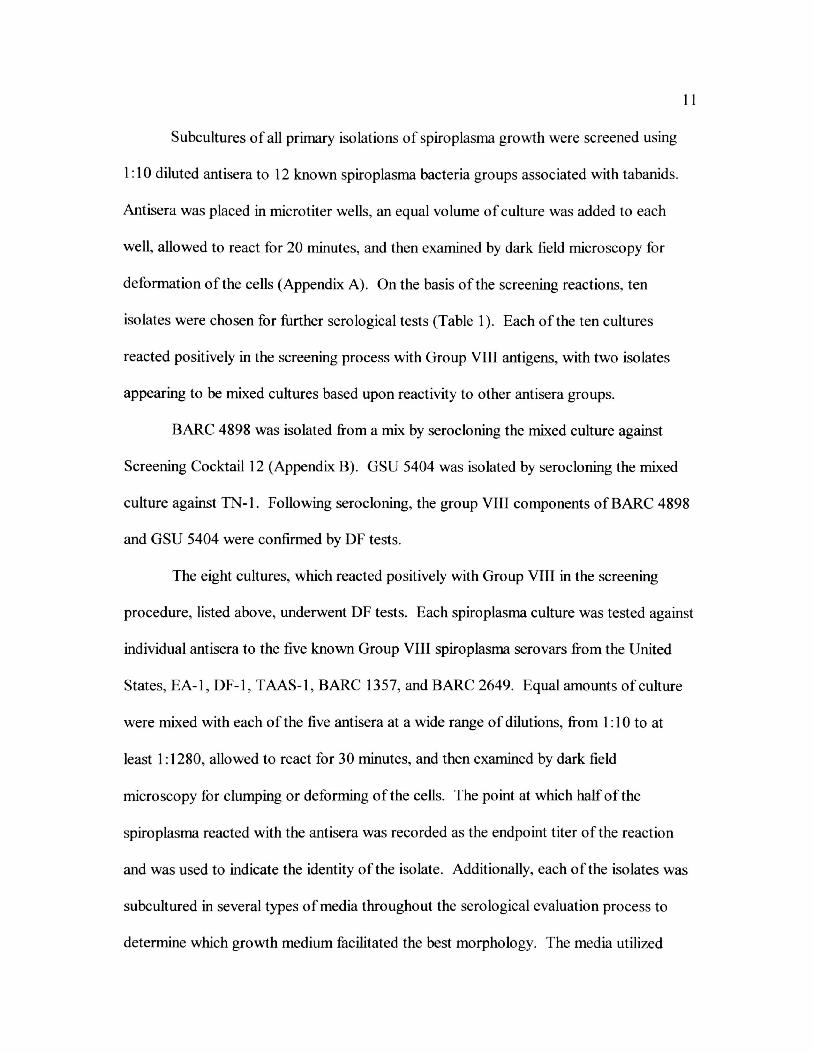
Table Page
1. Host and Host Location of Group VIII Isolates Chosen for Cloning 12
2. Results of One-way and Reciprocal Deformation Tests for Costa Rican
and Temperate North American Group VIII Spiroplasmas 21
xm
LIST OF FIGURES
Figure Page
1. Host and Isolation Sites for Spiroplasma in Costa Rica 10
2. Representative agarose gel of a DNA isolation. Lane 1- "k Hind III,
Lane 2- GSU 5431 15
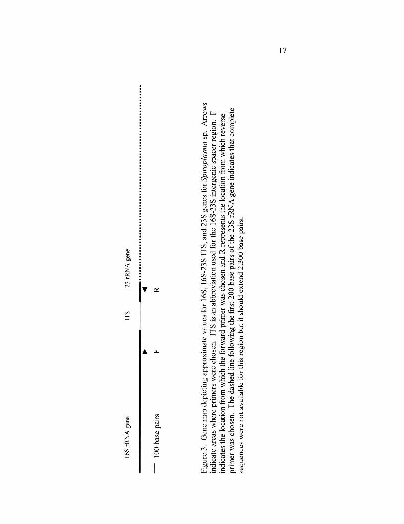
3. Gene map depicting approximate values for 16S, 16S-23S ITS, and 23S genes for
Spiroplasma sp. Arrows indicate areas where primers were chosen. ITS is an abbreviation used for the 16S-23S intergenic spacer region. F indicates the location from which the forward primer was chosen and R represents the location from which reverse primer was chosen. The dashed line following the
first 200 base pairs of the 23 S rRNA gene indicates that complete sequences were not available for this region but it should extend to 2,300 base pairs 17
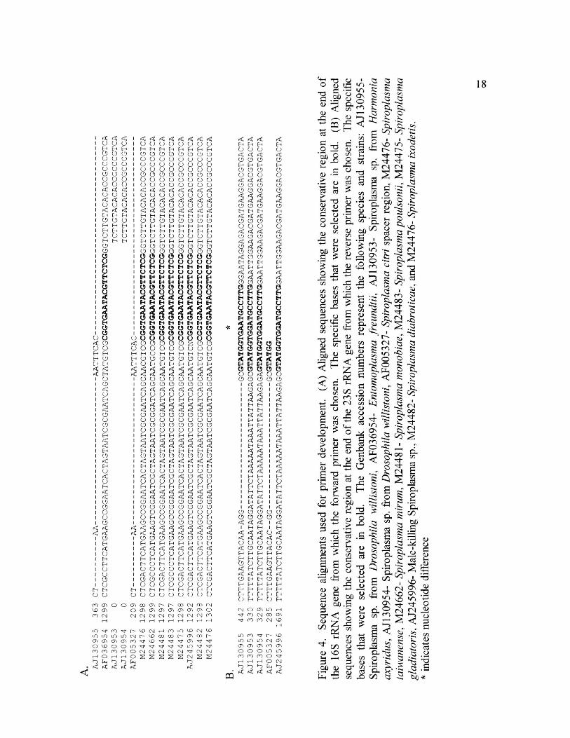
4. Sequence alignments used for primer development. (A) Aligned sequences showing the conservative region at the end of the 16S rRNA gene from which the forward primer was chosen. The specific bases that were selected are in bold. (B) Aligned sequences showing the conservative region at the end of the 23S rRNA gene from which the reverse primer was chosen. The specific bases that were selected are in bold. The Genbank accession numbers represent the following species and strains: AJ130955- Spiroplasma sp. from Drosophila willistoni, AF036954- Entomoplasma freundtii, AJ130953- Spiroplasma sp. from Harmonia axyridus, AJ130954- Spiroplasma sp. from Drosophila willistoni, AF005327- Spiroplasma citri spacer region, M24476- Spiroplasma taiwanense, M24662- Spiroplasma minim, M24481- Spiroplasma monobiae, M24483- Spiroplasma poulsonii, M24475- Spiroplasma gladiator is, AJ245996- Male-killing Spiroplasma sp., M24482- Spiroplasma diabroticae, and M24476- Spiroplasma ixodetis 18
xiv
Chapter I
Introduction
Spiroplasma bacteria are members of Division Tenericutes, Class Mollicutes,
Order Entomoplasmatales, Family Spiroplasmataceae, and Genus Spiroplasma (Tully et
al. 1993). They are prokaryotic organisms, lacking a cell wall, and thought to be
descendants of a branch of gram-positive bacteria (Woese 1987). At some point in their
life cycle, generally during the exponential growth phase, all spiroplasma exhibit a helical
morphology (Williamson et al. 1989, Vazeille-Falcoz 1997). Spiroplasma exhibit both
rotatory and flexional movement despite their lack of flagella, periplasmic fibrils, and
axial filaments (Razin 1978, Bove 1997). These organisms can be found as natural
pathogens or commensals in association with a wide variety of insects, plants, and ticks,
and as experimental pathogens in association with vertebrates (Clark 1982, Hackett and
Clark 1989, Bove 1997).
Spiroplasma were discovered through studies on two plant diseases, citrus
stubborn and com stunt disease. The agent of citrus stubborn disease, Spiroplasma citri,
was the first mollicute to be cultured (Saglio et al. 1973) and the first spiroplasma to be
characterized and named. Two other plant-pathogenic spiroplasmas have been identified
and classified since that time, Spiroplasma kunkelli, the agent of com stunt disease
(Whitcomb et al. 1986a), and Spiroplasma phoeniciem, the agent of periwinkle disease
(Saillard et al. 1987). The three plant-pathogenic spiroplasmas are restricted to the sieve
tube elements of the plant and are vectored by leafhoppers. Leafhoppers acquire
2
spiroplasmas by feeding on an infected plant. The spiroplasma multiply in the
hemolymph, gut, and other organs of the insect. During subsequent feeding, infected
saliva is transferred from the salivary glands of the leafhopper to the plant (Bove 1997).
Insects are bountiful sources of spiroplasma. Spiroplasmas have been isolated
from representatives of nine insect orders: Coleoptera; Diptera; Hemiptera; Homoptera;
Hymenoptera; Lepidoptera; Mecoptera; Odonata; and Trichoptera (Clark 1982, Hackett
et al. 1990, Bove 1997). Spiroplasma are found in the gut, hemolymph, and organs such
as salivary glands of most of these insects, however, isolations from Odonata came only
from the gut (Hackett et al. 1990). Tabanids (Diptera: Tabanidae) are particularly
abundant sources of spiroplasma (Clark et al. 1984, French et al. 1992). Multiple species
of spiroplasma have been isolated from a single tabanid fly (Whitcomb and Hackett
1996). Adult tabanids are easy to maintain under laboratory conditions and can be
artificially infected with spiroplasma rather easily, which makes them exceptional
candidates for study (Wedincamp et al. 1997). The majority of the characterized and
described spiroplasma have been isolated from tabanids; 11 of the 34 recognized
spiroplasma groups were isolated from tabanids (Williamson et al. 1998, Whitcomb et al.
1999).
The life cycle of spiroplasma bacteria is not fully understood and there are
varying opinions on modes of infection in insects. Hackett et al. (1990), after an analysis
of spiroplasma isolations from representative Odonata that came only from the gut,
concluded that the infections were contaminants acquired through predation rather than
true infections. Wedincamp et al. (1997) tested the suggestion that spiroplasmas could be
acquired through predation by infecting mealworm beetle pupae with spiroplasma and
3
then offering them to predaceous firefly larvae. The firefly larvae failed to become
infected after feeding on the infected pupae. In further studies involving tabanids and
fireflies, Wedincamp et al. (1996, 1997) demonstrated that spiroplasmas could be
exchanged between the two species at common carbohydrate feeding sites. The fact that
Spiroplasma apis could be cultured from plant surfaces up to 200 m away from infected
beehives suggests that the spiroplasmas found on plant surfaces were most likely
deposited there by other insects (Bove 1997), thus supporting the suggestion by
Wedincamp et al. (1996) of oral transmission during carbohydrate feeding by tabanids.
Several spiroplasmas are entomopathogens. Spiroplasma apis and melliferum
both infect honeybees. Spiroplasma melliferum was first isolated from the hemolymph of
"dead and moribund bees" (Clark 1977, Clark et al. 1985). Spiroplasma apis was
cultured in 1981 from honeybees carrying "May disease" as well as from plants growing
around beehives with infected bees (Bove 1997). Spiroplasma poulsonii is found in four
related species of Neotropical fruit flies, vertically transmitted, and lethal to male
progeny (Williamson et al. 1999).
Spiroplasma mirum is an experimental pathogen of chicken embryos and suckling
rats and mice. When injected with S. mirum, suckling rats and mice experienced
neurological and ocular disease. Embryonated chicken eggs injected with S. mirum
developed lethal infections (Tully et al. 1982).
Currently spiroplasma are classified according to a group system. To merit group
status in the Genus Spiroplasma, the following requirements must be met (Anonymous
1995, Whitcomb et al. 1999): (1) triple cloning of the organism, (2) demonstration that
the organism belongs to the Class Mollicutes, (3) confirmation that the organism belongs
4
to the genus Spiroplasma, Family Spiroplasmataceae, (4) demonstration that the
organism is serologicaUy distinct from other spiroplasma groups and species by
performing reciprocal DF tests and at least one other serological test, (5) determination of
guanine-plus-cytosine content is optional for group status but should be determined if
there are plans to designate the organism as a new species, (6) deposition of the candidate
in a nationally recognized culture collection so that it is available to other workers, and
(7) determination of species according to the requirements established by the "minimal
standards for description of new mollicute species" as previously established by the
Subcommittee on the Taxonomy of Mollicutes (Anonymous 1995). Spiroplasma, which
exhibit moderate amounts of DNA-DNA homo logy and serological interactions, can be
designated as subgroups. Subgroups exhibit less than 70% homology with the related
subgroups and therefore, like groups are eligible for species designation (Whitcomb et al.
1999).
In addition to the requirements for group designation, requirements for species
designation as outlined by the International Subcommittee on Systematic Bacteriology
are: (1) G+C base composition less than 40%, determination of genome size, and
sequence analysis (16S ribosomal); (2) order and family determination which includes
determination of sterol requirements, cellular and colony morphology, determination of
the usage of aerobisis or anaerobisis, and the organisms usage of the UGA codon; (3)
genus determination which for Spiroplasmataceae would include organisms cultured
from arthropods, plant surfaces, or phloem that are helical, do not require anaerobic
conditions, may or may not require sterols, and have a genome size ranging from 940 to
2,200 kbp; (4) determination of species which includes a species description and DNA-
5
DNA homo logy of less than 70% (Anonymous 1995). Genetically, the most defining
characteristic for a species is less than 70% shared DNA-DNA homology (Gasparich et
al. 1998).
Williamson et al. (1998) and Whitcomb et al. (1999) list 34 groups, 14 subgroups,
and 34 published species descriptions. Group VIII contains three subgroups with two
designated species, along with three serovars (strains), which are awaiting species
designation. Members of this group have less than six turns per helix, reside in dipterans,
require temperatures ranging from 30-370C for growth, have the G+C content ranging
from 29 to 30 +/- 1 mol%, and specific antigenic requirements (Gasparich et al. 1993).
When the 16S rRNA gene was sequenced for Group VIII spiroplasmas, it failed to
separate this tightly knit clade (Dodge et al. 1998).
In order to make evaluations of the phylogeny, biogeography, ecology, host
distribution, and pathogenicity of spiroplasmas, they have to be identified and classified.
They are identified through a series of serological tests that normally consists of
screening, one-way deformation tests, cloning, antisera production, and reciprocal
deformation tests (Whitcomb et al. 1999). Serological tests on the spiroplasmas are
followed by molecular analysis.
Screening is a serological technique in which a spiroplasma is tested against
antisera directed against known spiroplasmas. The screening process places the isolate
into one of twelve recognized antigenic groups (Williamson et al. 1998). After screening,
the deformation (DF) test (Williamson et al. 1978) is the most widely used method for
further serological evaluation of spiroplasmas. Organisms to be tested are mixed with
antisera at a wide range of dilutions. The mixtures are allowed to react and then
6
examined by dark field microscopy for clumping or deforming of the cells. The point at
which half of the spiroplasmas are reacting with the antisera and half are not is called the
endpoint titer of the reaction and should indicate the identity of the organism (Williamson
et al. 1978, Whitcombet al. 1999).
Dilution cloning is done when the identity of a spiroplasma isolate has not been
clearly resolved through the screening and one-way DF tests. The purpose of dilution
cloning a spiroplasma is to obtain a culture that originates from a single cell so that it
reacts homogeneously with the antisera. Cloning is essential in many cases because the
results of serological tests performed on mixed cultures (cultures containing more than
one type of spiroplasma) are worthless. The dilution cloning procedure is normally
repeated three times (Whitcomb et al. 1986b) followed by one-way DF tests to determine
if the identity has been resolved. If an isolate has been triply-cloned and the identity
cannot be determined with one-way DF tests, then it is assumed to represent a novel
antigenic class and antisera is produced from the clone. A triply-cloned culture is the
antigen used for antisera production. The new antisera is used to evaluate serologically
the unidentified culture through homologous and reciprocal DF tests run against
previously typed spiroplasma cultures (Williamson et al. 1978).
Molecular analysis is used to support identifications made through serological
tests and to estimate phylogeny (Dodge et al. 1998). Spiroplasma subgroups with less
than 70% shared DNA-DNA reassociation values with putative sister taxa are eligible for
species designation. Previous experiments have shown that some Group VIII subgroups
indeed share less than 70% genomic similarities among the matrix of species therefore
making them candidates for molecular analysis based on 16S rRNA gene sequence
7
analysis (Whitcomb et al. 1999). However, when the 16S rRNA analyses were done,
they demonstrated more than 99% sequence similarity, thus showing that the 16S rRNA
is too conservative in the case of Group VIII spiroplasmas and therefore not effective for
distinguishing between pairs of species that are closely related (Dodge et al. 1998).
Similar problems with the inability of 16S rRNA analysis to successfully resolve
phylogenetic relationships were indicated by Schulenberg et al. (2000) through sequence
analysis of three male-killing spiroplasmas. Spiroplasma poulsonii, from the Drosophila
willistoni species group of fruit flies, and two other unnamed spiroplasmas found in the
beetles, Adalia bipimctata and Harmonia axyridus, are lethal to male embryos. Analysis
of 16S rRNA sequences from these spiroplasmas demonstrated that the male-killer
spiroplasmas found in the beetles formed a monophyletic clade with Spiroplasma
ixodetis, the representative strain for Group VI, distinct from S. poulsonii of Group II.
The two male-killing spiroplasmas from the beetles and S. poulsonii fell into related
clades. Although the 16S rRNA analysis indicated that these spiroplasmas did fall into
related clades, it failed to provide enough detail to resolve exact relationships. The 16S-
23 S intergenic spacer region was then sequenced in an attempt to make evaluations of the
specific relationships among the clade. The results suggest the sequence variation and
length of the spacer region can be used in identification of spiroplasma infections as well
as species-specific markers (Schulenburg et al. 2000). The 16S-23S intergenic spacer
region has been demonstrated to be species-specific in studies conducted on ureaplasmas
and mycoplasmas as well (Harawasa 1996, 1999, Harawasa et al. 1999).
The primary goal of my thesis was to serologically evaluate Group VIII
spiroplasmas isolated from Costa Rican tabanids and to compare them to temperate North

American Group VIII spiroplasma. A secondary goal was to evaluate both the temperate
and tropical Group VIII strains through sequencing of the 16S-23S rRNA spacer region.
Chapter II
Materials and Methods
The hosts for the Group VIII spiroplasmas of this project were females, live
trapped in a Gressitt Malaise trap in Costa Rica by Frank E. French in 1995 or 1998.
From the 1995 sample of 13 flies, 12 isolations were obtained and from the 1998 sample
of 99 flies, 66 isolations were transported safely to Georgia Southern University.
Spiroplasma isolations were made as previously described by Markham et al. (1983) and
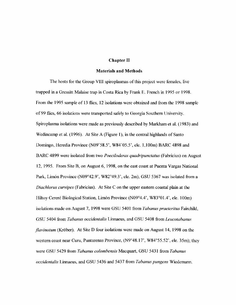
Wedincamp et al. (1996). At Site A (Figure 1), in the central highlands of Santo
Domingo, Heredia Province (N09°58.5', W84°05.5', ele. 1,100m) BARC 4898 and
BARC 4899 were isolated from two Poeciloderas quadripunctatus (Fabricius) on August
12, 1995. From Site B, on August 6, 1998, on the east coast at Puenta Vargas National
Park, Limon Province (N09o42.9', W82049.3', ele. 2m), GSU 5367 was isolated from a
Diachlorus curvipes (Fabricius). At Site C on the upper eastern coastal plain at the
Hiltoy Cerere Biological Station, Limon Province (N09o4.4', W83o01.4', ele. 100m)
isolations made on August 7, 1998 were GSU 5401 from Tabanus praeteritus Fairchild,
GSU 5404 from Tabanus occidentalis Linnaeus, and GSU 5408 from Leucotabanus
jlavinotum (Krober). At Site D four isolations were made on August 14, 1998 on the
western coast near Cum, Puntarenas Province, (N9048.17', W84055.52,, ele. 35m); they
were GSU 5429 from Tabanus colombensis Macquart, GSU 5431 from Tabanus
occidentalis Linnaeus, and GSU 5436 and 5437 from Tabanuspungens Wiedemann.
u c 4s RS "u 0}
u r> o
C5
s
& O u
qj u c So; > o o _ .E £ a. o = 2 <£ 1 *a^ □ (Z! 73
U K o 42 « X 0- sn 0) O cs on c c o
CZ3 -z -S C Q
C3 2
_« 2 o VI
E o a o ^ i: so c Js
O) > •a ' © S R "O 3 ^ S fx S® -C c3 50 w
. *-5 8 ^ "c3 S ^ i: ^ u c ^ a <d « .SdU w cZ < CO
v u > o
c -o
2
"cd u '5b _o o
bo g V(U c
OJ > S3 2 CJ 0-
2 S — 1) X S «§ c o-
"co 3
CO CJ CO V c
cc o u
CJ CL ^
"ZD •£ I I
u a
11
Subcultures of all primary isolations of spiroplasma growth were screened using
1:10 diluted antisera to 12 known spiroplasma bacteria groups associated with tabanids.
Antisera was placed in microtiter wells, an equal volume of culture was added to each
well, allowed to react for 20 minutes, and then examined by dark field microscopy for
deformation of the cells (Appendix A). On the basis of the screening reactions, ten
isolates were chosen for further serological tests (Table 1). Each of the ten cultures
reacted positively in the screening process with Group VIII antigens, with two isolates
appearing to be mixed cultures based upon reactivity to other antisera groups.
BARC 4898 was isolated from a mix by serocloning the mixed culture against
Screening Cocktail 12 (Appendix B). GSU 5404 was isolated by serocloning the mixed
culture against TN-1. Following serocloning, the group VIII components of BARC 4898
and GSU 5404 were confirmed by DF tests.
The eight cultures, which reacted positively with Group VIII in the screening
procedure, listed above, underwent DF tests. Each spiroplasma culture was tested against
individual antisera to the five known Group VIII spiroplasma serovars from the United
States, EA-1, DF-1, TAAS-1, BARC 1357, and BARC 2649. Equal amounts of culture
were mixed with each of the five antisera at a wide range of dilutions, from 1:10 to at
least 1:1280, allowed to react for 30 minutes, and then examined by dark field
microscopy for clumping or deforming of the cells. The point at which half of the
spiroplasma reacted with the antisera was recorded as the endpoint titer of the reaction
and was used to indicate the identity of the isolate. Additionally, each of the isolates was
subcultured in several types of media throughout the sero logical evaluation process to
determine which growth medium facilitated the best morphology. The media utilized
12
13
included MID (Whitcomb 1983), and R2 (Liao and Chen 1977), and derivatives of these
two including V2 R2, '/z MID, and MID Horse Serum (MID HS). Both 1/2 R2 and V2 MID
contain one half the amount of horse serum or fetal bovine serum, respectively, used to
make R2 and MID. In the MID HS, horse serum was substituted for fetal bovine serum.
Following the serocloning and initial DF tests, the identity of two of the isolates
was resolved. The eight unresolved cultures were dilution-cloned in broth medium
utilizing serial dilutions of 10"6 through 10"" in microtiter plates (Appendix C)
(Whitcomb et al. 1986b). The isolates were dilution-cloned at least three times and
deformation tests were made after each cloning. Following the dilution cloning, the
identities of only three triply cloned isolates, GSU 5367, GSU 5408, and GSU 5431, were
not satisfactorily resolved and antisera was produced for these spiroplasma clones as
previously described (Williamson et al. 1978). Spiroplasma cells for antigen were grown
in at least 500 ml broth medium from a triply cloned culture, that was centrifuged at
21,000 x g, combined with RIBI® adjuvant (Sigma Chemical Co., St. Louis, MO), and
injected into a rabbit. GSU 5367 antigen was grown in R2 and GSU 5431 antigen was
grown in V2 R2. GSU 5408 antigen was grown in two different batches of MID and one
batch of V2 HS R2. Each of the 500 ml samples for GSU 5408 was centrifuged, and the
pellets were combined for the antigen. The animals were cared for in accordance with
approved guidelines set forth in the Guide for the Care and Use of Laboratory Animals
and their use was reviewed and approved by the Institutional Animal Care and Use
Committee of Georgia Southern University. The antisera was recovered when
homologous liters of 1:320 or higher were observed and used in reciprocal DF tests
14
against the five Group VIII type cultures and against all of the Group VIII isolates from
Costa Rica.
Following the serological evaluation process, six strains were chosen for
molecular analysis, GSU 5367 and GSU 5431 from Costa Rica, and four United States
Group VIII strains, DF-1, TAAS-1, BARC 2649, and BARC 1357. DNA was isolated
and extracted from the six isolates as previously described (Appendix D) (Duret et al.
1999). The candidate cultures were passed into two 10 ml aliquots of MID medium and
incubated at 30°C until growth was observed. The cultures were then examined by dark
field microscopy to determine morphology and concentration. When prolific cells with
good morphology were observed, the cultures were transferred to 50 ml centrifuge tubes
and harvested by centrifugation in a Fisher Scientific Marathon 21K/BR centrifuge at
12, 000 x g. The pellets obtained were resuspended in STE buffer and the cells were
lysed with sodium dodecyl sulfate. The lysate was heated to 65°C, treated with RNase,
and then subjected to phenol-chloroform-isoamyl extraction, and ethanol precipitation.
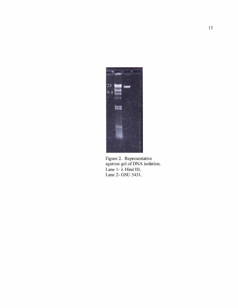
The purified products were run on an agarose gel, using X Hind III as a standard, to
demonstrate DNA had been extracted (Figure 2).
Primers were developed for the 16S-23S intergenic spacer region. For primer
development, the 16S, 23S, and 16S-23S spacer region sequences from Genbank for the
isolates from Drosophila willistoni, Entomoplasma freundtii, Harmonia axyridus, and
Ixodes pacificus and sequences from the spiroplasma S. citri, S. diabroticae, S.
gladiator is, S. ixodetis, S. mirum, S. monobiae, S. poulsonii, and S. taiwanense were
aligned using the Genetic Data Environment Program (Smith et al. 1994). The aligned
sequences were then compared and primers were developed
*
23 B — 9.4 55 r
tm
15
Figure 2. Representative agarose gel of DNA isolation.
Lane 1- X Hind III, Lane 2- GSU 5431.
16
manually from the most conserved areas of the 16S and 23S rRNA genes. The forward
primer, 5'GCACTTATGCAAGAGC, was chosen from the most conserved region near
the end of the 16S rRNA. The reverse primer, 5'CAAGGCATTCACCATAC, was
chosen from the most conservative region of the 23 S rRNA gene (Figures 3-4). The
primers were used in PGR amplification under the conditions outlined by Duret et al.
1999. Specifically, amplification was carried out in 50 (il reaction mixtures containing 1
(al (9-20 ng) of target DNA, IX Cloned Pfu Buffer (200 mM Tris-HCL, pH 8.8, 20 mM
MgS04, 100 mM KC1, 100 mM (NFLt^SC^, 1% Triton X-100, 1 mg/ml nuclease-free
BSA) (Stratagene Cloning Systems, La Jolla, CA), 0.2 mM deoxynucleoside
triphosphates (Promega Corp., Madison, WL), 1 |iM of each primer, and 2.5 U of Cloned
Pfu DNA polymerase (Stratagene Cloning Systems, La Jolla, CA). PGR reactions were
performed in a PE 2400 Thermal Cycler (Perkin-Elmer, Foster City, CA). The
parameters for the PGR were an initial denaturation at 92°C for 2 minutes, followed by 35
cycles of 920C for 45 seconds, 48°C for 45 seconds, and 12°C for 1 minute with a final
extension at 72°C for 10 minutes. When these conditions did not amplify the targeted
product, the conditions were adjusted which included altering annealing temperatures,
DNA concentrations, and MgCb concentrations. Concentrations of MgCh were varied
from 2.0 mM to 4.0 mM and 8.0 mM using the Cloned Pfu DNA Polymerase and 10X
Cloned Pfu Buffer. Additionally, Taq DNA Polymerase (Promega Corp., Madison, WL)
was ordered along with a 10X buffer (100 mM Tris-HCL, pH 9.0 at 250C, 500 mM KCL,
1% Triton® X-100) (Promega Corp., Madison, WL), which did not contain MgCh, and
MgCb titrations ranged from 1.0 mM to 3.0 mM. Additional annealing temperatures
17
<u c o
< §
fN ▼ «
H t—I C/5
C/3 h-
▲ ^
<U
cu S o o g ^ TD Dh_
0) c <D 00 <
§ C/D
00 .fcj Q- O tfi a
x- o o
g c3 ^3 3 .a .a ^ *-3 b .S .S Oh C/3
Cl Cy < < < < < < <C < < < < o O u O O O O U O O 0 EH Eh EH EH fH H Eh Eh Eh Eh o o O O O O 0 0 0 0 0 CJ O o o u O o 0 U 0 0 o O o O o U u U O 0 0 o U CJ CJ u CJ CJ 0 0 0 0 o O O O 0 0 0 0 0 0 0 o U O O CJ O o U O O O u CJ O U CJ O u O U 0 0 < < < < < C < < < < < L) u V o O O u U 0 0 0 < < < < c < c < < < < o CJ o o u U CJ u 0 0 0 < < < c < c < c <c < < H Eh Eh Eh Eh Eh Eh Eh Eh Eh fri o O O O O 0 0 0 0 0 0 Eh Eh Eh H Eh Eh Eh Eh Eh H Eh Eh Eh Eh H Eh Eh Eh H Eh Eh O o O O O CJ CJ 0 0 0 0 Eh Eh Eh H Eh Eh Eh Fh H H Eh e> O O 0 0 0 0 0 0 o o O 0 0 0 0 0 0 o o o e> o O O o 0 u o u u o U O u U H H H Eh Eh H Eh Eh U u O U U U O U U Eh H Eh Eh Eh Eh Eh Eh H EH H EH H Eh Eh Eh O O O O O O C5 O U U U Q U U U U U U < < ri! < < •< rf; < EH Eh EH H Eh Eh tH Eh
H O O u a < o o o
E-» H
O o o o H g H O < H U < u H
C5 U U O O g o H C u E-H H O (J O U H H O O
t-» H 88 o u u COO o o o H H O E-< O O Eh o EH
U U o o o o EH E-t O O (H E-i
O O O O < < o o Eh < < < o o o o o o o o o frH H
E-» t-« O Q o 3 u u O 2 u o EH eh O O
S ' u o o o < < u o
o o o o < < o u H Eh
O O U L) O O O U H H
H EH O O < C H EH O O < O u u Eh H < C 2 < o o o o u u u ^ o o
o o Eh H < c o o H H Eh O O U < (J O O O L> Eh EH H
U O O
H Eh O O < < H Eh U U < O (J U Eh R
o o o o o o u u o o
o o o o o o o a Eh H 2 EH H CD O < < EH H O O < u o o H eh
o o o o o o O Eh O O
Eh ^ Q C5 8 O U U o o u a H EH o o Eh ^ S ' cj a o o C < o CJ Eh EH ss o o a o o o o o H EH
Eh Eh O O < < EH EH O O < O U U Eh Eh gg o o o o u u u ^ o o C C C C C (=£ < < < C <c ^ F=d
e» o H H < < u o Eh H Eh U D U < CJ O O U O Eh H O O
o o H EH < < O U Eh B Eh H O O <C < O O O CJ EH EH O O
O O EH H c c o o FH H O CJ c < e> o u u H H u o
ro CTv o o o\ *X) <J\ o ro CM eg T 1 m CO r- m m m m CNJ cr. cr\ o> <y\ ro o o o m ro ro CO ro o i—j o i—i i—i O h) Ui Eu < < < < <C
cocrir-r-oocvjcocM <T»<Tv0%<J»(TS<T>Cr»O CSlCMCMCNJCMCMOsirO *—I i—I rH i—I i—1 <—I i—I <—I \£>C\]«—IfOin^DCN^X) r--,x>aooor-cT\cDr- ■^isOKT^^cri'^r^r CSJCSJCSICMCSI^CNCSI S 2 2 2 2 2 2
O O < < o o
o o Eh H < < O O O O
O O H H
e> o o o
o o EH £H
88
CD O O < O C5 u o
m
H EH EH 0 0 0 EH EH Eh Eh Eh H < < < Eh Eh Eh < < <
0 0 0 0 0 0 0 0 0 0 < < < < Eh Eh
< < 0 < < < < < 0 0 0 0 0 < 0 0 < 0 Eh Eh H Eh Eh Eh Eh Eh Eh H 0 O 0 0 0 Eh Eh Eh 2 < < 2 < 0 Eh Eh 0 Eh Eh H Eh H Eh R Eh Eh Eh Eh Eh Eh Eh Eh Eh c.; H H 0 H CNJ O cr> m <—I
ro CM 00 CTi ro 00 CsJ T 1
lO ro r- VD m m m CM (Ti cr. o> cr> ro CTi o o o m m ro ro ro o t—< rH «—J o CM ♦o
< < < <
<u <L>
) srH ^ C 2 <
c3 J GO (U
IX) 6c *

19
used included 370C, 390C, 410C, 430C, 450C, 470C, and 540C. DNA concentrations of
50 ng and 100 ng were also used.
Chapter III
Results
Ten strains of Group VIII were examined serologically in this study (BARC 4898,
BARC 4899, GSU 5367, GSU 5401, GSU 5404, GSU 5408, GSU 5429, GSU 5431, GSU
5436, and GSU 5437) and compared serologically with five recognized strains of Group
VIII spiroplasmas from the United States. Cultures examined by dark field microscopy
revealed short helices with six or less turns and were often in excess of 1010per ml broth.
All strains grew well in MID broth medium. Additionally, each strain grew in R2 broth
medium; however, the DF test in R2 was more difficult to read perhaps due to a
combination of a different refractive index and morphology. Some strains also grew in V2
HS MID and Vz R2. Antisera pellets for GSU 5367, GSU 5408, and GSU 5431 were
grown in R2, R2 '/i HS MID and MID, and Vi R2 respectively.
The ten strains examined are serologically related to five serovars previously
reported for temperate North American Spiroplasma. The homologous DF test titer for
GSU 5367 was 1:2560 (Table 2) and the reciprocal tests with BARC 2649 were both
strong with titers of 1:640. GSU 5367 cells reacted at very low levels (1:20 to none)
with antisera of GSU 5431 and the five strains from the United States.
GSU 5431 had a homologous DF titer of 1:1280. The GSU 5431 antisera DF titers
were strong against cells from GSU 5429 and GSU 5437 (1:1280 and 1:640,
respectively), which also were from the same collection at Curu. GSU 5431 cells had
strong one-way DF titers against the antisera of TAAS-1 (1:640) and BARC 1357
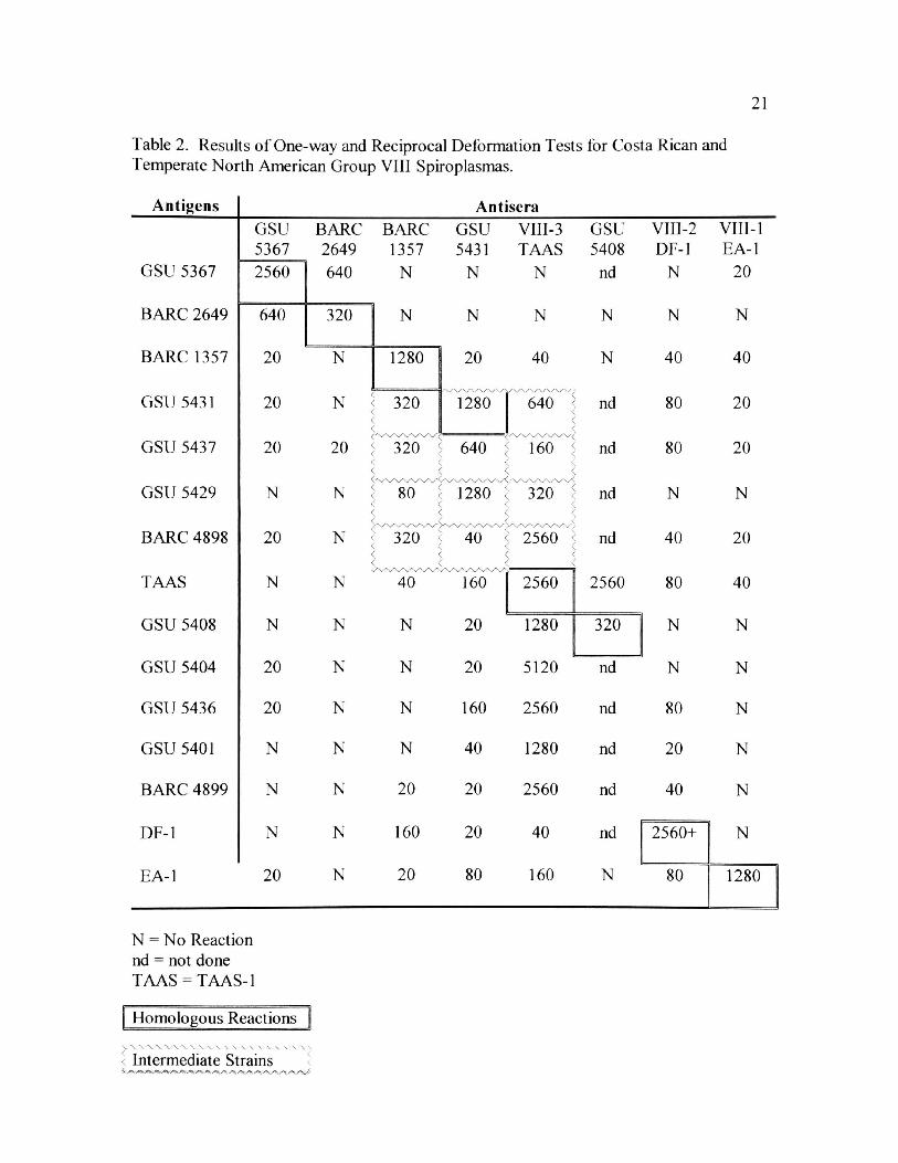
21
Table 2. Results of One-way and Reciprocal Deformation Tests for Costa Rican and
Temperate North American Group VIII Spiroplasmas.
Antigens Antisera
GSU 5367
BARC 2649
BARC 1357
GSU 5431
GSU 5437
GSU 5429
BARC 4898
GSU BARC BARC 5367 2649 1357
640 N
GSU VIII-3 5431 TAAS
N N
GSU 5408
nd
N
N
nd
160 5 nd
nd
nd
TAAS N N 40 160 2560 2560
GSU 5408 N N N 20 1280 320
GSU 5404 20 N N 20 5120 nd
GSU 5436 20 N N 160 2560 nd
GSU 5401 N N N 40 1280 nd
BARC 4899 N N 20 20 2560 nd
DF-1 N N 160 20 40 nd
EA-1 20 N 20 80 160 N
VII1-2 DF-1
N
N
40
80
80
N
40
80
N
N
80
20
40
2560+
80
VII1-1
EA-1
20
N
40
20
20
N
20
40
N
N
N
N
N
N
1280
N = No Reaction nd = not done TAAS = TAAS-1
| Homologous Reactions
v. . , • - - - X Intermediate Strains
22
(1:320). However, the reciprocal DF titers were only 1:160 for TAAS-1 cells, and 1:20
for BARC 1357. For cells of other strains from Costa Rica or the United States, only
moderate (1:160) to no DF titers were obtained with the GSU 5431 antisera. Cells of
GSU 5431 had DF titers of 1:20 or none to the other three antisera (Table 2).
In addition to strong one-way reactions with antisera GSU 5431, GSU 5437 cells
had DF titers of 1:320 (BARC 1357) and 1:160 (TAAS-1), and GSU 5429 cells had a
titer of only 1:80 for BARC 1357 but a 1:320 titer with antisera of TAAS-1.
TAAS-1 antisera reacted very strongly with cells of BARC 4898, BARC 4899,
GSU 5401, GSU 5404, GSU 5408, and GSU 5436, with titers of 1:1280-1:5120.
Reciprocal DF titers of GSU 5408 and TAAS-1 were 1:1280 and 1:2560, respectively.
DNA was extracted and purified from GSU 5367, GSU 5431, BARC 1357, BARC
2649, DF-1, and TAAS-1. The primers chosen were used for PCR amplification and the
PCR conditions were altered numerous times; however, only primer dimers were
obtained.
Chapter IV
Discussion
Serology, in particular the DF test, has been the main method by which new
spiroplasma isolates have been characterized (Williamson et al. 1998). The five Group
VIII strains from the United States have been repeatedly differentiated by reciprocal DF
tests in this research project and others (Whitcomb et al. 1997, 1999, Williamson et al.
1998). When antiserum is not available for an isolate, only one-way DF tests are
possible. While one-way DF tests are not always indicative of close relationships, titers
of 1:1280 or greater have not been associated with reciprocal titers of less than 1:320.
Reciprocal DF tests among the five Group VIII strains (EA-1, DF-1, TAAS-1, BARC
1357, and BARC 2649) and 16 other strains associated with tabanids from the United
States produced no reactions above 1:160 except for homologous reactions (Whitcomb et
al. 1997). The one-way DF reactions of 1:1280 to 1:5120 for cells of BARC 4899, GSU
5401, GSU 5404, and GSU 5436 with TAAS-1 antisera are indicative of a close
relationship with TAAS-1.
GSU 5367 shares a close serological relationship to BARC 2649. BARC 2649
was isolated from Tabanus lineola Fabricius, Bulloch Co., Georgia and is considered rare
with an 8.6% frequency of isolation from 104 of the type host and locality (F. E. French,
unpublished). GSU 5408, although initially obscure, shows reciprocally, a close
serological relationship with TAAS-1. Four other isolates, BARC 4899, GSU 5401, GSU
5404, and GSU 5436 had very high one-way DF titers of 1:1280 to 1:5120 to TAAS-1
24
antisera. Among the Group VIII isolates from Costa Rica, five of ten reacted strongly
with TAAS-1 antisera; thus, the strain TAAS-1 is perhaps the most prevalent Group VIII
form in Costa Rica. These five cultures were obtained from five different tabanid species
from three sites. Cum, Hiltoy Cerere, and Santo Domingo with a range in elevation from
35 to 1,100m. In Bulloch Co. Georgia, 7.7% of Group VIII spiroplasmas isolated from
104 T. lineola, reacted strongly with TAAS-1 (F. E. French, unpublished).
GSU 5431, with a homologous titer of 1:1280, reacted strongly with both TAAS-
1 (1:640) and BARC 1357 (1:320) in one-way DF tests but in reciprocal DF tests it only
reacted at 1:20 with BARC 1357 and moderately with TAAS-1(1:160). GSU 5431 may
represent an intermediate strain between BARC 1357 and TAAS-1, and perhaps is a more
serologically basic strain. GSU 5431 and three other strains, BARC 4898, GSU 5429,
and GSU 5437 appear to form a small cluster of bridge strains that tie BARC 1357, GSU
5431, and TAAS-1 to a common near ancestor (Table 2).
At the outset of this research project the prediction was made that there would be
many new spiroplasma strains in the tropics. However, the evaluation of the samples in
this study failed to reveal any new Group VIII strains. The ten isolates from Group VIII
showed at least a strong one-way relationship with isolates from the United States, unlike
the long form spiroplasmas from Costa Rica in which there are at least seven isolates that
show moderate to no reciprocal reactions with any strain from the United States (R.F.
Whitcomb and F.E. French, unpublished). Since the Group VIII spiroplasma isolations
were obtained from seven different species of tabanids, captured in four different
locations in Costa Rica in two separate years, it is likely that there is no widely
distributed undiscovered Group VIII spiroplasma in Costa Rica.
25
DNA was isolated from GSU 5367, GSU 5431, BARC 1357, BARC 2649, DF-1,
and TAAS-1. Amplification with the first set of primers was not achieved despite
varying annealing temperatures, DNA concentrations, and MgCh concentrations.
Therefore, a second set of primers was designed. Amplification with a hot start was
attempted concurrently for both sets of primers. A dominant product, approximately 900
base pairs in length, was obtained at 1.0, 1.5, and 2.5 mM MgC^ concentration with the
first set of primers. The second set of primers produced a dominant product only with the
2.5 mM MgCk concentration; the product was approximately 900 base pairs long. This
has led to the assumption that perhaps the spacer region is longer than the approximately
300 base pairs previously estimated. The MgCfe concentrations will be altered once
again and if the same size product is obtained, then the five remaining samples will be
PGR amplified. Following amplification the products for each of the six samples will be
purified on an agarose gel, excised, and then sent to the appropriate facilities for
sequencing. After the sequences are obtained, they will be analyzed by alignment with
the sequences used for primer development to determine if the 16S-23S spacer region has
been amplified. If the spacer region has indeed been amplified, the sequences will then
be compared to determine if the spacer region is species specific within the Group VIII
spiroplasmas.
26
Literature Cited
Anonymous. (1995). International Committee on Systematic Bacteriology,
Subcommittee on the taxonomy of the Mollicutes. Revised minimum standards for description of new species of the class Mollicutes (Division Tenericutes). Int
JSy.st Bacteriol 45, 605-612.
Bove, J.M. (1997). Spiroplasmas - infectious agents of plants, arthropods, and
vertebrates. Wien Klin Wochenschr 109, 604-612.
Clark, T.B. (1977). Note: Spiroplasma sp., a new pathogen in honey bees. J
Invertebr Pathol 29, 112-113.
Clark, T.B. (1982). Spiroplasmas: diversity of arthropod reservoirs and host-parasite
relationships. Science 217, 57-59.
Clark, T.B., Peterson, B.V., Whitcomb, R.F., Henegar, R.B., Hackett, K.J., and Tully, J.G. (1984). Spiroplasma in the Tabanidae. hr J Med Sci 20, 1002-1005.
Clark, T.B., Whitcomb, R.F., Tully, J.G., Mouches, C., Saillard, C., Bove, J.M., Wroblewski, H., Carle, P., Rose, D.L., Henegar, R.B., and Williamson, D.L. (1985). Spiroplasma melliferum, a new species from the honeybee (Apis mellifera). IntJSyst Bacteriol 35, 296-308.
Dodge, D.E., Ellis, N.E., Collins, P.C., Bost, D.A., Gasparich, G., Williamson, D.L., Tully, J.G., and Whitcomb, R.F. (1998). Taxonomic classification of members
of the genus Spiroplasma by fluorescent DNA sequencing of the 16S rDNA gene. Abstract. Congress of the International Organization of Mycoplasmology. Sydney, Australia, July 1998.
Duret, S., Danet, J.L., Garnier, M., and Renaudin, J. (1999). Gene disruption
through homologous recombination in Spiroplasma citri: and scml - disrupted motility mutant is pathogenic. J Bacteriol 24, 7449-7456.
French, F.E., Whitcomb, R.F., Hagan, D.V., Rafter, J.A., Konai, M., and Clark, E.A. (1992). Dynamics of Spiroplasma infections in tabanid (Diptera: Tabanidae)
flies, laboratory transmission, and in vitro tests. IOM Letter 2, 114.

27
Gasparich, G.E., Hackett, K.J., French, F.E., and Whitcomb, R.F. (1998). Serologic and genomic relatedness of group XIV spiroplasma isolates from a lampyrid
beetle and tabanid flies: an ecologic paradox. Int JSyst Bacterial 48, 321-324.
Gasparich, G.E., Saillard, C., Clark, E.A., Konai, M., French, F.E., Tully, J.G., Hackett, K.J., and Whitcomb, R.F. (1993). Serologic and genomic relatedness of group VIII and group XVII spiroplasmas and subdivision of spiroplasma group VIII into subgroups. Int J Syst Bacterial 43, 338-341.
Hackett, K.J. and Clark, T.B. (1989). Ecology of spiroplasmas. In The Mycaplasmas,
vol. 3, pp. 175-208. Edited by R.F. Whitcomb & J.G. Tully. New York: Academic Press.
Hackett, K.J., Lynn, D.E., and Whitcomb, R.F. (1990). Spiroplasma and other
mollicutes: possible application to plant pest control. In Biotechnology, Biological Pesticides and Novel Plant Resistance for Insect Pest Management, pp. 93-97. Edited by D.W. Roberts & R.R. Granados.
Harawasa, R. (1996). PCR: application of nested PCR to detection of mycoplasmas. In Molecular and Diagnostic Procedures in Mycoplasmology, vol. 2, pp. 75-79.
Edited by S. Razin & J.G. Tully. New York: Academic Press.
Harawasa, R. (1999). Genetic Relationships among Mycoplasmas based on the 16S- 23S rRNA Spacer Sequence. Microbiology and Immunology 43, 127-132.
Harawasa, R., Lefkowitz, E.J., Glass, J.I., and Cassell, G.H. (1999). Phylogenetic
analysis of thel6S-23S rRNA intergenic spacer region of the genus Ureaplasma. J Vet Med Sci 58, 191-195.
Liao, C.H., and T.A. Chen. (1977). Culture of com stunt spiroplasma in a simple
medium. Phytopathology 67, 802-807.
Markham, P., Clark, T.B., and Whitcomb, R.F. (1983). Culture media for spiroplasmas from arthropods. In Methods in Mycoplasmology, vol. 2,
pp. 217-223. Edited by J.G. Tully «fe S. Razin. New York: Academic Press.
Razin, S. (1978). The mycoplasmas. Microbiological Review 42, 414-470.
Saglio, P., Lhospital, M., La flee he, D., Dupont, G., Bove, J.M., Tully, J.G., and Freundt, E.A. (1973). Spiroplasma citri gen. and sp.n.: a mycoplasma-like
organism associated with "stubborn" disease in citrus. Int J Syst Bacteriol 23, 191-204.
28
Saillard, C., Vignault, J.C., Bove, J.M., Raie, A., Tully, J.G., Williamson, D.L., Fos, A., Garnier, M., Gadeau, A., Carle, P., and Whitcomb, R.F. (1987).
Spiroplasma phoenicium sp. nov., a New Plant-Pathogenic Species from Syria. Int J Syst Bacterial 37, 106-115.
Schulenburg, J.H.G.v.d., Majerus, T.M.O., Dorzhu, C.M., Zakharov, LA., Hurst, G.D.D., and Majerus, M.E.N. (2000). Evolution of male-killing Spiroplasma
(Procaryotae: Mollicutes) inferred from ribosomal spacer sequences. The Journal of General and Applied Microbiology 46, 95-98.
Smith, S.W., Overbeek, R., Woese, C.R., Gilbert, W., and Gillvet, P.M. (1994). The
genetic environment and expandable GUI for multiple sequence analysis.
Computational Applied Bioscience 1, 6710-6715.
Tully, J.G., Bove, J.M., Laigret, F., and Whitcomb, R.F. (1993). Revised taxonomy
of the class Mollicutes: Proposed elevation of a monophyletic cluster of
arthropod-associated mollicutes to ordinal rank (Entomoplasmatales ord. Nov.), with provision for familial rank to separate species with nonhelical morphology (Entomoplasmataceae fam. nov.) from helical species (Spiroplasmataceae), and emended descriptions of the order Mycoplasmatales, family Mycoplamataceae. Int J Syst Bacterial 43, 378-385.
Tully, J.G., Whitcomb, R.F., Rose, D.L., and Bove, J.M. (1982). Spiroplasma mirum, a new species from the rabbit tick (Haemaphysalis leporispalustris). Int J Syst Bacterial 32,1 92-100.
Vazeille-Falcoz, M., Helias, C., Le Goff, F., Rodhain, F., and Chastel, C. (1997).
Three spiroplasmas isolated from Haematopota sp. (Diptera: Tabanidae) in France. Entomological Society of America 34, 238-241.
Wedincamp, J.Jr., French, F.E., Whitcomb, R.F., and Henegar, R.B. (1996). Spiroplasmas and Entomoplasmas (Procaryotae: Mollicutes) Associated with Tabanids (Diptera: Tabanidae) and Fireflies (Coleoptera: Lampyridae). J Invert Patho 68, 183-186.
Wedincamp, J. Jr., French, F.E., and R.F. Whitcomb, R.F. (1997). Anew
spiroplasma (Entomoplasmatales: Spiroplasmataceae) record for Georgia and attempted horizontal transmission via predation. Entomological News 108, 209-
212.
Whitcomb, R.F. (1983). Culture media for spiroplasmas, p. 147-158. In Methods in
Mycoplasmology, pp. 147-158. Edited by S., Razin & J.G. Tully. New York: Academic Press.
29
Whitcomb, R.F., Chen, T.A., Williamson, D.L., Liao, C., Tully, J.G., Bove, J.M., Mouches, C., Rose, D.L., Coan, M.E., and Clark, T.B. (1986a). Spiroplasma kunkelli sp. no v.: Characterization of the Etio logical Agent of Com Stunt Disease. Int J Syst Bacterial 36, 170-178.
Whitcomb, R.F., French, F.E., Tully, J.G., Carle, P., Henegar, R., Hackett, K.J., Gasparich, G.E., and Williamson, D.L. (1997). Spiroplasma Species, Groups, and Subgroups from North American Tabanidae. Curr Microbiol 35: 287-293.
Whitcomb, R.F., and Hackett, K.J. (1996). Identification of mollicutes from insects. In Molecular and Diagnostic Procedures in Mycoplasmology, vol. 2, pp. 313-322.
Edited by S. Razin & J.G. Tully. San Diego, CA: Academic Press.
Whitcomb, R.F., Hackett, K.J., and Clark, E.A. 1986b. Cloning by limiting dilution in liquid Media: an improved alternative for cloning mollicute species.
th Proceedings of the 6 International Congress of Mycoplasmology, Birmingham, Alabama, August 26-31, p. 167.
Whitcomb, R.F., Williamson, D.L., Gasparich, G.E., Tully, J.G., and French, F.E. (1999). Spiroplasma Taxonomy. First Internet Conference on Phytopathogenic
Mollicutes, http;//www.unid.it.phytoplasma/conf.html. May 24, 1999, p. 16-33.
Williamson, D.L., Sakaguchi, B., Hackett, K.J., Whitcomb, R.F., Tully, J.G., Carle, P., Bove, J.M., Adams, J.R., Konai, M., and Henegar, R.B. (1999). Spiroplasma poulsonii sp. nov., a new species associated with male-lethality in
Drosophila willistoni, a neotropical species of fruit fly. Int J Syst Bacteriol 49: 611-618.
Williamson, D.L., Whitcomb, R.F., and Tully, J.G. (1978). The spiroplasma
deformation test, a new serological method. Curr Microbiol 1: 203-207.
Williamson, D.L., Whitcomb, R.F., and Tully, J.G. (1989). The genus Spiroplasma.
In The Mycoplasmas, vol. 5, pp. 71-111. Edited by R.F. Whitcomb & J.G. Tully. San Diego, CA: Academic Press.
Williamson, D.L., Whitcomb, R.F., Tully, J.G., Gasparich, G.E., Rose, D.L., Carle, P., Bove, J.M., Hackett, K.J., Adams, J.R., Henegar, R.B., Konai, M., Chastel, C., and French, F.E. (1998). Revised group classification of the genus Spiroplasma. Int J Syst Bacteriol 48:1-12.
Woese, C.R. (1987). Bacterial evolution. Microbiological Review 51: 221-271.

31
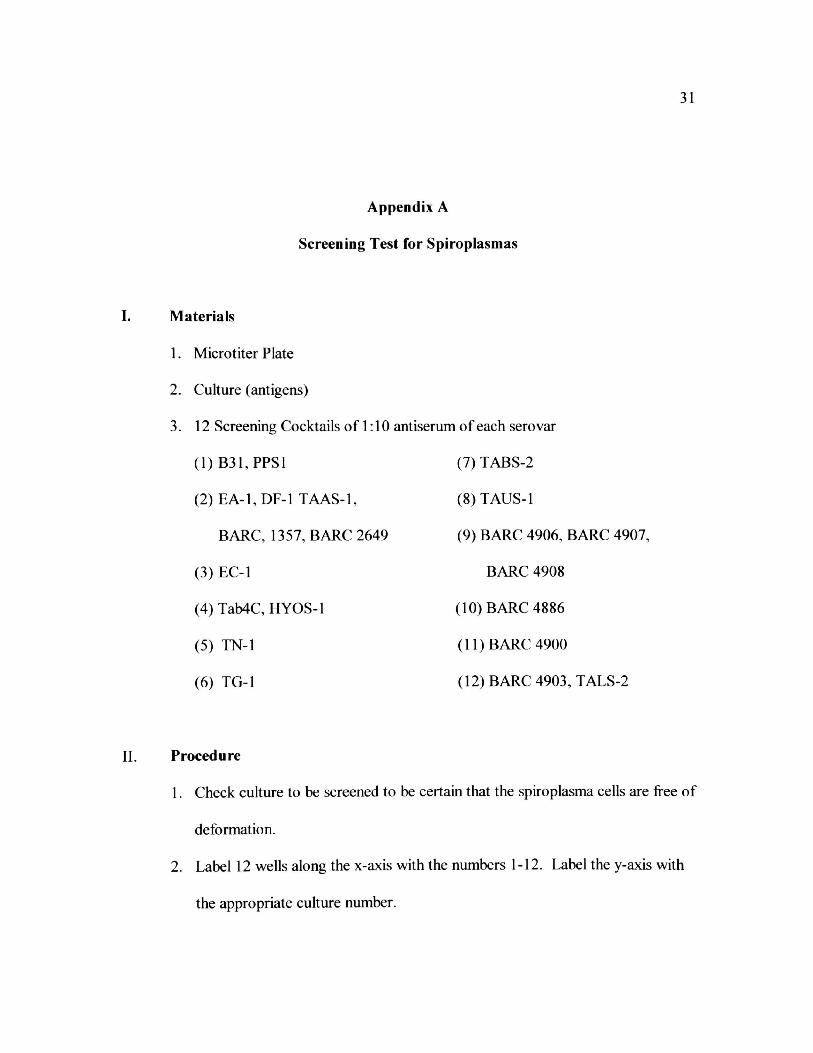
Appendix A
Screening Test for Spiroplasmas
Materials
1. Microtiter Plate
2. Culture (antigens)
3. 12 Screening Cocktails of 1:10 antiserum of each serovar
(1) B31, PPS1 (7) TABS-2
(2) EA-1, DF-1 TAAS-1, (8) TAUS-1
BARC, 1357, BARC 2649 (9) BARC 4906, BARC 4907,
(3) EC-1 BARC 4908
(4) Tab4C, HYOS-1 (10) BARC 4886
(5) TN-1 (11) BARC 4900
(6) TG-1 (12) BARC 4903, TALS-2
Procedure
1. Check culture to be screened to be certain that the spiroplasma cells are free of
deformation.
2. Label 12 wells along the x-axis with the numbers 1-12. Label the y-axis with
the appropriate culture number.

32
3. Add 20 |il of the appropriate screening cocktail to the microtiter well whose
number corresponds to the number on the cocktail.
4. Add 20 [al of the appropriate culture to each well.
5. Cover the plate and allow it to react for 30 minutes at room temperature
before beginning the readings.
6. Make slides and observe cells for deformation at 1200 magnification with
dark field illumination.

33
Appendix B
Serocloning
(Protocol based on R. F. Whitcomb personal communication)
50^1 of culture
50^.1 of antisera 1:10 (single or combined screen)
1. Mix, after one hour add about one ml of medium.
2. Gently pass through 0.45|im filter without foaming and examine microscopically for
estimate of concentration.
3. Serially dilute by 1/10, i.e. 10 -l =1.8 ml medium and 0.2ml of filtered culture (or 0.9
ml medium and 0.1 ml culture).
For very low concentrations, do 10 1 through 10~5 tubes.
For short spiroplasmas in heavy concentration, do 10"' through 10"'1 tubes.

34
Appendix C
Triple Cloning Spiroplasmas
(Protocol based on Whitcomb et al. 1986 and R. F. Whitcomb personal communication)
1. Select cultures to be cloned carefully.
2. Resuscitate chosen culture, and serially pass it until the spiroplasmas exhibit
morphology free of deformation.
3. When the culture has stabilized and generates 200 helices per field or so, it is ready to
be cloned.
4. If the culture has serological reactivity, it is a good idea to do a confirmatory
deformation titer.
Prepare dilution series for cloning.
The first part of the dilution series will be in 3.0 ml snap-cap tubes, and the final tubes
will be large sterile centrifuge tubes.
For dilutions of: 10"', 10~2, 10~3, 10"4, and 10~5, add 2.7 ml of broth medium to 3.0 ml
tubes.
For dilutions of: 10"6, 10"7, 10"8, 10"9, 10"'°, and 10"11, add 18 ml of broth medium.
Filtration
Filter the culture through a 0.45|im filter.
35
Dilutions
Now prepare the dilution series. Make tenfold dilutions (0.3 filtrate to 2.7 ml of medium,
and then serially dilute). Specifically, transfer 0.3 ml filtrate by a sterile 1 ml pipette,
discard pipette, mix with vortex mixer for 30 seconds, and then transfer 0.3ml to next
dilution. When you get to the 10"6 tube, add 2 ml of diluted culture (using a sterile 2ml
pipette) from the 10"5 tube to the 18 ml of medium in the centrifuge tubes.
[It is vital to change pipettes after each transfer. If pipettes are not changed after each
transfer, "Pipettenfehlen" will occur in which organisms are retained on the pipette
surface, and the endpoint goes out (irregularly) one, two, or maybe even three tenfold
dilutions farther than it ought. You could miss the endpoint, and the data are no good for
analysis.]
Preparing slides
The purpose of this step is to do an enumeration, so the number of organisms in the
filtrate can be imperically determined. This step is usually done after loading the
microtiter plates, if only one person is doing the cloning. Obviously it is best to do it as
soon as possible. Prepare slides of the filtrate; examine microscopically to determine the
number of organisms per field. Then prepare four slides of the filtrate, or of a dilution of
the filtrate, so there are no more than 25 helices per field. Usually the filtrate itself, or the
10"' dilution is used but if it is Group VIII, it might be necessary to use the 10 2 dilution.
Carefully place, without bubbles, a 22mm square no.l cover slide over a 5^1 drop of
culture. Record quantity of drop, size of cover glass, and magnification used for
36
counting. Be sure to record what dilution is counted. With a hand-counter, count 25
fields of each of the four slides for helix number and then determine an average number
of helices per field for the prep.
Loading the microtiter plates
To load the microtiter plates, use 25()(il pipette tips with aerosol filters and an 8-channel
pipettor set to 200|il. Place the spiroplasma sample in a sterile trough and distribute the
sample into a 96 well microtiter plate. Usually a few wells are empty (maybe 88 or 89
wells). The plates should be loaded in reverse sequence starting with the ICT11 plate.
Sealing the plates
The plates have to be covered with a semitransparent plastic to keep them from drying
out. There is an expensive lab product designed for this, but transparent 3-M Tape ® will
also work.
Storing the Plates
The plates are stacked and incubated at 30oC.
Examining the Plates
The plates should be examined daily for color change in any of the microtiter wells. The
medium used in the cloning procedure contains phenol red that will change from red to
yellow when cell growth is present. It generally takes 4-5 days for the first wells to
exhibit color changes. The 10"5 plate should exhibit color changes first; almost certainly
37
all of the wells will exhibit spiroplasma growth. The other plates will undergo color
changes in turn, but as soon as below 100% of the wells on the plates exhibit growth, the
wells exhibiting growth will more and more tend to be initiated by single helices; of
course, these will all exhibit color changes at the same time, so there will be a final flood
of growth, and then darkness.
Selection of Clones
This is an art. It is necessary to watch the plates, and pick at least one clone early on to
be sure you don't miss out altogether. Obviously, it is ideal to have one plate with only
one clone. A well most remote from other wells exhibiting growth, on the plate that
contains the fewest wells exhibiting spiroplasma growth should be selected. To pull the
well contents, pierce the plastic with a sterile dental tool, withdraw well contents with a
pipettor, and inoculate a tube of fresh medium.
Counting the Wells
It is good to keep a daily record of number of wells exhibiting a color change. Also keep
track of the first tubes of the series; that will alert you when to expect the wells to change
color. A point will quickly be reached where everything that will exhibit growth has
done so. Then quit. Don't wait for a final straggler. If the spiroplasma has struggled to
grow, there is something abnormal about it. Maybe it is not even a spiroplasma, it could
be a mutant that grows slowly. Neither of those things would be welcome.

38
Triple-cloning
Obviously, this procedure must be repeated twice, for a total of three clonings. Always
save the clones, all of them, until the final clone is at hand. When the final clone is at
hand, the early candidates can be discarded. Next, concentrate on growing up and
lyophilizing the final. It is a good idea to DF or screen the final clone, to see if it reacts
the way the primary culture did. Be sure to lyophilize a lot of vials of the final product.
39
Appendix D
DNA Extraction and Purification for 16S-23S rRNA Sequencing
(Procedure modified from Duret et al. 1999 and Molecular Biotechniques Notebook)
I. Materials
1. STE Buffer (1 OOmM NaCl, 1 OmM Tris-HCL [pH 8.0], 1 mM EDTA)
2. 10% Sodium Dodecyl Sulfate
3. Phenol
4. Chloroform-Isoamyl
5. 95% Ethanol
6. 70% Ethanol
7. Sodium Acetate
8. Microcentrifuge tubes
9. Medium
II. Methods
1. Grow 20 mis of appropriate culture.
2. Examine culture by dark field microscopy (5p.l under 22mm cover glass). If
spiroplasmas exhibit good morphology and are prolific, then proceed with
extraction and purification immediately.
3. Place 1.5 mis in each of 6 microcentrifuge tubes.
40
4. Collect cells by centrifugation. Spin at 12,000-14,000 x g for 5 minutes. (If
using a Fisher Scientific Marathon 21K/BR centrifuge, spin at 12,200 RPM
and 40C).
5. After centrifugation, pour off supernatant being careful not to disturb the
pellet. Repeat process until all of culture has been harvested.
6. Resuspend each pellet in 600|u.l of STE buffer.
7. Lyse cells by adding 61\i\ of 10% SDS per tube.
8. Heat the lysate at 670C for 15 minutes.
9. Treat lysate with 2.3^1 (lOmg/ml concentration) of RNase for 30 minutes at
370C.
10. Add cold phenol in an amount equivalent to 1 /2 of the total volume of the tube
to each tube. Mix gently and pulse spin.
11. Add an equivalent amount of chloroform-isoamyl (24:1 concentration) to each
tube and spin at 12,000-14,000 x g for 10 minutes.
12. Remove the top layer of each tube after centrifugation and place in new,
sterilized microcentrifuge tubes. (If particles remain in the top layer of the
solution then the chloroform-isoamyl precipitation can be repeated.)
13. Add 0.1 volumes of 3M Sodium Acetate to each tube and mix thoroughly by
vortexing.
14. Add 2.0 volumes of ice-cold 95% ethanol to each tube and mix thoroughly.
Allow to sit at room temperature or on ice for 10 minutes.
15. Spin at 12,000-14,000 x g for 10 minutes. Be sure that the hinge on the
microcentrifuge tube is pointing out because you might not be able to see the
pellet after the spin.
16. Carefully decant the supernatant and remove the last traces with a
micropipettor. DO NOT DISTURB THE PELLET!
17. Add 0.5-1.0 ml of 70% ethanol to each tube and gently rock to rinse the pellet.
Centrifuge at 12,000-14,000 RPM for 5 minutes.
18. Remove the supernatant as described in step 16. Set the tubes on benchtop for
at least an hour to ensure that the pellets are completely dry.
19. Dissolve each pellet in 50(j.l of TE buffer and then combine the contents of the
tubes into one tube.
20. Run the products out on an agarose gel at 100V for 45 minutes. Load 8|al of
X Hind III in well one as the standard. Load 10^1 of sample plus 2\i\ of
loading dye in well 2. The product in well 2 should be 23 KB.
Related Documents