Female Reproductive System

Welcome message from author
This document is posted to help you gain knowledge. Please leave a comment to let me know what you think about it! Share it to your friends and learn new things together.
Transcript

Female Reproductive
System
1. To know the different parts of the female reproductive system, as compared to the male counterpart2. To understand the relations of these organs to neighboring structures4. To enumerate the common clinical conditions of these organs as to its embryology, anatomy and histology5. To know the recent technology used in its study
OBJECTIVES
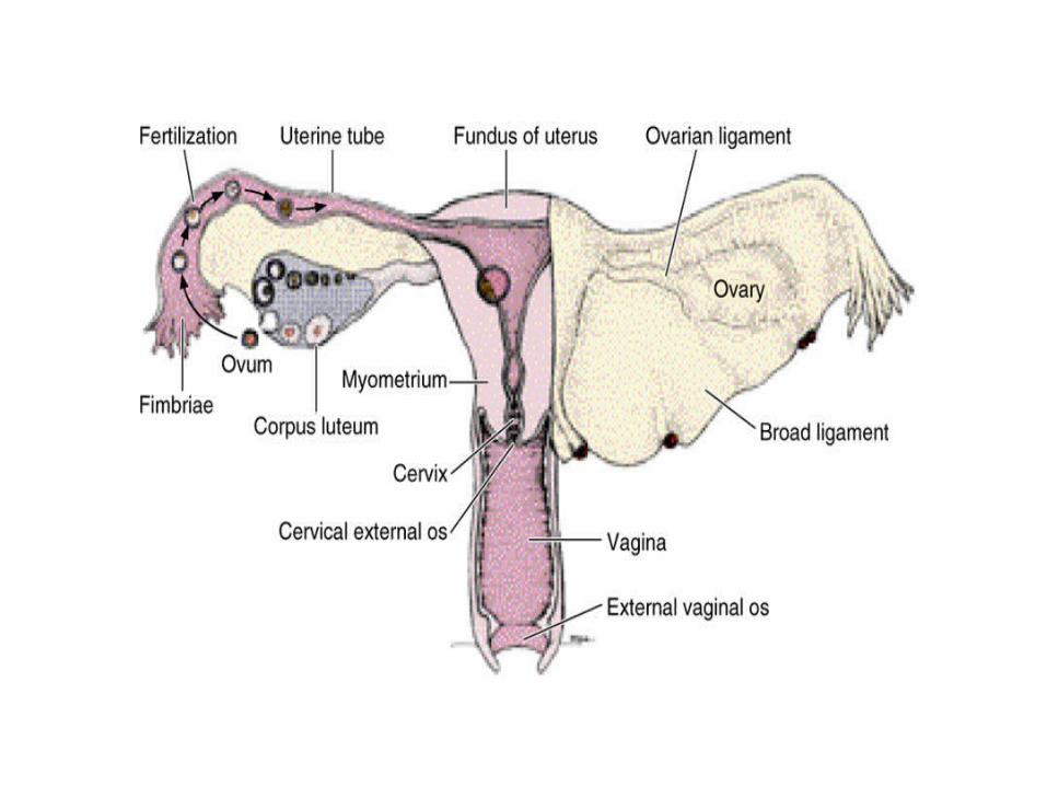

Broad Ligament
• Fold of peritoneum that encloses uterus
• Extends from side of uterus to lateral pelvic wall and floor
• Uterine tubes found in free edge
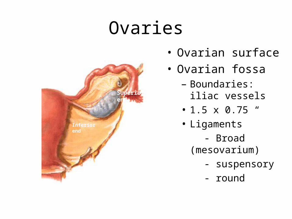
Ovaries• Ovarian surface• Ovarian fossa
– Boundaries: iliac vessels
• 1.5 x 0.75 “
• Ligaments
- Broad (mesovarium)
- suspensory
- round
Superiorend
Inferiorend
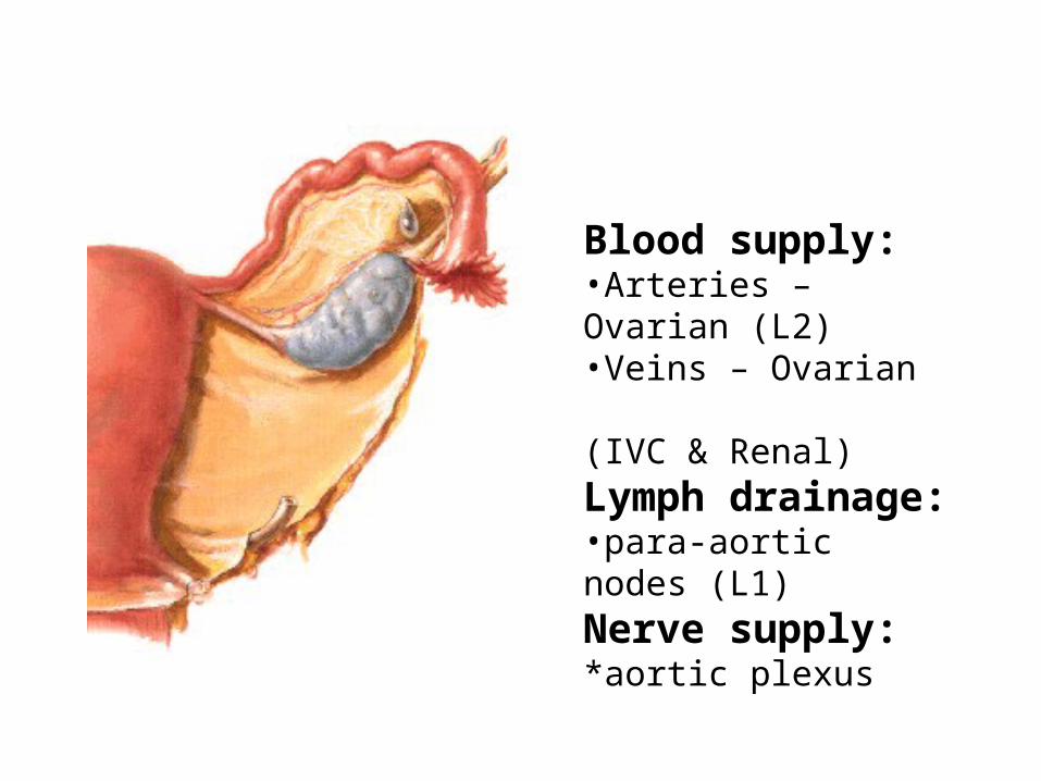
Blood supply:•Arteries – Ovarian (L2)•Veins – Ovarian (IVC & Renal)Lymph drainage:•para-aortic nodes (L1)Nerve supply:*aortic plexus
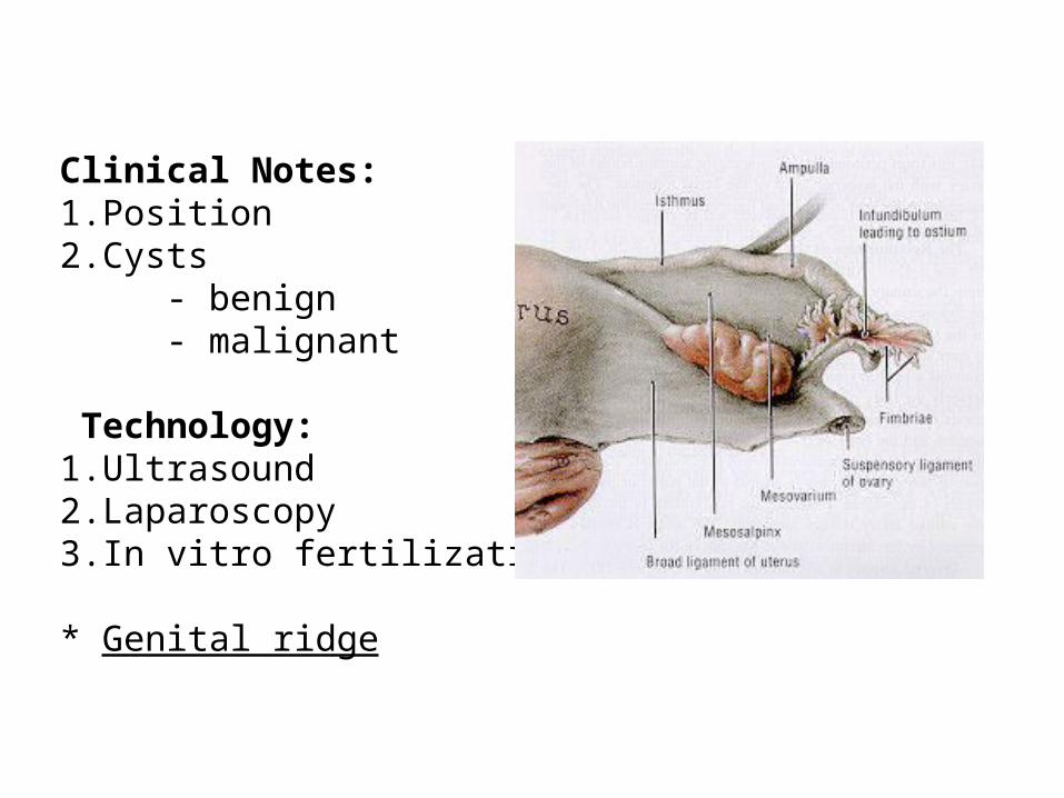
Clinical Notes:1.Position2.Cysts
- benign- malignant
Technology: 1.Ultrasound2.Laparoscopy3.In vitro fertilization
* Genital ridge
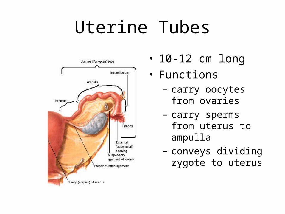
Uterine Tubes
• 10-12 cm long• Functions
– carry oocytes from ovaries
– carry sperms from uterus to ampulla
– conveys dividing zygote to uterus
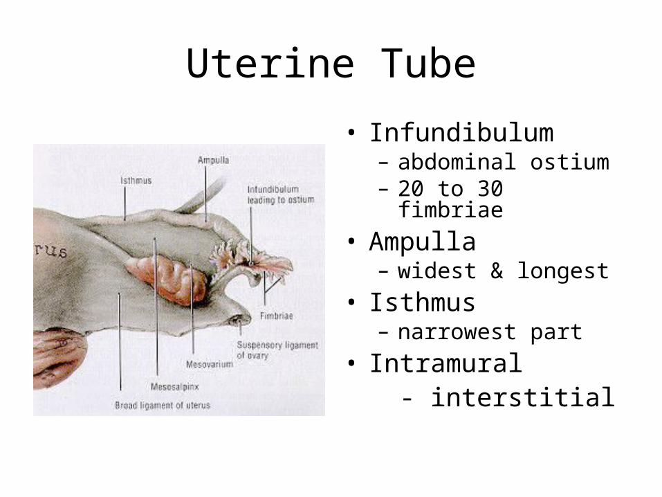
Uterine Tube
• Infundibulum– abdominal ostium– 20 to 30 fimbriae
• Ampulla– widest & longest
• Isthmus– narrowest part
• Intramural - interstitial
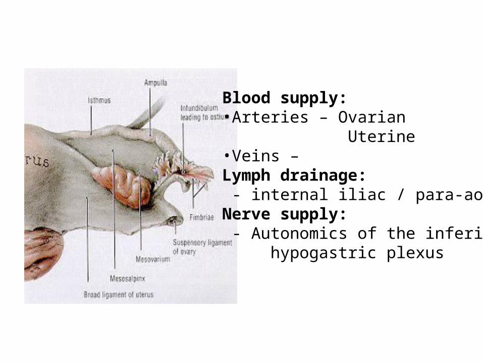
Blood supply:•Arteries – Ovarian
Uterine•Veins – Lymph drainage: - internal iliac / para-aorticNerve supply: - Autonomics of the inferior hypogastric plexus
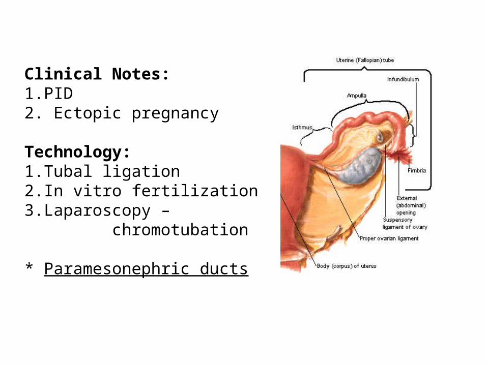
Clinical Notes:1.PID2. Ectopic pregnancy
Technology:1.Tubal ligation2.In vitro fertilization3.Laparoscopy – chromotubation
* Paramesonephric ducts
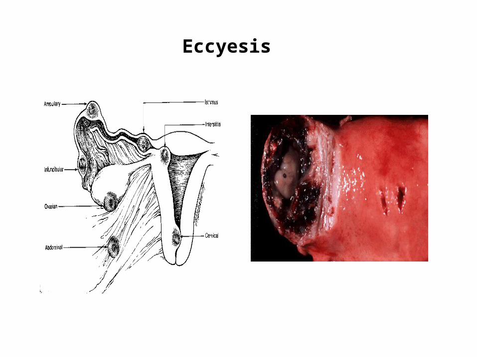
Eccyesis

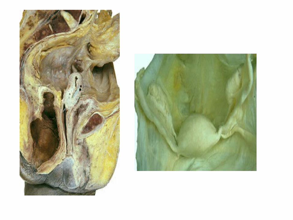
Uterus
• Located between bladder and rectum
• Anterior – bladder• Posterior – rectum• Lateral – adnexae
Vesicouterinepouch
Rectouterinepouch

Uterus
• Two main portions– body - superior two
thirds– cervix - inferior third
• Isthmus– junction between the
body and cervix
• Fundus• Cornua
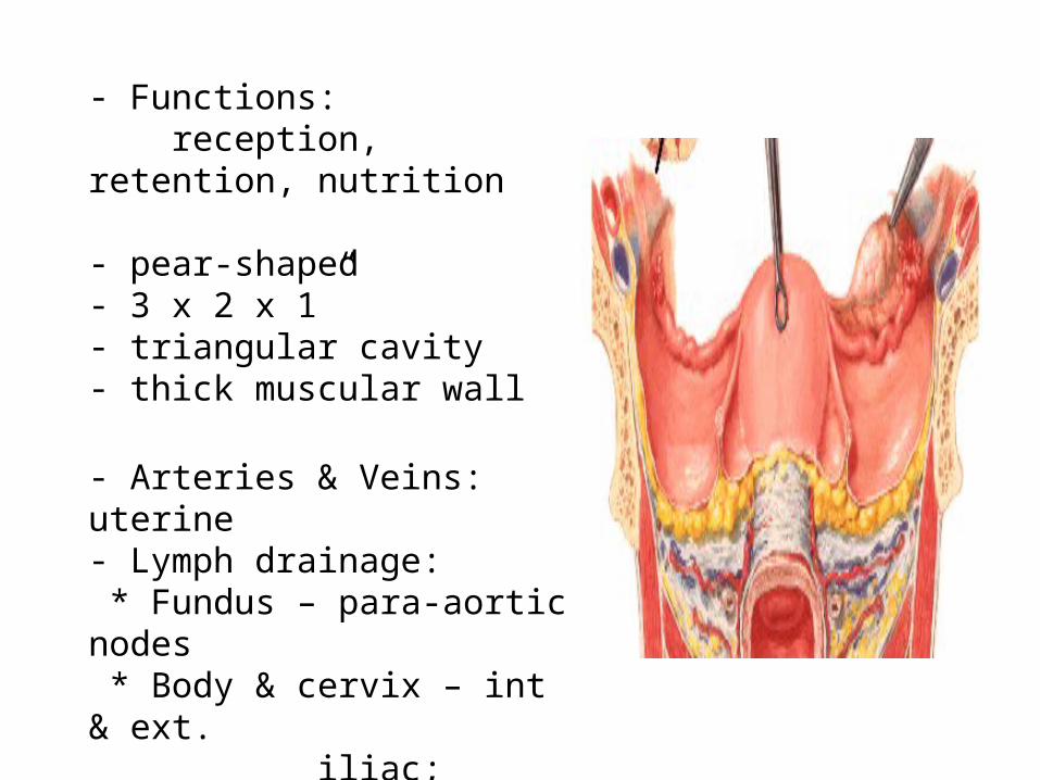
- Functions: reception, retention, nutrition
- pear-shaped- 3 x 2 x 1”- triangular cavity - thick muscular wall
- Arteries & Veins: uterine- Lymph drainage: * Fundus – para-aortic nodes * Body & cervix – int & ext. iliac; superficial inguinal nodes * Nerve supply – as the tubes

Uterine positions
Vesicouterinepouch
Rectouterinepouch
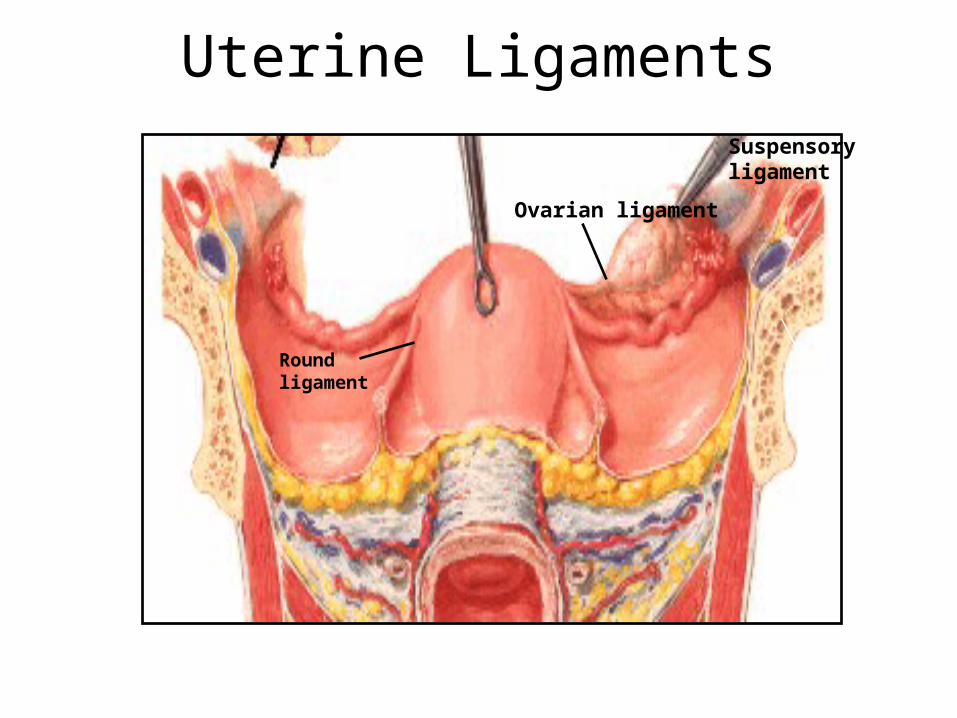
Uterine Ligaments
Ovarian ligament
Suspensory ligament
Round ligament

Uterine Support
• Pelvic Diaphragm– levator ani muscles– coccygeus
• Urogenital diaphragm• Cardinal ligament• Uterosacral ligament• Pubocervical ligament
LEVATORANI
UG DIAPH
FASCIA
< OBT. INT.
< PERINEAL BODY

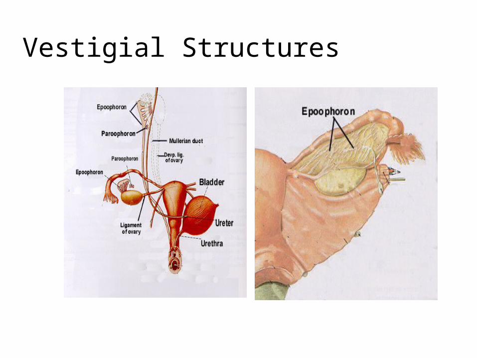
Vestigial Structures
Clinical Correlations:1.Uterus at different ages 2.Pregnancy & labor
Cesarean section3. Pelvic examination4. Conditions:
* Prolapse / Procidentia* Leiomyoma* Malignancy * Embryological
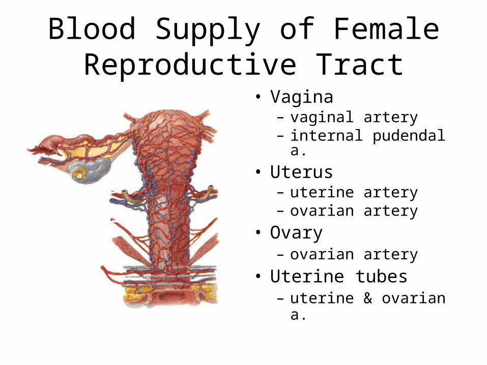
Blood Supply of Female Reproductive Tract
• Vagina– vaginal artery– internal pudendal a.
• Uterus– uterine artery– ovarian artery
• Ovary– ovarian artery
• Uterine tubes– uterine & ovarian a.
Vaginal a.
Internal pudendal a.
Uterine a.
Ovarian a.
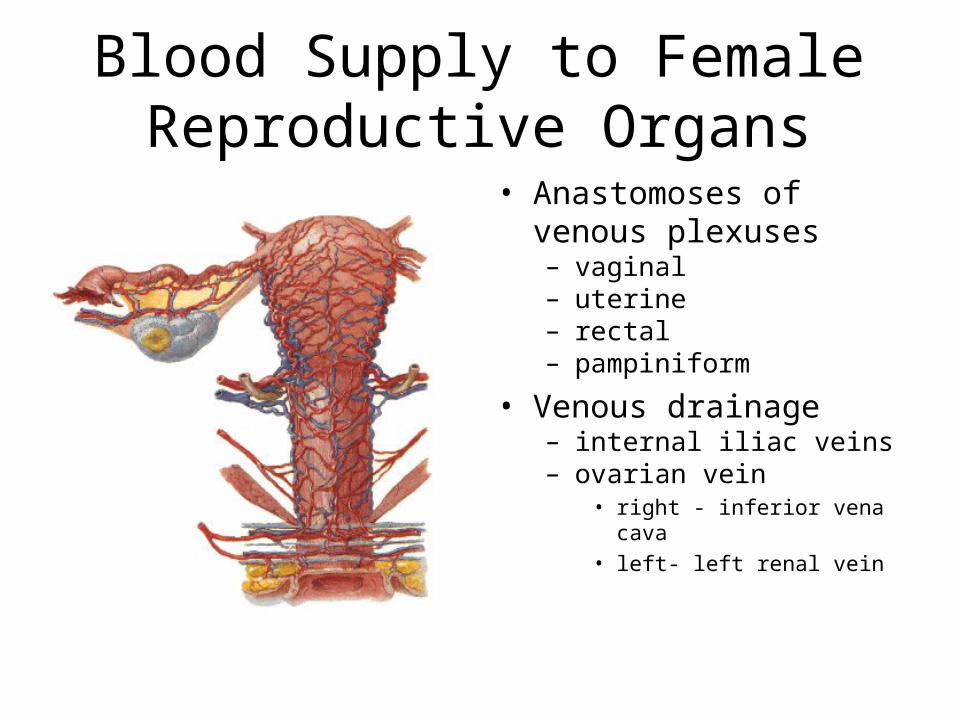
Blood Supply to FemaleReproductive Organs
• Anastomoses of venous plexuses– vaginal– uterine– rectal– pampiniform
• Venous drainage– internal iliac veins– ovarian vein
• right - inferior vena cava• left- left renal vein
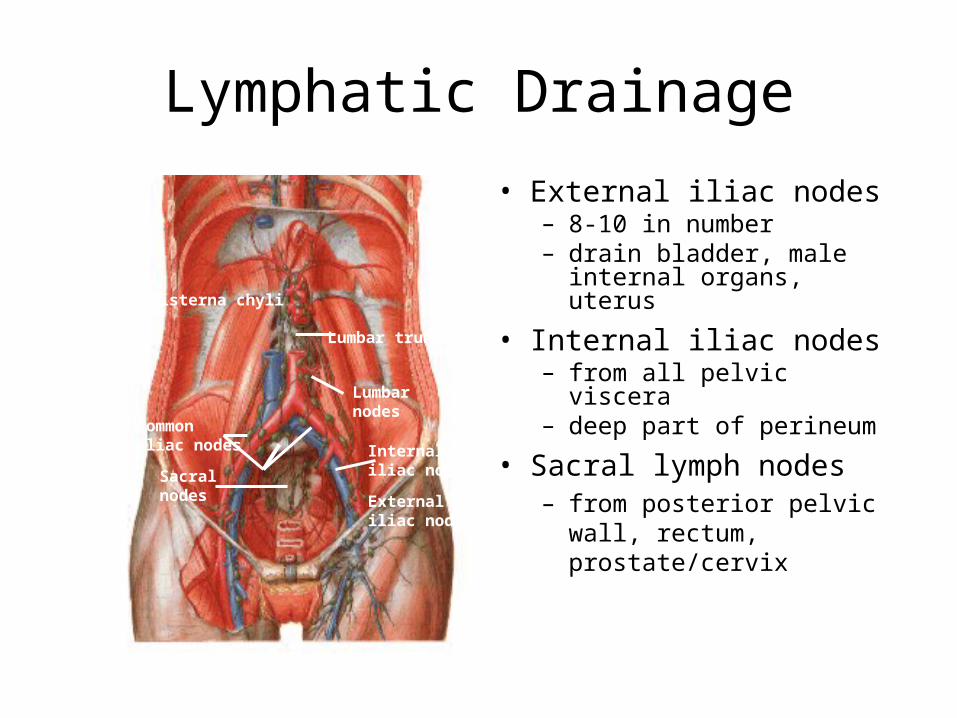
Lymphatic Drainage
• External iliac nodes– 8-10 in number– drain bladder, male internal
organs, uterus
• Internal iliac nodes– from all pelvic viscera– deep part of perineum
• Sacral lymph nodes– from posterior pelvic wall,
rectum, prostate/cervixExternaliliac nodes
Internaliliac nodes
Commoniliac nodes
Lumbarnodes
Sacral nodes
Cisterna chyli
Lumbar trunk

Lymphatic Drainage
• Common iliac nodes– lateral group
• common iliac vessels• lymph from external &
internal nodes– median group
• angle between vessels• lymph directly from
pelvic viscera
• Lumbar aortic nodes– along abdominal aorta– lymph from common iliac
nodes , fundus of uterus, ovary & tubes
Externaliliac nodes
Internaliliac nodes
Commoniliac nodes
Lumbarnodes
Sacral nodes
Cisterna chyli
Lumbar trunk
Lymphatic Drainage
Externaliliac nodes
Internaliliac nodes
Commoniliac nodes
Lumbarnodes
Sacral nodes
Cisterna chyli
Lumbar trunk
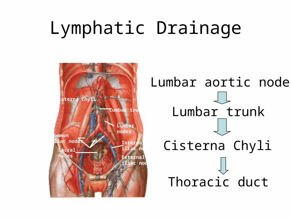
Lumbar aortic nodes
Lumbar trunk
Cisterna Chyli
Thoracic duct
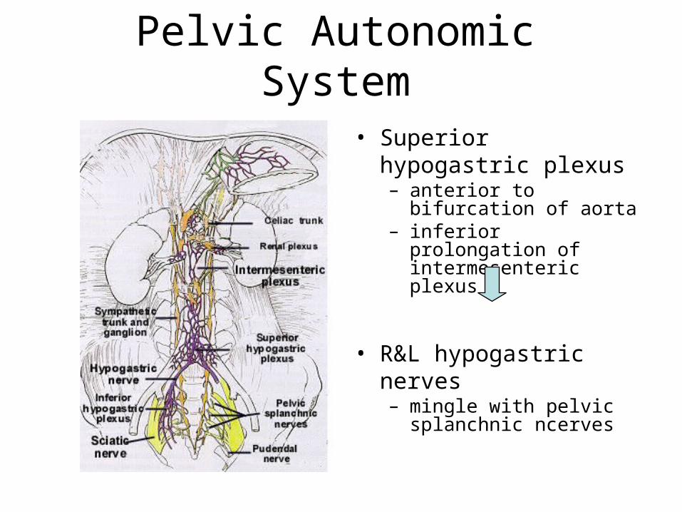
Pelvic Autonomic System
• Superior hypogastric plexus– anterior to bifurcation of
aorta– inferior prolongation of
intermesenteric plexus
• R&L hypogastric nerves– mingle with pelvic
splanchnic ncerves
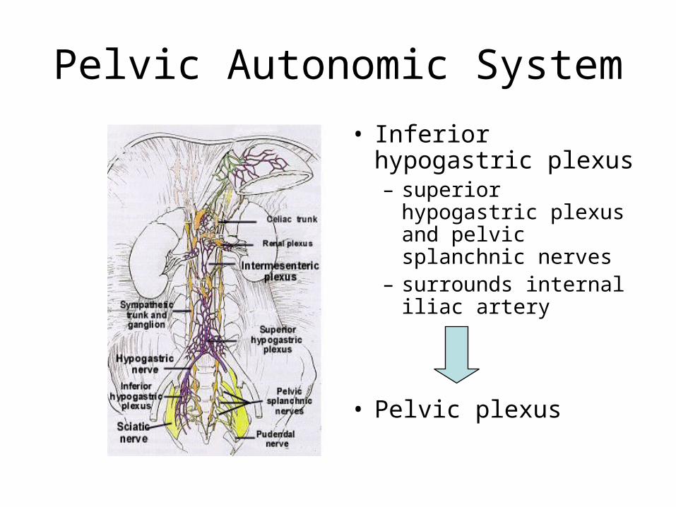
Pelvic Autonomic System
• Inferior hypogastric plexus– superior hypogastric
plexus and pelvic splanchnic nerves
– surrounds internal iliac artery
• Pelvic plexus

Pelvic Plexus
• Middle rectal plexus– innervates rectum
• Vesical plexus– innervates urinary
bladder
• Prostatic plexus– innervates male
internal reproductive organs
• Uterovaginal plexus
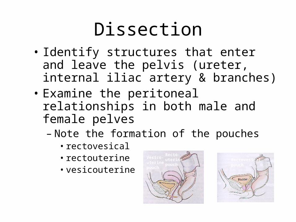
Dissection• Identify structures that enter and leave
the pelvis (ureter, internal iliac artery & branches)
• Examine the peritoneal relationships in both male and female pelves– Note the formation of the pouches
• rectovesical• rectouterine• vesicouterine
Vesico-uterinepouch
Recto-uterinepouch
Rectovesicalpouch

Dissection
– Identify all the different ligaments of each individual pelvis that can be visualized
• posterior surface of anterior abdominal wall• females
– uterosacral and cardinal ligaments– broad , round, suspensory ligament

Dissection
• Remove the peritoneum from the pelvic cavity and inspect the pelvic viscera – Pull the apex of the bladder upward and backward to easily
detach the bladder from the pelvic wall exposing the retropubic space
• With a bone saw cut through the symphysis pubis and retract it laterally
• Cut through the midline of the bladder (Sagittal section) Inspect bladder wall

Related Documents