Published: March 29, 2011 r2011 American Chemical Society 1167 dx.doi.org/10.1021/ct100576m | J. Chem. Theory Comput. 2011, 7, 1167–1176 ARTICLE pubs.acs.org/JCTC GRIFFIN: A Versatile Methodology for Optimization of Protein-Lipid Interfaces for Membrane Protein Simulations Ren e Staritzbichler, † Claudio Anselmi, ‡ Lucy R. Forrest,* ,† and Jos e D. Faraldo-G omez* ,‡,§ † Computational Structural Biology Group and ‡ Theoretical Molecular Biophysics Group, Max Planck Institute of Biophysics, Frankfurt am Main, Germany § Cluster of Excellence Macromolecular Complexes, Frankfurt am Main, Germany ABSTRACT: As new atomic structures of membrane proteins are resolved, they reveal increasingly complex transmembrane topologies and highly irregular surfaces with crevices and pores. In many cases, specific interactions formed with the lipid membrane are functionally crucial, as is the overall lipid composition. Compounded with increasing protein size, these characteristics pose a challenge for the construction of simulation models of membrane proteins in lipid environments; clearly, that these models are sufficiently realistic bears upon the reliability of simulation-based studies of these systems. Here, we introduce GRIFFIN (GRId- based Force Field INput), which uses a versatile framework to automate and improve a widely used membrane-embedding protocol. Initially, GRIFFIN carves out lipid and water molecules from a volume equivalent to that of the protein, to conserve the system density. In the subsequent optimization phase GRIFFIN adds an implicit grid-based protein force field to a molecular dynamics simulation of the precarved membrane. In this force field, atoms inside the implicit protein volume experience an outward force that will expel them from that volume, whereas those outside are subject to electrostatic and van der Waals interactions with the implicit protein. At each step of the simulation, these forces are updated by GRIFFIN and combined with the intermolecular forces of the explicit lipid-water system. This procedure enables the construction of realistic and reproducible starting configurations of the protein-membrane interface within a reasonable time frame and with minimal intervention. GRIFFIN is a stand-alone tool designed to work alongside any existing molecular dynamics package, such as NAMD or GROMACS. ’ INTRODUCTION Membrane proteins constitute around a third of all proteins encoded in a typical genome, 1-3 and yet the microscopic mechanisms of their functions are only just beginning to be described. Major advances in such understanding have been made through the elucidation of the three-dimensional atomic structure of some of these proteins by, e.g., X-ray crystallography. Indeed, the exponential increase in the number of structures being reported 4 highlights the very significant progress made in this area. However, crystallographic structures capture a single state of what is typically a dynamic conformational equilibrium, intrinsic to the functional mechanism of the protein. A variety of structure-based computational approaches focus on these dynamic properties, in order to complement the experimental data. Prominent among these approaches is molecular dynamics (MD) simulation, for its ability to provide insights at atomic resolution, and its robust and versatile theore- tical framework. A surprising feature of many of the newly discovered mem- brane protein structures is their complex transmembrane topol- ogy, and the highly irregular interfaces they appear to form with the surrounding lipid bilayer. Indeed, for some proteins, such as the voltage-gated and mechanosensitive channels, or osmoregu- latory transporters, regulatory mechanisms dependent on lipid composition are likely to be conveyed precisely by this protein- lipid interface. 5-8 As the structure of the protein-membrane interface is not known experimentally, such complexity poses a serious challenge for the construction of molecular-simulation models. At the same time, whether this interface is realistically modeled will clearly influence the ability of simulation-based studies to derive plausible mechanistic hypotheses. A number of methodologies have been proposed for the construction of lipid-protein complexes for simulation. 9-14 These adopt one of two general strategies, namely either to assemble a lipid bilayer around the protein de novo, or to adapt existing and well-optimized membrane models to the protein structure. The second strategy is an appealing and efficient option, because it builds upon much effort and considerable success in the construction of realistic membrane models for MD simulations, for a range of different lipids or mixtures thereof. Atomic coordinates and force fields for such systems are publicly available. In one of these adaptive strategies, the protein and hydrated lipid bilayer systems are first superimposed in the same coordinate space, and some overlapping lipids and waters are removed, as necessary to preserve the system density. 12,14 However, the cavity thus carved in the bilayer system does not normally match the shape of the protein, because of the very irregular conformations adopted by lipid hydrocarbon tails. To resolve this problem, one of the authors helped develop a methodology 12 in which the surface of the protein is used in the course of a molecular dynamics simulation to define additional forces that expel these tails from the protein volume. In this way, lipid and solvent atoms become adapted perfectly to the shape of the protein, and there is minimal perturbation of the existing, preoptimized membrane model. Received: October 7, 2010

Welcome message from author
This document is posted to help you gain knowledge. Please leave a comment to let me know what you think about it! Share it to your friends and learn new things together.
Transcript

Published: March 29, 2011
r 2011 American Chemical Society 1167 dx.doi.org/10.1021/ct100576m | J. Chem. Theory Comput. 2011, 7, 1167–1176
ARTICLE
pubs.acs.org/JCTC
GRIFFIN: A Versatile Methodology for Optimization of Protein-LipidInterfaces for Membrane Protein SimulationsRen�e Staritzbichler,† Claudio Anselmi,‡ Lucy R. Forrest,*,† and Jos�e D. Faraldo-G�omez*,‡,§
†Computational Structural Biology Group and ‡Theoretical Molecular Biophysics Group, Max Planck Institute of Biophysics, Frankfurtam Main, Germany§Cluster of Excellence Macromolecular Complexes, Frankfurt am Main, Germany
ABSTRACT: As new atomic structures of membrane proteins are resolved, they reveal increasingly complex transmembranetopologies and highly irregular surfaces with crevices and pores. In many cases, specific interactions formed with the lipid membraneare functionally crucial, as is the overall lipid composition. Compounded with increasing protein size, these characteristics pose achallenge for the construction of simulation models of membrane proteins in lipid environments; clearly, that these models aresufficiently realistic bears upon the reliability of simulation-based studies of these systems. Here, we introduce GRIFFIN (GRId-based Force Field INput), which uses a versatile framework to automate and improve a widely usedmembrane-embedding protocol.Initially, GRIFFIN carves out lipid and water molecules from a volume equivalent to that of the protein, to conserve the systemdensity. In the subsequent optimization phase GRIFFIN adds an implicit grid-based protein force field to a molecular dynamicssimulation of the precarved membrane. In this force field, atoms inside the implicit protein volume experience an outward force thatwill expel them from that volume, whereas those outside are subject to electrostatic and van der Waals interactions with the implicitprotein. At each step of the simulation, these forces are updated by GRIFFIN and combined with the intermolecular forces of theexplicit lipid-water system. This procedure enables the construction of realistic and reproducible starting configurations of theprotein-membrane interface within a reasonable time frame and with minimal intervention. GRIFFIN is a stand-alone tooldesigned to work alongside any existing molecular dynamics package, such as NAMD or GROMACS.
’ INTRODUCTION
Membrane proteins constitute around a third of all proteinsencoded in a typical genome,1-3 and yet the microscopicmechanisms of their functions are only just beginning to bedescribed. Major advances in such understanding have beenmade through the elucidation of the three-dimensional atomicstructure of some of these proteins by, e.g., X-ray crystallography.Indeed, the exponential increase in the number of structuresbeing reported4 highlights the very significant progress madein this area. However, crystallographic structures capture asingle state of what is typically a dynamic conformationalequilibrium, intrinsic to the functional mechanism of the protein.A variety of structure-based computational approaches focuson these dynamic properties, in order to complement theexperimental data. Prominent among these approaches ismolecular dynamics (MD) simulation, for its ability to provideinsights at atomic resolution, and its robust and versatile theore-tical framework.
A surprising feature of many of the newly discovered mem-brane protein structures is their complex transmembrane topol-ogy, and the highly irregular interfaces they appear to form withthe surrounding lipid bilayer. Indeed, for some proteins, such asthe voltage-gated and mechanosensitive channels, or osmoregu-latory transporters, regulatory mechanisms dependent on lipidcomposition are likely to be conveyed precisely by this protein-lipid interface.5-8 As the structure of the protein-membraneinterface is not known experimentally, such complexity poses aserious challenge for the construction of molecular-simulationmodels. At the same time, whether this interface is realistically
modeled will clearly influence the ability of simulation-basedstudies to derive plausible mechanistic hypotheses.
A number of methodologies have been proposed for theconstruction of lipid-protein complexes for simulation.9-14
These adopt one of two general strategies, namely either toassemble a lipid bilayer around the protein de novo, or to adaptexisting and well-optimized membrane models to the proteinstructure. The second strategy is an appealing and efficientoption, because it builds upon much effort and considerablesuccess in the construction of realistic membrane models for MDsimulations, for a range of different lipids or mixtures thereof.Atomic coordinates and force fields for such systems are publiclyavailable.
In one of these adaptive strategies, the protein and hydrated lipidbilayer systems are first superimposed in the same coordinate space,and some overlapping lipids andwaters are removed, as necessary topreserve the system density.12,14 However, the cavity thus carved inthe bilayer systemdoes not normallymatch the shape of the protein,because of the very irregular conformations adopted by lipidhydrocarbon tails. To resolve this problem, one of the authorshelped develop a methodology12 in which the surface of the proteinis used in the course of a molecular dynamics simulation to defineadditional forces that expel these tails from the protein volume. Inthis way, lipid and solvent atoms become adapted perfectly to theshape of the protein, and there is minimal perturbation of theexisting, preoptimized membrane model.
Received: October 7, 2010

1168 dx.doi.org/10.1021/ct100576m |J. Chem. Theory Comput. 2011, 7, 1167–1176
Journal of Chemical Theory and Computation ARTICLE
Here, we present an improved andmore general version of thissurface-based approach, based upon a newly developed toolnamed GRIFFIN (GRId-based Force Field INput). GRIFFINprovides several significant advantages over the earlier imple-mentation. First, forces acting on lipid atoms due to Coulomband van der Waals interactions with the protein volume are nowincluded along with the repulsive surface-guided forces; thisresults in chemical specificity at the protein-lipid interface, inaddition to shape complementarity. Second, new features areincluded to selectively guide lipids or other molecules into or outof user-specified regions of the system, and also to preventtrapping of lipid tails in regions of high irregularity. Thesefeatures thus provide a means to handle the kind of complexprotein topologies increasingly uncovered by structural studies.
Third, GRIFFIN is designed to be a stand-alone, fully integratedapplication, compatible with anyMDengine that provides a suitableinterface, such as NAMD,15 GROMACS,16 or CHARMM.17 Last,its object-oriented grid-based algorithm, and the resulting computa-tional efficiency, extends the applicability of themethod to very largemembrane protein complexes.
We demonstrate the effectiveness of this improved methodol-ogy with four examples, namely, the secondary transporterCaiT,18 two membrane rotors from the ATPase family,19,20
and the AcrB multidrug efflux pump21 (Figure 1). In all cases,irregular features of the protein-lipid interface make otherapproaches either unsuitable or impractical. These featuresinclude a central pore in the ATPase rings, and intersubunitchannels and water-filled crevices in the trimeric CaiT and AcrB;AcrB is also one of the largest and most complex membraneproteins structures resolved to date. We will show howGRIFFINenables the construction of physically realistic starting config-urations of the lipid-protein interface for membrane proteinsimulations, following a reproducible procedure and within anaffordable time frame, even for these very challenging cases.
’METHODOLOGY
The overall protocol is described in the scheme in Figure 2.Initially, a cavity is carved into a hydrated lipid bilayer system, inwhich the membrane protein will ultimately be embedded. Theprotein structure, or rather, its surface, is used to estimate thenumber of lipid and water molecules to be carved out. In mostcases, however, the resulting protein-lipid interface will beunsuitable for energy minimization, let alone simulation. Thesame surface is therefore employed to define a grid-based forcefield designed to expel the remaining lipid and/or water atomsfrom that volume. This force field also includes physical interac-tions (electrostatic and van der Waals), but only in the region ofthe grid outside the protein volume. During a subsequentmolecular dynamics simulation of the precarved membranesystem, the implicit protein force field is added to the standardinteraction forces, thus optimizing the protein-lipid and pro-tein-water interfaces. Geometric objects may be used as addi-tional, lipid and/or water specific exclusion volumes, during thecarving and/or the simulation.Initial Carving of the Lipid Membrane. GRIFFIN provides
an integrated tool for constructing the surface of the protein andfor the initial carving of the membrane. The surface is con-structed using a rolling-sphere method,22 with a probe ofadjustable radius, typically set to 1.4 Å. Atomic radii are derivedfrom the force field to be used in the simulation, e.g.,CHARMM27.23 The protein surface is used to estimate thevolume occupied by the protein within each leaflet of the lipidmembrane, as well in the hydration layer. The boundaries ofthese regionsmay be user-defined or determined by the program.Based on this volume estimation and the lipid density, thenumber of lipid molecules to be deleted from each leaflet isdetermined. These are selected from a list of all lipids that overlapwith the protein volume, ranked according to the number ofoverlapping atoms and their distance to the protein surface. Alloverlapping water molecules are also deleted. Specific values forthese parameters may be also provided by the user, overridingthose calculated by the program.Additional Molecule Type Specific Exclusion Volumes.
GRIFFIN offers the possibility of using geometric objects(spheres, rectangular cuboids, and cones) as additional exclusion
Figure 1. Representative membrane proteins with complex protein-lipid interfaces, used here as test cases for GRIFFIN. Each structure isdisplayed in cartoon format with different colors for each proteinsubunit, and viewed either from the plane of the membrane (upperpanels) or normal to the membrane (lower panels). The approximatelimits of the hydrophobic core of a hypothetical lipid membrane are alsoindicated (green lines). (A) The K10 rotor ring of the V-type Naþ-ATPase from Enterococcus hirae. (B) The c11 rotor ring of the F-typeNaþ-ATP synthase from Ilyobacter tartaricus. (C) The carnitine/γ-butyrobetaine antiporter CaiT from Proteus mirabilis. (D) The multi-drug efflux pump AcrB from E. coli.

1169 dx.doi.org/10.1021/ct100576m |J. Chem. Theory Comput. 2011, 7, 1167–1176
Journal of Chemical Theory and Computation ARTICLE
regions in the membrane carving, as well as in the optimizationstage (see below). These objects are integrated in the volumeestimation described above, andmay be specific to a user-definedmolecule type, e.g., to exclude lipid from a channel pore.Calculation of the Implicit Protein Force Field. A second
tool within GRIFFIN enables the calculation of the implicit forcefield for use during the subsequent molecular dynamics optimi-zation stage. This force field is stored on a three-dimensional grid(Figure 3) of user-defined grid-point spacing (with a defaultvalue of 0.5 Å). Within the protein volume (which may bemodified to include theoptional exclusion regionsmentioned above),each grid point stores a force (of magnitude 1 kcal/mol/Å) directedtoward the nearest point on the protein surface (NPS; Figure 4A).These so-called surface forces will be applied to lipid and wateratoms inside the volume during the optimization. Grid pointsoutside of the protein surface map the Coulombic and van derWaals interaction forces between the protein and a probe particleof q = 1e, ε = 1 kcal/mol, and σ = 1 Å (Figure 3). User-definedcutoff distances may be specified for each of these interactions(default values are 18, 12, and 8 Å, respectively). The calculationof the force grid is carried out only once during the protocol;however, as it may be time-consuming, this calculation may bedistributed over an adjustable number of processors.Calculation of GRIFFIN Forces duringMolecular Dynamics
Simulations. GRIFFIN supplies a molecular dynamics simula-tion with atomic forces derived from the precomputed grid.These are calculated at every simulation step, based on theatomic coordinates of the explicit system (Figure 3F). To mapthe grid-point forces onto the actual atoms, GRIFFIN uses atrilinear interpolation method. By default, the physical forcesare scaled by the actual charge and van der Waals parameters ofeach atom (a geometric-mean combination rule is used for both
σ and ε). Surface forces may also be scaled by a constant; inpractice this constant is typically increased in a stepwisemanner, as the surface forces ultimately become balanced outby the external pressure (see Results). A user-specified constantfactor may also be used to further scale each of the physicalinteraction forces.To improve the performance and stability of the algorithm, no
surface forces are applied to hydrogen atoms by default. Bycontrast, all atoms may experience physical forces if outside theprotein volume. However, for greater efficiency, physical inter-actions are computed only for atoms neighboring the protein; alist of such atoms is updated every few steps during the simula-tion (typically 10), and includes all atoms for which the GRIF-FIN force is nonzero at the update step. Lastly, it should be notedthat by construction no reaction forces on the protein areconsidered; it is thus advisable that GRIFFIN is employedalongside stochastic dynamics, with tight temperature and pres-sure coupling.A feature specific to lipid molecules is the possibility of
redirection of the surface forces. This feature is important whenatoms within the same lipid chain are driven in oppositedirections (see Results). Redirection is applied when the surfaceforce acting on a given atom is directed away from the overallgeometric center of the lipid molecule to which it belongs(Figure 4). An adjustable threshold defines the maximum angleby which the surface force may diverge; this is typically <110�.Simulation Details. All GRIFFIN optimizations described
here were carried out with NAMD 2.715 with the CHARMM27protein/lipid force field.23 The lipid types used were palmitoyloleyl phosphatidylcholine (POPC; 234 for c11, 542 for K10, and532 for AcrB) and palmitoyl oleyl phosphatidylethanolamine(POPE; 470 for CaiT). The simulations were carried out at
Figure 2. Schematic of the protocol for optimization of a protein-membrane interface using GRIFFIN. In the first step, a hypothetical membraneprotein with a central pore (top left) is overlaid with a cylindrical geometric object (orange). The combined volume (yellow) of the protein and object iscarved from an explicit system (bottom left) containing water (red) and lipidmolecules (hydrocarbon chains are colored green and polar head groups areshown as orange spheres). After carving, some lipid and water atoms typically overlap the protein volume. The combined exclusion volume, and theactual protein structure, are also used to construct a force grid (top right) consisting of surface-directed expelling forces inside the volume and physicalinteraction forces outside. This force grid is overlaid on themolecular system to compute, at each time step of amolecular dynamics simulation, the actualforces on individual atoms; these are then added to the interaction energies between explicit atoms computed by the molecular dynamics package. In thelast step, the GRIFFIN-optimized hydrated membrane is merged with the atomistic model of the membrane protein, and the entire system is energy-minimized to obtain the initial configuration for simulation (bottom right).
1170 dx.doi.org/10.1021/ct100576m |J. Chem. Theory Comput. 2011, 7, 1167–1176
Journal of Chemical Theory and Computation ARTICLE
constant temperature (298 and 310 K for POPC and POPE,respectively), using a Langevin thermostat (collision frequencyof 100 ps-1). The pressure was maintained along the membranenormal using a Nos�e-Hoover Langevin piston barostat (1 atm),while the surface area of the membrane was kept constant.Electrostatic interactions were calculated using the particle meshEwald method (PME) with a real-space cutoff of 12 Å. A cutoffdistance of 12 Å was also used for the van der Waals interactions.The MD integration time step was 1 fs; bonds involvinghydrogen atoms were constrained with SETTLE. The numberof lipid/water atoms in each optimization was 80 172 for c11 ring,187 585 for K10 ring, 163 666 for CaiT, and 319 790 for AcrB.
’RESULTS
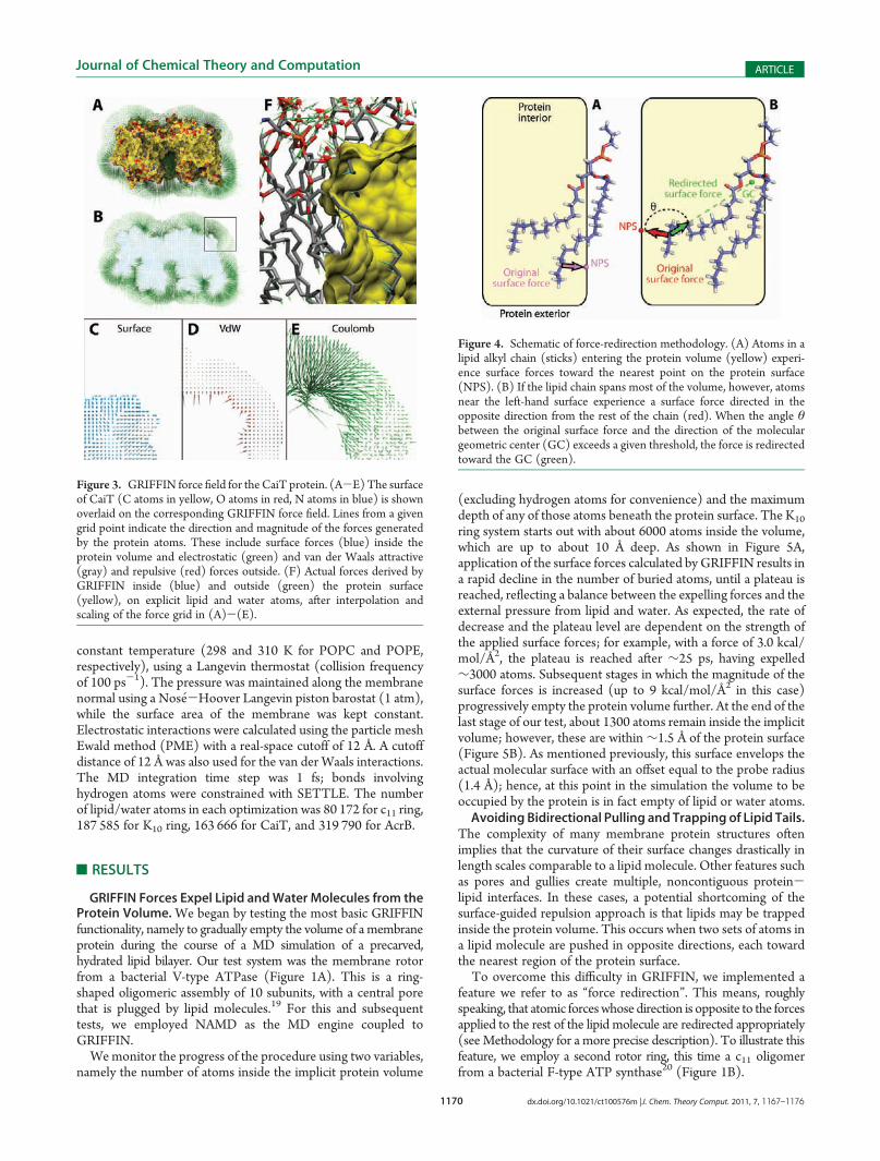
GRIFFIN Forces Expel Lipid andWater Molecules from theProtein Volume.We began by testing the most basic GRIFFINfunctionality, namely to gradually empty the volume of a membraneprotein during the course of a MD simulation of a precarved,hydrated lipid bilayer. Our test system was the membrane rotorfrom a bacterial V-type ATPase (Figure 1A). This is a ring-shaped oligomeric assembly of 10 subunits, with a central porethat is plugged by lipid molecules.19 For this and subsequenttests, we employed NAMD as the MD engine coupled toGRIFFIN.Wemonitor the progress of the procedure using two variables,
namely the number of atoms inside the implicit protein volume
(excluding hydrogen atoms for convenience) and the maximumdepth of any of those atoms beneath the protein surface. The K10
ring system starts out with about 6000 atoms inside the volume,which are up to about 10 Å deep. As shown in Figure 5A,application of the surface forces calculated by GRIFFIN results ina rapid decline in the number of buried atoms, until a plateau isreached, reflecting a balance between the expelling forces and theexternal pressure from lipid and water. As expected, the rate ofdecrease and the plateau level are dependent on the strength ofthe applied surface forces; for example, with a force of 3.0 kcal/mol/Å2, the plateau is reached after ∼25 ps, having expelled∼3000 atoms. Subsequent stages in which the magnitude of thesurface forces is increased (up to 9 kcal/mol/Å2 in this case)progressively empty the protein volume further. At the end of thelast stage of our test, about 1300 atoms remain inside the implicitvolume; however, these are within ∼1.5 Å of the protein surface(Figure 5B). As mentioned previously, this surface envelops theactual molecular surface with an offset equal to the probe radius(1.4 Å); hence, at this point in the simulation the volume to beoccupied by the protein is in fact empty of lipid or water atoms.Avoiding Bidirectional Pulling and Trapping of Lipid Tails.
The complexity of many membrane protein structures oftenimplies that the curvature of their surface changes drastically inlength scales comparable to a lipid molecule. Other features suchas pores and gullies create multiple, noncontiguous protein-lipid interfaces. In these cases, a potential shortcoming of thesurface-guided repulsion approach is that lipids may be trappedinside the protein volume. This occurs when two sets of atoms ina lipid molecule are pushed in opposite directions, each towardthe nearest region of the protein surface.To overcome this difficulty in GRIFFIN, we implemented a
feature we refer to as “force redirection”. This means, roughlyspeaking, that atomic forceswhose direction is opposite to the forcesapplied to the rest of the lipid molecule are redirected appropriately(see Methodology for a more precise description). To illustrate thisfeature, we employ a second rotor ring, this time a c11 oligomerfrom a bacterial F-type ATP synthase20 (Figure 1B).
Figure 3. GRIFFIN force field for the CaiT protein. (A-E)The surfaceof CaiT (C atoms in yellow, O atoms in red, N atoms in blue) is shownoverlaid on the corresponding GRIFFIN force field. Lines from a givengrid point indicate the direction and magnitude of the forces generatedby the protein atoms. These include surface forces (blue) inside theprotein volume and electrostatic (green) and van der Waals attractive(gray) and repulsive (red) forces outside. (F) Actual forces derived byGRIFFIN inside (blue) and outside (green) the protein surface(yellow), on explicit lipid and water atoms, after interpolation andscaling of the force grid in (A)-(E).
Figure 4. Schematic of force-redirection methodology. (A) Atoms in alipid alkyl chain (sticks) entering the protein volume (yellow) experi-ence surface forces toward the nearest point on the protein surface(NPS). (B) If the lipid chain spans most of the volume, however, atomsnear the left-hand surface experience a surface force directed in theopposite direction from the rest of the chain (red). When the angle θbetween the original surface force and the direction of the moleculargeometric center (GC) exceeds a given threshold, the force is redirectedtoward the GC (green).

1171 dx.doi.org/10.1021/ct100576m |J. Chem. Theory Comput. 2011, 7, 1167–1176
Journal of Chemical Theory and Computation ARTICLE
As shown in Figure 6A, several phospholipids in a membraneprecarved for this protein span most of the distance between theinner and outer surfaces of the ring. Application of surface forceswithout the redirection functionality fails to expel these lipid tailsfrom the protein volume; instead, as Figure 6B shows, the lipidtails are stretched in opposite directions. In this simple case, thesituation can be somewhat resolved by increasing the magnitudeof the surface forces (Figure 6D), as the lipid tail ultimatelycommits to one of the two surfaces. However, in general this willnot be the case; moreover, large surface forces (>3 kcal/mol/Å2)should be avoided in the first stages of the procedure, as theyotherwise result in unrealistic lipid-tail conformations, whichsubsequent simulations will be unlikely to correct.By contrast, the redirection of problematic atomic forces
rapidly resolves any conflicting lipid configurations, and allowsGRIFFIN to empty the protein volume gradually and control-lably (Figure 6C-E).GRIFFIN Generates Lipid-Protein Interfaces with Interac-
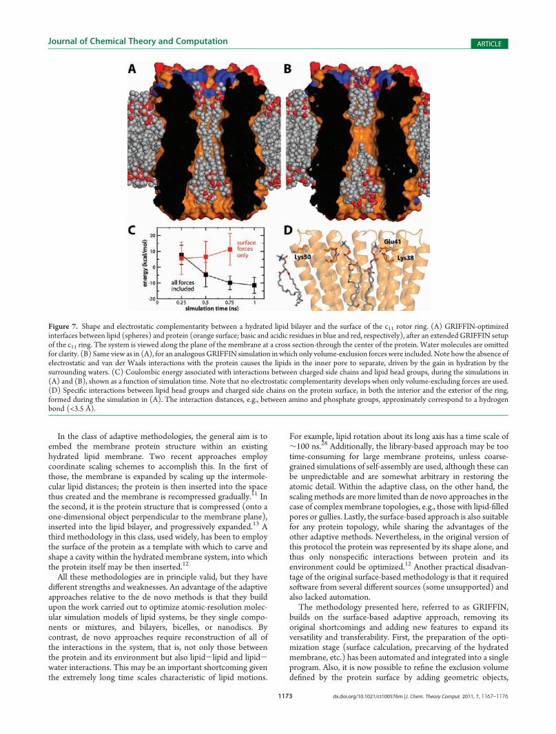
tion Specificity. An important improvement of GRIFFINcompared to its predecessor12 is that it attains specificity in theprotein-lipid interface, in addition to shape complementarity.To achieve this, the implicit protein force field overlaid on theexplicit membrane system includes both electrostatic and van derWaals interactions; these act on lipid and water atoms outside theprotein volume, in contrast to the surface forces, which onlyapply inside that volume (see Methodology; Figures 2 and 3).Figure 7 illustrates both the degree of shape complementarity
and the electrostatic specificity that may be attained with
GRIFFIN, using again the c11 rotor ring as an example. Therepulsive surface-directed forces alone cause the lipid moleculesto adapt neatly to the shape of the protein volume.12 However,the electrostatic interactions in particular help to guide lipidgroups to appropriate regions of the protein-membrane inter-face (Figure 7A cf. 7B), and thus reduce its overall electrostaticenergy (Figure 7C). As illustrated in Figure 7D, during thisoptimization specific interactions are formed, for example,between the (explicit) polar head groups and oppositely chargedatoms in the (implicit) protein. Arguably, that such interactionsare already present in the starting configuration of a simulation isadvantageous, as it implies that the subsequent equilibration ofthe system will be more realistic and efficient.Geometric Objects for Exclusion or Inclusion of Specific
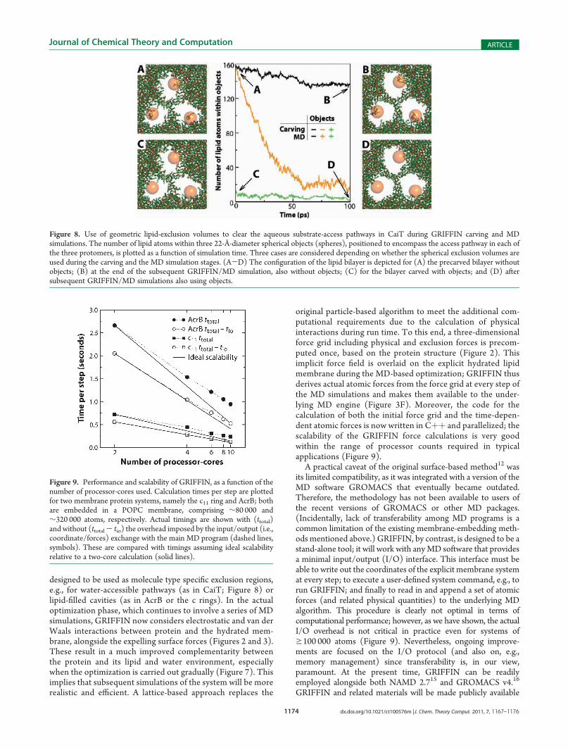
Molecule Types in Desired Locations. Transmembrane poresand access pathways are characteristic features of membraneproteins that function as channels or transporters; many otherscontain large crevices in the lipid interface (Figure 1). Such casespresent an additional challenge to the setup of a membraneprotein simulation, since lipid molecules must be excluded from,e.g., hydrated pores, while water should be excluded from, e.g., lipid-filled crevices. To overcome these difficulties, GRIFFIN supportsthe use of geometric objects that define additional, molecule-typespecific exclusion volumes. These objects are added to the proteinvolume during the initial membrane carving and/or during thesubsequent MD/GRIFFIN simulation (see Methodology).To illustrate this functionality, we consider as an example the
trimeric carnitine/γ-butyrobetaine antiporter CaiT from Proteusmirabilis,18 each protomer of which contains an aqueous vestibuleleading toward the center of the membrane (Figures 1C and 8).Without using any additional exclusion volumes during the initialmembrane carving, several lipid molecules are found inside theseaccess pathways, amounting to ∼160 atoms, not countinghydrogens (Figure 8A). Throughout a subsequent MD/GRIF-FIN simulation—also without the objects—the majority of theseatoms remain stubbornly in place (Figure 8B). If during the MDsimulation, however, a spherical exclusion object is overlaid oneach protomer so that it encompasses the access pathway(spheres, Figure 8), these regions are progressively emptied oflipid molecules (Figure 8, orange line). This result shows that thegeometric objects are indeed integrated correctly into the proteinvolume, and that the expelling forces are calculated accordingly.Nevertheless, it should be noted that to use these additionalexclusion volumes during the MD stage, but not the carvingstage, implies that the overall area per lipid will be too small,particularly in the vicinity of the protein interface. Thus, it isrecommended that the same objects be used also during theinitial carving of the membrane. In our example, this excludes allbut ∼15 of the lipid atoms originally within those volumes(Figure 8C). At the end of the subsequent MD/GRIFFINsimulation, the access pathways in CaiT are entirely clear of lipidmolecules (Figure 8D).Performance and Portability. GRIFFIN is designed to be
a stand-alone tool; i.e., it is intended to be compatible with anyMD package, provided a minimal input/output interface. Thisinterface, already included in, e.g., NAMD and GROMACS,allows for an external, accessory program (in this case GRIFFIN)to be executed at every time step of the simulation, to which thecurrent coordinates of themolecular system aremade available. Ifthe program returns a set of atomic forces (and related quantities,e.g., energy, virial, etc), these will be added to the main algorithmand reflected in the simulation.
Figure 5. Progressive expulsion of phospholipid and water moleculesfrom the volume of the K10 rotor ring due to GRIFFIN surface forces.(A) Number of atoms inside the protein volume, as a function ofsimulation time. Themagnitude of the expelling surface forces calculatedwith GRIFFIN is increased in stages, at the time points marked witharrows. (B) Maximum depth inside the protein surface of any atom, as afunction of simulation time. Hydrogen atoms are not considered in theseor subsequent plots.

1172 dx.doi.org/10.1021/ct100576m |J. Chem. Theory Comput. 2011, 7, 1167–1176
Journal of Chemical Theory and Computation ARTICLE
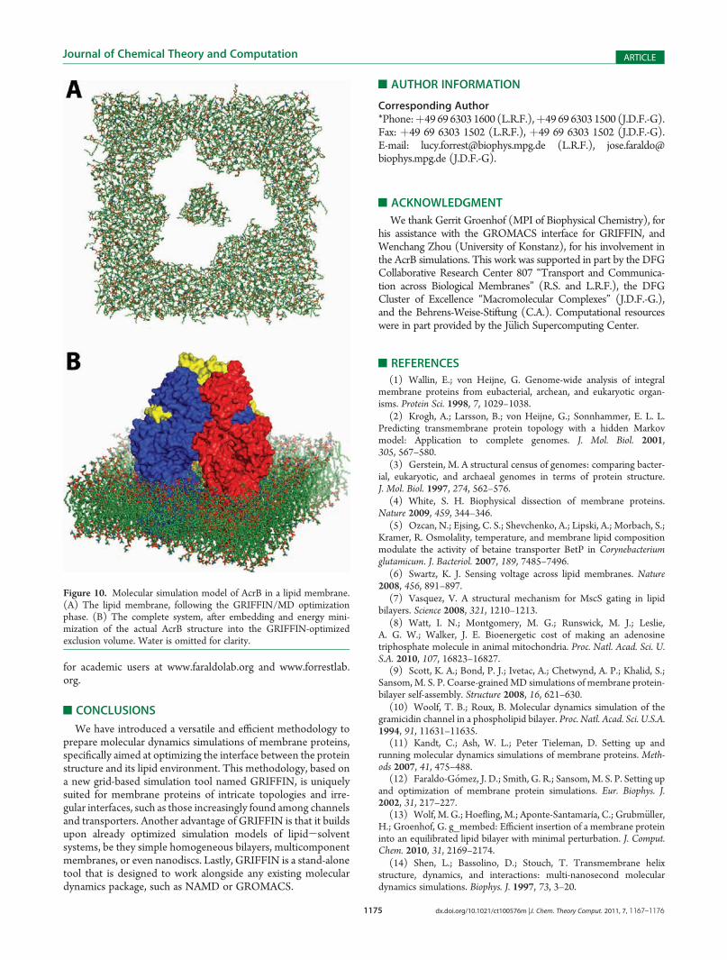
As described previously, GRIFFIN precomputes a three-dimensional force grid before the start of the optimization phase,based on the membrane protein structure and its interactionswith a probe particle positioned throughout the grid. During theGRIFFIN/MD simulation, the grid-point forces are interpolatedand scaled to yield actual atomic forces. As mentioned, the initialforce-grid calculation can be distributed among an adjustablenumber of computers, by portioning the grid accordingly. Thecomputation of GRIFFIN atomic forces during run time can alsobe parallelized, using anMPI-compatible version of the program.As shown in Figure 9, the scalability of the atomic-force calcula-tions is very good, especially for systems of about ∼100 000atoms, such as the c11 ring. This scalability is, however, limited;the limit is imposed by the time required to exchange coordinatesand forces with the MD software, which is a fixed overhead thatdepends on the computing infrastructure. Nevertheless, the totalexecution time, including the input/output of information, is stillsignificantly reduced as the number of processor increases, atleast within the range typically required. This allows GRIFFINoptimizations to be feasible within an affordable time frame evenfor very large systems such as AcrB, which comprises ∼300 000atoms (Figure 10).
’DISCUSSION
As new technologies lead to ever-growing computationalpower, and as algorithms improve in efficiency and accuracy,molecular dynamics simulations will be increasingly capable of
providing unique insights into the molecular mechanisms ofmembrane proteins. The strength of this theoretical tool—founded on statistical thermodynamics—lies in the atomicresolution at which the energetics and dynamics of themolecular system is simulated. These qualities, however, alsopose a challenge in practice, as in most cases the structure ofthe environment of the membrane protein of interest is notknown in atomic (or even molecular) detail. If this environ-ment is modeled unrealistically to begin with, the results of thesubsequent calculations will be questionable, as simulationsare not designed to overcome large systematic errors. There-fore, it is generally advisable that a simulation research projectcomprises an initial stage focused not on the membraneprotein but on the optimization of its lipid and solventenvironment.
Current methodologies for preparing membrane proteinsimulations fall in two classes: those in which a lipid mem-brane is constructed around the protein of interest de novo,and those in which an existing lipid membrane model issomehow adapted. In the former class, one method uses alibrary of lipid conformations (derived from simulations ofhydrated lipid bilayers) and progressively assembles indivi-dual molecules at designated locations around the protein;this is followed by a series of rigid-body rotations, translations,and energy minimizations.10,24-27 A second methodology inthis class involves coarse-grained self-assembly simulations ofa protein-lipid-water system, to which atomic detail isultimately added.9
Figure 6. Effect of force redirection on the expulsion of lipid tails from the volume of the c11 rotor ring. (A-C) Several configurations of the lipids(green sticks) in the lipid bilayer, viewed along the normal to the membrane. Hydrogen and water atoms are omitted for clarity. Snapshots are taken (A)after carving, at t = 0 ps, and after the first stage of the GRIFFIN simulations, at t = 100 ps, either (B) without or (C) with force redirection. (D) Themaximal depth inside the protein surface of any atom, as a function of simulation time. (E) Number of atoms inside the protein volume, as a function ofsimulation time. The magnitude of the forces was increased from 3.0 to 6.0 kcal/mol/Å2 (at t = 50 ps) and 9.0 kcal/mol/Å2 (at t = 100 ps). The forceredirection angle was set to 110� (see Methodology).
1173 dx.doi.org/10.1021/ct100576m |J. Chem. Theory Comput. 2011, 7, 1167–1176
Journal of Chemical Theory and Computation ARTICLE
In the class of adaptive methodologies, the general aim is toembed the membrane protein structure within an existinghydrated lipid membrane. Two recent approaches employcoordinate scaling schemes to accomplish this. In the first ofthose, the membrane is expanded by scaling up the intermole-cular lipid distances; the protein is then inserted into the spacethus created and the membrane is recompressed gradually.11 Inthe second, it is the protein structure that is compressed (onto aone-dimensional object perpendicular to the membrane plane),inserted into the lipid bilayer, and progressively expanded.13 Athird methodology in this class, used widely, has been to employthe surface of the protein as a template with which to carve andshape a cavity within the hydrated membrane system, into whichthe protein itself may be then inserted.12
All these methodologies are in principle valid, but they havedifferent strengths and weaknesses. An advantage of the adaptiveapproaches relative to the de novo methods is that they buildupon the work carried out to optimize atomic-resolution molec-ular simulation models of lipid systems, be they single compo-nents or mixtures, and bilayers, bicelles, or nanodiscs. Bycontrast, de novo approaches require reconstruction of all ofthe interactions in the system, that is, not only those betweenthe protein and its environment but also lipid-lipid and lipid-water interactions. This may be an important shortcoming giventhe extremely long time scales characteristic of lipid motions.
For example, lipid rotation about its long axis has a time scale of∼100 ns.28 Additionally, the library-based approach may be tootime-consuming for large membrane proteins, unless coarse-grained simulations of self-assembly are used, although these canbe unpredictable and are somewhat arbitrary in restoring theatomic detail. Within the adaptive class, on the other hand, thescaling methods are more limited than de novo approaches in thecase of complex membrane topologies, e.g., those with lipid-filledpores or gullies. Lastly, the surface-based approach is also suitablefor any protein topology, while sharing the advantages of theother adaptive methods. Nevertheless, in the original version ofthis protocol the protein was represented by its shape alone, andthus only nonspecific interactions between protein and itsenvironment could be optimized.12 Another practical disadvan-tage of the original surface-based methodology is that it requiredsoftware from several different sources (some unsupported) andalso lacked automation.
The methodology presented here, referred to as GRIFFIN,builds on the surface-based adaptive approach, removing itsoriginal shortcomings and adding new features to expand itsversatility and transferability. First, the preparation of the opti-mization stage (surface calculation, precarving of the hydratedmembrane, etc.) has been automated and integrated into a singleprogram. Also, it is now possible to refine the exclusion volumedefined by the protein surface by adding geometric objects,
Figure 7. Shape and electrostatic complementarity between a hydrated lipid bilayer and the surface of the c11 rotor ring. (A) GRIFFIN-optimizedinterfaces between lipid (spheres) and protein (orange surface; basic and acidic residues in blue and red, respectively), after an extended GRIFFIN setupof the c11 ring. The system is viewed along the plane of the membrane at a cross section-through the center of the protein. Water molecules are omittedfor clarity. (B) Same view as in (A), for an analogousGRIFFIN simulation inwhich only volume-exclusion forces were included. Note how the absence ofelectrostatic and van der Waals interactions with the protein causes the lipids in the inner pore to separate, driven by the gain in hydration by thesurrounding waters. (C) Coulombic energy associated with interactions between charged side chains and lipid head groups, during the simulations in(A) and (B), shown as a function of simulation time. Note that no electrostatic complementarity develops when only volume-excluding forces are used.(D) Specific interactions between lipid head groups and charged side chains on the protein surface, in both the interior and the exterior of the ring,formed during the simulation in (A). The interaction distances, e.g., between amino and phosphate groups, approximately correspond to a hydrogenbond (<3.5 Å).
1174 dx.doi.org/10.1021/ct100576m |J. Chem. Theory Comput. 2011, 7, 1167–1176
Journal of Chemical Theory and Computation ARTICLE
designed to be used as molecule type specific exclusion regions,e.g., for water-accessible pathways (as in CaiT; Figure 8) orlipid-filled cavities (as in AcrB or the c rings). In the actualoptimization phase, which continues to involve a series of MDsimulations, GRIFFIN now considers electrostatic and van derWaals interactions between protein and the hydrated mem-brane, alongside the expelling surface forces (Figures 2 and 3).These result in a much improved complementarity betweenthe protein and its lipid and water environment, especiallywhen the optimization is carried out gradually (Figure 7). Thisimplies that subsequent simulations of the system will be morerealistic and efficient. A lattice-based approach replaces the
original particle-based algorithm to meet the additional com-putational requirements due to the calculation of physicalinteractions during run time. To this end, a three-dimensionalforce grid including physical and exclusion forces is precom-puted once, based on the protein structure (Figure 2). Thisimplicit force field is overlaid on the explicit hydrated lipidmembrane during the MD-based optimization; GRIFFIN thusderives actual atomic forces from the force grid at every step ofthe MD simulations and makes them available to the under-lying MD engine (Figure 3F). Moreover, the code for thecalculation of both the initial force grid and the time-depen-dent atomic forces is now written in Cþþ and parallelized; thescalability of the GRIFFIN force calculations is very goodwithin the range of processor counts required in typicalapplications (Figure 9).
A practical caveat of the original surface-based method12 wasits limited compatibility, as it was integrated with a version of theMD software GROMACS that eventually became outdated.Therefore, the methodology has not been available to users ofthe recent versions of GROMACS or other MD packages.(Incidentally, lack of transferability among MD programs is acommon limitation of the existing membrane-embedding meth-ods mentioned above.) GRIFFIN, by contrast, is designed to be astand-alone tool; it will work with anyMD software that providesa minimal input/output (I/O) interface. This interface must beable to write out the coordinates of the explicit membrane systemat every step; to execute a user-defined system command, e.g., torun GRIFFIN; and finally to read in and append a set of atomicforces (and related physical quantities) to the underlying MDalgorithm. This procedure is clearly not optimal in terms ofcomputational performance; however, as we have shown, the actualI/O overhead is not critical in practice even for systems ofg100 000 atoms (Figure 9). Nevertheless, ongoing improve-ments are focused on the I/O protocol (and also on, e.g.,memory management) since transferability is, in our view,paramount. At the present time, GRIFFIN can be readilyemployed alongside both NAMD 2.715 and GROMACS v4.16
GRIFFIN and related materials will be made publicly available
Figure 8. Use of geometric lipid-exclusion volumes to clear the aqueous substrate-access pathways in CaiT during GRIFFIN carving and MDsimulations. The number of lipid atoms within three 22-Å-diameter spherical objects (spheres), positioned to encompass the access pathway in each ofthe three protomers, is plotted as a function of simulation time. Three cases are considered depending on whether the spherical exclusion volumes areused during the carving and the MD simulation stages. (A-D) The configuration of the lipid bilayer is depicted for (A) the precarved bilayer withoutobjects; (B) at the end of the subsequent GRIFFIN/MD simulation, also without objects; (C) for the bilayer carved with objects; and (D) aftersubsequent GRIFFIN/MD simulations also using objects.
Figure 9. Performance and scalability of GRIFFIN, as a function of thenumber of processor-cores used. Calculation times per step are plottedfor two membrane protein systems, namely the c11 ring and AcrB; bothare embedded in a POPC membrane, comprising ∼80 000 and∼320 000 atoms, respectively. Actual timings are shown with (ttotal)and without (ttotal- tio) the overhead imposed by the input/output (i.e.,coordinate/forces) exchange with the main MD program (dashed lines,symbols). These are compared with timings assuming ideal scalabilityrelative to a two-core calculation (solid lines).
1175 dx.doi.org/10.1021/ct100576m |J. Chem. Theory Comput. 2011, 7, 1167–1176
Journal of Chemical Theory and Computation ARTICLE
for academic users at www.faraldolab.org and www.forrestlab.org.
’CONCLUSIONS
We have introduced a versatile and efficient methodology toprepare molecular dynamics simulations of membrane proteins,specifically aimed at optimizing the interface between the proteinstructure and its lipid environment. This methodology, based ona new grid-based simulation tool named GRIFFIN, is uniquelysuited for membrane proteins of intricate topologies and irre-gular interfaces, such as those increasingly found among channelsand transporters. Another advantage of GRIFFIN is that it buildsupon already optimized simulation models of lipid-solventsystems, be they simple homogeneous bilayers, multicomponentmembranes, or even nanodiscs. Lastly, GRIFFIN is a stand-alonetool that is designed to work alongside any existing moleculardynamics package, such as NAMD or GROMACS.
’AUTHOR INFORMATION
Corresponding Author*Phone:þ49 69 6303 1600 (L.R.F.),þ49 69 6303 1500 (J.D.F.-G).Fax: þ49 69 6303 1502 (L.R.F.), þ49 69 6303 1502 (J.D.F.-G).E-mail: [email protected] (L.R.F.), [email protected] (J.D.F.-G).
’ACKNOWLEDGMENT
We thank Gerrit Groenhof (MPI of Biophysical Chemistry), forhis assistance with the GROMACS interface for GRIFFIN, andWenchang Zhou (University of Konstanz), for his involvement inthe AcrB simulations. This work was supported in part by the DFGCollaborative Research Center 807 “Transport and Communica-tion across Biological Membranes” (R.S. and L.R.F.), the DFGCluster of Excellence “Macromolecular Complexes” (J.D.F.-G.),and the Behrens-Weise-Stiftung (C.A.). Computational resourceswere in part provided by the J€ulich Supercomputing Center.
’REFERENCES
(1) Wallin, E.; von Heijne, G. Genome-wide analysis of integralmembrane proteins from eubacterial, archean, and eukaryotic organ-isms. Protein Sci. 1998, 7, 1029–1038.
(2) Krogh, A.; Larsson, B.; von Heijne, G.; Sonnhammer, E. L. L.Predicting transmembrane protein topology with a hidden Markovmodel: Application to complete genomes. J. Mol. Biol. 2001,305, 567–580.
(3) Gerstein, M. A structural census of genomes: comparing bacter-ial, eukaryotic, and archaeal genomes in terms of protein structure.J. Mol. Biol. 1997, 274, 562–576.
(4) White, S. H. Biophysical dissection of membrane proteins.Nature 2009, 459, 344–346.
(5) Ozcan, N.; Ejsing, C. S.; Shevchenko, A.; Lipski, A.; Morbach, S.;Kramer, R. Osmolality, temperature, and membrane lipid compositionmodulate the activity of betaine transporter BetP in Corynebacteriumglutamicum. J. Bacteriol. 2007, 189, 7485–7496.
(6) Swartz, K. J. Sensing voltage across lipid membranes. Nature2008, 456, 891–897.
(7) Vasquez, V. A structural mechanism for MscS gating in lipidbilayers. Science 2008, 321, 1210–1213.
(8) Watt, I. N.; Montgomery, M. G.; Runswick, M. J.; Leslie,A. G. W.; Walker, J. E. Bioenergetic cost of making an adenosinetriphosphate molecule in animal mitochondria. Proc. Natl. Acad. Sci. U.S.A. 2010, 107, 16823–16827.
(9) Scott, K. A.; Bond, P. J.; Ivetac, A.; Chetwynd, A. P.; Khalid, S.;Sansom, M. S. P. Coarse-grainedMD simulations of membrane protein-bilayer self-assembly. Structure 2008, 16, 621–630.
(10) Woolf, T. B.; Roux, B. Molecular dynamics simulation of thegramicidin channel in a phospholipid bilayer. Proc. Natl. Acad. Sci. U.S.A.1994, 91, 11631–11635.
(11) Kandt, C.; Ash, W. L.; Peter Tieleman, D. Setting up andrunning molecular dynamics simulations of membrane proteins. Meth-ods 2007, 41, 475–488.(12) Faraldo-G�omez, J. D.; Smith, G. R.; Sansom,M. S. P. Setting up
and optimization of membrane protein simulations. Eur. Biophys. J.2002, 31, 217–227.
(13) Wolf, M. G.; Hoefling,M.; Aponte-Santamaría, C.; Grubm€uller,H.; Groenhof, G. g_membed: Efficient insertion of a membrane proteininto an equilibrated lipid bilayer with minimal perturbation. J. Comput.Chem. 2010, 31, 2169–2174.
(14) Shen, L.; Bassolino, D.; Stouch, T. Transmembrane helixstructure, dynamics, and interactions: multi-nanosecond moleculardynamics simulations. Biophys. J. 1997, 73, 3–20.
Figure 10. Molecular simulation model of AcrB in a lipid membrane.(A) The lipid membrane, following the GRIFFIN/MD optimizationphase. (B) The complete system, after embedding and energy mini-mization of the actual AcrB structure into the GRIFFIN-optimizedexclusion volume. Water is omitted for clarity.
1176 dx.doi.org/10.1021/ct100576m |J. Chem. Theory Comput. 2011, 7, 1167–1176
Journal of Chemical Theory and Computation ARTICLE
(15) Phillips, J.; Braun, R.; Wang, W.; Gumbart, J.; Tajkhorshid, E.;Villa, E.; Chipot, C.; Skeel, R.; Kale, L.; Schulten, K. Scalable moleculardynamcs in NAMD. J. Comput. Chem. 2005, 26, 1781–1802.(16) Hess, B.; Kutzner, C.; van der Spoel, D.; Lindahl, E. GRO-
MACS 4: Algorithms for Highly Efficient, Load-Balanced, and ScalableMolecular Simulation. J. Chem. Theory Comput. 2008, 4, 435–447.(17) Brooks, B. R.; Brooks, C. L.; Mackerell, A. D.; Nilsson, L.;
Petrella, R. J.; Roux, B.; Won, Y.; Archontis, G.; Bartels, C.; Boresch, S.;Caflisch, A.; Caves, L.; Cui, Q.; Dinner, A. R.; Feig, M.; Fischer, S.; Gao,J.; Hodoscek,M.; Im,W.; Kuczera, K.; Lazaridis, T.;Ma, J.; Ovchinnikov,V.; Paci, E.; Pastor, R. W.; Post, C. B.; Pu, J. Z.; Schaefer, M.; Tidor, B.;Venable, R. M.; Woodcock, H. L.; Wu, X.; Yang, W.; York, D. M.;Karplus, M. CHARMM: The biomolecular simulation program.J. Comput. Chem. 2009, 30, 1545–1614.
(18) Schulze, S.; Koester, S.; Geldmacher, U.; Terwisscha vanScheltinga, A. C.; Kuehlbrandt, W. Structural basis of cooperativesubstrate binding and Naþ-independent transport in the carnitine/butyrobetaine antiporter CaiT. Nature 2010, 467, 233–237.
(19) Murata, T.; Yamato, I.; Kakinuma, Y.; Leslie, A. G. W.; Walker,J. E. Structure of the Rotor of the V-Type Naþ-ATPase from Enter-ococcus hirae. Science 2005, 308, 654–659.
(20) Meier, T.; Krah, A.; Bond, P. J.; Pogoryelov, D.; Diederichs, K.;Faraldo-G�omez, J. D. Complete ion-coordination structure in the rotorring of Naþ-dependent F-ATP synthases. J. Mol. Biol. 2009,391, 498–507.(21) Seeger, M. A.; Schiefner, A.; Eicher, T.; Verrey, F.; Diederichs,
K.; Pos, K. M. Structural asymmetry of AcrB trimer suggests a peristalticpump mechanism. Science 2006, 313, 1295–1298.(22) Connolly, M. L. Analytical molecular surface calculations.
J. Appl. Crystallogr. 1983, 16, 548–558.(23) MacKerrell, A. D.; Bashford, D.; Bellott, M.; Dunbrack, R. L.;
Evanseck, J. D.; Field, M. J.; Fischer, S.; Gao, J.; Guo, H.; Ha, S.; Joseph-McCartney, D.; Kuchnir, L.; Kuczera, K.; Lau, F. T. K.; Mattos, C.;Michnick, S.; Ngo, T.; Nguyen, D. T.; Prodhom, B.; Reiher, W. E.; Roux,B.; Schlenkrich, M.; Smith, J. C.; Stote, R.; Straub, J.; Watanabe, M.;Wiorkiewicz-Kuczera, J.; Yin, D.; Karplus, M. All-atom empirical poten-tial for molecular modeling and dynamics studies of proteins. J. Phys.Chem. B 1998, 102, 3586–3616.(24) Woolf, T.; Roux, B. Structure, energetics, and dynamics of lipid-
protein interactions: A molecular dynamics study of the gramicidin Achannel in a DMPC bilayer. Proteins: Struct., Funct., Genet. 1996,24, 92–114.(25) Roux, B.;Woolf, T., Molecular dynamics of Pf1 coat protein in a
phospholipid bilayer. In Biological Membranes; Merz, K. M. J., Roux, B.,Eds.; Birkhauser: Boston, 1996; pp 555-587.(26) Petrache, H.; Grossfield, A.; MacKenzie, K. R.; Engelman, D.;
Woolf, T. B. Modulation of glycophorin A transmembrane helix inter-actions by lipid bilayers: molecular dynamics calculations. J. Mol. Biol.2000, 302, 727–746.(27) Jo, S.; Kim, T.; Im, W. Automated builder and database of
protein/membrane complexes for molecular dynamics simulations.PLoS One 2007, 2, e880.(28) Klauda, J. B.; Roberts, M. F.; Redfield, A. G.; Brooks, B. R.;
Pastor, R. W. Rotation of lipids in membranes: molecular dynamicssimulation, 31P spin-lattice relaxation, and rigid-body dynamics. Biophys.J. 2008, 94, 3074–3083.
Related Documents