766 | Page Green Synthesis of Silver Nanoparticles (Ag-NPs) Using Plant Extract For Antimicrobial and Antioxidant Applications : A Review Jasmeet Kaur Sohal 1 , Ashish Saraf 2 ,Kamlesh Kumar Shukla 3 1,2 Faculty of Biological and Chemical Sciences, MATS University, Raipur, C.G, (India) 3 SoS Biotechnology, Pt. Ravishankar Shukla University, Raipur, C.G,( India) ABSTRACT Development of environmental friendly technique for the synthesis of nanoparticles has emerged as a significant step in the field of nanotechnology. Nanotechnology is the branch of science that deals with the framing of materials at atomic level to achieve unique properties, which can be then manipulated for desired applications. Out of all the metallic nanoparticles silver nanoparticles grabs more attention because of its unique physical, chemical and biological properties. To overcome the limitation of conventional methods used for synthesizing nanoparticles green chemistry has emerged as an alternative. Use of plants in synthesis of nanoparticles among all the green methods available is by far considered as most suitable methods because of wide variability of bio- molecules present in them which not only act as reducting but as stabilizing/capping agents and thus increases the rate of reaction. Moreover unlike microbial culture they are easy to handle, widely distributed and easily available. The present review explores a wide variety of plants to be used for rapid and single step protocol for synthesis of silver nanoparticles and also describes its antimicrobial and antioxidant properties. Keywords : Silver nanoparticle synthesis, Bioreduction, Plant extract, Antimicrobial, Antioxidant I. INTRODUCTION Nanotechnology is the field of science that deals with the synthesis, manipulation and use of particles ranging in size 1 to 100 nm. Such particles are termed as nanoparticles. Nanoparticles show a unique and significantly modified physical, chemical and biological properties, as compared to their macroscaled counterparts, which make them of particular interest. Day by day increasing incidence of microbial challenges, multiple drug resistance (MDR) micro-organisms, poor dietary intake and serious health hazardous drugs call on new site for researchers to work on prominent antimicrobial active metabolites with good antioxidant activity to boost metabolism of an individual and overcome the problem of clinically significant microorganisms including MDR microorganisms. With this respect silver and silver based compounds has been long known for its toxicity against microorganisms including bacteria and fungi. For over centuries, silver and silver based compounds have been used as non-hazardous, inorganic, and antibacterial agents in many applications like wood preservatives or for water purification in hospitals because of their biocidal properties. The recent advances in the field of nanotechnology have a strong impact in many scientific areas and the synthesis of silver nanoparticles has also followed this tendency. Various literatures depict many ways to synthesize silver nanoparticles which include physical, chemical, and biological methods. The physical and chemical methods

Welcome message from author
This document is posted to help you gain knowledge. Please leave a comment to let me know what you think about it! Share it to your friends and learn new things together.
Transcript
766 | P a g e
Green Synthesis of Silver Nanoparticles (Ag-NPs)
Using Plant Extract For
Antimicrobial and Antioxidant Applications : A Review
Jasmeet Kaur Sohal1, Ashish Saraf
2,Kamlesh Kumar Shukla
3
1,2Faculty of Biological and Chemical Sciences, MATS University, Raipur, C.G, (India)
3SoS Biotechnology, Pt. Ravishankar Shukla University, Raipur, C.G,( India)
ABSTRACT
Development of environmental friendly technique for the synthesis of nanoparticles has emerged as a significant
step in the field of nanotechnology. Nanotechnology is the branch of science that deals with the framing of
materials at atomic level to achieve unique properties, which can be then manipulated for desired applications.
Out of all the metallic nanoparticles silver nanoparticles grabs more attention because of its unique physical,
chemical and biological properties. To overcome the limitation of conventional methods used for synthesizing
nanoparticles green chemistry has emerged as an alternative. Use of plants in synthesis of nanoparticles among
all the green methods available is by far considered as most suitable methods because of wide variability of bio-
molecules present in them which not only act as reducting but as stabilizing/capping agents and thus increases
the rate of reaction. Moreover unlike microbial culture they are easy to handle, widely distributed and easily
available. The present review explores a wide variety of plants to be used for rapid and single step protocol for
synthesis of silver nanoparticles and also describes its antimicrobial and antioxidant properties.
Keywords : Silver nanoparticle synthesis, Bioreduction, Plant extract, Antimicrobial, Antioxidant
I. INTRODUCTION
Nanotechnology is the field of science that deals with the synthesis, manipulation and use of particles ranging in
size 1 to 100 nm. Such particles are termed as nanoparticles. Nanoparticles show a unique and significantly
modified physical, chemical and biological properties, as compared to their macroscaled counterparts, which
make them of particular interest. Day by day increasing incidence of microbial challenges, multiple drug
resistance (MDR) micro-organisms, poor dietary intake and serious health hazardous drugs call on new site for
researchers to work on prominent antimicrobial active metabolites with good antioxidant activity to boost
metabolism of an individual and overcome the problem of clinically significant microorganisms including MDR
microorganisms. With this respect silver and silver based compounds has been long known for its toxicity
against microorganisms including bacteria and fungi. For over centuries, silver and silver based compounds
have been used as non-hazardous, inorganic, and antibacterial agents in many applications like wood
preservatives or for water purification in hospitals because of their biocidal properties. The recent advances in
the field of nanotechnology have a strong impact in many scientific areas and the synthesis of silver
nanoparticles has also followed this tendency. Various literatures depict many ways to synthesize silver
nanoparticles which include physical, chemical, and biological methods. The physical and chemical methods
767 | P a g e
used for the synthesis of nanoparticles are not only energy consuming but also non eco-friendly due to the use of
toxic solvents and stringent techniques. Thus efforts has been made for the development of eco-friendly and cost
effective technique for synthesizing nanoparticles. So, use of plant extracts is the most adopted green and rapid
method for nanoparticle synthesis because they are widely distributed, easy and safe to handle and contain
several metabolites required for reduction and stabilization of nanoparticles.
This review article draws the attention regarding the potential of plant metabolites for the biosynthesis of silver
nanoparticles and provides a database for the future researchers in the field of biosynthesis of nanoparticles
using plant extracts and also describes its antimicrobial and antioxidant properties.
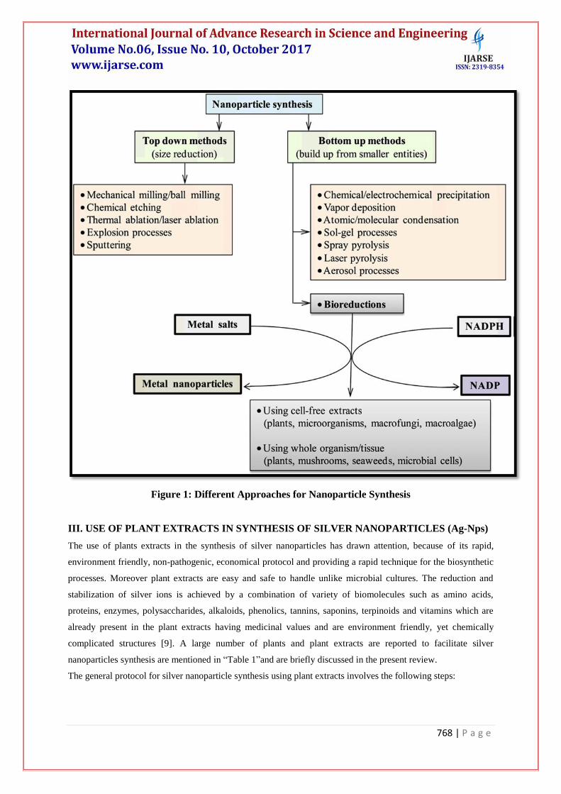
II. METHODS FOR SYNTHESIS OF NANOPARTICLES
Many techniques for the synthesis of metallic nanoparticles are now available. Synthesis of nanoparticles
generally involve either “top to bottom” approach or “bottom to up” approach [1]. In top down method of
synthesis, the nanoparticles are produced by size reduction from an appropriate starting material [2]. Variety of
physical and chemical treatments are used for the achievement of size reduction, physical approaches include
techniques such as evaporation-condensation and laser ablation whereas chemical approaches include chemical
reduction by use of organic and inorganic reducing agents. Top down fabrication methods introduce
imperfections in the surface structure of the product and this is a major drawback because the surface chemistry
and the other physical properties of nanoparticles are greatly dependent on the surface structure [3].
In bottom up method of synthesis, the nanoparticles are built from smaller entities, for example by fusion of
atoms, molecules and smaller particles [4]. In bottom up synthesis, the nanostructured building blocks of the
nanoparticles are produced first and then assembled to manufacture the final particle [3]. The bottom up
synthesis mostly achieved by chemical and biological methods.
The biological methods can be used to synthesize nanoparticles without the use of any harsh, toxic and
expensive chemical substances. Out of all the biological methods used for the synthesis of nanoparticles, the
methods based on microorganisms have been frequently reported [5,6]. Various advantages of microbial
synthesis is that its readily scalable, environment friendly and compatible with the use of the product for
medical applications but production of microorganisms is often more expensive than the production of plant
extracts. Plant mediated nanoparticles synthesis using whole plant extract or by living plants was also reported
in literature [7,8]. Different methods for synthesis of metallic nanoparticles were depicted in “Fig.1”.
768 | P a g e
Figure 1: Different Approaches for Nanoparticle Synthesis
III. USE OF PLANT EXTRACTS IN SYNTHESIS OF SILVER NANOPARTICLES (Ag-Nps)
The use of plants extracts in the synthesis of silver nanoparticles has drawn attention, because of its rapid,
environment friendly, non-pathogenic, economical protocol and providing a rapid technique for the biosynthetic
processes. Moreover plant extracts are easy and safe to handle unlike microbial cultures. The reduction and
stabilization of silver ions is achieved by a combination of variety of biomolecules such as amino acids,
proteins, enzymes, polysaccharides, alkaloids, phenolics, tannins, saponins, terpinoids and vitamins which are
already present in the plant extracts having medicinal values and are environment friendly, yet chemically
complicated structures [9]. A large number of plants and plant extracts are reported to facilitate silver
nanoparticles synthesis are mentioned in “Table 1”and are briefly discussed in the present review.
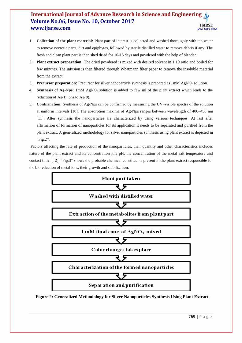
The general protocol for silver nanoparticle synthesis using plant extracts involves the following steps:
769 | P a g e
1. Collection of the plant material: Plant part of interest is collected and washed thoroughly with tap water
to remove necrotic parts, dirt and epiphytes, followed by sterile distilled water to remove debris if any. The
fresh and clean plant part is then shed dried for 10-15 days and powdered with the help of blender.
2. Plant extract preparation: The dried powdered is mixed with desired solvent in 1:10 ratio and boiled for
few minutes. The infusion is then filtered through Whatmann filter paper to remove the insoluble material
from the extract.
3. Precursor preparation: Precursor for silver nanoparticle synthesis is prepared as 1mM AgNO3 solution.
4. Synthesis of Ag-Nps: 1mM AgNO3 solution is added to few ml of the plant extract which leads to the
reduction of Ag(I) ions to Ag(0).
5. Confirmation: Synthesis of Ag-Nps can be confirmed by measuring the UV–visible spectra of the solution
at uniform intervals [10]. The absorption maxima of Ag-Nps ranges between wavelength of 400–450 nm
[11]. After synthesis the nanoparticles are characterized by using various techniques. At last after
affirmation of formation of nanoparticles for its application it needs to be separated and purified from the
plant extract. A generalized methodology for silver nanoparticles synthesis using plant extract is depicted in
“Fig.2”.
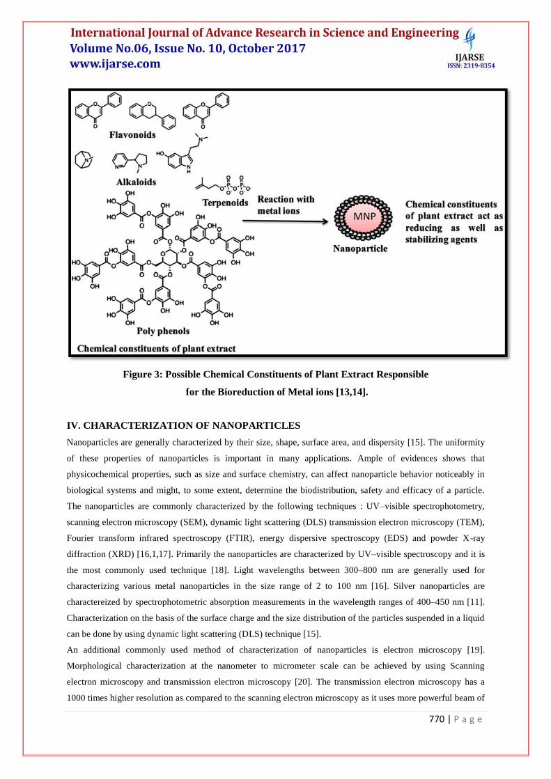
Factors affecting the rate of production of the nanoparticles, their quantity and other characteristics includes
nature of the plant extract and its concentration ,the pH, the concentration of the metal salt temperature and
contact time. [12]. “Fig.3” shows the probable chemical constituents present in the plant extract responsible for
the bioreduction of metal ions, their growth and stabilization.
Figure 2: Generalized Methodology for Silver Nanoparticles Synthesis Using Plant Extract
770 | P a g e
Figure 3: Possible Chemical Constituents of Plant Extract Responsible
for the Bioreduction of Metal ions [13,14].
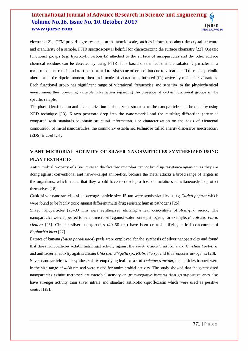
IV. CHARACTERIZATION OF NANOPARTICLES
Nanoparticles are generally characterized by their size, shape, surface area, and dispersity [15]. The uniformity
of these properties of nanoparticles is important in many applications. Ample of evidences shows that
physicochemical properties, such as size and surface chemistry, can affect nanoparticle behavior noticeably in
biological systems and might, to some extent, determine the biodistribution, safety and efficacy of a particle.
The nanoparticles are commonly characterized by the following techniques : UV–visible spectrophotometry,
scanning electron microscopy (SEM), dynamic light scattering (DLS) transmission electron microscopy (TEM),
Fourier transform infrared spectroscopy (FTIR), energy dispersive spectroscopy (EDS) and powder X-ray
diffraction (XRD) [16,1,17]. Primarily the nanoparticles are characterized by UV–visible spectroscopy and it is
the most commonly used technique [18]. Light wavelengths between 300–800 nm are generally used for
characterizing various metal nanoparticles in the size range of 2 to 100 nm [16]. Silver nanoparticles are
charactereized by spectrophotometric absorption measurements in the wavelength ranges of 400–450 nm [11].
Characterization on the basis of the surface charge and the size distribution of the particles suspended in a liquid
can be done by using dynamic light scattering (DLS) technique [15].
An additional commonly used method of characterization of nanoparticles is electron microscopy [19].
Morphological characterization at the nanometer to micrometer scale can be achieved by using Scanning
electron microscopy and transmission electron microscopy [20]. The transmission electron microscopy has a
1000 times higher resolution as compared to the scanning electron microscopy as it uses more powerful beam of
771 | P a g e
electrons [21]. TEM provides greater detail at the atomic scale, such as information about the crystal structure
and granularity of a sample. FTIR spectroscopy is helpful for characterizing the surface chemistry [22]. Organic
functional groups (e.g. hydroxyls, carbonyls) attached to the surface of nanoparticles and the other surface
chemical residues can be detected by using FTIR. It is based on the fact that the subatomic particles in a
molecule do not remain in intact position and transist some other position due to vibrations. If there is a periodic
alteration in the dipole moment, then such mode of vibration is Infrared (IR) active by molecular vibrations.
Each functional group has significant range of vibrational frequencies and sensitive to the physiochemical
environment thus providing valuable information regarding the presence of certain functional groups in the
specific sample.
The phase identification and characterization of the crystal structure of the nanoparticles can be done by using
XRD technique [23]. X-rays penetrate deep into the nanomaterial and the resulting diffraction pattern is
compared with standards to obtain structural information. For characterization on the basis of elemental
composition of metal nanoparticles, the commonly established technique called energy dispersive spectroscopy
(EDS) is used [24].
V.ANTIMICROBIAL ACTIVITY OF SILVER NANOPARTICLES SYNTHESIZED USING
PLANT EXTRACTS
Antimicrobial property of silver owes to the fact that microbes cannot build up resistance against it as they are
doing against conventional and narrow-target antibiotics, because the metal attacks a broad range of targets in
the organisms, which means that they would have to develop a host of mutations simultaneously to protect
themselves [18].
Cubic silver nanoparticles of an average particle size 15 nm were synthesized by using Carica papaya which
were found to be highly toxic against different multi drug resistant human pathogens [25].
Silver nanoparticles (20–30 nm) were synthesized utilizing a leaf concentrate of Acalypha indica. The
nanoparticles were appeared to be antimicrobial against water borne pathogens, for example, E. coli and Vibrio
cholera [26]. Circular silver nanoparticles (40–50 nm) have been created utilizing a leaf concentrate of
Euphorbia hirta [27].
Extract of banana (Musa paradisiaca) peels were employed for the synthesis of silver nanoparticles and found
that these nanoparticles exhibit antifungal activity against the yeasts Candida albicans and Candida lipolytica,
and antibacterial activity against Escherichia coli, Shigella sp., Klebsiella sp. and Enterobacter aerogenes [28].
Silver nanoparticles were synthesized by employing leaf extract of Ocimum sanctum, the particles formed were
in the size range of 4-30 nm and were tested for antimicrobial activity. The study showed that the synthesized
nanoparticles exhibit increased antimicrobial activity on gram-negative bacteria than gram-positive ones also
have stronger activity than silver nitrate and standard antibiotic ciprofloxacin which were used as positive
control [29].
772 | P a g e
Silver nanoparticles created utilizing peel concentrate of Citrus sinensis were found to have a wide range
antibacterial movement. The particles framed at 60 °C had a normal size of around 10 nm yet lessening the
response temperature to 25 °C expanded the normal size to 35 nm [30].
A tuber concentrate of Dioscorea bulbifera was utilized to create gold also, silver nanoparticles of different
shapes [31,32]. These nanoparticles in mix with antibiotics were found to have a synergistic antibacterial action
against test microorganisms, especially against Pseudomonas aeruginosa, Escherichia coli furthermore,
Acinetobacter baumannii [32]. Utilization of antibiotics in blend with silver nanoparticles has been accounted
for powerful control of generally antibiotic resistant safe microorganisms.
Dried fruit body extract of Tribulus terrestris was employed for the formation of silver nanoparticles, size range
of 16-28 nm was achieved with antibacterial property against multi-drug resistant bacteria such as Streptococcus
pyogens, Pseudomonas aeruginosa, Bacillus subtilis, Escherichia coli and Staphylococcus aureus [33].
Leaf concentrate of Calotropis gigantean was utilized to create silver nanoparticles and these nanoparticles
showed antibacterial activity against Vibrio alginolyticus [34].
Profoundly stabilized silver nanoparticles (25–40 nm) were delivered utilizing a leaf concentrate of Ocimum
tenuiflorum. The particles were antibacterial towards Gram-negative and Gram-positive microorganisms
[35,36].
The plant extract of Boerhaavia diffusa was utilized for the synthesis of silver nanoparticles, an average particle
size of 25 nm was obtained. These nanopraticles were tested for their antibacterial activity against three fish
pathogens namely Pseudomonas fluorescens, Aeromonas hydrophila and Flavobacterium branchiophilum and
found to have highest activity against F. branchiophilum [37].
Spherical silver nanoparticles of an average diameter 8-10 nm were synthesized by using fruit extract of
cucumber (Cucumis sativus) and analyzed their photocatalytic and antimicrobial activity. The Photo catalytic
study suggests the efficiency of these biosynthesized nanoparticles in degrading organic dyes methylene blue
under solar radiation. And moreover, the result of antibacterial assay (tested against- Staphylococcus aureus,
Klebsiella pneumoniae and Escherichia coli) showed that these nanoparticles possess effective bactericidal
property [38].
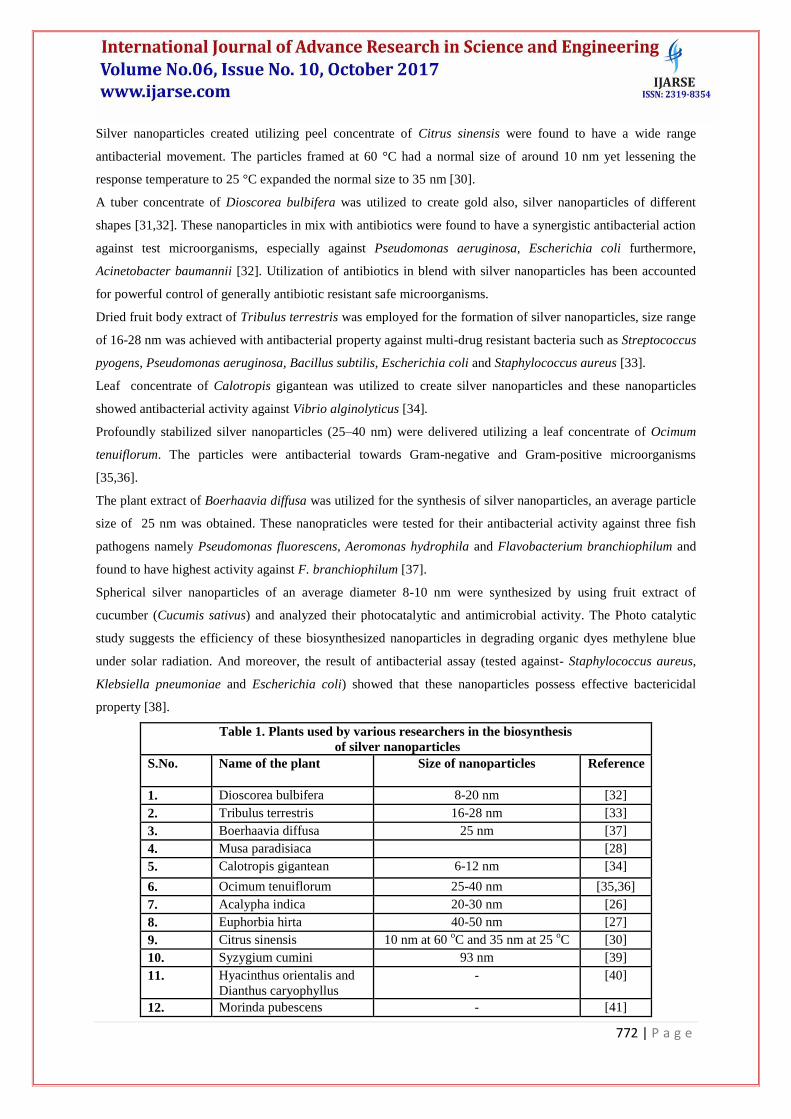
Table 1. Plants used by various researchers in the biosynthesis
of silver nanoparticles
S.No. Name of the plant Size of nanoparticles Reference
1. Dioscorea bulbifera 8-20 nm [32]
2. Tribulus terrestris 16-28 nm [33]
3. Boerhaavia diffusa 25 nm [37]
4. Musa paradisiaca [28]
5. Calotropis gigantean 6-12 nm [34]
6. Ocimum tenuiflorum 25-40 nm [35,36]
7. Acalypha indica 20-30 nm [26]
8. Euphorbia hirta 40-50 nm [27]
9. Citrus sinensis 10 nm at 60 oC and 35 nm at 25
oC [30]
10. Syzygium cumini 93 nm [39]
11. Hyacinthus orientalis and
Dianthus caryophyllus
- [40]
12. Morinda pubescens - [41]
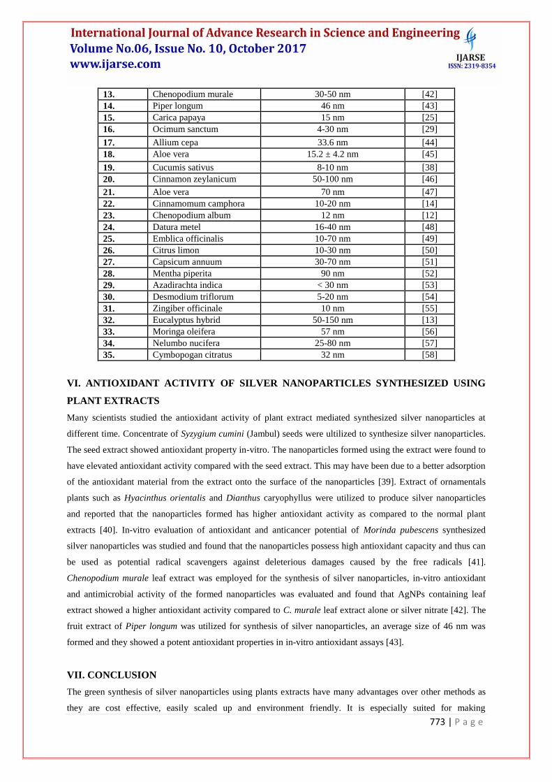
773 | P a g e
13. Chenopodium murale 30-50 nm [42]
14. Piper longum 46 nm [43]
15. Carica papaya 15 nm [25]
16. Ocimum sanctum 4-30 nm [29]
17. Allium cepa 33.6 nm [44]
18. Aloe vera 15.2 ± 4.2 nm [45]
19. Cucumis sativus 8-10 nm [38]
20. Cinnamon zeylanicum 50-100 nm [46]
21. Aloe vera 70 nm [47]
22. Cinnamomum camphora 10-20 nm [14]
23. Chenopodium album 12 nm [12]
24. Datura metel 16-40 nm [48]
25. Emblica officinalis 10-70 nm [49]
26. Citrus limon 10-30 nm [50]
27. Capsicum annuum 30-70 nm [51]
28. Mentha piperita 90 nm [52]
29. Azadirachta indica < 30 nm [53]
30. Desmodium triflorum 5-20 nm [54]
31. Zingiber officinale 10 nm [55]
32. Eucalyptus hybrid 50-150 nm [13]
33. Moringa oleifera 57 nm [56]
34. Nelumbo nucifera 25-80 nm [57]
35. Cymbopogan citratus 32 nm [58]
VI. ANTIOXIDANT ACTIVITY OF SILVER NANOPARTICLES SYNTHESIZED USING
PLANT EXTRACTS
Many scientists studied the antioxidant activity of plant extract mediated synthesized silver nanoparticles at
different time. Concentrate of Syzygium cumini (Jambul) seeds were ultilized to synthesize silver nanoparticles.
The seed extract showed antioxidant property in-vitro. The nanoparticles formed using the extract were found to
have elevated antioxidant activity compared with the seed extract. This may have been due to a better adsorption
of the antioxidant material from the extract onto the surface of the nanoparticles [39]. Extract of ornamentals
plants such as Hyacinthus orientalis and Dianthus caryophyllus were utilized to produce silver nanoparticles
and reported that the nanoparticles formed has higher antioxidant activity as compared to the normal plant
extracts [40]. In-vitro evaluation of antioxidant and anticancer potential of Morinda pubescens synthesized
silver nanoparticles was studied and found that the nanoparticles possess high antioxidant capacity and thus can
be used as potential radical scavengers against deleterious damages caused by the free radicals [41].
Chenopodium murale leaf extract was employed for the synthesis of silver nanoparticles, in-vitro antioxidant
and antimicrobial activity of the formed nanoparticles was evaluated and found that AgNPs containing leaf
extract showed a higher antioxidant activity compared to C. murale leaf extract alone or silver nitrate [42]. The
fruit extract of Piper longum was utilized for synthesis of silver nanoparticles, an average size of 46 nm was
formed and they showed a potent antioxidant properties in in-vitro antioxidant assays [43].
VII. CONCLUSION
The green synthesis of silver nanoparticles using plants extracts have many advantages over other methods as
they are cost effective, easily scaled up and environment friendly. It is especially suited for making
774 | P a g e
nanoparticles that must be free of toxic contaminants as required in therapeutic applications. Green synthesized
silver nanoparticles have noteworthy aspects of nanotechnology through unmatched applications and synthesis
of nanoparticles using plants can be beneficial over other other biological methods because plant products are
easier and safer to handle, widely distributed and easily available. The present review by conferring various
literatures reported recently has showed the importance of plant extract mediated synthesis of silver
nanoparticles and describes them as a promising antimicrobial and antioxidant agent.
VIII. ACKNOWLEDGEMENT
The author, Jasmeet Kaur Sohal gratefully acknowledges all the teaching and non-teaching staff of Faculty of
Biological and Chemical Sciences, MATS University, Raipur (C.G.)
REFERENCES
[1] Sepeur, S. (2008). Nanotechnology: technical basics and applications. Hannover: Vincentz.
[2] Meyers, M.A., Mishra, A. and Benson, D.J. (2006). Mechanical properties of nanocrystalline materials. Prog
Mater Sci, 51:427–556.
[3] Thakkar, K.N., Mhatre, S.S. and Parikh, R.Y. (2010). Biological synthesis of metallic nanoparticles.
Nanomed Nanotechnol Biol Med, 6:257–62.
[4] Mukherjee, P., Ahmad, A., Mandal, D., Senapati, S., Sainkar, S.R. and Khan, M.I. (2001). Fungus mediated
synthesis of silver nanoparticles and their immobilization in the mycelia matrix: a novel biological
approach to nanoparticle synthesis. Nano Lett, 1:515–9.
[5] Dhillon, G.S., Brar, S.K., Kaur, S. and Verma, M. (2012). Green approach for nanoparticle biosynthesis by
fungi: current trends and applications. Crit Rev Biotechnol, 32:49–73.
[6] Mohanpuria, P., Rana, N.K. and Yadav, S.K. (2008). Biosynthesis of nanoparticles: technological concepts
and future applications. J Nanopart Res, 10:507–17.
[7] Gardea-Torresdey, J.L., Gomez, E., Peralta-Videa, J.R., Parsons, J.G., Troiani, H. and Jose- Yacaman, M.
(2003). Alfalfa sprouts: a natural source for the synthesis of silver nanoparticles. Langmuir, 19:1357–61.
[8] Park, Y., Hong, Y.N., Weyers, A., Kim, Y.S and Linhardt, R.J. (2011). Polysaccharides and phytochemicals:
a natural reservoir for the green synthesis of gold and silver nanoparticles. IET Nanobiotechnol, 5:69–78.
[9] Kulkarni, N. and Muddapur, U. (2014). Biosynthesis of Metal Nanoparticles: A Review. J. Nanotech, 1-8.
[10] Sahayaraj, K. and Rajesh, S. (2011). Bionanoparticles: synthesis and antimicrobial applications, science
against microbial pathogens: communicating current research and technological advances. In: Me´ndez-
Vilas A, editor, FORMATEX, p.228–44.
[11] Huang, H. and Yang, X. (2004). Synthesis of polysaccharide-stabilized gold and silver nanoparticles: a
green method. Carbohydrate Res, 339:2627–31.
[12] Dwivedi, A.D. and Gopal, K. (2010). Biosynthesis of silver and gold nanoparticles using Chenopodium
album leaf extract. Colloids Surf A, 369:27–33.
[13] Dubey, M., Bhadauria, S. and Kushwah, B. (2009). Green synthesis of nanosilver particles from extract of
Eucalyptus hybrida (safeda) leaf. Dig J Nanomater Biostruct, 4:537–43.
775 | P a g e
[14] Huang, J.L., Li, Q.B., Sun, D.H., Lu, Y.H., Su, Y.B., Yang, X., et al. (2007). Biosynthesis of silver and
gold nanoparticles by novel sundried Cinnamomum camphora leaf. Nanotechnology, 18.
[15] Jiang, J., Oberdorster, G. and Biswas, P. (2009). Characterization of size, surface charge, and
agglomeration state of nanoparticle dispersions for toxicological studies. J Nanopart Res, 11:77–89.
[16] Feldheim, D.L. and Foss, C.A. (2002). Metal nanoparticles: synthesis, characterization, and applications.
Boca Raton, FL: CRC Press.
[17] Shahverdi, A.R., Shakibaie, M. and Nazari, P. (2011). Basic and practical procedures for microbial
synthesis of nanoparticles. In: Rai M, Duran N, editors. Metal nanoparticles in microbiology. Berlin:
Springer, p. 177–97.
[18] Pal, S., Tak, Y.K. and Song, J.M. (2007). Does the antibacterial activity of silver nanoparticles depend on
the shape of the nanoparticle? A study of the Gram-negative bacterium Escherichia coli. Appl Environ
Microbiol, 73:1712–20.
[19] Cao, G. (2004). Nanostructures and nanomaterials: synthesis, properties and applications. London: Imperial
College Press.
[20] Schaffer, B., Hohenester, U., Trugler, A. and Hofer, F. (2009). High-resolution surface plasmon imaging of
gold nanoparticles by energy-filtered transmission electron microscopy. Phys Rev B, 79. [article 041401].
[21] Eppler, A.S., Rupprechter, G., Anderson, E.A. and Somorjai, G.A. (2000). Thermal and chemical stability
and adhesion strength of Pt nanoparticle arrays supported on silica studied by transmission electron
microscopy and atomic force microscopy. J Phys Chem B, 104:7286–92.
[22] Chithrani, B.D., Ghazani, A.A. and Chan, W.C.W. (2006). Determining the size and shape dependence of
gold nanoparticle uptake into mammalian cells. Nano Lett, 6:662–8.
[23] Sun, S., Murray, C., Weller, D., Folks, L. and Moser, A. (2000). Monodisperse FePt nanoparticles and
ferromagnetic FePt nanocrystal superlattices. Science, 287:1989–92.
[24] Strasser, P., Koh, S., Anniyev, T., Greeley, J., More, K. and Yu, C. (2010). Lattice-strain control of the
activity in dealloyed core–shell fuel cell catalysts. Nat Chem, 2:454–60.
[25] Jain, D., Kumar, Daima. S., Kachhwaha, S. and Kothari, S.L. (2009). Synthesis of plant mediated silver
nanoparticles using Papaya Fruit Extract and Evaluation of their Antimicrobial Activities. Dig J
Nanomater Bios, 4: 557-56.
[26] Krishnaraj, C., Jagan, E., Rajasekar, S., Selvakumar, P., Kalaichelvan, P. and Mohan, N. (2010). Synthesis
of silver nanoparticles using Acalypha indica leaf extracts and its antibacterial activity against water borne
pathogens. Colloids Surf B Biointerfaces, 76:50–6.
[27] Elumalai, E.K., Prasad, T.N.V.K.V., Hemachandran, J., Vivivan Therasa, S., Thirumalai, T. and David, E.
(2010). Extracellular synthesis of silver nanoparticles using leaves of Euphorbia hirta and their
antibacterial activities. J Phram Sci, (9):549-554.
[28] Bankar, A., Joshi, B., Kumar, A.R. and Zinjarde, S. (2010). Banana peel extract mediated novel route for
the synthesis of silver nanoparticles. Colloids Surf A, 368:58–63.
[29] Singhal, G., Bhavesh, R., Kasariya, K., Sharma, A.R. and Singh, R.P. (2011). Biosynthesis of silver
nanoparticles using Ocimum sanctum (Tulsi) leaf extract and screening its antimicrobial activity. J
Nanopart Res, 13: 2981– 2988.
776 | P a g e
[30] Kaviya, S., Santhanalakshmi, J., Viswanathan, B., Muthumary, J. and Srinivasan, K. (2011b). Biosynthesis
of silver nanoparticles using Citrus sinensis peel extract and its antibacterial activity. Spectrochim Acta A
Mol Biomol Spectrosc, 79:594-8.
[31] Ghosh, S., Patil, S., Ahire, M., Kitture, R., Jabgunde, A., Kale, S., et al. (2011). Synthesis of gold nano-
anisotrops using Dioscorea bulbifera tuber extract. J Nanomater.[article ID 354793 2011].
[32] Ghosh, S., Patil, S., Ahire, M., Kitture, R., Kale, S., Pardesi, K., et al. (2012). Synthesis of silver
nanoparticles using Dioscorea bulbifera tuber extract and evaluation of its synergistic potential in
combination with antimicrobial agents. Int J Nanomed, 7: 483–96.
[33] Gopinatha, V., Ali, M.D., Priyadarshini, S., MeeraPriyadharsshini, N., Thajuddinb, N. and Velusamy, P.
(2012). Biosynthesis of silver nanoparticles from Tribulus terrestris and its antimicrobial activity: a novel
biological approach. Colloid Surf B: Biointerface, 96:69–74.
[34] Baskaralingam, V., Sargunar, C.G., Lin, Y.C. and Chen, J.C. (2012) Green synthesis of silver nanoparticles
through Calotropis gigantea leaf extracts and evaluation of antibacterial activity against Vibrio
alginolyticus. Nanotechnol Dev, 2:e3.
[35] Patil, S.V., Borase, H.P., Patil, C.D and Salunke, B.K. (2012a). Biosynthesis of silver nanoparticles using
latex from few euphorbian plants and their antimicrobial potential. Appl Biochem Biotechnol, 167:776–
90.
[36] Patil, R., Kokate, M. and Kolekar, S. (2012b) Bioinspired synthesis of highly stabilized silver nanoparticles
using Ocimum tenuiflorum leaf extract and their antibacterial activity. Spectrochim Acta A Mol Biomol
Spectrosc, 91:234–8.
[37] Nakkala, J.R., Mata, R., Gupta, A.K. and Sadras, S.R. (2014). Green synthesis and characterization of
silver nanoparticles using Boerhaavia diffusa plant extract and their antibacterial activity. Indus Crop
Prod, 52:562–6.
[38] Roy, K., Sarkar, C.K. and Ghosh, C.K. (2015). Single-step novel biosynthesis of silver nanoparticles using
Cucumis sativus fruit extract and study of its photcatalytic and antibacterial activity. Dig J Nanomater
Bios, 10: 107–115.
[39] Banerjee, J. and Narendhirakannan, R. (2011). Biosynthesis of silver nanoparticles from Syzygium cumini
(L.) seed extract and evaluation of their in vitro antioxidant activities. Dig J Nanomater Biostruct, 6:961–8.
[40] Bunghez, I. R., Barbinta Patrascu, M. E., Badea, N. M., Doncea, S. M., Popescu, A. and Ion, R. M. (2012).
Antioxidant silver nanoparticles green synthesized using ornamental plants. Journal of optoelectronics and
advanced materials, 14(11), 1016.
[41] Inbathamizh, L., Mekalai Ponnu, T. and Jancy Mary, E. (2013). In vitro evaluation of antioxidant and
anticancer potential of Morinda pubescens synthesized silver nanoparticles. Journal of pharmacy research,
6:32-38
[42] Abdel-Aziz, M. S., Shaheen, M. S., El-Nekeety, A. A. and Abdel-Wahhab, M. A. (2014). Antioxidant and
antibacterial activity of silver nanoparticles biosynthesized using Chenopodium murale leaf extract.
Journal of Saudi Chemical Society, 18(4), 356-363.

777 | P a g e
[43] Reddy, N.J., Vali, D.N., Rani, M. and Rani, S.S. (2014). Evaluation of antioxidant, antibacterial and
cytotoxic effects of green synthesized silver nanoparticles by Piper longum fruit. Materials Science and
Engineering C 34:115–122.
[44] Saxena, A., Tripathi, R.M. and Singh, R.P. (2010). Biological synthesis of silver nanoparticles by using
onion (Allium cepa) extract and their antibacterial activity. Dig J Nanomater Bios, 5: 427 – 432.
[45] Chandran, S.P., Chaudhary, M., Pasricha, R., Ahmad, A. and Sastry, M. (2006). Synthesis of gold
nanotriangles and silver nanoparticles using Aloe vera plant extract. Biotechnol Prog, 22: 577-583.
[46] Sathishkumar, M., Sneha, K., Won, S., Cho, C.W., Kim, S. and Yun, Y.S. (2009b). Cinnamon zeylanicum
bark extract and powder mediated green synthesis of nano-crystalline silver particles and its bactericidal
activity. Colloids Surf B Biointerfaces, 73:332–8.
[47] Medda, S., Hajra, A., Dey,U., Bose, P. and Monda, N.K. (2015). Biosynthesis of silver nanoparticles from
Aloe vera leaf extract and antifungal activity against Rhizopus sp. And Aspergillus sp., Appl Nanosci;
5:875–880.
[48] Kesharwani, J., Yoon, K.Y., Hwang, J. and Rai, M. (2009). Phytofabrication of silver nanoparticles by leaf
extract of Datura metel: hypothetical mechanism involved in synthesis J Bionanosci, 3:39–44.
[49] Ramesh, P.S., Kokila, T. and Geetha, D. (2015). Plant mediated green synthesis and antibacterial activity of
silver nanoparticles using Emblica Officinalis fruit extract. Spectrochimica Acta A, 142: 339–343.
[50] Mohapatra, B., Kaintura, R., Singh, J., Kuriakose, S. and Mohapatra. S. (2015). Biosynthesis of high
concentration, stable aqueous dispersions of silver nanoparticles using Citrus limon extract. Adv. Mater.
Lett, 6: 228-234.
[51] Li, S., Shen, Y., Xie, A., Yu, X., Qiu, L., Zhang, L., et al. (2007). Green synthesis of silver nanoparticles
using Capsicum annuum L. extract. Green Chem, 9: 852–858.
[52] MubarakAli, D., Thajuddin, N., Jeganathan, K. and Gunasekaran, M. (2011). Plant extract mediated
synthesis of silver and gold nanoparticles and its antibacterial activity against clinically isolated pathogens.
Colloids and Surfaces B: Biointerfaces, 85:360–365.
[53] Velusamy, P., Das, J., Pachaiappan, R., Vaseeharan, B. and Pandian, K. (2015). Greener approach for
synthesis of antibacterial silver nanoparticles using aqueous solution of neem gum (Azadirachta indica L.).
Industrial Crops and Products, 66:103–109.
[54] Ahmad, N., Sharma, S., Singh, V. N., Shamsi, S. F., Fatma, A. and Mehta, B.R. (2011). Biosynthesis of
Silver Nanoparticles from Desmodium triflorum: A Novel Approach Towards Weed Utilization.
Biotechnol Res Int. 454090.
[55] Singh, C., Sharma, V., Naik, K.R.P., Khandelwal, V. and Singh, H. (2011). A green biogenic approach for
synthesis of gold and silver nanoparticles using Zingiber officinale. Dig J Nanomater Bios, 6: 535-542.
[56] Prasad, T.N.V.K.V. and Elumalai, E. (2011). Biofabrication of Ag nanoparticles using Moringa oleifera
leaf extract and their antimicrobial activity. Asian Pac J Trop Biomed, 1:439–42.
[57] Santhoshkumar, T., Rahuman, A.A., Rajakumar, G., Marimuthu, S., Bagavan, A. and Jayaseelan, C.
(2011). Synthesis of silver nanoparticles using Nelumbo nucifera leaf extract and its larvicidal activity
against malaria and filariasis vectors. Parasitol Res, 108: 693–702.
Related Documents