Bahrulolum et al. J Nanobiotechnol (2021) 19:86 https://doi.org/10.1186/s12951-021-00834-3 REVIEW Green synthesis of metal nanoparticles using microorganisms and their application in the agrifood sector Howra Bahrulolum 1† , Saghi Nooraei 1† , Nahid Javanshir 1 , Hossein Tarrahimofrad 2 , Vasighe Sadat Mirbagheri 1,3 , Andrew J. Easton 4 and Gholamreza Ahmadian 1* Abstract The agricultural sector is currently facing many global challenges, such as climate change, and environmental prob- lems such as the release of pesticides and fertilizers, which will be exacerbated in the face of population growth and food shortages. Therefore, the need to change traditional farming methods and replace them with new technolo- gies is essential, and the application of nanotechnology, especially green technology offers considerable promise in alleviating these problems. Nanotechnology has led to changes and advances in many technologies and has the potential to transform various fields of the agricultural sector, including biosensors, pesticides, fertilizers, food pack- aging and other areas of the agricultural industry. Due to their unique properties, nanomaterials are considered as suitable carriers for stabilizing fertilizers and pesticides, as well as facilitating controlled nutrient transfer and increas- ing crop protection. The production of nanoparticles by physical and chemical methods requires the use of hazardous materials, advanced equipment, and has a negative impact on the environment. Thus, over the last decade, research activities in the context of nanotechnology have shifted towards environmentally friendly and economically viable ‘green’ synthesis to support the increasing use of nanoparticles in various industries. Green synthesis, as part of bio- inspired protocols, provides reliable and sustainable methods for the biosynthesis of nanoparticles by a wide range of microorganisms rather than current synthetic processes. Therefore, this field is developing rapidly and new methods in this field are constantly being invented to improve the properties of nanoparticles. In this review, we consider the latest advances and innovations in the production of metal nanoparticles using green synthesis by different groups of microorganisms and the application of these nanoparticles in various agricultural sectors to achieve food security, improve crop production and reduce the use of pesticides. In addition, the mechanism of synthesis of metal nanopar- ticles by different microorganisms and their advantages and disadvantages compared to other common methods are presented. Keywords: Agriculture, Metal nanoparticles, Green synthesis, Microorganisms, Nanopesticides, Nanofungicides, Nanofertilizers, Nanobiosensors, Crop protection © The Author(s) 2021. This article is licensed under a Creative Commons Attribution 4.0 International License, which permits use, sharing, adaptation, distribution and reproduction in any medium or format, as long as you give appropriate credit to the original author(s) and the source, provide a link to the Creative Commons licence, and indicate if changes were made. The images or other third party material in this article are included in the article’s Creative Commons licence, unless indicated otherwise in a credit line to the material. If material is not included in the article’s Creative Commons licence and your intended use is not permitted by statutory regulation or exceeds the permitted use, you will need to obtain permission directly from the copyright holder. To view a copy of this licence, visit http://creativeco mmons.org/licenses/by/4.0/. The Creative Commons Public Domain Dedication waiver (http://creativecommons.org/publicdomain/ zero/1.0/) applies to the data made available in this article, unless otherwise stated in a credit line to the data. Background Nanoparticles now play a key role in most technologies, including medicine, cosmetics, agriculture and the food sciences [1]. Recently, the synthesis of metal nanoparti- cles (MtNPs) using microorganisms and plants has been recognized as an efficient and green method for further exploitation of microorganisms as nanofactories [2]. Open Access Journal of Nanobiotechnology *Correspondence: [email protected] † Howra Bahrulolum and Saghi Nooraei contributed equally to this work 1 Department of Industrial Environmental and Biotechnology, National Institute of Genetic Engineering and Biotechnology (NIGEB), P.O.BOX: 14155-6343, 1497716316 Tehran, Iran Full list of author information is available at the end of the article

Welcome message from author
This document is posted to help you gain knowledge. Please leave a comment to let me know what you think about it! Share it to your friends and learn new things together.
Transcript

Bahrulolum et al. J Nanobiotechnol (2021) 19:86 https://doi.org/10.1186/s12951-021-00834-3
REVIEW
Green synthesis of metal nanoparticles using microorganisms and their application in the agrifood sectorHowra Bahrulolum1†, Saghi Nooraei1†, Nahid Javanshir1, Hossein Tarrahimofrad2, Vasighe Sadat Mirbagheri1,3, Andrew J. Easton4 and Gholamreza Ahmadian1*
Abstract
The agricultural sector is currently facing many global challenges, such as climate change, and environmental prob-lems such as the release of pesticides and fertilizers, which will be exacerbated in the face of population growth and food shortages. Therefore, the need to change traditional farming methods and replace them with new technolo-gies is essential, and the application of nanotechnology, especially green technology offers considerable promise in alleviating these problems. Nanotechnology has led to changes and advances in many technologies and has the potential to transform various fields of the agricultural sector, including biosensors, pesticides, fertilizers, food pack-aging and other areas of the agricultural industry. Due to their unique properties, nanomaterials are considered as suitable carriers for stabilizing fertilizers and pesticides, as well as facilitating controlled nutrient transfer and increas-ing crop protection. The production of nanoparticles by physical and chemical methods requires the use of hazardous materials, advanced equipment, and has a negative impact on the environment. Thus, over the last decade, research activities in the context of nanotechnology have shifted towards environmentally friendly and economically viable ‘green’ synthesis to support the increasing use of nanoparticles in various industries. Green synthesis, as part of bio-inspired protocols, provides reliable and sustainable methods for the biosynthesis of nanoparticles by a wide range of microorganisms rather than current synthetic processes. Therefore, this field is developing rapidly and new methods in this field are constantly being invented to improve the properties of nanoparticles. In this review, we consider the latest advances and innovations in the production of metal nanoparticles using green synthesis by different groups of microorganisms and the application of these nanoparticles in various agricultural sectors to achieve food security, improve crop production and reduce the use of pesticides. In addition, the mechanism of synthesis of metal nanopar-ticles by different microorganisms and their advantages and disadvantages compared to other common methods are presented.
Keywords: Agriculture, Metal nanoparticles, Green synthesis, Microorganisms, Nanopesticides, Nanofungicides, Nanofertilizers, Nanobiosensors, Crop protection
© The Author(s) 2021. This article is licensed under a Creative Commons Attribution 4.0 International License, which permits use, sharing, adaptation, distribution and reproduction in any medium or format, as long as you give appropriate credit to the original author(s) and the source, provide a link to the Creative Commons licence, and indicate if changes were made. The images or other third party material in this article are included in the article’s Creative Commons licence, unless indicated otherwise in a credit line to the material. If material is not included in the article’s Creative Commons licence and your intended use is not permitted by statutory regulation or exceeds the permitted use, you will need to obtain permission directly from the copyright holder. To view a copy of this licence, visit http:// creat iveco mmons. org/ licen ses/ by/4. 0/. The Creative Commons Public Domain Dedication waiver (http:// creat iveco mmons. org/ publi cdoma in/ zero/1. 0/) applies to the data made available in this article, unless otherwise stated in a credit line to the data.
BackgroundNanoparticles now play a key role in most technologies, including medicine, cosmetics, agriculture and the food sciences [1]. Recently, the synthesis of metal nanoparti-cles (MtNPs) using microorganisms and plants has been recognized as an efficient and green method for further exploitation of microorganisms as nanofactories [2].
Open Access
Journal of Nanobiotechnology
*Correspondence: [email protected]†Howra Bahrulolum and Saghi Nooraei contributed equally to this work1 Department of Industrial Environmental and Biotechnology, National Institute of Genetic Engineering and Biotechnology (NIGEB), P.O.BOX: 14155-6343, 1497716316 Tehran, IranFull list of author information is available at the end of the article

Page 2 of 26Bahrulolum et al. J Nanobiotechnol (2021) 19:86
Given the challenges facing the international commu-nity, especially in terms of population growth and climate change, nanotechnology can have positive effects on improving the quality of agricultural products, minimiz-ing the adverse effects of agricultural pesticides on the environment and human health, and increasing produc-tivity and food security. Unique properties of nanoscale materials make them an excellent candidate for using in the design and development of new tools for supporting agriculture and related industries. Nanotechnology can improve agricultural processes such as soil quality and the quality of agricultural products by using nanoparti-cle-based fertilizers or by stimulating plant growth. In addition, the use of fertilizers and pesticides using nan-oparticle-based carriers and compounds is reduced with-out reducing productivity [3]. Nanotechnology can also minimize waste by fabricating products that are more efficient. Applications of nanosensor technology can lead to the development of precision agriculture and efficient management of resources, including energy and materi-als used [4]. In particular, the goal of developing green nanotechnology, which utilizes biological pathways for the synthesis of nanomaterials is minimizing the produc-tion of hazardous substances. Meanwhile, the amount of energy input in green nanotechnology is much lower than in other technologies; almost no toxic chemicals are produced during synthesis, and their environmental compatibility is very high. Therefore, green nanomateri-als produced can be widely used in various industries [5]. Depending on the application required, different types of nanomaterials are used in agriculture. For example, for use in pesticides, nanoparticles are used as carriers, which gradually release the active ingredient(s) to reduce their overall consumption. When the goal is to improve the packaging of agricultural products, the nanomate-rials used are selected to be biocompatible and do not have negative effects on human health while increas-ing the shelf life of food. Alternatively, high-sensitivity nanosensors with plasmonic properties such as silver or gold nanoparticles can be used to measure environmen-tal conditions, report changes in a timely way, and intel-ligently control plant needs in greenhouses. In all cases, the small size and unique physical and chemical proper-ties of the MtNPs make them attractive for use in various agricultural sector [1]. To date, a broad range of nano-technology applications have emerged in the agrifood sector, such as nanosensors, tracking devices, targeted delivery of required components, food safety and intelli-gent packaging which can affect different aspects of our lives [6–8].
Several advanced techniques are available to improve precision breeding methods and enable precise control of the green synthesis process at the nanometer scale.
Nanotechnology can also be an alternative source for generating fertilizer, as MtNPs have been shown to be able to increase germination in agricultural seeds. Other applications include the use of nanoscale carriers for effective delivery of fertilizers, pesticides, plant growth regulators, and other similar compounds. These pro-cesses improve the stability of these materials to environ-mental degradation and ultimately reduce their amount used, which in turn leads to reductions in chemical run-off and associated environmental problems. Carriers can also be designed to increase the communication between plant roots and the surrounding soil structure [9]. Modi-fied nanoparticles can be added to conventional fertiliz-ers for improving nitrogen storage capacity which leads to reduced nitrogen loss and better nutrition for agricul-tural products. Several nanoemulsions have also been formulated to increase the biological compatibility of herbicides and pesticides [10].
Microorganisms are important nanofactories that are able to accumulate and detoxify heavy metals due to the presence of various reductase enzymes that are able to reduce metal salts to MtNP [2]. In recent research, bac-teria such as Pseudomonas deptenis [11], Visella oriza [12] Bacillus methylotrophicus [13], Bhargavaea indica and Brevibacterium frigoritolerans have been shown to be able to synthesize silver (Ag) and gold (Au) nano-particles. MtNPs have also been synthesized by various genera of microorganisms such as Lactobacillus, Bacil-lus, Pseudomonas, Streptomyces, Klebsiella, Enterobac-ter, Escherichia, Aeromonas, Corynebacterium, Weissella, Rhodobacter, Rhodococcus, Brevibacterium, Trichoderma, Desulfovibrio, Sargassum, Shewanella, Plectonemabo-ryanum, Pyrobaculum and Rhodopseudomonas [2]. The synthesis of nanoparticles by actinomycetes has not yet been well studied, although studies to date have shown that nanoparticles produced by actinomycetes have very good dispersion and stability and have significant lethal activity against various pathogens [14]. In particular, var-ious microorganisms, such as bacteria, fungi, yeasts and microalgae have been shown to produce MtNPs either intra- or extracellularly. These microorganisms are able to produce organic matter inside, and to transport it to the outside of their cells [15]. Microorganisms as nano-factories have great potential as environmentally friendly, inexpensive, and non-toxic tools that do not require much energy for MtNPs synthesis compared to phys-icochemical methods. Among the various mechanisms for the green synthesis of MtNPs, those that perform extracellular synthesis are of great interest because the extracellular location of the material eliminates the need for costly and complex downstream processing steps to recover intracellular nanoparticles [2]. Green synthesis of MtNPs using microorganisms has several advantages
Page 3 of 26Bahrulolum et al. J Nanobiotechnol (2021) 19:86
compared to conventional physicochemical methods. In particular it offers a rapid, cost-effective, clean, non-toxic and environmentally friendly method for the synthesis of MtNPs with a wide range of sizes, shapes, composi-tions and physicochemical properties [16, 17]. However, the main drawbacks of microorganism-based synthesis of MtNPs includes complicated steps such as microbial sampling, isolation, culturing and storage. In addition, the recovery of MtNPs produced by this method requires downstream processing [2].
In this review, we explore the various potential appli-cations of green synthesized MtNPs with an empha-sis on agriculture. This includes consideration of advantages of green synthesis of MtNPs using different microorganisms.
Green synthesis of MtNPs by microorganisms and their characterizationVarious approaches have been used for MtNP synthesis, such as physical, chemical, and biological methods. The physical and chemical methods for MtNP synthesis have many disadvantages including the use of expensive equip-ment, high heat generation, high energy consumption and low production yield [18, 19]. The main drawback of these methods is the use of toxic chemicals, which pre-sent several environmental problems [19, 20]. This has generated a need for an environmentally friendly option for the synthesis of MtNPs, the current focus of which is the green synthesis of MtNPs from biological routes such as microorganisms, plants, microbial enzymes, polysac-charides and degradable polymers [21]. Green synthesis methods are more beneficial than traditional physical and chemical methods because they are simple, cost-effective, free of toxic and environmentally unfriendly chemicals, and as a result they have gained considerable importance in recent years [20].
The innovative and diverse applications of MtNPs in various fields including medical sciences, environmental sciences and agriculture, research on MtNPs and differ-ent approaches of their synthesis has increased rapidly over recent years [18, 22]. The synthesis of MtNPs is gen-erally performed using one of two different approaches, broadly considered as top-down and bottom-up approaches. In top-down approaches, bulk materials are broken down into nano-sized particles to form MtNPs, based on their reduction in size, using various physical and chemical techniques [18, 23]. The main drawback of this method is the production of nanoparticles with imperfect surface structures. Also, it is an expensive and time consuming approach so it is not appropriate for large-scale production [23]. In bottom-up approaches, nanoparticles are produced by self-assembly of structures at the atomic and molecular scales, resulting in a more
precise size, shape and molecular composition [24]. This method includes chemical and biological methods of production [18].
Among the various biological sources for the green synthesis of MtNPs, green synthesis mediated by micro-organisms has acquired a special place due to their high growth rate, ease of cultivation and ability to grow in ambient conditions of temperature, pH and pressure [25]. Different microorganisms can serve as potential biofacto-ries for the eco-friendly and inexpensive synthesis of vari-ous MtNPs containing metals such as silver, gold, copper, zinc, titanium, palladium and nickel. This can be achieved to generate MtNPs with a defined shape, size, composi-tion and monodispersity of particles [18, 22, 26]. The biosynthetic mechanism of MtNPs in microorganisms can be carried out by trapping target metal ions from the surrounding environment and enzymatically converting them into elemental form, following a reduction mech-anism [26]. Not all microorganisms are able to produce MtNPs because they are produced through metabolic pathways and through cellular enzymes that may not be present in some organisms. The synthesis of MtNPs also is dependent on the capacity of microorganisms for tol-erating heavy metals. High metal stresses can affect vari-ous microbial activities and some microorganisms are able to reduce metal ions to the respective metals under stress condition. In general, microorganisms that live in metal-rich habitats are highly resistant to those metals due to their uptake and chelation of by intracellular and extracellular proteins. Consequently, this method, which mimics the natural bio-mineralization process, could be a favorable approach for the MtNPs synthesis [27]. Figure 1 shows a schematic illustration of intracellular and extra-cellular mechanisms of MtNPs biosynthesis. Intracellular biosynthesis involves unique transport systems in micro-organisms in which the cell wall plays an important role due to its negative charge: positively charged metal ions are deposited in negatively charged cell walls through electrostatic interactions. After transport into the cells of the microorganism, ions are reduced using metabolic reactions mediated by enzymes such as nitrate reductase to forms MtNPs. The MtNPs accumulated in the peri-plasmic space can then be passed through the cell wall [28, 29].
The extracellular biosynthesis of MtNPs is also a nitrate reductase-mediated synthesis in which the MtNPs are produced by reductase enzymes which are either located in the cell wall or secreted from the cell to the growth medium. In this process the nitrate reductase reduces metal ions to the metallic forms [27, 29].
The presence of diverse components such as enzymes, proteins, and other biological molecules in microor-ganisms also play an important role in the process of

Page 4 of 26Bahrulolum et al. J Nanobiotechnol (2021) 19:86
reducing MtNPs [27]. Studies have shown that NADH-dependent enzymes are responsible for the MtNP syn-thesis. The reduction mechanisms seem to begin by transferring an electron from NADH by NADH-depend-ent reductases as the electron carrier [30]. In addition, proteins secreted by microorganisms can act primarily as a stabilizing agent and provides colloidal stability while preventing agglomeration of MtNPs [27].
For intracellular synthetic approaches microorgan-isms are cultured in a suitable growth medium with favorable pH and temperature conditions [23]. The bio-mass is harvested after an optimal incubation period and washed thoroughly with sterile water to minimize potentially undesirable effects of the culture medium. The resulting biomass is then incubated with metal salt solution. In addition to the use of whole microorgan-isms for intracellular synthesis of MtNPs an alternative is the use of cell-free (CF) approaches using either cul-ture supernatant or cell-free extracts (CFE) [22]. In the CF approach using medium supernatant, after culturing
the microorganisms in a liquid culture medium, the mix-ture containing the culture medium and biomass is cen-trifuged and the supernatant collected and incubated with an aqueous metal salt solution to synthesize the MtNPs. In this method, the compounds of the culture medium containing the appropriate enzymes and other essential secretory components produced by the micro-organism are used to synthesize the MtNPs and also to act as reducing and capping agents. In approaches using cell-free extracts, the microorganisms are removed from the culture medium and resuspended in sterile distilled water for an approriate time. The resulting CFE is col-lected after centrifugation and is incubated with metal salt solutions, leading to the generation of MtNPs. In this approach the microorganisms and culture medium are removed through repeated washings, and only biomol-ecules released by cells due to autolysis or starvation con-ditions mediate synthesis of the MtNPs [19, 22, 25, 31]. In all cell free processes a color change in the reaction mixture is frequently the first indication of nanoparticle
Fig. 1 Schematic representation of the mechanisms of extracellular and intracellular biosynthesis of MtNPs. Extracellular biosynthesis of MtNPs carried out by trapping metal ions on the cell wall and reducing them in the presence of secreted enzymes or metabolite. In the intracellular biosynthesis of MtNPs, after transfer of metal ions into cell cytoplasm, the metal ions are reduced as a result of metabolic reactions with enzymes, such as nitrate reductase
Page 5 of 26Bahrulolum et al. J Nanobiotechnol (2021) 19:86
synthesis with the color change being dependent on the precise nature of the MtNP being produced. For example, a change in color from pale yellow to dark purple indi-cates the formation of gold nanoparticles, a pale yellow to deep brown color change indicates the formation of silver nanoparticles and a yellow to yellowish-white color change indicates the formation of manganese and zinc nanoparticles [19, 25, 32].
Various physiological factors including microbial source, reaction temperature, pH, pressure, incubation time and metal salt concentration affect the synthesis of various MtNPs. Optimization of these physiologi-cal parameters is required for synthesis of nanoparticles with accurate size, morphology and chemical compo-sitions [33, 34]. After synthesis of MtNPs, purification before their use in any application is essential. Typically, repeated washing and high-speed centrifugation are per-formed to separate and enrich the produced MtNPs and to eliminate unreacted bioactive molecules [34]. In-cell synthesized nanoparticles require additional purification steps such as ultrasonication or reaction with appropriate detergents, which release the MtNPs after breakdown of the cell wall. These additional steps reduce the economic benefit of this approach [19].
Characterization of MtNPs synthesized from micro-organisms is performed using various analytical tech-niques. UV–visible spectroscopy is generally used to confirm the synthesis and stability of MtNPs. Fourier-transform infrared (FTIR) spectroscopy is used to measure the properties of MtNPs such as chemical con-centration, surface chemistry, surface functional groups and atomic arrangement [33] and transmission electron microscopy (TEM), scanning electron microscopy (SEM) and atomic force microscopy (AFM) can be used to visu-alize the position, size and morphology of MtNPs [35]. X-ray powder diffraction (XRD) is used to determine the crystallographic structure [33]. The elemental composi-tion of MtNPs is usually examine by energy dispersive x-ray spectroscopy (EDS) [36]. Dynamic light scattering (DLS) method is mainly used to evaluate the size as well as surface charge of MtNPs [33].
Application of green synthesized MtNPs in agricultureGreen-synthesized MtNPs have many potential appli-cations in agriculture to increase the productivity of agricultural products. MtNPs are commonly used for generating products such as nanopesticides, nanofungi-cides, nanobiosensors and nanofertilizers. These nano-based products can help increase the quality and yield of agricultural products, reduce chemical pollution or even protect crops from environmental pressures [37].
The use of biosensors has revolutionized agricultural systems to increase the production of quality agricultural products due to their ability to quickly identify pathogens as well as their powerful monitoring and analytical capa-bilities [38]. Nanobiosensors are a modified version of a biosensor that can be described as an analytical unit by incorporating a biological sensitive element with a phys-icochemical transducer [39]. Nanobiosensors including enzymatic biosensors, genosensors, aptasensors, and immunosensors are made using a wide range of electro-chemical, biological or physicochemical transducers. The use of these sensors has received much attention due to their fast, specific and selective performance in detection of toxins and plant pathogens [38]. Pesticides are used to protect plants from harmful agents such as plant patho-gens and insects, to increase crop yield [40]. One of the most important challenges of using existing chemical pesticides is their negative effects on agricultural prod-ucts in the food chain and ultimately on human health [37].
Nanopesticides represent an emerging nanobiotech-nological development to encapsulate pesticides for con-trolled release and to improve the selectivity and stability of pesticides [37, 41]. These nanopesticides can offer a wide range of benefits including increased efficiency, durability and reduced amount of active ingredient required in their formulation [42, 43]. The nano-formu-lation of pesticides with MtNPs has shown a stronger effect against phytopathogens, insects and other pests that threaten crops. Fungi are the most common plant pathogens and cause more than 70% of major crop dam-age [40, 44]. To control this damage common fungicides are currently used, the widespread use of which for long-term disease management leads to environmental pol-lution and dangerous effects on the ecosystem. The use of nanofungicides is an effective strategy against fungal pathogens. The use of MtNPs in the formulation of nano-fungicides is the most common of their applications. These nanofungicides offer targeted delivery and greater bioavailability due to higher solubility and permeability, lower doses, lower dose-dependent toxicity, and con-trolled release [45].
Fertilizers are natural or synthetic substances that contain chemical elements necessary to improve plant growth and productivity and improve natural fertility by overcoming micronutrient deficiencies. The main prob-lem of excessive and long-term use of chemical fertilizers in the agricultural sector is the reduction of soil fertility, which ultimately affects the production of agricultural products. Nanofertilizers are environmentally friendly fertilizers or smart fertilizers that deliver nutrients in small but effective amounts to plants. Nutrient uptake can be increased by encapsulating nanofertilizers, which
Page 6 of 26Bahrulolum et al. J Nanobiotechnol (2021) 19:86
ultimately reduces nutrient loss, promotes proper plant growth and improves crop quality [40, 41, 44]. Nano-formulations provide gradual and controlled release of nutrients to the target sites through direct internaliza-tion of products, which prevents nutrients from interact-ing with soil, water, air and microorganisms resulting in minimizing the risk of environmental degradation [43]. It has been frequently observed that the use of MtNP-based nanofertilizers has significant potential to increase crop productivity.
The application of synthesized green nanoparticle tech-nology in the food or agricultural sector gives flexibility to conventional crop production systems, as it allows the controlled release of pesticides and fertilizers, as well as the targeted delivery of biological molecules. Interac-tions between MtNPs and plant responses are manifested by increase in breeding, and ultimately, it improves the quality and productivity of products [46]. In the follow-ing subsections, different species of microorganisms used for biosynthesis of MtNPs, and their perspective in agri-cultural applications are discussed.
Biosynthesis of MtNPs by probiotic bacteria and their application in agricultureThe use of probiotic microorganisms to produce MtNPs is an environmentally friendly as well as commercially attractive approach [47]. This is due to lower energy input, environmental sustainability, low costs, scalability and sta-bility of MtNPs compared to the use of chemical synthesis methods. The non-pathogenicity of probiotics and their capacity to grow rapidly, regulating the expression of genes to produce various proteins and enzymes involved in the production of MtNPs is useful in many ways. Lactobacil-lus and Bifidobacterium are the most popular probiotics found in dairy products and natural flora in various parts of the body. These non-pathogenic gram-positive bacteria can be used in the production of a wide range of products [48]. The green synthesis of MtNPs, metal oxide nanopar-ticles (MONPs) and non-MtNPs by probiotics has been studied [49]. Probiotics exert their beneficial effects in a variety of ways, including direct effects on living cells and indirect effects on a wide range of metabolites. Probiotics have a negative electrokinetic potential that freely attracts cations, similar to other bacteria, which can be the starting point for the NP biosynthesis process [50].
The negative surface electrokinetic potential of Lac-tobacilli causes the rapid absorption of cations, which in turn plays a key role in the biosynthesis of MtNPs. Pre-vious studies have reported biological adsorption and reduction of silver iodide by Lactobacillus sp. A09 [51] The tendency of lactobacilli to grow even in the presence of oxygen makes them metabolically highly viable. The bacterial redox potential decreases with the addition of
reducing agents such as glucose. The oxidation–reduction potential represents the quantitative state of the degree of aerobiosis with the unit defined as rH2 (negative logarithm of the partial pressure of hydrogen gas). By adjusting the redox potential in the culture medium, the conditions can be changed in the desired direction. For example, suitable conditions can be created by lowering the rH2 for anaero-bic conditions in the presence of oxygen, or by increasing the pH of the medium for creating aerobic conditions in an anaerobic environment. In this way, changing the different conditions of the culture medium plays an important role in the biosynthesis of MtNPs and/or MONPs. Various fac-tors such as energy efficiency, glucose (which controls the value of rH2), ionic mean, pH, and total oxidation capac-ity (rH2) play an important role in the synthesis of NPs by Lactobacillus strains. Although Lactobacilli have a rela-tively weak metal detoxification system, a slightly acidic pH and a decrease in rH2 activates membrane-bound oxidoreductases and the metabolic pathway involved in MtONPs synthesis [52].
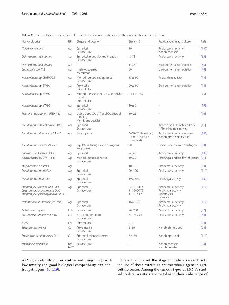
MtNPs such as silver, gold, cadmium, copper, zinc, iron and selenium have applications in agriculture such as plant growth stimulation, antimicrobial and antifun-gal effects, nanofertilizers, nanobiosensors, plant micro-nutrients and plant disease control [53]. Table 1 shows a collection of probiotic species used for the synthesis of different MtNPs and their potential application in agri-culture. Silver NPs (AgNPs) are amongst the most stud-ied in biological systems and their various inhibitory and antimicrobial effects have long been known [54]. Vari-ous probiotics including gram-positive bacteria such as lactic acid bacteria, bacillus, Staphylococcus, Brevibac-terium and gram-negative sp. Including Pseudomonas and E. coli, used for AgNP production. Lactobacillus sp. have been studied significantly as potential systems for AgNP production and Sásková and colleagues have dem-onstrated high extracellular production of AgNPs from silver ions by Lactobacillus casei sp. [55]. Similarly AgNP synthesis by Lactobacillus acidophilus have been shown to provide capping and reducing activities [56]. Gold NPs (AuNPs) are widely used in agriculture as antifungal and antibacterial agents and as delivery vehicles of ferti-lizer and pesticide sensors. The use of probiotics in the synthesis of AgNPs and AuNPs also eliminates the use of toxic chemicals and solvents, thus following the prin-ciples of green chemistry [57]. Cadmium sulfide (CdS) NPs are used in a wide variety of approaches such as biological sensors that have applications in medicine as well as in agriculture [58]. CdSNPs for use as nanosen-sors can be synthesized by probiotic bacteria. Nanosen-sors are useful in pesticide residue detection and can also detect soil moisture and soil nutrient levels [58, 59]. Cop-per is an essential micronutrient that is combined with

Page 7 of 26Bahrulolum et al. J Nanobiotechnol (2021) 19:86
many proteins and metalloenzymes and have a substan-tial role in plant metabolism and nutrition. CuNPs also have higher performance than bulk copper particles due to properties such as very small size and high surface-to-volume ratio compared to materials made from larger particles. The antifungal and antibacterial activity of CuNPs against gram-positive and gram-negative bacteria and pathogenic fungi has given them many applications in health and agriculture [60]. CuNPs have antifungal activity against plant pathogenic fungi such as Fusarium oxysporum, Fusarium culmorum, Fusarium gramine-arum and Phytophthora infestans [61]. They have also been reported to act as germinators and growth stimu-lants in some plants at concentrations below 100 ppm. So far, various chemical, physical and green synthesis meth-ods have been used to synthesize CuNPs with different amounts, shapes and morphologies. Kouhkan et al. [62] reported that Lactobacillus casei is a promising source for the biosynthesis of CuNPs. Selenium is essential for the
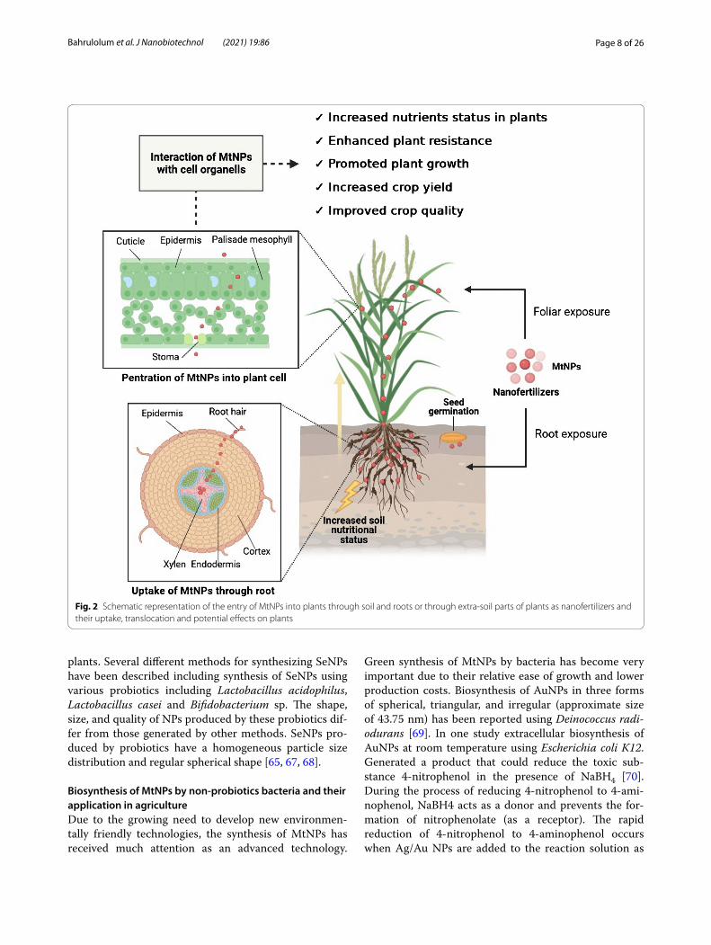
functions of most living organisms and is found in soil, water, seeds, livestock and food. Since SeNPs improve the plant’s ability to inhibit pathogens and activate antifun-gal properties, it is necessary to modify the Se content in plant nutrients by adding Se fertilizer to the soil and to balance Se in food [63]. Se-balanced food processing technology is a rapid process which helps to solve the Se imbalance issue in agriculture. Standardization of Se concentration in soil is very important and to achieve this pure Se compounds are used as fertilizer [64]. However, Se fertilizers remain in fertile topsoil during only one or few harvests and over a short period inorganic Se com-pounds are washed away by rain into the infertile hori-zons below the soil. Although the organic Se compounds are not actively leached, they are degraded quickly after applying. The advantage of SeNPs as nanofertilizers is that they do not leach slowly from the soil and do not dissolve in water or aqueous solutions [65, 66]. Figure 2 shows the potential effect of MtNPs as nanofertilizers on
Table 1 Nanoparticles synthesized by probiotic bacteria and their applications in agriculture
Probiotics NPs Production NP size (nm) Application in agriculture Refs
Lactobacillus. casei ssp. casei CCM 7088 Ag Extracellular 12–27 Plant-growth stimulator, antimicrobial effect, antifungal effect [53]
L. acidophilus Ag Extracellular 4–40 – [54]
Pseudomonas stutzeri Ag Intracellular Up to 200 Plant-growth stimulator, antimicrobial effect, antifungal effect [65]
Staphylococcus aureus Ag Extracellular 160–180 Plant-growth stimulator, antimicrobial effect, antifungal effect [83]
Brevibacterium casei Ag Extracellular 10–50 – [188]
Escherichia coli Ag Extracellular 100 Plant-growth stimulator, antimicrobial effect, antifungal effect [189]
Bacillus cereus SZT1 Ag Extracellular 4 and 5 – [190]
Bacillus licheniformis Dahb1 Ag Extracellular 18.69–63.42 Antifungal effect [191]
Lactobacillus fermentum Ag Extracellular 11.2 – [192]
Intracellular 15–40 –
Intracellular 60–80 –
Lactobacillus plantarum Ag Extracellular 19.92 ± 3.4 – [193]
Lactobacillus rhamnosus Ag Extracellular 233 – [194]
L. acidophilus 58p Ag Extracellular 30.65 ± 5.81 – [193]
Lactobacillus sp. from Yoghurt cells Ag Extracellular 15–25 – [237]
L. delbrueckii isolated from probiotic curd Ag Extracellular 54.3–112.7 – [195]
Actinobacter spp. Au Intracellular 5–500 Antimicrobial effect, antifungal effect, nano fertilizer [196]
Bacillus subtilis Au Extracellular 80 ± 0.18 – [197]
Escherichia coli k12 Au Extracellular 50 – [70]
L. casei (strain JCM1134) Au Intracellular ca.29.6 – [198]
Lactobacillus kimchicus DCY51T isolated from Korean kimchi
Au Intracellular 5–30 – [57]
Lactobacillus acidophilus DSMZ 20079T CdS Extracellular 2.5–5.5 Nanobiosensors [58]
Escherichia coli ATCC 8739Bacillus subtilis ATCC 6633
Lactobacillus casei Cop-per
Extracellular 30–75 Plant micronutrient [62]
Lactobacillus acidophilusLactobacillus caseiBifidobacterium sp.
Se Extracellular 50–50050–500400–500
Plant disease enhancerNanofertilizerNanofertilizer
[68]
Page 8 of 26Bahrulolum et al. J Nanobiotechnol (2021) 19:86
plants. Several different methods for synthesizing SeNPs have been described including synthesis of SeNPs using various probiotics including Lactobacillus acidophilus, Lactobacillus casei and Bifidobacterium sp. The shape, size, and quality of NPs produced by these probiotics dif-fer from those generated by other methods. SeNPs pro-duced by probiotics have a homogeneous particle size distribution and regular spherical shape [65, 67, 68].
Biosynthesis of MtNPs by non‑probiotics bacteria and their application in agricultureDue to the growing need to develop new environmen-tally friendly technologies, the synthesis of MtNPs has received much attention as an advanced technology.
Green synthesis of MtNPs by bacteria has become very important due to their relative ease of growth and lower production costs. Biosynthesis of AuNPs in three forms of spherical, triangular, and irregular (approximate size of 43.75 nm) has been reported using Deinococcus radi-odurans [69]. In one study extracellular biosynthesis of AuNPs at room temperature using Escherichia coli K12. Generated a product that could reduce the toxic sub-stance 4-nitrophenol in the presence of NaBH4 [70]. During the process of reducing 4-nitrophenol to 4-ami-nophenol, NaBH4 acts as a donor and prevents the for-mation of nitrophenolate (as a receptor). The rapid reduction of 4-nitrophenol to 4-aminophenol occurs when Ag/Au NPs are added to the reaction solution as
Fig. 2 Schematic representation of the entry of MtNPs into plants through soil and roots or through extra-soil parts of plants as nanofertilizers and their uptake, translocation and potential effects on plants

Page 9 of 26Bahrulolum et al. J Nanobiotechnol (2021) 19:86
a catalyst, which can be confirmed using the visible UV spectrum [71]. 4-Nitrophenol is a highly toxic organic compound and one of the most resistant contaminants in the effluents of various industries such as textile and dyeing. By spreading to the environment, this compound can contaminate soil and water leading to adverse effects on the central nervous system, liver and blood after ingestion of food grown in the contaminated areas. The development of a simple and effective method for the elimination or reduction of non-biodegradable bio pol-lutants into non-hazardous products is one of the serious challenges in environmental studies and agricultural sys-tems. The product of chemical reduction of 4-nitrophenol is a useful and important compound called 4-aminophe-nol, which does not pose the risks of toxicity of 4-nitro-phenol to the environment. The use of environmentally friendly green synthesis for produce nanoparticles as low-cost catalysts is a convenient method to chemically reduce toxic dyes such as 4-nitrophenol. MtNPs derive their catalytic capacity from their high surface-to-vol-ume ratio. Due to their high adsorption level, MtNPs can provide conditions that increase the adsorption of the reactants on their surface and thus increase the reac-tion rate and reduce the activation energy level [72]. An Acinetobacter sp. species was able to synthesize AuNPs at 37 °C, pH 7, when treated with tetra-chloroauric acid (HAuCl4). These AuNPs were monodisperse or spheri-cal and had antioxidant activity [73]. In a study of the biosynthesis of AuNPs using Acinetobacter sp. SW30 addition of HAuCl4 resulted in the biosynthesis of 10 to 20 nm polyhedral AuNPs. As the pH was increased to 9 and the temperature increased to 50 °C, more AuNPs were released into the solution [74]. Acinetobacter sp. SW30 has also been used at 30 °C and pH 7 to produce AuNPs with a monodisperse spherical shape and size of approximately 19 nm [75]. Reports indicate that filamen-tous cyanobacteria can biosynthesize AuNPs structures in various shapes, such as cubic, spherical, and octagonal, from the complexes of Au+-S2O−2
3 and Au3+-NaCl [76, 77]. A Cyanothece sp. was able to synthesis AuNPs in the size range of 80 to 129 nm [78]. The first step in the inter-action of cyanobacterium with Au3+ aqueous Cl− is the deposition of NP sulfur Au+ on the cell wall and in the next step octagonal platelets forms of Au3+ are formed in solutions close to cell surfaces [77]. Plectonema bory-anum UTEX 485, in the presence of S2O3, was able to biosynthesize cubic form (sizes ranged from 10 to 25 nm) AuNPs in membrane vesicles. These bacteria also pre-cipitated AuNPs in the form of octahedral platelets when incubated with AuCl4− [76]. Electron transfer in the pro-cess of photosynthesis affects the biosynthesis of AuNPs in cyanobacterium cell wall. Cell membrane composi-tions in cyanobacteria can produce AuNPs by affecting
the re-accumulation of gold in the cell wall. In general, at neutral pH, the biosynthesis of AuNPs takes place mostly in the periplasmic region of cyanobacteria. As the pH becomes more acidic, the more the synthesized AuNPs show different sizes and morphologies. Small AuNPs are deposited on bacterial cell walls at pH 2.0, while larger particles could be observed in the extracellular matrix. In general, changes in solution pH are a very influential factor in appearance and structure, as well as deposition location (extracellular or intracellular) of AuNPs [79]. Extracellular AgNP biosynthesis was demonstrated using Pseudomonas DC5 and Pseudomonas CA 417 [11]. In one study, the specificity of metal ion accumulation in the biosynthesis of AgNPs by Pseudomonas stutzeri AG259 was used to produce a range of shapes and sizes [80]. In one study, Acinetobacter sp. GWRFH45 biosynthesized AgNps [81]. Rapid biosynthesis of AgNps by Enterobacte-riaceae has also been reported [82]. The reduction of Ag+ ions in Staphylococcus aureus led to the biosynthesis of AgNPs [83]. The use of bacterial cell culture supernatant to generate AgNPs of various shapes and sizes has been reported in several other studies [84]. In general, in the AgNPs biosynthesis cycle, the presence of nitrate ions in the presence of NADPH-dependent nitrate reductase enzymes (for free electron transfer) reduces the bioavail-ability of silver ions and ultimately causes spherical bio-synthesis of AgNPs [79]. Au–Ag bimetallic NPs produced by a Deinococcus radiodurans synthesis system with a size of 149.8 nm showed the ability to decompose toxic triphenylmethane dye malachite green (MG) and con-vert it to the less toxic substance dimethylamino (ben-zophenone) [85]. The rapid and easy biosynthesis of a silver-gold double NPs functionalized with extremophilic Deinococcus radiodurans proteins (Drp-Au-AgNPs) led to the development of an environmentally friendly method for reducing polyphenyl from wastewater [85]. The ability of functionalized Drp-Au–Ag bimetallic MtNPs to degrade and reduce malachite green is attrib-uted to a redox reaction as well as the alkaline conditions that amplify the electrostatic force between the function-alized Drp-Au–Ag bimetallic MtNPs and the malachite green molecules. Malachite green is a group of polyphe-nolic chemical dyes that are widely used in fishponds to repel pests and insects. Malachite green effluents, if released into the environment, in addition to proven mutagenic and carcinogenic effects in humans, can cause permanent dangerous and toxic effects. Neverthe-less, the low price of green malachite is still a tempting factor to use this compound, so it can be considered an environmental problem. Although physical and chemical methods are used to remove polyphenyl compounds, the ability of nanoparticles as potential catalysts to absorb and then degrade polyphenol dyes is an efficient and

Page 10 of 26Bahrulolum et al. J Nanobiotechnol (2021) 19:86
environmentally friendly method for remediation [86]. In fact, nanobioremediation, is a new and efficient approach to clean up and remove contaminants and toxic com-pounds from the environment.
Extracellular biosynthesis of CdSNPs has been reported using Klebsiella aerogenes. The MtNPs ranged in diameter from 20 to 100 nm and their formation was highly dependent on the composition of the culture medium [87]. With the photosynthetic bacterium Rho-dopseudomonas palustris, the extracellular biosynthesis of CdSNPs of approximately about 8 nm in diameter was dependent on cell growth stage and utilized the cysteine desulfhydrase located in the cytoplasmic space to stabi-lize the CdSNPs [88]. The results of a study on an intra-cellular CdSNP biosynthesized by E. coli showed that changes in growth phases affect the rate of biosynthesis and the size of CdSNPs. The biosynthesis rate of CdSNPs with a diameter of 2 to 5 nm in the stationary phase of E. coli was about 20 times higher than found in the loga-rithmic phase [89]. Extracellular biosynthesis of spherical CuNPs of 5–50 nm in size by Streptomyces griseus and 3.6–59 nm in size in endophytic actinomycetes has been reported [90]. A new species of Desulfuromonas palmi-tatis SDBY1 converts polycarbonate organic compounds to oxidized form in the presence of F3+, because F3+ can play the role of H2 receptor and be reduced [91]. Iron-reducing bacteria need electron-donating compounds during extracellular deposition of magnetite [92]. She-wanella oneidensis was used for the biosynthesis of Fe2+ and Fe3+ as extracellular magnetite. FeCl2, along with other salts, was used to reduce Fe2+ and Fe3+. The reduc-tion of Fe2+ and Fe3+ seems to be facilitated by the trans-fer of salts by electron donation [93].
Although bacteria, viruses, and fungi are used to pro-duce nanobiosensors with different MtNPs, nanoparticles produced of bacterial origin are mostly used as nanobio-sensors in agricultural systems due to advantages such as production control, lower cost and high quality [94]. Bacterial NP-based biosensors, such as nanowires, nano-particles and nanocapsule substrates are used specifically to diagnose plant diseases and are also used in cleaning strategies related to the accumulation of pesticides and insecticides in the food sector. Quantitative detection of insecticides containing dangerous and prohibited com-pounds such as organophosphorus, carbamate com-pounds is also done using biosensors [19]. In a study on a SeNP-based agricultural sensor to detect heavy metal toxicity, Stenotrophomonas acidaminiphila was used for SeNPs biosynthesis. This study presented a colorimetric method for the detection of heavy metals during biore-mediation. In the absence of heavy metals, this pro-cess takes place naturally and the color changes to red, but in the presence of toxic heavy metals the process of
selenium green synthesis to SeNPs is inhibited and the color changes. This synthesis is dependent on NADH reductase and increasing the concentration of toxic heavy metals causes a gradual decrease in enzyme activity and discoloration [95].
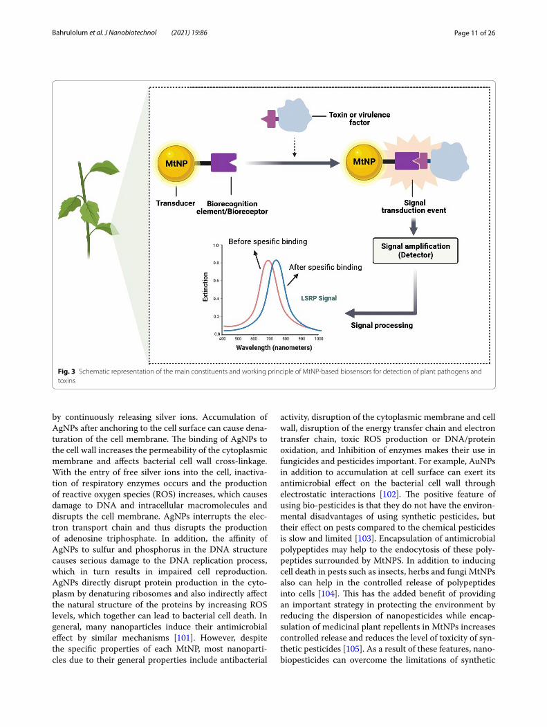
Several studies have examined the importance of using NPs as a diagnostic tool to identify a wide range of pathogenic bacteria in plants [96]. The application of nanoparticles in new technologies used in non-labora-tory rapid screening methods for the detection of plant pathogens has a significant impact on the quality of agri-cultural products. In a study by Panferov et al. [97], an enhanced and rapid method based on lateral flow immu-noassay (LFIA) was developed to detect low levels of potato leaf roll virus (PLRV) in contaminated fields. In this method, AuNPs were used as labels and silver ions were reduced at the AuNP surface to increase sensitivity [97]. In another report, infection of potato tubers with Ralstonia solanacearum was detected using an AuNP-based immunoassay. In this study, enhanced AuNP bio-synthesized approach was used to increase sensitivity in lateral flow immunoassay (LFIA). The special feature of this method was a significant reduction in time to diag-nose the cause of the infection [98]. In another study, the diagnosis of Phytophthora infestans, the causative agent of late blight in potatoes and tomatoes was performed using a combination of AuNPs-based lateral stream bio-sensor and asymmetric PCR to amplify the portion of the Ph. infestans genome. This showed that rapid detection of Phytophthora infestans in the early stages of infection can lead to appropriate management decisions to prevent the progression and spread of infection [99]. In another report, a rapid and inexpensive biosensing method was developed to identify the tomato yellow leaf ring virus genome using a AuNP-based probe and the local sur-face plasmon resonance (LSPR) method. Color changes were detected by UV–Vis spectroscopy, which indicates the presence of viral infection in the sample, eliminating the need for PCR and ELISA-dependent methods [100]. Although there are reports of successful use of MtNPs synthesized by non-biological methods in agricultural-related nanosensors, the importance of environmen-tal protection has given priority to the development of methods for green MtNPs synthesis. The working prin-ciples of MtNP-based sensors for the detection of plant pathogens and toxins shown in Fig. 3.
Bacterial-synthesized NPs such as AgNPs have shown remarkable antibacterial effects and their application increases crop productivity, reduces waste genera-tion, and saves energy and water when compared with common pesticides [37]. AgNPs are well-known anti-bacterial agents that can penetrate the bacterial cell wall and change the structure of the cell membrane
Page 11 of 26Bahrulolum et al. J Nanobiotechnol (2021) 19:86
by continuously releasing silver ions. Accumulation of AgNPs after anchoring to the cell surface can cause dena-turation of the cell membrane. The binding of AgNPs to the cell wall increases the permeability of the cytoplasmic membrane and affects bacterial cell wall cross-linkage. With the entry of free silver ions into the cell, inactiva-tion of respiratory enzymes occurs and the production of reactive oxygen species (ROS) increases, which causes damage to DNA and intracellular macromolecules and disrupts the cell membrane. AgNPs interrupts the elec-tron transport chain and thus disrupts the production of adenosine triphosphate. In addition, the affinity of AgNPs to sulfur and phosphorus in the DNA structure causes serious damage to the DNA replication process, which in turn results in impaired cell reproduction. AgNPs directly disrupt protein production in the cyto-plasm by denaturing ribosomes and also indirectly affect the natural structure of the proteins by increasing ROS levels, which together can lead to bacterial cell death. In general, many nanoparticles induce their antimicrobial effect by similar mechanisms [101]. However, despite the specific properties of each MtNP, most nanoparti-cles due to their general properties include antibacterial
activity, disruption of the cytoplasmic membrane and cell wall, disruption of the energy transfer chain and electron transfer chain, toxic ROS production or DNA/protein oxidation, and Inhibition of enzymes makes their use in fungicides and pesticides important. For example, AuNPs in addition to accumulation at cell surface can exert its antimicrobial effect on the bacterial cell wall through electrostatic interactions [102]. The positive feature of using bio-pesticides is that they do not have the environ-mental disadvantages of using synthetic pesticides, but their effect on pests compared to the chemical pesticides is slow and limited [103]. Encapsulation of antimicrobial polypeptides may help to the endocytosis of these poly-peptides surrounded by MtNPS. In addition to inducing cell death in pests such as insects, herbs and fungi MtNPs also can help in the controlled release of polypeptides into cells [104]. This has the added benefit of providing an important strategy in protecting the environment by reducing the dispersion of nanopesticides while encap-sulation of medicinal plant repellents in MtNPs increases controlled release and reduces the level of toxicity of syn-thetic pesticides [105]. As a result of these features, nano-biopesticides can overcome the limitations of synthetic
Fig. 3 Schematic representation of the main constituents and working principle of MtNP-based biosensors for detection of plant pathogens and toxins

Page 12 of 26Bahrulolum et al. J Nanobiotechnol (2021) 19:86
pesticides and biopesticides. With the use of nanopar-ticles, the active ingredients can be stabilized and made available through sustained-released giving effective and sustainable management for a long time without the haz-ards of using synthetic chemicals [106].
Several reports have evaluated the successful use of biological nanoparticles against pests. In one such study, spherical AuNPs and AgNPs biosynthesized from Haloferax volcanii were successfully used for antibac-terial applications against two gram-negative bacteria [107]. Extracellular biosynthesis of AgNPs with high antimicrobial properties has also been reported using Sporosarcina koreensis DC4 [108]. The antifungal activ-ity against Fusarium graminearum of an AgNPs biosyn-thesized by Endophytic bacteria has also been reported. In one study, biosynthesis of AgNPs was performed using Pseudomonas poae strain CO, in which the AgNPs with a diameter of approximately 20–50 nm showed antifun-gal activity [109]. Successful biosynthesis of AgNPs was reported in three strains of Endophytic Streptomyces spp. The biosynthesized NPs were spherical in shape, varying in size from at least 11 to a maximum of 63 nm, and acted against a wide range of single-celled fungi [110]. AgNPs (20 to 100 nm) biosynthesized using Pseudomonas rhode-siae culture medium supernatant showed strong antibac-terial activity against Dickeya dadantii infection in sweet potato roots [111]. A haloalkaliphilic bacterium Strep-tomyces sp. was able to biosynthesize spherical AgNPs (diameter 16 nm) with high fungicidal properties against Fusarium verticillioides, one of the main causes of infec-tion in cornfields by inhibiting ergosterol biosynthesis leading to inhibition of conidia germination and destruc-tion of the F. verticillioides membrane [112].
CuNPs biosynthesized by an actinomycetes sp. iso-lated from Convolvulus arvensis also showed significant antifungal and antibacterial activity [113]. In one study, the effect of foliar application of different concentrations of CuNPs on the accumulation of bioactive compounds and antioxidant capacity in tomato fruits was estimated. CuNPs reduced the formation of ROS by increasing the activity of superoxide dismutase and catalase enzymes. In addition, the content of vitamin C, lycopene and phenol was increased in the presence of CuNPs. The results of this study also showed that CuNPs increased the strength of tomato fruits [114]. To investigate the effect of CuNPs biosynthesized by Streptomyces griseus on fungi that cause red root rot disease, experiments were performed on infected tea plantations. Comparison of tea plants treated with the chemical fungicide carbendazim, bio-synthesized CuNPs or bulk copper showed that fungal resistance and leaf yield were higher in tea plants treated with biosynthesized CuNPs than in tea plants treated with carbendazim or bulk copper. Soil nutrients were
also increased after the use of CuNPs. This study suggests that these CuNPs can be used as fungicides in the formu-lation of nanobiofertilizers [46, 90].
Several studies have examined the effect of MtNP size on their toxicity. Although factors such as size, concen-tration and zeta potential of MtNPs show various effects on different plants, there is a significant relationship between the size of MtNPs and the degree of toxicity cre-ated for the plant with the larger MtNPs being less toxic to plants than smaller ones. In addition, studies have shown that the concentration of nanoparticles also has a significant effect on their toxicity, for example, a con-centration of more than 0.2 mg/ml CuNPs impairs plant growth and physiology [40].
The various MtNPs synthesized by non-probiotic bac-teria with their potential applications in agriculture are summarized in Table 2.
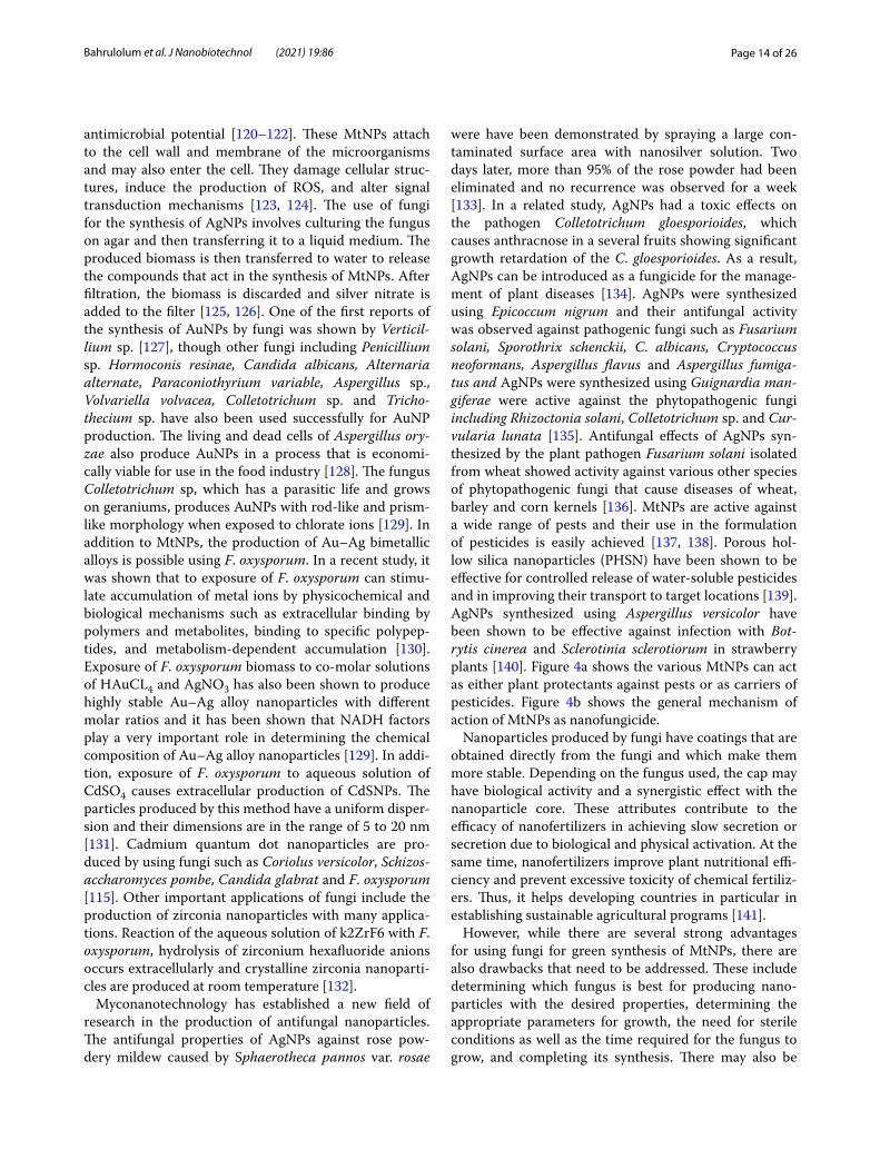
Biosynthesis of MtNPs by Fungi and their application in agricultureNanotechnology touches many fields, including agricul-ture and plant disease management. In recent years, fungi have been added to the list of microorganisms used in the production of nanoparticles. Among the various micro-organisms used to synthesize nanoparticles, fungi are effective candidates for making intracellular and extracel-lular MtNPs. Nanoparticles made using fungi have good dispersion and stability characteristics. The attractive-ness of using fungi in the production of nanoparticles is due to the presence of significant amounts of specific enzymes in these microorganisms, ease of working with them in the laboratory, scalability and financially eco-nomic growth of fungi even on an industrial scale mak-ing myconanotechnology an environmentally friendly and cost-effective option [115, 116]. Although there are several methods for synthesizing MtNPs from fungi, lit-tle is currently known about potential drawbacks and limitations. Filamentous fungi can produce a wide range of MtNPs such as gold, silver, iron oxide, and even bime-tallic nanoparticles [117, 118]. Research has shown that several different species of fungi can be used in the green synthesis MtNPs with the desired size, surface charge and morphology, and desirable properties including Pestaloti-opsis sp., Phoma sp., Humicola sp., Fusarium oxysporum, Aspergillus niger, Trichoderma sp., Hormoconis resinae, Phaenerochaete chrysosporium and Penicillium. Using fungi as reducing and stabilizing agents for the biosyn-thesis of AgNPs has been considered due to their high efficiency, ease of operation and low residual toxicity. The mechanisms of synthesis are not yet fully understood, but synthesis can be optimized by adjusting parameters such as silver salt concentration, biomass, temperature, pH and fungal cultivation time. As with bacterial produced
Page 13 of 26Bahrulolum et al. J Nanobiotechnol (2021) 19:86
AgNPs, similar structures synthesized using fungi, with low toxicity and good biological compatibility, can con-trol pathogens [40, 119].
These findings set the stage for future research into the use of these MtNPs as antimicrobials agent in agri-culture sector. Among the various types of MtNPs stud-ied to date, AgNPs stand out due to their wide range of
Table 2 Non-probiotic resources for the biosynthesis nanoparticles and their applications in agriculture
Non-probiotics NPs Shape and location Size (nm) Applications in agriculture Refs.
Haloferax volcanii Au SphericalExtracellular
10 Antibacterial activityNanobiosensors
[107]
Deinococcus radiodurans Au Spherical, triangular and irregularExtracellular
43.75 Antibacterial activity [69]
Deinococcus radiodurans Au - 149.8 Environmental remediation [85]
Escherichia coli K12 Au Highly dispersedMembrane
50 Environmental remediation [70]
Acinetobacter sp. GWRVA25 Au Monodispersed and sphericalExtracellular
15 ± 10 Antioxidant activity [73]
Acinetobacter sp. SW30 Au PolyhedralIntracellular
20 ± 10 Environmental remediation [74]
Acinetobacter sp. SW30 Au Monodispersed spherical and polyhe-dral
Intracellular
~ 19 to ~ 39 – [75]
Acinetobacter sp. SW30 Au SphericalExtracellular
10 ± 2 – [199]
Plectonemaboryanum UTEX 485 Au Cubic [Au (S2O3)23−] and Octahedral
[AuCl4−]
Membrane vesicles
10–25 – [76]
Pseudomonas deceptionensis DC5 Ag SphericalExtracellular
– Antimicrobial activity and bio-film inhibition activity
[11]
Pseudomonas fluorescens CA 417 Ag Polydisperse 5–50 (TEM method) and 20.66 (DLS method)
Antibacterial activity againstNanobiopesticide feature
[200]
Pseudomonas stutzeri AG259 Ag Equilateral triangles and hexagonsPeriplasmic
200 Biocide and antimicrobial agent [80]
Sporosarcina koreensis DC4 Ag Spherical varied Antibacterial activity [108]
Acinetobacter sp. GWRFH 45 Ag Monodispersed sphericalExtracellular
10 ± 5 Antifungal and biofilm inhibition [81]
Staphylococcus aureus Ag - 10–15 Antibacterial activity [83]
Pseudomonas rhodesiae Ag SphericalExtracellular
20–100 Antibacterial activity [111]
Pseudomonas poae CO Ag SphericalExtracellular
19.8–44.9 Antifungal activity [109]
Streptomyces capillispiralis Ca-1Streptomyces zaomyceticus Oc-5Streptomyces pseudogriseolus Acv-11
Ag SphericalExtracellular
23.77–63.1411.32–36.7211.70–44.73
Antibacterial activityAntifungal activityBiocatalystsLarvicidal
[110]
Haloalkaliphilic Streptomyces spp. Ag SphericalExtracellular
16.4 ± 2.2 Antibacterial activityAntifungal activity
[112]
Klebsiella aerogenes CdS Extracellular 20–200 Antibacterial activity [87]
Rhodopseudomonas palustris Cd Face-centered cubicExtracellular
8.01 ± 0.25 Antibacterial activity [88]
E. coli Cd Intracellular 2–5 – [89]
Streptomyces griseus Cu PolydisperseExtracellular
5–50 Nanobiofungicides [90]
Endophytic actinomycetes Ca-1 Cu Spherical-monodispersedExtracellular
3.6–59 Nanobiopesticide [113]
Shewanella oneidensis Fe2+
Fe3+Extracellular – Nanobiosensors
Nanobiomarker[93]
Page 14 of 26Bahrulolum et al. J Nanobiotechnol (2021) 19:86
antimicrobial potential [120–122]. These MtNPs attach to the cell wall and membrane of the microorganisms and may also enter the cell. They damage cellular struc-tures, induce the production of ROS, and alter signal transduction mechanisms [123, 124]. The use of fungi for the synthesis of AgNPs involves culturing the fungus on agar and then transferring it to a liquid medium. The produced biomass is then transferred to water to release the compounds that act in the synthesis of MtNPs. After filtration, the biomass is discarded and silver nitrate is added to the filter [125, 126]. One of the first reports of the synthesis of AuNPs by fungi was shown by Verticil-lium sp. [127], though other fungi including Penicillium sp. Hormoconis resinae, Candida albicans, Alternaria alternate, Paraconiothyrium variable, Aspergillus sp., Volvariella volvacea, Colletotrichum sp. and Tricho-thecium sp. have also been used successfully for AuNP production. The living and dead cells of Aspergillus ory-zae also produce AuNPs in a process that is economi-cally viable for use in the food industry [128]. The fungus Colletotrichum sp, which has a parasitic life and grows on geraniums, produces AuNPs with rod-like and prism-like morphology when exposed to chlorate ions [129]. In addition to MtNPs, the production of Au–Ag bimetallic alloys is possible using F. oxysporum. In a recent study, it was shown that to exposure of F. oxysporum can stimu-late accumulation of metal ions by physicochemical and biological mechanisms such as extracellular binding by polymers and metabolites, binding to specific polypep-tides, and metabolism-dependent accumulation [130]. Exposure of F. oxysporum biomass to co-molar solutions of HAuCL4 and AgNO3 has also been shown to produce highly stable Au–Ag alloy nanoparticles with different molar ratios and it has been shown that NADH factors play a very important role in determining the chemical composition of Au–Ag alloy nanoparticles [129]. In addi-tion, exposure of F. oxysporum to aqueous solution of CdSO4 causes extracellular production of CdSNPs. The particles produced by this method have a uniform disper-sion and their dimensions are in the range of 5 to 20 nm [131]. Cadmium quantum dot nanoparticles are pro-duced by using fungi such as Coriolus versicolor, Schizos-accharomyces pombe, Candida glabrat and F. oxysporum [115]. Other important applications of fungi include the production of zirconia nanoparticles with many applica-tions. Reaction of the aqueous solution of k2ZrF6 with F. oxysporum, hydrolysis of zirconium hexafluoride anions occurs extracellularly and crystalline zirconia nanoparti-cles are produced at room temperature [132].
Myconanotechnology has established a new field of research in the production of antifungal nanoparticles. The antifungal properties of AgNPs against rose pow-dery mildew caused by Sphaerotheca pannos var. rosae
were have been demonstrated by spraying a large con-taminated surface area with nanosilver solution. Two days later, more than 95% of the rose powder had been eliminated and no recurrence was observed for a week [133]. In a related study, AgNPs had a toxic effects on the pathogen Colletotrichum gloesporioides, which causes anthracnose in a several fruits showing significant growth retardation of the C. gloesporioides. As a result, AgNPs can be introduced as a fungicide for the manage-ment of plant diseases [134]. AgNPs were synthesized using Epicoccum nigrum and their antifungal activity was observed against pathogenic fungi such as Fusarium solani, Sporothrix schenckii, C. albicans, Cryptococcus neoformans, Aspergillus flavus and Aspergillus fumiga-tus and AgNPs were synthesized using Guignardia man-giferae were active against the phytopathogenic fungi including Rhizoctonia solani, Colletotrichum sp. and Cur-vularia lunata [135]. Antifungal effects of AgNPs syn-thesized by the plant pathogen Fusarium solani isolated from wheat showed activity against various other species of phytopathogenic fungi that cause diseases of wheat, barley and corn kernels [136]. MtNPs are active against a wide range of pests and their use in the formulation of pesticides is easily achieved [137, 138]. Porous hol-low silica nanoparticles (PHSN) have been shown to be effective for controlled release of water-soluble pesticides and in improving their transport to target locations [139]. AgNPs synthesized using Aspergillus versicolor have been shown to be effective against infection with Bot-rytis cinerea and Sclerotinia sclerotiorum in strawberry plants [140]. Figure 4a shows the various MtNPs can act as either plant protectants against pests or as carriers of pesticides. Figure 4b shows the general mechanism of action of MtNPs as nanofungicide.
Nanoparticles produced by fungi have coatings that are obtained directly from the fungi and which make them more stable. Depending on the fungus used, the cap may have biological activity and a synergistic effect with the nanoparticle core. These attributes contribute to the efficacy of nanofertilizers in achieving slow secretion or secretion due to biological and physical activation. At the same time, nanofertilizers improve plant nutritional effi-ciency and prevent excessive toxicity of chemical fertiliz-ers. Thus, it helps developing countries in particular in establishing sustainable agricultural programs [141].
However, while there are several strong advantages for using fungi for green synthesis of MtNPs, there are also drawbacks that need to be addressed. These include determining which fungus is best for producing nano-particles with the desired properties, determining the appropriate parameters for growth, the need for sterile conditions as well as the time required for the fungus to grow, and completing its synthesis. There may also be

Page 15 of 26Bahrulolum et al. J Nanobiotechnol (2021) 19:86
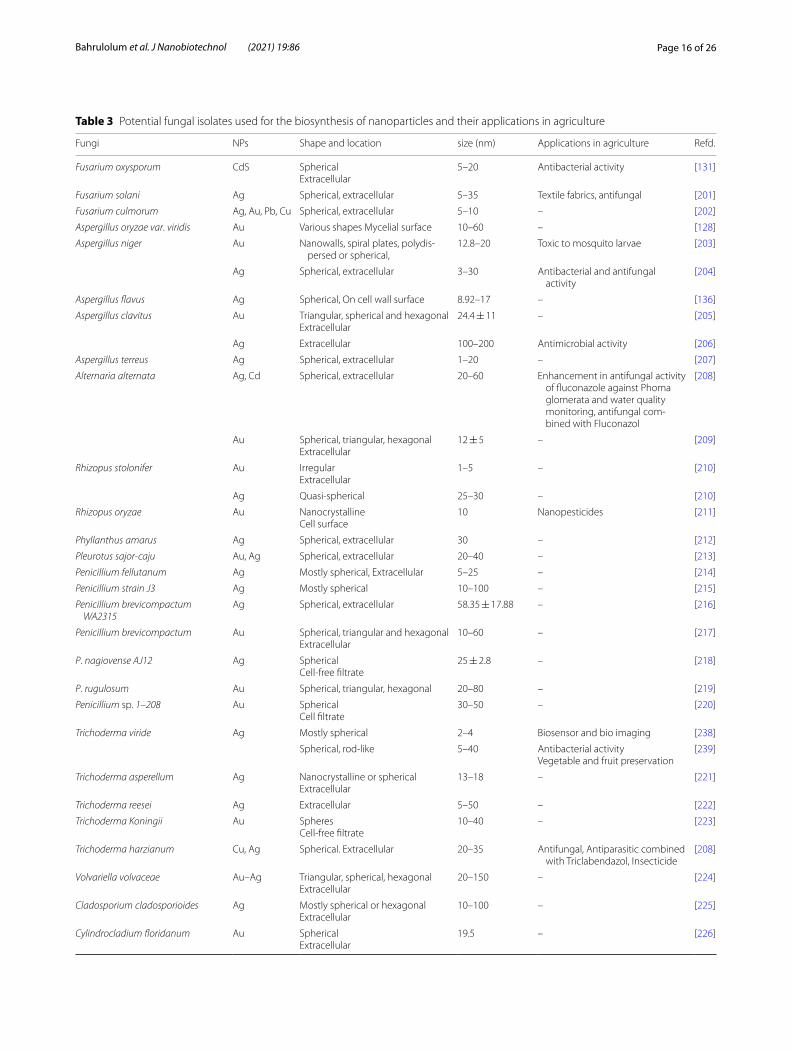
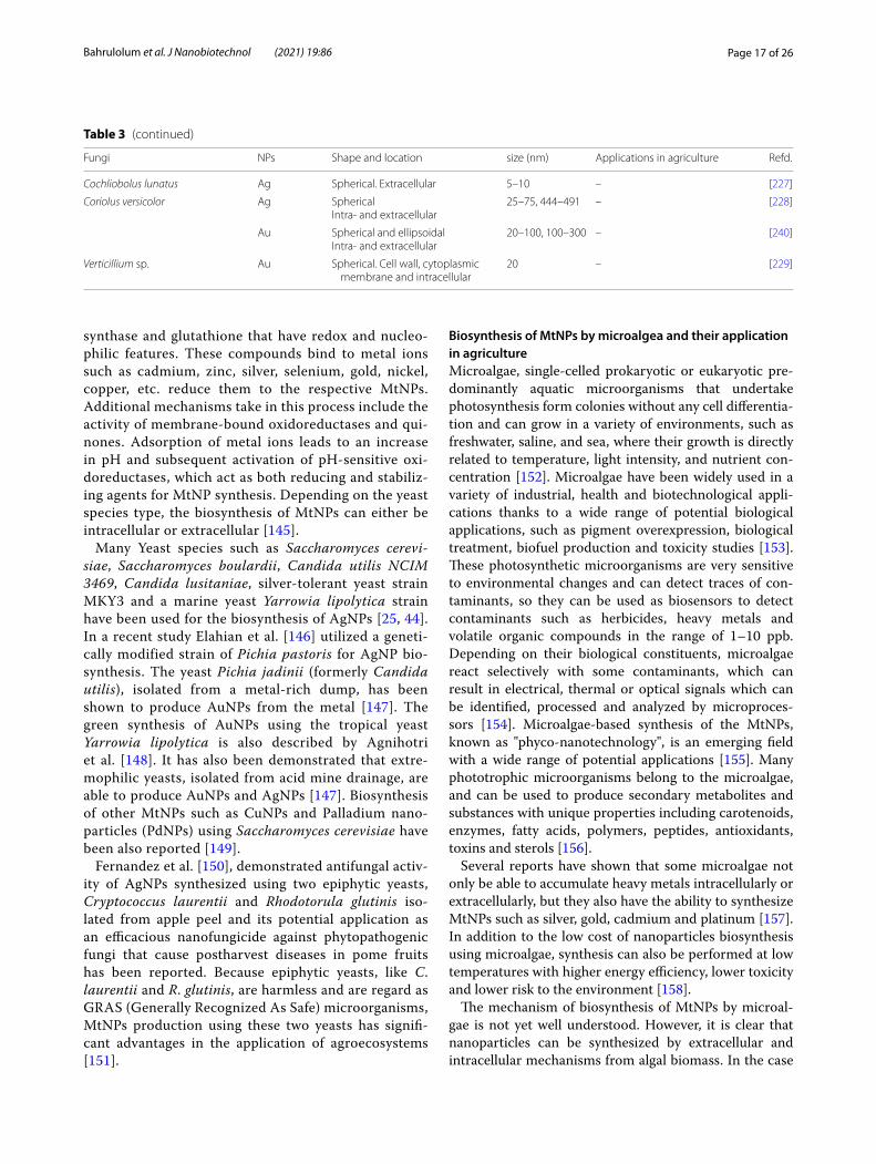
problems with scale-up production, including the need to further investigate the mechanisms by which cap layers are formed and the molecules contained in them. While more research is needed, studies showed that using fungi for the green synthesis of MtNPs has the potential to address a wide range of possible applications especially for the control of pests [135]. A summary of some fun-gal sources for the production of MtNPs with specific characteristics and potential applications in agriculture is shown in Table 3.
Biosynthesis of MtNPs by yeasts and their application in agricultureYeasts are the unicellular microorganisms that repro-duce during an asymmetric cell division process called budding and can be categorized as Ascomycetes such as Saccharomyces and Candida or Basidiomycetes such as Filobasidiella and Rhodotorula [142]. In addi-tion to traditionally use of yeasts for production of sev-eral fermented food such as alcoholic beverages and
bakery products modern application of yeasts include the production of heterologous compounds, single cell protein (SCP) and their use in the biofuels industry [142]. Yeasts also play an important role in agricul-ture as biological control agents, biological treatments and as indicators of a quality environment [143]. They grow easily on low-cost media and can adapt to harsh environmental conditions such as a wide range of tem-perature and pH and high concentrated organic and inorganic pollutants. Yeasts have the inherent abil-ity to absorb and accumulate large concentrations of toxic metal ions from the environment and can adapt themselves to this environmental stress using vari-ous detoxification mechanisms such as mobilization, immobilization or metals transformation. These biore-mediation mechanism of yeasts can play key roles for the green synthesis of MtNPs [144]. The stress caused by the presence of metal ions leads to activate a meta-bolic cascade of chemical reactions for the synthesis of stress-relieving compounds such as phytochelatin
Fig. 4 Application of MtNPs as nanopesticides: a MtNPs act as nanopesticides targeting a wide range of pests and phytopathogenic agents and as a carrier for pesticides to provide crop protection, b Mechanisms of action of MtNPs as nanofungicides. MtNPs act on the fungus cell wall, leading to membrane damage. Disruption of the membrane by MtNPs causes pore formation. After internalization, MtNPs target main cellular organs such as the nucleus, ribosomes and mitochondria, causing cell death
Page 16 of 26Bahrulolum et al. J Nanobiotechnol (2021) 19:86
Table 3 Potential fungal isolates used for the biosynthesis of nanoparticles and their applications in agriculture
Fungi NPs Shape and location size (nm) Applications in agriculture Refd.
Fusarium oxysporum CdS SphericalExtracellular
5–20 Antibacterial activity [131]
Fusarium solani Ag Spherical, extracellular 5–35 Textile fabrics, antifungal [201]
Fusarium culmorum Ag, Au, Pb, Cu Spherical, extracellular 5–10 – [202]
Aspergillus oryzae var. viridis Au Various shapes Mycelial surface 10–60 – [128]
Aspergillus niger Au Nanowalls, spiral plates, polydis-persed or spherical,
12.8–20 Toxic to mosquito larvae [203]
Ag Spherical, extracellular 3–30 Antibacterial and antifungal activity
[204]
Aspergillus flavus Ag Spherical, On cell wall surface 8.92–17 – [136]
Aspergillus clavitus Au Triangular, spherical and hexagonalExtracellular
24.4 ± 11 – [205]
Ag Extracellular 100–200 Antimicrobial activity [206]
Aspergillus terreus Ag Spherical, extracellular 1–20 – [207]
Alternaria alternata Ag, Cd Spherical, extracellular 20–60 Enhancement in antifungal activity of fluconazole against Phoma glomerata and water quality monitoring, antifungal com-bined with Fluconazol
[208]
Au Spherical, triangular, hexagonalExtracellular
12 ± 5 – [209]
Rhizopus stolonifer Au IrregularExtracellular
1–5 – [210]
Ag Quasi-spherical 25–30 – [210]
Rhizopus oryzae Au NanocrystallineCell surface
10 Nanopesticides [211]
Phyllanthus amarus Ag Spherical, extracellular 30 – [212]
Pleurotus sajor-caju Au, Ag Spherical, extracellular 20–40 – [213]
Penicillium fellutanum Ag Mostly spherical, Extracellular 5–25 – [214]
Penicillium strain J3 Ag Mostly spherical 10–100 – [215]
Penicillium brevicompactum WA2315
Ag Spherical, extracellular 58.35 ± 17.88 – [216]
Penicillium brevicompactum Au Spherical, triangular and hexagonalExtracellular
10–60 – [217]
P. nagiovense AJ12 Ag SphericalCell-free filtrate
25 ± 2.8 – [218]
P. rugulosum Au Spherical, triangular, hexagonal 20–80 – [219]
Penicillium sp. 1–208 Au SphericalCell filtrate
30–50 – [220]
Trichoderma viride Ag Mostly spherical 2–4 Biosensor and bio imaging [238]
Spherical, rod-like 5–40 Antibacterial activityVegetable and fruit preservation
[239]
Trichoderma asperellum Ag Nanocrystalline or sphericalExtracellular
13–18 – [221]
Trichoderma reesei Ag Extracellular 5–50 – [222]
Trichoderma Koningii Au SpheresCell-free filtrate
10–40 – [223]
Trichoderma harzianum Cu, Ag Spherical. Extracellular 20–35 Antifungal, Antiparasitic combined with Triclabendazol, Insecticide
[208]
Volvariella volvaceae Au–Ag Triangular, spherical, hexagonalExtracellular
20–150 – [224]
Cladosporium cladosporioides Ag Mostly spherical or hexagonalExtracellular
10–100 – [225]
Cylindrocladium floridanum Au SphericalExtracellular
19.5 – [226]
Page 17 of 26Bahrulolum et al. J Nanobiotechnol (2021) 19:86
synthase and glutathione that have redox and nucleo-philic features. These compounds bind to metal ions such as cadmium, zinc, silver, selenium, gold, nickel, copper, etc. reduce them to the respective MtNPs. Additional mechanisms take in this process include the activity of membrane-bound oxidoreductases and qui-nones. Adsorption of metal ions leads to an increase in pH and subsequent activation of pH-sensitive oxi-doreductases, which act as both reducing and stabiliz-ing agents for MtNP synthesis. Depending on the yeast species type, the biosynthesis of MtNPs can either be intracellular or extracellular [145].
Many Yeast species such as Saccharomyces cerevi-siae, Saccharomyces boulardii, Candida utilis NCIM 3469, Candida lusitaniae, silver-tolerant yeast strain MKY3 and a marine yeast Yarrowia lipolytica strain have been used for the biosynthesis of AgNPs [25, 44]. In a recent study Elahian et al. [146] utilized a geneti-cally modified strain of Pichia pastoris for AgNP bio-synthesis. The yeast Pichia jadinii (formerly Candida utilis), isolated from a metal-rich dump, has been shown to produce AuNPs from the metal [147]. The green synthesis of AuNPs using the tropical yeast Yarrowia lipolytica is also described by Agnihotri et al. [148]. It has also been demonstrated that extre-mophilic yeasts, isolated from acid mine drainage, are able to produce AuNPs and AgNPs [147]. Biosynthesis of other MtNPs such as CuNPs and Palladium nano-particles (PdNPs) using Saccharomyces cerevisiae have been also reported [149].
Fernandez et al. [150], demonstrated antifungal activ-ity of AgNPs synthesized using two epiphytic yeasts, Cryptococcus laurentii and Rhodotorula glutinis iso-lated from apple peel and its potential application as an efficacious nanofungicide against phytopathogenic fungi that cause postharvest diseases in pome fruits has been reported. Because epiphytic yeasts, like C. laurentii and R. glutinis, are harmless and are regard as GRAS (Generally Recognized As Safe) microorganisms, MtNPs production using these two yeasts has signifi-cant advantages in the application of agroecosystems [151].
Biosynthesis of MtNPs by microalgea and their application in agricultureMicroalgae, single-celled prokaryotic or eukaryotic pre-dominantly aquatic microorganisms that undertake photosynthesis form colonies without any cell differentia-tion and can grow in a variety of environments, such as freshwater, saline, and sea, where their growth is directly related to temperature, light intensity, and nutrient con-centration [152]. Microalgae have been widely used in a variety of industrial, health and biotechnological appli-cations thanks to a wide range of potential biological applications, such as pigment overexpression, biological treatment, biofuel production and toxicity studies [153]. These photosynthetic microorganisms are very sensitive to environmental changes and can detect traces of con-taminants, so they can be used as biosensors to detect contaminants such as herbicides, heavy metals and volatile organic compounds in the range of 1–10 ppb. Depending on their biological constituents, microalgae react selectively with some contaminants, which can result in electrical, thermal or optical signals which can be identified, processed and analyzed by microproces-sors [154]. Microalgae-based synthesis of the MtNPs, known as "phyco-nanotechnology", is an emerging field with a wide range of potential applications [155]. Many phototrophic microorganisms belong to the microalgae, and can be used to produce secondary metabolites and substances with unique properties including carotenoids, enzymes, fatty acids, polymers, peptides, antioxidants, toxins and sterols [156].
Several reports have shown that some microalgae not only be able to accumulate heavy metals intracellularly or extracellularly, but they also have the ability to synthesize MtNPs such as silver, gold, cadmium and platinum [157]. In addition to the low cost of nanoparticles biosynthesis using microalgae, synthesis can also be performed at low temperatures with higher energy efficiency, lower toxicity and lower risk to the environment [158].
The mechanism of biosynthesis of MtNPs by microal-gae is not yet well understood. However, it is clear that nanoparticles can be synthesized by extracellular and intracellular mechanisms from algal biomass. In the case
Table 3 (continued)
Fungi NPs Shape and location size (nm) Applications in agriculture Refd.
Cochliobolus lunatus Ag Spherical. Extracellular 5–10 – [227]
Coriolus versicolor Ag SphericalIntra- and extracellular
25–75, 444–491 – [228]
Au Spherical and ellipsoidalIntra- and extracellular
20–100, 100–300 – [240]
Verticillium sp. Au Spherical. Cell wall, cytoplasmic membrane and intracellular
20 – [229]
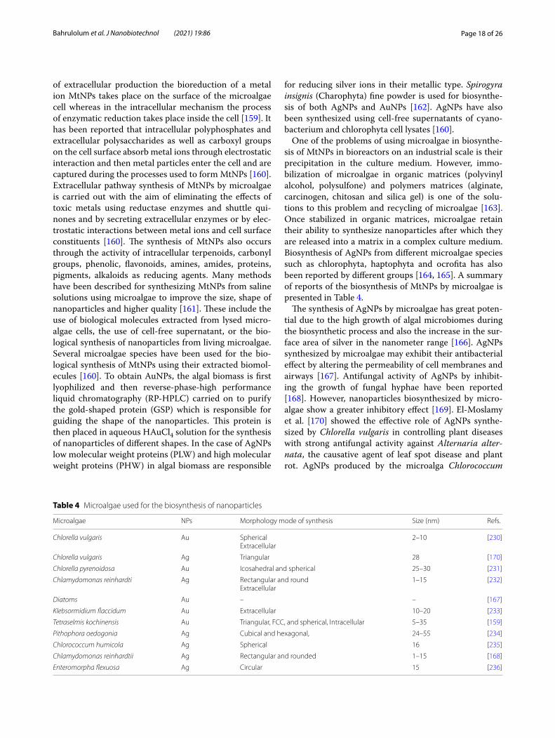
Page 18 of 26Bahrulolum et al. J Nanobiotechnol (2021) 19:86
of extracellular production the bioreduction of a metal ion MtNPs takes place on the surface of the microalgae cell whereas in the intracellular mechanism the process of enzymatic reduction takes place inside the cell [159]. It has been reported that intracellular polyphosphates and extracellular polysaccharides as well as carboxyl groups on the cell surface absorb metal ions through electrostatic interaction and then metal particles enter the cell and are captured during the processes used to form MtNPs [160]. Extracellular pathway synthesis of MtNPs by microalgae is carried out with the aim of eliminating the effects of toxic metals using reductase enzymes and shuttle qui-nones and by secreting extracellular enzymes or by elec-trostatic interactions between metal ions and cell surface constituents [160]. The synthesis of MtNPs also occurs through the activity of intracellular terpenoids, carbonyl groups, phenolic, flavonoids, amines, amides, proteins, pigments, alkaloids as reducing agents. Many methods have been described for synthesizing MtNPs from saline solutions using microalgae to improve the size, shape of nanoparticles and higher quality [161]. These include the use of biological molecules extracted from lysed micro-algae cells, the use of cell-free supernatant, or the bio-logical synthesis of nanoparticles from living microalgae. Several microalgae species have been used for the bio-logical synthesis of MtNPs using their extracted biomol-ecules [160]. To obtain AuNPs, the algal biomass is first lyophilized and then reverse-phase-high performance liquid chromatography (RP-HPLC) carried on to purify the gold-shaped protein (GSP) which is responsible for guiding the shape of the nanoparticles. This protein is then placed in aqueous HAuCl4 solution for the synthesis of nanoparticles of different shapes. In the case of AgNPs low molecular weight proteins (PLW) and high molecular weight proteins (PHW) in algal biomass are responsible
for reducing silver ions in their metallic type. Spirogyra insignis (Charophyta) fine powder is used for biosynthe-sis of both AgNPs and AuNPs [162]. AgNPs have also been synthesized using cell-free supernatants of cyano-bacterium and chlorophyta cell lysates [160].
One of the problems of using microalgae in biosynthe-sis of MtNPs in bioreactors on an industrial scale is their precipitation in the culture medium. However, immo-bilization of microalgae in organic matrices (polyvinyl alcohol, polysulfone) and polymers matrices (alginate, carcinogen, chitosan and silica gel) is one of the solu-tions to this problem and recycling of microalgae [163]. Once stabilized in organic matrices, microalgae retain their ability to synthesize nanoparticles after which they are released into a matrix in a complex culture medium. Biosynthesis of AgNPs from different microalgae species such as chlorophyta, haptophyta and ocrofita has also been reported by different groups [164, 165]. A summary of reports of the biosynthesis of MtNPs by microalgae is presented in Table 4.
The synthesis of AgNPs by microalgae has great poten-tial due to the high growth of algal microbiomes during the biosynthetic process and also the increase in the sur-face area of silver in the nanometer range [166]. AgNPs synthesized by microalgae may exhibit their antibacterial effect by altering the permeability of cell membranes and airways [167]. Antifungal activity of AgNPs by inhibit-ing the growth of fungal hyphae have been reported [168]. However, nanoparticles biosynthesized by micro-algae show a greater inhibitory effect [169]. El-Moslamy et al. [170] showed the effective role of AgNPs synthe-sized by Chlorella vulgaris in controlling plant diseases with strong antifungal activity against Alternaria alter-nata, the causative agent of leaf spot disease and plant rot. AgNPs produced by the microalga Chlorococcum
Table 4 Microalgae used for the biosynthesis of nanoparticles
Microalgae NPs Morphology mode of synthesis Size (nm) Refs.
Chlorella vulgaris Au SphericalExtracellular
2–10 [230]
Chlorella vulgaris Ag Triangular 28 [170]
Chlorella pyrenoidosa Au Icosahedral and spherical 25–30 [231]
Chlamydomonas reinhardti Ag Rectangular and roundExtracellular
1–15 [232]
Diatoms Au – – [167]
Klebsormidium flaccidum Au Extracellular 10–20 [233]
Tetraselmis kochinensis Au Triangular, FCC, and spherical, Intracellular 5–35 [159]
Pithophora oedogonia Ag Cubical and hexagonal, 24–55 [234]
Chlorococcum humicola Ag Spherical 16 [235]
Chlamydomonas reinhardtii Ag Rectangular and rounded 1–15 [168]
Enteromorpha flexuosa Ag Circular 15 [236]

Page 19 of 26Bahrulolum et al. J Nanobiotechnol (2021) 19:86
humicola with the help of microalgal biomass activity against Candida albicans, Aspergillus niger and Aspergil-lus flavus showed significant growth inhibition against C. albicans. Biomass containing Chlorella sp. and Haemato-coccus Candida albicanspluvialis inhibited the growth of Penicillium expansum, the main cause of loss of quality and quantity of fruit after harvest [152, 171].
Challenges and future direction of using MtNPs in agricultureGreen synthesis of MtNPs using microorganisms is a promising and environmentally friendly approach for agricultural applications such as nanofertilizers, nanope-sticides and nanobiosensors. Given their potential wide-spread use in the future it is likely that large volumes of MtNPs produced by different methods will enter ecosys-tems [172]. Despite the favorable physical and chemical properties of MtNPs, the complexity of soil-crop ecosys-tems means that the environmental behaviors of these nanoparticles are not yet fully predictable after use, and this remains an important challenge [173]. Therefore, before fully utilizing their potential, it is necessary to evaluate the effects and interaction with living systems. At this stage, screening of nanomaterials is essential to assess their potential toxicity and to understand their mechanisms of action to prevent their adverse effects in the future [174].
The nanoscale dimensions of MtNPs, which deter-mines many of their beneficial properties, can poten-tially also increase their potential adverse effects [172]. The toxicity of MtNPs is influenced by various factors such as solubility and their binding specificity to biologi-cal sites [175]. Several studies have shown the unpleas-ant aspect of long-term exposure to some MtNPs such as AuNPs and AgNPs. In a study by Vecchio et al. [174] the in vivo toxicity of AuNPs in Drosophila melanogaster was evaluated. Due to the mutations that can be passed on to offspring, significant phenotypic changes were observed in later generations of Drosophila after treatment with AuNPs, indicating the potential severity of AuNP tox-icity. These findings provide important evidence of the adverse effects of AuNPs on the growth and development of organisms. These studies also demonstrate the need for reliable evaluation of the toxicological properties of nanomaterials and the need for significant efforts by the nanoscience community to produce biocompatible nano-materials without any adverse effects on human health and the environment [174].
AgNPs are primarily produced for antiseptic applica-tions and have potential antimicrobial activity against many pathogenic microorganisms. However, together with this favorable feature, AgNPs also show imper-missible toxic effects on human health and ecosystems.
Ecologists have warned that if these nano-antimicrobials are released into the environment, their spread could have serious negative consequences for other microor-ganisms in natural ecosystems. There is ample evidence that AgNPs are not only toxic to bacteria, but also to the cells of other organisms such as brain cells, liver cells, and stem cells, which can lead to severe damage [175]. MtNPs cause toxicity through important cellular processes such as increased levels of ROS, decreased intracellular glutathione levels, and decreased mitochondrial mem-brane potential. AgNPs can adversely affect on cells and embryos of freshwater fish. In one study, the toxic effects of AgNPs on adult Japanese rice fish (Medaka, Oryzias latipes) were evaluated by exposure to these nanoparti-cles. The results showed a decrease in the activity of lac-tate dehydrogenase and antioxidant enzymes in the liver, glutathione depletion and lipid peroxidation in the liver and gills, with varying degrees of histological lesions in the tissues [176].
Several studies have shown that MtNPs can also have an adverse effects on key major elements (plant, soil and water) in agroecosystems [25]. Generally MtNPs can enter the agricultural ecosystem through both direct and indirect routes [173]. MtNPs used for agricultural appli-cations can enter soil, climate, and atmosphere through washing, rainfall, airflow, and trophic transfer. Various studies have shown that these MtNPs may be absorbed by microorganisms in the soil, sediments and plant roots. These MtNPs are then transferred from the roots to other parts of the plants where they can accumulate [25]. Accumulation as a key behavior of MtNPs can sig-nificantly affect their fate and toxicity in the agricultural system [173]. Standardization of MtNPs use is therefore required for their safe and sustainable use in agriculture [25]. Biogenic MtNPs can be potentially toxic directly to plants, to plant-related beneficial microbes and eventu-ally to human. Therefore, when using MtNPs directly in crops special attention must be paid to the interaction between nanoparticles and the treated plants [25, 172, 177]. The interaction between MtNPs and plants leads to numerous physiological, morphological and genotoxic changes that must be fully understood to ensure effec-tive application of nanotechnology in agriculture. The effects of MtNPs on plants vary according to the growth stage of the plant, the time of exposure to nanoparticles, the adsorption method as well as the different physical and chemical properties of the plants themselves [178]. However, some MtNPs have a positive effect on the plant system and can improve seed germination and stimu-late growth parameters, though these effects can differ between different plants [178]. Several studies have also reported significant phytotoxicity of a group of MtNPs such as AgNPs, AuNPs, and CuONPs to certain plant

Page 20 of 26Bahrulolum et al. J Nanobiotechnol (2021) 19:86
species by inhibiting germination and root growth [173, 179, 180]. Different MtNPs have been assessed for plant toxicity based on their uptake, deposition and accumu-lation in plant cells or organs [25]. The results showed that the uptake and deposition of MtNPs depended on various factors including MtNP characteristics such as size, composition, surface characteristics, dose, delivery methods and plant species. The results also showed that bioaccumulation may affect plant physiology and plant growth [25, 181]. Deposition of MtNPs in the edible part of plants can cause a risk to human and animal health [173, 182, 183].
At the cellular level, MtNPs can enters to various orga-nelles and interfere with the mitochondrial and chloro-plast electron transport chains. In these cases they can activate metabolic pathways related to oxidative stress, which is associated with increased concentrations of reactive oxygen species and leads to cytotoxicity and genotoxic effects such as membrane damage, chlorophyll degradation, vacuole shrinkage, DNA damage and chro-mosomal aberrations [182, 184]. Excessive exposure of MtNPs to crop plants such as tomatoes, wheat, onions, etc. may cause oxidative bursts by interference with the electron transfer chain and can disrupt the ROS detoxi-fying, resulting in genotoxic implication. As a result, the production of secondary metabolites and phyto-hormones are affected and plant growth retardation occurs [25]. The phytotoxity and side effects of MtNPs that have been reported so far in crops include distur-bances in water transfer, decreased photosynthetic rate, decreased growth hormone production, metabolic dis-orders, increased oxidative stress, chromosomal abnor-malities, decreased growth, transcriptional changes in several genes and hypersensitivity to natural toxins such as arsenic [172, 185]. MtNPs can also affect beneficial plant-associated microbes in the surrounding soil when used to control phytopathogens. Microbes are associ-ated epiphytically and endophytically with plants in the rhizosphere and soils near the plant root and may signifi-cantly promote plant growth through nitrogen fixation and phosphate solubilization [25, 186]. MtNPs used for plants crops and soil may have toxic effects on these ben-eficial microbes in the same way that they have on plant pathogens. These effects on the soil microbial community can be evaluated by measuring respiration and enzymatic activities in the soil [25]. For example, AgNPs have been shown to have potential antibacterial activity against soil microbial growth at levels below the concentrations of other heavy metals. Studies have also shown that AgNPs have toxic effects on beneficial microbial communities, including nitrogen-fixing bacteria, ammonifying bacteria and chemolithotrophic bacteria. These bacteria are able to form symbiotic relationship with leguminous plants
and in addition to fixing nitrogen, affect plant yield and growth by secreting substances [175].
One of the main sources of indirect input of MtNPs, particularly AgNPs, is through discharge into wastewater which then leads to accumulation of these molecules into sewage sludge [173]. The main concern is the land appli-cation of this sewage sludge for agricultural or remedia-tion purposes since the soil may receive a large source of silver contamination which can then affect plants and crops. Exposure of soil to MtNPs may lead to changes in microbial biomass, which in turn can affect plant growth and have physiological, biochemical, and molecular effects on them [172, 187]. With this risk of increased concentrations of potentially damaging materials, sus-tainable use of green synthesized NPs in agriculture will require further work to identify and address these issues. The development of less phytotoxic MtNPs must be examined in future studies and the effects of different MtNPs on plant growth at working concentrations must be determined coupled with clarification of the different effects of MtNPs application on plants and soil microbi-ota. Further research is also needed on the removal and clearance of MtNPs from agricultural soils and sewage sludge linked with experimental studies to understand the long-term effects of MtNPs on ecosystems and plant physiology.
ConclusionsGreen synthesis technology offers a potentially easy, efficient, clean, non-toxic and environmentally friendly method for the synthesis of MtNPs and has received much attention in recent years due to its economic pros-pects. A variety of microorganisms and plant extracts can be used for the efficient biosynthesis of MtNPs. While the synthesis of MtNPs using plants extracts is easier than that of microorganisms, the use of microorganisms to produce MtNPs is more cost-effective. Changing atti-tude of the international community towards sustainable development, improving environmental conditions and minimizing harmful man-made waste, provides a prom-ising future for green synthesis of MtNPs and their appli-cation in various technologies, including agriculture.
Nanotechnology is an effective tool for improving the agricultural industry. The implementation of nanotech-nology in modern agriculture, helps to boost the global economy. Given the various challenges posed by popula-tion growth and global climate change, the use of MtNPs in agriculture significantly helps to overcome the damage caused by excessive use of pesticides and chemical ferti-lizers for increasing crop production. More appropriate use of pesticides and fertilizers enclosed in various nano-formulations provides better application and controlled release and prevents environmental pollution. There are

Page 21 of 26Bahrulolum et al. J Nanobiotechnol (2021) 19:86
numerous studies on the successful use of various MtNPs in agriculture sector as nanobiosensors, nanopesticides and nanofertilizers. However, there is still not much knowledge about the adsorption capacity, permissible limit and environmental toxicity of these MtNPs.
Regardless of their origin as products with a specific purpose for agriculture as or the possibility of introduc-ing them into the environment through the misman-agement of wastes containing MtNPs, it is necessary to carefully evaluate the toxicological effects of the MtNPs on the ecosystem. Therefore, in-depth studies are needed to investigate and determine their long-term effects, and if proven safe, they can be valuable as alternatives to con-ventional products used in agriculture. Nanotechnology is considered as one of the main components of sustain-able agricultural development, but the promise of sig-nificant use of nanotechnology can only be achieved if ecotoxicity of these nanomaterials are fully assessed and properly managed.
AbbreviationsMtNPs: Metal nanoparticles; AuNPs: Gold nanoparticles; AgNPs: Silver nanopar-ticle; MtONPs: Metal oxide nanoparticles; CF: Cell-free; CFE: Cell-free extract; FTIR: Fourier transform infrared; TEM: Transmission electron microscopy; SEM: Scanning electron microscopy; AFM: Atomic force microscopy; XRD: X-ray dif-fraction; EDS: Energy-dispersive spectroscopy; DLS: Dynamic light scattering; CdNPs: Cadmium NPs; CuNPs: Copper NPs; SeNPs: Selenium NPs; MG: Mala-chite green; CdSNPs: Cadmium sulfide NPs; LFIA: Lateral flow immunoassay; PLRV: Potato leaf roll virus; LSPR: Local surface plasmon resonance; CuONPs: Copper oxide NPs; PHSN: Hollow silica nanoparticles; SCP: Single cell protein; PdNPs: Palladium nanoparticles; GRAS: Generally recognized as safe; RP-HPLC: Reverse-phase-high performance liquid chromatography; GSP: Gold-shaped protein; ROS: Reactive oxygen species.
AcknowledgementsThis research was supported by the National Institute of Genetic Engineering and Biotechnology, Tehran, Iran.
Authors’ contributionsHB and SN, equally involved in drafting the original version as well as writing the revised versions. NJ, HT and VSM, participated in some part of the final manuscript. AJE edited the final version of the manuscript. GA has a role in the design and collection of data, writing review and editing. All authors read and approved the final manuscript.
FundingNot applicable.
Availability of data and materialsNot applicable.
Declarations
Ethics approval and consent to participateNot applicable.
Competing interestsThe authors declare that they have no conflict of interests.
Author details1 Department of Industrial Environmental and Biotechnology, National Insti-tute of Genetic Engineering and Biotechnology (NIGEB), P.O.BOX: 14155-6343,
1497716316 Tehran, Iran. 2 Department of Animal Biotechnology, National Institute of Genetic Engineering and Biotechnology (NIGEB), Tehran, Iran. 3 Fac-ulty of Fisheries and Environment Science, Gorgan University of Agriculture Science and Natural Resources, Gorgan, Iran. 4 School of Life Sciences, Gibbet Hill Campus, University of Warwick, Coventry, UK.
Received: 3 January 2021 Accepted: 14 March 2021
References 1. Kingsley JD, Ranjan S, Dasgupta N, Saha P. Nanotechnology for tis-
sue engineering: need, techniques and applications. J Pharm Res. 2013;7(2):200–4.
2. Singh P, Kim Y-J, Zhang D, Yang D-C. Biological synthesis of nano-particles from plants and microorganisms. Trends Biotechnol. 2016;34(7):588–99.
3. Duhan JS, Kumar R, Kumar N, Kaur P, Nehra K, Duhan S. Nanotechnol-ogy: the new perspective in precision agriculture. Biotechnol Rep. 2017;15:11–23.
4. Fraceto LF, Grillo R, de Medeiros GA, Scognamiglio V, Rea G, Bartolucci C. Nanotechnology in agriculture: which innovation potential does it have? Front Environ Sci. 2016;4:20.
5. Bartolucci C, Antonacci A, Arduini F, Moscone D, Fraceto L, Campos E, et al. Green nanomaterials fostering agrifood sustainability. Trends Anal Chem. 2020;125:115840.
6. Dasgupta N, Ranjan S, Mundekkad D, Ramalingam C, Shanker R, Kumar A. Nanotechnology in agro-food: from field to plate. Food Res Int. 2015;69:381–400.
7. Rizwan M, Singh M, Mitra CK, Morve RK. Ecofriendly application of nanomaterials: nanobioremediation. J Nanopart. 2014. https:// doi. org/ 10. 1155/ 2014/ 431787.
8. Dasgupta N, Ramalingam C. Silver nanoparticle antimicrobial activity explained by membrane rupture and reactive oxygen generation. Environ Chem Lett. 2016;14(4):477–85.
9. Dhillon N, Mukhopadhyay S. Nanotechnology and allelopathy: syner-gism in action. J Crop Weed. 2015;11(2):187–91.
10. Pant M, Dubey S, Patanjali P, Naik S, Sharma S. Insecticidal activity of eucalyptus oil nanoemulsion with karanja and jatropha aqueous filtrates. Int Biodeterior Biodegrad. 2014;91:119–27.
11. Jo JH, Singh P, Kim YJ, Wang C, Mathiyalagan R, Jin C-G, et al. Pseu-domonas deceptionensis DC5-mediated synthesis of extracellular silver nanoparticles. Artif Cells Nanomed Biotechnol. 2016;44(6):1576–81.
12. Singh B, Garg T, Goyal AK, Rath G. Development, optimization, and characterization of polymeric electrospun nanofiber: a new attempt in sublingual delivery of nicorandil for the management of angina pectoris. Artif Cells Nanomed Biotechnol. 2016;44(6):1498–507.
13. Wang C, Kim YJ, Singh P, Mathiyalagan R, Jin Y, Yang DC. Green synthesis of silver nanoparticles by Bacillusmethylotrophicus, and their antimicro-bial activity. Artif Cells Nanomed Biotechnol. 2016;44(4):1127–32.
14. Golinska P, Wypij M, Ingle AP, Gupta I, Dahm H, Rai M. Biogenic synthesis of metal nanoparticles from actinomycetes: biomedical applications and cytotoxicity. Appl Microbiol Biotechnol. 2014;98(19):8083–97.
15. Mandal D, Bolander ME, Mukhopadhyay D, Sarkar G, Mukherjee P. The use of microorganisms for the formation of metal nanoparticles and their application. Appl Microbiol Biotechnol. 2006;69(5):485–92.
16. Shah M, Fawcett D, Sharma S, Tripathy SK, Poinern GEJ. Green synthesis of metallic nanoparticles via biological entities. Materials. 2015;8(11):7278–308.
17. Ovais M, Khalil AT, Ayaz M, Ahmad I, Nethi SK, Mukherjee S. Biosynthesis of metal nanoparticles via microbial enzymes: a mechanistic approach. Int J Mol Sci. 2018;19(12):4100.
18. Gahlawat G, Choudhury AR. A review on the biosynthesis of metal and metal salt nanoparticles by microbes. RSC Adv. 2019;9(23):12944–67.
19. Soni M, Mehta P, Soni A, Goswami GK. Green nanoparticles: Synthesis and applications. IOSR J Biotechnol Biochem. 2018;4:78–83.
20. Pal G, Rai P, Pandey A. Green synthesis of nanoparticles: A greener approach for a cleaner future. In: Green synthesis, characterization and applications of nanoparticles: Elsevier; 2019. p. 1–26.

Page 22 of 26Bahrulolum et al. J Nanobiotechnol (2021) 19:86
21. Roychoudhury A. Yeast-mediated green synthesis of nanoparticles for biological applications. Indian J Pharm Biol Res. 2020;8(03):26–31.
22. Kato Y, Suzuki M. Synthesis of metal nanoparticles by microorgan-isms. Crystals. 2020;10(7):589.
23. Prasad R. Microbial nanobionics. Cham: Springer; 2019. 24. Su SS, Chang I. Review of production routes of nanomaterials. In:
Brabazon D, Pellicer E, Zivic F, Sort J, Baró MD, Grujovic N, Choy K-L, editors. Commercialization of nanotechnologies—a case study approach. Cham: Springer; 2018. p. 15–29.
25. Ali M, Ahmed T, Wu W, Hossain A, Hafeez R, Islam Masum M, et al. Advancements in plant and microbe-based synthesis of metallic nanoparticles and their antimicrobial activity against plant patho-gens. Nanomaterials. 2020;10(6):1146.
26. Khan T, Abbas S, Fariq A, Yasmin A. Microbes: Nature’s cell factories of nanoparticles synthesis. In: Prasad R, Jha AK, Prasad K, editors. Explor-ing the Realms of Nature for Nanosynthesis. Cham: Springer; 2018. p. 25–50.
27. Yusof HM, Mohamad R, Zaidan UH. Microbial synthesis of zinc oxide nanoparticles and their potential application as an antimicrobial agent and a feed supplement in animal industry: a review. J Anim Sci Biotechnol. 2019;10(1):1–22.
28. Tiquia-Arashiro S, Rodrigues D. Nanoparticles synthesized by microorganisms. In: Extremophiles: applications in nanotechnology. Springer; 2016. p. 1–51.
29. Hulkoti NI, Taranath T. Biosynthesis of nanoparticles using microbes—a review. Colloids Surf, B. 2014;121:474–83.
30. Jain N, Bhargava A, Majumdar S, Tarafdar J, Panwar J. Extracellular biosynthesis and characterization of silver nanoparticles using Aspergillus flavus NJP08: a mechanism perspective. Nanoscale. 2011;3(2):635–41.
31. Singh R, Shedbalkar UU, Wadhwani SA, Chopade BA. Bacteriagenic silver nanoparticles: synthesis, mechanism, and applications. Appl Microbiol Biotechnol. 2015;99(11):4579–93.
32. Kalimuthu K, Babu RS, Venkataraman D, Bilal M, Gurunathan S. Bio-synthesis of silver nanocrystals by Bacilluslicheniformis. Colloids Surf B. 2008;65(1):150–3.
33. Jeyaraj M, Gurunathan S, Qasim M, Kang M-H, Kim J-H. A comprehen-sive review on the synthesis, characterization, and biomedical appli-cation of platinum nanoparticles. Nanomaterials. 2019;9(12):1719.
34. Singh A, Gautam PK, Verma A, Singh V, Shivapriya PM, Shivalkar S, et al. Green synthesis of metallic nanoparticles as effective alterna-tives to treat antibiotics resistant bacterial infections: a review. Biotechnol Rep. 2020;25:e00427.
35. Ingale AG, Chaudhari A. Biogenic synthesis of nanoparticles and potential applications: an eco-friendly approach. J Nanomed Nano-techol. 2013;4(165):1–7.
36. Strasser P, Koh S, Anniyev T, Greeley J, More K, Yu C, et al. Lattice-strain control of the activity in dealloyed core–shell fuel cell catalysts. Nat Chem. 2010;2(6):454–60.
37. Usman M, Farooq M, Wakeel A, Nawaz A, Cheema SA, ur Rehman H, et al. Nanotechnology in agriculture: current status, challenges and future opportunities. Sci Tot Environ. 2020;721:137778.
38. Mocan T, Matea CT, Pop T, Mosteanu O, Buzoianu AD, Puia C, et al. Development of nanoparticle-based optical sensors for pathogenic bacterial detection. J Nanobiotechnol. 2017;15(1):1–14.
39. Rai V, Acharya S, Dey N. Implications of nanobiosensors in agriculture. J Biomater Nanobiotechnol. 2012;3:315–24.
40. Mittal D, Kaur G, Ali S. Nanoparticle-based sustainable agriculture and food science: recent advances and future outlook. Front Nano-technol. 2020;2:579954. https:// doi. org/ 10. 3389/ fnano. 2020. 579954.
41. Paramo LA, Feregrino-Pérez AA, Guevara R, Mendoza S, Esquivel K. Nanoparticles in agroindustry: applications, toxicity, challenges, and trends. Nanomaterials. 2020;10(9):1654.
42. Kookana RS, Boxall AB, Reeves PT, Ashauer R, Beulke S, Chaudhry Q, et al. Nanopesticides: guiding principles for regulatory evaluation of environmental risks. J Agric Food Chem. 2014;62(19):4227–40.
43. Shang Y, Hasan M, Ahammed GJ, Li M, Yin H, Zhou J. Applications of nanotechnology in plant growth and crop protection: a review. Molecules. 2019;24(14):2558.
44. Salem SS, Fouda A. Green synthesis of metallic nanoparticles and their prospective biotechnological applications: an overview. Biol Trace Elem Res. 2020;199:344–70.
45. Haq IU, Ijaz S. Use of metallic nanoparticles and nanoformulations as nanofungicides for sustainable disease management in plants. In: Nanobiotechnology in bioformulations. Springer; 2019. p. 289–316.
46. Mishra VK, Kumar A. Impact of metal nanoparticles on the plant growth promoting rhizobacteria. Dig J Nanomater Biostruct. 2009;4:587–92.
47. Kulkarni N, Muddapur U. Biosynthesis of metal nanoparticles: a review. J Nanotechnol. 2014. https:// doi. org/ 10. 1155/ 2014/ 510246.
48. Jha AK, Prasad K, Kulkarni A. Synthesis of TiO2 nanoparticles using microorganisms. Colloids Surf B. 2009;71(2):226–9.
49. Prasad K, Jha AK, Kulkarni A. Lactobacillus assisted synthesis of titanium nanoparticles. Nanoscale Res Lett. 2007;2(5):248–50.
50. Khosravi-Darani K, da Cruz A, Mozafari M, Abdi Z, Ahmadi N. Biosynthe-sis of metal nanoparticles by probiotic bacteria. Lett Appl NanoBioSci. 2019;8:619–26.
51. Pingying FJLYG, Bingxin TDLZY, Shengzhou W. Spectroscopic characteri-zation on the biosorption and bioreduction of Ag (I) by Lactobacillus sp. A09. Acta Physico-chim Sin. 2000;9:779–82.
52. Prasad K, Jha AK. Biosynthesis of CdS nanoparticles: an improved green and rapid procedure. J Colloid Interface Sci. 2010;342(1):68–72.
53. Singh S, Singh B, Yadav S, Gupta A. Applications of nanotechnology in agricultural and their role in disease management. Res J Nanosci Nanotechnol. 2014;5:1–5.
54. Chhipa H. Nanofertilizers and nanopesticides for agriculture. Environ Chem Lett. 2017;15(1):15–22.
55. Denisa S, Pořízka J, Kulich P, Španová A, Diviš P, Rittich B. Silver nano-particles production with probiotic bacteria. Int Mater Sci Forum. 2016;851:32–6.
56. Rajesh S, Dharanishanthi V, Kanna AV. Antibacterial mechanism of biogenic silver nanoparticles of Lactobacillusacidophilus. J Exp Nanosci. 2015;10(15):1143–52.
57. Markus J, Mathiyalagan R, Kim Y-J, Abbai R, Singh P, Ahn S, et al. Intra-cellular synthesis of gold nanoparticles with antioxidant activity by probiotic Lactobacillus kimchicus DCY51T isolated from Korean kimchi. Enzyme Microb Technol. 2016;95:85–93.
58. Riaz S, Raza ZA, Majeed MI. Preparation of cadmium sulfide nanopar-ticles and mediation thereof across poly (hydroxybutyrate) nanocom-posite. Polym Bull. 2020;77(2):775–91.
59. Sabir S, Arshad M, Chaudhari SK. Zinc oxide nanoparticles for revo-lutionizing agriculture: synthesis and applications. Sci World J. 2014. https:// doi. org/ 10. 1155/ 2014/ 925494.
60. Kasana RC, Panwar NR, Kaul RK, Kumar P. Copper nanoparticles in agri-culture: biological synthesis and antimicrobial activity. In: Nanoscience in food and agriculture 3. Springer; 2016. p. 129–43.
61. Rai M, Ingle AP, Pandit R, Paralikar P, Shende S, Gupta I, et al. Copper and copper nanoparticles: role in management of insect-pests and pathogenic microbes. Nanotechnol Rev. 2018;7(4):303–15.
62. Kouhkan M, Ahangar P, Babaganjeh LA, Allahyari-Devin M. Biosyn-thesis of copper oxide nanoparticles using Lactobacillus casei subsp. casei and its anticancer and antibacterial activities. Curr Nanosci. 2020;16(1):101–11.
63. Kieliszek M. Selenium–fascinating microelement, properties and sources in food. Molecules. 2019;24(7):1298.
64. Gissel-Nielsen G. Selenium content of some fertilizers and their influence on uptake of selenium in plants. J Agric Food Chem. 1971;19(3):564–6.
65. Gudkov SV, Shafeev GA, Glinushkin AP, Shkirin AV, Barmina EV, Rakov II, et al. Production and use of selenium nanoparticles as fertilizers. ACS Omega. 2020;5(28):17767–74.
66. Pouri S, Motamedi H, Honary S, Kazeminezhad I. Biological synthesis of selenium nanoparticles and evaluation of their bioavailability. Braz Archiv Biol Technol. 2017. https:// doi. org/ 10. 1590/ 1678- 4324- 20171 60452.
67. Hosnedlova B, Kepinska M, Skalickova S, Fernandez C, Ruttkay-Nedecky B, Peng Q, et al. Nano-selenium and its nanomedicine applications: a critical review. Int J Nanomed. 2018;13:2107.
68. Eszenyi P, Sztrik A, Babka B, Prokisch J. Elemental, nano-sized (100–500 nm) selenium production by probiotic lactic acid bacteria. Int J Biosci Biochem Bioinform. 2011;1(2):148.

Page 23 of 26Bahrulolum et al. J Nanobiotechnol (2021) 19:86
69. Li J, Li Q, Ma X, Tian B, Li T, Yu J, Dai S, Weng Y, Hua Y. Biosynthesis of gold nanoparticles by the extreme bacterium Deinococcusradiodurans and an evaluation of their antibacterial properties. Int J Nanomed. 2016;11:5931–44.
70. Srivastava SK, Yamada R, Ogino C, Kondo A. Biogenic synthesis and characterization of gold nanoparticles by Escherichiacoli K12 and its heterogeneous catalysis in degradation of 4-nitrophenol. Nanoscale Res Lett. 2013;8:70.
71. Gangula A, Podila R, Karanam L, Janardhana C, Rao AM. Catalytic reduc-tion of 4-nitrophenol using biogenic gold and silver nanoparticles derived from Breynia rhamnoides. Langmuir. 2011;27(24):15268–74.
72. Zhang W, Xiao X, An T, Song Z, Fu J, Sheng G, et al. Kinetics, degradation pathway and reaction mechanism of advanced oxidation of 4-nitro-phenol in water by a UV/H2O2 process. J Chem Technol Biotechnol. 2003;78(7):788–94.
73. Nadhe SB, Wadhwani SA, Singh R, Chopade BA. Green synthesis of AuNPs by Acinetobacter sp. GWRVA25: optimization, characterization, and its antioxidant activity. Front Chem. 2020;8:474.
74. Wadhwani SA, Shedbalkar UU, Singh R, Karve MS, Chopade BA. Novel polyhedral gold nanoparticles: green synthesis, optimization and char-acterization by environmental isolate of Acinetobacter sp. SW30. World J Microbiol Biotechnol. 2014;30(10):2723–31.
75. Wadhwani SA, Shedbalkar UU, Singh R, Vashisth P, Pruthi V, Chopade BA. Kinetics of synthesis of gold nanoparticles by Acinetobacter sp. SW30 isolated from environment. Indian J Microbiol. 2016;56:439–44.
76. Lengke MF, Fleet ME, Southam G. Morphology of gold nanoparticles synthesized by filamentous cyanobacteria from gold (I)—thiosulfate and gold (III)—chloride complexes. Langmuir. 2006;22:2780–7.
77. Lengke MF, Ravel B, Fleet ME, Wanger G, Gordon RA, Southam G. Mech-anisms of gold bioaccumulation by filamentous cyanobacteria from gold (III)— chloride complex. Environ Sci Technol. 2006;40(20):6304–9.
78. Younis NS, Bakir EM, Mohamed ME, El Semary NA. Cyanobacteria as nanogold factories II: chemical reactivity and anti-myocardial infraction properties of customized gold nanoparticles biosynthesized by Cyan-othece sp. Mar Drugs. 2019;17(7):402.
79. Iravani S, Varma RS. Bacteria in heavy metal remediation and nanoparti-cle biosynthesis. ACS Sustain Chem Eng. 2020;8(14):5395–409.
80. Klaus T, Joerger R, Olsson E, Granqvist C-G. Silver-based crystalline nano-particles, microbially fabricated. Proc Natl Acad Sci. 1999;96:13611–4.
81. Nadhe SB, Singh R, Wadhwani SA, Chopade BA. Acinetobacter sp. mediated synthesis of AgNPs, its optimization, characterization and synergistic antifungal activity against C. albicans. J Appl Microbiol. 2019;127:445–58.
82. Shahverdi AR, Minaeian S, Shahverdi HR, Jamalifar H, Nohi A-A. Rapid synthesis of silver nanoparticles using culture supernatants of Enterobacteria: a novel biological approach. Process Biochem. 2007;42(5):919–23.
83. Nanda A, Saravanan M. Biosynthesis of silver nanoparticles from Staphy-lococcus aureus and its antimicrobial activity against MRSA and MRSE. Nanomed Nanotechnol Biol Med. 2009;5:452–6.
84. Shivaji S, Madhu S, Singh S. Extracellular synthesis of antibacterial silver nanoparticles using psychrophilic bacteria. Process Biochem. 2011;46(9):1800–7.
85. Weng Y, Li J, Ding X, Wang B, Dai S, Zhou Y, et al. Functionalized gold and silver bimetallic nanoparticles using deinococcus radiodurans protein extract mediate degradation of toxic dye malachite green. Int J Nanomed. 2020;15:1823–35.
86. Jia J, Yan S, Lai X, Xu Y, Liu T, Xiang Y. Colorimetric aptasensor for detection of malachite green in fish sample based on RNA and gold nanoparticles. Food Anal Methods. 2018;11(6):1668–76.
87. Holmes JD, Smith PR, Evans-Gowing R, Richardson DJ, Russell DA, Sodeau JR. Energy-dispersive X-ray analysis of the extracellular cadmium sulfide crystallites of Klebsiellaaerogenes. Arch Microbiol. 1995;163:143–7.
88. Bai HJ, Zhang ZM, Guo Y, Yang GE. Biosynthesis of cadmium sulfide nanoparticles by photosynthetic bacteria Rhodopseudomonas palustris. Colloids Surf B. 2009;70:142–6. https:// doi. org/ 10. 1016/j. colsu rfb. 2008. 12. 025.
89. Sweeney RY, Mao C, Gao X, Burt JL, Belcher AM, Georgiou G, et al. Bacterial biosynthesis of cadmium sulfide nanocrystals. Chem Biol. 2004;11:1553–9.
90. Ponmurugan P, Manjukarunambika K, Elango V, Gnanamangai BM. Antifungal activity of biosynthesised copper nanoparticles evaluated against red root-rot disease in tea plants. J Exp Nanosci. 2016;11:1019–31.
91. Coates JD, Lonergan DJ, Philips EJ, Jenter H, Lovley DR. Desulfuromonas palmitatis sp. nov., a marine dissimilatory Fe (III) reducer that can oxidize long-chain fatty acids. Arch Microbiol. 1995;164(6):406–13.
92. Esther J, Sukla LB, Pradhan N, Panda S. Fe (III) reduction strategies of dis-similatory iron reducing bacteria. Korean J Chem Eng. 2015;32(1):1–14.
93. Perez-Gonzalez T, Jimenez-Lopez C, Neal AL, Rull-Perez F, Rodriguez-Navarro A, Fernandez-Vivas A, et al. Magnetite biomineralization induced by Shewanella oneidensis. Geochim Cosmochim Acta. 2010;74:967–79.
94. Yuan P, Ding X, Yang YY, Xu QH. Metal nanoparticles for diagnosis and therapy of bacterial infection. Adv Healthc Mater. 2018;7(13):1701392.
95. Ahmed F, Dwivedi S, Shaalan NM, Kumar S, Arshi N, Alshoaibi A, et al. Development of selenium nanoparticle based agriculture sensor for heavy metal toxicity detection. AGRICULTURE-BASEL. 2020. https:// doi. org/ 10. 3390/ agric ultur e1012 0610.
96. Arora K. Advances in nano based biosensors for food and agriculture. In: Nanotechnology, food security and water treatment. Springer; 2018. p. 1–52.
97. Panferov VG, Safenkova IV, Byzova NA, Varitsev YA, Zherdev AV, Dzantiev BB. Silver-enhanced lateral flow immunoassay for highly-sensitive detection of potato leafroll virus. Food Agric Immunol. 2018;29(1):445–57.
98. Razo SC, Panferova NA, Panferov VG, Safenkova IV, Drenova NV, Varitsev YA, et al. Enlargement of gold nanoparticles for sensitive immunochro-matographic diagnostics of potato brown rot. Sensors. 2019;19(1):153.
99. Zhan F, Wang T, Iradukunda L, Zhan J. A gold nanoparticle-based lateral flow biosensor for sensitive visual detection of the potato late blight pathogen Phytophthora infestans. Anal Chim Acta. 2018;1036:153–61.
100. Razmi A, Golestanipour A, Nikkhah M, Bagheri A, Shamsbakhsh M, Malekzadeh-Shafaroudi S. Localized surface plasmon resonance bio-sensing of tomato yellow leaf curl virus. J Virol Methods. 2019;267:1–7.
101. Yin IX, Zhang J, Zhao IS, Mei ML, Li Q, Chu CH. The antibacterial mechanism of silver nanoparticles and its application in dentistry. Int J Nanomed. 2020;15:2555.
102. Shaikh S, Nazam N, Rizvi SMD, Ahmad K, Baig MH, Lee EJ, et al. Mechanistic insights into the antimicrobial actions of metallic nano-particles and their implications for multidrug resistance. Int J Mol Sci. 2019;20(10):2468.
103. Rai M, Ingle A. Role of nanotechnology in agriculture with special reference to management of insect pests. Appl Microbiol Biotechnol. 2012;94(2):287–93.
104. Tarvirdipour S, Huang X, Mihali V, Schoenenberger C-A, Palivan CG. Peptide-based nanoassemblies in gene therapy and diagnosis: paving the way for clinical application. Molecules. 2020;25(15):3482.
105. Oliveira JLd, Campos EnV, Pereira AE, Pasquoto T, Lima R, Grillo R, et al. Zein nanoparticles as eco-friendly carrier systems for botani-cal repellents aiming sustainable agriculture. J Agric Food Chem. 2018;66(6):1330–40.
106. Kah M, Tufenkji N, White JC. Nano-enabled strategies to enhance crop nutrition and protection. Nat Nanotechnol. 2019;14(6):532–40.
107. Costa MI, Álvarez-Cerimedo MS, Urquiza D, Ayude MA, Hoppe CE, Fasce DP, De Castro RE, Giménez MI. Synthesis, characterization and kinetic study of silver and gold nanoparticles produced by the archaeon Haloferax volcanii. J Appl Microbiol. 2020;129:1297–308.
108. Singh P, Singh H, Kim YJ, Mathiyalagan R, Wang C, Yang DC. Extracellular synthesis of silver and gold nanoparticles by Sporosarcina koreensis DC4 and their biological applications. Enzyme Microb Technol. 2016;86:75–83. https:// doi. org/ 10. 1016/j. enzmi ctec. 2016. 02. 005.
109. Ibrahim E, Zhang M, Zhang Y, Hossain A, Qiu W, Chen Y, Wang Y, Wu W, Sun G, Li B. Green-synthesization of silver nanoparticles using endophytic bacteria isolated from garlic and its antifungal activity against wheat Fusarium head blight pathogen Fusarium graminearum. Nanomaterials. 2020;10:219.
110. Fouda A, Hassan SE, Abdo AM, El-Gamal MS. Antimicrobial, antioxidant and larvicidal activities of spherical silver nanoparticles synthesized by endophytic Streptomyces Spp. Biol Trace Elem Res. 2019;195:707–24.

Page 24 of 26Bahrulolum et al. J Nanobiotechnol (2021) 19:86
111. Hossain A, Hong X, Ibrahim E, Li B, Sun G, Meng Y, Wang Y, An Q. Green synthesis of silver nanoparticles with culture supernatant of a bacterium Pseudomonas rhodesiae and their antibacterial activity against soft rot pathogen Dickeya dadantii. Molecules. 2019;24:2303.
112. Marathe K, Naik J, Maheshwari V. Biogenic synthesis of silver nano-particles using Streptomyces spp. and their antifungal activity against Fusarium verticillioides. J Clust Sci. 2020. https:// doi. org/ 10. 1007/ s10876- 020- 01894-5.
113. Hassan SELD, Salem SS, Fouda A, Awad MA, El-Gamal MS, Abdo AM. New approach for antimicrobial activity and bio-control of various pathogens by biosynthesized copper nanoparticles using endo-phytic actinomycetes. J Radiat Res Appl Sci. 2018;11:262–70.
114. López-Vargas ER, Ortega-Ortíz H, Cadenas-Pliego G, de Alba Romenus K, Cabrera de la Fuente M, Benavides-Mendoza A, et al. Foliar application of copper nanoparticles increases the fruit quality and the content of bioactive compounds in tomatoes. Appl Sci. 2018;8(7):1020.
115. Youssef K, Hashim AF, Hussien A, Abd-Elsalam KA. Fungi as ecosyn-thesizers for nanoparticles and their application in agriculture. In: Fungal nanotechnology. Springer; 2017. p. 55–75.
116. Gade A, Ingle A, Whiteley C, Rai M. Mycogenic metal nanoparticles: progress and applications. Biotechnol Lett. 2010;32(5):593–600.
117. Molnár Z, Bódai V, Szakacs G, Erdélyi B, Fogarassy Z, Sáfrán G, et al. Green synthesis of gold nanoparticles by thermophilic filamentous fungi. Sci Rep. 2018;8(1):1–12.
118. Sastry M, Ahmad A, Khan MI, Kumar R. Biosynthesis of metal nanopar-ticles using fungi and actinomycete. Curr Sci. 2003;85:162–70.
119. Rai M, Bonde S, Golinska P, Trzcińska-Wencel J, Gade A, Abd-Elsalam K, et al. Fusarium as a novel fungus for the synthesis of nanoparticles: mechanism and applications. J Fungi. 2021;7(2):139.
120. Prabhu S, Poulose EK. Silver nanoparticles: mechanism of antimicro-bial action, synthesis, medical applications, and toxicity effects. Int Nano Lett. 2012;2(1):1–10.
121. Dos Santos CA, Seckler MM, Ingle AP, Gupta I, Galdiero S, Galdiero M, et al. Silver nanoparticles: therapeutical uses, toxicity, and safety issues. J Pharm Sci. 2014;103(7):1931–44.
122. Gupta RK, Kumar V, Gundampati RK, Malviya M, Hasan SH, Jagannad-ham MV. Biosynthesis of silver nanoparticles from the novel strain of Streptomyces sp. BHUMBU-80 with highly efficient electroanalytical detection of hydrogen peroxide and antibacterial activity. J Environ Chem Eng. 2017;5(6):5624–35.
123. Kim S-H, Lee H-S, Ryu D-S, Choi S-J, Lee D-S. Antibacterial activity of silver-nanoparticles against Staphylococcusaureus and Escherichiacoli. Microbiol Biotechnol Lett. 2011;39(1):77–85.
124. Dakal TC, Kumar A, Majumdar RS, Yadav V. Mechanistic basis of anti-microbial actions of silver nanoparticles. Front Microbiol. 2016;7:1831. https:// doi. org/ 10. 3389/ fmicb. 2016. 01831.
125. Silva LPC, Oliveira JP, Keijok WJ, da Silva AR, Aguiar AR, Guimarães MCC, et al. Extracellular biosynthesis of silver nanoparticles using the cell-free filtrate of nematophagous fungus Duddingtonia flagrans. Int J Nanomed. 2017;12:6373.
126. Mekkawy AI, El-Mokhtar MA, Nafady NA, Yousef N, Hamad MA, El-Shanawany SM, et al. In vitro and in vivo evaluation of biologically synthesized silver nanoparticles for topical applications: effect of sur-face coating and loading into hydrogels. Int J Nanomed. 2017;12:759.
127. Gericke M, Pinches A. Microbial production of gold nanoparticles. Gold Bull. 2006;39(1):22–8.
128. Binupriya AR, Sathishkumar M, Vijayaraghavan K, Yun SI. Bioreduction of trivalent aurum to nano-crystalline gold particles by active and inactive cells and cell-free extract of Aspergillus oryzae var. viridis. J Hazard Mater. 2010;177:539–45.
129. Shankar SS, Rai A, Ahmad A, Sastry M. Rapid synthesis of Au, Ag, and bimetallic Au core–Ag shell nanoparticles using Neem (Azadirachta indica) leaf broth. J Colloid Interface Sci. 2004;275(2):496–502.
130. Zn H, Bn V. Accumulation of cadmium, lead, and nickel by fungal and wood biosorbents. Appl Biochem Biotechnol. 1995;53(2):133–46.
131. Ahmad A, Mukherjee P, Mandal D, Senapati S, Khan MI, Kumar R, et al. Enzyme mediated extracellular synthesis of CdS nanoparticles by the fungus, Fusarium oxysporum. J Am Chem Soc. 2002;124:12108–9.
132. Bansal V, Rautaray D, Ahmad A, Sastry M. Biosynthesis of zirconia nanoparticles using the fungus Fusarium oxysporum. J Mater Chem. 2004;14(22):3303–5.
133. Kim HS, Kang HS, Chu GJ, Byun HS, editors. Antifungal effectiveness of nanosilver colloid against rose powdery mildew in greenhouses. In: Solid state phenomena. Trans Tech Publ. 2008.
134. Aguilar-Méndez MA, San Martín-Martínez E, Ortega-Arroyo L, Cobián-Portillo G, Sánchez-Espíndola E. Synthesis and characterization of silver nanoparticles: effect on phytopathogen Colletotrichum gloesporioides. J Nanopart Res. 2011;13(6):2525–32.
135. Guilger-Casagrande M, Lima RD. Synthesis of silver nanoparticles medi-ated by fungi: a review. Front Bioeng Biotechnol. 2019;7:287.
136. Vigneshwaran N, Ashtaputre NM, Varadarajan PV, Nachane RP, Paralikar KM, Balasubramanya RH. Biological synthesis of silver nanoparticles using the fungus Aspergillus flavus. Mater Lett. 2007;61:1413–8.
137. Barik T, Sahu B, Swain V. Nanosilica—from medicine to pest control. Parasitol Res. 2008;103(2):253–8.
138. Goswami A, Roy I, Sengupta S, Debnath N. Novel applications of solid and liquid formulations of nanoparticles against insect pests and pathogens. Thin Solid Films. 2010;519(3):1252–7.
139. Liu F, Wen L-X, Li Z-Z, Yu W, Sun H-Y, Chen J-F. Porous hollow silica nanoparticles as controlled delivery system for water-soluble pesticide. Mater Res Bull. 2006;41(12):2268–75.
140. Qian Y, Yu H, He D, Yang H, Wang W, Wan X, et al. Biosynthesis of silver nanoparticles by the endophytic fungus Epicoccum nigrum and their activity against pathogenic fungi. Bioprocess Biosyst Eng. 2013;36(11):1613–9.
141. Parisi C, Vigani M, Rodríguez-Cerezo E. Agricultural nanotechnologies: what are the current possibilities? Nano Today. 2015;10(2):124–7.
142. Żymańczyk-Duda E, Brzezińska-Rodak M, Klimek-Ochab M, Duda M, Zerka A. Yeast as a versatile tool in biotechnology. Yeast Ind Appl. 2017;1:3–40.
143. Satyanarayana T, Kunze G. Yeast biotechnology: diversity and applica-tions. Cham: Springer; 2009.
144. Siddiquee S, Rovina K, Azad SA, Naher L, Suryani S, Chaikaew P. Heavy metal contaminants removal from wastewater using the potential filamentous fungi biomass: a review. J Microb Biochem Technol. 2015;7(6):384–93.
145. Rana A, Yadav K, Jagadevan S. A comprehensive review on green syn-thesis of nature-inspired metal nanoparticles: mechanism, application and toxicity. J Clean Prod. 2020:122880.
146. Elahian F, Heidari R, Charghan VR, Asadbeik E, Mirzaei SA. Genetically modified Pichia pastoris, a powerful resistant factory for gold and pal-ladium bioleaching and nanostructure heavy metal biosynthesis. Artif Cells Nanomed Biotechnol. 2020;48(1):259–65.
147. Mourato A, Gadanho M, Lino AR, Tenreiro R. Biosynthesis of crystalline silver and gold nanoparticles by extremophilic yeasts. Bioinorg Chem Appl. 2011. https:// doi. org/ 10. 1155/ 2011/ 546074.
148. Agnihotri M, Joshi S, Kumar AR, Zinjarde S, Kulkarni S. Biosynthesis of gold nanoparticles by the tropical marine yeast Yarrowialipolytica NCIM 3589. Mater Lett. 2009;63(15):1231–4.
149. do Nascimento JM, de Oliveira JD, de LimaRizzo AC, Leite SGF. Biogenic Production of copper nanoparticles by Saccharomycescerevisiae. J Bionanosci. 2018;12(5):689–93.
150. Fernández JG, Fernández-Baldo MA, Berni E, Camí G, Durán N, Raba J, et al. Production of silver nanoparticles using yeasts and evaluation of their antifungal activity against phytopathogenic fungi. Process Biochem. 2016;51(9):1306–13.
151. Sukhwal A, Jain D, Joshi A, Rawal P, Kushwaha HS. Biosynthesised silver nanoparticles using aqueous leaf extract of Tagetespatula L. and evalu-ation of their antifungal activity against phytopathogenic fungi. IET Nanobiotechnol. 2016;11(5):531–7.
152. Terra ALM, Kosinski RdC, Moreira JB, Costa JAV, Morais MGd. Microalgae biosynthesis of silver nanoparticles for application in the control of agricultural pathogens. J Environ Sci Health Part B. 2019;54(8):709–16.
153. Dahoumane SA, Mechouet M, Alvarez FJ, Agathos SN, Jeffryes C. Microalgae: an outstanding tool in nanotechnology. Bionatura. 2016;1(4):196–201.
154. Brayner R, Couté A, Livage J, Perrette C, Sicard C. Micro-algal biosensors. Anal Bioanal Chem. 2011;401(2):581–97.

Page 25 of 26Bahrulolum et al. J Nanobiotechnol (2021) 19:86
155. Chaudhary R, Nawaz K, Khan AK, Hano C, Abbasi BH, Anjum S. An overview of the algae-mediated biosynthesis of nanoparticles and their biomedical applications. Biomolecules. 2020;10(11):1498.
156. de Morais MG, Vaz BdS, de Morais EG, Costa JAV. Biologically active metabolites synthesized by microalgae. BioMed Res Int. 2015;2015.
157. Sathishkumar R, Sundaramanickam A, Srinath R, Ramesh T, Saranya K, Meena M, et al. Green synthesis of silver nanoparticles by bloom form-ing marine microalgae Trichodesmiumerythraeum and its applications in antioxidant, drug-resistant bacteria, and cytotoxicity activity. J Saudi Chem Soc. 2019;23(8):1180–91.
158. Agarwal P, Gupta R, Agarwal N. Advances in synthesis and applications of microalgal nanoparticles for wastewater treatment. J Nanotechnol. 2019. https:// doi. org/ 10. 1155/ 2019/ 73927 13.
159. Senapati S, Syed A, Moeez S, Kumar A, Ahmad A. Intracellular synthesis of gold nanoparticles using alga Tetraselmis kochinensis. Mater Lett. 2012;79:116–8.
160. Govindaraju K, Basha SK, Kumar VG, Singaravelu G. Silver, gold and bimetallic nanoparticles production using single-cell protein (Spirulina platensis) Geitler. J Mater Sci. 2008;43(15):5115–22.
161. Joshi M, Sarup R, Behl K, Sharma M, Nigam S. Applications of algal nanoparticles in agriculture. In: Pudake RN, Chauhan N, Kole C, editors. Nanoscience for sustainable agriculture. Cham: Springer; 2019. p. 265–80.
162. Li Y, Tang X, Song W, Zhu L, Liu X, Yan X, et al. Biosynthesis of silver nano-particles using Euglena gracilis, Euglena intermedia and their extract. IET Nanobiotechnol. 2014;9(1):19–26.
163. Ruiz-Martinez A, Garcia NM, Romero I, Seco A, Ferrer J. Microalgae cultivation in wastewater: nutrient removal from anaerobic membrane bioreactor effluent. Bioresour Technol. 2012;126:247–53.
164. Mohseniazar M, Barin M, Zarredar H, Alizadeh S, Shanehbandi D. Poten-tial of microalgae and lactobacilli in biosynthesis of silver nanoparticles. BioImpacts. 2011;1(3):149.
165. Dahoumane SA, Djediat C, Yéprémian C, Couté A, Fiévet F, Coradin T, et al. Species selection for the design of gold nanobioreactor by photo-synthetic organisms. J Nanopart Res. 2012;14(6):1–17.
166. Rauwel P, Küünal S, Ferdov S, Rauwel E. A review on the green synthesis of silver nanoparticles and their morphologies studied via TEM. Adv Mater Sci Eng. 2015. https:// doi. org/ 10. 1155/ 2015/ 682749.
167. Schröfel A, Kratošová G, Bohunická M, Dobročka E, Vávra I. Biosynthesis of gold nanoparticles using diatoms—silica-gold and EPS-gold biona-nocomposite formation. J Nanopart Res. 2011;13(8):3207–16.
168. Rao D, Gautam P. A facile one-pot synthesis of gold nanoparticles by Chlamydomonas reinhardtii. Asian J Microbiol Biotechnol Environm Sci. 2014;16:633–9.
169. Krishnaraj C, Ramachandran R, Mohan K, Kalaichelvan P. Optimization for rapid synthesis of silver nanoparticles and its effect on phy-topathogenic fungi. Spectrochim Acta Part A Mol Biomol Spectrosc. 2012;93:95–9.
170. El-Moslamy S, Kabeil S, Hafez E. Bioprocess development for Chlorella vulgaris cultivation and biosynthesis of anti-phytopathogens silver nanoparticles. J Nanomater Mol Nanotechnol. 2016;5:1.
171. Najdenski HM, Gigova LG, Iliev II, Pilarski PS, Lukavský J, Tsvetkova IV, et al. Antibacterial and antifungal activities of selected microalgae and cyanobacteria. Int J Food Sci Technol. 2013;48(7):1533–40.
172. Castillo-Henríquez L, Alfaro-Aguilar K, Ugalde-Álvarez J, Vega-Fernández L, Montes de Oca-Vásquez G, Vega-Baudrit JR. Green synthesis of gold and silver nanoparticles from plant extracts and their possible applica-tions as antimicrobial agents in the agricultural area. Nanomaterials. 2020;10(9):1763.
173. Chen H. Metal based nanoparticles in agricultural system: behavior, transport, and interaction with plants. Chem Speciat Bioavailab. 2018;30(1):123–34.
174. Vecchio G, Galeone A, Brunetti V, Maiorano G, Rizzello L, Sabella S, Cingolani R, Pompa PP. Mutagenic effects of gold nanoparticles induce aberrant phenotypes in Drosophila melanogaster. Nanomedicine. 2012;8(1):1–7. https:// doi. org/ 10. 1016/j. nano. 2011. 11. 001.
175. Rana S, Kalaichelvan P. Ecotoxicity of nanoparticles. ISRN Toxicology. 2013;1:2013.
176. Wu Y, Zhou Q. Silver nanoparticles cause oxidative damage and histo-logical changes in medaka (Oryzias latipes) after 14 days of exposure. Environ Toxicol Chem. 2013;32(1):165–73.
177. Singh A, Singh N, Hussain I, Singh H, Singh S. Plant-nanoparticle interaction: an approach to improve agricultural practices and plant productivity. Int J Pharm Sci Invent. 2015;4(8):25–40.
178. Nair R. Effects of nanoparticles on plant growth and development. In: Plant nanotechnology. Springer; 2016. p. 95–118.
179. Ma X, Quah B. Effects of surface charge on the fate and phytotoxicity of gold nanoparticles to phaseolus vulgaris. J Food Chem Nanotechnol. 2016;2:57–65.
180. Peng C, Duan D, Xu C, Chen Y, Sun L, Zhang H, et al. Translocation and biotransformation of CuO nanoparticles in rice (Oryzasativa L.) plants. Environ Pollut. 2015;197:99–107.
181. Wang S, Kurepa J, Smalle JA. Ultra-small TiO2 nanoparticles disrupt microtubular networks in Arabidopsisthaliana. Plant Cell Environ. 2011;34(5):811–20.
182. Yan A, Chen Z. Impacts of silver nanoparticles on plants: a focus on the phytotoxicity and underlying mechanism. Int J Mol Sci. 2019;20(5):1003.
183. Rico CM, Majumdar S, Duarte-Gardea M, Peralta-Videa JR, Gardea-Torresdey JL. Interaction of nanoparticles with edible plants and their possible implications in the food chain. J Agric Food Chem. 2011;59(8):3485–98.
184. Rastogi A, Zivcak M, Sytar O, Kalaji HM, He X, Mbarki S, et al. Impact of metal and metal oxide nanoparticles on plant: a critical review. Front Chem. 2017;5:78.
185. Morales-Díaz AB, Ortega-Ortíz H, Juárez-Maldonado A, Cadenas-Pliego G, González-Morales S, Benavides-Mendoza A. Application of nanoele-ments in plant nutrition and its impact in ecosystems. Adv Nat Sci Nanosci Nanotechnol. 2017;8(1):013001.
186. Abdallah Y, Yang M, Zhang M, Masum MM, Ogunyemi SO, Hossain A, et al. Plant growth promotion and suppression of bacterial leaf blight in rice by Paenibacillus polymyxa Sx3. Lett Appl Microbiol. 2019;68(5):423–9.
187. Courtois P, Rorat A, Lemiere S, Guyoneaud R, Attard E, Levard C, et al. Ecotoxicology of silver nanoparticles and their derivatives introduced in soil with or without sewage sludge: a review of effects on microorgan-isms, plants and animals. Environ Pollut. 2019;253:578–98.
188. Kalishwaralal K, Deepak V, Pandian SRK, Kottaisamy M, BarathManiKanth S, Kartikeyan B, et al. Biosynthesis of silver and gold nanoparticles using Brevibacteriumcasei. Colloids Surf B. 2010;77(2):257–62.
189. Kannan N, Selvaraj S, Murty RV. Microbial production of silver nanopar-ticles. Dig J Nanomater Biostruct. 2010;5(1):135–40.
190. Ahmed T, Shahid M, Noman M, Niazi MBK, Mahmood F, Manzoor I, et al. Silver nanoparticles synthesized by using Bacillus cereus SZT1 amelio-rated the damage of bacterial leaf blight pathogen in rice. Pathogens. 2020;9(3):160.
191. Shanthi S, Jayaseelan BD, Velusamy P, Vijayakumar S, Chih CT, Vasee-haran B. Biosynthesis of silver nanoparticles using a probiotic Bacillus licheniformis Dahb1 and their antibiofilm activity and toxicity effects in Ceriodaphnia cornuta. Microb Pathog. 2016;93:70–7.
192. Sintubin L, De Windt W, Dick J, Mast J, Van Der Ha D, Verstraete W, et al. Lactic acid bacteria as reducing and capping agent for the fast and efficient production of silver nanoparticles. Appl Microbiol Biotechnol. 2009;84(4):741–9.
193. Garmasheva I, Kovalenko N, Voychuk S, Ostapchuk A, Livins’ka O, Ole-schenko L. Lactobacillus species mediated synthesis of silver nanopar-ticles and their antibacterial activity against opportunistic pathogens in vitro. BioImpacts. 2016;6(4):219. https:// doi. org/ 10. 15171/ bi. 2016. 29.
194. Mousavi SMAA, Mirhosseini SA, Panahi MRS, Hosseini HM. Charac-terization of biosynthesized silver nanoparticles using lactobacillus rhamnosus GG and its in vitro assessment against colorectal cancer cells. Probiotics Antimicrob Proteins. 2020;12(2):740–6.
195. Saravanan M, Nanda A, Kingsley S, editors. Lactobacillus delbrueckii mediated synthesis of silver nanoparticles and their evaluation of antibacterial efficacy against MDR clinical pathogens. In: International conference on nanoscience, engineering and technology (ICONSET 2011); IEEE. 2011.
196. Bharde A, Kulkarni A, Rao M, Prabhune A, Sastry M. Bacterial enzyme mediated biosynthesis of gold nanoparticles. J Nanosci Nanotechnol. 2007;7(12):4369–77.
197. Tariq F, Ahmed N, Afzal M, Khan MAU, Zeshan B. Synthesis, charac-terization and antimicrobial activity of Bacillussubtilis-derived silver

Page 26 of 26Bahrulolum et al. J Nanobiotechnol (2021) 19:86
nanoparticles against multidrug-resistant bacteria. Jundishapur J Microbiol. 2020;13(5):e91934.
198. Kikuchi F, Kato Y, Furihata K, Kogure T, Imura Y, Yoshimura E, et al. Forma-tion of gold nanoparticles by glycolipids of Lactobacillus casei. Sci Rep. 2016;6:34626.
199. Wadhwani SA, Shedbalkar UU, Singh R, Chopade BA. Biosynthesis of gold and selenium nanoparticles by purified protein from Acinetobacter sp. SW 30. Enzyme Microb Technol. 2018;111:81–6.
200. Syed B, Nagendra Prasad MN, Dhananjaya BL, Mohan Kumar K, Yallappa S, Satish S. Synthesis of silver nanoparticles by endosymbiont Pseudomonas-fluorescens CA 417 and their bactericidal activity. Enzyme Microb Technol. 2016;95:128–36.
201. Ingle A, Rai M, Gade A, Bawaskar M. Fusarium solani: a novel biological agent for the extracellular synthesis of silver nanoparticles. J Nanopart Res. 2009;11:2079–85. https:// doi. org/ 10. 1007/ s11051- 008- 9573-y.
202. Bharde A, Rautaray D, Bansal V, Ahmad A, Sarkar I, Yusuf SM, et al. Extracellular biosynthesis of magnetite using fungi. Small. 2006;2(1):135–41.
203. Bhambure R, Bule M, Shaligram N, Kamat M, Singhal R. Extracellular biosyn-thesis of gold nanoparticles using Aspergillusniger—its characterization and stability. Chem Eng Technol Ind Chem Plant Equip Process Eng Biotechnol. 2009;32(7):1036–41.
204. Jaidev L, Narasimha G. Fungal mediated biosynthesis of silver nanoparticles, characterization and antimicrobial activity. Colloids Surf B. 2010;81:430–3.
205. Verma VC, Singh SK, Solanki R, Prakash S. Biofabrication of anisotropic gold nanotriangles using extract of endophytic Aspergillus clavatus as a dual functional reductant and stabilizer. Nanoscale Res Lett. 2011;6:16–22
206. Saravanan M, Nanda A. Extracellular synthesis of silver bionanoparticles from Aspergillusclavatus and its antimicrobial activity against MRSA and MRSE. Colloids Surf B. 2010;77(2):214–8.
207. Li G, He D, Qian Y, Guan B, Gao S, Cui Y, Yokoyama K, Wang L. Fungus-medi-ated green synthesis of silver nanoparticles using Aspergillus terreus. Int J Mol Sci. 2012;13:466–76.
208. Gajbhiye MB, Kesharwani JG, Ingle AP, Gade AK, Rai MK. Fungus-mediated synthesis of silver nanoparticles and their activity against pathogenic fungi in combination with fluconazole. Nanomed Nanotechnol Biol Med. 2009;5:382–6.
209. Sarkar J, Ray S, Chattopadhyay D, Laskar A, Acharya K. Mycogenesis of gold nanoparticles using a phytopathogen Alternaria alternata. Bioprocess Biosyst Eng. 2012;35:637–43. https:// doi. org/ 10. 1007/ s00449- 011- 0646-4.
210. Binupriya A, Sathishkumar M, Yun S-I. Biocrystallization of silver and gold ions by inactive cell filtrate of Rhizopus stolonifer. Colloids Surf B. 2010;79:531–4.
211. Das SK, Das AR, Guha AK. Gold nanoparticles: microbial synthesis and appli-cation in water hygiene management. Langmuir. 2009;25:8192–9.
212. Chen J, Lin Z, Ma X. Evidence of the production of silver nanoparticles via pretreatment of Phoma sp. 3.2883 with silver nitrate. Lett Appl Microbiol. 2003;37:105–8.
213. Chaturvedi VK, Yadav N, Rai NK et al. Pleurotus sajor-caju-mediated synthesis of silver and gold nanoparticles active against colon cancer cell lines: a new era of herbonanoceutics. Molecules. 2020;25(13):3091. https:// doi. org/ 10. 3390/ molec ules2 51330 91
214. Kathiresan K, Manivannan S, Nabeel M, Dhivya B. Studies on silver nanopar-ticles synthesized by a marine fungus, Penicillium fellutanum isolated from coastal mangrove sediment. Colloids Surf B. 2009;71:133–7.
215. Maliszewska I, Szewczyk K, Waszak K. Biological synthesis of silver nanopar-ticles. J Phys Conf Ser. 2009. https:// doi. org/ 10. 1088/ 1742- 6596/ 146/1/ 012025.
216. Shaligram NS, Bule M, Bhambure R, Singhal RS, Singh SK, Szakacs G, Pandey A. Biosynthesis of silver nanoparticles using aqueous extract from the compactin producing fungal strain. Process Biochem. 2009;44(8):939–43.
217. Mishra A, Tripathy SK, Wahab R, Jeong SH, Hwang I, Yang YB, Kim YS, Shin HS, Yun SII. Microbial synthesis of gold nanoparticles using the fungus Penicil-lium brevicompactum and their cytotoxic effects against mouse mayo blast cancer C2C12 cells. Appl Microbiol Biotechnol. 2011;92:617–30.
218. Maliszewska I, Juraszek A, Bielska K. Green synthesis and characterization of silver nanoparticles using ascomycota fungi Penicillium nalgiovense AJ12. J Clust Sci. 2014;25:989–1004.
219. Mishra A, Tripathy SK, Yun SI. Fungus mediated synthesis of gold nanoparti-cles and their conjugation with genomic DNA isolated from Escherichia coli and Staphylococcusaureus. Process Biochem. 2012;47:701–11.
220. Du L, Xian L, Feng JX. Rapid extra-/intracellular biosynthesis of gold nanopar-ticles by the fungus Penicillium sp. J Nanopart Res. 2011;13:921–30.
221. Mukherjee P, Roy M, Mandal BP, Dey GK, Mukherjee P, Ghatak J, Tyagi AK, Kale SP. Green synthesis of highly stabilized nanocrystalline silver particles by a non-pathogenic and agriculturally important fungus T. asperellum. Nanotechnology. 2008;19:075103.
222. Vahabi K, Mansoori GA, Karimi S. Biosynthesis of silver nanoparticles by fungus Trichoderma reesei (a route for large-scale production of AgNPs). Insciences J. 2011;1:65–79.
223. Maliszewska I. Microbial mediated synthesis of gold nanoparticles: prepara-tion, characterization and cytotoxicity studies. Dig J Nanomater Biostruct. 2013;8:1123–31.
224. Philip D. Biosynthesis of Au, Ag and Au–Ag nanoparticles using edible mushroom extract. Spectrochim Acta Part A Mol Biomol Spectrosc. 2009;73:374–81.
225. Balaji DS, Basavaraja S, Deshpande R, Mahesh DB, Prabhakar BK, Venkatara-man A. Extracellular biosynthesis of functionalized silver nanoparticles by strains of Cladosporiumcladosporioides fungus. Colloids Surf B. 2009;68:88–92.
226. Narayanan KB, Sakthivel N. Mycocrystallization of gold ions by the fungus Cylindrocladium floridanum. World J Microbiol Biotechnol. 2013;29:2207–11.
227. Salunkhe RB, Patil SV, Patil CD, Salunke BK. Larvicidal potential of silver nanoparticles synthesized using fungus Cochliobolus lunatus against Aedes aegypti (Linnaeus, 1762) and Anopheles stephensi Liston (Diptera; Culicidae). Parasitol Res. 2011;109(3):823–31.
228. Sanghi R, Verma P. Biomimetic synthesis and characterisation of protein capped silver nanoparticles. Bioresour Technol. 2009;100(1):501–4.
229. Mukherjee P, Ahmad A, Mandal D, Senapati S, Sainkar SR, Khan MI, et al. Bioreduction of AuCl4− ions by the fungus, Verticillium sp. and surface trapping of the gold nanoparticles formed. Angew Chem Int Ed. 2001;40:3585–8.
230. Annamalai J, Nallamuthu T. Characterization of biosynthesized gold nanopar-ticles from aqueous extract of Chlorella vulgaris and their anti-pathogenic properties. Appl Nanosci. 2015;5(5):603–7.
231. Oza G, Pandey S, Mewada A, Kalita G, Sharon M, Phata J, Ambernath W, Sharon M. Facile biosynthesis of gold nanoparticles exploiting optimum pH and temperature of fresh water algae Chlorellapyrenoidusa. Adv Appl Sci Res. 2012;3:1405–12.
232. Barwal I, Ranjan P, Kateriya S, Yadav SC. Cellular oxido-reductive proteins of Chlamydomonas reinhardtii control the biosynthesis of silver nanoparti-cles. J Nanobiotechnol. 2011;9:56.
233. Sicard C, Brayner R, Margueritat J, Hémadi M, Couté A, Yéprémian C, Djediat C, Aubard J, Fiévet F, Livage J. Nano-gold biosynthesis by silica-encapsulated micro-algae: a “living” bio-hybrid material. J Mater Chem. 2010;20:9342–7.
234. Sinha SN, Paul D, Halder N, Sengupta D, Patra SK. Green synthesis of silver nanoparticles using fresh water green alga Pithophoraoedogonia (Mont.) Wittrock and evaluation of their antibacterial activity. Appl Nanosci. 2015;5:703–9.
235. Jena J, Pradhan N, Nayak RR, Dash BP, Sukla LB, Panda PK, Mishra BK. Microalga Scenedesmus sp.: a potential low-cost green machine for silver nanoparticle synthesis. J Microbiol Biotechnol. 2014;24:522–33.
236. Yousefzadi M, Rahimi Z, Ghafori V. The green synthesis, characterization and antimicrobial activities of silver nanoparticles synthesized from green alga Enteromorphaflexuosa (wulfen). J Agardh Mater Lett. 2014;137:1–4.
237. Manjunatha S, Biradar D, Aladakatti YR. Nanotechnology and its applications in agriculture: a review. J farm Sci. 2016;29(1):1–13.
238. Fayaz M, Tiwary CS, Kalaichelvan PT, Venkatesan R. Blue orange light emission from biogenic synthesized silver nanoparticles using Trichoderma viride. Colloids Surf B. 2010;75(1):175–8. https:// doi. org/ 10. 1016/j. colsu rfb. 2009. 08. 028.
239. Fayaz AM, Balaji K, Girilal M, Kalaichelvan PT, Venkatesan R. Mycobased synthesis of silver nanoparticles and their incorporation into sodium alginate films for vegetable and fruit preservation. J Agric Food Chem. 2009;57:6246–52.
240. Sanghi R, Verma P. pH dependant fungal proteins in the “green” synthe-sis of gold nanoparticles. Adv Mater Lett. 2010;1:193–9.
Publisher’s NoteSpringer Nature remains neutral with regard to jurisdictional claims in pub-lished maps and institutional affiliations.
Related Documents


