Research paper Green fluorescent protein – Tagged HCV non-enveloped capsid like particles: Development of a new tool for tracking HCV core uptake Konstantina Katsarou a, 1 , Elisavet Serti a, 1 , Panagiota Tsitoura a , Alexandros A. Lavdas b , Agoritsa Varaklioti c , Angela-Maria Pickl-Herk d , Dieter Blaas d , Devrim Oz-Arslan a , Rong Zhu e , Peter Hinterdorfer e , Penelope Mavromara a , Urania Georgopoulou a, * a Molecular Virology Laboratory, Hellenic Pasteur Institute,11521 Athens, Greece b Laboratory of Cellular and Molecular Neurobiology, Hellenic Pasteur Institute, Greece c 2nd Regional Blood Transfusion and Haemophilia Centre, Laikon General Hospital, Greece d Max F. Perutz Laboratories, Medical University of Vienna, Austria e Institute for Biophysics, J. Kepler University, A-4040 Linz, Austria article info Article history: Received 9 January 2009 Accepted 15 April 2009 Available online 4 May 2009 Keywords: Hepatitis C virus Fluorescent non-enveloped capsid-like particle Cellular uptake abstract Circulating ‘free’ non-enveloped Hepatitis C virus (HCV) core protein has been demonstrated in HCV- infected patients, and HCV subgenomes with deletions of the envelope proteins have been previously identified. Initial studies from our laboratory, previously published, indicated that expression of HCV core in insect cells can direct the formation of capsid-like particles lacking the envelope glycoproteins. These protein nanospheres, morphologically similar to natural capsids, were shown to be taken up by human hepatic cells and to produce cell-signalling events. To follow the intracellular fate of these particles we fused the core protein to eGFP. We demonstrate that the chimeric proteins core 173 -eGFP, eGFP-core 191 and eGFP-core 173 can be efficiently expressed, self-assembled, and form fluorescent non-enveloped capsid-like particles. By using confocal microscopy and FACS analysis, we provide evidence that the fluorescent nanospheres can not only enter human hepatic cells – the main target of HCV – but also human immune cells such as T and B lymphocytes, as well as human myeloid leukaemia cells differ- entiated along the monocyte/macrophage-like pathway. The fluorescent particles might thus be used to trace the intracellular trafficking of naked HCV capsids as showed by live microscopy and to further understand their biological significance. Ó 2009 Elsevier Masson SAS. All rights reserved. 1. Introduction Hepatitis C virus (HCV) is an enveloped virus belonging to the Flaviviridae family [1]. Its nucleocapsid is surrounded by a lipid bilayer containing the glycoproteins E1 and E2. However, different forms of HCV particles have been found in the circulation of infected individuals, among them naked capsids [2,3]. A plausible explanation for the presence of non-enveloped particles in the blood is their release into the circulation by the lysis of infected hepatocytes that accompanies liver inflammation. Non-enveloped particles have also been detected as viral inclusions in the cyto- plasm of liver cells of infected patients [40]. Furthermore, HCV subgenomes with in-frame deletions of both envelope proteins were identified with relatively high abundance in the liver as well as in the serum of HCV infected individuals. The biological signifi- cance of these findings remains unclear although recently it was suggested that defective HCV clones might be associated with poor response to combination therapy [4–6]. Defective RNA viral genomes lacking the envelope coding sequence or possessing a natural stop codon have also been recently reported for other flaviviruses with important implications for their evolutionary dynamics and viral persistence [35]. Recently, Tsitoura et al. [7] reported the generation of recombinant non-enveloped HCV core particles in the absence of other HCV proteins and, more importantly, demonstrated that these naked capsids can be taken up by cells and induce cell-sig- nalling phenomena. These intriguing properties of HCV core protein can be of great interest as a recent report highlights the possibility that subgenomic RNA resembling natural occurring deletion mutants are efficiently trans-packaged into virus-like particles by helper virus or helper cell lines [36]. For these reasons we aimed at developing a strategy for the labelling of the HCV non-enveloped capsid by using enhanced * Corresponding author. Tel.: þ30 210 6478876; fax: þ30 210 6426323. E-mail address: [email protected] (U. Georgopoulou). 1 Authors contributed equally. Contents lists available at ScienceDirect Biochimie journal homepage: www.elsevier.com/locate/biochi 0300-9084/$ – see front matter Ó 2009 Elsevier Masson SAS. All rights reserved. doi:10.1016/j.biochi.2009.04.016 Biochimie 91 (2009) 903–915

Welcome message from author
This document is posted to help you gain knowledge. Please leave a comment to let me know what you think about it! Share it to your friends and learn new things together.
Transcript
lable at ScienceDirect
Biochimie 91 (2009) 903–915
Contents lists avai
Biochimie
journal homepage: www.elsevier .com/locate/biochi
Research paper
Green fluorescent protein – Tagged HCV non-enveloped capsid like particles:Development of a new tool for tracking HCV core uptake
Konstantina Katsarou a,1, Elisavet Serti a,1, Panagiota Tsitoura a, Alexandros A. Lavdas b,Agoritsa Varaklioti c, Angela-Maria Pickl-Herk d, Dieter Blaas d, Devrim Oz-Arslan a,Rong Zhu e, Peter Hinterdorfer e, Penelope Mavromara a, Urania Georgopoulou a,*
a Molecular Virology Laboratory, Hellenic Pasteur Institute, 11521 Athens, Greeceb Laboratory of Cellular and Molecular Neurobiology, Hellenic Pasteur Institute, Greecec 2nd Regional Blood Transfusion and Haemophilia Centre, Laikon General Hospital, Greeced Max F. Perutz Laboratories, Medical University of Vienna, Austriae Institute for Biophysics, J. Kepler University, A-4040 Linz, Austria
a r t i c l e i n f o
Article history:Received 9 January 2009Accepted 15 April 2009Available online 4 May 2009
Keywords:Hepatitis C virusFluorescent non-enveloped capsid-likeparticleCellular uptake
* Corresponding author. Tel.: þ30 210 6478876; faxE-mail address: [email protected] (U. Georgopou
1 Authors contributed equally.
0300-9084/$ – see front matter � 2009 Elsevier Masdoi:10.1016/j.biochi.2009.04.016
a b s t r a c t
Circulating ‘free’ non-enveloped Hepatitis C virus (HCV) core protein has been demonstrated in HCV-infected patients, and HCV subgenomes with deletions of the envelope proteins have been previouslyidentified. Initial studies from our laboratory, previously published, indicated that expression of HCV corein insect cells can direct the formation of capsid-like particles lacking the envelope glycoproteins. Theseprotein nanospheres, morphologically similar to natural capsids, were shown to be taken up by humanhepatic cells and to produce cell-signalling events. To follow the intracellular fate of these particles wefused the core protein to eGFP. We demonstrate that the chimeric proteins core173-eGFP, eGFP-core191
and eGFP-core173 can be efficiently expressed, self-assembled, and form fluorescent non-envelopedcapsid-like particles. By using confocal microscopy and FACS analysis, we provide evidence that thefluorescent nanospheres can not only enter human hepatic cells – the main target of HCV – but alsohuman immune cells such as T and B lymphocytes, as well as human myeloid leukaemia cells differ-entiated along the monocyte/macrophage-like pathway. The fluorescent particles might thus be used totrace the intracellular trafficking of naked HCV capsids as showed by live microscopy and to furtherunderstand their biological significance.
� 2009 Elsevier Masson SAS. All rights reserved.
1. Introduction
Hepatitis C virus (HCV) is an enveloped virus belonging to theFlaviviridae family [1]. Its nucleocapsid is surrounded by a lipidbilayer containing the glycoproteins E1 and E2. However, differentforms of HCV particles have been found in the circulation ofinfected individuals, among them naked capsids [2,3]. A plausibleexplanation for the presence of non-enveloped particles in theblood is their release into the circulation by the lysis of infectedhepatocytes that accompanies liver inflammation. Non-envelopedparticles have also been detected as viral inclusions in the cyto-plasm of liver cells of infected patients [40]. Furthermore, HCVsubgenomes with in-frame deletions of both envelope proteinswere identified with relatively high abundance in the liver as well
: þ30 210 6426323.lou).
son SAS. All rights reserved.
as in the serum of HCV infected individuals. The biological signifi-cance of these findings remains unclear although recently it wassuggested that defective HCV clones might be associated with poorresponse to combination therapy [4–6]. Defective RNA viralgenomes lacking the envelope coding sequence or possessinga natural stop codon have also been recently reported for otherflaviviruses with important implications for their evolutionarydynamics and viral persistence [35].
Recently, Tsitoura et al. [7] reported the generation ofrecombinant non-enveloped HCV core particles in the absence ofother HCV proteins and, more importantly, demonstrated thatthese naked capsids can be taken up by cells and induce cell-sig-nalling phenomena. These intriguing properties of HCV coreprotein can be of great interest as a recent report highlights thepossibility that subgenomic RNA resembling natural occurringdeletion mutants are efficiently trans-packaged into virus-likeparticles by helper virus or helper cell lines [36].
For these reasons we aimed at developing a strategy for thelabelling of the HCV non-enveloped capsid by using enhanced
K. Katsarou et al. / Biochimie 91 (2009) 903–915904
green fluorescent protein (eGFP). GFP and derivatives from it suchas eGFP have been fused to a great number of proteins in order tostudy their intracellular trafficking and localization; this techniqueprovides an attractive tool for viral tracking [8–13].
We here demonstrate that chimeric proteins core173-eGFP,eGFP-core191 and eGFP-core173 can be efficiently expressed and selfassembled in the baculovirus system. The three fusion proteins allform fluorescent non-enveloped capsid-like particles that canefficiently be taken up by human cells, allowing for the study oftheir intracellular trafficking to further understand the biologicalsignificance of naked HCV capsids.
2. Materials and methods
2.1. Plasmids
The HCV genotype 1a (H) cDNA [14] was used to generate theplasmid constructs used in this study. Core sequences encodingthe full-length C191 and the truncated form C173 (this form isgenerated after cleavage by signal peptide peptidase [15]) wereamplified by PCR using pHPI 1327 [7] as a template with thefollowing primers: C191 forward 50 ggaagatctatgacgaatcctaaacct 30,C191 reverse 50 ggaagatctttagcctgaagcgggcacggtcaggca 30, C173
reverse 50 ggaagatctttaagagcaaccaggaaggttccctgt 30 (BglII sitesunderlined, start and stop codons in bold). The PCR fragmentswere digested with BglII and ligated with BamHI-digested pEGFP/C1 to generate plasmid pHPI 1739 with the eGFP sequence fusedin frame with C173 and pHPI 1738 with the C191 fused to the C-terminus of eGFP. Core173 was fused to the N-terminus of eGFP viaPCR by using pHPI 1327 as template with the following primers:C173-c forward 50 cgcggatccatgacgaatcctaaacctcaa 30 and C173-c
reverse 50 gcgggatccagagcaaccaggaaggttccctgt 30 (BamHI under-lined and start codon in bold). The PCR fragments were digestedwith BamHI and ligated with BamHI-digested pEGFP/N3 togenerate plasmid pHPI 1735 with C173 fused in frame to the aminoterminus of eGFP.
To generate the recombinant baculoviruses (Bac 1736, Bac1740and Bac 1741) expressing the HCV core-eGFP fusions under thepolyhedrin promoter, the BacPAK8 (Clonetech) vector was used.The NotI-EcoRI C173-eGFP cassette obtained from pHPI 1735 wascloned into the NotI – EcoRI site of BacPaK8 yielding pHPI 1736.Similarly, NheI – EcoRI fragments released from pHPI 1738 andpHPI 1739 (eGFP-C191 and eGFP-C173 respectively) were cloned intothe XbaI – EcoRI sites of BacPAK8 yielding pHPI1740 and pHPI1741,respectively. For generation of the recombinant pHPI 1746 controlbaculovirus, the eGFP coding region was isolated from pEGFP/N3 asEcoRI–NotI fragment and subcloned into the EcoRI–NotI sites of theBacPAK8 plasmid.
The GFP-ERF plasmid [44] was used for transient expression ofERF, and transfection in Huh7 cells was performed as previouslydescribed [7].
2.2. Cells and construction of recombinant baculoviruses
The Spodoptera frugiperda Sf9 cell line was used for the gener-ation and propagation of recombinant baculoviruses Bac1736,Bac1740, Bac 1741 and Bac 1746. Cells were maintained in Sf900IISFM medium (Gibco) supplemented with 5% fetal bovine serum at27 �C. Recombinant baculoviruses were obtained following stan-dard protocols using Baculogold linearized DNA (BD Biosciences),and viruses were propagated in Sf900II SFM medium supple-mented with 5% fetal bovine serum and 50 mg/ml gentamicin(Gibco). The titers of the viral stocks were determined by limiteddilution on Sf9 cells. Bac 1432 was previously described [7].
2.3. SDS-PAGE and immunoblotting analysis
The time-course of expression of the different forms of theHCV core protein fused to GFP in Sf9 cells infected with therecombinant baculovirus Bac1736, Bac 1741 and Bac 1740 (orBac1746 as a negative control) was determined by immuno-blotting with polyclonal [7] or monoclonal (Alexis Biochemicals)anti-core and anti-GFP (Santa Cruz Biotechnology Inc.)antibodies. Cells grown in 6-well plates were infected ata multiplicity of infection (MOI) of w7 and harvested at differenttimes post infection (p.i.). An aliquot of the lysates was subjectedto 12% SDS-polyacrylamide gel electrophoresis followed byelectroblotting to nitrocellulose membrane (Schleicher &Schuell). Membranes were stained with Ponceau S (Sigma) andblocked with 5% nonfat dry milk in phosphate-buffered saline(PBS), 0.01% Tween 20 (Sigma) for 1 h, before the addition of theprimary antibodies. The blots were then probed with horse-radish peroxidase-conjugated goat anti-rabbit immunoglobulins(Chemicon) and were finally visualized with the ChemiLucentdetection system (Chemicon).
2.4. Sucrose density gradient analysis
Sf9 cells (approximately 4 � 107) were infected with theBac1736, Bac 1740 and Bac 1741, respectively, at MOI ¼ 5 and har-vested at 2 day p.i. The capsid-like particles were isolated from celllysates, according to the procedure described before [7,16]. Briefly,the cleared cell lysate, after treatment with RNAse (Sigma) andDNAse (Promega Corp.) for 30 min, was pelleted through a 30% w/vsucrose cushion for 3h at 30,000 rpm. The pellet was resuspendedin 50 mM Tris, 100 mM NaCl and layered onto a discontinuous 20–60% w/w sucrose gradient and ultracentrifuged at 35,000 rpm for22 h (SW41 Beckman rotor).
Fractions (600 ml each) were collected from the top of thegradient, the density was determined by refractometry, and HCVantigen was analyzed both with the Ortho HCV core antigen ELISAtest system (dilution 1:1000 in PBS), and by SDS-PAGE followed byimmunoblotting. Total protein concentrations were measured withthe Bio-Rad protein assay. The fractions were stored at �80 �C.Lysates from cells infected with the Bac 1746 control virus werefractionated by the same procedure.
2.5. Visualization of chimeric recombinant capsids
2.5.1. ImmunofluorescenceImmunofluorescence microscopy was used to visualize putative
HCV capsids, to estimate their diameter, and to confirm theirantigenic nature. As standard for calibration, we used red fluores-cent microspheres (Molecular Probes), 0.1 mm in diameter at1:10,000 dilution.
A mixture of the different GFP positive fractions (1 ml) and ofproperly diluted microspheres (5 ml) was spread and air-dried ona glass slide. After mounting with mowiol, the slides were exam-ined by confocal microscopy.
For colocalization of the GFP-tagged particles with core antigena small quantity of the GFP positive gradient fraction from thevarious baculovirus extracts was spread and air-dried on a glassslide and processed for immunostaining as described before [7].After fixation, the sample was incubated with polyclonal anti-coreIgG followed by incubation with the anti-rabbit Alexa Fluor568-conjugated secondary antibody (Molecular Probes). Aftermounting, the slides were examined with a Leica TCS-SP5 confocalmicroscope equipped with a 63� objective. Quantification wasproceed with Image-Pro plus (Media Cybernetics).
K. Katsarou et al. / Biochimie 91 (2009) 903–915 905
2.5.2. Electron microscopySucrose gradient fractions positive for HCV core were pooled,
dialyzed against PBS to remove sucrose and further concentrated bycentrifugation with a Vivaspin 4 ml concentrator filter (Sartorius).A drop of the sample was applied to the grid for 1–2 min, andnegatively stained with phosphotungstic acid or uranyl acetate.
2.5.3. Atomic force microscopy (AFM)Bac 1741 GFP/core positive fraction (5 ml) was applied to clean
silicon chip with 5 ml Tris-Ni buffer (50 mM Tris, 1 mM NiCl2, pH7.4).After 30 min incubation, the sample was rinsed with 250 ml Tris-Nibuffer for 10 times. A Mac-mode PicoSPM magnetically drivendynamic-force microscope (Molecular Imaging, Phoenix, AZ) wasused. The topography images were recorded in the Magnetic AC(Mac) mode with MacLevers (Molecular Imaging) of about 0.03 N/m nominal spring constant in Tris-Ni buffer at room temperature.Measurements were performed with about 10 nm oscillationamplitude at 5 kHz driving frequency. Images were measured at1.3 Hz lateral scan rate.
2.6. Analysis of cellular uptake
2.6.1. Huh7 cellsHuh7 cells were maintained in Dulbecco’s MEM (Biochrom AG)
containing 10% fetal calf serum. To study uptake, cells grown oncoverslips in 24-well plates were incubated with 5 ml (approxi-mately 10 ng core protein) of purified HCV capsid-like particles(peak fractions). Incubation was performed at 37 �C, in 300 ml ofDMEM for 60 min. Where indicated, cells were incubated at 4 �C.Heat-denatured (1 h at 95 �C) core-fraction, soluble GFP proteinand control preparation (the corresponding fraction of controlvirus-infected cells) were used as negative controls. Uptake wasanalyzed by immunofluorescence. Briefly, after washing to removenon-bound material, cells were fixed with 4% paraformaldehyde inPBS for 30 min at RT and remaining reactive groups were blockedwith 100 mM glycine. In some slides nuclei and actin were coun-terstained for 15 min with To-Pro3 (1:1000 dilution in PBS) andphalloidin Alexa 546 (1:1000 dilution in PBS) (Molecular Probes),respectively. Cells were permeabilized with 0.02–0.1% TX100, andincubated with the anti-core antibody [7] at a dilution of 1:100 for45 min, followed by anti-rabbit Alexa Fluor 568-conjugatedsecondary antibody (Molecular Probes) diluted 1:1000 for 45 min.Slides were examined with a Leica TCS-SP5 confocal microscope.Internalisation of the capsids by the host cells and cytoplasmictransport were monitored at the indicated times by time lapse liveimaging microscope.
For live cell imaging we used an inverted time-lapse OlympusX181 Cell-R microscope equipped with a Hamamatsu CCD ORCA/AG camera and a 100� objective. Cells were maintained in 37�c and70% humidity. Procession of the pictures was made with the Cell-Rsoftware.
2.6.2. Jurkat, BJAB and U937 cellsJurkat (human T cells), BJAB (human B cells) and U937 (human
myeloid –monocytes) were maintained in RPMI (Gibco) containing10% fetal calf serum. 5�105 cells per assay were incubated in 400 mlRPMI without fetal calf serum with increasing volumes of dialyzedfractions from Bac1736, Bac1740 and Bac 1741 in PBS (approxi-mately 80 and 160 ng/ml core protein) for 40 min at 4 �C and60 min at 37 �C. Non-bound material was removed by three washesin PBS. The cell pellet was then resuspended in 500 ml of PBScontaining 2% FCS. Cell-bound fluorescence was analyzed witha FACS-calibur flow cytometer (Becton Dickinson) by using Cell-quest 3.11 software. This program produces histograms of eachsample and calculates the mean fluorescence intensity of the gated
population. Using gating in each cell line, we were able to subtractdead cells and cells that were non-specifically fluorescent. Themean fluorescence intensity directly relates to the bound/inter-nalized GFP tagged non-enveloped particles. Values of each samplewere compared with the autofluorescence of mock treated cellsand with values from negative controls. All mean fluorescenceintensity values were obtained using histogram statistic tools viathis software. Autofluorescence, as determined from mock-treatedcells, was subtracted. Non-specific fluorescence was determined byincubating the cells with increasing volumes of the correspondingfractions of the control Bac1746. As negative controls we performedincubation of 5 � 105 cells with the same fraction from baculovi-ruses but heat denatured. As an alternative control incubation of5 � 105cells with 20 ng of soluble GFP protein was done. Further-more cells permeabilized with 0.1% saponin (Fluka), incubated withanti-GFP antibody at a dilution of 1:500 for 60 min followed byincubation with anti-rabbit Alexa Fluor 488 conjugated secondaryantibody diluted 1:2000 for 45 min, were used as an alternativenegative control for non-specific uptake. The mean fluorescenceintensity of bound particles was determined after subtraction ofnon-specific fluorescence.
2.7. Proteinase K assay
To remove particles that were bound but not internalized, thecells were treated for 35 min at 4 �C with 50 mg/ml of proteinase K(Sigma), washed once with PBS containing 1 mM PMSF and 0.2%(w/v) BSA, and three times with PBS/PMSF [42]. Cells wereanalyzed directly by FACS or lysates subjected to electrophoresisfollowed by electroblotting and probed with the appropriateantibodies.
3. Results
3.1. Expression of the chimeric HCV core proteins in insect cellsinfected with recombinant baculoviruses
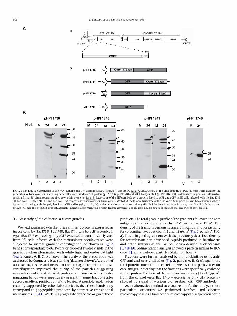
A schematic representation of the plasmid constructs and thechimeric HCV core protein expressed in Sf9 cells upon infectionwith Bac 1736 (core173-eGFP), Bac 1740 (eGFP-core191), Bac 1741(eGFP-core173) are given in Fig. 1A. Bac 1746 lacking core sequencesbut expressing GFP protein was used as control. Cell lysates werecollected at various times p.i. and subjected to immunoblot analysisusing monoclonal and polyclonal antibodies directed against HCVcore and eGFP proteins.
At 24 h p.i. the fusion proteins were probed with the anti-GFPantibody (Fig. 1, panel B). They migrated with an apparent molec-ular mass of 46 kDa (Bac 1736 and Bac 1741) corresponding tocore173 – eGFP (Ia) and eGFP-core173 (IIIa), and at 48 kDa for Bac1740 corresponding to eGFP-core191 (IIa). As expected, Bac 1746showed a strong band at 26 kDa corresponding to eGFP (IVa). Thesame proteins were also recognized by the core-specific mono-clonal antiserum (Ib, IIb, IIIb). HCV core antibody and in some casesGFP antibody recognized additional polypeptides (asterisks)migrating faster than the fusion proteins; these might representproteins produced by alternative translational mechanisms[17,32,38,43]. As they did not disappear after addition of E64(inhibitor of cysteine proteases) and pepstatin A (inhibitor of acidproteases) immediately following expression in insect cells it isunlikely that they originate from proteolysis (data not shown) butrather produced by an unknown mechanism. The fragments of corein Bac 1740 and 1741 (eGFP amino terminal to core) revealed mostlyby core antibody might stem from translational start at the naturalHCV core AUG codon (double asterisks in Fig. 1, panel B: II, III).
5’ UTR C+1
191
NS4B NS5AC E1 E2 NS2 NS3 NS5B4Ap7
3’ UTR
CORE
1
STRUCTURAL NONSTRUCTURAL
pHPI 1736 PPH Core (173aa) GFP
pHPI 1741 PPH
pHPI 1740 PPH
pHPI 1746 PPH
Core (173aa)
Core (191aa)
GFP
GFP
GFP
I
M 24 M 24
pHPI 1736
H p.i.
1 2 3 4
45
36
29
24
20
66
a b
IV
M 24
pHPI 1746
1 2
29
24
a
II
M 24 M 24
pHPI 1740
1 2 3 4
66
45
36
2429
20
a b
III
M 24 M 24
pHPI 1741
1 2 3 4
66
45
36
2924
20
a b
a
b
SS
A
B
Fig. 1. Schematic representation of the HCV genome and the plasmid constructs used in this study. Panel A: a) Structure of the viral genome b) Plasmid constructs used for thegeneration of baculoviruses expressing either HCV core fused to eGFP protein (pHPI 1736, pHPI 1740 and pHPI 1741) or eGFP (pHPI 1746). UTR, untranslated region; cþ1, alternativereading frame; SS, signal sequence; pPH, polyhedrin promoter. Panel B: Expression of the different HCV core proteins fused to eGFP and eGFP in Sf9 cells infected with the Bac 1736(I), Bac 1740 (II), Bac 1741 (III) and Bac 1746 (IV) recombinant baculoviruses. Baculovirus-infected Sf9 cells were harvested at the indicated time point p.i. and lysates were analyzedby immunoblotting with the polyclonal anti-GFP antibody (Ia, IIa, IIIa, IV) or the monoclonal anti-core antibody (Ib, IIb, IIIb), lane 1 and lane 3: mock; lanes 2 and 4: 24 h p.i. Longarrows indicate the expected product, asterisks indicate faster migrating protein fragments/forms (see results), double asterisks indicate the presence of core protein.
K. Katsarou et al. / Biochimie 91 (2009) 903–915906
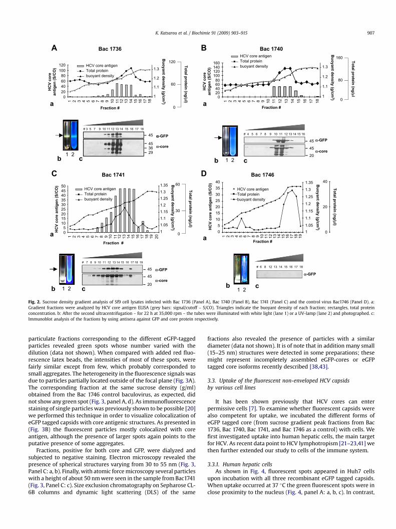
3.2. Assembly of the chimeric HCV core proteins
We next examined whether these chimeric proteins expressed ininsect cells by Bac1736, Bac1740, Bac1741 can be self-assembled.Again Bac1746 expressing only eGFP was used as control. Cell lysatesfrom Sf9 cells infected with the recombinant baculoviruses weresubjected to sucrose gradient centrifugation. As shown in Fig. 2bands corresponding to eGFP-core or core-eGFP were visible in thegradients when illuminated with white light and under UV light(Fig. 2 Panels A, B, C: b arrows). The purity of the preparation wasaddressed by Coomassie blue staining (data not shown). Addition of0.1% NP-40, DNase and RNase to the homogenate prior to ultra-centrifugation improved the purity of the particles suggestingassociation with host derived proteins and nucleic acids. Fastermigrating bands were repetitively present in some fractions aftersucrose gradient purification of the lysates. A possible explanationrecently supported by other laboratories is that these bands maycorrespond to polypeptides produced by alternative translationalmechanisms [38,43]. Work is in progress to define the origin of these
products. The total protein profile of the gradients followed the coreantigen profile as determined by HCV core antigen ELISA. Thedensity of the fractions demonstrating significant immunoreactivityfor core antigen was between 1.2 and 1.3 g/cm3 (Fig. 2, panels A, B, C:a). This is in good agreement with the previously described densityfor recombinant non-enveloped capsids produced in baculovirusand other systems as well as for serum-derived nucleocapsids[3,7,18,19]. Sedimentation analysis showed a pattern similar to HCVcore [7] non-enveloped particles (data not shown).
Fractions were further analyzed by immunoblotting using anti-GFP and anti-core antibodies (Fig. 2, panels A, B, C: c). Again, thetotal protein concentration correlated well with the peak values forcore antigen indicating that the fractions were specifically enrichedin core protein. Fractions of the same sucrose density (1.2–1.3 g/cm3)from the control virus Bac 1746 – expressing only GFP protein -showed no signal in western blot probed with GFP antibody.
As an alternative method to visualize and further analyze theseparticulate structures we performed confocal and electronmicroscopy studies. Fluorescence microscopy of a suspension of the
453629
αα-GFP
α-core
020406080
100120
1 2 53 4 6 7 8 9 10 11 12 13 14 15 16 17 18 1 2 53 4 6 7 8 9 10 11 12 13 14 15 16 17 18
1
1.1
1.2
1.3
Fraction #
Bu
oyan
t d
en
sity (g
/cm
3)
HC
V co
re
an
tig
en
(S
/C
O)
HCV core antigenTotal proteinbuoyant density
Bac 1736
0
To
tal p
ro
tein
(n
g/μ
l)
60
120
a
# 7 8 9 10 11 12 13 14 15 16 17 18 19
b1 2
# 6 8 12 13 14 15 16 17 18
α-GFP
c
05
10152025303540
41 2 3 5 6 7 8 9 10 11 12 13 14 15 16 17 18 19
11.05
1.1
1.15
1.2
1.25
1.3
1.35
Fraction #
0
20
40
To
tal p
ro
tein
(n
g/μ
l)
Bu
oyan
t d
en
sity (g
/cm
3)
HC
V co
re an
tig
en
(S
/C
O)
Bac 1746
a
HCV core antigenTotal proteinbuoyant density
a
HC
V co
re
an
tig
en
(S
/C
O)
Bac 1740
020406080
100120140160
1
1.1
1.2
1.3
Fraction #
HCV core antigenTotal proteinbuoyant density
Bu
oyan
t d
en
sity (g
/cm
3)
To
tal p
ro
tein
(n
g/ μ
l
0
80
160
b 1 2
# 4 5 6 7 8 9 10 11 12 13 14 15 16
45
45
c
20
α-GFP
α-core
b c
1 2
# 3 5 7 9 10 11 12 13 14 15 16 17 18
45
b c1 2
45
45
20
α-GFP
α-core
a
HC
V co
re an
tig
en
(S
/C
O)
Bu
oyan
t d
en
sity (g
/cm
3)
To
tal p
ro
tein
(n
g/μ
l)
Fraction #
Bac 1741
0
30
60
05
101520253035404550
1 2 3 4 5 6 7 8 9 10 11 12 13 14 15 16 17 18 19 20
11.051.11.151.21.251.31.35
HCV core antigenTotal proteinbuoyant density
A
C
B
D
Fig. 2. Sucrose density gradient analysis of Sf9 cell lysates infected with Bac 1736 (Panel A), Bac 1740 (Panel B), Bac 1741 (Panel C) and the control virus Bac1746 (Panel D). a:Gradient fractions were analyzed by HCV core antigen ELISA (grey bars: signal/cutoff – S/CO). Triangles indicate the buoyant density of each fraction; rectangles, total proteinconcentration. b: After the second ultracentrifugation – for 22 h at 35,000 rpm – the tubes were illuminated with white light (lane 1) or a UV-lamp (lane 2) and photographed. c:Immunoblot analysis of the fractions by using antisera against GFP and core protein respectively.
K. Katsarou et al. / Biochimie 91 (2009) 903–915 907
particulate fractions corresponding to the different eGFP-taggedparticles revealed green spots whose number varied with thedilution (data not shown). When compared with added red fluo-rescence latex beads, the intensities of most of these spots, werefairly similar except from few, which probably corresponded tosmall aggregates. The heterogeneity in the fluorescence signals wasdue to particles partially located outside of the focal plane (Fig. 3A).The corresponding fraction at the same sucrose density (g/ml)obtained from the Bac 1746 control baculovirus, as expected, didnot show any green spot (Fig. 3, panel A, d). As immunofluorescencestaining of single particles was previously shown to be possible [20]we performed this technique in order to visualize colocalization ofeGFP tagged capsids with core antigenic structures. As presented in(Fig. 3B) the fluorescent particles mostly colocalized with coreantigen, although the presence of larger spots again points to theputative presence of some aggregates.
Fractions, positive for both core and GFP, were dialyzed andsubjected to negative staining. Electron microscopy revealed thepresence of spherical structures varying from 30 to 55 nm (Fig. 3,Panel C: a, b). Finally, with atomic force microscopy several particleswith a height of about 50 nm were seen in the sample from Bac1741(Fig. 3, Panel C: c). Size exclusion chromatography on Sepharose CL-6B columns and dynamic light scattering (DLS) of the same
fractions also revealed the presence of particles with a similardiameter (data not shown). It is of note that in addition many small(15–25 nm) structures were detected in some preparations; thesemight represent incompletely assembled eGFP-cores or eGFPtagged core isoforms recently described [38,43].
3.3. Uptake of the fluorescent non-enveloped HCV capsidsby various cell lines
It has been shown previously that HCV cores can enterpermissive cells [7]. To examine whether fluorescent capsids werealso competent for uptake, we incubated the different forms ofeGFP tagged core (from sucrose gradient peak fractions from Bac1736, Bac 1740, Bac 1741, and Bac 1746 as a control) with cells. Wefirst investigated uptake into human hepatic cells, the main targetfor HCV. As recent data point to HCV lymphotropism [21–23,41] wethen further extended our study to cells of the immune system.
3.3.1. Human hepatic cellsAs shown in Fig. 4, fluorescent spots appeared in Huh7 cells
upon incubation with all three recombinant eGFP tagged capsids.When uptake occurred at 37 �C the green fluorescent spots were inclose proximity to the nucleus (Fig. 4, panel A: a, b, c). In contrast,
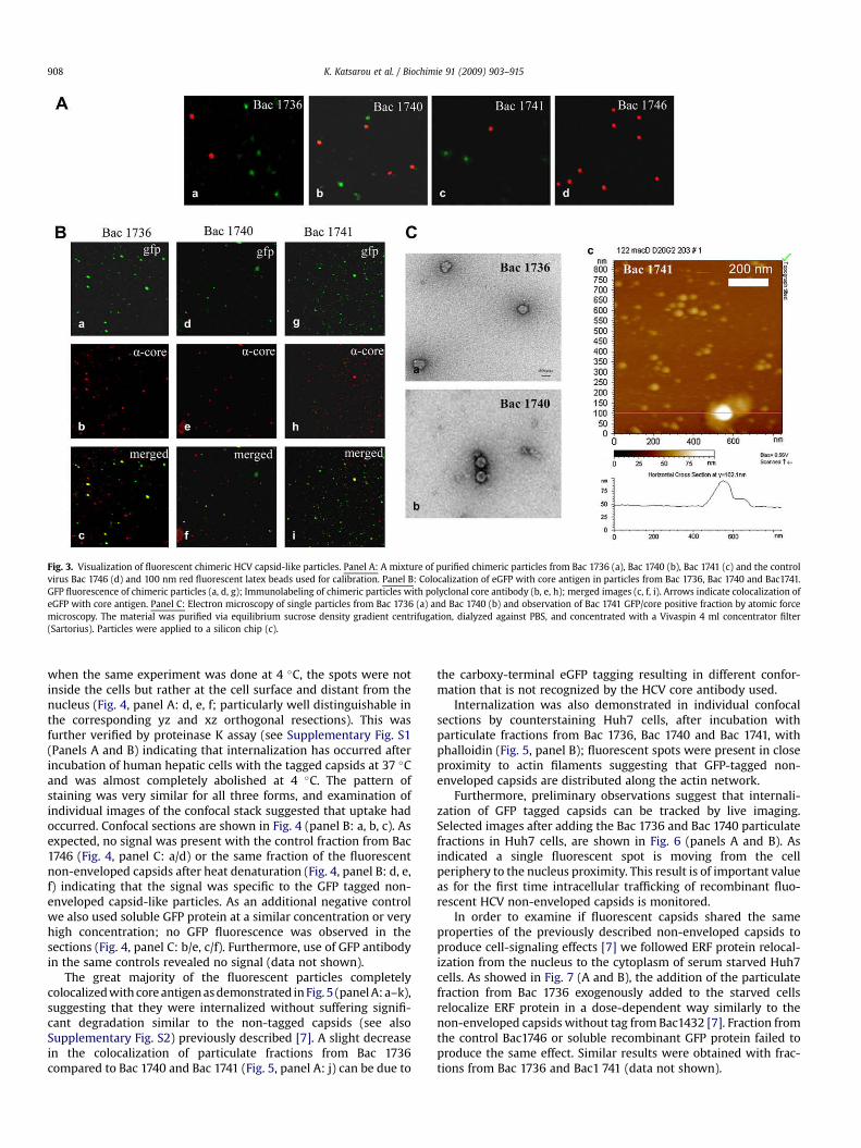
Fig. 3. Visualization of fluorescent chimeric HCV capsid-like particles. Panel A: A mixture of purified chimeric particles from Bac 1736 (a), Bac 1740 (b), Bac 1741 (c) and the controlvirus Bac 1746 (d) and 100 nm red fluorescent latex beads used for calibration. Panel B: Colocalization of eGFP with core antigen in particles from Bac 1736, Bac 1740 and Bac1741.GFP fluorescence of chimeric particles (a, d, g); Immunolabeling of chimeric particles with polyclonal core antibody (b, e, h); merged images (c, f, i). Arrows indicate colocalization ofeGFP with core antigen. Panel C: Electron microscopy of single particles from Bac 1736 (a) and Bac 1740 (b) and observation of Bac 1741 GFP/core positive fraction by atomic forcemicroscopy. The material was purified via equilibrium sucrose density gradient centrifugation, dialyzed against PBS, and concentrated with a Vivaspin 4 ml concentrator filter(Sartorius). Particles were applied to a silicon chip (c).
K. Katsarou et al. / Biochimie 91 (2009) 903–915908
when the same experiment was done at 4 �C, the spots were notinside the cells but rather at the cell surface and distant from thenucleus (Fig. 4, panel A: d, e, f; particularly well distinguishable inthe corresponding yz and xz orthogonal resections). This wasfurther verified by proteinase K assay (see Supplementary Fig. S1(Panels A and B) indicating that internalization has occurred afterincubation of human hepatic cells with the tagged capsids at 37 �Cand was almost completely abolished at 4 �C. The pattern ofstaining was very similar for all three forms, and examination ofindividual images of the confocal stack suggested that uptake hadoccurred. Confocal sections are shown in Fig. 4 (panel B: a, b, c). Asexpected, no signal was present with the control fraction from Bac1746 (Fig. 4, panel C: a/d) or the same fraction of the fluorescentnon-enveloped capsids after heat denaturation (Fig. 4, panel B: d, e,f) indicating that the signal was specific to the GFP tagged non-enveloped capsid-like particles. As an additional negative controlwe also used soluble GFP protein at a similar concentration or veryhigh concentration; no GFP fluorescence was observed in thesections (Fig. 4, panel C: b/e, c/f). Furthermore, use of GFP antibodyin the same controls revealed no signal (data not shown).
The great majority of the fluorescent particles completelycolocalized with core antigen as demonstrated in Fig. 5 (panel A: a–k),suggesting that they were internalized without suffering signifi-cant degradation similar to the non-tagged capsids (see alsoSupplementary Fig. S2) previously described [7]. A slight decreasein the colocalization of particulate fractions from Bac 1736compared to Bac 1740 and Bac 1741 (Fig. 5, panel A: j) can be due to
the carboxy-terminal eGFP tagging resulting in different confor-mation that is not recognized by the HCV core antibody used.
Internalization was also demonstrated in individual confocalsections by counterstaining Huh7 cells, after incubation withparticulate fractions from Bac 1736, Bac 1740 and Bac 1741, withphalloidin (Fig. 5, panel B); fluorescent spots were present in closeproximity to actin filaments suggesting that GFP-tagged non-enveloped capsids are distributed along the actin network.
Furthermore, preliminary observations suggest that internali-zation of GFP tagged capsids can be tracked by live imaging.Selected images after adding the Bac 1736 and Bac 1740 particulatefractions in Huh7 cells, are shown in Fig. 6 (panels A and B). Asindicated a single fluorescent spot is moving from the cellperiphery to the nucleus proximity. This result is of important valueas for the first time intracellular trafficking of recombinant fluo-rescent HCV non-enveloped capsids is monitored.
In order to examine if fluorescent capsids shared the sameproperties of the previously described non-enveloped capsids toproduce cell-signaling effects [7] we followed ERF protein relocal-ization from the nucleus to the cytoplasm of serum starved Huh7cells. As showed in Fig. 7 (A and B), the addition of the particulatefraction from Bac 1736 exogenously added to the starved cellsrelocalize ERF protein in a dose-dependent way similarly to thenon-enveloped capsids without tag from Bac1432 [7]. Fraction fromthe control Bac1746 or soluble recombinant GFP protein failed toproduce the same effect. Similar results were obtained with frac-tions from Bac 1736 and Bac1 741 (data not shown).
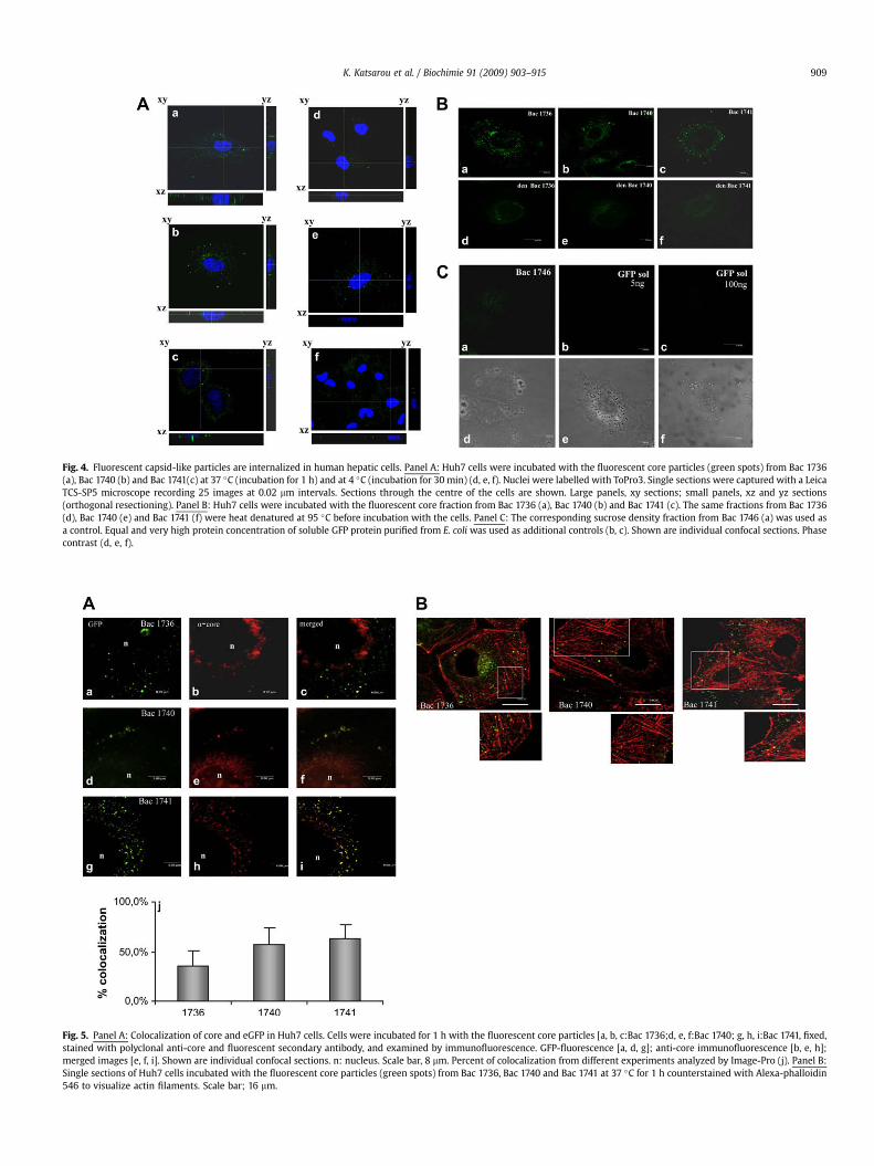
Fig. 4. Fluorescent capsid-like particles are internalized in human hepatic cells. Panel A: Huh7 cells were incubated with the fluorescent core particles (green spots) from Bac 1736(a), Bac 1740 (b) and Bac 1741(c) at 37 �C (incubation for 1 h) and at 4 �C (incubation for 30 min) (d, e, f). Nuclei were labelled with ToPro3. Single sections were captured with a LeicaTCS-SP5 microscope recording 25 images at 0.02 mm intervals. Sections through the centre of the cells are shown. Large panels, xy sections; small panels, xz and yz sections(orthogonal resectioning). Panel B: Huh7 cells were incubated with the fluorescent core fraction from Bac 1736 (a), Bac 1740 (b) and Bac 1741 (c). The same fractions from Bac 1736(d), Bac 1740 (e) and Bac 1741 (f) were heat denatured at 95 �C before incubation with the cells. Panel C: The corresponding sucrose density fraction from Bac 1746 (a) was used asa control. Equal and very high protein concentration of soluble GFP protein purified from E. coli was used as additional controls (b, c). Shown are individual confocal sections. Phasecontrast (d, e, f).
Fig. 5. Panel A: Colocalization of core and eGFP in Huh7 cells. Cells were incubated for 1 h with the fluorescent core particles [a, b, c:Bac 1736;d, e, f:Bac 1740; g, h, i:Bac 1741, fixed,stained with polyclonal anti-core and fluorescent secondary antibody, and examined by immunofluorescence. GFP-fluorescence [a, d, g]; anti-core immunofluorescence [b, e, h];merged images [e, f, i]. Shown are individual confocal sections. n: nucleus. Scale bar, 8 mm. Percent of colocalization from different experiments analyzed by Image-Pro (j). Panel B:Single sections of Huh7 cells incubated with the fluorescent core particles (green spots) from Bac 1736, Bac 1740 and Bac 1741 at 37 �C for 1 h counterstained with Alexa-phalloidin546 to visualize actin filaments. Scale bar; 16 mm.
K. Katsarou et al. / Biochimie 91 (2009) 903–915 909
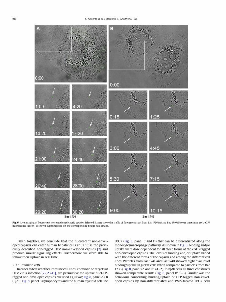
Fig. 6. Live imaging of fluorescent non-enveloped capsid uptake. Selected frames show the traffic of fluorescent spot from Bac 1736 (A) and Bac 1740 (B) over time (min. sec). eGFPfluorescence (green) is shown superimposed on the corresponding bright field image.
K. Katsarou et al. / Biochimie 91 (2009) 903–915910
Taken together, we conclude that the fluorescent non-envel-oped capsids can enter human hepatic cells at 37 �C as the previ-ously described non-tagged HCV non-enveloped capsids [7] andproduce similar signalling effects. Furthermore we were able tofollow their uptake in real time.
3.3.2. Immune cellsIn order to test whether immune cell lines, known to be targets of
HCV virus infection [22,23,41], are permissive for uptake of eGFP-tagged non-enveloped capsids, we used T (Jurkat; Fig. 8, panel A), B(BJAB; Fig. 8, panel B) lymphocytes and the human myeloid cell line
U937 (Fig. 8, panel C and D) that can be differentiated along themonocyte/macrophage pathway. As shown in Fig. 8, binding and/oruptake were dose dependent for all three forms of the eGFP-taggednon-enveloped capsids. The levels of binding and/or uptake variedwith the different forms of the capsids and among the different celllines. Particles from Bac 1741 and Bac 1740 showed higher values ofbinding/uptake in Jurkat cells when compared to particles from Bac1736 (Fig. 8, panels A and B: a1–2). In BJAb cells all three constructsshowed comparable results (Fig. 8, panel B: 1–3). Similar was thebehaviour concerning binding/uptake of GFP-tagged non-envel-oped capsids by non-differentiated and PMA-treated U937 cells
Starved cells +EGF +GFP sol Bac 1746 Bac 1432 Bac 1740
0
10
20
30
40
50
60
1 2 3 4 5 6
% cyto
plasm
ic E
RF
-G
FP
d
istrib
utio
n
starved cells
EGF
GFP
1746
1432
1740
Fraction quantity
A
B
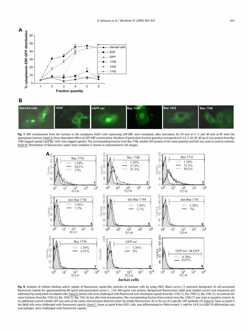
Fig. 7. ERF translocation from the nucleus to the cytoplasm. Huh7 cells expressing GFP-ERF, were incubated, after starvation, for 20 min at 4 �C and 40 min at RT with theappropriate fraction. Panel A: Dose-dependent effect on GFP-ERF translocation. Numbers of particulate fraction quantity correspond to 0, 2.5, 5, 10, 20, 40 ng of core protein from Bac1740 (tagged capsids) and Bac 1432 (non-tagged capsids). The corresponding fraction from Bac 1746, soluble GFP protein of the same quantity and EGF was used as used as controls.Panel B: Distribution of fluorescence under each condition is shown in representative cell images.
Fig. 8. Analysis of cellular binding and/or uptake of fluorescent capsid-like particles in immune cells by using FACS. Black curves [-] represent histograms of cell-associatedfluorescent capsids for approximatively 80 ng/ml and punctuated curves [.] for 160 ng/ml core protein. Background fluorescence (light grey shaded curves) was measured andsubtracted by using mock-incubated cells. Panel A: Jurkat cells were challenged with fluorescent non-enveloped capsids from Bac 1736 (1), Bac 1740 (2), Bac 1741 (3). As controls thesame fractions from Bac 1736 (4), Bac 1740 (5), Bac 1741 (6) but after heat denaturation. The corresponding fraction from control virus Bac 1746 (7) was used as negative control. Asan additional control soluble GFP was used at the same concentration detected either by simple fluorescence (8) or by use of a specific GFP antibody (9). Panel B: Same as panel Abut BJAB cells were challenged with fluorescent capsids. Panel C: Same as panel B but U937 cells, non-differentiated or PMA-treated (1 mM for 24 h) in order to differentiate intomacrophages, were challenged with fluorescent capsids.
K. Katsarou et al. / Biochimie 91 (2009) 903–915 911
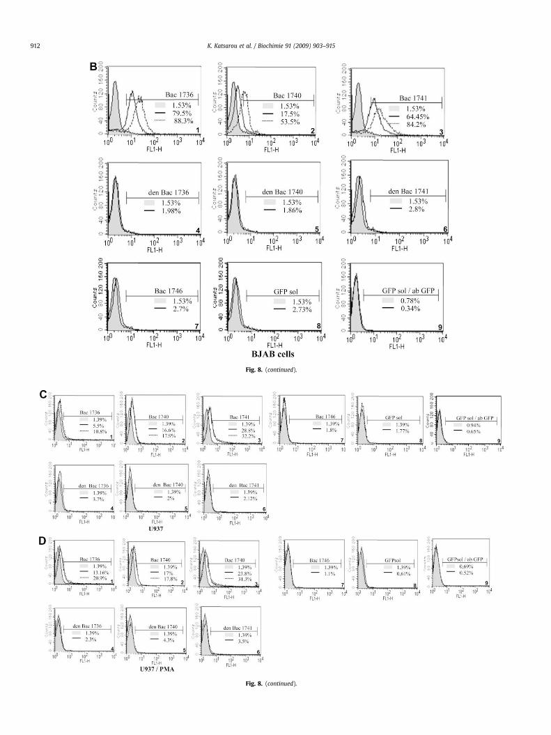
Fig. 8. (continued).
Fig. 8. (continued).
K. Katsarou et al. / Biochimie 91 (2009) 903–915912
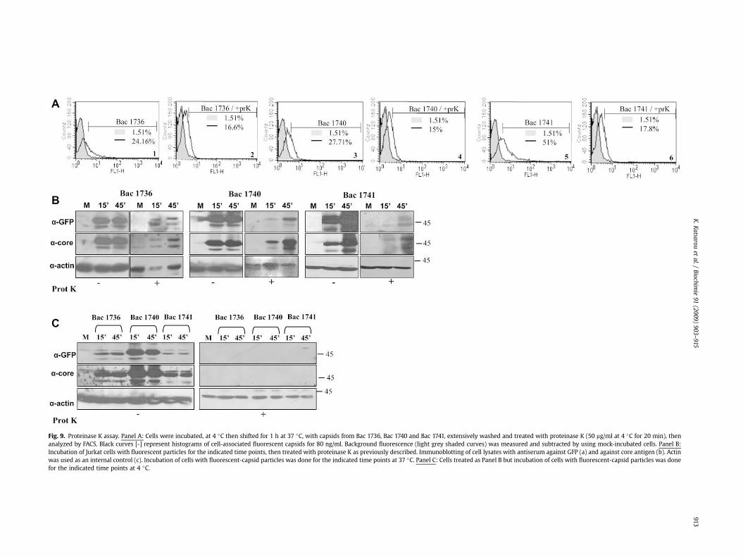
Fig. 9. Proteinase K assay. Panel A: Cells were incubated, at 4 �C then shifted for 1 h at 37 �C, with capsids from Bac 1736, Bac 1740 and Bac 1741, extensively washed and treated with proteinase K (50 mg/ml at 4 �C for 20 min), thenanalyzed by FACS. Black curves [-] represent histograms of cell-associated fluorescent capsids for 80 ng/ml. Background fluorescence (light grey shaded curves) was measured and subtracted by using mock-incubated cells. Panel B:Incubation of Jurkat cells with fluorescent particles for the indicated time points, then treated with proteinase K as previously described. Immunoblotting of cell lysates with antiserum against GFP (a) and against core antigen (b). Actinwas used as an internal control (c). Incubation of cells with fluorescent-capsid particles was done for the indicated time points at 37 �C. Panel C: Cells treated as Panel B but incubation of cells with fluorescent-capsid particles was donefor the indicated time points at 4 �C.
K.K
atsarouet
al./Biochim
ie91
(2009)903–915
913
K. Katsarou et al. / Biochimie 91 (2009) 903–915914
although signal was less strong (Fig. 8, panel C: 1, 2, 3). As controlsidentical samples were used that had been heat denatured beforeincubation with all the immune cell lines used (Fig. 8, panels A, B, C,D:4, 5, 6). These samples were totally negative. In addition the cor-responding fraction from the control virus Bac 1746 (Fig. 8, panels A,B, C, D:7), and soluble GFP with the same protein concentrationshowed no binding/uptake (Fig. 8, panels A, B, C, D:8). This lastcontrol was also analyzed by using a GFP antibody (Fig. 8, panels A, B,C, D:9). No signal was observed in these controls, indicating that onlythe eGFP tagged cores can bind and/or enter the cells. These datashowed that uptake of fluorescent non-enveloped capsids wassimilar to the cellular uptake of core non-enveloped capsids underthe same experimental conditions (Supplementary Fig. S3).Furthermore, cells that had been incubated with the GFP taggednon-enveloped capsids were subjected to proteinase K treatment, asexemplified for Jurkat T cells. FACS analysis showed that an efficientinternalization has occurred (Fig. 9, panel A:1–6). After digestion ofsurface-attached particles with proteinase K, immunoreactive coreand the GFP signal were clearly detectable in the cells as evidencedby western blotting (Fig. 9, panel B). Again, most efficient internal-ization at late time points was observed with fractions from Bac 1740and Bac 1741. Conversely, when incubation was at 4 �C, binding oftagged non-enveloped capsids was evident but no intracellularsignal was recorded (Fig. 9, panel C) suggesting that at thistemperature internalization had not occurred.
4. Discussion
Our data demonstrate that fusion of GFP to the amino- or car-boxy terminus of the HCV protein core173 and to the aminoterminus of core191, does not alter the autoassembly properties ofthe protein. The particles obtained were very similar to the previ-ously described recombinant HCV non-enveloped capsid-likeparticles [7] and are internalized into human hepatic cells and cellsof immune origin. The fluorescent particles are particularlyattractive for monitoring non-enveloped particle uptake in realtime. Successful labelling of viruses and VLPs via fusion with eGFPby using similar approaches has been reported for human immu-nodeficiency virus, herpes simplex virus, rotavirus, rabies virus,simian virus and parvovirus [8–11,24,25, 31, 34].
The eGFP fusion proteins were abundantly expressed from allconstructs and had the expected relative molecular mass. However,smaller immunoreactive products were also observed even aftersucrose density gradient purification. This raises the question ofwhether these forms of the core protein produced via cleavage, oralternative translational mechanisms are still able to assemble intoparticulate cores and to what extent the GFP moiety is inhibitory tothe assembly process. It is also not clear whether forms of the coreprotein are incorporated into core particles giving rise to the non-uniform particle size observed in EM. Supporting this hypothesis isthe recently reported evidence that the N-terminal half of HCV coreis an intrinsically unstructured protein [37] as well as the produc-tion of four N-terminally truncated isoforms of the nucleocapsid(core) protein ranging from 8 to 14 kDa recently described by EngJ. et al. [38,43]. In addition unusual lability of the nucleocapsidprotein was observed in SARS virus during the purification processwhere the appearance of two other protein/protein fragment bandson SDS-PAGE was detected [39].
Expression of all constructs gave rise to small spherical capsidswith a diameter range of 30–55 nm found in the fractions fromsucrose density gradient centrifugation with densities between 1.2and 1.3 g/cm3. Nevertheless, a more detailed structural analysis isrequired in order to study the conformation of these particles.Nucleocapsids, isolated by detergent treatment of virions present inpatient serum, have been reported to have a density of 1.35 g/ml in
caesium chloride and of 1.23–1.27 g/ml in sucrose [3,29,30]. Thedifferent densities might be due to association of the highly lipo-philic HCV core protein with lipids [33]. While the issue of HCVcapsid density requires further investigation, density valueobtained for the fluorescent recombinant non-enveloped capsidscorresponds closely to the 1.25 g/ml density seen for at least onepopulation of natural HCV capsids produced in cells and with the1.17–1.24 g/ml previously described [3]. In some cases the capsidswere irregular in size and often imperfectly assembled, mostprobably because of steric problems caused by the presence of theeGFP. Despite their heterogeneous size, they were efficientlyinternalized by the cell lines investigated.
The presented data demonstrate for the first time that non-enveloped capsids, rendered fluorescent via fusion with eGFP areinternalized into cells of hepatic and immune origin. Although therecent development of an infectious HCV system was of great impactfor the study of viral uptake, a number of limitations still exist.Fastidious host cell requirements, limitation to a single replicatingstrain, and no evidence for the production of the different forms ofparticles present in the sera of infected patients, mean that the invitro produced HCV capsids will remain an important tool for studiesof virus uptake. Furthermore, recent reports provided evidence thatnot only B cells but also in vitro infected T-cells support HCV repli-cation and predominantly non-enveloped core particles are detec-ted in the supernatants [21]. In this context it is of interest thatdefective viruses may play a significant role in the establishmentand maintenance of chronic infection in vivo [5,26–28].
Our data on the uptake of fluorescent non-enveloped particlesby human hepatic cells, T cells, B cells as well as monocytes pro-gressing to macrophages pave the way for a better understanding ofthe biological importance of naked capsids in HCV infection.
Results of binding and internalization assays also suggest thatwe have produced fluorescent particles with morphological andphysical properties as well as producing cell-signalling effectssimilar to those of the untagged non-enveloped capsids. The fluo-rescent non-enveloped capsid-like particles are safe to handle andof great utility as a visualization tool in studies related to nakedHCV capsid trafficking mechanisms. They also enable real timeimaging, providing new answers on the potential role of nakedcapsids in HCV pathogenesis. Taken together, our results demon-strate that fusions between eGFP and HCV core protein assembleinto core-like particles, with the most efficient tagging being at thecarboxyterminus of core173. These fluorescent capsid-like particlesare internalized into human hepatic cells and cells of immuneorigin by a so far unknown mechanism and pathway. Experimentsaimed at unravelling these are currently underway.
Acknowledgements
We thank Pr. G. Mavrothalassitis (University of Crete) forprovision of the GFP-ERF plasmid. We also thank our colleaguesM. Zouridakis (Department of Biochemistry, Hellenic PasteurInstitute) for dynamic light scattering (DLS), P. Foka and E. Tsitourafor useful discussions. R.Z. and P.H. were partially supported byAustria Nano-Initiative (NABIOS).
This work was supported by PENED 03ED297, co-financed byE.U.-European Social Fund (75%) and the Greek Ministry ofDevelopment-GSRT (25%).
Appendix. Supplementary data
Supplementary data associated with this article can be found, inthe online version, at doi:10.1016/j.biochi.2009.04.016
K. Katsarou et al. / Biochimie 91 (2009) 903–915 915
References
[1] M. Houghton, Hepatitis C Viruses, in: D.M. Knipe, B.N. Fields, P.M. Howley(Eds.), third ed., Fields Virology, vol. 1 Lippincott-Raven Publishers, Phila-delphia, 1996, pp. 1035–1058.
[2] P. Andre, G. Perlemuter, A. Budkowska, C. Brechot, V. Lotteau, Hepatitis Cvirus particles and lipoprotein metabolism, Semin. Liver Dis. 25 (2005)93–104.
[3] P. Maillard, K. Krawczynski, J. Nitkiewicz, C. Bronnert, M. Sidorkiewicz,P. Gounon, J. Dubuisson, G. Faure, R. Crainic, A. Budkowska, Nonenvelopednucleocapsids of hepatitis C virus in the serum of infected patients, J. Virol.75 (2001) 8240–8250.
[4] A. Iwai, H. Marusawa, Y. Takada, H. Egawa, K. Ikeda, M. Nabeshima,S. Uemoto, T. Chiba, Identification of novel defective HCV clones in livertransplant recipients with recurrent HCV infection, J. Viral Hepat. 13 (2006)523–531.
[5] S. Noppornpanth, S.L. Smits, T.X. Lien, Y. Poovorawan, A.D. Osterhaus,B.L. Haagmans, Characterization of hepatitis C virus deletion mutantscirculating in chronically infected patients, J. Virol. 81 (2007) 12496–12503.
[6] S. Yagi, K. Mori, E. Tanaka, A. Matsumoto, F. Sunaga, K. Kiyosawa,K. Yamaguchi, Identification of novel HCV subgenome replicating persis-tently in chronic active hepatitis C patients, J. Med. Virol. 77 (2005) 399–413.
[7] P. Tsitoura, U. Georgopoulou, S. Petres, A. Varaklioti, A. Karafoulidou,D. Vagena, C. Politis, P. Mavromara, Evidence for cellular uptake ofrecombinant hepatitis C virus non-enveloped capsid-like particles, FEBS Lett.581 (2007) 4049–4057.
[8] A. Charpilienne, M. Nejmeddine, M. Berois, N. Parez, E. Neumann, E. Hewat,G. Trugnan, J. Cohen, Individual rotavirus-like particles containing 120molecules of fluorescent protein are visible in living cells, J. Biol. Chem. 276(2001) 29361–29367.
[9] S. Finke, K. Brzozka, K.K. Conzelmann, Tracking fluorescence-labeled rabiesvirus: enhanced green fluorescent protein-tagged phosphoprotein Psupports virus gene expression and formation of infectious particles, J. Virol.78 (2004) 12333–12343.
[10] L. Gilbert, J. Toivola, E. Lehtomaki, L. Donaldson, P. Kapyla, M. Vuento,C. Oker-Blom, Assembly of fluorescent chimeric virus-like particles of canineparvovirus in insect cells, Biochem. Biophys. Res. Commun 313 (2004)878–887.
[11] L. Gilbert, J. Toivola, O. Valilehto, T. Saloniemi, C. Cunningham, D. White,A.R. Makela, E. Korhonen, M. Vuento, C. Oker-Blom, Truncated forms of viralVP2 proteins fused to EGFP assemble into fluorescent parvovirus-like parti-cles, J. Nanobiotechnology 4 (2006) 13.
[12] C. Lambert, N. Thome, C.J. Kluck, R. Prange, Functional incorporation of greenfluorescent protein into hepatitis B virus envelope particles, Virology 330(2004) 158–167.
[13] K. Lux, N. Goerlitz, S. Schlemminger, L. Perabo, D. Goldnau, J. Endell, K. Leike,D.M. Kofler, S. Finke, M. Hallek, H. Buning, Green fluorescent protein-taggedadeno-associated virus particles allow the study of cytosolic and nucleartrafficking, J. Virol. 79 (2005) 11776–11787.
[14] G. Inchauspe, S. Zebedee, D.H. Lee, M. Sugitani, M. Nasoff, A.M. Prince,Genomic structure of the human prototype strain H of hepatitis C virus:comparison with American and Japanese isolates, Proc. Natl. Acad. Sci. U.S.A.88 (1991) 10292–10296.
[15] J. McLauchlan, M.K. Lemberg, G. Hope, B. Martoglio, Intramembrane prote-olysis promotes trafficking of hepatitis C virus core protein to lipid droplets,EMBO J. 21 (2002) 3980–3988.
[16] T.F. Baumert, S. Ito, D.T. Wong, T.J. Liang, Hepatitis C virus structural proteinsassemble into viruslike particles in insect cells, J. Virol. 72 (1998) 3827–3836.
[17] A.D. Branch, D.D. Stump, J.A. Gutierrez, F. Eng, J.L. Walewski, The hepatitis Cvirus alternate reading frame (ARF) and its family of novel products: thealternate reading frame protein/F-protein, the double-frameshift protein, andothers, Semin. Liver Dis. 25 (1) (2005) 105–117.
[18] K.C. Klein, S.J. Polyak, J.R. Lingappa, Unique features of hepatitis C virus capsidformation revealed by de novo cell-free assembly, J. Virol. 78 (2004)9257–9269.
[19] C. Vauloup-Fellous, V. Pene, J. Garaud-Aunis, F. Harper, S. Bardin, Y. Suire,E. Pichard, A. Schmitt, P. Sogni, G. Pierron, P. Briand, A.R. Rosenberg, Signalpeptide peptidase-catalyzed cleavage of hepatitis C virus core protein isdispensable for virus budding but destabilizes the viral capsid, J. Biol. Chem.281 (2006) 27679–27692.
[20] M. Pizzato, S.A. Marlow, E.D. Blair, Y. Takeuchi, Initial binding of murineleukemia virus particles to cells does not require specific Env-receptorinteraction, J. Virol. 73 (1999) 8599–8611.
[21] S.A. MacParland, T.N. Pham, S.A. Gujar, T.I. Michalak, De novo infection andpropagation of wild-type Hepatitis C virus in human T lymphocytes in vitro,J. Gen. Virol. 87 (2006) 3577–3586.
[22] T.N. Pham, T.I. Michalak, Occult persistence and lymphotropism of hepatitis Cvirus infection, World J. Gastroenterol. 14 (2008) 2789–2793.
[23] T.N. Pham, D. King, S.A. Macparland, J.S. McGrath, S.B. Reddy, F.R. Bursey,T.I. Michalak, Hepatitis C virus replicates in the same immune cell subsets inchronic hepatitis C and occult infection, Gastroenterology 134 (2008)812–822.
[24] P. Desai, S. Person, Incorporation of the green fluorescent protein into theherpes simplex virus type 1 capsid, J. Virol. 72 (1998) 7563–7568.
[25] D. McDonald, M.A. Vodicka, G. Lucero, T.M. Svitkina, G.G. Borisy,M. Emerman, T.J. Hope, Visualization of the intracellular behavior of HIV inliving cells, J. Cell Biol. 159 (2002) 441–452.
[26] A.S. Huang, D. Baltimore, Defective viral particles and viral disease processes,Nature 226 (1970) 325–327.
[27] M.U. Lancaster, S.I. Hodgetts, J.S. Mackenzie, N. Urosevic, Characterization ofdefective viral RNA produced during persistent infection of Vero cells withMurray Valley encephalitis virus, J. Virol. 72 (1998) 2474–2482.
[28] S.W. Yoon, S.Y. Lee, S.Y. Won, S.H. Park, S.Y. Park, Y.S. Jeong, Characterizationof homologous defective interfering RNA during persistent infection of Verocells with Japanese encephalitis virus, Mol. Cells 21 (2006) 112–120.
[29] M. Kaito, M.S. Watanabe, K. Tsukiyama-Kohara, K. Yamaguchi, Y. Kobayashi,M. Konishi, M. Yokoi, S. Ishida, S. Suzuki, M. Kohara, Hepatitis C virus particledetected by immunoelectron microscopic study, J. Gen. Virol. 75 (1994)1755–1760.
[30] T. Kanto, T.N. Hayashi, T. Takehara, H. Hagiwara, E. Mita, M. Naito,A. Kasahara, H. Fusamoto, T. Kamada, Buoyant density of hepatitis C virusrecovered from infected hosts: two different features in sucrose equilibriumdensity-gradient centrifugation related to degree of liver inflammation,Hepatology 19 (1994) 296–302.
[31] L. Pelkmans, J. Kartenbeck, A. Helenius, Caveolar endocytosis of simian virus40 reveals a new two-step vesicular-transport pathway to the ER, Nat. CellBiol. 3 (2001) 473–484.
[32] M. Ratinier, S. Boulant, C. Combet, P. Targett-Adams, J. McLauchlan,J.P. Lavergne, Transcriptional slippage prompts recoding in alternate readingframes in the hepatitis C virus (HCV) core sequence from strain HCV-1, J. Gen.Virol. 89 (2008) 1569–1578.
[33] A. Shavinskaya, S. Boulant, F. Penin, J. McLauchlan, R. Bartenschlager, Thelipid droplet binding domain of hepatitis C virus core protein is a majordeterminant for efficient virus assembly, J. Biol. Chem. 282 (51) (2007)37158–37169.
[34] M. Dundr, J. McNally, J. Cohen, T. Misteli, Quantitation of GFP-fusion proteinsin single living cells, J. Struct. Biol. 140 (2002) 92–99.
[35] J. Aaskov, K. Buzacott, H.M. Thu, K. Lowry, E.C. Holmes, Long-term trans-mission of defective RNA viruses in humans and Aedes mosquitoes, Science311 (2006) 236–238.
[36] L. Pacini, R. Graziani, L. Bartholomeu, R. De Francesco, G. Paonessa, HepatitisC virus sub-genomic RNA resembling natural occurring deletion mutants areefficiently trans-packaged into virus-like particles by helper virus or helpercell lines. In: Abstract presented in 15th International Symposium onHepatitis C virus & Related Viruses, (2008) San Antonio Texas.
[37] J.B. Duvignaud, C. Davard, R. Fromentin, N. Majeau, D. Leclerc, S. Gagne,Structure and dynamics of the N-terminal half of hepatitis C virus coreprotein: an intrinsically unstructured protein, Biochem. Res. Commun. 378(1) (2009) 27–31.
[38] F. Eng, A.L. Klepper, J. Walewski, S.L. Fishman, L. McMullan, M.J. Evans, C.M.Rice, S. Desai, A.D. Branch, The hepatitis C virus core gene expresses a familyof N-terminally truncated isoforms of the nucleocapsid (core) protein. In:Abstract presented in 15th International Symposium on Hepatitis C virus &Related Viruses, (2008) San Antonio Texas.
[39] J. Mark, X. Li, T. Cyr, S. Fournier, B. Jaentschke, M.A. Hefford, SARS corona-virus: unusual lability of the nucleocapsid protein, Biochem. Res. Commun.377 (2) (2008) 429–433.
[40] V. Falcon, N. Acosta-Rivero, G. Chinea, 12 other authors, Ultrastructuralevidences of HCV infection in hepatocytes of chronically HCV-infectedpatients, Biochem. Biophys. Res. Commun. 305 (2003) 1085–1090.
[41] Y. Kondo, V.M.H. Sung, K. Machida, M. Liu, M.M.C. Lai, Hepatitis C infects Tcells and affects interferon –g signaling in T cell lines, Virology 361 (2007)161–173.
[42] A. Helenius, J. Kartenbeck, K. Simons, E. Fries, On the entry of Semliki forestvirus into BHK-21 cells, J. Cell Biol. 84 (1980) 404–420.
[43] F. Eng, A.L. Klepper, J. Walewski, S.L. Fishman, S. Desai, L. McMullan,M.J. Evans, C.M. Rice, A.D. Branch, Internal initiation stimulates production ofp8 mini-core, a member of a newly discovered family of hepatitis C viruscore protein isoforms, J. Virol. 83 (2009) 3104–3114.
[44] L. Le Gallic, L. Virgilio, P. Cohen, B. Biteau, G. Mavrothalassitis, ERF nuclearshuttling, a continuous monitor of ERK activity that links it to cell cycleprogression, Mol. Cell Biol. 24 (2004) 1206–1218.
Related Documents