Graphic Integration of Causal Factors of Pelvic Floor Disorders: An Integrated Lifespan Model John O. L. DeLANCEY, MD a,b , Lisa KANE LOW, PhD, RN, CNM a,b,c , Janis M. MILLER, PhD, RN, ANP a,b,c , Divya A. PATEL, PhD b , and Julie A. TUMBARELLO, MA a a Pelvic Floor Research Group, University of Michigan, Ann Arbor, MI b Department of Obstetrics and Gynecology, University of Michigan, Ann Arbor, MI c School of Nursing, University of Michigan, Ann Arbor, MI Abstract There is growing interest in causal factors for pelvic floor disorders. These conditions include pelvic organ prolapse and urinary and fecal incontinence and are affected by a myriad of factors that increase occurrence of symptomatic disease. Unraveling the complex causal network of genetic factors, birth- induced injury, connective tissue aging, lifestyle and co-morbid factors is challenging. We describe a graphical tool to integrate the factors affecting pelvic floor disorders. It plots pelvic floor function in 3 major life phases: 1) development of functional reserve during an individual’s growth, 2) variations in the amount of injury and potential recovery that occur during and after vaginal birth, and 3) deterioration that occurs with advancing age. This graphical tool accounts for changes in different phases to be integrated to form a disease model to help assess the overlap of different causal factors. Keywords Vaginal birth; pelvic floor; urinary incontinence; pelvic organ prolapse Introduction A woman has an 11% chance of experiencing pelvic floor dysfunction (PFD) so severely during her life that she will require surgery, based on data from a managed care population (1). Pelvic floor disorders (PFD), the umbrella term for conditions such as incontinence and pelvic organ prolapse, leads to over 300,000 operations each year (2) (3). Understanding the cause of these common conditions is critical to improving treatment and prevention; but clarifying causation is complex due to their multifactorial nature. Factors such as instrumented vaginal delivery, heavy lifting, rapid decline in tissue strength, damaged levator ani muscle, and nerve injury are cited as increasing the likelihood that a woman may develop prolapse or experience recurrence of PFD after surgery (4) (5). Rather than a single factor, it is more probable that combinations of anatomical, physiological, genetic, lifestyle, and reproductive factors interact throughout a woman’s lifespan to contribute to PFD. Understanding these complex causal Corresponding Author: John O. L. DeLancey, MD, Dept Obstetrics & Gynecology, L4000 Women’s Hospital, 1500 E Medical Center Drive, Ann Arbor, MI 48109-0276, Phone: (734) 764-8429, Fax: (734) 647-9727, Email: [email protected]. Publisher's Disclaimer: This is a PDF file of an unedited manuscript that has been accepted for publication. As a service to our customers we are providing this early version of the manuscript. The manuscript will undergo copyediting, typesetting, and review of the resulting proof before it is published in its final citable form. Please note that during the production process errors may be discovered which could affect the content, and all legal disclaimers that apply to the journal pertain. NIH Public Access Author Manuscript Am J Obstet Gynecol. Author manuscript; available in PMC 2009 December 1. Published in final edited form as: Am J Obstet Gynecol. 2008 December ; 199(6): 610.e1–610.e5. doi:10.1016/j.ajog.2008.04.001. NIH-PA Author Manuscript NIH-PA Author Manuscript NIH-PA Author Manuscript

Welcome message from author
This document is posted to help you gain knowledge. Please leave a comment to let me know what you think about it! Share it to your friends and learn new things together.
Transcript
Graphic Integration of Causal Factors of Pelvic Floor Disorders:An Integrated Lifespan Model
John O. L. DeLANCEY, MDa,b, Lisa KANE LOW, PhD, RN, CNMa,b,c, Janis M. MILLER, PhD,RN, ANPa,b,c, Divya A. PATEL, PhDb, and Julie A. TUMBARELLO, MAaa Pelvic Floor Research Group, University of Michigan, Ann Arbor, MIb Department of Obstetrics and Gynecology, University of Michigan, Ann Arbor, MIc School of Nursing, University of Michigan, Ann Arbor, MI
AbstractThere is growing interest in causal factors for pelvic floor disorders. These conditions include pelvicorgan prolapse and urinary and fecal incontinence and are affected by a myriad of factors that increaseoccurrence of symptomatic disease. Unraveling the complex causal network of genetic factors, birth-induced injury, connective tissue aging, lifestyle and co-morbid factors is challenging. We describea graphical tool to integrate the factors affecting pelvic floor disorders. It plots pelvic floor functionin 3 major life phases: 1) development of functional reserve during an individual’s growth, 2)variations in the amount of injury and potential recovery that occur during and after vaginal birth,and 3) deterioration that occurs with advancing age. This graphical tool accounts for changes indifferent phases to be integrated to form a disease model to help assess the overlap of different causalfactors.
KeywordsVaginal birth; pelvic floor; urinary incontinence; pelvic organ prolapse
IntroductionA woman has an 11% chance of experiencing pelvic floor dysfunction (PFD) so severely duringher life that she will require surgery, based on data from a managed care population (1). Pelvicfloor disorders (PFD), the umbrella term for conditions such as incontinence and pelvic organprolapse, leads to over 300,000 operations each year (2) (3). Understanding the cause of thesecommon conditions is critical to improving treatment and prevention; but clarifying causationis complex due to their multifactorial nature. Factors such as instrumented vaginal delivery,heavy lifting, rapid decline in tissue strength, damaged levator ani muscle, and nerve injuryare cited as increasing the likelihood that a woman may develop prolapse or experiencerecurrence of PFD after surgery (4) (5). Rather than a single factor, it is more probable thatcombinations of anatomical, physiological, genetic, lifestyle, and reproductive factors interactthroughout a woman’s lifespan to contribute to PFD. Understanding these complex causal
Corresponding Author: John O. L. DeLancey, MD, Dept Obstetrics & Gynecology, L4000 Women’s Hospital, 1500 E Medical CenterDrive, Ann Arbor, MI 48109-0276, Phone: (734) 764-8429, Fax: (734) 647-9727, Email: [email protected]'s Disclaimer: This is a PDF file of an unedited manuscript that has been accepted for publication. As a service to our customerswe are providing this early version of the manuscript. The manuscript will undergo copyediting, typesetting, and review of the resultingproof before it is published in its final citable form. Please note that during the production process errors may be discovered which couldaffect the content, and all legal disclaimers that apply to the journal pertain.
NIH Public AccessAuthor ManuscriptAm J Obstet Gynecol. Author manuscript; available in PMC 2009 December 1.
Published in final edited form as:Am J Obstet Gynecol. 2008 December ; 199(6): 610.e1–610.e5. doi:10.1016/j.ajog.2008.04.001.
NIH
-PA Author Manuscript
NIH
-PA Author Manuscript
NIH
-PA Author Manuscript
questions can be facilitated by having a framework that integrates how different factors interactto result in the development of PFD.
We will present a conceptual model, The Lifespan Model, to help understand how PFDs mightresult from different combinations of biologic and lifestyle factors, and we will demonstratehow these conditions can be better understood by considering the individual elements“independently, interactively, and cumulatively during… childhood, adolescence, and adultlife” (6). We will provide some examples of factors that may be involved, recognizing thatthese are only exemplars and not a complete list of involved factors.
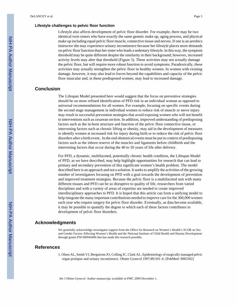
Lifespan ModelThe pelvic floor grows and develops during childhood reaching a maximal capability in earlylife. At this point, there is considerable functional reserve and symptoms are rare. Beyond thispoint, there is a normal decline in functional reserve with increasing age. In certain womenthis reserve is depleted to the point that a threshold is crossed and symptoms begin to occur.When, and if, this occurs will depend on the amount of reserve originally achieved indevelopment, the rate of decline, the degree of stress that her lifestyle places on the pelvic floor,and the effects of any inciting events.
The Lifespan Model for Pelvic Floor Disorders is a simple graphical display that portrays atheoretical parameter, “pelvic floor function,” and tracks it over a woman’s life (Figure 1).There are three basic life phases in this analysis: Phase I, Predisposing Factors (e.g., growthand development); Phase II, Inciting Factors (e.g., birth induced changes); and Phase III,Intervening Factors (e.g., age-related changes).
This “pelvic floor function,” as an abstract concept, can have several different meaningsdepending on the condition being considered. Generally, it can be viewed as the sum of theactivities of many elements of the pelvic floor that each contribute in different ways to overallpelvic organ support and urinary and fecal control. In this sense, it is a concept rather than onespecific measurable parameter. The concept can include the combined actions of the levatorani muscles, urethra, bladder, anal sphincters with their controlling nerves and supportingconnective tissue, or alternatively can also be used to consider any single parameter (forexample, levator ani muscle maximal strength, anterior vaginal wall support, or urethral closurepressure).
In the Lifespan Model for Pelvic Floor Disorders, we consider the composite function of thepelvic floor as a conceptual entity. Although the pelvic floor is a single unit, it is comprised ofseveral separate structures that relate to clinical symptoms and problems such as prolapse andincontinence. These involve muscles that have different origins and insertions, specific levelsat which neural control occurs, and unique connective tissue features that connect specific partsof the pelvic floor to the bony pelvis and to one another that allows the actions of individualstructural units to be integrated. Many of the structures that relate to stress incontinence aredifferent than those involved with fecal incontinence. On the other hand there are elementssuch as the levator ani muscles and the pelvic nerves that affect all aspects of the pelvic floor.
Each of these structures has its own tissue composition. Dense regular connective tissue of thearcus tendineus fascia pelvis is similar to tendon and very different than the smooth muscleand irregular connective tissue makeup of the vaginal wall, or the striated muscle of the levatorani. The injury, recovery, and disease mechanisms that affect each type of tissue and structuremust be individually considered. For example, during birth, the striated muscle could sustainirrecoverable injury due to rupture of the muscle from excess stretching, denervation fromnerve compression or elongation, or ischemic injury due to compartment syndrome.Connective tissue change with advancing age could be due to gradual stretching due to
DeLANCEY et al. Page 2
Am J Obstet Gynecol. Author manuscript; available in PMC 2009 December 1.
NIH
-PA Author Manuscript
NIH
-PA Author Manuscript
NIH
-PA Author Manuscript
mechanical deterioration, or it could rupture during a single episode of structural overload.Each type of tissue has specific types of injury; specifying the proposed mechanism ofimpairment can focus research on forward progress.
Women present with specific symptoms that relate to a subset of these structures. Each structuremust be considered in the role that it plays in pelvic organ support and urinary and fecal control.Additionally, each of these structures functions differently in the context of aging and lifestylefactors over time.
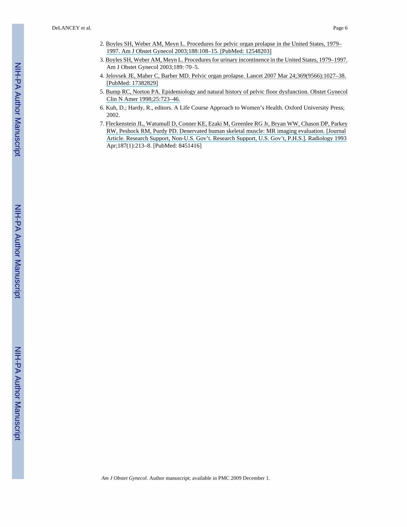
Phase I: Predisposing FactorsThere are normal variations in initial growth and development of all parts of the body. Variationin height is perhaps the most familiar. Pelvic floor development, like height, is influenced byfactors such as an individual’s genetic code, nutrition, and environment. An individual whodevelops excellent pelvic floor function may never have sufficient deterioration to developPFD symptoms throughout her lifespan, despite inciting or lifestyle events. However, anotherwoman whose growth and development are less robust may become symptomatic during herlifetime solely on the basis of normal age-related decline (Figure 2). Conceptually then, therole of genetics must be considered in the analysis of PFD causation.
Phase II: Inciting factorsThe pelvic floor may be altered by events such as pregnancy and childbirth. No other part ofa woman’s body has such dramatic and dynamic changes than the pelvic floor during the secondstage of labor. The degree of pelvic floor injury and recovery is shown in Figure 3. Birth of asmall baby (e.g., 2,400 grams) may cause few changes in pelvic floor structures, while deliveryby forceps of a 5,000 gram infant may cause considerable changes, and even damage. If littledamage occurs a woman’s body may completely recover and she may not experience symptomsof pelvic floor disorders. Even with substantial, dynamic changes to the pelvic floor duringchildbirth, return to normal functional capacity occurs for most women. The fact that onewoman can deliver several large babies without developing PFD while another can havesignificant problems after a single birth, or a relatively small infant, indicates how little weknow about this process. Depending on Phase I events, a woman may have no increase in therisk of developing pelvic floor disorders, despite the experience of potentially inciting events.
In another instance with the delivery of a macrosomic infant by forceps, there may be types ofinjury that cannot be fully recovered from. Permanent nerve injury, avulsion of a muscle, andrupture of connective tissue under the skin are examples of injuries that lead to lasting changesin the pelvic floor. Because the pelvic floor has excess capacity in young women, this damagemay not become immediately evident in all cases, however the loss of capacity, when addedto deterioration with advancing age, can lead to symptoms later in life. Certain instances, forexample neuromuscular damage to the urethra or its connective tissue supports, may result inimmediate symptoms.
To date, the cause of injury and pattern of recovery revealing the underlying origin of lastingstructural damage has not been well elucidated. It is possible to presume that if a torn muscle,myogenic injury, were to occur the defect would be visible immediately and persist. In contrast,a neurogenic injury in a muscle may appear normal early on, but then degenerate over timedue to denervation-related atrophy (7). However, there is limited understanding of the variedrecovery processes. What is certain is that, for most women, the experience of childbirth and,in particular, vaginal birth is a lifespan event from which they initially recover. The LifespanModel for PFD, however, offers a method of conceptualizing the process of initial recoveryand subsequent PFD as well as the opposite: no injury, yet subsequent PFD. The modelchallenges our research efforts to broadly consider the specific events or factors within the
DeLANCEY et al. Page 3
Am J Obstet Gynecol. Author manuscript; available in PMC 2009 December 1.
NIH
-PA Author Manuscript
NIH
-PA Author Manuscript
NIH
-PA Author Manuscript
larger context and reality of a woman’s lifespan in order to enhance our ability to accuratelyidentify prevention and treatment strategies.
There are several aspects of childbirth that have been cited as factors leading to PFD, but allrelate to the amount of injury that occurs and the ability of the body to recover from theseinjuries. Whether a woman sustains injury is influenced by her pelvic floor structure (e.g.,pelvic size and shape), strength of her soft tissues (e.g., fragile or strong), size of the baby (e.g.,small or large), mechanism of delivery (e.g., occipito-anterior or -posterior), obstetricalinterventions (e.g., forceps delivery), and potentially the management of second stage labor.It is necessary to be clear about which factors might be causative (e.g., large infant), and whichfactors are markers of difficult birth (e.g., anal sphincter laceration). For example, obstetricalmanagement of end stage bradycardia that requires performance of an episiotomy andsubsequent manual manipulation to accomplish delivery may also result in a fourth degreeextension. In addition to obstetrical factors, events such as spinal cord injury or hysterectomymight also play an inciting role.
To gain a more detailed understanding of birth-induced injury, the type of injury (e.g., muscleimpairment) and the mechanism of injury (stretch induced avulsion vs. nerve compression vs.nerve stretch) must be determined. Each of these factors will lead to different approaches forintervention and the degree to which the body can recover from these injuries needs to beestablished. For example, if the length of time a nerve is compressed is believed to be themechanism of injury, it might lead to forceps to shorten the second stage. If, however, it is therate and degree of nerve stretch that is most often actually responsible, then adding forcepsmay in fact decrease the length of the second stage, but increase injury. For this reason, theexact nature of the injury must be carefully considered.
Phase III: Intervening factorsWe have reviewed the development of initial pelvic floor functional reserve and the pelvicfloor injury and recovery that occur during vaginal birth. Most women live more than half theirlife after reproduction has been completed and it is during these 40 to 50 years when the rateof pelvic floor decline influences the likelihood of experiencing pelvic floor disorders. Pelvicorgan prolapse, for example, increases with age (1). Several factors influence the rate at whichdecline in pelvic floor function occurs and it should be noted that the effect of these factorsmay not occur for many years after they begin to influence pelvic floor function. The morerapidly age-related impairment occurs, the sooner symptom threshold occurs (Figure 4). Forexample, a woman may have a normally developed pelvic floor and may not have any birthrelated damage, yet may develop pelvic floor disorder as she ages. For any individual, the rateof aging may vary. These genetically programmed variations of aging influence when a womanreaches the symptom threshold.
The normal decline of the pelvic floor across the lifespan may be influenced by other factors.Chronic constipation is an example of a condition that affects pelvic floor load by increasingstress and strain over time. Obesity is associated with increased rates of pelvic floor disordersindependent of other associated factors. Finally scurvy, though rare today, impacts connectivetissue integrity and such treatments as chronic steroid use can accelerate loss of connectivetissue reserve. Generalized problems such as diabetes may affect peripheral motor and sensorynerves. Increased loading, such as occupations that involve heavy lifting or conditions thatelicit heavy coughing, such as chronic bronchitis, may result in a type of “repetitive motion”trauma that challenges the pelvic floor. Any of these factors, or combination of factors, canaccelerate loss of pelvic floor function over the course of a woman’s lifespan.
DeLANCEY et al. Page 4
Am J Obstet Gynecol. Author manuscript; available in PMC 2009 December 1.
NIH
-PA Author Manuscript
NIH
-PA Author Manuscript
NIH
-PA Author Manuscript
Lifestyle challenges to pelvic floor functionLifestyle also affects development of pelvic floor disorder. For example, there may be twoidentical twin sisters who have exactly the same genetic make up, aging process, and physicalmake up including equal pelvic floor muscle, connective tissue and nerves. If one is an aerobicsinstructor she may experience urinary incontinence because her lifestyle places more demandson pelvic floor function than her sister who leads a sedentary lifestyle. In this way, the symptomthreshold may be quite different despite the similarity in their background; however, increasedactivity levels may alter that threshold (Figure 5). These activities may not actually damagethe pelvic floor, but will require more robust function to avoid symptoms. Paradoxically, theseactivities may actually strengthen the pelvic floor in healthy women. In women with existingdamage, however, it may also lead to forces beyond the capabilities and capacity of the pelvicfloor muscular and, in these predisposed women, may lead to increased damage.
ConclusionThe Lifespan Model presented here would suggest that the focus on preventive strategiesshould be on more refined identification of PFD risk in an individual woman as opposed touniversal recommendations for all women. For example, focusing on specific events duringthe second stage management in individual women to reduce risk of muscle or nerve injurymay result in successful prevention strategies that avoid exposing women who will not benefitto interventions such as cesarean section. In addition, improved understanding of predisposingfactors such as the in-born structure and function of the pelvic floor connective tissue, orintervening factors such as chronic lifting or obesity, may aid in the development of measuresto identify women at increased risk for injury during birth or to reduce the risk of pelvic floordisorders after a birth event.. In the end obstetrical events must be put in context of predisposingfactors such as the inborn reserve of the muscles and ligaments before childbirth and theintervening factors that occur during the 40 to 50 years of life after delivery.
For PFD, a dynamic, multifaceted, potentially chronic health condition, the Lifespan Modelof PFD, as we have described, may help highlight opportunities for research that can lead toprimary and secondary prevention of this significant women’s health problem. The modeldescribed here is an approach and not a solution. It seeks to amplify the activities of the growingnumber of investigators focusing on PFD with a goal towards the development of preventionand improved treatment strategies. Because the pelvic floor is a multifaceted unit with manydifferent tissues and PFD can be so disruptive to quality of life, researchers from varieddisciplines and with a variety of areas of expertise are needed to create improvedinterdisciplinary approaches to PFD. It is hoped that this article can form a unifying model tohelp integrate the many important contributions needed to improve care for the 300,000 womeneach year who require surgery for pelvic floor disorder. Eventually, as data become available,it may be possible to quantify the degree to which each of these factors contributes todevelopment of pelvic floor disorders.
AcknowledgmentsWe gratefully acknowledge investigator support from the Office for Research on Women’s Health’s SCOR on Sexand Gender Factors Affecting Women’s Health and the National Institute of Child Health and Human Developmentthrough grants P50 HD044406 that has made this research possible.
References1. Olsen AL, Smith VJ, Bergstrom JO, Colling JC, Clark AL. Epidemiology of surgically managed pelvic
organ prolapse and urinary incontinence. Obstet Gynecol 1997;89:501–6. [PubMed: 9083302]
DeLANCEY et al. Page 5
Am J Obstet Gynecol. Author manuscript; available in PMC 2009 December 1.
NIH
-PA Author Manuscript
NIH
-PA Author Manuscript
NIH
-PA Author Manuscript
2. Boyles SH, Weber AM, Meyn L. Procedures for pelvic organ prolapse in the United States, 1979–1997. Am J Obstet Gynecol 2003;188:108–15. [PubMed: 12548203]
3. Boyles SH, Weber AM, Meyn L. Procedures for urinary incontinence in the United States, 1979–1997.Am J Obstet Gynecol 2003;189:·70–5.
4. Jelovsek JE, Maher C, Barber MD. Pelvic organ prolapse. Lancet 2007 Mar 24;369(9566):1027–38.[PubMed: 17382829]
5. Bump RC, Norton PA. Epidemiology and natural history of pelvic floor dysfunction. Obstet GynecolClin N Amer 1998;25:723–46.
6. Kuh, D.; Hardy, R., editors. A Life Course Approach to Women’s Health. Oxford University Press;2002.
7. Fleckenstein JL, Watumull D, Conner KE, Ezaki M, Greenlee RG Jr, Bryan WW, Chason DP, ParkeyRW, Peshock RM, Purdy PD. Denervated human skeletal muscle: MR imaging evaluation. [JournalArticle. Research Support, Non-U.S. Gov’t. Research Support, U.S. Gov’t, P.H.S.]. Radiology 1993Apr;187(1):213–8. [PubMed: 8451416]
DeLANCEY et al. Page 6
Am J Obstet Gynecol. Author manuscript; available in PMC 2009 December 1.
NIH
-PA Author Manuscript
NIH
-PA Author Manuscript
NIH
-PA Author Manuscript
Figure 1. Integrated lifespan analysis of pelvic floor functionThis graphical display of the abstract concept of pelvic floor function tracks the functionalreserve throughout different phases of a woman’s lifespan. Initially, pelvic floor structuregrowth in late teens leads to a fully developed pelvic floor. Vaginal birth affects pelvic floorfunction. Finally, age-related deterioration occurs until a symptom threshold is reached wherethe functional reserve present earlier in life is lost. (© DeLancey 2007)
DeLANCEY et al. Page 7
Am J Obstet Gynecol. Author manuscript; available in PMC 2009 December 1.
NIH
-PA Author Manuscript
NIH
-PA Author Manuscript
NIH
-PA Author Manuscript
Figure 2. Phase I Variation in Growth and DevelopmentDifferences in the degree of functional reserve development during early life, and the effect itcan have on when symptoms may develop are demonstrated. An individual with morefunctional reserve (A) may not develop symptoms during her lifespan while somewhat lessreserve may lead to symptoms late in life and even less reserve, symptoms early in life. (©DeLancey 2007)
DeLANCEY et al. Page 8
Am J Obstet Gynecol. Author manuscript; available in PMC 2009 December 1.
NIH
-PA Author Manuscript
NIH
-PA Author Manuscript
NIH
-PA Author Manuscript
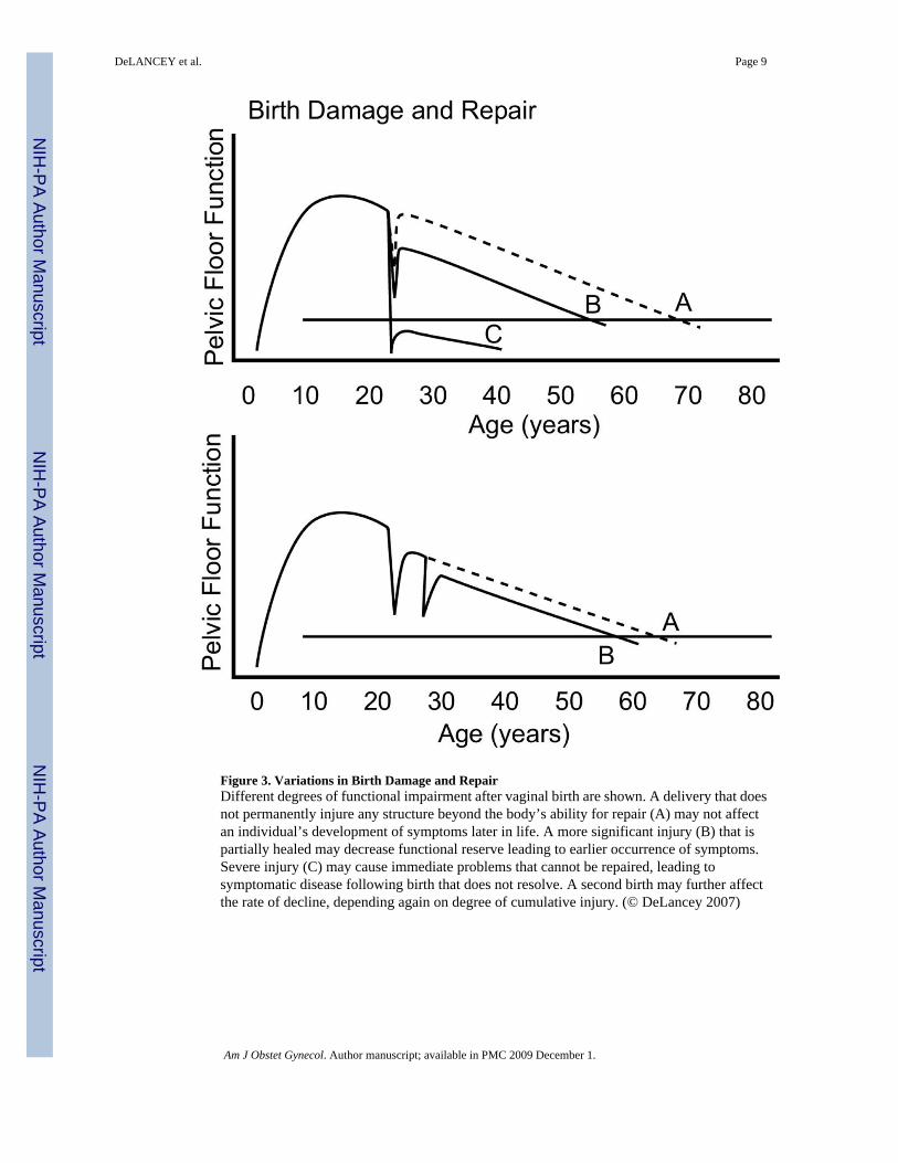
Figure 3. Variations in Birth Damage and RepairDifferent degrees of functional impairment after vaginal birth are shown. A delivery that doesnot permanently injure any structure beyond the body’s ability for repair (A) may not affectan individual’s development of symptoms later in life. A more significant injury (B) that ispartially healed may decrease functional reserve leading to earlier occurrence of symptoms.Severe injury (C) may cause immediate problems that cannot be repaired, leading tosymptomatic disease following birth that does not resolve. A second birth may further affectthe rate of decline, depending again on degree of cumulative injury. (© DeLancey 2007)
DeLANCEY et al. Page 9
Am J Obstet Gynecol. Author manuscript; available in PMC 2009 December 1.
NIH
-PA Author Manuscript
NIH
-PA Author Manuscript
NIH
-PA Author Manuscript
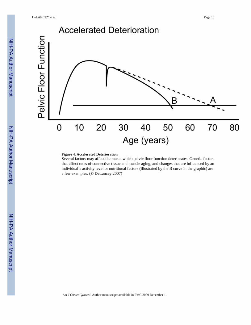
Figure 4. Accelerated DeteriorationSeveral factors may affect the rate at which pelvic floor function deteriorates. Genetic factorsthat affect rates of connective tissue and muscle aging, and changes that are influenced by anindividual’s activity level or nutritional factors (illustrated by the B curve in the graphic) area few examples. (© DeLancey 2007)
DeLANCEY et al. Page 10
Am J Obstet Gynecol. Author manuscript; available in PMC 2009 December 1.
NIH
-PA Author Manuscript
NIH
-PA Author Manuscript
NIH
-PA Author Manuscript
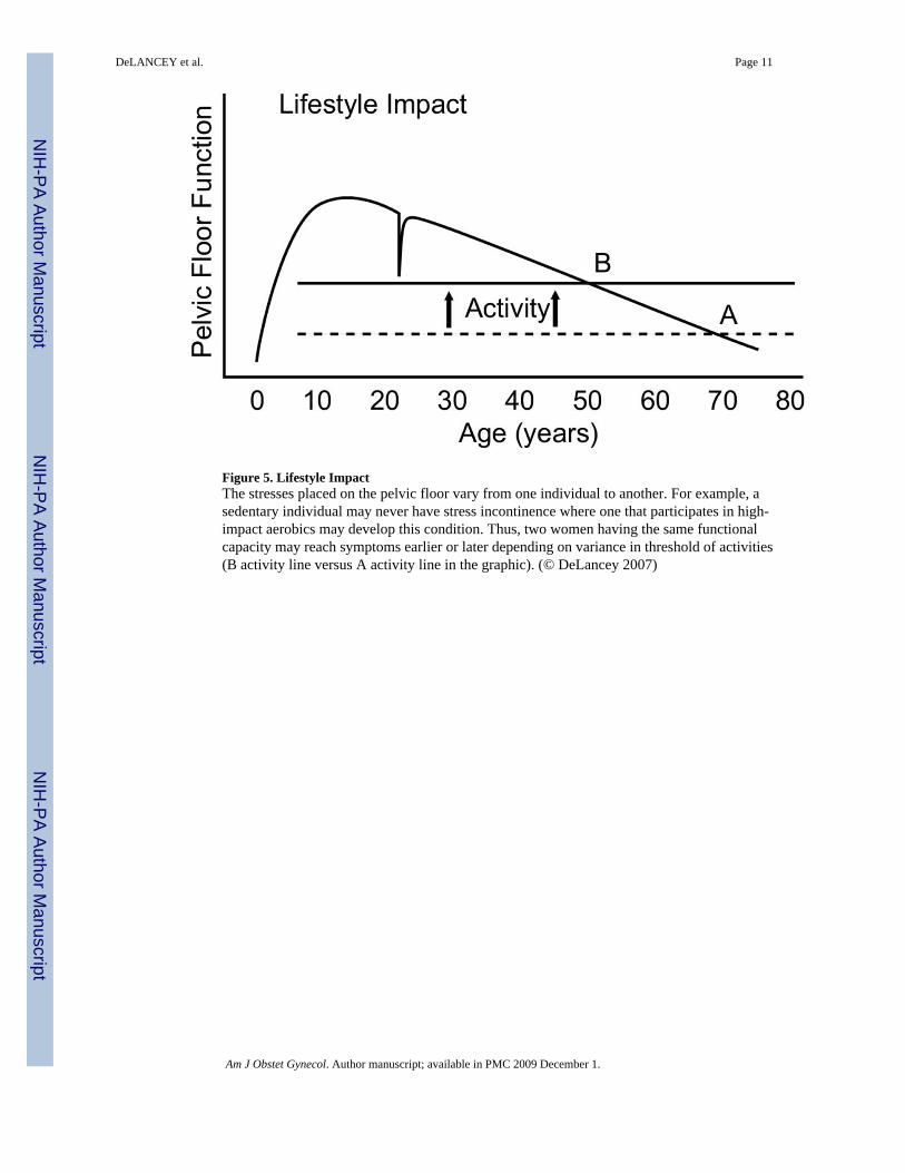
Figure 5. Lifestyle ImpactThe stresses placed on the pelvic floor vary from one individual to another. For example, asedentary individual may never have stress incontinence where one that participates in high-impact aerobics may develop this condition. Thus, two women having the same functionalcapacity may reach symptoms earlier or later depending on variance in threshold of activities(B activity line versus A activity line in the graphic). (© DeLancey 2007)
DeLANCEY et al. Page 11
Am J Obstet Gynecol. Author manuscript; available in PMC 2009 December 1.
NIH
-PA Author Manuscript
NIH
-PA Author Manuscript
NIH
-PA Author Manuscript
NIH
-PA Author Manuscript
NIH
-PA Author Manuscript
NIH
-PA Author Manuscript
DeLANCEY et al. Page 12
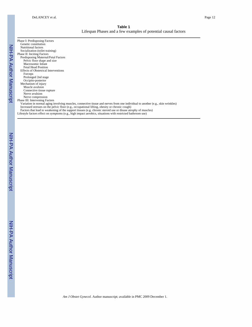
Table 1Lifespan Phases and a few examples of potential causal factors
Phase I: Predisposing Factors Genetic constitution Nutritional factors Socialization (toilet training)Phase II: Inciting Factors Predisposing Maternal/Fetal Factors Pelvic floor shape and size Macrosomic Infant Fetal Head Position Effects of Obstetrical Interventions Forceps Prolonged 2nd stage Occipito-posterior Mechanism of injury Muscle avulsion Connective tissue rupture Nerve avulsion Nerve compressionPhase III: Intervening Factors Variation in normal aging involving muscles, connective tissue and nerves from one individual to another (e.g., skin wrinkles) Increased stresses on the pelvic floor (e.g., occupational lifting, obesity or chronic cough) Factors that lead to weakening of the support tissues (e.g. chronic steroid use or disuse atrophy of muscles)Lifestyle factors effect on symptoms (e.g., high impact aerobics, situations with restricted bathroom use)
Am J Obstet Gynecol. Author manuscript; available in PMC 2009 December 1.
Related Documents