Graph-based tracking of the tongue contour in ultrasound sequences with adaptive temporal regularization Lisa Tang, Ghassan Hamarneh Medical Image Analysis Lab, Simon Fraser University, Canada {lisat,hamarneh}@cs.sfu.ca Abstract We propose a graph-based approach for semi-automatic tracking of the human tongue in 2D+time ultrasound im- age sequences. We construct a graph capturing the intra- (spatial) and inter-frame (temporal) relationships between the dynamic contour vertices. Tongue contour tracking is formulated as a graph-labeling problem, where each ver- tex is labeled with a displacement vector describing its mo- tion. The optimal displacement labels are those minimizing a multi-label Markov random field energy with unary, pair- wise, and ternary potentials, capturing image evidence and temporal and smoothness regularization, respectively. The regularization strength is designed to adapt to the reliability of images features. Evaluation based on real clinical data and comparative analyses with existing approaches demon- strate the accuracy and robustness of our method. 1. Introduction Ultrasound (US) imaging is the most effective technique to capture the motion of the human tongue during speech. Analysis of tongue US to extract information about the tongue’s shape and dynamics has numerous applications, e.g. studying the effects of aging and glossectomies on speech, studies in linguistics and phonetics, and speech per- ception and tongue modeling [4, 6, 21]. A crucial component to tongue shape analysis is the extraction of tongue contours from an US sequence. However, as the number of frames of a typical US sequence is very large, manual segmenta- tion of the tongue contour from each frame for subsequent shape analysis is unrealistic and thus techniques for auto- matic segmentation are urgently needed. The automatic segmentation of US images and track- ing of shapes within are generally difficult due to known problems [21]: 1) low signal-to-noise ratio; 2) high speckle noise corruption; 3) general US artifacts (e.g. acoustic shad- owing, mirroring, and refraction [4]); and 4) weak inter- frame relation due to partially decorrelated speckle noise. In US, parts of the tongue will often disappear between consecutive frames, such that matching based on anatom- ical landmarks, e.g. apex of the tongue, cannot be reliably performed. Further, structures within the tongue (e.g. ten- dons and blood vessels) may function as US wave reflec- tors, causing not only sporadic disappearances of the en- tire tongue contour, but also occurrences of bright profiles in non-interest regions [13, 4], rendering intensity informa- tion unreliable. Furthermore, unlike echocardiographs and breast US images, textures useful for matching are lacking in US tongue images, making the automatic segmentation of tongue contour in these images a challenging problem. Various segmentation approaches for US sequence have been proposed in the literature, including active contour ap- proaches (e.g. snakes); optical flow or registration-based; and tracking. Snakes depend on external and internal en- ergies to pull the contour estimate towards matching image features while constraining the contour to be smooth. In the tongue segmentation algorithm of Akgul et al. [4], the en- ergy functional used includes a similarity term that forces the final contour to be similar to a template contour, and two smoothness terms that either restrict the angles between consecutive points on the contour or constrain the stretching of the contour. In [5], Aron et al. introduced a snake-based approach that relies on preprocessing of US frames to en- hance edge information, and the use of electromagnetic sen- sors for contour-initialization based on an optical-flow for- mulation. More recently, Roussos et al. [24] incorporated models of shape variations and active appearance models. The use of these models, however, required additional in- formation: X-ray videos gathered during the same US scan were used to construct a motion model and expert’s man- ual annotations of US frames were used to build a texture model. Besides the need for a laborious annotation process, which may be erroneous as landmarks are lacking in US images, the acquisition of X-ray images is not always avail- able, making their approach fairly impractical. In [18], Li et al. also developed a snake-based tongue contour extraction software, called EdgeTrak, that accounts for local edge gra- dient, local region-based intensity information, and contour 1

Welcome message from author
This document is posted to help you gain knowledge. Please leave a comment to let me know what you think about it! Share it to your friends and learn new things together.
Transcript
Graph-based tracking of the tongue contourin ultrasound sequences with adaptive temporal regularization
Lisa Tang, Ghassan HamarnehMedical Image Analysis Lab, Simon Fraser University, Canada
{lisat,hamarneh}@cs.sfu.ca
Abstract
We propose a graph-based approach for semi-automatictracking of the human tongue in 2D+time ultrasound im-age sequences. We construct a graph capturing the intra-(spatial) and inter-frame (temporal) relationships betweenthe dynamic contour vertices. Tongue contour tracking isformulated as a graph-labeling problem, where each ver-tex is labeled with a displacement vector describing its mo-tion. The optimal displacement labels are those minimizinga multi-label Markov random field energy with unary, pair-wise, and ternary potentials, capturing image evidence andtemporal and smoothness regularization, respectively. Theregularization strength is designed to adapt to the reliabilityof images features. Evaluation based on real clinical dataand comparative analyses with existing approaches demon-strate the accuracy and robustness of our method.
1. Introduction
Ultrasound (US) imaging is the most effective techniqueto capture the motion of the human tongue during speech.Analysis of tongue US to extract information about thetongue’s shape and dynamics has numerous applications,e.g. studying the effects of aging and glossectomies onspeech, studies in linguistics and phonetics, and speech per-ception and tongue modeling [4,6,21]. A crucial componentto tongue shape analysis is the extraction of tongue contoursfrom an US sequence. However, as the number of framesof a typical US sequence is very large, manual segmenta-tion of the tongue contour from each frame for subsequentshape analysis is unrealistic and thus techniques for auto-matic segmentation are urgently needed.
The automatic segmentation of US images and track-ing of shapes within are generally difficult due to knownproblems [21]: 1) low signal-to-noise ratio; 2) high specklenoise corruption; 3) general US artifacts (e.g. acoustic shad-owing, mirroring, and refraction [4]); and 4) weak inter-frame relation due to partially decorrelated speckle noise.
In US, parts of the tongue will often disappear betweenconsecutive frames, such that matching based on anatom-ical landmarks, e.g. apex of the tongue, cannot be reliablyperformed. Further, structures within the tongue (e.g. ten-dons and blood vessels) may function as US wave reflec-tors, causing not only sporadic disappearances of the en-tire tongue contour, but also occurrences of bright profilesin non-interest regions [13, 4], rendering intensity informa-tion unreliable. Furthermore, unlike echocardiographs andbreast US images, textures useful for matching are lackingin US tongue images, making the automatic segmentationof tongue contour in these images a challenging problem.
Various segmentation approaches for US sequence havebeen proposed in the literature, including active contour ap-proaches (e.g. snakes); optical flow or registration-based;and tracking. Snakes depend on external and internal en-ergies to pull the contour estimate towards matching imagefeatures while constraining the contour to be smooth. In thetongue segmentation algorithm of Akgul et al. [4], the en-ergy functional used includes a similarity term that forcesthe final contour to be similar to a template contour, andtwo smoothness terms that either restrict the angles betweenconsecutive points on the contour or constrain the stretchingof the contour. In [5], Aron et al. introduced a snake-basedapproach that relies on preprocessing of US frames to en-hance edge information, and the use of electromagnetic sen-sors for contour-initialization based on an optical-flow for-mulation. More recently, Roussos et al. [24] incorporatedmodels of shape variations and active appearance models.The use of these models, however, required additional in-formation: X-ray videos gathered during the same US scanwere used to construct a motion model and expert’s man-ual annotations of US frames were used to build a texturemodel. Besides the need for a laborious annotation process,which may be erroneous as landmarks are lacking in USimages, the acquisition of X-ray images is not always avail-able, making their approach fairly impractical. In [18], Li etal. also developed a snake-based tongue contour extractionsoftware, called EdgeTrak, that accounts for local edge gra-dient, local region-based intensity information, and contour
1
orientation. Due to its public availability, this software hasbecome a popular tool [7,22] for tongue tracking in US andfor comparative analyses. e.g. [5, 24]. However, as notedin [24], because EdgeTrak uses a local approach (the con-tour of one frame is used to initialize the next), it tends tolose track of the contour after having segmented few framesand so, manual refinement is usually needed.
Pairwise image registration of consecutive US frameshas also been proposed. The obtained deformation fieldsare then applied to an input contour to generate the segmen-tations of all frames. For example, Ledesma-Carbayo etal. [16] applied parametric elastic registration to align con-secutive cardiac US frames. Nielsen [20] extracted specklepatterns for use in a block-matching registration algorithmto track the left-ventricle (LV) in US. Duan et al. [8] appliedcorrelation-based optical flow to track LV endocardial sur-faces in volumetric echocardiography. More recently, Le-ung et al. [17] proposed and validated a feature-matchingbased registration algorithm for real-time applications.
In Bayesian tracking, segmentation is usually formulatedas an estimation of a posterior probability of a segmenta-tion contour given all past observations (i.e. image frames).Two popular techniques include Kalman filtering and par-ticle filtering. In [28], Lin et al. proposed particle filter-ing for tracking the LV in echocardiography using a shapemodel built via Principle Component Analysis (PCA). Theyemployed particle filters to sample and constrain the allow-able space of shape transformations. The likelihood of eachsample shape given the observed image data is evaluated toestimate the target shape by a weighted combination of allshape samples. However, they have tested their method ona single US echocardiography only. Furthermore, it is un-clear how many manual segmentations are needed to con-struct a reliable PCA model, as it is not stated, and whethera linear (Gaussian) shape model is indeed valid. In [1],Abolmaesumi et al. extracted artery contours from US im-ages using an edge-based algorithm that was incorporatedwith temporal Kalman filtering. However, no analyses weredone to quantify the accuracy and robustness of their ap-proach. Lastly, Qin et al. [22] proposed a semi-automaticapproach to estimate mid-sagittal tongue contour using alearned radial basis function network that nonlinearly mapsthree landmark locations to a contour estimate. Parametersof a spline-interpolation were then optimized via numeri-cal minimization of reconstruction errors computed basedon their groundtruth segmentation data. Nevertheless, theirtest dataset was limited to an US sequence of one speaker.Their approach also required a high degree of intervention(selection of 3 or more landmarks per frame) and a separatetraining procedure per US sequence.
All of the aforementioned methods also have their limita-tions. Snake-based approaches demand good initializationand, in the absence of training data and presence of high
levels of noise corruption, require interactive procedures torefine the obtained segmentations. Accuracy is thus gen-erally sensitive to initialization and model parameters (e.g.number of curve segments or edge points used). Pairwiseregistration and optical-flow based approaches, which prop-agate results from one frame to initialize the segmentationin another, can easily accumulate errors and eventually losetrack of the subject. Lastly, Bayesian tracking and generaltracking methods based on velocity models or parametricmodels often require training data [21, 19].
In this paper, we propose a graph-based approach thatperforms tongue contour-tracking semi-automatically with-out the need for training data and tedious refinement pro-cedures. Using a single input of a contour extracted fromthe start of an US sequence, our method can generate accu-rate tongue contour segmentations for all subsequent framesin the whole sequence. Our approach casts the trackingproblem as a graph-labeling problem wherein each vertexof a graph corresponds to the final position of a controlpoint on a tongue boundary in a frame. Our goal is thento assign to each vertex a displacement label that maps thisparticular vertex to a reference contour point. In solvingthis labeling problem, we define data-likelihood terms de-signed specifically for US images and spatio-temporal reg-ularization terms that ensure each tongue contour estimateis smooth and that it evolves consistently over time. Theamount of regularization is also designed such that it adaptsaccording to the quality of the image data in each frame.
Our proposed method may be seen as most similar to[10] where Friedland and Adam performed ventricular seg-mentation on US image sequences. In their method, a 1-dimensional cyclic Markov Random Field (MRF) was usedto describe a configuration of radii values, each of whichgives a distance of a point on the detected cavity bound-ary to a pre-detected centroid. Simulated annealing wasused to optimize an energy function that: 1) forced eachdetected boundary point to be centered on an optimally de-tected edge; 2) ensured smoothness in the radii values ofneighbouring boundary points; 3) guided the overall seg-mentation to avoid secondary boundaries; and 4) enforcedone-way temporal continuity (the current and next frame) inthe radii configuration. While shown to be effective for USechocardiographs, their method assumed that the target ob-ject is generally elliptical and that a consistent centroid lo-cation can be calculated, assumptions which are inappropri-ate for the segmentation of the non-elliptical tongue contourthat also deforms irregularly and in biased directions. Ad-ditionally, we model the problem with a special graph andadapt the graph-cuts optimization software of Ishikawa [12]in order to solve our exact problem globally. While theuse of graph-cuts had been proposed by Xu and Ajika [27]for active contour-based image segmentation, it was not forsegmenting a sequence of images. As only image gradients
were used, their approach can only be applied to natural im-ages and, as confirmed by our experiments with their tool,would fail when applied to ultrasound images, which haverelatively worse signal-to-noise ratio. We also note thatFreedman and Turek [9] had previously proposed a graph-cuts based illumination-invariant tracking algorithm. As weshall present later, our method differs in that we employ acombination of domain-specific regularization terms (spa-tial and temporal) and incorporate adaptive regularizationso that the amount of temporal regularization is increasedor decreased depending on the absence or presence of reli-able local image features.
To this end, our contributions are: 1) we propose agraph-based approach for US sequence tracking that en-forces both spatial and temporal regularization on the con-tour segmentations; 2) we develop effective ways to encodetemporally-varying regularization; 3) we adapt the algo-rithm of Ishikawa in order to solve the exact problem glob-ally; 4) we examine various approaches to capture imageevidence for reliable contour-tracking; and 5) we conductthorough validation and comparisons with [18, 25] on realclinical data.
2. MethodsLet there be an image sequence I0:T−1, where the sub-
scripts denote frame numbers 0 to T − 1, and an initialsegmentation contour x0 that is represented by a vector ofspatial coordinates of length N, i.e. x0 = {x0,1, · · · ,x0,N},where x0,i denotes the spatial coordinates of the i-th con-trol point of x0. Our goal is to find, for each frame t, acontour xt that segments the tongue shape. We shall reachthis objective via a graph-labeling approach where we rep-resent the spatial coordinates of all segmentation contoursin frames 0 to t with a graph and seek to label each node iin the graph with a displacement vector dt,i such that uponthe termination of our algorithm, the spatial coordinates ofeach control point xt,i is calculated as x0,i + dt,i. Just asin many snake-based formulations [4, 18], the optimalityof each label assignment is computed as the weighted sumof data-driven and regularization penalties, where the for-mer attracts a segmentation to the desired features in eachframe and the latter penalizes excessive bending and dis-continuities in the estimated contours. Additionally, we im-pose temporal regularization to ensure that contours fromconsecutive frames would not deviate significantly. Witha graph-based approach, these penalties are encoded in aMRF energy in which unary potentials capture image ev-idence while pairwise and ternary potentials capture tem-poral and spatial regularization, respectively. In contrastto [18,4], where either the solution from a previous frame isused to initialize the next or where temporal regularizationis ensured via post-processing, our method solves the track-ing problem globally such that it considers all frames in a
sequence together and tries to find the most globally plausi-ble set of displacement vectors under both spatial and tem-poral constraints. We now present the details of our graph-based tracking approach.
2.1. Contour tracking via graph-labeling
We begin by constructing a two-dimensional graphG (V ,E ), where the set of control point coordinates xt,iconstitutes the set of graph vertices V . Our graph-labelingapproach aims to find a set of label assignments that rep-resent a set of displacement vectors D, where each of itselement dt,i spatially maps the i-th point on xt to the i-thpoint on x0. To enforce regularization on the assignments,the set of graph edges E has a grid-like topology such thatvertices along row t represent the segmentation contour offrame t, and edges along each column represent correspon-dence between two points in consecutive frames, e.g. (xt,i,xt+1,i). For brevity, we denote the column (temporal) edgesas et ∈ E T and row (spatial) edges as es ∈ E S such thatE T ⋃
E S = E . es represents intra-frame connectivity be-tween two controls points and is used to regularize the as-signed displacement vectors of neighbouring points withineach contour. et represents inter-frame connectivity and isused to regularize the assigned displacement vectors of aparticular control point across time.
As we introduced previously, the optimality of assign-ing a displacement vector to a vertex will be measured bydata and regularization energies. The data energy encour-ages each displaced control point xt,i to become attached toimage edges while the regularization energies ensure eachcontour remains smooth and continuous and that contoursof adjacent frames move coherently. With a graph-labelingapproach, the optimality of D can then be easily capturedby the MRF energy of labeling V . The unary, pairwise,and ternary potentials of the MRF energy represent external,temporal regularization, and spatial regularization energies,respectively.
Specifically, in edge-based formulations [4, 21, 3, 18], aunary potential may be designed to capture image forcesthat attract a control point xt,i to locations with high imagegradient. In this case, we wish to penalize the assignmentof dt,i to xt,i if dt,i displaces xt,i to locations of low imagegradients:
Eimage(xt,i,dt,i) = exp(0.2|∇I(xt,i +dt,i)|−1) (1)
Conversely, in template-based, feature-matching formula-tions, where image features around a contour estimate arematched to those of a template, the unary potential may in-stead be based directly on image-features. Thus, if F(xt,i, t)denotes a set of image features extracted at xt,i in frame t,then we could treat the input contour x0,i as the templateand seek to minimize the dissimilarity between F(x0,i,0)
and F(xt,i, t). Furthermore, if the dissimilarity between twofeature sets is defined as their L2-norms, then each unarypotential may alternatively be defined as:
EFimage(xt,i , dt,i) = ‖F(x0,i,0)−F(xt,i +di,t , t)‖ (2)
The appropriateness of these alternative data terms is ex-plored further in Sec. 3.
Similar to the approach of [4], pairwise potentials areused to impose temporal constraints over the segmentationcontours in two consecutive frames and is defined as:
Etemp(xt,i , xt+1,i , dt,i , dt+1, j) = ‖dt,i−dt+1,i‖ , (3)
which measures the L2-norm between two displacement la-bels that have been assigned to temporal neighbours xt,i andxt+1,i that are connected by et .
Finally, ternary potentials are used to impose smoothnesson each segmentation contour by measuring the amount ofbending due to three displaced neighbouring points on agiven contour estimate, and is estimated as [4, 3]:
Espat(xt,i , xt, j , xt,k , dt,i , dt, j , dt,k) = 1−ui j ·u jk
|ui j||u jk|(4)
where (xt,i,xt, j,xt,k) is an ordered triplets of spatially adja-cent vertices connected by es and ui j = xt, j +dt, j−xt,i−dt,i.
The overall MRF energy representing the above energiesof the set of contour segmentations implied by D is thendefined as:
E(D) = ∑xt,i∈V
α Eimage(xt,i , dt,i)
+ ∑(xt,i , xt+1,i)∈E T
β Etemp(xt,i , xt+1,i , dt,i , dt, j)
+ ∑(xt,i , xt, j , xt,k)∈E S
γ Espat(xt,i , xt, j , xt,k , dt,i , dt, j , dt,k)
(5)
where α , β , γ are weights for the respective energies.In solving for D, from which we compute the set of all
segmentation contours, we minimize (5) using the graph-cuts optimization technique of Ishikawa [12].
By formulating the contour tracking problem as a graph-labeling problem and representing intra-frame and inter-frame relationships via es and et , we can now track the en-tire US sequence without reinitialization of the contour. Theentire tracking problem is now performed in a single opti-mization procedure, that ensures an exact and global solu-tion [12]. Furthermore, as we explain in the next section,our framework easily allows for adaptive regularizationsuch that contour tracking in unreliable, noise-corruptedframes can now be guided by the more reliable adjacentframes.
2.2. Adaptive temporal regularization
As motivated in the introduction, image features likeridges and edges cannot be reliably extracted in US imagesas the tongue contour may partially or completely disap-pear in a frame. In these cases, we advocate the idea ofadaptively adjusting the amount of temporal regularizationsuch that when the region of a control point xt,i lacks infor-mative image features, meaningful information available inadjacent frames is used instead.
In our formulation, an increased emphasis on temporalinformation is effectively done by increasing the amount oftemporal regularization to be enforced on a graph node suchthat its label assignment is forced to become similar to theassignments obtained in adjacent frames. Encoding adap-tive regularization in a graph-based approach is done by ad-justing the weights of edges in E T according to the imagefeatures available around the local region of xt,i. Specifi-cally, for every node xt,i, we examine the distribution of allunary potentials of xt,i and assign a high weight when itsvariance σ is high or assign a low weight otherwise:
λ (xt,i) = η1
√σ(xt,i)+η2 (6)
where η1 and η2 are constants used to rescale λ to [0,1].The weight β in (5) is then replaced by λ .
2.3. Estimating data penalty with local-phasegradients or image features
What remains to be detailed in (5) is how exactly we cal-culate Eimage. We explored two ways of capturing image-based penalty: one that attracts segmentation estimates toimage edges and one that match features extracted aroundestimates to those extracted from a reference contour. Inthe former, instead of calculating the gradients on the origi-nal images, we calculated the gradients on their local-phasefeatures, which can be interpreted as a qualitative descrip-tion of salient regions in images such as edges or ridgesthat are invariant to changes in illumination or image con-trast [14, 26, 21]. These are based on a model that postu-lates that image features are perceived at points in an im-age where the Fourier components are maximal in phase.These features have shown to be useful for boundary de-tection of LV in echocardiographs [21] and recently pro-posed for bone segmentation and fracture detection in USimages [11]. In extracting these features, we employ theimplementation of [15] that uses monogenic filters.
In the feature-based approach (Section 1), we adoptedthe work of [17], where Leung et al. extracted the follow-ing set of attributes for feature-based registration of US liverimages: pixel intensity, gradient magnitude and the Lapla-cian of Gaussian (LoG). According to the authors, LoGhelps locate features that surround smooth or faded soft
transitions while the gradient magnitude helps detect im-age edges and ridges. Based on our experiments, we foundthat matching the gradient and LoG features extracted witha 3×3 spatial mask worked best. Prior to feature extraction,we applied anisotropic diffusion filtering using the approachof [8] where a linear model was used to control the gradientweight of the diffusion function. This allowed us to obtainmore reliable features.
3. Results3.1. Data acquisition & groundtruth segmentations
A General Electric Logiq Alpha 100 MP ultrasoundscanner (General Electric Medical Systems, Milwaukee,Wisconsin) with a model E72 6.5 MHz transducer that usesa 114◦ microconvex array was used for acquisition of 8datasets. The tongue movement of each participant wasrecorded using the protocol described in [23]. The videooutput from the US scanner was digitized to a video cam-era with a capture rate of 30 frames per second (fps) at aresolution of 240×320 pixels, each of size 0.482 mm2.
Two data sets were created from the collected sequences.The first, denoted by Φdense, contains 2 US sequences ran-domly selected from the entire set. For every sequence inthis set, a dense set of 60 segmentations was created (every2nd frame was segmented) to allow for precise assessmentof tracking accuracy. The second, denoted as Φsparse, con-tains the remaining 6 sequences from which only a sparseset of segmentations were created (every 5th to 8th frame).This set was used to assess the robustness of tracking.
Two experts were recruited to manually create the seg-mentations; each created either Φdense or Φsparse. For eachframe of an US sequence, a segmentation was manually cre-ated by positioning 15 to 23 points which the expert be-lieved would represent the contour of the tongue. A total of16-23 frames were segmented per sequence.
Intra and inter-rater reliability of the experts were as-sessed by repeating the extraction session on a segment of adata set. The intra-rater mean measurement error of the ex-pert who created Φdense was found to be 0.92 mm (0.72 mmstd) while the mean error of the other expert was found tobe 0.77 mm (0.69 mm std). Inter-rater difference was statis-tically insignificant (mean positional difference of 0.92mm±1.1 std). Because the accuracy of the US scanner was es-timated to be within 61 mm and that no single intra-ratermean measurement error exceeded 3 mm, their segmenta-tions were regarded as acceptable and thereafter treated asgroundtruth.
3.2. Validation
Following [24], we performed validation by comparingthe tracked contours obtained by the proposed method withthe groundtruth segmentations. The segmentation error on
each tracked contour was defined as the mean Euclideandistance (MED) between each point on a segmentation so-lution and the closest point on the groundtruth contour.
We used the QPBO-based graph-cuts optimization algo-rithm of Ishikawa [12]. Due to the small size of the graph(< 30 control points for every frame in T < 60 frames),the run-time of the algorithm depended mostly on the sizeof label set (label set of displacement vectors). In defin-ing this label set, we sampled the ℜ2 space in polar co-ordinates within a bounded range. The discretization ofthis space is thus parameterized by the number of steps ofalong the radial axis, Nrad , and the number of steps of alongthe angular axis, Nang, and the maximum radius Rmax. For2D US sequences, we observed that the maximal displace-ment required is no more than half the size of the imagewidth. Thus, for all our experiments, we set Rmax = 30 mm,Nang = 8◦, and Nrad = 2 mm to obtain < 100 labels. For aset of 150 labels, the optimization algorithm converged usu-ally in < 2 min, when run on a PC with 2.66 GHz Intel R©
CoreTM 2 Duo CPU. The overhead on the overall run-timeof our method thus chiefly depended on the calculation ofthe unary potentials (pre-filtering and feature-extraction).
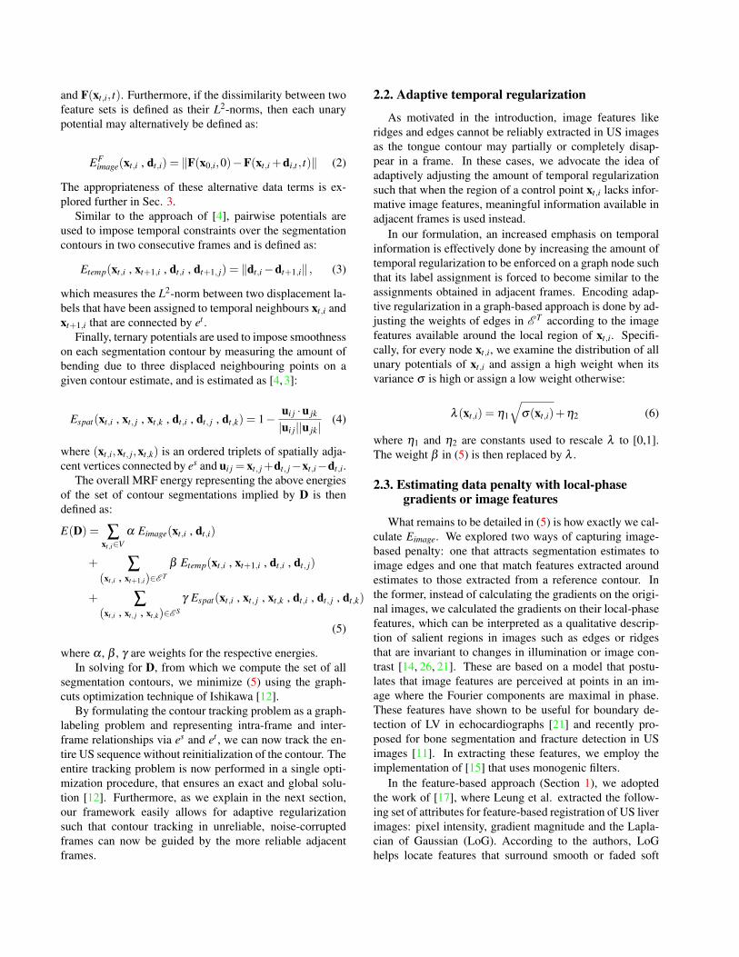
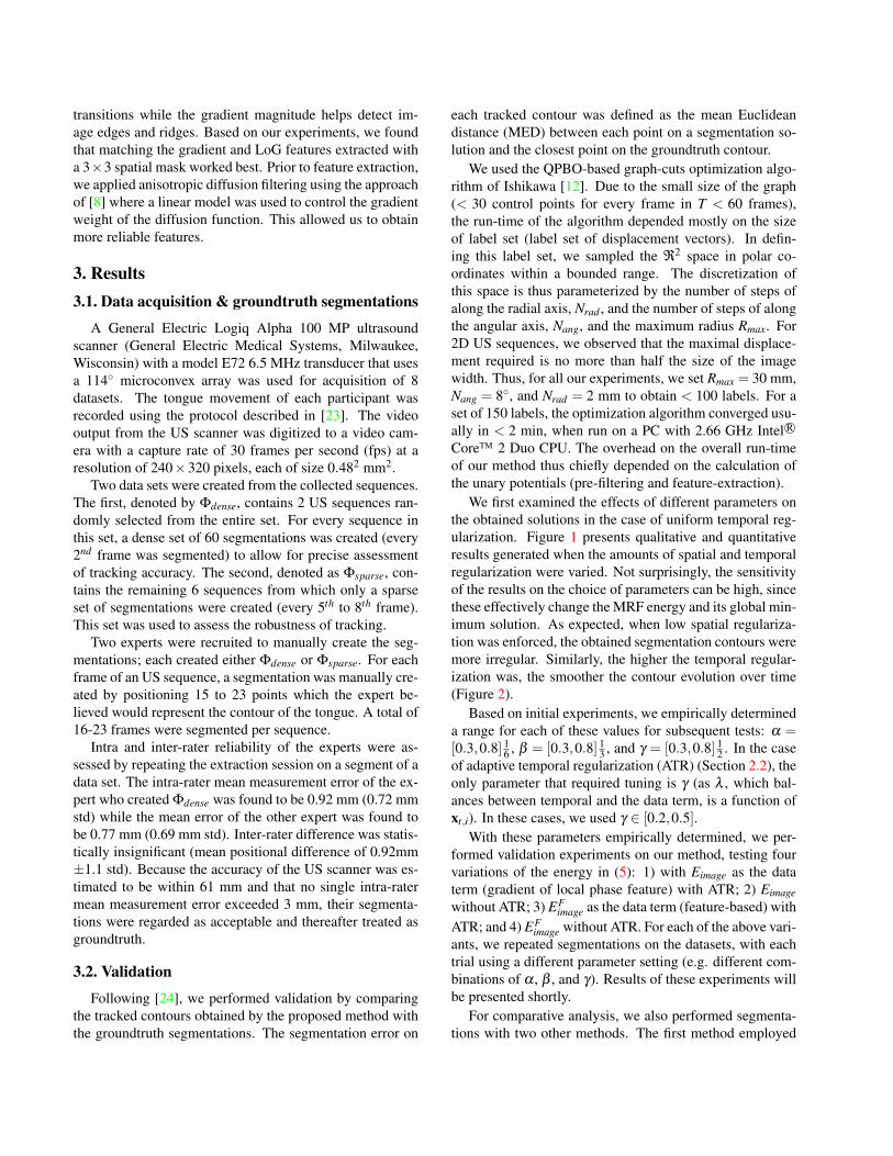
We first examined the effects of different parameters onthe obtained solutions in the case of uniform temporal reg-ularization. Figure 1 presents qualitative and quantitativeresults generated when the amounts of spatial and temporalregularization were varied. Not surprisingly, the sensitivityof the results on the choice of parameters can be high, sincethese effectively change the MRF energy and its global min-imum solution. As expected, when low spatial regulariza-tion was enforced, the obtained segmentation contours weremore irregular. Similarly, the higher the temporal regular-ization was, the smoother the contour evolution over time(Figure 2).
Based on initial experiments, we empirically determineda range for each of these values for subsequent tests: α =[0.3,0.8] 1
6 , β = [0.3,0.8] 13 , and γ = [0.3,0.8] 1
2 . In the caseof adaptive temporal regularization (ATR) (Section 2.2), theonly parameter that required tuning is γ (as λ , which bal-ances between temporal and the data term, is a function ofxt,i). In these cases, we used γ ∈ [0.2,0.5].
With these parameters empirically determined, we per-formed validation experiments on our method, testing fourvariations of the energy in (5): 1) with Eimage as the dataterm (gradient of local phase feature) with ATR; 2) Eimagewithout ATR; 3) EF
image as the data term (feature-based) withATR; and 4) EF
image without ATR. For each of the above vari-ants, we repeated segmentations on the datasets, with eachtrial using a different parameter setting (e.g. different com-binations of α , β , and γ). Results of these experiments willbe presented shortly.
For comparative analysis, we also performed segmenta-tions with two other methods. The first method employed
Figure 1. Segmentations of pairs of two consecutive frames (frame number and errors are given below each image). Contours obtainedby our method are shown in magenta; EdgeTrak in blue; and groundtruth in yellow. The top row (a-c) depicts the effect of changing theamount of spatial regularization in our method; β is varied with fixed α = 0.8 and β = 0.2: (a) γ = 0.8, (b) γ = 0.2, (c) γ = 0.1. The bottomrow (d-f) shows the effect of changing the amount of temporal regularization with α = 0.8 and γ = 0.4: (d) β = 0.1, (e) β = 0.6, (f) β = 1.Note that the smoothness of the contour is ensured in (a), but not in (b) and (c), as shown with red arrows, due to insufficient regularization.For (d-f), because of higher temporal regularization, the inter-frame difference between the two contours in (e) is more subtle than that in(d). The bounded cases appear to reflect the optimality of the respective parameters. The errors of EdgeTrak are 5.53 mm and 7.31 mm forthe respective frames.
Figure 2. Contours extracted from a US sequence are stacked alongthe vertical time axis. Results obtained with (left) minimal tempo-ral regularization (β = 0.05) and (right) adequate temporal regu-larization (β = 0.48).
was the snake-based EdgeTrack1 software developed byLi et al. [18]. The second method we performed waspairwise diffeomorphic demons registration using the algo-rithm developed by Vercauteren et al. [25] that is based onhistogram-matching of local regions. As the latter accountsfor spatial uncertainty on pixel-wise correspondences, webelieve that it is more robust than the algorithms presentedin [2], where optical-flow registrations were performed onconsecutive US frames. Results on pairwise registration
1We could not validate against the work of [24,5] as we do not have ad-ditional multi-modal data (e.g. electromagnetic sensors or X-ray images).
will help us approximate the performance of the correlation-based optical-flow method of [8].
In the demons algorithm, the parameters involvedare ζdemons1 and ζdemons2, which respectively control theamount of smoothing applied on the update field and onthe deformation field [25]. These were tuned empirically(ζdemons1 = [1,5] and ζdemons2 = [2,4]). For EdgeTrak, theonly parameter needed for tuning was the balance, ζET , be-tween the smoothness and image terms. Based on initialexperiments on 4 US sequences, we found that EdgeTrakperformed best when ζET ≤ 0.1 and failed miserably whenζET > 0.3. We also found EdgeTrak highly sensitive to thenumber of points provided by the input contour; its perfor-mance stabilized only when we interpolated the input seg-mentation to twice its original resolution. Thus, we did thisinterpolation for all subsequent tests and repeated the track-ing session with ζET = {0.01,0.03,0.05,0.1,0.3}.
In all methods, only the contour extracted from the firstframe of the groundtruth segmentations of each sequencewas used as input; the groundtruth segmentations of all sub-sequent frames were used for validation.
Overall accuracies of all methods evaluated using Φdenseare presented in Table 1. For all methods, only the regis-tration trial performed on each US sequence that gave thelowest MED was used in the comparison. Note that the re-sults of EdgeTrak had the highest MED while the demons
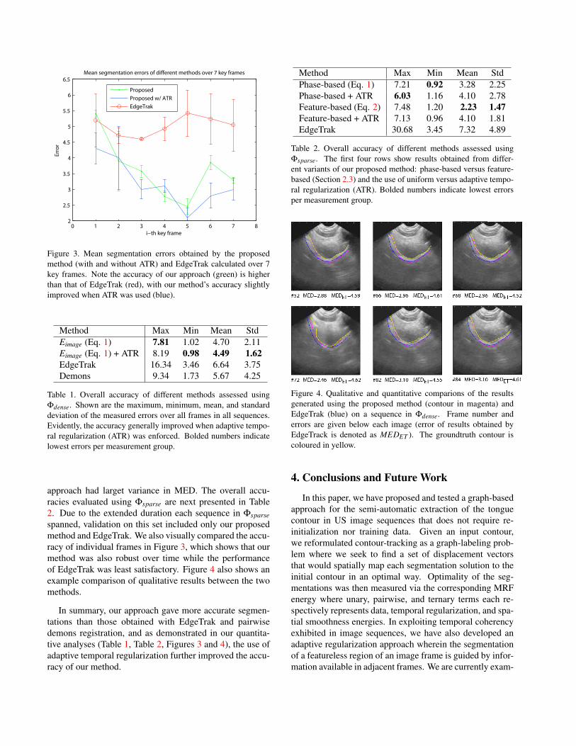
Figure 3. Mean segmentation errors obtained by the proposedmethod (with and without ATR) and EdgeTrak calculated over 7key frames. Note the accuracy of our approach (green) is higherthan that of EdgeTrak (red), with our method’s accuracy slightlyimproved when ATR was used (blue).
Method Max Min Mean StdEimage (Eq. 1) 7.81 1.02 4.70 2.11Eimage (Eq. 1) + ATR 8.19 0.98 4.49 1.62EdgeTrak 16.34 3.46 6.64 3.75Demons 9.34 1.73 5.67 4.25
Table 1. Overall accuracy of different methods assessed usingΦdense. Shown are the maximum, minimum, mean, and standarddeviation of the measured errors over all frames in all sequences.Evidently, the accuracy generally improved when adaptive tempo-ral regularization (ATR) was enforced. Bolded numbers indicatelowest errors per measurement group.
approach had larget variance in MED. The overall accu-racies evaluated using Φsparse are next presented in Table2. Due to the extended duration each sequence in Φsparsespanned, validation on this set included only our proposedmethod and EdgeTrak. We also visually compared the accu-racy of individual frames in Figure 3, which shows that ourmethod was also robust over time while the performanceof EdgeTrak was least satisfactory. Figure 4 also shows anexample comparison of qualitative results between the twomethods.
In summary, our approach gave more accurate segmen-tations than those obtained with EdgeTrak and pairwisedemons registration, and as demonstrated in our quantita-tive analyses (Table 1, Table 2, Figures 3 and 4), the use ofadaptive temporal regularization further improved the accu-racy of our method.
Method Max Min Mean StdPhase-based (Eq. 1) 7.21 0.92 3.28 2.25Phase-based + ATR 6.03 1.16 4.10 2.78Feature-based (Eq. 2) 7.48 1.20 2.23 1.47Feature-based + ATR 7.13 0.96 4.10 1.81EdgeTrak 30.68 3.45 7.32 4.89
Table 2. Overall accuracy of different methods assessed usingΦsparse. The first four rows show results obtained from differ-ent variants of our proposed method: phase-based versus feature-based (Section 2.3) and the use of uniform versus adaptive tempo-ral regularization (ATR). Bolded numbers indicate lowest errorsper measurement group.
Figure 4. Qualitative and quantitative comparions of the resultsgenerated using the proposed method (contour in magenta) andEdgeTrak (blue) on a sequence in Φdense. Frame number anderrors are given below each image (error of results obtained byEdgeTrack is denoted as MEDET ). The groundtruth contour iscoloured in yellow.
4. Conclusions and Future Work
In this paper, we have proposed and tested a graph-basedapproach for the semi-automatic extraction of the tonguecontour in US image sequences that does not require re-initialization nor training data. Given an input contour,we reformulated contour-tracking as a graph-labeling prob-lem where we seek to find a set of displacement vectorsthat would spatially map each segmentation solution to theinitial contour in an optimal way. Optimality of the seg-mentations was then measured via the corresponding MRFenergy where unary, pairwise, and ternary terms each re-spectively represents data, temporal regularization, and spa-tial smoothness energies. In exploiting temporal coherencyexhibited in image sequences, we have also developed anadaptive regularization approach wherein the segmentationof a featureless region of an image frame is guided by infor-mation available in adjacent frames. We are currently exam-
ining ways to automatically adjust the involved parametersand working on incorporating texture-based image featuresto devise an even more robust data energy term.
5. AcknoweldgmentsWe are grateful to Dr. Tim Bressmann from the De-
partment of Speech-Language Pathology, University ofToronto, for provision of the US data. We also thank BiancaHerold and Matt Toom for assistance in data preparation.
References[1] S. Abolmaesumi, P. Abolmaesumi, M. R. Sirouspour, and
S. E. Salcudean. Real-time extraction of carotid artery con-tours from ultrasound. In Computer-Based Medical Systems,pages 181–186, 2000.
[2] B. Achmad, M. M. Mustafa, and A. Hussain. Inter-frameenhancement of ultrasound images using optical flow. In In-ternational Visual Informatics Conference, pages 191–201,2009.
[3] Y. Akgul and C. Kambhamettu. A coarse-to-fine deformablecontour optimization framework. IEEE Transactions on Pat-tern Analysis and Machine Intelligence, 25:174–186, 2003.
[4] Y. Akgul, C. Kambhamettu, and M. Stone. Extraction andtracking of the tongue surface from ultrasound image se-quences. IEEE Conference on Computer Vision and PatternRecognition, page 298, 1998.
[5] M. Aron, A. Roussos, M. odile Berger, E. Kerrien, andP. Maragos. Multimodality acquisition of articulatory dataand processing. In European Conference on Signal Process-ing, 2008.
[6] T. Bressmann, C. Heng, and J. Irish. The application of2D and 3D ultrasound imaging in speech-language pathol-ogy. Speech-Language Pathology and Audiology, 29:158–168, 2005.
[7] L. Davidson. Comparing tongue shapes from ultrasoundimaging using smoothing spline analysis of variance. Acous-tical Society of America, 120(1):407–415, 2006.
[8] Q. Duan, E. Angelini, and O. Gerard. Comparing opticalflow based methods for quantification of myocardial defor-mations on RT3D ultrasound. In IEEE International Sympo-sium on Biomedical Imaging, pages 173–176, 2006.
[9] D. Freedman and M. W. Turek. Illumination-invariant track-ing via graph cuts. In IEEE Conference on Computer Visionand Pattern Recognition, pages 10–17, 2005.
[10] N. Friedland and D. Adam. Automatic ventricular cavityboundary detection from sequential ultrasound images usingsimulated annealing. IEEE Transactions on Medical Imag-ing, 8(4):344–353, 1989.
[11] I. Hacihaliloglu, R. Abugharbieh, A. Hodgson, andR. Rohling. Bone segmentation and fracture detection inultrasound using 3D local phase features. In InternationalConference on Medical Image Computing and Computer As-sisted Intervention, pages 287–295, 2008.
[12] H. Ishikawa. Higher-order clique reduction in binary graphcut. In IEEE Conference on Computer Vision and PatternRecognition, pages 20–25, 2009.
[13] K. Iskarous. Detecting the edge of the tongue: A tutorial.Clinical Linguistics and Phonetics, 19(6):555–565, 2005.
[14] P. Kovesi. Image features from phase congruency. Videre: AJournal of Computer Vision Research, 1(3):2–26, 1999.
[15] P. D. Kovesi. MATLAB and Octave functionsfor computer vision and image processing. URL:<http://www.csse.uwa.edu.au/∼pk/research/matlabfns/>.
[16] M. J. Ledesma-carbayo, J. Kybic, M. Desco, A. San-tos, S. Member, M. Shling, S. Member, P. Hunziker, andM. Unser. Spatio-temporal nonrigid registration for ultra-sound cardiac motion estimation. IEEE Transactions onMedical Imaging, 24(9):1113–1126, 2005.
[17] C. Leung, K. Hashtrudi-Zaad, P. Foroughi, and P. Abolmae-sumi. A real-time intrasubject elastic registration algorithmfor dynamic 2-D ultrasound images. Ultrasound in Medicineand Biology, 35(7):1159 – 1176, 2009.
[18] M. Li, C. Kambhamettu, and M. Stone. Automatic contourtracking in ultrasound images. Clinical Linguistics and Pho-netics, 19(6):545–554, 2005.
[19] S. Mitchell, J. Bosch, B. Lelieveldt, R. van der Geest,J. Reiber, and M. Sonka. 3-D active appearance models:segmentation of cardiac MR and ultrasound images. IEEETransactions on Medical Imaging, 21(9):1167–1178, 2002.
[20] K. E. Nielsen. Practical use of block-matching in 3D speckletracking. Master’s thesis, Norwegian University of Scienceand Technology, Department of Computer and InformationScience, 2009.
[21] J. A. Noble and D. Boukerroui. Ultrasound image segmen-tation: a survey. IEEE Transactions on Medical Imaging,25(8):987–1010, 2006.
[22] C. Qin, M. Carreira-Perpin, K. Richmond, A. Wrench, andS. Renals. Predicting tongue shapes from a few landmarklocations. In Proc. Interspeech, pages 2306–2309, 2008.
[23] O. Rastadmehr, T. Bressmann, R. Smyth, and J. C. Irish. In-creased midsagittal tongue velocity as indication of articu-latory compensation in patients with lateral partial glossec-tomies. Head and Neck, 30(6):718–726, 2008.
[24] A. Roussos, A. Katsamanis, and P. Maragos. Tongue track-ing in ultrasound images with active appearance models. InIEEE International Conference on Image Processing, pages1733–1736, 2009.
[25] T. Vercauteren, X. Pennec, A. Perchant, and N. Ayache. Dif-feomorphic demons using itk’s finite difference solver hier-archy. In Insight Journal – ISC/NA-MIC Workshop on OpenScience at MICCAI 2007, October 2007. Source code avail-able online.
[26] J. Woo, B.-W. Hong, C.-H. Hu, K. K. Shung, C. C. Kuo, andP. J. Slomka. Non-rigid ultrasound image registration basedon intensity and local phase information. J. Signal Process.Syst., 54(1-3):33–43, 2009.
[27] N. Xu, N. Ahuja, and R. Bansal. Object segmentation us-ing graph cuts based active contours. Computer Vision andImage Understanding, 107(3):210–224, 2007.
[28] L. Yang, B. Georgescu, Y. Zheng, P. Meer, and D. Co-maniciu. 3D ultrasound tracking of the left ventricle usingone-step forward prediction and data fusion of collaborativetrackers. IEEE Conference on Computer Vision and PatternRecognition, pages 1–8, 2008.
Related Documents