10 IEEE TRANSACTIONS ON NANOBIOSCIENCE, VOL. 4, NO. 1, MARCH 2005 Gramicidin Channels Olaf S. Andersen, Roger E. Koeppe, II, and Benoît Roux Abstract—Gramicidin channels are mini-proteins composed of two tryptophan-rich subunits. The conducting channels are formed by the transbilayer dimerization of nonconducting sub- units, which are tied to the bilayer/solution interface through hydrogen bonds between the indole NH groups and the phos- pholipid backbone and water. The channel structure is known at atomic resolution and the channel’s permeability characteristics are particularly well defined: gramicidin channels are selective for monovalent cations, with no measurable permeability to anions or polyvalent cations; ions and water move through a pore whose wall is formed by the peptide backbone; and the single-channel conductance and cation selectivity vary when the amino acid se- quence is varied, even though the permeating ions make no contact with the amino acid side chains. Given the amount of experimental information that is available—for both the wild-type channels and for channels formed by amino acid-substituted gramicidin analogues—gramicidin channels provide important insights into the microphysics of ion permeation through bilayer-spanning channels. For the same reason, gramicidin channels constitute the system of choice for evaluating computational strategies for obtaining mechanistic insights into ion permeation through the complex channels formed by integral membrane proteins. Index Terms—Kinetics of ion permeation, molecular dynamics, single-channel electrophysiology, structure–function studies. I. INTRODUCTION T HE LINEAR gramicidins are a family of antibiotics that exert their antibacterial activity by increasing the cation permeability of the target bacterial plasma membrane [1], due to the formation of bilayer-spanning channels [2]. Compared to channels formed by other antibiotics, the gramicidin chan- nels are exceptionally well behaved, which, together with their cation selectivity and the existence of an atomic-level model for the channel structure, the single-stranded (SS) -helical dimer [3], [4], makes gramicidin channels important tools for under- standing the microphysics of ion permeation through bilayer- spanning channels. Gramicidin channels are arguably the best understood ion channels. Atomic (or near-atomic) resolution structures have been reported for an increasing number of ion channels [5]–[13], but gramicidin channels continue to provide a unique combi- nation of advantages that sets them apart from other channels: the structure of the bilayer-spanning channel is known; there is a wealth of information about its ion permeability; the ion Manuscript received November 4, 2004; revised November 15, 2004. This work was supported in part by the National Institutes of Health (NIH) under Grants GM21342 (awarded to O. S. Andersen), RR15569 (awarded to R. E. Koeppe), and GM62342 (awarded to B. Roux). O. S. Andersen and B. Roux are with the Weill Medical College of Cor- nell University, New York, NY 10021 USA (e-mail: [email protected]; [email protected]). R. E. Koeppe, II, is with the University of Arkansas, Fayetteville, AR 72701 USA (e-mail: [email protected]). Digital Object Identifier 10.1109/TNB.2004.842470 permeability can be modulated by defined chemical modifica- tions whose influence on structure can be determined exper- imentally; and the wild-type and amino-substituted analogue channels are large enough to be nontrivial and yet small enough to be amenable to detailed computational studies. We review first the channel structure and dynamics and show that even though the linear gramicidins are conformationally polymorphic in organic solvents, the structure in lipid bilayers, or bilayer-like environments, is remarkably well defined. We then discuss the channels’ permeability properties; finally we show how molecular dynamics (MD) simulations have reached the point where there is semiquantitative agreement between observed and predicted ion permeabilities. II. STRUCTURE The linear gramicidins, exemplified by Val gramicidin A (gA), have an alternating L-D-amino acid sequence [14] which allows the molecule to fold as a helix with the side chains projecting from the exterior surface of a cylindrical tube formed by the peptide backbone [3], [15]. Many such folding patterns are possible, and gA is conformationally polymorphic [16]. Common to the observed conformers is that the helices are stabilized by hydrogen bonds between the backbone CO and NH functions. At least seven such structures have been described in organic solvents, where gA forms a variety of double-stranded (DS) intertwined dimers [17]–[21]. A. One Conducting Channel Conformation In lipid bilayers or bilayer-like environments, however, the channel’s electrophysiological “fingerprint” (single-channel current traces, Fig. 1, and current transition amplitude and lifetime distributions, Fig. 2) show that there is only a single conducting channel structure. The luminal diameter of the pore that is formed by peptide backbone is 4 , as determined from the channel’s permeability to alkali metal cations, H , and water [22]–[24]. These constraints allowed for a refine- ment of the -helical structure [4], which taken together with results from single-channel [25], [26] and nuclear magnetic resonance (NMR) studies [27], [28] established gA channels to be antiparallel SS -helical dimers [29]. B. Atomic Resolution Structure The atomic resolution structure was determined by solution and solid-state NMR [30]–[32]; the channel is an antiparallel formyl-NH-terminal-to-formyl-NH-terminal dimer formed by 1536-1241/$20.00 © 2005 IEEE

Welcome message from author
This document is posted to help you gain knowledge. Please leave a comment to let me know what you think about it! Share it to your friends and learn new things together.
Transcript
10 IEEE TRANSACTIONS ON NANOBIOSCIENCE, VOL. 4, NO. 1, MARCH 2005
Gramicidin ChannelsOlaf S. Andersen, Roger E. Koeppe, II, and Benoît Roux
Abstract—Gramicidin channels are mini-proteins composedof two tryptophan-rich subunits. The conducting channels areformed by the transbilayer dimerization of nonconducting sub-units, which are tied to the bilayer/solution interface throughhydrogen bonds between the indole NH groups and the phos-pholipid backbone and water. The channel structure is known atatomic resolution and the channel’s permeability characteristicsare particularly well defined: gramicidin channels are selective formonovalent cations, with no measurable permeability to anionsor polyvalent cations; ions and water move through a pore whosewall is formed by the peptide backbone; and the single-channelconductance and cation selectivity vary when the amino acid se-quence is varied, even though the permeating ions make no contactwith the amino acid side chains. Given the amount of experimentalinformation that is available—for both the wild-type channelsand for channels formed by amino acid-substituted gramicidinanalogues—gramicidin channels provide important insights intothe microphysics of ion permeation through bilayer-spanningchannels. For the same reason, gramicidin channels constitutethe system of choice for evaluating computational strategies forobtaining mechanistic insights into ion permeation through thecomplex channels formed by integral membrane proteins.
Index Terms—Kinetics of ion permeation, molecular dynamics,single-channel electrophysiology, structure–function studies.
I. INTRODUCTION
THE LINEAR gramicidins are a family of antibiotics thatexert their antibacterial activity by increasing the cation
permeability of the target bacterial plasma membrane [1], dueto the formation of bilayer-spanning channels [2]. Comparedto channels formed by other antibiotics, the gramicidin chan-nels are exceptionally well behaved, which, together with theircation selectivity and the existence of an atomic-level model forthe channel structure, the single-stranded (SS) -helical dimer[3], [4], makes gramicidin channels important tools for under-standing the microphysics of ion permeation through bilayer-spanning channels.
Gramicidin channels are arguably the best understood ionchannels. Atomic (or near-atomic) resolution structures havebeen reported for an increasing number of ion channels [5]–[13],but gramicidin channels continue to provide a unique combi-nation of advantages that sets them apart from other channels:the structure of the bilayer-spanning channel is known; thereis a wealth of information about its ion permeability; the ion
Manuscript received November 4, 2004; revised November 15, 2004.This work was supported in part by the National Institutes of Health (NIH)under Grants GM21342 (awarded to O. S. Andersen), RR15569 (awarded toR. E. Koeppe), and GM62342 (awarded to B. Roux).
O. S. Andersen and B. Roux are with the Weill Medical College of Cor-nell University, New York, NY 10021 USA (e-mail: [email protected];[email protected]).
R. E. Koeppe, II, is with the University of Arkansas, Fayetteville, AR 72701USA (e-mail: [email protected]).
Digital Object Identifier 10.1109/TNB.2004.842470
permeability can be modulated by defined chemical modifica-tions whose influence on structure can be determined exper-imentally; and the wild-type and amino-substituted analoguechannels are large enough to be nontrivial and yet small enoughto be amenable to detailed computational studies.
We review first the channel structure and dynamics and showthat even though the linear gramicidins are conformationallypolymorphic in organic solvents, the structure in lipid bilayers,or bilayer-like environments, is remarkably well defined. Wethen discuss the channels’ permeability properties; finally weshow how molecular dynamics (MD) simulations have reachedthe point where there is semiquantitative agreement betweenobserved and predicted ion permeabilities.
II. STRUCTURE
The linear gramicidins, exemplified by Val gramicidin A(gA), have an alternating L-D-amino acid sequence [14]
which allows the molecule to fold as a helix with the sidechains projecting from the exterior surface of a cylindrical tubeformed by the peptide backbone [3], [15]. Many such foldingpatterns are possible, and gA is conformationally polymorphic[16]. Common to the observed conformers is that the helicesare stabilized by hydrogen bonds between the backbone COand NH functions. At least seven such structures have beendescribed in organic solvents, where gA forms a variety ofdouble-stranded (DS) intertwined dimers [17]–[21].
A. One Conducting Channel Conformation
In lipid bilayers or bilayer-like environments, however, thechannel’s electrophysiological “fingerprint” (single-channelcurrent traces, Fig. 1, and current transition amplitude andlifetime distributions, Fig. 2) show that there is only a singleconducting channel structure. The luminal diameter of the porethat is formed by peptide backbone is 4 , as determinedfrom the channel’s permeability to alkali metal cations, H ,and water [22]–[24]. These constraints allowed for a refine-ment of the -helical structure [4], which taken together withresults from single-channel [25], [26] and nuclear magneticresonance (NMR) studies [27], [28] established gA channels tobe antiparallel SS -helical dimers [29].
B. Atomic Resolution Structure
The atomic resolution structure was determined by solutionand solid-state NMR [30]–[32]; the channel is an antiparallelformyl-NH-terminal-to-formyl-NH-terminal dimer formed by
1536-1241/$20.00 © 2005 IEEE
ANDERSEN et al.: GRAMICIDIN CHANNELS 11
Fig. 1. Single-channel current trace obtained with the gA analogue [D-Ala ,Ser ]gA in a diphytanoylphosphatidylcholine (DPhPC)/n-decane bilayer. Thenumbers to the right of the trace denote the number of conducting channels atthe different current levels. The analogue forms a single type of ion-permeablechannels with well-defined current transitions. The conducting state is “stable,”meaning that the current noise of the conducting state is indistinguishable fromthat of the baseline: the standard deviation (SD) of level 0 is 0.136 pA; for level1, 0.136 pA; for level 2, 0.133 pA. Electrolyte solution: 1.0 M NaCl, pH 7;applied potential 200 mV; current signal filtered at 500 Hz.
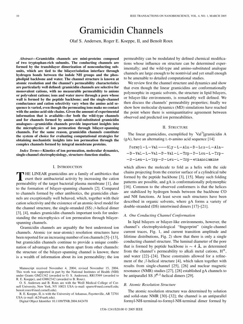
Fig. 2. Characterization of [D-Ala , Ser ]gA channel function. Left:current transition amplitude histogram. There is a single predominant peakat 1.9 pA, which comprises 1175 of the 1202 transitions. Right: lifetimedistribution, as normalized survivor plot. The stippled curve is a fit ofN(t)=N(0) = expf�t=�g to the results, where N(t) is the number ofchannels with lifetime longer than time t, and � the average lifetime (1410 ms).Experimental conditions as in Fig. 1.
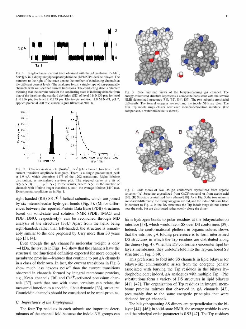
right-handed (RH) SS -helical subunits, which are joinedby six intermolecular hydrogen bonds (Fig. 3). (Minor differ-ences between the reported Protein Data Base (PDB) structuresbased on solid-state and solution NMR (PDB : 1MAG andPDB : 1JNO, respectively), can be reconciled through MDanalysis of the structures [33].) Apart from the helix beingright-handed, rather than left-handed, the structure is remark-ably similar to the one proposed by Urry more than 30 yearsago [3], [4].
Even though the gA channel’s molecular weight is only4 kDa, the results in Figs. 1–3 show that the channels have the
structural and functional definition expected for more complexmembrane proteins—features that continue to put gA channelsin a class of their own. In fact, the current transitions in Fig. 3show much less “excess noise” than the current transitionsobserved in channels formed by integral membrane proteins,e.g., KcsA channels [36] and Ca -activated potassium chan-nels [37], such that one with some certainty can relate themeasured function to a specific, albeit dynamic [33], structure.Gramicidin channels should be considered to be mini-proteins.
C. Importance of the Tryptophans
The four Trp residues in each subunit are important deter-minants of the channel fold because the indole NH groups can
Fig. 3. Side and end views of the bilayer-spanning gA channel. Theenergy-minimized structure represents a composite consistent with the severalNMR-determined structures [31], [32], [34], [35]. The two subunits are shadeddifferently. The formyl oxygens are red, and the indole NHs are blue. Thefour Trp indole rings cluster near each membrane/solution interface. (Forcomparison, a water molecule is shown).
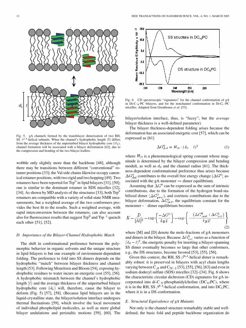
Fig. 4. Side views of two DS gA conformers crystallized from organicsolvents. (A) Structure crystallized from CsCl/methanol or from acetic acid[20]. (B) Structure crystallized from ethanol [19]. As in Fig. 3, the two subunitsare shaded differently: the formyl oxygens are red, and the indole NHs are blue.In contrast to Fig. 3, in the DS structures the Trp indole rings do not clusternear the ends, but are distributed rather evenly along the dimer.
form hydrogen bonds to polar residues at the bilayer/solutioninterface [38], which would favor SS over DS conformers [39].Indeed, the conformational plethora in organic solutes showsthat the intrinsic gA folding preference is to form intertwinedDS structures in which the Trp residues are distributed alongthe dimer (Fig. 4). When the DS conformers encounter lipid bi-layers membranes, they unfold/refold into the Trp-anchored SSstructure in Fig. 3 [40].
This preference to fold into SS channels in lipid bilayers (orbilayer-like environments) arises from the energetic penaltyassociated with burying the Trp residues in the bilayer hy-drophobic core; indeed, gA analogues with multiple Trp Phesubstitutions form a variety of DS structures in lipid bilayers[41], [42]. The organization of Trp residues in integral mem-brane proteins mirrors that observed in gA channels [43],presumably due to the same energetic principles that werededuced for gA channels.
The bilayer-spanning SS dimers are perpendicular to the bi-layer [44]–[46]; in solid-state NMR, the average wobble is zeroand the principal order parameter is 0.93 [47]. The Trp residues
12 IEEE TRANSACTIONS ON NANOBIOSCIENCE, VOL. 4, NO. 1, MARCH 2005
Fig. 5. gA channels formed by the transbilayer dimerization of two RH,SS � -helical subunits. When the channel’s hydrophobic length (l) differsfrom the average thickness of the unperturbed bilayer hydrophobic core (d ),channel formation will be associated with a bilayer deformation [62], due tothe compression and bending of the two bilayer leaflets.
wobble only slightly more than the backbone [48], althoughthere may be transitions between different “conventional” ro-tamer positions [33]; the Val side chains likewise occupy canon-ical rotamer positions, with two rigid and two hopping [49]. Tworotamers have been reported for Trp in lipid bilayers [31], [50];one is similar to the dominant rotamer in SDS micelles [32],[34]. As shown by MD analysis of the structures [33], both Trprotamers are compatible with a variety of solid-state NMR mea-surements; but a weighted average of the two conformers pro-vides the best fit to the results. Such a weighted average, withrapid interconversion between the rotamers, can also accountalso for fluorescence results that suggest Trp and Trp quencheach other [51], [52].
D. Importance of the Bilayer-Channel Hydrophobic Match
The shift in conformational preference between the poly-morphic behavior in organic solvents and the unique structurein lipid bilayers is but one example of environment-dependentfolding. The preference to fold into SS dimers depends on thehydrophobic “match” between bilayer thickness and channellength [53]. Following Mouritsen and Bloom [54], exposing hy-drophobic residues to water incurs an energetic cost [55], [56].A hydrophobic mismatch between the channel’s hydrophobiclength and the average thickness of the unperturbed bilayerhydrophobic core will, therefore, cause the bilayer todeform (Fig. 5) [57], [58]. (Because lipid bilayers are in theliquid-crystalline state, the bilayer/solution interface undergoesthermal fluctuations [59], which involve the local movementof individual phospholipid molecules, as well as more globalbilayer undulations and peristaltic motions [59], [60]. The
Fig. 6. CD spectroscopic “signatures” for the channel conformation of gAin Di-C -PC bilayers, and for the nonchannel conformation in Di-C -PCmicelles. Adapted from Greathouse et al. [53].
bilayer/solution interface, thus, is “fuzzy”; but the averagebilayer thickness is a well-defined parameter).
The bilayer thickness-dependent folding arises because thedeformation has an associated energetic cost [57], which can beexpressed as [61]
(1)
where is a phenomenological spring constant whose mag-nitude is determined by the bilayer compression and bendingmoduli, as well as and the channel radius [61]. The thick-ness-dependent conformational preference thus arises because
contributes to the overall free energy change as-sociated with the gA monomer dimer equilibrium.
Assuming that can be expressed as the sum of intrinsiccontributions, due to the formation of the hydrogen bond-sta-bilized dimer , and extrinsic contributions due to thebilayer deformation, , the equilibrium constant for themonomer dimer equilibrium becomes
(2)where [M] and [D] denote the mole-fractions of gA monomersand dimers in the bilayer. Because varies as a function of
, the energetic penalty for inserting a bilayer-spanningSS dimer eventually becomes so large that other conformers,e.g., the DS structures, become favored [53], [55], [56].
Given this context, the RH, SS -helical dimer is remark-ably robust: it is preserved in bilayers with acyl chain lengthsvarying between C and C [53], [55], [56], [63] and even insodium dodecyl sulfate (SDS) micelles [32]–[34]. Fig. 6 showsthe characteristic circular dichroism (CD) signatures for gA in-corporated into di-C -phosphatidylcholine DC PC , whereit is in the RH, SS -helical conformation, and into DC PC,where it is in a DS conformation.
E. Structural Equivalence of gA Mutants
Not only is the channel structure remarkably stable and well-defined, the basic fold and peptide backbone organization do
ANDERSEN et al.: GRAMICIDIN CHANNELS 13
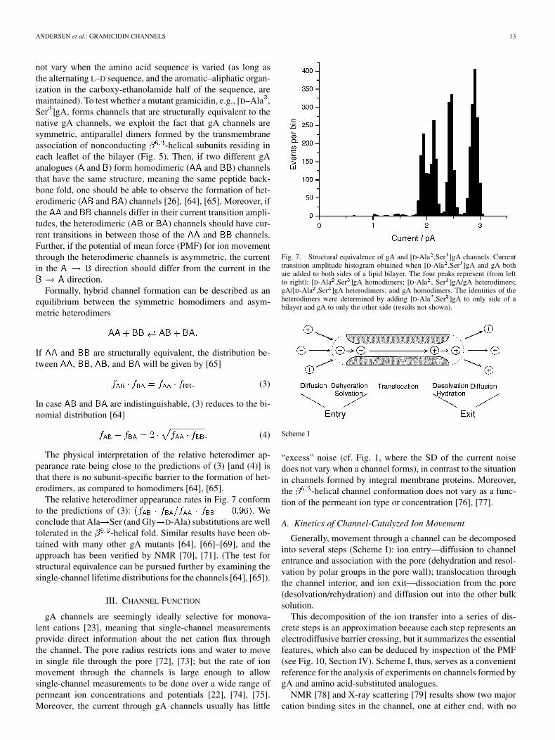
not vary when the amino acid sequence is varied (as long asthe alternating L–D sequence, and the aromatic–aliphatic organ-ization in the carboxy-ethanolamide half of the sequence, aremaintained). To test whether a mutant gramicidin, e.g., [D–Ala ,Ser ]gA, forms channels that are structurally equivalent to thenative gA channels, we exploit the fact that gA channels aresymmetric, antiparallel dimers formed by the transmembraneassociation of nonconducting -helical subunits residing ineach leaflet of the bilayer (Fig. 5). Then, if two different gAanalogues ( and ) form homodimeric ( and ) channelsthat have the same structure, meaning the same peptide back-bone fold, one should be able to observe the formation of het-erodimeric ( and ) channels [26], [64], [65]. Moreover, ifthe and channels differ in their current transition ampli-tudes, the heterodimeric ( or ) channels should have cur-rent transitions in between those of the and channels.Further, if the potential of mean force (PMF) for ion movementthrough the heterodimeric channels is asymmetric, the currentin the direction should differ from the current in the
direction.Formally, hybrid channel formation can be described as an
equilibrium between the symmetric homodimers and asym-metric heterodimers
If and are structurally equivalent, the distribution be-tween , , , and will be given by [65]
(3)
In case and are indistinguishable, (3) reduces to the bi-nomial distribution [64]
(4)
The physical interpretation of the relative heterodimer ap-pearance rate being close to the predictions of (3) [and (4)] isthat there is no subunit-specific barrier to the formation of het-erodimers, as compared to homodimers [64], [65].
The relative heterodimer appearance rates in Fig. 7 conformto the predictions of (3): . Weconclude that Ala Ser (and Gly D-Ala) substitutions are welltolerated in the -helical fold. Similar results have been ob-tained with many other gA mutants [64], [66]–[69], and theapproach has been verified by NMR [70], [71]. (The test forstructural equivalence can be pursued further by examining thesingle-channel lifetime distributions for the channels [64], [65]).
III. CHANNEL FUNCTION
gA channels are seemingly ideally selective for monova-lent cations [23], meaning that single-channel measurementsprovide direct information about the net cation flux throughthe channel. The pore radius restricts ions and water to movein single file through the pore [72], [73]; but the rate of ionmovement through the channels is large enough to allowsingle-channel measurements to be done over a wide range ofpermeant ion concentrations and potentials [22], [74], [75].Moreover, the current through gA channels usually has little
Fig. 7. Structural equivalence of gA and [D-Ala ,Ser ]gA channels. Currenttransition amplitude histogram obtained when [D-Ala ,Ser ]gA and gA bothare added to both sides of a lipid bilayer. The four peaks represent (from leftto right): [D-Ala ,Ser ]gA homodimers; [D-Ala , Ser ]gA/gA heterodimers;gA/[D-Ala ,Ser ]gA heterodimers; and gA homodimers. The identities of theheterodimers were determined by adding [D-Ala ,Ser ]gA to only side of abilayer and gA to only the other side (results not shown).
Scheme I
“excess” noise (cf. Fig. 1, where the SD of the current noisedoes not vary when a channel forms), in contrast to the situationin channels formed by integral membrane proteins. Moreover,the -helical channel conformation does not vary as a func-tion of the permeant ion type or concentration [76], [77].
A. Kinetics of Channel-Catalyzed Ion Movement
Generally, movement through a channel can be decomposedinto several steps (Scheme I): ion entry—diffusion to channelentrance and association with the pore (dehydration and resol-vation by polar groups in the pore wall); translocation throughthe channel interior, and ion exit—dissociation from the pore(desolvation/rehydration) and diffusion out into the other bulksolution.
This decomposition of the ion transfer into a series of dis-crete steps is an approximation because each step represents anelectrodiffusive barrier crossing, but it summarizes the essentialfeatures, which also can be deduced by inspection of the PMF(see Fig. 10, Section IV). Scheme I, thus, serves as a convenientreference for the analysis of experiments on channels formed bygA and amino acid-substituted analogues.
NMR [78] and X-ray scattering [79] results show two majorcation binding sites in the channel, one at either end, with no
14 IEEE TRANSACTIONS ON NANOBIOSCIENCE, VOL. 4, NO. 1, MARCH 2005
Scheme II
significant ion occupancy outside these two sites. The two sitescan be occupied simultaneously [80], such that a minimal ki-netic model for ion permeation becomes the three-barrier-two-site-two-ion (3B2S2I) model depicted in Scheme II: in which“ ” and “ ” denote whether a site (in the left or right half of thechannel) is empty or occupied. The rate constants (where thesuper- and subscripts denote the initial and final states, respec-tively) are functions of the applied voltage
(5)
where denotes the 0 mV value for a rate constant, which canbe determined from the PMF following Kramer [81] (see also[82], [83]), and its voltage-dependent change. Scheme IIneeds to be enhanced, however, because the diffusional entrystep (Scheme I) constitutes a significant barrier to ion movementthrough gA channels [84]. Diffusion limitation (DL) becomesimportant because incoming ions must “hit” the pore entrancerather precisely—meaning that the channel is “hidden” behinda diffusion resistance, which will complicate the mechanisticinterpretation of structure-function studies. Furthermore, a po-tential difference applied across the bilayer (and channel) willchange the interfacial ion concentrations; this interfacial polar-ization (IP) will in its own right have an impact on ion move-ment through the channel [85]. When the 3B2S2I kinetic modelis enhanced to incorporate both DL and interfacial limitation,the resulting 3B2S2I(DL,IP) model provides an acceptable, dis-crete-state, kinetic description of ion permeation through gAchannels (Section III-B).
B. Ion Permeation Through gA Channels
The amino acid side chains do not contact the permeatingions, but the gA channels’ permeability properties are modu-lated by amino acid substitutions throughout the sequence [66],[86]–[90]: nonpolar polar substitutions in the formyl–NH–ter-minal half of the sequence tend to reduce the conductance;polar nonpolar substations in the carboxy-ethanolamide halftend to reduce the conductance. The primary mechanism bywhich amino acid substitutions alter the ion permeability ap-pears to be electrostatic interactions between the permeatingions and the side chain dipoles [66], [90], [91].
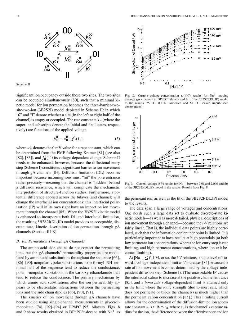
The kinetics of ion movement through gA channels havebeen studied using single-channel measurements in glycerol-monoleate [74], [92]–[94] or DPhPC [95] bilayers. Figs. 8and 9 show results obtained in DPhPC/n-decane with Na as
Fig. 8. Current–voltage–concentration (i-V-C) results for Na movingthrough gA channels in DPhPC bilayers and fit of the 3B2S2I(DL,IP) modelto the results. 25 C. (O. S. Andersen and M. D. Becker, unpublishedobservations).
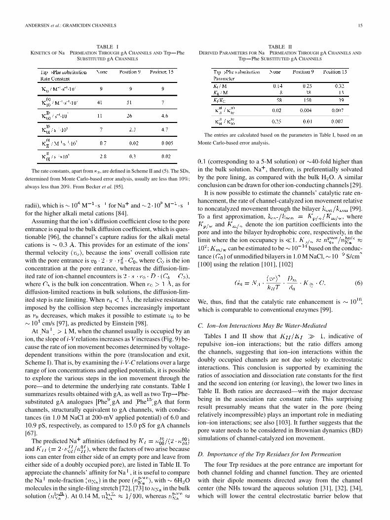
Fig. 9. Current-voltage (i-V) results for [Na ] between 0.01 and 2.0 M and fitsof the 3B2S2I(DL,IP) model to the results. Results from Fig. 8.
the permeant ion, as well as the fit of the 3B2S2I(DL,IP) modelto the results.
The data span a large range of voltages and concentrations.One needs such a large data set to evaluate discrete-state ki-netic models—as well as more detailed, physical descriptions ofion movement through a channel—because the i-V relations arefairly linear. That is, the individual data points are highly corre-lated, such that the information content per point is limited. It isparticularly important to have results at high potentials (at bothlow permeant ion concentrations, where the ion entry step is ratelimiting, and high permeant concentrations, where ion exit be-comes limiting).
At Na M, or so, the i-V relations tend to level off to-ward a voltage-independent limit as V increases [84] because therate of ion movement becomes determined by the voltage-inde-pendent diffusion step (Scheme I). (The unavoidable IP causesthe interfacial cation to increase at the positive channel entrance[85], and a bona fide voltage-dependent limit is attained onlyin the limit where the ionic strength (due to inert salt, whichdoes not permeate or block the channels) is much higher thanthe permeant cation concentration [85].) This limiting currentallows for the determination of the diffusion-limited ion accessrate constant ( , where is the channel’s capture ra-dius for the ion, the difference between the effective pore and ion
ANDERSEN et al.: GRAMICIDIN CHANNELS 15
TABLE IKINETICS OF Na PERMEATION THROUGH gA CHANNELS AND Trp!Phe
SUBSTITUTED gA CHANNELS
The rate constants, apart from � , are defined in Scheme II and (5). The SDs,
determined from Monte Carlo-based error analysis, usually are less than 10%;
always less than 20%. From Becker et al. [95].
radii), which is 10 M s for Na and 2 10 M sfor the higher alkali metal cations [84].
Assuming that the ion’s diffusion coefficient close to the poreentrance is equal to the bulk diffusion coefficient, which is ques-tionable [96], the channel’s capture radius for the alkali metalcations is 0.3 . This provides for an estimate of the ions’thermal velocity , because the ions’ overall collision ratewith the pore entrance is , where is the ionconcentration at the pore entrance, whereas the diffusion-lim-ited rate of ion-channel encounters is ,where is the bulk ion concentration. When , as fordiffusion-limited reactions in bulk solutions, the diffusion-lim-ited step is rate limiting. When , the relative resistanceimposed by the collision step becomes increasingly importantas decreases, which makes it possible to estimate to be
10 cm/s [97], as predicted by Einstein [98].At Na M, when the channel usually is occupied by an
ion, the slope of i-V relations increases as V increases (Fig. 9) be-cause the rate of ion movement becomes determined by voltage-dependent transitions within the pore (translocation and exit,Scheme I). That is, by examining the i-V-C relations over a largerange of ion concentrations and applied potentials, it is possibleto explore the various steps in the ion movement through thepore—and to determine the underlying rate constants. Table Isummarizes results obtained with gA, as well as two Trp Phe-substituted gA analogues Phe gA and Phe gA that formchannels, structurally equivalent to gA channels, with conduc-tances (in 1.0 M NaCl at 200-mV applied potential) of 6.0 and10.9 pS, respectively, as compared to 15.0 pS for gA channels[67].
The predicted Na affinities (defined byand , where the factors of two arise becauseions can enter from either side of an empty pore and leave fromeither side of a doubly occupied pore), are listed in Table II. Toappreciate the channels’ affinity for Na , it is useful to comparethe Na mole-fraction in the pore , with 6H Omolecules in the single-filing stretch [72], [73] to in the bulksolution . At 0.14 M, , whereas
TABLE IIDERIVED PARAMETERS FOR Na PERMEATION THROUGH gA CHANNELS AND
Trp!Phe SUBSTITUTED gA CHANNELS
The entries are calculated based on the parameters in Table I, based on an
Monte Carlo-based error analysis.
(corresponding to a 5-M solution) or 40-fold higher thanin the bulk solution. Na , therefore, is preferentially solvatedby the pore lining, as compared with the bulk H O. A similarconclusion can be drawn for other ion-conducting channels [29].
It is now possible to estimate the channels’ catalytic rate en-hancement, the rate of channel-catalyzed ion movement relativeto noncatalyzed movement through the bilayer [99].To a first approximation, , where
and denote the ion partition coefficients into thepore and into the bilayer hydrophobic core, respectively, in thelimit where the ion occupancy is 1.
; can be estimated to be 10 based on the conduc-tance of unmodified bilayers in 1.0 M NaCl, 10 S/cm[100] using the relation [101], [102]
(6)
We, thus, find that the catalytic rate enhancement is 10 ,which is comparable to conventional enzymes [99].
C. Ion–Ion Interactions May Be Water-Mediated
Tables I and II show that , indicative ofrepulsive ion–ion interactions; but the ratio differs amongthe channels, suggesting that ion–ion interactions within thedoubly occupied channels are not due solely to electrostaticinteractions. This conclusion is supported by examining theratios of association and dissociation rate constants for the firstand the second ion entering (or leaving), the lower two lines inTable II. Both ratios are decreased—with the major decreasebeing in the association rate constant ratio. This surprisingresult presumably means that the water in the pore (beingrelatively incompressible) plays an important role in mediatingion–ion interactions; see also [103]. It further suggests that thepore water needs to be considered in Brownian dynamics (BD)simulations of channel-catalyzed ion movement.
D. Importance of the Trp Residues for Ion Permeation
The four Trp residues at the pore entrance are important forboth channel folding and channel function. They are orientedwith their dipole moments directed away from the channelcenter (the NHs toward the aqueous solution [31], [32], [34],which will lower the central electrostatic barrier below that
16 IEEE TRANSACTIONS ON NANOBIOSCIENCE, VOL. 4, NO. 1, MARCH 2005
estimated using a structure-less dielectric model [91], [104],[105].
Consistent with this idea, gA analogues with one or moreTrp Phe replacements form channels with decreased ion per-meabilities [67]; see Section III-B. When all four Trp residuesare replaced by Phe, the Cs conductance is reduced sixfold[68]. The basis for the reduced conductance appears to be agreatly reduced rate constant for ion translocation through thechannel, [106], although ion entry is also altered [68]. Thelatter could arise because the amphipathic indoles may be ableto move (a little) out beyond the hydrophobic membrane core,which could be important as an incoming ion sheds most ofits hydration shell to become solvated by the peptide back-bone. The backbone deformation that is needed to optimizeion–oxygen contacts [107] will involve also side chain motions[108].
The changes in Na permeation through single Trp Phesubstituted gramicidins has been examined in detail [95]; seeTables I and II. The kinetic analysis provides information aboutrate constants for ion translocation, but no information about theabsolute barrier heights (and well depths) of the free energy pro-file for ion movement through the pore [82]. Such informationcan be extracted only through an appropriate physical theory,such as a hierarchical implementation of MD and BD simula-tions [83]. Nevertheless, it is possible to estimate the changes inbarrier heights and well depths asfor each of the transitions along the reaction coordinate [82]. Weconclude that single Trp Phe substitutions at the channel poreentrances (one in each subunit) may alter the barrier profile (de-crease well depths, increase barrier heights) by several .
Trp Phe substitutions increase in the height of the centralbarrier. Because the sequence substitutions to do not alter thestructure of the subunit interface [67], the conductance changesmost likely result from favorable electrostatic interactions be-tween the indole dipole and the permeant ion [91]. Surprisingly,the sign of the Trp Phe substitution-induced changes in theentry/exit barrier depends on the position where the substitu-tion is made, indicating that the side chain dynamics indeed areimportant for the rate of the ions’ hydration/solvation.
IV. MOLECULAR DYNAMICS ANALYSIS OF ION PERMEATION
To obtain better insights into the molecular basis for channel(or membrane protein) function, it is necessary to establish di-rect links between the atomic structure of a channel (or protein)and its observed function. Though MD simulations, in principle,should provide the desired tool [109], one cannot reach the nec-essary level of insight through “brute force” MD simulations[83], [110], because the ion flux typically corresponds to a netflux of one ion in 100 ns—much longer than typical MD tra-jectories [110]. Indeed, when using MD simulations to predictthe PMF for ion movement through gA channels, the barrierheights are too high by many kcal/mol [111]. This is of con-cern because the small and relatively well-behaved gA channelsshould be particularly amenable to in-depth theoretical analysisand computer simulations.
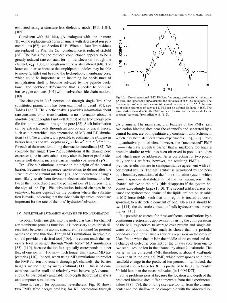
There is reason for optimism, nevertheless. Fig. 10 showstwo PMFs (free energy profiles) for K permeation through
Fig. 10. One-dimensional (1-D) PMF, or free energy profile, for K along thegA axis. The upper solid curve denotes the initial result of MD simulations. Thefree energy profile is not meaningful beyond the cuts at z = 15 �A becauseno absolute reference of such a 1-D PM can be defined for large z [83]. Thelower dashed curve denotes the PMF corrected for size, and membrane dielectricconstant (see text). From Allen et al. [112].
gA channels. The main structural features of the PMFs, i.e.,two cation binding sites near the channel’s end separated by acentral barrier, are both qualitatively consistent with Scheme I,which has been deduced from experiments [78], [79]. Froma quantitative point of view, however, the “uncorrected” PMF
displays a central barrier that is markedly too high, aproblem similar to what has been observed in previous studiesand which must be addressed. After correcting for two poten-tially serious artifacts, however, the resulting PMFpredicts results that are in semiquantitative agreement with ex-perimental results. The first artifact is introduced by the peri-odic boundary conditions of the finite simulation system, whichcause a spurious destabilization of the permeating ion in thechannel relative to the bulk (this disappears if the system be-comes exceedingly large) [113]. The second artifact arises be-cause the hydrocarbon chains of the lipids are not polarizablein MD force fields, such that this region is treated as corre-sponding to a dielectric constant of one, whereas it should betwo [114], the dielectric constant of bulk hydrocarbons, or evenhigher [115].
It is possible to correct for these artifactual contributions by acontinuum electrostatic approximation using the configurationsof the MD trajectories to average over protein and single-filewater configurations. This analysis shows that the periodicboundary conditions cause a spurious repulsion on the order of2 kcal/mole when the ion is in the middle of the channel and thata change of dielectric constant for the bilayer core from one totwo stabilizes the ion in the channel by about 2 kcal/mole. Thebarrier in the corrected PMF, therefore, is about 4 kcal/molelower than in the original PMF, which corresponds to a thou-sandfold change in the predicted ion permeability. Indeed, themaximal conductance for is predicted to be 0.8 pS, “only”30-fold less than the measured value (in 1.0 M KCl).
Some problems persist because the location and depth of thepredicted binding sites differ somewhat from the experimentalvalues [78], [79]: the binding sites are too far from the channelcenter and too shallow to be compatible with the observed ion
ANDERSEN et al.: GRAMICIDIN CHANNELS 17
affinities; but the predicted affinity M differs onlyby a factor of five from the one predicted from the kinetic anal-ysis of i-V-C results in gA channels ( M; O. S. An-dersen and M. D. Becker, unpublished results). Both problemsare likely to result from the use of nonpolarizable force fields toevaluate the ion-peptide interactions, which, therefore, will tendto be underestimated. Because the PMF effectively arises as thesum of contributions from the permeating ion’s interaction withthe channel peptide, the single-filing water in the pore, and thebilayer hydrophobic core [112], both the position and depth ofthe energy minima will be particularly sensitive to the choice offorce fields.
Given the progress that is taking place in terms of developingforce fields that include induced polarization [116]–[118], thereis every reason to be optimistic about the future. Indeed, eventhe present generation of force fields predicts changes in the cen-tral barrier (which should reflect primarily long-range electro-static interactions) that are in near-quantitative agreement withexperimental results (T. W. Allen, O. S. Andersen and B. Roux,unpublished results), which suggests that one with some confi-dence can begin to use MD to understand the basis for aminoacid substitution-induced changes in channel function.
V. CONCLUSION
The well-defined functional and structural features of grami-cidin channels provide detailed insights into the molecular basisfor channel function. The modest channel size further allowsfor detailed computational analysis, which promises to provideatomic-level insights into the microphysics underlying ion per-meation. This combination of features remains unprecedentedand suggests that the gA channels will become increasingly im-portant as test beds for theoretical models of channel-catalyzedion permeation.
ACKNOWLEDGMENT
The authors would like to thank T. W. Allen, M. D. Becker,A. E. Daily, D. V. Greathouse, S. Shobana, and S. E. Tape forstimulating discussions and technical assistance.
REFERENCES
[1] F. M. Harold and J. R. Baarda, “Gramicidin, valinomycin, and cationpermeability of Streptococcus faecalis,” J. Bacteriol., vol. 94, pp. 53–60,1967.
[2] S. B. Hladky and D. A. Haydon, “Discreteness of conductance changein bimolecular lipid membranes in the presence of certain antibiotics,”Nature, vol. 225, pp. 451–453, 1970.
[3] D. W. Urry, “The gramicidin A transmembrane channel: a proposed� helix,” Proc. Nat. Acad. Sci. USA, vol. 68, pp. 672–676, 1971.
[4] , “Protein conformation in biomembranes: optical rotation and ab-sorption of membrane suspensions,” Biochim. Biophys. Acta, vol. 265,pp. 115–168, 1972.
[5] M. S. Weiss, A. Kreusch, E. Schiltz, U. Nestel, W. Welte, J. Weckesser,and G. E. Schulz, “The structure of porin from Rhodobacter capsulatusat 1.8 �A resolution,” FEBS Lett., vol. 280, pp. 379–382, 1991.
[6] S. W. Cowan et al., “Crystal structures explain functional properties oftwo E. coli porins,” Nature, vol. 358, pp. 727–733, 1992.
[7] D. A. Doyle et al., “The structure of the potassium channel: molecularbasis of K conduction and selectivity,” Science, vol. 280, pp. 69–77,1998.
[8] G. Chang, R. H. Spencer, A. T. Lee, M. T. Barclay, and D. C. Rees,“Structure of the MscL homolog from Mycobacterium tuberculosis: agated mechanosensitive ion channel,” Science, vol. 282, pp. 2220–2226,1998.
[9] R. B. Bass, P. Strop, M. Barclay, and D. C. Rees, “Crystal structureof Escherichia coli MscS, a voltage-modulated and mechanosensitivechannel,” Science, vol. 298, pp. 1582–1587, 2002.
[10] Y. Jiang, A. Lee, J. Chen, M. Cadene, B. T. Chait, and R. MacKinnon,“The open pore conformation of potassium channels,” Nature, vol. 417,pp. 523–526, 2002.
[11] Y. Jiang, A. Lee, J. Chen, V. Ruta, M. Cadene, B. T. Chait, and R. MacK-innon, “X-ray structure of a voltage-dependent K channel,” Nature,vol. 423, pp. 33–41, 2003.
[12] A. Kuo et al., “Crystal structure of the potassium channel KirBac1.1 inthe closed state,” Science, vol. 300, pp. 1922–1926, 2003.
[13] A. Miyazawa, Y. Fujiyoshi, and N. Unwin, “Structure and gating mecha-nism of the acetylcholine receptor pore,” Nature, vol. 423, pp. 949–955,2003.
[14] R. Sarges and B. Witkop, “Gramicidin A. V. The structure of valine- andisoleucine-gramicidin A,” J. Amer. Chem. Soc., vol. 87, pp. 2011–2019,1965.
[15] G. N. Ramachandran and R. Chandrasekaran, “Studies on dipeptideconformation and on peptides with sequences of alternating L and Dresidues with special reference to antibiotic and ion transport peptides,”Prog. Peptides Res., vol. 2, pp. 195–215, 1972.
[16] W. R. Veatch, E. T. Fossel, and E. R. Blout, “The conformation of gram-icidin A,” Biochemistry, vol. 13, pp. 5249–5256, 1974.
[17] W. R. Veatch and E. R. Blout, “The aggregation of gramicidin A in so-lution,” Biochemistry, vol. 13, pp. 5257–5264, 1974.
[18] V. F. Bystrov and A. S. Arseniev, “Diversity of the gramicidin A spatialstructure: two-dimensional proton NMR study in solution,” Tetrahedron,vol. 44, pp. 925–940, 1988.
[19] D. A. Langs, “Three-dimensional structure at 0.86�A of the uncomplexedform of the transmembrane ion channel peptide gramicidin A,” Science,vol. 241, pp. 188–191, 1988.
[20] B. M. Burkhart, N. Li, D. A. Langs, W. A. Pangborn, and W. L. Duax,“The conducting form of gramicidin A is a right-handed double-strandeddouble helix,” Proc. Nat. Acad. Sci. USA, vol. 95, pp. 12 950–12 955,1998.
[21] N. Abdul-Manan and J. F. Hinton, “Conformational states of gramicidinA along the pathway to the formation of channels in model membranesdetermined by 2D NMR and circular dichroism spectroscopy,” Biochem-istry, vol. 33, pp. 6773–6783, 1994.
[22] S. B. Hladky and D. A. Haydon, “Ion transfer across lipid membranesin the presence of gramicidin A. I. Studies of the unit conductancechannel,” Biochim. Biophys. Acta, vol. 274, pp. 294–312, 1972.
[23] V. B. Myers and D. A. Haydon, “Ion transfer across lipid membranesin the presence of gramicidin A. II. Ion selectivity,” Biochim. Biophys.Acta, vol. 274, pp. 313–322, 1972.
[24] A. Finkelstein, “Aqueous pores created in thin lipid membranes by theantibiotics nystatin, amphotericin B and gramicidin A. Implications forpores in plasma membranes,” in Drugs and Transport Processes, B. A.Callingham, Ed. London, U.K.: MacMillan, 1974, pp. 241–250.
[25] E. Bamberg, H. J. Apell, and H. Alpes, “Structure of the gramicidinA channel: discrimination between the � and the � helix by elec-trical measurements with lipid bilayer membranes,” Proc. Nat. Acad.Sci. USA, vol. 74, pp. 2402–2406, 1977.
[26] W. Veatch and L. Stryer, “The dimeric nature of the gramicidin Atransmembrane channel: conductance and fluorescence energy transferstudies of hybrid channels,” J. Mol. Biol., vol. 113, pp. 89–102, 1977.
[27] S. Weinstein, B. A. Wallace, E. R. Blout, J. S. Morrow, and W. Veatch,“Conformation of gramicidin A channel in phospholipid vesicles: acarbon-13 and fluorine-19 nuclear magnetic resonance study,” Proc.Nat. Acad. Sci. USA, vol. 76, pp. 4230–4234, 1979.
[28] S. Weinstein, B. A. Wallace, J. S. Morrow, and W. R. Veatch, “Con-formation of the gramicidin A transmembrane channel: a 13C nuclearmagnetic resonance study of 13C-enriched gramicidin in phosphatidyl-choline vesicles,” J. Mol. Biol., vol. 143, pp. 1–19, 1980.
[29] O. S. Andersen and R. E. Koeppe II, “Molecular determinants of channelfunction,” Physiol. Rev., vol. 72, pp. S89–S158, 1992.
[30] A. S. Arseniev, I. L. Barsukov, V. F. Bystrov, and Y. A. Ovchinnikov,“Spatial structure of a gramicidin A transmembrane ion channel. NMRanalysis in micelles,” Biol. Membr., vol. 3, pp. 437–462, 1986.
[31] R. R. Ketchem, B. Roux, and T. A. Cross, “High-resolution polypeptidestructure in a lamellar phase lipid environment from solid state NMR de-rived orientational constraints,” Structure, vol. 5, pp. 1655–1669, 1997.
18 IEEE TRANSACTIONS ON NANOBIOSCIENCE, VOL. 4, NO. 1, MARCH 2005
[32] L. E. Townsley, W. A. Tucker, S. Sham, and J. F. Hinton, “Structuresof gramicidins A, B, and C incorporated into sodium dodecyl sulfatemicelles,” Biochemistry, vol. 40, pp. 11 676–11 686, 2001.
[33] T. W. Allen, O. S. Andersen, and B. Roux, “The structure of gramicidinA in a lipid bilayer environment determined using molecular dynamicssimulations and solid-state NMR data,” J. Amer. Chem. Soc., vol. 125,pp. 9868–9878, 2003.
[34] A. S. Arseniev, A. L. Lomize, I. L. Barsukov, and V. F. Bystrov, “Gram-icidin A transmembrane ion-channel. Three-dimensional structure re-construction based on NMR spectroscopy and energy refinement,” Biol.Membr., vol. 3, pp. 1077–1104, 1986.
[35] R. R. Ketchem, W. Hu, and T. A. Cross, “High-resolution conformationof gramicidin A in a lipid bilayer by solid-state NMR,” Science, vol. 261,pp. 1457–1460, 1993.
[36] C. M. Nimigean and C. Miller, “Na block and permeation in a Kchannel of known structure,” J. Gen. Physiol., vol. 120, p. 323, Aug 26,2002.
[37] J. B. Park, H. J. Kim, P. D. Ryu, and E. Moczydlowski, “Effect ofphosphatidylserine on unitary conductance and Ba block of theBK Ca -activated K channel: re-examination of the surface chargehypothesis,” J. Gen. Physiol., vol. 121, pp. 375–398, 2003.
[38] A. M. O’Connell, R. E. Koeppe II, and O. S. Andersen, “Kineticsof gramicidin channel formation in lipid bilayers: transmembranemonomer association,” Science, vol. 250, pp. 1256–1259, 1990.
[39] J. T. Durkin, L. L. Providence, R. E. Koeppe II, and O. S. Andersen,“Formation of non-�-helical gramicidin channels between sequence-substituted gramicidin analogues,” Biophys. J., vol. 62, pp. 145–159,1992.
[40] O. S. Andersen et al., “Gramicidin channel controversy—The structurein a lipid environment,” Nature Struct. Biol., vol. 6, p. 609, 1999.
[41] D. Salom, M. C. Bano, L. Braco, and C. Abad, “HPLC demonstrationthat an all Trp!Phe replacement in gramicidin A results in a confor-mational rearrangement from beta-helical monomer to double-strandeddimer in model membranes,” Biochem. Biophys. Res. Commun., vol.209, pp. 466–473, 1995.
[42] D. Salom, E. Perez-Paya, J. Pascal, and C. Abad, “Environment- and se-quence-dependent modulation of the double-stranded to single-strandedconformational transition of gramicidin A in membranes,” Biochemistry,vol. 37, pp. 14 279–14 291, 1998.
[43] J. A. Killian and G. von Heijne, “How proteins adapt to a membrane-water interface,” Trends Biochem. Sci., vol. 25, pp. 429–434, 2000.
[44] B. A. Cornell, F. Separovic, A. J. Baldassi, and R. Smith, “Conformationand orientation of gramicidin A in oriented phospholipid bilayers mea-sured by solid state carbon-13 NMR,” Biophys. J., vol. 53, pp. 67–76,1988.
[45] L. K. Nicholson, F. Moll, T. E. Mixon, P. V. LoGrasso, J. C. Lay, and T.A. Cross, “Solid-state 15N NMR of oriented lipid bilayer bound grami-cidin A ,” Biochemistry, vol. 26, pp. 6621–6626, 1987.
[46] J. A. Killian, M. J. Taylor, and R. E. Koeppe II, “Orientation of the va-line-1 side chain of the gramicidin transmembrane channel and implica-tions for channel functioning. A H NMR study,” Biochemistry, vol. 31,pp. 11 283–11 290, 1992.
[47] F. Separovic, R. Pax, and B. Cornell, “NMR order parameter analysis ofa peptide plane aligned in a lyotropic liquid crysta,” Mol. Phys., vol. 78,pp. 357–369, 1993.
[48] R. E. Koeppe II, H. Sun, P. C. van der Wel, E. M. Scherer, P. Pulay, andD. V. Greathouse, “Combined experimental/theoretical refinement of in-dole ring geometry using deuterium magnetic resonance and ab initiocalculations,” J. Amer. Chem. Soc., vol. 125, pp. 12 268–12 276, 2003.
[49] K. C. Lee, S. Huo, and T. A. Cross, “Lipid-peptide interface: valine con-formation and dynamics in the gramicidin channel,” Biochemistry, vol.34, pp. 857–867, 1995.
[50] R. E. Koeppe II, J. A. Killian, and D. V. Greathouse, “Orientations ofthe tryptophan 9 and 11 side chains of the gramicidin channel based ondeuterium nuclear magnetic resonance spectroscopy,” Biophys. J., vol.66, pp. 14–24, 1994.
[51] S. F. Scarlata, “The effects of viscosity on gramicidin tryptophan rota-tional motion,” Biophys. J., vol. 54, pp. 1149–1157, 1988.
[52] S. Mukherjee and A. Chattopadhyay, “Motionally restricted tryptophanenvironments at the peptide-lipid interface of gramicidin channels,” Bio-chemistry, vol. 33, pp. 5089–5097, 1994.
[53] D. V. Greathouse, J. F. Hinton, K. S. Kim, and R. E. Koeppe II, “Grami-cidin A/short-chain phospholipid dispersions: chain length dependenceof gramicidin conformation and lipid organization,” Biochemistry, vol.33, pp. 4291–4299, 1994.
[54] O. G. Mouritsen and M. Bloom, “Mattress model of lipid-protein inter-actions in membranes,” Biophys. J., vol. 46, pp. 141–153, 1984.
[55] N. Mobashery, C. Nielsen, and O. S. Andersen, “The conformationalpreference of gramicidin channels is a function of lipid bilayer thick-ness,” FEBS Lett., vol. 412, pp. 15–20, 1997.
[56] T. P. Galbraith and B. A. Wallace, “Phospholipid chain length alters theequilibrium between pore and channel forms of gramicidin,” FaradayDiscuss., pp. 159–164, 1998.
[57] H. W. Huang, “Deformation free energy of bilayer membrane and its ef-fect on gramicidin channel lifetime,” Biophys. J., vol. 50, pp. 1061–1070,1986.
[58] J. A. Lundbæk and O. S. Andersen, “Spring constants for channel-in-duced lipid bilayer deformations—estimates using gramicidin chan-nels,” Biophys. J., vol. 76, pp. 889–895, 1999.
[59] M. C. Wiener and S. H. White, “Structure of a fluid dioleoylphos-phatidylcholine bilayer determined by joint refinement of X-ray andneutron diffraction data. III. Complete structure,” Biophys. J., vol. 61,pp. 437–447, 1992.
[60] E. Lindahl and O. Edholm, “Mesoscopic undulations and thickness fluc-tuations in lipid bilayers from molecular dynamics simulations,” Bio-phys. J., vol. 79, p. 426, 2000.
[61] C. Nielsen, M. Goulian, and O. S. Andersen, “Energetics of inclusion-in-duced bilayer deformations,” Biophys. J., vol. 74, pp. 1966–1983, 1998.
[62] J. R. Elliott, D. Needham, J. P. Dilger, and D. A. Haydon, “The effectsof bilayer thickness and tension on gramicidin single-channel lifetime,”Biochim. Biophys. Acta, vol. 735, pp. 95–103, 1983.
[63] B. A. Cornell, F. Separovic, D. E. Thomas, A. R. Atkins, and R. Smith,“Effect of acyl chain length on the structure and motion of gramicidin Ain lipid bilayers,” Biochim. Biophys. Acta, vol. 985, pp. 229–232, 1989.
[64] J. T. Durkin, R. E. Koeppe II, and O. S. Andersen, “Energetics of grami-cidin hybrid channel formation as a test for structural equivalence. Side-chain substitutions in the native sequence,” J. Mol. Biol., vol. 211, pp.221–234, 1990.
[65] J. T. Durkin, L. L. Providence, R. E. Koeppe II, and O. S. Andersen, “En-ergetics of heterodimer formation among gramicidin analogues with anNH -terminal addition or deletion. Consequences of a missing residueat the join in channel,” J. Mol. Biol., vol. 231, pp. 1102–1121, 1993.
[66] J. L. Mazet, O. S. Andersen, and R. E. Koeppe II, “Single-channelstudies on linear gramicidins with altered amino acid sequences. Acomparison of phenylalanine, tryptophan, and tyrosine substitutions atpositions 1 and 11,” Biophys. J., vol. 45, pp. 263–276, 1984.
[67] M. D. Becker, D. V. Greathouse, R. E. Koeppe II, and O. S. Andersen,“Amino acid sequence modulation of gramicidin channel function. Ef-fects of tryptophan-to-phenylalanine substitutions on the single-channelconductance and duration,” Biochemistry, vol. 30, pp. 8830–8839, 1991.
[68] V. Fonseca, P. Daumas, L. Ranjalahy-Rasoloarijao, F. Heitz, R. Lazaro,Y. Trudelle, and O. S. Andersen, “Gramicidin channels that have no tryp-tophan residues,” Biochemistry, vol. 31, pp. 5340–5350, 1992.
[69] A. R. Jude, D. V. Greathouse, R. E. Koeppe II, L. L. Providence, and O.S. Andersen, “Modulation of gramicidin channel structure and functionby the aliphatic ‘spacer’ residues 10, 12, and 14 between the trypto-phans,” Biochemistry, vol. 38, pp. 1030–1039, 1999.
[70] G. L. Mattice, R. E. Koeppe II, L. L. Providence, and O. S. Andersen,“Stabilizing effect of D-alanine in gramicidin channels,” Biochemistry,vol. 34, pp. 6827–6837, 1995.
[71] S. S. Sham et al., “The structure, cation binding, transport, and conduc-tance of Gly -gramicidin A incorporated into SDS micelles and PC/PGvesicles,” Biochemistry, vol. 42, pp. 1401–1409, 2003.
[72] D. G. Levitt, S. R. Elias, and J. M. Hautman, “Number of watermolecules coupled to the transport of sodium, potassium and hydrogenions via gramicidin, nonactin or valinomycin,” Biochim. Biophys. Acta,vol. 512, pp. 436–451, 1978.
[73] P. A. Rosenberg and A. Finkelstein, “Interaction of ions and water ingramicidin A channels: streaming potentials across lipid bilayer mem-branes,” J. Gen. Physiol., vol. 72, pp. 327–340, 1978.
[74] E. Neher, J. Sandblom, and G. Eisenman, “Ionic selectivity, saturation,and block in gramicidin A channels. II. Saturation behavior of singlechannel conductances and evidence for the existence of multiple bindingsites in the channel,” J. Membr. Biol., vol. 40, pp. 97–116, 1978.
[75] O. S. Andersen, “Ion movement through gramicidin A channels. Single-channel measurements at very high potentials,” Biophys. J., vol. 41, pp.119–133, 1983.
[76] B. A. Wallace, W. R. Veatch, and E. R. Blout, “Conformation of grami-cidin A in phospholipid vesicles: circular dichroism studies of effects ofion binding, chemical modification, and lipid structure,” Biochemistry,vol. 20, pp. 5754–5760, 1981.
[77] F. Tian and T. A. Cross, “Cation transport: an example of structural basedselectivity,” J. Mol. Biol., vol. 285, pp. 1993–2003, 1999.
ANDERSEN et al.: GRAMICIDIN CHANNELS 19
[78] D. W. Urry, T. L. Trapane, C. M. Venkatachalam, and R. B. McMichens,“Ion interactions at membranous polypeptide sites using nuclear mag-netic resonance: determining rate and binding constants and site loca-tions,” Methods Enzymol., vol. 171, pp. 286–342, 1989.
[79] G. A. Olah, H. W. Huang, W. Liu, and Y. Wu, “Location of ion-bindingsites in the gramicidin channel by X-ray diffraction,” J. Mol. Biol., vol.218, pp. 847–858, 1991.
[80] L. V. Schagina, A. E. Grinfeldt, and A. A. Lev, “Interaction of cationfluxes in gramicidin A channels in lipid bilayer membranes,” Nature,vol. 273, pp. 243–245, 1978.
[81] H. A. Kramers, “Brownian motion in a field of force and the diffusionmodel of chemical reactions,” Physica, vol. 7, pp. 284–304, 1940.
[82] O. S. Andersen, “Kinetics of ion movement mediated by carriers andchannels,” Methods Enzymol., vol. 171, pp. 62–112, 1989.
[83] B. Roux, T. W. Allen, T. W. Bernèche, and W. Im, “Theoretical andcomputational models of biological ion channels,” Q. Rev. Biophys., vol.37, pp. 15–103, 2004.
[84] O. S. Andersen, “Ion movement through gramicidin A channels. Studieson the diffusion-controlled association step,” Biophys. J., vol. 41, pp.147–165, 1983.
[85] , “Ion movement through gramicidin A channels. Interfacial po-larization effects on single-channel current measurements,” Biophys. J.,vol. 41, pp. 135–146, 1983.
[86] E. Bamberg, K. Noda, E. Gross, and P. Läuger, “Single-channel param-eters of gramicidin A, B, and C,” Biochim. Biophys. Acta, vol. 419, pp.223–228, 1976.
[87] J. S. Morrow, W. R. Veatch, and L. Stryer, “Transmembrane channelactivity of gramicidin A analogs: effects of modification and deletion ofthe amino-terminal residue,” J. Mol. Biol., vol. 132, pp. 733–738, 1979.
[88] F. Heitz, G. Spach, and Y. Trudelle, “Single channels of 9,11,13,15-destryptophyl-phenylalanyl-gramicidin A,” Biophys. J., vol. 40, pp.87–89, 1982.
[89] E. W. B. Russell, L. B. Weiss, F. I. Navetta, R. E. Koeppe II, and O.S. Andersen, “Single-channel studies on linear gramicidins with alteredamino acid side chains. Effects of altering the polarity of the side chainat position 1 in gramicidin A,” Biophys. J., vol. 49, pp. 673–686, 1986.
[90] R. E. Koeppe II, J.-L. Mazet, and O. S. Andersen, “Distinction betweendipolar and inductive effects in modulating the conductance of grami-cidin channels,” Biochemistry, vol. 29, pp. 512–520, 1990.
[91] O. S. Andersen, D. V. Greathouse, L. L. Providence, M. D. Becker, andR. E. Koeppe II, “Importance of tryptophan dipoles for protein function:5-fluorination of tryptophans in gramicidin A channels,” J. Amer. Chem.Soc., vol. 120, pp. 5142–5146, 1998.
[92] C. D. Cole, A. S. Frost, N. Thompson, M. Cotten, T. A. Cross, andD. D. Busath, “Noncontact dipole effects on channel permeation. VI.5F-and 6F-Trp gramicidin channel currents,” Biophys. J., vol. 83, pp.1974–1986, 2002.
[93] B. W. Urban, S. B. Hladky, and D. A. Haydon, “Ion movements in gram-icidin pores. An example of single-file transport,” Biochim. Biophys.Acta, vol. 602, pp. 331–354, 1980.
[94] D. D. Busath et al., “Noncontact dipole effects on channel permeation.I. Experiments with (5F-indole) Trp gramicidin A channels,” Biophys.J., vol. 75, pp. 2830–2844, 1998.
[95] M. D. Becker, R. E. Koeppe II, and O. S. Andersen, “Amino acid sub-stitutions and ion channel function: model-dependent conclusions,” Bio-phys. J., vol. 62, pp. 25–27, 1992.
[96] S. König, E. Sackmann, D. Richter, R. Zorn, C. Carlile, and T. M. Bayerl,“Molecular dynamics of water in oriented DPPC multilayers studiedby quasielastic neutron scattering and deuterium-nuclear magnetic res-onance relaxation,” J. Chem. Phys., vol. 100, pp. 3307–3316, 1994.
[97] O. S. Andersen and S. W. Feldberg, “The heterogeneous collision ve-locity for hydrated ions in aqueous solutions is � 10 cm/s,” J. Phys.Chem., vol. 100, pp. 4622–4629, 1996.
[98] A. Einstein, “Theoretische Betrachtungen über der Brownsche Bewe-gungen,” Zeit. f. Elektrochemie, vol. 13, pp. 41–42, 1907.
[99] R. Wolfenden and M. J. Snider, “The depth of chemical time and thepower of enzymes as catalysts,” Acc. Chem. Res., vol. 12, pp. 938–945,2001.
[100] T. Hanai, D. A. Haydon, and J. Taylor, “The variation of capacitance andconductance of bimolecular lipid membranes with area,” J. Theor. Biol.,vol. 9, pp. 433–443, 1965.
[101] A. L. Hodgkin and B. Katz, “The effect of sodium ions on the electricalactivity of the giant axon of the squid,” J. Physiol., vol. 108, pp. 37–77,1949.
[102] O. S. Andersen, “Elementary aspects of acid-base permeation and pHregulation,” Ann. New York Acad. Sci., vol. 574, pp. 333–353, 1989.
[103] B. Roux, B. Prod’hom, and M. Karplus, “Ion transport in the gramicidinchannel: molecular dynamics study of single and double occupancy,”Biophys. J., vol. 68, pp. 876–892, 1995.
[104] P. C. Jordan, “The total electrostatic potential in a gramicidin channel,”J. Membr. Biol., vol. 78, pp. 91–102, 1984.
[105] M. Sancho and G. Martinez, “Electrostatic modeling of dipole-ion in-teractions in gramicidin like channels,” Biophys. J., vol. 60, pp. 81–88,1991.
[106] D. Caywood, J. Durrant, P. Morrison, and D. D. Busath, “The Trp po-tential deduced from gramicidin A/gramicidin M channels,” Biophys. J.,vol. 86, p. 55a, 2004.
[107] S. Y. Noskov, S. Bernèche, and B. Roux, “Control of ion selectivity inpotassium channels by electrostatic and dynamic properties of carbonylligands,” Nature, vol. 431, pp. 830–834, 2004.
[108] D. W. Urry, “Polypeptide conformation and biological function:�-helices (�L D-helices) as permselective transmembrane channels,”in Proc. Jerusalem Symp. Quant. Chem. Biochem., vol. 5, 1973, pp.723–736.
[109] S. Edwards, B. Corry, S. Kuyucak, and S.-H. Chung, “Continuum elec-trostatics fails to describe ion permeation in the gramicidin channel,”Biophys. J., vol. 83, p. 1348, 2002.
[110] B. Roux, “Theoretical and computational models of ion channels,” Curr.Opin. Struct. Biol., vol. 12, pp. 182–189, 2002.
[111] T. W. Allen, T. Bastug, S. Kuyucak, and S.-H. Chung, “Gramicidin Achannel as a test ground for molecular dynamics force fields,” Biophys.J., vol. 84, p. 2159, 2003.
[112] T. W. Allen, O. S. Andersen, and B. Roux, “Energetics of ion conductionthrough the gramicidin channel,” Proc. Nat. Acad. Sci. USA, vol. 101, pp.117–122, 2004.
[113] P. H. Hünenberger and J. A. McCammon, “Ewald artifacts in computersimulations of ionic solvation and ion–ion interaction: a continuum elec-trostatics study,” J. Chem. Phys., vol. 110, p. 1856, 1999.
[114] J. Åqvist and A. Warshel, “Energetics of ion permeation through mem-brane channels. Solvation of Na by gramicidin A,” Biophys. J., vol. 56,pp. 171–182, 1989.
[115] A. B. Mamanov, R. D. Coalson, A. Nitzan, and M. G. Kurnikova, “Therole of the dielectric barrier in narrow biological channels: a novel com-posite approach to modeling single-channel currents,” Biophys. J., vol.84, pp. 3646–3661, 2003.
[116] G. Lamoureux and B. Roux, “Modeling induced polarizability with clas-sical Drude oscillators: theory and molecular dynamics simulation algo-rithm.,” J. Chem. Phys., pp. 3025–3039, 2003.
[117] V. M. Anisimov, I. V. Vorobyov, G. Lamoureux, S. Noskov, B. Roux,and A. D. MacKerell Jr., “CHARMM all-atom polarizable force fieldparameter development for nucleic acids,” Biophys. J., vol. 86, p. 415a,2004.
[118] G. Lamoureux, A. D. MacKerell Jr., and B. Roux, “A simple polarizablewater model based on classical Drude oscillators,” J. Chem. Phys., pp.5185–5197, 2003.
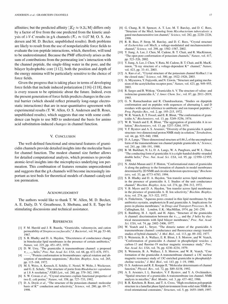
Olaf S. Andersen received the M.D. degree fromUniversity of Copenhagen, Copenhagen, Denmarkin 1971.
After postdoctoral training at University ofCopenhagen and Rockefeller University, New York(1971–1973), he joined the faculty in the Departmentof Physiology and Biophysics at Cornell UniversityMedical College (now Weill Medical College ofCornell University), New York, where he is cur-rently Thomas H. Meikle, Jr., Professor of MedicalEducation. He is Editor of the Journal of General
Physiology. His primary research interests involve channel-mediated ion move-ment across lipid bilayers, the elastic properties of lipid bilayer membranes,and the bilayer-dependent regulation of membrane protein function.
Dr. Andersen is a Foreign Member of the Royal Danish Academy of Sciencesand Letters and Recipient of the K. S. Cole Medal from the Biophysical Society(1999).
20 IEEE TRANSACTIONS ON NANOBIOSCIENCE, VOL. 4, NO. 1, MARCH 2005
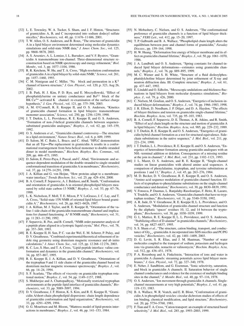
Roger E. Koeppe, II, received the A.B. degree inchemistry from Haverford College, Haverford, PA, in1971, working with C. MacKay, and the Ph.D. degreein chemistry from the California Institute of Tech-nology, Pasadena, in 1976, working with R. Stroud.
He did postdoctoral work in structural biology withL. Stryer at Stanford University, Stanford, CA, be-fore joining the faculty at the University of Arkansas,Fayetteville, in 1979, where he is currently Univer-sity Professor of Chemistry and Biochemistry. Hisprimary research interests involve the design and use
of model peptides that will shed light on mechanistic details of protein/lipid in-teractions at the molecular and atomic levels.
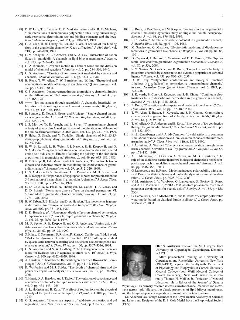
Benoît Roux received the B.S. degree in physics andthe M.S. degree in biophysics from the Université deMontréal, Montréal, QC, Canada, in 1981 and 1984,respectively, and the Ph.D. degree in biophysicsfrom Harvard University, Cambridge, MA, in 1990,working under the supervision of M. Karplus.
After a year of postdoctoral research at theCommissariat a l’Energie Atomique, Paris, Francehe joined the faculty in the Physics and ChemistryDepartments at the Université de Montréal. Since1999, he has been Professor in the Departments of
Biochemistry and Physiology and Biophysics at the Weill Medical College,Cornell University, New York. He is one of the developers of the biomolecularsimulation program CHARMM. His work has mostly focused on the inves-tigations of the function of ion channels and the development of statisticalmechanical methods for computing the solvation-free energy of biologicalmolecules.
In 1998, Dr. Roux was awarded the Rutherford Medal from the Royal So-ciety of Canada and the Noranda Lecture Award from the Chemical Institute ofCanada.
Related Documents