RESEARCH ARTICLE Open Access GP88 (PC-Cell Derived Growth Factor, progranulin) stimulates proliferation and confers letrozole resistance to aromatase overexpressing breast cancer cells Tesfom Abrhale 1,2 , Angela Brodie 3,4 , Gauri Sabnis 3,4 , Luciana Macedo 3 , Changsheng Tian 1 , Binbin Yue 1 and Ginette Serrero 1,4* Abstract Background: Aromatase inhibitors (AI) that inhibit breast cancer cell growth by blocking estrogen synthesis have become the treatment of choice for post-menopausal women with estrogen receptor positive (ER + ) breast cancer. However, some patients display de novo or acquired resistance to AI. Interactions between estrogen and growth factor signaling pathways have been identified in estrogen-responsive cells as one possible reason for acquisition of resistance. Our laboratory has characterized an autocrine growth factor overexpressed in invasive ductal carcinoma named PC-Cell Derived Growth Factor (GP88), also known as progranulin. In the present study, we investigated the role GP88 on the acquisition of resistance to letrozole in ER + breast cancer cells Methods: We used two aromatase overexpressing human breast cancer cell lines MCF-7-CA cells and AC1 cells and their letrozole resistant counterparts as study models. Effect of stimulating or inhibiting GP88 expression on proliferation, anchorage-independent growth, survival and letrozole responsiveness was examined. Results: GP88 induced cell proliferation and conferred letrozole resistance in a time- and dose-dependent fashion. Conversely, naturally letrozole resistant breast cancer cells displayed a 10-fold increase in GP88 expression when compared to letrozole sensitive cells. GP88 overexpression, or exogenous addition blocked the inhibitory effect of letrozole on proliferation, and stimulated survival and soft agar colony formation. In letrozole resistant cells, silencing GP88 by siRNA inhibited cell proliferation and restored their sensitivity to letrozole. Conclusion: Our findings provide information on the role of an alternate growth and survival factor on the acquisition of aromatase inhibitor resistance in ER + breast cancer. Background Estrogen plays a crucial role in breast and endometrial carcinomas in addition to being involved in normal phy- siological processes [1]. The primary site of estrogen biosynthesis in premenopausal women is the ovaries [2]. After menopause however, peripheral adipose tissue becomes the main source of estrogen synthesis [2,3]. The principal enzyme responsible for the conversion of androgens to estrogens is the cytochrome P450 complex known as aromatase (CYP 19) [1,2]. Several studies have demonstrated that this in situ estrogen production plays a more important role than circulating estrogen in breast tumor growth [4,5]. Estrogen produces a variety of cellular responses, such as stimulation of cell prolif- eration, inhibition of apoptosis, and enhancement of various peptide growth factor/growth factor receptor expression in ER + breast cancer cells [2,3]. Treatment options for patients with hormone-depen- dent ER + breast cancers are estrogen antagonist such as tamoxifen, estrogen receptor down regulator such as Fulvestrant or inhibitors of estrogen biosynthesis such as aromatase inhibitors [6,7]. Tamoxifen has been the major agent used to inhibit breast cancer growth until * Correspondence: [email protected] 1 A&G Pharmaceutical Inc. 9130 Red Branch Rd. Columbia, MD, USA Full list of author information is available at the end of the article Abrhale et al. BMC Cancer 2011, 11:231 http://www.biomedcentral.com/1471-2407/11/231 © 2011 Abrhale et al; licensee BioMed Central Ltd. This is an Open Access article distributed under the terms of the Creative Commons Attribution License (http://creativecommons.org/licenses/by/2.0), which permits unrestricted use, distribution, and reproduction in any medium, provided the original work is properly cited.

Welcome message from author
This document is posted to help you gain knowledge. Please leave a comment to let me know what you think about it! Share it to your friends and learn new things together.
Transcript
RESEARCH ARTICLE Open Access
GP88 (PC-Cell Derived Growth Factor,progranulin) stimulates proliferation and confersletrozole resistance to aromatase overexpressingbreast cancer cellsTesfom Abrhale1,2, Angela Brodie3,4, Gauri Sabnis3,4, Luciana Macedo3, Changsheng Tian1, Binbin Yue1 andGinette Serrero1,4*
Abstract
Background: Aromatase inhibitors (AI) that inhibit breast cancer cell growth by blocking estrogen synthesis havebecome the treatment of choice for post-menopausal women with estrogen receptor positive (ER+) breast cancer.However, some patients display de novo or acquired resistance to AI. Interactions between estrogen and growthfactor signaling pathways have been identified in estrogen-responsive cells as one possible reason for acquisitionof resistance. Our laboratory has characterized an autocrine growth factor overexpressed in invasive ductalcarcinoma named PC-Cell Derived Growth Factor (GP88), also known as progranulin. In the present study, weinvestigated the role GP88 on the acquisition of resistance to letrozole in ER+ breast cancer cells
Methods: We used two aromatase overexpressing human breast cancer cell lines MCF-7-CA cells and AC1 cellsand their letrozole resistant counterparts as study models. Effect of stimulating or inhibiting GP88 expression onproliferation, anchorage-independent growth, survival and letrozole responsiveness was examined.
Results: GP88 induced cell proliferation and conferred letrozole resistance in a time- and dose-dependent fashion.Conversely, naturally letrozole resistant breast cancer cells displayed a 10-fold increase in GP88 expression whencompared to letrozole sensitive cells. GP88 overexpression, or exogenous addition blocked the inhibitory effect ofletrozole on proliferation, and stimulated survival and soft agar colony formation. In letrozole resistant cells,silencing GP88 by siRNA inhibited cell proliferation and restored their sensitivity to letrozole.
Conclusion: Our findings provide information on the role of an alternate growth and survival factor on theacquisition of aromatase inhibitor resistance in ER+ breast cancer.
BackgroundEstrogen plays a crucial role in breast and endometrialcarcinomas in addition to being involved in normal phy-siological processes [1]. The primary site of estrogenbiosynthesis in premenopausal women is the ovaries [2].After menopause however, peripheral adipose tissuebecomes the main source of estrogen synthesis [2,3].The principal enzyme responsible for the conversion ofandrogens to estrogens is the cytochrome P450 complexknown as aromatase (CYP 19) [1,2]. Several studies have
demonstrated that this in situ estrogen production playsa more important role than circulating estrogen inbreast tumor growth [4,5]. Estrogen produces a varietyof cellular responses, such as stimulation of cell prolif-eration, inhibition of apoptosis, and enhancement ofvarious peptide growth factor/growth factor receptorexpression in ER+ breast cancer cells [2,3].Treatment options for patients with hormone-depen-
dent ER+ breast cancers are estrogen antagonist such astamoxifen, estrogen receptor down regulator such asFulvestrant or inhibitors of estrogen biosynthesis suchas aromatase inhibitors [6,7]. Tamoxifen has been themajor agent used to inhibit breast cancer growth until
* Correspondence: [email protected]&G Pharmaceutical Inc. 9130 Red Branch Rd. Columbia, MD, USAFull list of author information is available at the end of the article
Abrhale et al. BMC Cancer 2011, 11:231http://www.biomedcentral.com/1471-2407/11/231
© 2011 Abrhale et al; licensee BioMed Central Ltd. This is an Open Access article distributed under the terms of the Creative CommonsAttribution License (http://creativecommons.org/licenses/by/2.0), which permits unrestricted use, distribution, and reproduction inany medium, provided the original work is properly cited.
the development and clinical application of estrogenreceptor (ER) antagonists such as ICI 182,780, alsocalled Faslodex or Fulvestrant [6]. Aromatase inhibitoris now the treatment of choice for post-menopausalbreast cancer patients. The inhibitory effect of anti-estrogen therapy is observed almost exclusively in breasttumors that are ER+. However, after prolonged anti-estrogen therapy, breast carcinoma often progress andbecome estrogen-insensitive and refractory to treatment[8,9]. It is widely documented that the inappropriateactivation of growth factor signaling cascades, eitherthrough overexpression of growth factor, or via up-regu-lation and increased activation of their target growthfactor receptors or their recruited downstream signalingelements, can readily promote anti-hormone failure inbreast cancer cells [10-12]. This phenomenon wasdemonstrated for the overexpression of multiple growthfactors and their receptors, including heregulins actingthrough HER3 and HER4 [13,14], epidermal growth fac-tor [15] and transforming growth factor (TGF)-a actingthrough the epidermal growth factor receptor (EGFR)[16,17], insulin-like growth factors IGF-I and IGF-II act-ing through the IGF-IR [18,19], and HER2 receptor con-tributing to anti-hormone failure either directly whenoverexpressed [20-23] or indirectly through heterodi-merization with other erbB receptor family members[17].Increased autocrine or paracrine growth factor signal-
ing network could then bypass the need for ER-mediated growth stimulation in human breast cancercells and would make anti-estrogen therapy ineffective.Such an elevated or sustained growth factor signalingwithin the anti-estrogen-resistant models would even-tually lead to endocrine insensitivity, or to ER signalingbeing circumvented [10-12]. Autocrine and paracrinegrowth factor signaling cross-talk with estrogen receptor(ER) signaling to facilitate tumor growth [24]. Therefore,increase in autocrine growth factor signaling that med-iates proliferation signals may induce resistance to endo-crine therapy [25].One autocrine growth factor under study in our
laboratory and that has been implicated in the develop-ment of tamoxifen resistance and estrogen indepen-dence is PC-cell Derived Growth Factor (GP88) [26].GP88 also called granulin/epithelin precursor or progra-nulin, is the largest member of a unique family ofgrowth modulators characterized by 7.5 cysteine-rich6KDa polypeptide repeats [27-29]. Published reportshave shown that GP88 was expressed in human breastcancer cells in a positive correlation with tumorigenesis,and estradiol (E2) stimulated GP88 expression transcrip-tionally in ER+ cells [26]. Inhibition of GP88 expressionby antisense GP88 cDNA transfection in human breastadenocarcinoma MDA-MB-468 cells led to inhibition of
tumor formation in vitro and in vivo in mouse xenograftstudies [30]. In pathological studies of 203 human breastcancer biopsies, GP88 was expressed in 80% of invasiveductal carcinomas in correlation with parameters ofpoor prognosis [31] whereas it was negative in benignlesion and normal mammary epithelial tissues. In addi-tion, circulating GP88 was found in serum of breastcancer patients at an increased level when compared tohealthy volunteers [32]. GP88 overexpression in thehuman breast cancer cell line, MCF-7, conferred estro-gen-independent growth, tamoxifen resistance andincreased invasive properties [26,33,34]. Previous studiesalso demonstrated that tamoxifen-resistant MCF-7 cells,selected by culturing cells in the continuous presence oftamoxifen, expressed higher level of GP88 than tamoxi-fen-sensitive cells. This suggests that GP88 plays a criti-cal role in breast cancer tumorigenesis and that there isa direct relationship between GP88 overexpression andtamoxifen resistance exists.Based on the above information, it would be interest-
ing to examine the effect of GP88 on breast cancer cellsresponse to aromatase inhibitors. Using cellular modelsystems established from two independently derivedMCF-7 cell lines overexpressing aromatase activity, weexamined the effect of GP88 on letrozole responsive-ness. One model consisted of the MCF-7-CA cell lineobtained by transfection of an vector expressing aroma-tase cDNA [4] and its letrozole resistant counterpart(LTLT cell line) derived from an MCF-7CA tumordeveloped in mice treated with letrozole [23,35]. Thesecond model included the aromatase overexpressingbreast cancer AC1 cells [36] and letrozole resistant deri-vative AC1-LetR obtained by continuous cultivation ofAC1 cells in the continuous presence of letrozole.
MethodsThe following reagents were obtained: Lipofectamineand Plus reagent from Life Technologies, Inc. (St. Paul,MN); agar and zeocin from InVitrogen, (Carlsbad, CA),androstenedione and b-Estradiol (E2) from Sigma (St.Louis, MO);, DME/F12 nutrient medium and G418from Gibco, (La Jolla, CA), fetal bovine serum (FBS)from Hyclone, (Salt Lake City, Utah); CellTiter-GloReagent from Promega, (Pittsburgh, PA); GP88 SiRNAand control SiRNAs (smart pool duplex), from Dharma-con (Chicago, Il). Letrozole was provided to Dr. AngelaBrodie by Novartis. Purified GP88 and all anti-GP88antibodies and EIA kit to measure GP88 expressionwere developed by the Precision Antibody Division ofA&G Pharmaceutical.
Cell lines and culture conditionHuman ER+ breast cancer cells lines MCF-7CA, MCF-7AC1, LTLT-CA and UMB1-CA cell lines were derived
Abrhale et al. BMC Cancer 2011, 11:231http://www.biomedcentral.com/1471-2407/11/231
Page 2 of 10
from MCF-7 cells stably transfected with aromatasecDNA [35,36]. MCF-7AC1 were maintained in DMEM/F12 medium supplemented with 5% FBS in the presenceof 650 μg/ml G418. LTLT and UMB1 cells [35] werecultured in steroid-depleted medium consisting of phe-nol red-free (PRF) DMEM/F12 medium supplementedwith 5% charcoal-stripped (ChX) FBS (PRF-ChX med-ium) in the presence of 750 μg/ml G418. In addition,LTLT were cultivated in the presence of 1 μMLetrozole.
Anchorage-independent growth in Soft AgarCells were plated in 6-well plates at 1 × 10⁴ cells per wellin 0.33% agarose in PRF-ChX medium layered on top of0.6% agarose in the same medium with the indicatedtreatments. Colony formation was observed after 14-21days in culture. Following staining with 0.005% crystalviolet, colonies were counted under a microscope.
Cell proliferation assayCells were plated at 2 × 10³ cells/well in 96 wells platein PRF medium supplemented with 5% ChX FBS (PRF-ChX medium). The next day, medium was removed andPRF medium supplemented with the intended treat-ments and growth factors was added and incubated forspecified times. Cell growth was measured using CellTi-ter-Glo Reagent following manufacturer’s instructions(Promega). Recombinant human GP88 produced inCHO cells was prepared and affinity purified in ourlaboratory as described previously [26,33,34].
GP88 cDNA Transfection in MCF-7AC1 cellsStable transfection of MCF-7AC1 cells with plasmidDNA of pSectag expression vector containing humanGP88 cDNA [26] was conducted using lipofectamineand Plus reagent according to manufacturer’s protocol.After transfection, selection of transfected clones wasperformed in the presence of 80 μg/ml of selection drugZeocin. After two weeks, single clones were picked andtested for GP88 expression by immunoprecipitation/western blot as described previously [26] and GP88 pro-duction in culture medium with GP88 EIA kit developedin our laboratory.
Aromatase activity assayCells were plated at a density of 1.5 × 10⁵ cells/well in 6wells plate in PRF-ChX medium. The next day, cellswere washed with PBS and PRF medium supplementedwith 1% ChX FBS (no G418). Radiolabeled [1 beta3H]-androstenedione (23.5 Ci/mmol, Perkin-Elmer) wasadded and incubated for the indicated times. Tritiumreleased from the C-1beta during aromatization to form3H2O was then measured in the supernatant [34], afteraddition of trichloroacetic acid to stop the reaction,
centrifugation, and addition of chloroform. After centri-fugation, 700 μL of the upper phase was transferred to atube to which 700 μL of 2.5% activated charcoal wasadded and spun once more. Then 700 μL of the super-natant was taken and 4 ml scintillation liquid was addedand the radioactivity was measured in a liquid scintilla-tion counter.
Silencing of GP88 by siRNA in Letrozole resistant cellsLTLT cells or AC1-LetR cells (5,000 cells/well) wereplated in 96 wells plate overnight in ChX-PRF medium.The next day transfection of GP88 or control SiRNA(smart pool duplex) (1 μM) was carried out followingthe protocol of the manufacturer (DHARMACON). Cellviability was measured by cell Titer-Glo assay and thelevel of GP88 was measured both by western blot analy-sis and EIA as indicated below.
Measurement of GP88 expression by Western Blotanalysis and by EIAGP88 expression in breast cancer cells and culturemedia was measured using two methods: Western blotanalysis and EIA. For western blot analysis of GP88expression in cell lysates, cells treated with GP88 orcontrol siRNA (1 μM) were washed with ice cold PBStwice and lysed with RIPA buffer containing proteaseinhibitors (150 mM NaCl, 50 mM Tris-HCL, 1% NP-40,0.25% Na-Deoxycholate, 1 mM EDTA, 1 μg/ml Aproti-nin, Leupeptin, and Pepstatin, 1 mM Na3VO4, 1 mMPMSF and 1 mM NaF. 50 μg of total protein lysate wereanalyzed by 10% SDS gel electrophoresis. Western blotanalysis was performed as described previously [26]. ForWestern blot analysis of GP88 in culture medium, con-ditioned medium normalized to cell number was col-lected and brought up to 1 ml volume and incubatedwith 1 μg of anti-human GP88 rabbit polyclonal anti-body in the presence of 30 μl of protein G agarose over-night at 4°C. The next day, the agarose beads were spunand washed three times with ice cold PBS and resus-pended in 40 μL of 2x sample buffer to perform SDS-PAGE on a 7.5% polyacrylamide gel followed by westernblot analysis using anti-human GP88 mouse monoclonalantibody also developed in our laboratory [26]. Quantifi-cation of GP88 in culture medium was also carried outusing an GP88 EIA kit developed in our laboratory.
Determination of Bcl-2 expression in response to GP88and letrozoleThe ability of letrozole to down regulate bcl-2 expressionwas examined for MCF-7-AC1 cells. AC1 cells were pla-ted at 1.5 × 10⁵ cells in 35 mm plate and incubated inPRF medium with 5% ChX-FBS (ChX-PRF). The nextday, the medium was removed and fresh medium wasadded with the treatments: 25 nM of androstenedione
Abrhale et al. BMC Cancer 2011, 11:231http://www.biomedcentral.com/1471-2407/11/231
Page 3 of 10
(AD) or AD with 5 nM letrozole (Let) in the absence orpresence of GP88 at the indicated concentrations. Con-trol corresponded to cells cultivated in ChX-PRF. 48hours later, total RNA was extracted using Trizol™reagent (InVitrogen) and resuspended in DEPC-treatedwater for measuring bcl-2 and GAPDH mRNA expres-sion by RT-PCR. PCR conditions and the oligonucleotidesequences of the specific forward and reverse primers forbcl-2 and GAPDH have been described previously [33].
Statistical AnalysisAll experiments were performed in triplicates andrepeated at least three times. Data are expressed asmean with standard deviation. Student’s t-test was usedto determine the significance of results with p < 0.05 asstatistically significant.
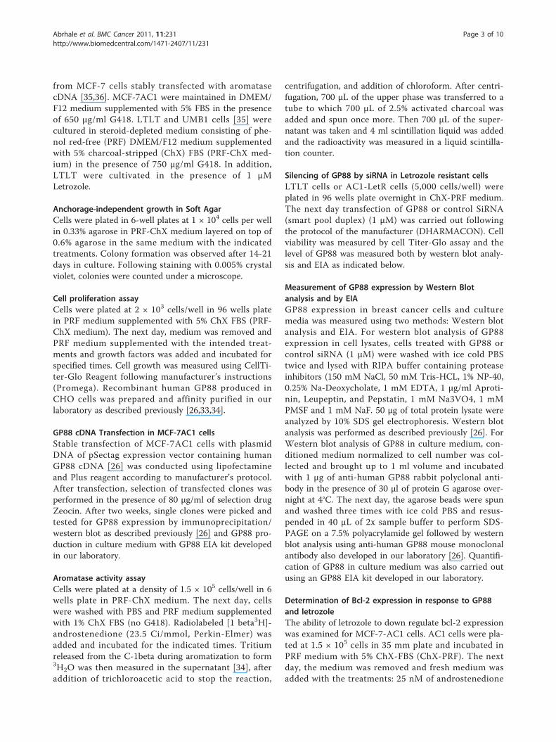
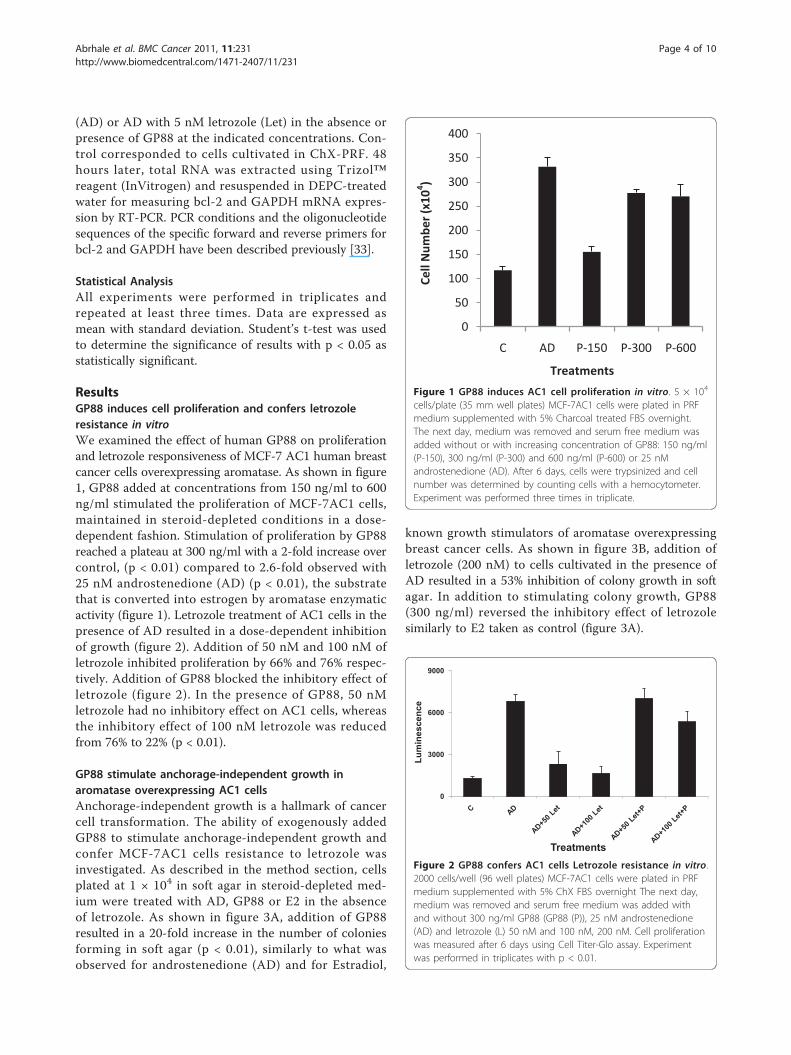
ResultsGP88 induces cell proliferation and confers letrozoleresistance in vitroWe examined the effect of human GP88 on proliferationand letrozole responsiveness of MCF-7 AC1 human breastcancer cells overexpressing aromatase. As shown in figure1, GP88 added at concentrations from 150 ng/ml to 600ng/ml stimulated the proliferation of MCF-7AC1 cells,maintained in steroid-depleted conditions in a dose-dependent fashion. Stimulation of proliferation by GP88reached a plateau at 300 ng/ml with a 2-fold increase overcontrol, (p < 0.01) compared to 2.6-fold observed with25 nM androstenedione (AD) (p < 0.01), the substratethat is converted into estrogen by aromatase enzymaticactivity (figure 1). Letrozole treatment of AC1 cells in thepresence of AD resulted in a dose-dependent inhibitionof growth (figure 2). Addition of 50 nM and 100 nM ofletrozole inhibited proliferation by 66% and 76% respec-tively. Addition of GP88 blocked the inhibitory effect ofletrozole (figure 2). In the presence of GP88, 50 nMletrozole had no inhibitory effect on AC1 cells, whereasthe inhibitory effect of 100 nM letrozole was reducedfrom 76% to 22% (p < 0.01).
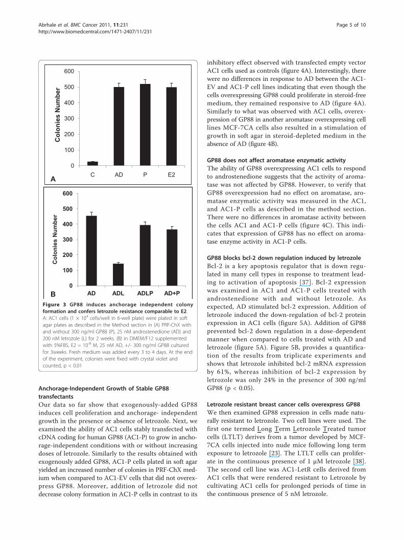
GP88 stimulate anchorage-independent growth inaromatase overexpressing AC1 cellsAnchorage-independent growth is a hallmark of cancercell transformation. The ability of exogenously addedGP88 to stimulate anchorage-independent growth andconfer MCF-7AC1 cells resistance to letrozole wasinvestigated. As described in the method section, cellsplated at 1 × 10⁴ in soft agar in steroid-depleted med-ium were treated with AD, GP88 or E2 in the absenceof letrozole. As shown in figure 3A, addition of GP88resulted in a 20-fold increase in the number of coloniesforming in soft agar (p < 0.01), similarly to what wasobserved for androstenedione (AD) and for Estradiol,
known growth stimulators of aromatase overexpressingbreast cancer cells. As shown in figure 3B, addition ofletrozole (200 nM) to cells cultivated in the presence ofAD resulted in a 53% inhibition of colony growth in softagar. In addition to stimulating colony growth, GP88(300 ng/ml) reversed the inhibitory effect of letrozolesimilarly to E2 taken as control (figure 3A).
0
50
100
150
200
250
300
350
400
C AD P-150 P-300 P-600
Cell
Num
ber (
x104 )
Treatments
Figure 1 GP88 induces AC1 cell proliferation in vitro. 5 × 10⁴
cells/plate (35 mm well plates) MCF-7AC1 cells were plated in PRFmedium supplemented with 5% Charcoal treated FBS overnight.The next day, medium was removed and serum free medium wasadded without or with increasing concentration of GP88: 150 ng/ml(P-150), 300 ng/ml (P-300) and 600 ng/ml (P-600) or 25 nMandrostenedione (AD). After 6 days, cells were trypsinized and cellnumber was determined by counting cells with a hemocytometer.Experiment was performed three times in triplicate.
0
3000
6000
9000
Lum
ines
cenc
e
Treatments Figure 2 GP88 confers AC1 cells Letrozole resistance in vitro.2000 cells/well (96 well plates) MCF-7AC1 cells were plated in PRFmedium supplemented with 5% ChX FBS overnight The next day,medium was removed and serum free medium was added withand without 300 ng/ml GP88 (GP88 (P)), 25 nM androstenedione(AD) and letrozole (L) 50 nM and 100 nM, 200 nM. Cell proliferationwas measured after 6 days using Cell Titer-Glo assay. Experimentwas performed in triplicates with p < 0.01.
Abrhale et al. BMC Cancer 2011, 11:231http://www.biomedcentral.com/1471-2407/11/231
Page 4 of 10
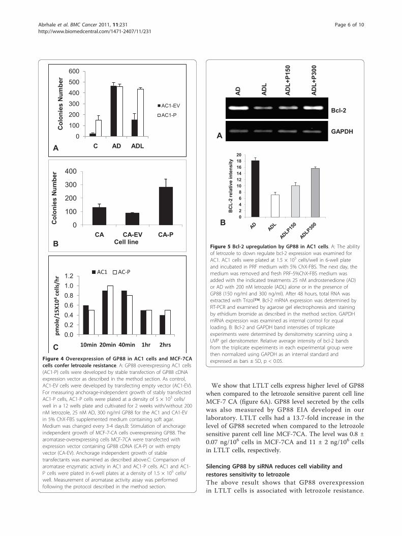
Anchorage-Independent Growth of Stable GP88transfectantsOur data so far show that exogenously-added GP88induces cell proliferation and anchorage- independentgrowth in the presence or absence of letrozole. Next, weexamined the ability of AC1 cells stably transfected withcDNA coding for human GP88 (AC1-P) to grow in ancho-rage-independent conditions with or without increasingdoses of letrozole. Similarly to the results obtained withexogenously added GP88, AC1-P cells plated in soft agaryielded an increased number of colonies in PRF-ChX med-ium when compared to AC1-EV cells that did not overex-press GP88. Moreover, addition of letrozole did notdecrease colony formation in AC1-P cells in contrast to its
inhibitory effect observed with transfected empty vectorAC1 cells used as controls (figure 4A). Interestingly, therewere no differences in response to AD between the AC1-EV and AC1-P cell lines indicating that even though thecells overexpressing GP88 could proliferate in steroid-freemedium, they remained responsive to AD (figure 4A).Similarly to what was observed with AC1 cells, overex-pression of GP88 in another aromatase overexpressing celllines MCF-7CA cells also resulted in a stimulation ofgrowth in soft agar in steroid-depleted medium in theabsence of AD (figure 4B).
GP88 does not affect aromatase enzymatic activityThe ability of GP88 overexpressing AC1 cells to respondto androstenedione suggests that the activity of aroma-tase was not affected by GP88. However, to verify thatGP88 overexpression had no effect on aromatase, aro-matase enzymatic activity was measured in the AC1,and AC1-P cells as described in the method section.There were no differences in aromatase activity betweenthe cells AC1 and AC1-P cells (figure 4C). This indi-cates that expression of GP88 has no effect on aroma-tase enzyme activity in AC1-P cells.
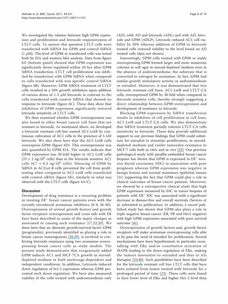
GP88 blocks bcl-2 down regulation induced by letrozoleBcl-2 is a key apoptosis regulator that is down regu-lated in many cell types in response to treatment lead-ing to activation of apoptosis [37]. Bcl-2 expressionwas examined in AC1 and AC1-P cells treated withandrostenedione with and without letrozole. Asexpected, AD stimulated bcl-2 expression. Addition ofletrozole induced the down-regulation of bcl-2 proteinexpression in AC1 cells (figure 5A). Addition of GP88prevented bcl-2 down regulation in a dose-dependentmanner when compared to cells treated with AD andletrozole (figure 5A). Figure 5B, provides a quantifica-tion of the results from triplicate experiments andshows that letrozole inhibited bcl-2 mRNA expressionby 61%, whereas inhibition of bcl-2 expression byletrozole was only 24% in the presence of 300 ng/mlGP88 (p < 0.05).
Letrozole resistant breast cancer cells overexpress GP88We then examined GP88 expression in cells made natu-rally resistant to letrozole. Two cell lines were used. Thefirst one termed Long Term Letrozole Treated tumorcells (LTLT) derives from a tumor developed by MCF-7CA cells injected into nude mice following long termexposure to letrozole [23]. The LTLT cells can prolifer-ate in the continuous presence of 1 μM letrozole [38].The second cell line was AC1-LetR cells derived fromAC1 cells that were rendered resistant to Letrozole bycultivating AC1 cells for prolonged periods of time inthe continuous presence of 5 nM letrozole.
0
100
200
300
400
500
600
C AD P E2
Col
onie
s N
umbe
r
0
100
200
300
400
500
600
AD ADL ADLP AD+P
Col
onie
s N
umbe
r
A
BFigure 3 GP88 induces anchorage independent colonyformation and confers letrozole resistance comparable to E2.A: AC1 cells (1 × 10⁴ cells/well in 6-well plate) were plated in softagar plates as described in the Method section in (A) PRF-ChX withand without 300 ng/ml GP88 (P), 25 nM androstenedione (AD) and200 nM letrozole (L) for 2 weeks. (B) in DMEM/F12 supplementedwith 5%FBS, E2 = 10-8 M, 25 nM AD, +/- 300 ng/ml GP88 culturedfor 3weeks. Fresh medium was added every 3 to 4 days. At the endof the experiment, colonies were fixed with crystal violet andcounted, p < 0.01
Abrhale et al. BMC Cancer 2011, 11:231http://www.biomedcentral.com/1471-2407/11/231
Page 5 of 10
We show that LTLT cells express higher level of GP88when compared to the letrozole sensitive parent cell lineMCF-7 CA (figure 6A). GP88 level secreted by the cellswas also measured by GP88 EIA developed in ourlaboratory. LTLT cells had a 13.7-fold increase in thelevel of GP88 secreted when compared to the letrozolesensitive parent cell line MCF-7CA. The level was 0.8 ±0.07 ng/106 cells in MCF-7CA and 11 ± 2 ng/106 cellsin LTLT cells, respectively.
Silencing GP88 by siRNA reduces cell viability andrestores sensitivity to letrozoleThe above result shows that GP88 overexpressionin LTLT cells is associated with letrozole resistance.
0
100
200
300
400
500
600
C AD ADL
Col
onie
s N
umbe
r
AC1-EV
AC1-P
0
100
200
300
400
CA CA-EV CA-P
Col
onie
s N
umbe
r
Cell line
0.00.20.40.60.81.01.2
10min 20min 40min 1hr 2hrs
pmole/15
X104
cells/hr
AC1 AC-P
A
B
CFigure 4 Overexpression of GP88 in AC1 cells and MCF-7CAcells confer letrozole resistance. A: GP88 overexpressing AC1 cells(AC1-P) cells were developed by stable transfection of GP88 cDNAexpression vector as described in the method section. As control,AC1-EV cells were developed by transfecting empty vector (AC1-EV).For measuring anchorage-independent growth of stably transfectedAC1-P cells, AC1-P cells were plated at a density of 5 × 10³ cells/well in a 12 wells plate and cultivated for 2 weeks with/without 200nM letrozole, 25 nM AD, 300 ng/ml GP88 for the AC1 and CA1-EVin 5% ChX-FBS supplemented medium containing soft agar.Medium was changed every 3-4 days.B: Stimulation of anchorageindependent growth of MCF-7-CA cells overexpressing GP88. Thearomatase-overexpressing cells MCF-7CA were transfected withexpression vector containing GP88 cDNA (CA-P) or with emptyvector (CA-EV). Anchorage independent growth of stabletransfectants was examined as described above.C: Comparison ofaromatase enzymatic activity in AC1 and AC1-P cells. AC1 and AC1-P cells were plated in 6-well plates at a density of 1.5 × 10⁵ cells/well. Measurement of aromatase activity assay was performedfollowing the protocol described in the method section.
AD ADL
ADL+
P150
ADL+
P300
Bcl-2
GAPDHA
02468
101214161820
BC
L-2
rela
tive
inte
nsity
B
Figure 5 Bcl-2 upregulation by GP88 in AC1 cells. A: The abilityof letrozole to down regulate bcl-2 expression was examined forAC1. AC1 cells were plated at 1.5 × 10⁵ cells/well in 6-well plateand incubated in PRF medium with 5% ChX-FBS. The next day, themedium was removed and fresh PRF-5%ChX-FBS medium wasadded with the indicated treatments 25 nM androstenedione (AD)or AD with 200 nM letrozole (ADL) alone or in the presence ofGP88 (150 ng/ml and 300 ng/ml). After 48 hours, total RNA wasextracted with Trizol™. Bcl-2 mRNA expression was determined byRT-PCR and examined by agarose gel electrophoresis and stainingby ethidium bromide as described in the method section. GAPDHmRNA expression was examined as internal control for equalloading. B: Bcl-2 and GAPDH band intensities of triplicateexperiments were determined by densitometry scanning using aUVP gel densitometer. Relative average intensity of bcl-2 bandsfrom the triplicate experiments in each experimental group werethen normalized using GAPDH as an internal standard andexpressed as bars ± SD, p < 0.05.
Abrhale et al. BMC Cancer 2011, 11:231http://www.biomedcentral.com/1471-2407/11/231
Page 6 of 10
0
1000
2000
3000
4000
5000
6000
24 48 72 96
Lum
ines
cenc
e
Time (hrs)
Control
siRNA
0
1000
2000
3000
4000
5000
6000
7000
8000
C 1 M 2 M 3 M
Lum
ines
cenc
e
Letrozole
Control
siRNA
A
B
D
C
E
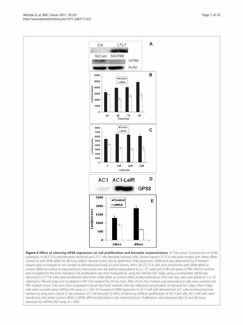
Figure 6 Effect of silencing GP88 expression on cell proliferation and letrozole responsiveness. A: Top panel: Comparison of GP88expression in MCF-7CA cells (letrozole sensitive) and LTLT cells (letrozole resistant cells). Bottom panel: LTLT-CA cells were treated with either siRNAcontrol or with GP88 siRNA for 48 hours before harvesting the cells to determine GP88 expression. GP88 level was determined by IP-Westernanalysis and normalized to cell number as described previously (Lu and Serrero, 2001). (B) LTLT-CA cells were transfected with GP88 siRNA orcontrol siRNA according to manufacturer’s instructions one day before being plated at 2 × 10³ cells/well in 96 well plates in PRF-5%ChX mediumand incubated for the time indicated. Cell proliferation was then measured by using the cellTiter-GloR assay using a luminometer (MolecularDevices).(C) LTLT-CA cells were transfected with either GP88 siRNA or Control siRNA as described above. The next day, cells were plated at 2 × 10³
cells/well in 96-well plate and incubated in PRF-ChX medium for 24 hrs more. After 24 hrs, the medium was removed and cells were washed withPRF medium twice. Cells were then incubated in serum free fresh medium with the indicated concentration of letrozole for 3 days. After 3 dayscells were counted using cellTiter-Glo assay, p < 0.05. D) Increase of GP88 expression in AC1-LetR cells derived from AC1 cells rendered letrozoleresistant by long term culture in the presence of 5 nM letrozole. E) Effect of silencing GP88 on proliferation of AC1-LetR cells. AC1-LetR cells weretransfected with either control siRNA or GP88 siRNA as described in the method section. Proliferation was measured after 24 and 48 hoursexposure by cellTiter-GloR assay, p < 0.05.
Abrhale et al. BMC Cancer 2011, 11:231http://www.biomedcentral.com/1471-2407/11/231
Page 7 of 10
We investigated the relation between high GP88 expres-sion and proliferation and letrozole responsiveness ofLTLT cells. To answer this question LTLT cells weretransfected with SiRNA for GP88 and control SiRNA(1 μM). The level of GP88 in transfected cells was testedboth by EIA and western blot analysis. Data from figure6A (bottom panel) showed that GP88 expression wassignificantly down regulated within 24 hrs after GP88SiRNA transfection. LTLT cell proliferation was inhib-ited by transfection with GP88 SiRNA when comparedto cells transfected with non specific control SiRNA(figure 6B). Moreover, GP88 SiRNA treatment of LTLTcells resulted in a 50% growth inhibition upon additionof various doses of 1-3 μM letrozole in contrast to thecells transfected with control SiRNA that showed noresponse to letrozole (figure 6C). These data show thatinhibition of GP88 expression significantly restoredletrozole sensitivity of LTLT cells.We then examined whether GP88 overexpression was
also found in other breast cancer cell lines that areresistant to letrozole. As mentioned above, we developeda letrozole resistant cell line named AC1-LetR by con-tinuous cultivation of AC1 cells in the presence of 5 nMletrozole. We also show here that the AC1-LetR cellsoverexpress GP88 (figure 6D). This overexpression wasalso quantified by GP88 EIA. The results indicate thatGP88 expression was 30-fold higher in AC1-LetR cells(23 ± 2 ng/106 cells) than in the letrozole sensitive AC1cells (0.7 ± 0.2 ng/106 cells). Silencing of GP88 bySiRNA in AC1Let-R cells prevented the cell from prolif-erating when compared to AC1-LetR cells transfectedwith control siRNA (figure 6E), similarly to what wasobserved with the LTLT cells (figure 6A-C).
DiscussionDevelopment of drug resistance is a recurring problemin treating ER+ breast cancer patients even with therecently introduced aromatase inhibitors [8-9; 36-40].Overexpression of several growth factors and growthfactor receptors overexpression and cross talk with ERhave been described as some of the major changes asassociated or causing such resistance [17,22,39]. Weshow here that an alternate growth/survival factor GP88(progranulin), previously identified as playing a role inbreast cancer tumorigenesis [28,29], is involved in con-ferring letrozole resistance using two aromatase overex-pressing breast cancer cells as study models. Thepresent study demonstrates that exogenously addedGP88 induces AC1 and MCF-7CA growth in steroid-depleted medium in both anchorage-dependent andindependent conditions. In addition, Letrozole induceddown regulation of bcl-2 expression whereas GP88 pre-vented such down regulation. We have also measuredviability of the cells treated with androstenedione only
(AD), with AD and letrozole (ADL) and with AD, letro-zole and GP88 (ADLP). Letrozole reduced AC1 cell via-bility by 50% whereas addition of GP88 to letrozoletreated cells restored viability to the level found on ADtreated cells (data not shown).Interestingly, GP88 cells treated with GP88 or stably
overexpressing GP88 formed larger and more numerouscolonies in soft agar in steroid-depleted medium even inthe absence of androstenedione, the substrate that isconverted to estrogen by aromatase. In fact, GP88 hadsimilar growth stimulatory activity as androstenedioneor estradiol. Moreover, it was demonstrated that twoletrozole resistant cell lines, AC1-LetR and LTLT-CAcells, overexpressed GP88 by 30-fold when compared toletrozole sensitive cells, thereby strongly suggesting adirect relationship between GP88 overexpression anddevelopment of resistance to letrozole.Blocking GP88 expression by SiRNA transfection
results in inhibition of cell proliferation in cell lines,AC1-LetR and LTLT-CA cells. We also demonstratethat SiRNA treatment partially restores LTLT-CA cellssensitivity to letrozole. These data provide additionalsupport to our previous findings that GP88 could substi-tute for estradiol to stimulate proliferation, in steroid-depleted medium and confer tamoxifen resistance toMCF-7 cells both in vitro and in vivo [32]. Our previouspathological study with paraffin-embedded breast cancerbiopsies has shown that GP88 is expressed in ER+ inva-sive ductal carcinoma (IDC) in association with poorprognosis whereas GP88 expression was negative inbenign lesions and normal mammary epithelial tissues[31] supporting the fact that GP88 could play a role inclinical outcomes of breast cancer patients. Moreover,we showed by a retrospective clinical study that highGP88 expression measured by IHC in tumor biopsies ofpatients with ER+ IDC was associated with a significantdecrease in disease-free and overall survivals (Serrero etal, submitted to publication). In addition, a recent pub-lished study has shown that GP88 also plays a role intriple negative breast cancer (ER, PR and Her2 negative)with high GP88 expression associated with poor survivaloutcome [41].Overexpression of growth factors and, growth factor
receptors will make aromatase overexpressing cells ableto by-pass the need of estradiol for proliferation. Severalmechanisms have been hypothesized, in particular cross-talking with ERa and/or constitutive activation ofMAPK leading to the down regulation of ERa, makingthe tumors insensitive to estradiol and thus to AIstherapies [23,40]. Such possibilities have been describedfor the letrozole resistant cell line LTLT cells that havebeen isolated from tumor treated with letrozole for aprolonged period of time [23]. These cells were foundto have lower level of ERa and higher Her-2 level than
Abrhale et al. BMC Cancer 2011, 11:231http://www.biomedcentral.com/1471-2407/11/231
Page 8 of 10
their letrozole sensitive counterparts MCF7-CA cells.The cells appear to survive estrogen deprivation by theactivation of Her-2/MAPK pathway [42]. In fact, forthese letrozole resistant cells, the combination of letro-zole and trastuzumab resulted in superior outcome ininhibiting tumor growth in vivo over letrozole or trastu-zumab alone leading to inhibition of tumor growth, incontrast to MCF-7CA cells where letrozole alone was aseffective as trastuzumab in combination with letrozole.This indicates that overexpression of Her-2 is a majorcontributor of letrozole resistance of LTLT cells. Weshow here that the autocrine growth factor GP88 con-fers letrozole resistance to MCF-CA cells and that inLTLT cells that overexpress GP88, inhibition of GP88expression by SiRNA partially restores letrozole respon-siveness. In addition, GP88 also conferred letrozoleresistance to AC1 cell line, another aromatase overex-pressing cell line. Concerning the possible mechanismsby which GP88 confers letrozole resistance, we showhere that AC1 cells overexpressing GP88 can still be sti-mulated by androstenedione (figure 4A) in steroid-depleted medium and by estradiol ( data not shown)indicating that the letrozole resistance caused by GP88is not due to the cells becoming estrogen independentor not expressing estrogen receptors. Moreover, asshown in figure 4C, aromatase enzymatic activity wasnot affected by GP88 overexpression thereby suggestingthat GP88 expression did not result in a defect in aro-matase expression or function. The fact that GP88 hasbeen shown to be a potent proliferation and anti-apop-totic factor for breast cancer cells [26,33,34] would sug-gest that GP88 confers resistance to the killing effect ofletrozole by providing growth and survival advantage tothe cells rather than by blocking estrogen response orby inhibiting aromatase activity.It is also interesting to note that we have shown pre-
viously that GP88 activates Her-2 phosphorylation inbreast cancer cells through the ERK1/2 and Akt path-ways [43]. This would suggest the association of GP88and Her-2 in conferring resistance to letrozole. How-ever, our pathological studies with breast cancer tumorbiopsies have indicated that GP88 and Her-2 were inde-pendent biomarkers [31], suggesting that not all ofGP88 effects on letrozole resistance are necessarilymediated via GP88’s ability to cross-talk with Her-2.
ConclusionsUnderstanding the role and mechanisms of GP88 in thedevelopment of resistance to anti-hormonal treatmentscould contribute to improving the treatment of breastcancer. Based on our pathological studies, it would beinteresting to examine if overexpression of GP88 intumor tissue s is associated with poor response to AI.Moreover, combination of drugs that include the
inhibition of growth factors and anti-hormones couldprovide new avenues to treat breast cancer patients andovercome resistance. Our findings provide a novel para-digm for the acquisition of aromatase inhibitor resistantbreast cancer and could suggest new therapeuticapproaches.
List of AbbreviationsAD: androstenedione; AI: aromatase inhibitors; E2: 17-β estradiol; ER:estrogen receptor; FBS: fetal bovine serum; ChX-FBS: charcoal extracted fetalbovine serum; Let: letrozole; PRF medium: phenol red free medium; PRF-ChX: phenol red free DME-F12 medium supplemented with 5% Charcoalextracted fetal bovine serum; SDS: sodium dodecyl sulfate; PAGE:polyacrylamide gel electrophoresis
AcknowledgementsThe authors wish to thank Dr. Jun Hayashi for helpful discussion. This studywas supported by grants 1R43CA124179-01 and 2R44CA124179-02 from theNational Cancer Institute and 07-2007-064 from the Avon Foundation BreastCare Fund.
Author details1A&G Pharmaceutical Inc. 9130 Red Branch Rd. Columbia, MD, USA.2Department of Pharmaceutical Sciences, University of Maryland School ofPharmacy, 20 N Pine Street, Baltimore MD 21201, USA. 3Department ofPharmacology and Experimental Therapeutics, University of Maryland Schoolof Medicine, HSF-I, 685 W. Baltimore Street, Baltimore, MD 21201, USA.4Program in Oncology, Marlene and Stewart Greenebaum Cancer Center,University of Maryland School of Medicine, 22 S. Greene St. Baltimore, MD21201, USA.
Authors’ contributionsTA carried out the GP88 studies and drafted the manuscript. AB, and LMdeveloped the cell lines used in these studies and helped in data analysisand thoroughly reviewed and revised the manuscript. GaSa carried out thearomatase studies and helped in data input and data analysis. CT designedand develop conditions for the SiRNA experiments and the RT-PCRexperiments. BY did all the EIA GP88 measurements for all cell lines studied.GS conceived the study, drafted the manuscript, directly guided TA in hisstudies and provided the input on experimental design and datainterpretation.All authors have given final approval of the submitted manuscript.
Competing interestsDrs. Ginette Serrero, Changsheng Tian and Ms. Binbin Yue have financialcompeting interests as employees and stock holders of A&G PharmaceuticalInc. Drs. Tesfom Abrhale, Angela Brodie, Luciana Macedo, and Gauri Sabnisdo not have competing interests.
Received: 31 March 2011 Accepted: 9 June 2011 Published: 9 June 2011
References1. Brueggemeier RW, Hackett JC, Diaz-Cruz ES: Aromatase inhibitors in the
treatment of breast cancer. Endocr Rev 2005, 26:331-45.2. Grodin JM, Siiteri PK, McDonald PC: Source of estrogen production in
postmenopausal women. J Clin Endocrinol Metab 1973, 36:207-14.3. Burak WE Jr, Quinn AL, Farrar WB, Brueggemeier RW: Androgens influence
estrogen-induced responses in human breast carcinoma cells throughcytochrome P450 aromatase. Breast Cancer Res Treat 1997, 44:57-64.
4. Chen S, Zhou D, Okubo T, Kao YC, Yang C: Breast tumor aromatase:functional role and transcriptional regulation. Endocr Relat Cancer 1999,6:149-56.
5. Shupnik MA: Crosstalk between steroid receptors and the c-Src-receptortyrosine kinase pathways: implications for cell proliferation. Oncogene2004, 23:7979-89.
6. Jordan VC, Brodie AM: Development and evolution of therapies targetedto the estrogen receptor for the treatment and prevention of breastcancer. Steroids 2007, 72:7-25.
Abrhale et al. BMC Cancer 2011, 11:231http://www.biomedcentral.com/1471-2407/11/231
Page 9 of 10
7. Jelovac D, Macedo L, Goloubeva OG, Handratta V, Brodie AM: Activation ofmitogen-activated protein kinase in xenografts and cells duringprolonged treatment with aromatase inhibitor letrozole. Cancer Res 2005,65:5380-9.
8. Chen S, Masri S, Wang X, Phung S, Yuan YC, Wu X: What do we knowabout the mechanisms of aromatase inhibitor resistance? J SteroidBiochem Mol Biol 2006, 102:232-40.
9. Dowsett M, Martin LA, Smith I, Johnston S: Mechanisms of resistance toaromatase inhibitors. J Steroid Biochem Mol Biol 2005, 95:167-172.
10. Nicholson RI, Gee JM: Oestrogen and growth factor cross-talk andendocrine insensitivity and acquired resistance in breast cancer. Br JCancer 2000, 82:501-13.
11. Nicholson RI, Hutcheson IR, Britton D, Knowlden JM, Jones HE, Harper ME,Hiscox SE, Barrow D, Gee JM: Growth factor signalling networks in breastcancer and resistance to endocrine agents: new therapeutic strategies.J Steroid Biochem Mol Biol 2005, 93:257-62.
12. Nicholson RI, Hutcheson IR, Knowlden JM, Jones HE, Harper ME, Jordan N,Hiscox SE, Barrow D, Gee JM: Nonendocrine pathways and endocrineresistance: observations with antiestrogens and signal transductioninhibitors in combination. Clin Cancer Res 2004, 10:346S-54S.
13. Pietras RJ, Arboleda J, Reese DM, Wongvipat N, Pegram MD, Ramos L,Gorman CM, Parker MG, Sliwkowski MX, Slamon DJ: HER-2 tyrosinekinase pathway targets estrogen receptor and promotes hormone-independent growth in human breast cancer cells. Oncogene 1995,10:2435-46.
14. Lupu R, Cardillo M, Cho C, Harris L, Hijazi M, Perez C, Rosenberg K, Yang D,Tang C: The significance of heregulin in breast cancer tumor progressionand drug resistance. Breast Cancer Res Treat 1996, 38:57-66.
15. Tang CK, Perez C, Grunt T, Waibel C, Cho C, Lupu R: Involvement ofheregulin-beta2 in the acquisition of the hormone-independentphenotype of breast cancer cells. Cancer Res 1996, 56:3350-8.
16. McClelland RA, Barrow D, Madden TA, Dutkowski CM, Pamment J,Knowlden JM, Gee JM, Nicholson RI: Enhanced epidermal growth factorreceptor signaling in MCF7 breast cancer cells after long-term culture inthe presence of the pure antiestrogen ICI 182,780 (Faslodex).Endocrinology 2001, 142:2776-88.
17. Knowlden JM, Hutcheson IR, Jones HE, Madden T, Gee JM, Harper ME,Barrow D, Wakeling AE, Nicholson RI: Elevated levels of epidermal growthfactor receptor/c-erbB2 heterodimers mediate an autocrine growthregulatory pathway in tamoxifen-resistant MCF-7 cells. Endocrinology2003, 144:1032-44.
18. Guvakova MA, Surmacz E: Overexpressed IGF-I receptors reduce estrogengrowth requirements, enhance survival, and promote E-cadherin-mediated cell-cell adhesion in human breast cancer cells. Exp Cell Res1997, 231:149-62.
19. Stephen RL, Shaw LE, Larsen C, Corcoran D, Darbre PD: Insulin-like growthfactor receptor levels are regulated by cell density and by long termestrogen deprivation in MCF7 human breast cancer cells. J Biol Chem2001, 276:40080-6.
20. Benz CC, Scott GK, Sarup JC, Johnson RM, Tripathy D, Coronado E,Shepard HM, Osborne CK: Estrogen-dependent, tamoxifen-resistanttumorigenic growth of MCF-7 cells transfected with HER2/neu. BreastCancer Res Treat 1992, 24:85-95.
21. Liu Y, el-Ashry D, Chen D, Ding IY, Kern FG: MCF-7 breast cancer cellsoverexpressing transfected c-erbB-2 have an in vitro growth advantagein estrogen-depleted conditions and reduced estrogen-dependence andtamoxifen-sensitivity in vivo. Breast Cancer Res Treat 1995, 34:97-117.
22. Kurokawa H, Lenferink AE, Simpson JF, Pisacane PI, Sliwkowski MX,Forbes JT, Arteaga CL: Inhibition of HER2/neu (erbB-2) and mitogen-activated protein kinases enhances tamoxifen action against HER2-overexpressing, tamoxifen-resistant breast cancer cells. Cancer Res 2000,60:5887-94.
23. Sabnis GJ, Jelovac D, Long B, Brodie A: The role of growth factor receptorpathways in human breast cancer cells adapted to long-term estrogendeprivation. Cancer Res 2005, 65:3903-10.
24. Oh AS, Lorant LA, Holloway JN, Miller DL, Kern FG, El-Ashry D:Hyperactivation of MAPK induces loss of ERalpha expression in breastcancer cells. Mol Endocrinol 2001, 15:1344-59.
25. Su B, Wong C, Hong Y, Chen S: Growth factor signaling enhancesaromatase activity of breast cancer cells via post-transcriptionalmechanisms. J Steroid Biochem Mol Biol 2010.
26. Lu R, Serrero G: Mediation of estrogen mitogenic effect in human breastcancer MCF-7 cells by PC-cell-derived growth factor (PCDGF/granulinprecursor). Proc Natl Acad Sci USA 2001, 98:142-7.
27. He Z, Bateman A: Progranulin (granulin-epithelin precursor, PC-cell-derived growth factor, acrogranin) mediates tissue repair andtumorigenesis. J Mol Med 2003, 81:600-12.
28. Ong CH, Bateman A: Progranulin (granulin-epithelin precursor, PC-cellderived growth factor, acrogranin) in proliferation and tumorigenesis.Histol Histopathol 2003, 18:1275-88.
29. Serrero G: Autocrine growth factor revisited: PC-cell-derived growthfactor (progranulin), a critical player in breast cancer tumorigenesis.Biochem Biophys Res Commun 2003, 308:409-13.
30. Lu R, Serrero G: Inhibition of PC cell-derived growth factor (PCDGF,epithelin/granulin precursor) expression by antisense PCDGF cDNAtransfection inhibits tumorigenicity of the human breast carcinoma cellline MDA-MB-468. Proc Natl Acad Sci USA 2000, 97:3993-8.
31. Serrero G, Ioffe O: Expression of the novel autocrine growth factor PC-Cell Derived Growth Factor in human breast cancer tissue. HumanPathology 2003, 34:1148-54.
32. Tkaczuk KR, Yue B, Zhan M, Tait N, Yarlagadda L, Dai H, Serrero G:Increased circulating level of the survival factor GP88 (Progranulin) inthe serum of breast cancer patients when compared to healthysubjects. Breast Cancer Basic and Clinical Research 2011.
33. Tangkeangsirisin W, Hayashi J, Serrero G: PC cell-derived growth factormediates tamoxifen resistance and promotes tumor growth of humanbreast cancer cells. Cancer Res 2004, 64:1737-43.
34. Tangkeangsirisin W, Serrero G: PC-Cell Derived Growth Factor (PCDGF/GP88) Stimulates Migration, Invasiveness and VEGF expression in BreastCancer Cells. Carcinogenesis 2004, 25:1587-89.
35. Long BJ, Tilghman SL, Yue W, Thiantanawat A, Grigoryev DN, Brodie AM:The steroidal antiestrogen ICI 182,780 is an inhibitor of cellulararomatase activity. J Steroid Biochem Mol Biol 1998, 67:293-304.
36. Macedo LF, Guo Z, Tilghman SL, Sabnis GJ, Qiu Y, Brodie A: Role ofandrogens on MCF-7 breast cancer cell growth and on the inhibitoryeffect of letrozole. Cancer Res 2006, 66:7775-7782.
37. Diel P, Smolnikar K, Michna H: The pure antiestrogen ICI 182780 is moreeffective in the induction of apoptosis and down regulation of bcl-2than tamoxifen in MCF-7 cells. Breast Cancer Res Treat 1999, 58:87-97.
38. Brodie A, Jelovac D, Sabnis G, Long B, Macedo L, Goloubeva O: Modelsystems: mechanisms involved in the loss of sensitivity to letrozole. JSteroid Biochem Mol Biol 2005, 95:41-8.
39. Brodie A, Jelovac D, Macedo L, Sabnis G, Tilghman S, Goloubeva O:Therapeutic observations in MCF-7 aromatase xenografts. Clin Cancer Res2005, 11:884s-8s.
40. Miller TW, Hennessy BT, González-Angulo AM, Fox EM, Mills GB, Chen H,Higham C, García-Echeverría C, Shyr Y, Arteaga CL: Hyperactivation ofPhosphatidylinositol-3-kinase promotes escape frpm hormonedependence in estrogen receptor-positive human breast cancer. J ClinInvest 2010, 120:2406-13.
41. Elkabets M, Gifford AM, Scheel C, Nilsson B, Reinhardt F, Bray MA,Carpenter AE, Jirström K, Magnusson K, Ebert BL, Pontén F, Weinberg RA,McAllister SS: Human tumors instigate granulin-expressing hematopoieticcells that promote malignancy by activating stromal fibroblasts in mice.J Clin Invest 2011, 121:784-99.
42. Sabnis G, Schayowitz A, Goloubeva O, Macedo L, Brodie A: Trastuzumabreverses letrozole resistance and amplifies the sensitivity of breastcancer cells to estrogen. Cancer Res 2009, 69:1416-1428.
43. Kim WE, Serrero G: PC cell-derived growth factor (PCDGF/GP88)stimulates proliferation and confers Trastuzumab resistance to Her-2-overexpressing breast cancer cells. Clin Cancer Res 2006, 15:4192-9.
Pre-publication historyThe pre-publication history for this paper can be accessed here:http://www.biomedcentral.com/1471-2407/11/231/prepub
doi:10.1186/1471-2407-11-231Cite this article as: Abrhale et al.: GP88 (PC-Cell Derived Growth Factor,progranulin) stimulates proliferation and confers letrozole resistance toaromatase overexpressing breast cancer cells. BMC Cancer 2011 11:231.
Abrhale et al. BMC Cancer 2011, 11:231http://www.biomedcentral.com/1471-2407/11/231
Page 10 of 10
Related Documents


