G.P. Review Al'lide FLAT FOOT: II* G. F. DOMMISSE, F.R.C.S. (EDIN.), Pretoria SUMMARY The most important varieties of 'flat foot' are described and are classified into the 'congenital' and the 'acquired' groups. A foot which on casual inspection appears to be 'flat' will on careful examination prove to be within the limits of normal in the majority of cases. The hasty labelling of a patient as 'flat-footed', whether child or adult, is severely condemned and is generally not justified. Both knowledge and skill are required in the diagnosis of a condition which is as important to the individual as wheels to a carriage and wings to a bird. General rules in the choice of footwear suitable fOI; children and adults in their various daily activities are laid down. Pedicure is strongly urged, so that the humble feet may enjoy the same degree of fragrance and pride of ownership as the hands. Let our motto be: 'Pale feet, pink-tipped, [loved beside sweet Shalimar' (with apologies). * Date received: 4 February 1971. 'Flat-foot' is a generic term which covers a number of con- ditions involving the feet, conditions which differ in degree, in aetiology, in pathology, in prognosis, in treatment and in incidence as well as in age of incidence. The use of the terms 'pes planus' or 'pes plana-valgus' offers no particular advantage over 'fiat-foot', and in either event assumes significance only when suitably qualified, e.g. 'congenital rigid flat-foot with vertical talus', or 'congenital rigid pes planus with vertical talus'. There are many kinds of flat- foot, of which only the most important are briefly des- cribed below. They are classified under two main groups, namely congenital and acquired fiat-foot. The term 'fiat-foot' is deceptive because of its simplicity, and has led to a faulty diagnosis in countless normal indi- vidua:Is whose feet merely appeared to be flat. Worse, there are those individuals who, although they suffer from flat- foot in the incipient stages, are denied treatment on the grounds that the arches of the feet have not yet fallen. I! would be illogical to attempt a definition in the know-

Welcome message from author
This document is posted to help you gain knowledge. Please leave a comment to let me know what you think about it! Share it to your friends and learn new things together.
Transcript
726 S.-A. MEDIESE TYDSKRIF 3 Julie 1971
Fig. 1: Bassini's original description of herniorrhaphy.(A) subcutaneous tissue, (B) external oblique, (C) fasciatransversalis, (E) spermatic cord, (F) transversus, internaloblique and fascia transversus, (G) hernia sac. (FromBassini's (j ber die Behandlung des Leisten-bruches,Langenbecks Arch. klin. Chir., Vo\. 40.)
Bassini appreciated the importance of strengthening theposterior abdominal wall. He advised that the hernia sacshould be excised and ligated as high as possible. The nextstep in the operation was to separate the external obliquefrom the internal oblique, and the transversus muscle andfascia transversalis from the preperitoneal fat. The herniorrhaphy consisted of uniting the three layers (internaloblique, transversus and fascia transversalis) to the inguinal
G.P. Review Al'lide
ligament below with interrupted black silk sutures. Thesutures located at the most medial part also passed throughthe outer edge of the rectus sheath. The internal ring wasreconstituted so that it was situated lateral to the externalring to ensure that the obliquity of the inguinal canalwas maintained (Fig. I).
In 1890, Bassini reported his experience with 262 consecutive cases who had had a hernia repaired accordingto the new method he described. Out of 251 cases wherestrangulation had not occurred, all survived the operation.The recurrence rate was less than 3% of those cases whohad been followed up for between one month and fouryears.
The operation described by Bassini has made a greatimpact on the surgery of inguinal herniae, and althoughmany different procedures have been described, none hashad a similar world-wide acceptance. The Bassini operation has withstood the test of time, and proof of its valuelies in the fact that it is still used today by many surgeonsin different parts of the globe.
I wish to thank Professor J. M. Mynors and Mrs P. Fergusonfor assistance in preparing this paper, and Mr G. Davie, forthe photograph.
REFERENCES
1. Celsus, 1st Century, AD (1938): De Medicine. Vo!. Ill, Book VIT. TheLoeb Classical Library. Cambridge: Harvard University Press.
2. Galen, 2nd Century AD (1929): Greek Medicine. London: T. M. Dent& Sons.
3. Paul of Aegira, 7th Century AD (1844): Seven books, translated fromthe Greek. Vo!. 1. Printed for the Sydenham Society. London: FrancisAdams.
4. Pierre Franco, 16th Century AD 0895): Irate des Hernies. Paris:Librairie Germer Bailliere.
5. Stromayr, 16th Century AD in Van Brunn, W., ed. (1925): PracticaCapiosa. Berlin: Idra-Verlagsanstalt.
6. Bull, W. T. ill Zimrnerman, L. 1. and 1. Keith, ed,. (1890): GreatIdeas in {he History of Surgery. Baltimore: Williams & \Vilkins.
7. Bassini, E. (1890): Arch. Klin. Chit., 40, 429.
FLAT FOOT: II*G. F. DOMMISSE, F.R.C.S. (EDIN.), Pretoria
SUMMARY
The most important varieties of 'flat foot' are describedand are classified into the 'congenital' and the 'acquired'groups.
A foot which on casual inspection appears to be 'flat'will on careful examination prove to be within the limitsof normal in the majority of cases. The hasty labelling ofa patient as 'flat-footed', whether child or adult, is severelycondemned and is generally not justified. Both knowledgeand skill are required in the diagnosis of a condition whichis as important to the individual as wheels to a carriageand wings to a bird.
General rules in the choice of footwear suitable fOI;children and adults in their various daily activities are laiddown. Pedicure is strongly urged, so that the humble feetmay enjoy the same degree of fragrance and pride ofownership as the hands. Let our motto be: 'Pale feet,pink-tipped, [loved beside sweet Shalimar' (with apologies).* Date received: 4 February 1971.
'Flat-foot' is a generic term which covers a number of conditions involving the feet, conditions which differ in degree,in aetiology, in pathology, in prognosis, in treatment andin incidence as well as in age of incidence. The use of theterms 'pes planus' or 'pes plana-valgus' offers no particularadvantage over 'fiat-foot', and in either event assumessignificance only when suitably qualified, e.g. 'congenitalrigid flat-foot with vertical talus', or 'congenital rigid pesplanus with vertical talus'. There are many kinds of flatfoot, of which only the most important are briefly described below. They are classified under two main groups,namely congenital and acquired fiat-foot.
The term 'fiat-foot' is deceptive because of its simplicity,and has led to a faulty diagnosis in countless normal individua:Is whose feet merely appeared to be flat. Worse, thereare those individuals who, although they suffer from flatfoot in the incipient stages, are denied treatment on thegrounds that the arches of the feet have not yet fallen.
I! would be illogical to attempt a definition in the know-
3 July 1971 S.A. MEDICAL JOUR 'AL 727
ledge that 'flat-foot' is a generic term referring to numerousaetiological and morphological types of abnormality. Thereis, however, a more or less uniform clinical picture whichis the end-result of every type of flat-foot, irrespective ofthe cause:
1. The medial border of the foot presents as a convexityrather than a concavity. The convexity is attributableto the head of the talus which 'bulges' both in amedial direct;on and into the sole of the foot (Fig.I (a)).
2. The mid-tarsal joints are everted (pronated), prominent and tender. Rigidity of these joints will be notedin the final stage of the condition, with pain onattempted active or passive movement (Fig. I (b)).
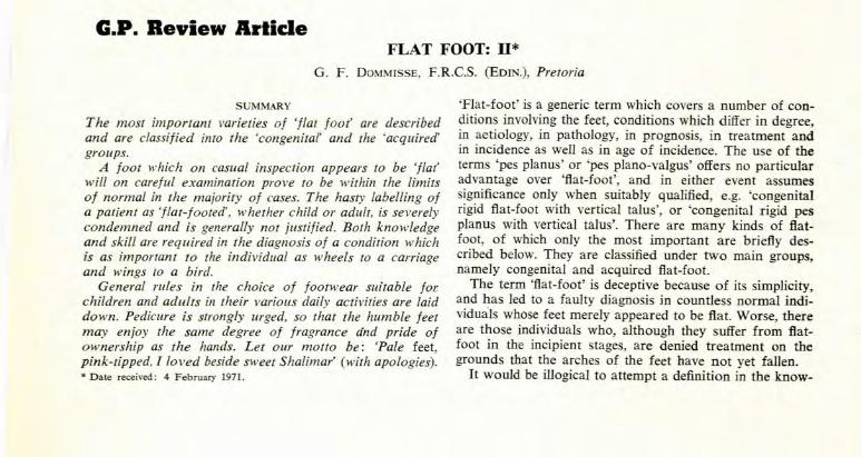
3. The heel is everted (Fig. 1 (c)) and efforts at inversionare painful.
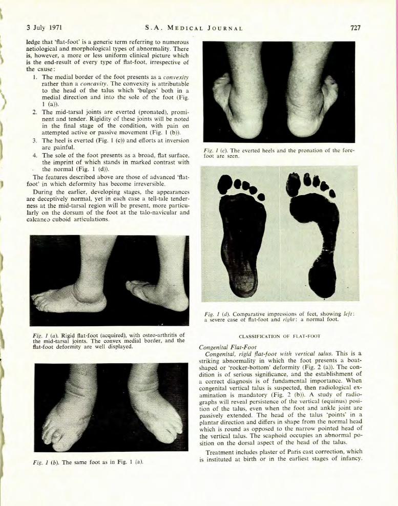
4. The sole of the foot presents as a broad, flat surface,the imprint of which stands in marked contrast withthe normal (Fig. I (d)).
The features described above are those of advanced 'flatfoot' in which deformity has become irreversible.
During the earlier, developing stages, the appearancesare deceptively normal, yet in each case a tell-tale tenderness at the mid-tarsal region will be present, more particularly on the dorsum of the foot at the talo-navicular andcalcanea cuboid art~culations.
Fig. 1 (a). Rigid fiat-foot (acquired), with osteo-arthritis ofthe mid-tarsal joints. The convex medial border, and thefiat-foot deformity are well displayed.
Fig. 1 (b). The same foot as in Fig. I (a).
Fig. 1 (c). The evened heels and the pronation of the forefoot are seen.
Fig. J (d). Comparative impressions of feet, showing lell:a severe case of flat-foot and righT: a normal foot.
CLASSIFICATION OF FLAT-FOOT
Congenital Flat-FootCongenital, rigid flat-foot with vertical talus. This is a
striking abnormality in which the foot presents a boatshaped or 'rocker-bottom' deformity (Fig. 2 (a)). The condition is of serious significance, and the establishment ofa correct diagnosis is of fundamental importance. Whencongenital vertical talus is suspected, then radiological examination is mandatory (Fig. 2 (b)). A study of radiographs will reveal persistence of the vertical (equinus) position of the talus, even when the foot and ankle joint arepassively extended. The head of the talus 'points' in aplantar direction and differs in shape from the normal headwhich is round as opposed to the narrow pointed head ofthe vertical tal us. The scaphoid occupies an abnormal position on the dorsal aspect of the head of the tal us.
Treatment includes plaster of Paris cast correction, whichis instituted at birth or in the earliest stages of infancy.
728 S.-A. MEDIESE TYDSKRIF 3 Julie 1971
CFig. 2 (a). Congenital rigid flat-foot with vertical talus,showing the clinical appearances in an infant. (From Ferguson').
Fig. 2 (b). Radiological appearance of an infant foot.(Courtesy Prof. I. S. de Wet, Orthopaedic Hospital, Pretoria.)
Operative correction is generally indicated and is undertaken early, at 3 months if possible, and in all cases beforewalking commences. Failure to correct this serious deformity will result in crippling disability.
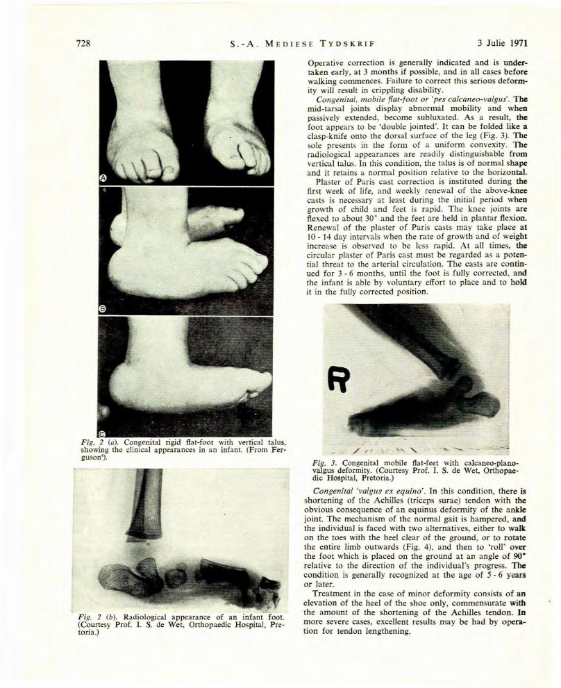
Congenital, mobile flat-foot or 'pes calcanea-valgus'. Themid-tarsal joints display abnormal mobility and whenpassively extended, become subluxated. As a result, thefoot appears to be 'double jointed'. It can be folded like aclasp-knife onto the dorsal surface of the leg (Fig. 3). Thesole presents in the form of a uniform convexity. Theradiological appearances are readily distinguishable fromvertical talus. In this condition, the talus is of normal shapeand it retains a normal position relative to the horizontal.
Plaster of Paris cast correction is instituted during thefirst week of life, and weekly renewal of the above-kneecasts is necessary at least during the initial period whengrowth of child and feet is rapid. The knee joints areflexed to about 30° and the feet are held in plantar flexion.Renewal of the plaster of Paris casts may take place at10 - 14 day intervals when the rate of growth and of weightincrease is observed to be less rapid. At all times, thecircular plaster of Paris cast must be regarded as a potential threat to the arterial circulation. The casts are continued for 3 - 6 months, until the foot is fully corrected, andthe infant is able by voluntary effort to place and to holdit in the fully corrected position.
,.J/.',( \ ,,,' ,..,,,-
Fig. 3. Congenital mobile flat-feet with calcaneo-planovalgus deformity. (Courtesy Prof. 1. S. de Wet, Orthopaedic Hospital, Pretoria.)
Congenital 'valgus ex equina'. In this condition, there isshortening of the Achilles (triceps surae) tendon with theobvious consequence of an equinus deformity of the anklejoint. The mechanism of the normal gait is hampered, andthe individual is faced with two alternatives, either to walkon the toes with the heel clear of the ground, or to rotatethe entire limb outwards (Fig. 4), and then to 'roll' overthe foot which is placed on the ground at an angle of 90relative to the direction of the individual's progress. Thecondition is generally recognized at the age of 5 - 6 yearsor later.
Treatment in the case of minor deformity consists of anelevation of the heel of the shoe only, commensurate withthe amount of the shortening of the Achilles tendon. Inmore severe cases, excellent results may be had by operation for tendon lengthening.

3 July 1971 S.A. MEDICAL JOURNAL 729
.Fig. 4. Congenital valgus ex equino The left foot only ISmvolved and the secondary valgus deformity of the footis clearly demonstrated.
Congenital accessory scaphoid or 'pre-hallux'. This is acommon condition an~ is readily recognized by the presence of a bony promlllence on the medial aspect of thefoot, at m~d-tarsal joint level. The prominence consists ofan acc~ssory ossicle (see Fig. 10'), which replaces the tuberosity of the scaphoid. There is a false joint between theaccessory and the parent scaphoid. The condition is notusually serious but should be kept under observation fora number of years.~wo complications may develop, namely: a painful cal
lOSity over the bony prominence, due to pressure exertedby the shoe; and flat-foot, with eversion at m;d-tarsal jointlevel. The latter is due to the loss of the action of the tibialisposterior muscle which expends its action on the accessoryrather than on the parent scaphoid. Operation for theexcision of the ossicle and reattachment of the tendon tothe scaphoid is sometimes desirable.
Congenital tarsal synostosis. While a number of abnormalities may be encountered, there are only 2 types whichoccur with relative frequency: calcaneo-navicular 'bar' or'bridge',> and talo-calcaneal fusion.'
Fig. 5. Congenital calcaneo-navicular synostosis. (FromFerguson4
).
Pain and painful peroneal spasm are common to bothtypes, and the presenting sign in each case is a painful footw: th partial or total rigidity, and with peroneal spasm.
Diagnosis i by radiological examination and the treatmentis determined by a study of each case on its own merits.
Flal-fOOI as lhe resull of spurious correclion of club-fOOl(congeniral ralipes equino-varus). Club-foot is relativelycommon, with an incidence of approximately 2·5 casesper I 000 live births. (The condition will be the subject ofa later publication.) The failures of treatment are in ourmidst, and present with 'rocker-bottom feet' which canbe distinguished from congenital vertical talus' only after afull consideration of the history. the clinical findings andthe radiographic appearances of the case. Referral to aspecial clinic is essential, in order that salvage proceduresmay be instituted without delay.
Acquired FIar-FoOlFOOl slrain. This is the most common of all disabilities
relating to the foot, and it constitutes a condition whichalthough eminently amenable to treatment will if untreated'progress to the ultimate stage of flat-foot 'With' osteo-arthri~tis of the mid-tarsal joints. Basically. it may be referred toas a breakdown of the balance between the lioaments andthe capsules of the tarsal joints (in particular the mid-tarsaland sub-taloid joints), and the muscles which act uponthese joints. There are many factors which contribute tothe breakdown, the commonest among them being the excessive demands which are often made on the feet and theunsuitable footwear in which the feet are enca~ed. Thecondition is encountered more frequently in women thanIII men.
The onset may be acute, with the rapid development ofsevere disability and of the clinical picture of 'spastic flatfoot". On the other hand, it is usually of a less dramaticand more insidious nature which takes the form of pain,tenderness and swelling on the dorsal aspect of the midtarsal joints. It is rapidly relieved by rest in bed or by theapplication of a plaster of Paris cast. In the acute form,manipulation under general anaesthetic followed by diathermy, massage and non-weightbearing exercises will befound to give rapid relief.
Relapses are common, and with each attack the midtarsal and sub-taloid joints are swollen, tender and stiff.Osteo-arthritic changes will ultimately occur.
Chronic flat-foor u'ith osteo-arthritis of the mid-tarsaland sub-taloid joints. At this stage of the development ofacquired flat-foot the medial border of the foot will beseen to be convex in outline, while the swelling on thedorsum of the foot over the mid-tarsal joints will persist.Tenderness and rigidity of the feet will be found and thepatient will complain of pain even at rest. The conditionis characterized by osteo-arthritis with rigidity of the midtarsal joints, and by the outward appearances of feet whichare literally 'flat' (Fig. 1).
Palliative measures only can be applied. Diathermy,gentle passive manipulation and the use of rigid, in olearch supports may give relief. Operative measures such asstabilization by means of triple arthrodesis and pan-talararthrodesis are rarely advised. The tragedy of this condition is that it is a readily preventable deformity provideddue care is given to the feet and to footwear during theearly, active stage of a working-person's career. WoodJones' deplores the common practice of meticulous attention to the hands while neglecting the feet: 'For the most

730 S.-A. MEDIESE TYDSKRIF 3 Julie 1971
part, we have but little pride in our feet and it is a pitythat this is so ... '. The implication is obvious, namelythat because the demands made upon the feet are muchgreater than those made upon the hands, it is to pedicurerather than to manicure that man (not to mention woman)should pay due attention.
Spastic flat-foot or spasmodic flat-foot. Acquired spasticflat-foot, so called on account of the protective spasm ofthe peroneal muscles, occurs in the presence of infectiveor rheumatoid arthritis of the sub-taloid and/ or the midtarsal joints. Considerable disability accompanies the condition, which may be relieved by rest in bed, splinting ofthe foot and medication appropriate to the type of arthritispresent. The diagnosis is suspected when pain, tendernessand spasticity of the foot predominate. It is confirmed byradiological and laboratory tests.
TraumQIic flat-foot. This is a condition which closelyresembles chronic flat-foot, described above. It is not infrequently seen in young men on exposure to unaccustomed and excessive physical exercise, and to the use offootwear which is strange to their feet. Conscripts intomilitary service are sometimes affected during the earlydays of training and of parade-ground drill. Treatment isby a spell of relative rest, combined with physiotherapy andgraduated exercises. It may also be encountered in persons called upon to resume activities of a relatively strenuous nature, after a prolonged period of enforced idlenessdue to illness or other factors. The diagnosis is straightforward, and the treatment is as described above.
Occasionally, when trauma has been severe and whenirreversible changes in the articular surfaces of the midtarsal and sub-taloid joints have taken place, then resolution cannot be complete and osteo-arthritis, or fibrousankylosis or bony ankylosis will ensue. Operation for stabilization of the foot may prove necessary to afford relief.
ParalyTic flat-foot. This may be caused by anterior poliomyelitis, spina bifida, traumatic or infective neuritis and ahost of other lesions involving the invertors of the feet.The appearances vary with the degree of paralysis, and theexact nature of the neurological lesion involved. Deviations from the features common to all flat-feet and described above will occur but the broad pattern of deformity remains the same.
The general practitioner who is aware of the existenceof this type of flat-foot will realize that detailed recordingof the musculature of the foot and the digits constitutes anintegral part of the examination of the foot.
The 'atrophic feet' of civilized society.' Commonly occurring among women, the picture is a familiar one-thereare bunions with hallux valgus, there are hammertoe deformities with callosities on the dorsum of the toes andbeneath the metatarsal heads. Subluxation or dislocation ofthe metatarso-phalangeal joints is associated with dorsaldisplacement of the proximal phalanx. Paralysis of the intrinsic muscles is partial or complete and the mid-tarsaljoints are rigid, tender, osteo-arthritic and everted. Thereis littla joy in these feet, neither to their owners nor to theirmedical advisers. At best, palliative measures directed atremoval of painful bunions and corns may afford somerelief. At worst, circulatory disturbances, devitalized skinand infected corns place the victims beyond surgical aid.
It is encouraging to note that modern fashionable foot-
wear bows to the form and function of the feet and grantspriority where priority is due. Undoubtedly, the long-continued survival of current modes will profoundly affect thehealth of the feet of the female section of the community.It remains to be seen whether or not the fashion will last.
Secondary (acquired) flat-feet in association WiTh developmental genu valgum. This is a common type of abnormalitywhich is frequently overlooked because the complaint is of'knock-knees' and not of the feet. In this condition; thereis no structural abnormality of the feet in the early developmental stages, and the successful management of theknock-knee deformity will result in spontaneous resolution of the foot problem.
In mild cases, and especially in those under 6 years ofage, boots with 'crooked and elongated heels' and i-inchmedial wedge on both heels will generally suffice. In severecases with an inter-malleolar distance of more than 4
F!g. 6. Secondary (acquired) mobile flat-feet, in associatioIllWith developmental genu valgum. (FrOJIl. FeJl;\uson').
3 July 1971 S.A. MEDICAL JOURNAL 731
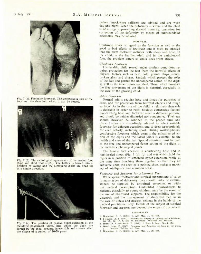
Fig. 7 (a). Feminine footwear. The comparative size of thefoot and the shoe into which it c?n be forced.
Fig. 7 (b). The radiological appearanc~ of the unshod foot(lefT) and shod foot (righT). The hallux is forced into aposition of valgus and the r~maining d:gits are lined upin a single direction.
Fig. 7 (c). The posillOn of passive hyper-extension at themetatarso-phalangeal joints, into which the dlg!tS areforced by the shoe, becomes irreversible and chromc afterthe elapse of a period of 10-20 years.
inches, knock-knee calliper- are advised and are wornday and night. When the deformity is evere and the childis of an age approaching skeletal maturity. operation forcorrection of the deformity by means of supracondylarosteotomy may be advi ed.
FOOTWE....R
Confusion exist in regard to the function as well a thegood or bad effect of footwear and it must be stressedthat the term footwear includes both shoes and ho e. Inthe child, in the healthy adult, and in the pathologicalfoot, the problem differ as chalk does from chee e.
Children's F OOTh'earThe healthy child reared under modern conditions re
quires protection for the feet from the harmful effects ofphysical factors such as heat, cold, granite chips, stone,broken glass and thorns. Sandal which protect the oleof the feet and permit the unhampered action of the digitsas well as the tarsal joints are ideal. Those which constrictthe free movement of the digits is harmful, especially inthe case of the growing child.
AdulT FooTh'earNormal adults require hose and shoe for purposes of
dress, and for protection from harmful objects and roughsurfaces. As in the case of the child, a relatively firm solei desirable in order to resi t noxious extraneous factor.Eye-catching hose and footwear serve a different purpose,and should be neither discarded nor condemned. Their useshould. however, be confined to the proper time andplace. Ladies are accordingly advised to select suitablefootwear for different occasions, and to dress appropriatelyfor each activity, including port. During working-hour,comfortable footwear which permits the unhampered action of the digits and the tarsal joints is essential to thehealth and care of the feet. Special attention must be paidto the free and unhampered flexor action of the digits atthe metatarsophalangeal joints.
The female foot encased in constricting hose and inhigh-heeled shoes (Fig. 7 (a), (b) and (c)) which hold thedigits in a position of enforced hyper-extension, while atthe same time bunching them together 0 that they allconverge upon the apex of a pointed shoe, makes a mockery of intelligence and common sense.
FooTh'ear and SupporTs for A bnorma/ FeeTWhile special footwear and urgical supports are of value
in many types of deformity. they hould under no circumstances be supplied by untrained personnel or without medical prescription. Undoubted disadvantages topatients, especially to young children, may be the result ofthe use of ill-advi ed supports. The responsibility for thediagnosis and the management of abnormal feet, as inthe case of illness and disease, belongs in the hands of themedical practitioner only. Details of the subject of surgicalfootwear and supports are beyond the cope of this article.
REFERENCES
I. Dommisse. G. F. (1971): S. Afr. Med. J .. 45. 663.2. Ferguson. A. B. (1968): Orthopaedic Surgery in Infancy and Childhood,
3rd ed., pp. 17, 52 and 54. Baltimore: Williams and Wilkins.3. Harris, R. l. and Beath, T. (194 ): J. Bone JI Surg., 30 - 8, 624.4. \Vood Janes. F. (1943): Structure and Function as Seen in the Foot.
p. I. London: Balliere and Cox.5. Dommisse, G. F. (1964): S. Afr. Med. J .. 38, 5.
Related Documents