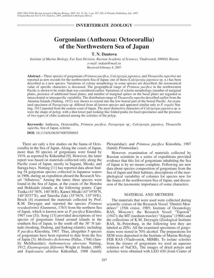
ISSN 1063-0740, Russian Journal of Marine Biology, 2007, Vol. 33, No. 5, pp. 297–304. © Pleiades Publishing, Ltd., 2007. Original Russian Text © T.N. Dautova, 2007, published in Biologiya Morya. 297 There are only a few studies on the fauna of Octo- corallia in the Sea of Japan. Along the coasts of Japan, more than 50 species of gorgonians were found by 1919, as reported by Kükenthal [9]. However, the latter report was based on materials collected only along the Pacific coast of Japan, mostly in Sagami, Misaki, and Suruga bays. Nutting [11] has reported data about find- ing 54 gorgonian species collected in Japanese waters in 1906, during an expedition aboard the Research Ves- sel “Albatross.” Among the latter, three species were found in the Sea of Japan, at the coasts of the Honshu and Hokkaido islands, at the following points: Cape Tsuika (41°36'N, 140°36'E), Kamoi Misaki (43°19'58''N, 140°20'57''E), and Tateisha Zaki (35°56'N, 135°39'E). Broch [4] examined the materials collected by Prof. K.M. Deryugin and reported the species Primnoa resedaeformis (Gunnerus, 1763) from the northern Sea of Japan, which is known now as P. pacifica Kinoshita, 1907 (see [5]). Song [13] provided descriptions of two species of gorgonians found around islands in the southern Sea of Japan, to the south off the 38° N lati- tude (Jeodong, Dodong, and Sadong islands), including P. pacifica Kinoshita, 1907. Thus, altogether 5 species of gorgonians have been reported to date from the Sea of Japan [5]: Melitodes dichotoma (Pallas, 1766) (fam- ily Melithaeidae); Anthomuricea aberrans Nutting, 1912; Elasmogorgia filiformis Wright et Studer, 1889; and Euplexaura abietina Kükenthal, 1908 (family Plexauridae); and Primnoa pacifica Kinoshita, 1907 (family Primnoidae). However, examination of materials collected by Russian scientists in a series of expeditions provided evidence that this list of gorgonians inhabiting the Sea of Japan is by no means complete. Following are new data about species composition of gorgonians from the Sea of Japan and their habitats, descriptions of the mor- phological variability of colonies for species new for the fauna of the northwestern Sea of Japan, and discus- sion of the taxonomic importance of some characters. MATERIAL AND METHODS The materials that were used were collected during scientific cruises of the Research Vessel “Dmitrii Men- deleev” (35th cruise, 1985; Institute of Oceanology RAS, Moscow); the Research Vessel “Toporok” (1947); the MT (medium trawler) “Algama” (1966) and the collections of K.M. Deryugin (Zoological Institute RAS, St.-Petersburg, in the following text they are labeled as ZIN). All the examined specimens of gorgo- nians were stored in 70% alcohol. The preparations for SEM were deposited in the Institute of Marine Biology FEB RAS (Vladivostok, MIMB). To isolate sclerites from the tissues of gorgonians we used an aqueous solution of NaClO 2 . The images of dried polyps and sclerites were obtained with LEO 430 (Joint Center of INVERTEBRATE ZOOLOGY Gorgonians (Anthozoa: Octocorallia) of the Northwestern Sea of Japan T. N. Dautova Institute of Marine Biology, Far East Division, Russian Academy of Sciences, Vladivostok, 690041 Russia e-mail: [email protected] Received February 8, 2007 Abstract—Three species of gorgonians (Primnoa pacifica, Calcigorgia japonica, and Thouarella superba) are reported as new records for the northwestern Sea of Japan; one of them (Calcigorgia japonica sp. n.) has been described as a new species. Variations of colony morphology in some species are described; the taxonomical value of specific characters is discussed. The geographical range of Primnoa pacifica in the northwestern Pacific is shown to be wider than was considered earlier. Variations of sclerite morphology (number of marginal plates, presence of additional basal plates, and number of marginal spines on the basal plate) are regarded as intracolonial or intraspecific variability. The distribution range of Thouarella superba described earlier from the Aleutian Islands (Nutting, 1912) was shown to extend into the low-boreal part of the boreal Pacific. An exam- ined specimen of Paragorgia sp. differed from all known species and appeared similar only to P. regalis Nut- ting, 1912 reported from the eastern coast of Japan. The most distinctive characters of Calcigorgia japonica sp. n. were the shape of polyp, with a thin lower part looking like folded podia (in fixed specimens) and the presence of two types of clubs scattered among the sclerites of the polyp. Keywords: Anthozoa, Octocorallia, Primnoa pacifica, Paragorgia sp., Calcigorgia japonica, Thouarella superba, Sea of Japan, sclerite. DOI: 10.1134/S1063074007050045

Welcome message from author
This document is posted to help you gain knowledge. Please leave a comment to let me know what you think about it! Share it to your friends and learn new things together.
Transcript
ISSN 1063-0740, Russian Journal of Marine Biology, 2007, Vol. 33, No. 5, pp. 297–304. © Pleiades Publishing, Ltd., 2007.Original Russian Text © T.N. Dautova, 2007, published in Biologiya Morya.
297
There are only a few studies on the fauna of Octo-corallia in the Sea of Japan. Along the coasts of Japan,more than 50 species of gorgonians were found by1919, as reported by Kükenthal [9]. However, the latterreport was based on materials collected only along thePacific coast of Japan, mostly in Sagami, Misaki, andSuruga bays. Nutting [11] has reported data about find-ing 54 gorgonian species collected in Japanese watersin 1906, during an expedition aboard the Research Ves-sel “Albatross.” Among the latter, three species werefound in the Sea of Japan, at the coasts of the Honshuand Hokkaido islands, at the following points: CapeTsuika (41
°
36'N, 140
°
36'E), Kamoi Misaki (43
°
19'58''N,140
°
20'57''E), and Tateisha Zaki (35
°
56'N, 135
°
39'E).Broch [4] examined the materials collected by Prof.K.M. Deryugin and reported the species
Primnoaresedaeformis
(Gunnerus, 1763) from the northern Seaof Japan, which is known now as
P. pacifica
Kinoshita,1907 (see [5]). Song [13] provided descriptions of twospecies of gorgonians found around islands in thesouthern Sea of Japan, to the south off the 38
°
N lati-tude (Jeodong, Dodong, and Sadong islands), including
P. pacifica
Kinoshita, 1907. Thus, altogether 5 speciesof gorgonians have been reported to date from the Seaof Japan [5]:
Melitodes dichotoma
(Pallas, 1766) (fam-ily Melithaeidae);
Anthomuricea aberrans
Nutting,1912;
Elasmogorgia filiformis
Wright et Studer, 1889;and
Euplexaura abietina
Kükenthal, 1908 (family
Plexauridae); and
Primnoa pacifica
Kinoshita, 1907(family Primnoidae).
However, examination of materials collected byRussian scientists in a series of expeditions providedevidence that this list of gorgonians inhabiting the Seaof Japan is by no means complete. Following are newdata about species composition of gorgonians from theSea of Japan and their habitats, descriptions of the mor-phological variability of colonies for species new forthe fauna of the northwestern Sea of Japan, and discus-sion of the taxonomic importance of some characters.
MATERIAL AND METHODS
The materials that were used were collected duringscientific cruises of the Research Vessel “Dmitrii Men-deleev” (35th cruise, 1985; Institute of OceanologyRAS, Moscow); the Research Vessel “Toporok”(1947); the MT (medium trawler) “Algama” (1966) andthe collections of K.M. Deryugin (Zoological InstituteRAS, St.-Petersburg, in the following text they arelabeled as ZIN). All the examined specimens of gorgo-nians were stored in 70% alcohol. The preparations forSEM were deposited in the Institute of Marine BiologyFEB RAS (Vladivostok, MIMB). To isolate scleritesfrom the tissues of gorgonians we used an aqueoussolution of NaClO
2
. The images of dried polyps andsclerites were obtained with LEO 430 (Joint Center of
INVERTEBRATE ZOOLOGY
Gorgonians (Anthozoa: Octocorallia)of the Northwestern Sea of Japan
T. N. Dautova
Institute of Marine Biology, Far East Division, Russian Academy of Sciences, Vladivostok, 690041 Russiae-mail: [email protected]
Received February 8, 2007
Abstract
—Three species of gorgonians (
Primnoa pacifica, Calcigorgia japonica
, and
Thouarella superba
) arereported as new records for the northwestern Sea of Japan; one of them (
Calcigorgia japonica
sp. n.) has beendescribed as a new species. Variations of colony morphology in some species are described; the taxonomicalvalue of specific characters is discussed. The geographical range of
Primnoa pacifica
in the northwesternPacific is shown to be wider than was considered earlier. Variations of sclerite morphology (number of marginalplates, presence of additional basal plates, and number of marginal spines on the basal plate) are regarded asintracolonial or intraspecific variability. The distribution range of
Thouarella superba
described earlier from theAleutian Islands (Nutting, 1912) was shown to extend into the low-boreal part of the boreal Pacific. An exam-ined specimen of
Paragorgia
sp. differed from all known species and appeared similar only to
P. regalis
Nut-ting, 1912 reported from the eastern coast of Japan. The most distinctive characters of
Calcigorgia japonica
sp. n.were the shape of polyp, with a thin lower part looking like folded podia (in fixed specimens) and the presenceof two types of clubs scattered among the sclerites of the polyp.
Keywords:
Anthozoa, Octocorallia,
Primnoa pacifica, Paragorgia
sp.,
Calcigorgia japonica, Thouarellasuperba
, Sea of Japan, sclerite.
DOI:
10.1134/S1063074007050045
298
RUSSIAN JOURNAL OF MARINE BIOLOGY
Vol. 33
No. 5
2007
DAUTOVA
IMB FEB RAS) and EVO 50 Zeiss (Analytic Center ofFar-Eastern Geological Institute FEB RAS) micro-scopes using the optimum magnification for each typeof the sclerites.
The terms to designate different types of the scler-ites, i.e., capstan and spindle, were used following themodern terminological system [3]. In the text, the max-imum length and width of sclerites (or only the length,if not specified) are provided in parentheses. The mea-surements were made using the scanning electronmicroscopes or with a light microscope, under magnifi-cations of 200 and 400.
RESULTS AND DISCUSSION
Family
Primnoidae
Gray, 1857
Genus
Primnoa
Lamouroux, 1812
Primnoa pacifica
Kinoshita, 1907
Primnoa pacifica
Kinoshita, 1907: 232; Cairns,Bayer, 2005: 233–239, figs. 4–6 (including synonymy).
Materials. RV “Dmitrii Mendeleev,” Stn. 3126,MIMB 16531, MIMB 16532 (1 specimen; between40
°
16'N, 133
°
25'E and 42
°
23'N, 131
°
53'E; 1350–900 mdeep; a dredge; July 16, 1985); RV “Dmitrii Men-deleev,” Stn. 3107 (3 specimens; 40
°
13'N, 133
°
58'E;1000–800 m deep; a dredge; July 12, 1985); ZIN 2, ZIN 3(2 specimens; 39
°
35'N. 135
°
01'E; 832–736 m deep;silted sand; August 8, 1933; coll. A.M. Deryugin); ZIN 4(1 specimen; Peter the Great Bay, Sea of Japan, 1600–900 m deep; June 12, 1932); ZIN 5 (1 specimen, atFurugel’ma Island, Peter the Great Bay; 42
°
10'N,131
°
03'E; 780–730 m deep).Diagnosis. See Cairns and Bayer [5].Description. Zooids are usually bent toward the
lower parts of branches; only some of them are directedacross the axis of the branch or toward the base of thelatter. Tips of branches are conical in shape; distal pol-yps are located by 4–6 mm beneath branch apex andbent toward the base of colony.
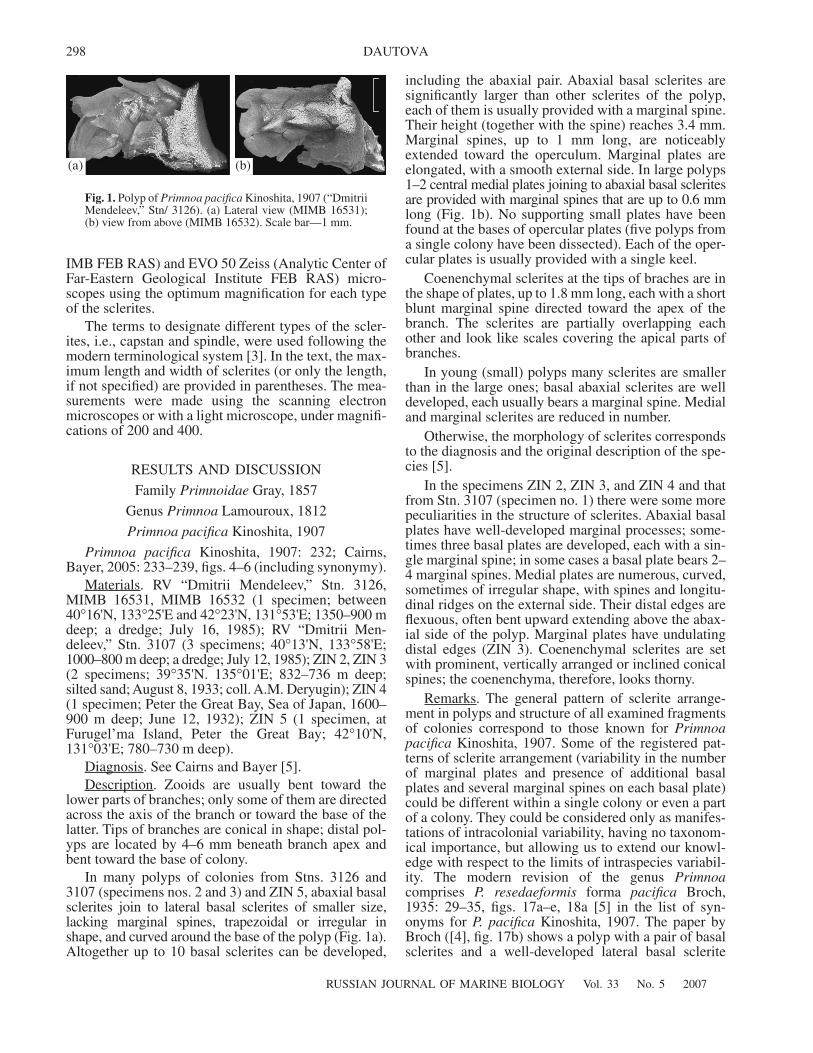
In many polyps of colonies from Stns. 3126 and3107 (specimens nos. 2 and 3) and ZIN 5, abaxial basalsclerites join to lateral basal sclerites of smaller size,lacking marginal spines, trapezoidal or irregular inshape, and curved around the base of the polyp (Fig. 1a).Altogether up to 10 basal sclerites can be developed,
including the abaxial pair. Abaxial basal sclerites aresignificantly larger than other sclerites of the polyp,each of them is usually provided with a marginal spine.Their height (together with the spine) reaches 3.4 mm.Marginal spines, up to 1 mm long, are noticeablyextended toward the operculum. Marginal plates areelongated, with a smooth external side. In large polyps1–2 central medial plates joining to abaxial basal scleritesare provided with marginal spines that are up to 0.6 mmlong (Fig. 1b). No supporting small plates have beenfound at the bases of opercular plates (five polyps froma single colony have been dissected). Each of the oper-cular plates is usually provided with a single keel.
Coenenchymal sclerites at the tips of braches are inthe shape of plates, up to 1.8 mm long, each with a shortblunt marginal spine directed toward the apex of thebranch. The sclerites are partially overlapping eachother and look like scales covering the apical parts ofbranches.
In young (small) polyps many sclerites are smallerthan in the large ones; basal abaxial sclerites are welldeveloped, each usually bears a marginal spine. Medialand marginal sclerites are reduced in number.
Otherwise, the morphology of sclerites correspondsto the diagnosis and the original description of the spe-cies [5].
In the specimens ZIN 2, ZIN 3, and ZIN 4 and thatfrom Stn. 3107 (specimen no. 1) there were some morepeculiarities in the structure of sclerites. Abaxial basalplates have well-developed marginal processes; some-times three basal plates are developed, each with a sin-gle marginal spine; in some cases a basal plate bears 2–4 marginal spines. Medial plates are numerous, curved,sometimes of irregular shape, with spines and longitu-dinal ridges on the external side. Their distal edges areflexuous, often bent upward extending above the abax-ial side of the polyp. Marginal plates have undulatingdistal edges (ZIN 3). Coenenchymal sclerites are setwith prominent, vertically arranged or inclined conicalspines; the coenenchyma, therefore, looks thorny.
Remarks. The general pattern of sclerite arrange-ment in polyps and structure of all examined fragmentsof colonies correspond to those known for
Primnoapacifica
Kinoshita, 1907. Some of the registered pat-terns of sclerite arrangement (variability in the numberof marginal plates and presence of additional basalplates and several marginal spines on each basal plate)could be different within a single colony or even a partof a colony. They could be considered only as manifes-tations of intracolonial variability, having no taxonom-ical importance, but allowing us to extend our knowl-edge with respect to the limits of intraspecies variabil-ity. The modern revision of the genus
Primnoa
comprises
P. resedaeformis
forma
pacifica
Broch,1935: 29–35, figs. 17a–e, 18a [5] in the list of syn-onyms for
P. pacifica
Kinoshita, 1907. The paper byBroch ([4], fig. 17b) shows a polyp with a pair of basalsclerites and a well-developed lateral basal sclerite
(‡) (b)
Fig. 1.
Polyp of
Primnoa pacifica
Kinoshita, 1907 (“DmitriiMendeleev,” Stn/ 3126). (a) Lateral view (MIMB 16531);(b) view from above (MIMB 16532). Scale bar—1 mm.
RUSSIAN JOURNAL OF MARINE BIOLOGY
Vol. 33
No. 5
2007
GORGONIANS (ANTHOZOA: OCTOCORALLIA) 299
(ZIN 3). In their diagnosis the authors of the revisiondescribed
P. pacifica
Kinoshita, 1907 as follows: “mostpolyps with a pair of massive basal scales, each basalbearing a prominent marginal spine” (see [5]). There-fore, the presence of additional lateral basal sclerites ofsmall size does not contradict the diagnosis of the spe-cies and can be considered as a manifestation ofintraspecies variability. Complicated structure of scler-ites in the polyps and the presence of spines on coenen-chymal sclerites in some specimens (Stn. 3107, speci-men no. 1; ZIN 2, ZIN 3, ZIN 4) differ these animalsfrom all the samples discussed here. Similar modifica-tions of surface sculpture in polyp sclerites, registeredin specimens collected in the Alexander Archipelago, tothe south off Alaska, were referred to as intraspeciesvariability [5]. In the specimens from Russian waters ofthe Sea of Japan, the structure of coenenchymal scler-ites is also complicated. However, without examinationof additional materials, these arguments could hardlybe used as evidence to erect a new species. The findingof
P. pacifica
in Russian waters of the Sea of Japanextends the species range of this species.
Genus Thouarella Gray, 1870
Thouarella superba
(Nutting, 1912)
Primnodendron superbum
Nutting
,
1912: 71–72, pl. 9, Fig. 2, 2a; pl. 19, Fig. 4.
Thouarella
(
Amphilaphis
)
superba
Kükenthal,1919: 412; Kükenthal, 1924: 291.
Materials. ZIN 1 (46
°
50'N, 143
°
29'E; Penina Cape;Sakhalin Island; 29 m deep; RV “Toporok”; Stn. 49;1947), MIMB 16534.
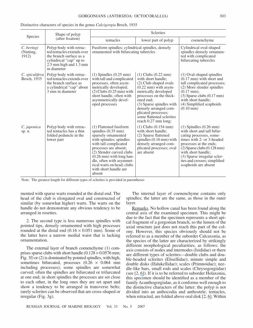
Diagnosis. See Nutting [11].Description. The branching is plumate; polyps are
bent toward the axis of the branch; they are arrangedirregularly on all sides of the latter. The polyp is 1–1.6 mm long and up to 1 mm in diameter. There areeight triangular opercular sclerites with outer spines(Fig. 2:
1
,
4
,
5
), each bearing a well-developed keelalong the internal surface (Fig. 2:
2
,
3
). Five–six abaxialopercular sclerites (0.384
×
0.288 mm; Fig. 2:
1
,
2
) aregreater than the adaxial ones (0.256
×
0.16 mm; Fig. 2:
4
).In well-developed polyps there are 8 marginal sclerites.Among the latter, 6 sclerites are scales (0.32
×
0.256 mm; Fig. 2:
5
); the others are scaphoids curvedaround operculum (0.162
×
0.28 mm; Fig. 2:
6
). Medialsclerites are scales; in shape and size they are similar toabaxial marginal sclerites (Fig. 2:
7
–
9
) and are arrangedin vertical rows, each comprising 6–8 plates. Moreover,there are small medial sclerites of irregular shape. Theouter surface of all sclerites in the polyp is set withspines tilted toward its apex. On the internal side ofpolyp sclerites there are rows of tubercles that radiateoutward from one point located in the lower one thirdof the sclerite (Fig. 2:
2
,
3
,
9
). Coenenchymal scleritesare plates of irregular shape, 0.32 mm long (Fig. 2:
10
–
14
), ornamented by spines along the external side. The
internal surface of coenenchymal sclerites is often con-vex and ornamented with tubercles (Fig. 2:
11
–
12
).Remarks. Earlier this species was registered at
Semisopochnyi Island (the Aleutian Islands), at a depthof 60–79 m [11]. The new findings extend the geo-graphical range of the species and allow us to refer it tothe Low-Boreal Subprovince of the Boreal PacificProvince.
Family
Paragorgiidae
Kükenthal, 1916
Genus
Paragorgia
Milne Edwards et Haime, 1857
Paragorgia
sp.
Materials. MT “Algama,” 1 specimen, southwesternSakhalin, 170 m deep, March 4, 1966, ZIN.
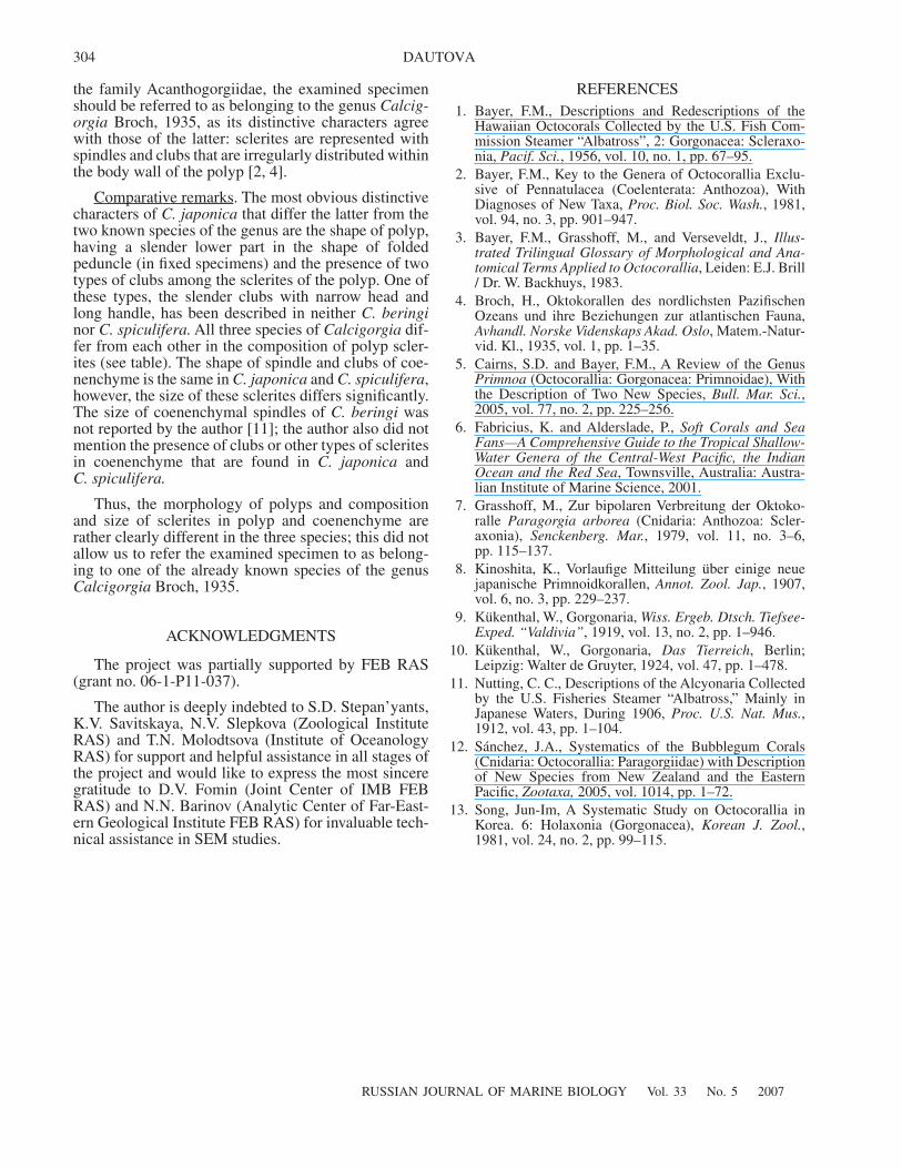
Description. This is a fragment of a colony, 15 cmlong, oval in transverse section, the greatest width is3 cm. The outer layer of coenenchyme is up to 0.5 mmthick. It contains mostly 8-radiate capstans of three dif-ferent shape patterns and different size, as follows:compact capstans 0.0384
×
0.032 to 0.0448
×
0.032 mmin size ornamented with lobulate processes (Fig. 1a:
1
);slender capstans 0.048
×
0.0352 to 0.0576
×
0.0384 mmin size, with processes, where lobules take conicalshape (Fig. 3a:
2
); and larger slender capstans 0.064
×
0.0416 to 0.0768
×
0.064 mm in size ornamented withprocesses bearing conical elevations (Fig. 3a:
2
).Seven-radiate capstans (0.0384
× 0.032 mm in size)occur rarely (approximately one for 15 examined cap-stans) and are characterized by a certain asymmetry inthe size and shape of processes (Fig. 3a: 2). No 6-radi-ate forms were found among 50 examined capstanschosen randomly. Also, no capstans were found bearingprocesses decorated with pattern consisting of numer-ous grooves. Rarely scattered grooves were observedonly on one of the smallest 8-radiate capstans.
Moreover, in the coenenchyme there are capstanswith weakly developed processes (Fig. 3a: 4) and shortspindles each bearing an additional belt of tubercles(0.14 × 0.051 mm; Fig. 3b: 5, 6); the latter could beconsidered as a transitional form between the capstansof external coenenchyme and the spindles of medulla.
Medulla contains spindles (0.43 mm; Fig. 3b) withrarely scattered sharp slender spines and also conicalspines arising from wide base or bearing bifurcatedprocesses. Some spindles are provided with 2–3 pro-cesses arising at the tips (Fig. 3b: 3, 5, 6).
Remarks. The family Paragorgiidae comprises twogenera, a cosmopolitan genus Paragorgia MilneEdwards et Haime, 1857 and a genus SibogagorgiaStiasny, 1937, whose members have been found only intropical zone. The diagnostic characters for species ofthe genus Paragorgia are, first of all, the shape of col-ony and type, size and ornamentation pattern of cap-stans of external layer. To date, 14 species of the genusParagorgia have been described, including 6 Pacificspecies that are considered as endemic for New
300
RUSSIAN JOURNAL OF MARINE BIOLOGY Vol. 33 No. 5 2007
DAUTOVA
1 2
3
4
5
6
7
8
9
10 11 12
13
14
Fig. 2. Sclerites of Thouarella superba (Nutting, 1912). (1) abaxial opercular, external side; (2, 3) abaxial opercular, internal side;(4) adaxial opercular, external side; (5) abaxial marginal, external side; (6) adaxial marginal, external side; (7, 8) medial, externalside; (9) medial, internal side; (10) coenenchymal, external side; (11) coenenchymal, lateral view; (12) coenenchymal, internal side;(13) coenenchymal, lateral view; (14) coenenchymal, internal side. Scale bar—0.2 mm.
RUSSIAN JOURNAL OF MARINE BIOLOGY Vol. 33 No. 5 2007
GORGONIANS (ANTHOZOA: OCTOCORALLIA) 301
1
(a) (b)
(c)
(d) (e)
(f)
(g)
2 3 4 56
1
2 3 45
6
7
1
23
4
5
Fig. 3. Paragorgia sp. (a—sclerites of the surface layer of coenenchyme; b—sclerites of the underlying layer of coenenchyme) andCalcigorgia japonica sp. n., ZIN 1/10706 (c, d—sclerites of tentacles and upper part of polyp body wall; e—sclerites of the lower partof polyp and its peduncle; f, g—sclerites of coenenchyme). Scale bar: (a) 0.05 mm; (b), (c), (d), (f) 0.15 mm; (e), (g) 0.1 mm.
302
RUSSIAN JOURNAL OF MARINE BIOLOGY Vol. 33 No. 5 2007
DAUTOVA
Zealand and 2 species found only at the western coastof North America [12]. Together with the two latter spe-cies, the total number of species of the genus Paragor-gia registered in the Northern Pacific reaches four.The above described specimen cannot be identified asbelonging to any of these four species: P. regalis Nut-ting, 1912 has absolutely different capstans in externallayer [12], while P. arborea (Linnaeus, 1758) is charac-terized by presence in external layer of capstans(mostly 6-radiate) with numerous grooves on their pro-cesses [7, 12]. Two North American species, P. yutlinuxSánchez, 2005 and P. stephencairnsi Sánchez, 2005differ in the composition of capstans in external layer[12]. The type materials of P. tenuis Kinoshita, 1913and P. granulosa Kinoshita, 1913 found at the easterncoast of Honshu (Japan) is now considered to be lost[1]. The incompleteness of the available material did notallow us to describe a new species of the genus Para-gorgia, as there is no information about the shape of thecolony and sclerite composition of the polyp.
Family Acanthogorgiidae Gray, 1859
Calcigorgia Broch, 1935
Calcigorgia japonica sp. n.
Materials. ZIN 1/10706, MIMB 16533 (1 specimen,39°35'N, 135°01'E; Sea of Japan; 832–736 m deep;silted sand; August 8, 1933; coll. K.M. Deryugin).
Diagnosis. Polyps are up to 5 mm high and 2.2 mmwide, arranged irregularly. The body of the polyp has8 folds, its lower part either takes the shape of a narrowtwisted folded peduncle or makes up a cup. In the ten-tacle, sclerites make up a longitudinal bundle extendingwithin polyp body wall down to the lower one third ofits wide part. Horizontal bundles of spicules are locatedwithin polyp, in the gaps between the folds. In the lowerone third and peduncle of the polyp the sclerites arearranged irregularly.
Characteristic sclerite patterns are as follows: (1)curved clubs with a long pointed handle ornamentedwith rarely scattered warts (the clubs are up to 0.26 mmlong; located in the upper part of polyp); (2) clubs witha short handle densely ornamented with high processes(the clubs are up to 0.15 mm long; located in the lowerpart of polyp and surface layer of coenenchyme);(3) flattened spindles, straight or curved, ornamented
with rarely scattered warts or spinules (the spindles areup to 0.35 mm long; located in the upper part of polyp);(4) straight or weakly curved spindles with high, oftenbifurcated, processes, which are often arranged in belts(the spindles are up to 0.26 mm long; located in coenen-chyme); and (5) sparse sclerites of irregular shape orcross-shaped.
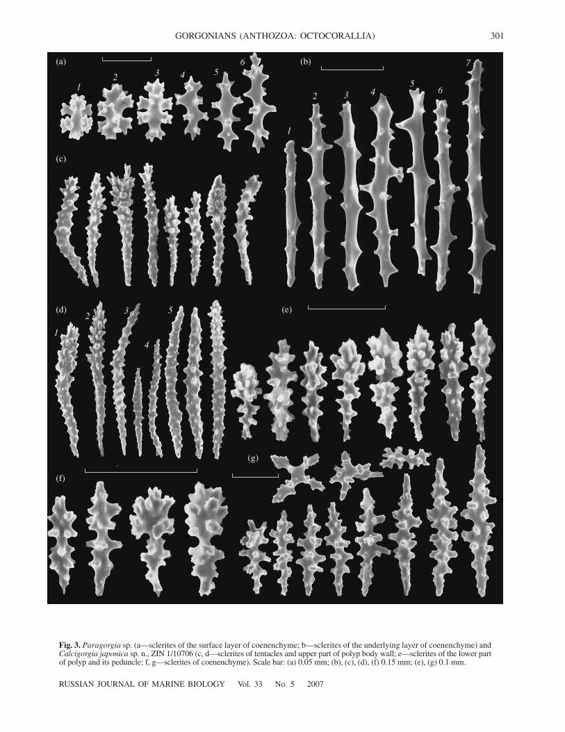
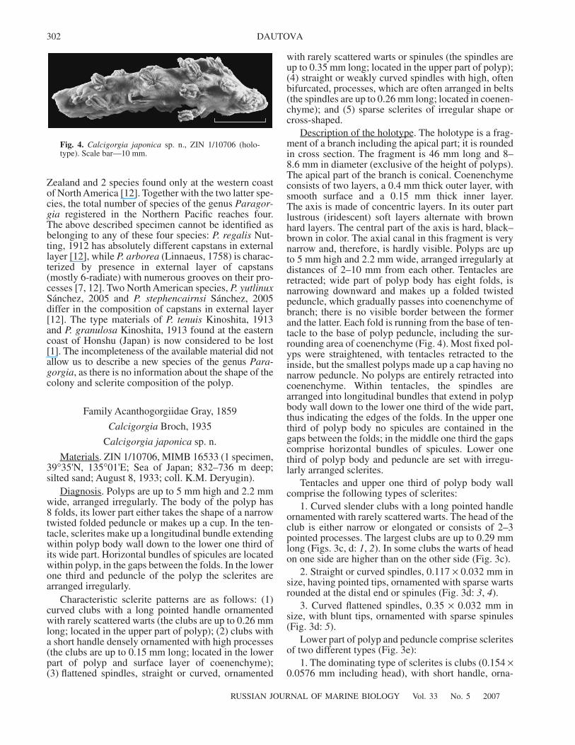
Description of the holotype. The holotype is a frag-ment of a branch including the apical part; it is roundedin cross section. The fragment is 46 mm long and 8–8.6 mm in diameter (exclusive of the height of polyps).The apical part of the branch is conical. Coenenchymeconsists of two layers, a 0.4 mm thick outer layer, withsmooth surface and a 0.15 mm thick inner layer.The axis is made of concentric layers. In its outer partlustrous (iridescent) soft layers alternate with brownhard layers. The central part of the axis is hard, black–brown in color. The axial canal in this fragment is verynarrow and, therefore, is hardly visible. Polyps are upto 5 mm high and 2.2 mm wide, arranged irregularly atdistances of 2–10 mm from each other. Tentacles areretracted; wide part of polyp body has eight folds, isnarrowing downward and makes up a folded twistedpeduncle, which gradually passes into coenenchyme ofbranch; there is no visible border between the formerand the latter. Each fold is running from the base of ten-tacle to the base of polyp peduncle, including the sur-rounding area of coenenchyme (Fig. 4). Most fixed pol-yps were straightened, with tentacles retracted to theinside, but the smallest polyps made up a cap having nonarrow peduncle. No polyps are entirely retracted intocoenenchyme. Within tentacles, the spindles arearranged into longitudinal bundles that extend in polypbody wall down to the lower one third of the wide part,thus indicating the edges of the folds. In the upper onethird of polyp body no spicules are contained in thegaps between the folds; in the middle one third the gapscomprise horizontal bundles of spicules. Lower onethird of polyp body and peduncle are set with irregu-larly arranged sclerites.
Tentacles and upper one third of polyp body wallcomprise the following types of sclerites:
1. Curved slender clubs with a long pointed handleornamented with rarely scattered warts. The head of theclub is either narrow or elongated or consists of 2–3pointed processes. The largest clubs are up to 0.29 mmlong (Figs. 3c, d: 1, 2). In some clubs the warts of headon one side are higher than on the other side (Fig. 3c).
2. Straight or curved spindles, 0.117 × 0.032 mm insize, having pointed tips, ornamented with sparse wartsrounded at the distal end or spinules (Fig. 3d: 3, 4).
3. Curved flattened spindles, 0.35 × 0.032 mm insize, with blunt tips, ornamented with sparse spinules(Fig. 3d: 5).
Lower part of polyp and peduncle comprise scleritesof two different types (Fig. 3e):
1. The dominating type of sclerites is clubs (0.154 ×0.0576 mm including head), with short handle, orna-
Fig. 4. Calcigorgia japonica sp. n., ZIN 1/10706 (holo-type). Scale bar—10 mm.
RUSSIAN JOURNAL OF MARINE BIOLOGY Vol. 33 No. 5 2007
GORGONIANS (ANTHOZOA: OCTOCORALLIA) 303
mented with sparse warts rounded at the distal end. Thehead of the club is elongated oval and constructed ofsimilar (by somewhat higher) warts. The warts on thehandle do not demonstrate any obvious tendency to bearranged in rosettes.
2. The second type is less numerous spindles withpointed tips, densely ornamented with high processesrounded at the distal end (0.16 × 0.051 mm). Some ofthe latter have a narrow medial waist that is lackingornamentation.
The external layer of branch coenenchyme (1) com-prises sparse clubs with short handle (0.128 × 0.0576 mm;Fig. 3f) or (2) is dominated by pointed spindles, with high,sometimes bifurcated, processes (0.26 × 0.064 mmincluding processes); some spindles are somewhatcurved; often the spindles are bifurcated or trifurcatedat one end; in short spindles the processes are set closeto each other, in the long ones they are set apart andshow a tendency to be arranged in transverse belts;rarely sclerites can be observed that are cross-shaped orirregular (Fig. 3g).
The internal layer of coenenchyme contains onlyspindles; the latter are the same, as those in the outerlayer.
Remarks. No hollow canal has been found along thecentral axis of the examined specimen. This might bedue to the fact that the specimen represents a short api-cal fragment of a gorgonian branch, so the lumen of theaxial structure just does not reach this part of the col-ony. However, this species obviously should not bereferred to as a member of the suborder Calcaxonia, asthe species of the latter are characterized by strikinglydifferent morphological peculiarities, as follows: theaxis consists of nodes and internodes (Isididae) or thereare different types of sclerites—double clubs and dou-ble-headed sclerites (Elisellidae); minute simple anddouble disks (Ifalukellidae); scales (Primnoidae); nee-dle-like bars, small rods and scales (Chrysogorgiidae)(see [2, 6]). If it is to be referred to suborder Holaxonia,this specimen should be identified as a member of thefamily Acanthogorgiidae, as it conforms well enough tothe distinctive characters of the latter: the polyp is notdivided into an anthocodia and anthostele; tentacles,when retracted, are folded above oral disk [2, 6]. Within
Distinctive characters of species in the genus Calcigorgia Broch, 1935
Species Shape of polyp(after fixation)
Sclerites
tentacles lower part of polyp coenenchyme
C. beringi(Nutting, 1912)
Polyp body with retrac-ted tentacles extends over the branch surface as a cylindrical “cap” up to 2.5 mm high and 1.3 mm in diameter
Fusiform spindles; cylindrical spindles, densely ornamented with bifurcating tubercles
Cylindrical oval-shaped spindles densely ornamen-ted with complicated bifurcating tubercles
C. spiculiferaBroch, 1935
Polyp body with retrac-ted tentacles extends over the branch surface as a cylindrical “cap” about 3 mm in diameter
(1) Spindles (0.25 mm) with tall and complicated processes, often asym-metrically developed;(2) Clubs (0.25 mm) with short handle, often with asymmetrically devel-oped processes
(1) Clubs (0.22 mm) with short handle;(2) Club-shaped ovals (0.22 mm) with asym-metrically developed processes on the thick-ened end;(3) Sparse spindles with densely arranged com-plicated processes; some flattened sclerites reach 0.27 mm long;
(1) Oval-shaped spindles (0.17 mm) with short and tall complicated processes;(2) More slender spindles (0.17 mm);(3) Sparse clubs (0.17 mm) with short handle;(4) Simplified scaphoids (0.10 mm)
C. japonicasp. n.
Polyp body with retrac-ted tentacles has a thin folded peduncle in the lower part
(1) Flattened fusiform spindles (0.35 mm) sparsely ornamented with spinules; spindles with tall complicated processes are absent;(2) Slender curved clubs (0.26 mm) with long han-dle, often with asymmet-rical warts on head; clubs with short handle are absent
(1) Clubs (0.154 mm) with short handle;(2) Sparse flattened spindles (0.16 mm) with densely arranged com-plicated processes; oval are absent
(1) Spindles (0.26 mm) with short and tall bifur-cating processes, some-times with 2- or 3-headed processes at the ends;(2) Sparse clubs (0.128 mm) with short handle;(3) Sparse irregular scler-ites and crosses; simplified scaphoids are absent
Note: The greatest length for different types of sclerites is provided in parentheses
304
RUSSIAN JOURNAL OF MARINE BIOLOGY Vol. 33 No. 5 2007
DAUTOVA
the family Acanthogorgiidae, the examined specimenshould be referred to as belonging to the genus Calcig-orgia Broch, 1935, as its distinctive characters agreewith those of the latter: sclerites are represented withspindles and clubs that are irregularly distributed withinthe body wall of the polyp [2, 4].
Comparative remarks. The most obvious distinctivecharacters of C. japonica that differ the latter from thetwo known species of the genus are the shape of polyp,having a slender lower part in the shape of foldedpeduncle (in fixed specimens) and the presence of twotypes of clubs among the sclerites of the polyp. One ofthese types, the slender clubs with narrow head andlong handle, has been described in neither C. beringinor C. spiculifera. All three species of Calcigorgia dif-fer from each other in the composition of polyp scler-ites (see table). The shape of spindle and clubs of coe-nenchyme is the same in C. japonica and C. spiculifera,however, the size of these sclerites differs significantly.The size of coenenchymal spindles of C. beringi wasnot reported by the author [11]; the author also did notmention the presence of clubs or other types of scleritesin coenenchyme that are found in C. japonica andC. spiculifera.
Thus, the morphology of polyps and compositionand size of sclerites in polyp and coenenchyme arerather clearly different in the three species; this did notallow us to refer the examined specimen to as belong-ing to one of the already known species of the genusCalcigorgia Broch, 1935.
ACKNOWLEDGMENTS
The project was partially supported by FEB RAS(grant no. 06-1-P11-037).
The author is deeply indebted to S.D. Stepan’yants,K.V. Savitskaya, N.V. Slepkova (Zoological InstituteRAS) and T.N. Molodtsova (Institute of OceanologyRAS) for support and helpful assistance in all stages ofthe project and would like to express the most sinceregratitude to D.V. Fomin (Joint Center of IMB FEBRAS) and N.N. Barinov (Analytic Center of Far-East-ern Geological Institute FEB RAS) for invaluable tech-nical assistance in SEM studies.
REFERENCES1. Bayer, F.M., Descriptions and Redescriptions of the
Hawaiian Octocorals Collected by the U.S. Fish Com-mission Steamer “Albatross”, 2: Gorgonacea: Scleraxo-nia, Pacif. Sci., 1956, vol. 10, no. 1, pp. 67–95.
2. Bayer, F.M., Key to the Genera of Octocorallia Exclu-sive of Pennatulacea (Coelenterata: Anthozoa), WithDiagnoses of New Taxa, Proc. Biol. Soc. Wash., 1981,vol. 94, no. 3, pp. 901–947.
3. Bayer, F.M., Grasshoff, M., and Verseveldt, J., Illus-trated Trilingual Glossary of Morphological and Ana-tomical Terms Applied to Octocorallia, Leiden: E.J. Brill/ Dr. W. Backhuys, 1983.
4. Broch, H., Oktokorallen des nordlichsten PazifischenOzeans und ihre Beziehungen zur atlantischen Fauna,Avhandl. Norske Videnskaps Akad. Oslo, Matem.-Natur-vid. Kl., 1935, vol. 1, pp. 1–35.
5. Cairns, S.D. and Bayer, F.M., A Review of the GenusPrimnoa (Octocorallia: Gorgonacea: Primnoidae), Withthe Description of Two New Species, Bull. Mar. Sci.,2005, vol. 77, no. 2, pp. 225–256.
6. Fabricius, K. and Alderslade, P., Soft Corals and SeaFans—A Comprehensive Guide to the Tropical Shallow-Water Genera of the Central-West Pacific, the IndianOcean and the Red Sea, Townsville, Australia: Austra-lian Institute of Marine Science, 2001.
7. Grasshoff, M., Zur bipolaren Verbreitung der Oktoko-ralle Paragorgia arborea (Cnidaria: Anthozoa: Scler-axonia), Senckenberg. Mar., 1979, vol. 11, no. 3–6,pp. 115–137.
8. Kinoshita, K., Vorlaufige Mitteilung über einige neuejapanische Primnoidkorallen, Annot. Zool. Jap., 1907,vol. 6, no. 3, pp. 229–237.
9. Kükenthal, W., Gorgonaria, Wiss. Ergeb. Dtsch. Tiefsee-Exped. “Valdivia”, 1919, vol. 13, no. 2, pp. 1–946.
10. Kükenthal, W., Gorgonaria, Das Tierreich, Berlin;Leipzig: Walter de Gruyter, 1924, vol. 47, pp. 1–478.
11. Nutting, C. C., Descriptions of the Alcyonaria Collectedby the U.S. Fisheries Steamer “Albatross,” Mainly inJapanese Waters, During 1906, Proc. U.S. Nat. Mus.,1912, vol. 43, pp. 1–104.
12. Sánchez, J.A., Systematics of the Bubblegum Corals(Cnidaria: Octocorallia: Paragorgiidae) with Descriptionof New Species from New Zealand and the EasternPacific, Zootaxa, 2005, vol. 1014, pp. 1–72.
13. Song, Jun-Im, A Systematic Study on Octocorallia inKorea. 6: Holaxonia (Gorgonacea), Korean J. Zool.,1981, vol. 24, no. 2, pp. 99–115.
Related Documents