Good, better, best. Never let it rest. Until your good is better and your better is best Tim Duncan

Welcome message from author
This document is posted to help you gain knowledge. Please leave a comment to let me know what you think about it! Share it to your friends and learn new things together.
Transcript

Good, better, best.
Never let it rest.
Until your good is better and your better is best
Tim Duncan
Intracellular localization and regulation of
Gelatinase-A in zebrafish skeletal muscle
by
Amina Mohammed Ahmed Fallata
Bachelor of Science, King Abdul-Aziz University, 2009
A Thesis Submitted in Partial Fulfillment of the Requirements for the Degree of
Master of Science
in the Graduate Academic Unit of Biology
Supervisor: Bryan D. Crawford, Ph. D., Department of Biology
Examining Board: Les Cwynar, Ph. D., Department of Biology, Chair Shawn R. MacLellan, Ph. D., Department of Biology David Lentz, Ph. D., Department of Geology
This thesis is accepted by the Dean of Graduate Studies
THE UNIVERSITY OF NEW BRUNSWICK
November, 2015
©Amina M. Fallata, 2016
ii
ABSTRACT
Matrix metalloproteinase (MMPs) are class-I secreted proteins known to function in
extracellular matrix remodeling. However, studies in the last decade and a half revealed
the unexpected presence of MMP-2 (a.k.a. gelatinase-A) intracellularly, within
cardiomyocytes and implicated them in the pathology of ischemia/reperfusion injury
(IRI). Furthermore, the activity of this protease in mammals is controlled by
phosphorylation implicating the existence of unknown kinases and phosphatases, and
possibly a signalling system that modulate MMP-2 activity inside cells. Two questions
that emerge from these discoveries are (1) is the intracellular localization of gelatinase-A
is something common in striated muscle, and (2) is its regulation by phosphorylation of
physiological significance? Answering these questions is the objective of this thesis.
Using immunofluorescence, confocal microscopy, and ultrathin sectioning, I have
confirmed the intracellular localization of Mmp2 in zebrafish skeletal muscle. However, I
observed zebrafish Mmp2 accumulating on M-bands within sarcomeres, rather than in the
Z-discs as has been reported for mammalian MMP-2 within cardiomyocytes. I also note
that the signal sequence that directs this protease into the secretory pathway is
consistently poorly recognized, indicating a selective pressure for maintaining a
significant intracellular portion of this enzyme. While I was unable to determine the
phosphorylation status of Mmp2 purified from zebrafish muscle, there are high
probability phosphorylation sites in the Mmp2 sequence that are well- conserved among
homologues of this protease for which sequence is available. Thus I show that the
intracellular localization of gelatinase-A proteases within the sarcomere of striated

iii
muscle is not unique to mammalian cardiomyocytes, and that its regulation by
phosphorylation is likely an evolutionarily conserved characteristic of physiological
significance. I speculate that this protease is a previously unrecognized component of the
mechanism that regulates protein turnover within the contractile apparatus of striated
muscle.

iv
DEDICATION
To my father and to my mom
Thanks for everything

v
ACKNOWLEDGEMENTS
First of all, I would like to give my sincere appreciation to my supervisor Dr. Bryan
Crawford for his advice to help me complete my thesis. Without him, I would not have
been able to finish my work. Also I would like to thank my committee members Dr.
Tillmann Benfey and Dr. Katherine Barclay for their help to complete my thesis. A super
thanks goes to my friend Sheila Thompson, for her support and suggestions in writing my
thesis. I would also like to thank Christopher Small, Kelsey Katherine Mann, Aaron
Frenette, and all my friends in Bryan’s lab for their help, as well as Robyn O’Keefe and
Robyn Shortt for their care of the fish.
I would like to thank King Abdullah Bin Abdul-Aziz of the Kingdom of Saudi
Arabia for giving me the chance to achieve my dreams and enrol in his Scholarship
program. Appreciation is also due to the Saudi Cultural Bureau in Canada for everything
they helped me with during my study in Canada. A special thanks is in order for my
supervisor in the Embassy Dr. Maha Abou-Elghit for her help and advice during my
study time.
Finally, I would like to give big thanks to my family: my mother (Fatima), brothers
(Nizar and Raed), sisters (Dr. Eman and Ebtehal), and grandpas (Dr. Abdul Razzaq
Fallatah and Abdul Rahim Fallatah), as well as to my friend Eman Mohsen Basaheeh and
to other friends and relatives back home, for their encouragement, love, support and
prayers.

vi
!
شك! & تق#"!
<تق!' بج;1: 2لشك$ & 2لإمتنا( %لى 2لمش$6 2ل!$2سي 2ل!كت&$ ب$12( ك$&ف&$! لما ق!م- لي م( !ع' &%$شا!
! الله ب. عب+*لع&'& (%حم" الله), 0ج;! م: 9ج7 ن-7 !,جة )لماجست-,. ك3ل$ )لشك, 0 )لتق!-, لبعثة )لمل$ عب!
. #لملحق9ة #لثقاف9ة #لسع5%9ة بكن%# لما ق%م45 لي م1 %ع/ .ثناء فت$' #ل%$#سة!0'/. 'لتعل+* 'لعالي 'لسع!#", !
لأمي *لس-': فا8مة # لإخ#*تي #ك2ل1 لجم-ع *لأ+! # *لأص'قاء #ك! )لس%" محم" ! لأبي .لشك* م'ص'& %$ضا
م2 ساع(ني ' ت/ك-ني ب(ع'& صالحة.
!
vii
TABLE OF CONTENTS
Abstract .............................................................................................................................. ii
Dedication ......................................................................................................................... iv
Acknowledgements ........................................................................................................... v
Table of contents ............................................................................................................. vii
List of figures ..................................................................................................................... x
List of abbreviations ........................................................................................................ xi
Chapter 1: Introduction ................................................................................................... 1
1.1 Matrix Metalloproteinases (MMPs) .......................................................................... 1
1.1.1 Matrix Metalloproteinase structure and biochemistry ....................................... 3
1.1.2 The function of Matrix Metalloproteinases ....................................................... 7
1.1.2.1 Extracellular matrix remodelling ................................................................ 7
1.1.2.2 Matrix Metalloproteinase in development and disease ............................... 8
1.2 Intracellular localization of MMPs ........................................................................... 8
1.3 Muscle structure and function ................................................................................. 10
1.4 MMPs in ischemia-reperfusion injury .................................................................... 14
1.5 Zebrafish as model system for the study of muscle ................................................ 15
1.6 Objectives ............................................................................................................... 16
1.7 Overall hypothesis .................................................................................................. 17
1.8 References ............................................................................................................... 19
viii
Chapter 2: Intracellular localization of gelatinase-A (Mmp2) in zebrafish skeletal
muscle ............................................................................................................................... 24
2.1 Introduction ............................................................................................................. 24
2.2 Materials and methods ............................................................................................ 29
2.2.1 Spawning zebrafish and collecting embryos .................................................... 29
2.2.2 Immunostaining and confocal microscopy ...................................................... 29
2.2.3 Cryo-sectioning ................................................................................................ 30
2.2.4 Prediction the signal sequence cleavage sites in Gelatinase-A, Gelatinase-B
and BiP (Binding immunoglobulin protein) using SignalP ...................................... 30
2.2.5 Statistical analysis ............................................................................................ 31
2.3 Results ..................................................................................................................... 32
2.4 Discussion ............................................................................................................... 38
2.5 References ............................................................................................................... 40
Chapter 3: Phosphorylation status of Mmp2 in zebrafish myocytes ......................... 42
3.1 Introduction ............................................................................................................. 42
3.2 Materials and methods ............................................................................................ 46
3.2.1 Tissue preparation ............................................................................................ 46
3.2.2 Isolation of Gelatinases using gelatin-affinity chromatography ...................... 46
3.2.3 SDS-polyacrylaminde gel electrophoresis and phospho-protein identification48
3.2.4 Immunoblots .................................................................................................... 48
3.2.5 Gelatin zymography ......................................................................................... 49
3.2.6 Silver staining .................................................................................................. 50
ix
3.2.7 Prediction of phosphorylation sites within vertebrate Gelatinase-A homologues
................................................................................................................................... 51
3.3 Results ..................................................................................................................... 52
3.4 Discussion ............................................................................................................... 62
3.5 References ............................................................................................................... 64
Chapter 4: General Discussion and Conclusions ......................................................... 66
4.1 Mmp2 is unequivocally localized intracellularly within skeletal muscle in zebrafish
....................................................................................................................................... 67
4.2 Evolutionary argument for an important physiological function of intracellular
gelatinase-A .................................................................................................................. 70
4.3 What role(s) does gelatinase-A play in the sarcomere? .......................................... 74
4.4 Conclusion .............................................................................................................. 78
4.5 References ............................................................................................................... 80
Appendix A ....................................................................................................................... 84
Appendix B ....................................................................................................................... 91
Curriculum Vitae

x
LIST OF FIGURES
Chapter 1 figures
Figure 1.1 .............................................................................................................. 4!
Figure 1.2 ............................................................................................................ 12!
Chapter 2 figures
Figure 2.1 ............................................................................................................ 27!
Figure 2.2 ............................................................................................................ 35!
Figure 2.3 ............................................................................................................ 35!
Figure 2.4 ............................................................................................................ 36!
Figure 2.5 ............................................................................................................ 37!
Chapter 3 figures
Figure 3.1 ............................................................................................................ 55!
Figure 3.2 ............................................................................................................ 56!
Figure 3.3 ............................................................................................................ 57!
Figure 3.4 ............................................................................................................ 58!
Figure 3.5 ............................................................................................................ 59!
Figure 3.6 ............................................................................................................ 60!
Figure 3.7 ............................................................................................................ 60!
Figure 3.8 ............................................................................................................ 61!
xi
LIST OF ABBREVIATIONS
α-actinin Alpha-actinin
ATP Adenosine 5’-triphosphate
ADP Adenosine 5’-diphosphate
BiP Binding immunoglobulin protein
BSA Bovine serum albumin
DMSO Dimethyl sulfoxide
DNA Deoxyribonucleic acid
dNTP Deoxyribonucleotide triphosphate dT Deoxy thymine nucleotides
ECL Enhanced Chemiluminescence
ECM Extracellular matrix
ERM Embryo rearing medium
ER Endoplasmic reticulum
g Gravity force
GPI Glycosylphosphatidylinositol
hpf Hours post-fertilization
HRP Horse-radish peroxidase
H2O2 Hydrogen peroxide
IRI Ischemia and reperfusion injury
MMP Matrix metalloproteinase
MT-MMP Membrane type matrix metalloproteinase
Mmp2 Matrix metalloproteinase 2 protein from zebrafish
mmp2 Matrix metalloproteinase 2 gene in zebrafish
MMP-2 Matrix metalloproteinase-2 protein from human (Homo sapiens)
xii
mmp-2 Matrix metalloproteinase-2 gene in human
MMP2 Matrix metalloproteinase 2 protein from rat, or mouse
MuRF Muscle-specific RING finger
NO· Nitric oxide
PBS Phosphate Buffered Saline
PBSTx Phosphate buffered saline solution with tritonX-100
PBSTw Phosphate buffered saline with Tween-20
ONOO- Peroxynitrite
PVDF Polyvinylidene difluoride
ROS Reactive oxygen species RECK Reversion-inducing-cysteine-rich protein with kazal motifs RT-PCR Reverse transcriptase polymerase chain reaction
RNA Ribonucleic acid
SSs Signal sequences
SRP Signal recognition particle
SRPR Signal recognition particle receptor
SDS-PAGE Sodium dodecyl sulfate- polyacrylamide gel electrophoresis
SERCA Sarco(endo)plasmic reticulum Ca+2- ATPase
·O2 - Superoxide
TAILS Terminal Amine Isotopic Labeling of Substrates
TEMED Tetramethylethylenediamine
TEM Transmission Electron Microscope
TIMP Tissue Inhibitor of metalloproteinase
!!

xiii
!*Note on nomenclature: In this thesis, I have used the standard nomenclature of matrix metalloproteinases. When referring to zebrafish proteins, normal font is used with the first letter capitalized; for their genes or transcripts lowercase italics are used with no hyphen between the protein/gene name and its number (e.g., the zebrafish mmp2 gene and the zebrafish Mmp2 protein).When referring to mouse/rat and human proteins, the names are written in capital letters, with a hyphen used only for human proteins (e.g., the mouse/rat MMP2 and the human MMP-2). In the transcripts for both mouse/rat and human, lowercase italics are used (e.g., the mouse/rat mmp2 and the human mmp-2).
1
Chapter 1: Introduction
1.1 Matrix Metalloproteinases (MMPs)
Matrix metalloproteinases (MMPs) are zinc-dependent endopeptidases whose
importance to living organisms is profound. MMPs play significant roles in diverse
physiological and pathological processes, many of which have been discovered in the
five decades since the discovery of the first MMP (Gross and Lapiere 1962; reviewed in
Lapière 2005; Page-McCaw et al. 2007; Kessenbrock et al. 2010). ‘Matrix
metalloproteinases’, ‘matrixins’ or ‘matrix degrading metalloenzymes’ are all terms used
to describe the same family of enzymes, although matrix metalloproteinases is the most
popular (reviewed in Amălinei et al. 2007). These proteases were first recognized in
experiments investigating the degradation of collagen fibres during tadpole
metamorphosis (Gross and Lapière, 1962). Since then, dozens of MMPs have been
characterized in animals ranging from Cnidarians to Vertebrates, and genes encoding
related metalloproteinases have been identified in plants, viruses and prokaryotes
(reviewed in Gomis-Ruth 2009). All MMPs are synthesized in a latent 'pro-enzyme' form
and need to be activated post-translationally in order to function. In addition to post-
translational activation, MMPs are subject to reversible and irreversible post-translational
inhibition, as the result of forming complexes with endogenous inhibitors such as the
tissue inhibitors of metalloproteinases (TIMPs) or reversion-inducing cysteine-rich
protein with Kazal motifs (RECK), and ultimately proteolytic degradation of the enzyme
(reviewed in Tallant et al. 2010). The complement of MMPs encoded by chordate
2
genomes is quite diverse. The ascidian Ciona intestinalis has only 7 MMPs (Huxley-
Jones et al. 2007), whereas vertebrates generally have two-dozen or more. Zebrafish
(Danio rerio), for example, have 25 MMPs (Wyatt et al. 2009), humans have 24 and
mice have 25 (reviewed in Jackson et al. 2010), Xenopus leavis has 29 and X. tropicalis
has 27 (Fu et al. 2009). The larger number of MMP genes in the genomes of vertebrates
may be the result of duplication and expansion of the ancestral chordate MMP genes
during vertebrate evolution, or the loss of ancestral deuterostome MMP genes during
ascidian evolution, as the sea urchin Strongylocentrotus purpuratus has 26 MMPs
(reviewed in Fanjul-Fernández et al. 2010), or a combination of both.
Mammalian MMPs have been classified on the basis of their substrate affinities,
including gelatinases (MMP-2 and MMP-9), collagenases (MMP-1, 8 and 13) and
stromelysins (MMP-3, 10 and 11), matrilysins (MMP-7 and MMP-26), as well as on the
basis of their subcellular localization, as secreted (MMP-1, 2, 7, 8, 9, 10, 11, 12, 13, 18,
19, 20, 21, 23, 27, 28), membrane type (MMP-14 (MT1-MMP), MMP-15 (MT2-MMP),
MMP-16 (MT3-MMP) and MMP-24 (MT5-MMP)), or the glycosyl-phosphatidyl-
inositol (GPI) membrane tethered MMPs (MMP-17 (MT4-MMP) and MMP-25 (MT6-
MMP)) (reviewed in Visse and Nagase 2003). The gelatinases degrade the basement
membrane (laminin), denatured collagen (a.k.a. gelatin), and collagen types IV, V and XI
(reviewed in Murphy and Nagase 2008). The collagenases degrade fibrillar collagens
such as collagen type I, II and III, as well as other extracellular matrix proteins (reviewed
in Murphy and Nagase 2008). Stromelysins break down many extracellular matrix
proteins except collagens (reviewed in Klein and Bischoff 2011). The membrane type
MMPs digest type I, II, and III collagens, and most of them degrade fibronectin and
3
laminin as well (reviewed in Amălinei et al. 2007). While these overlapping substrate
specificities are interesting and consistent with the evolutionary divergence of the MMPs
as a family, it is worth noting that most of what is known about MMP-substrate
interactions is based on biochemical analysis in vitro, and may not provide an accurate
picture of their biologically relevant proteolytic activities.
1.1.1 Matrix Metalloproteinase structure and biochemistry
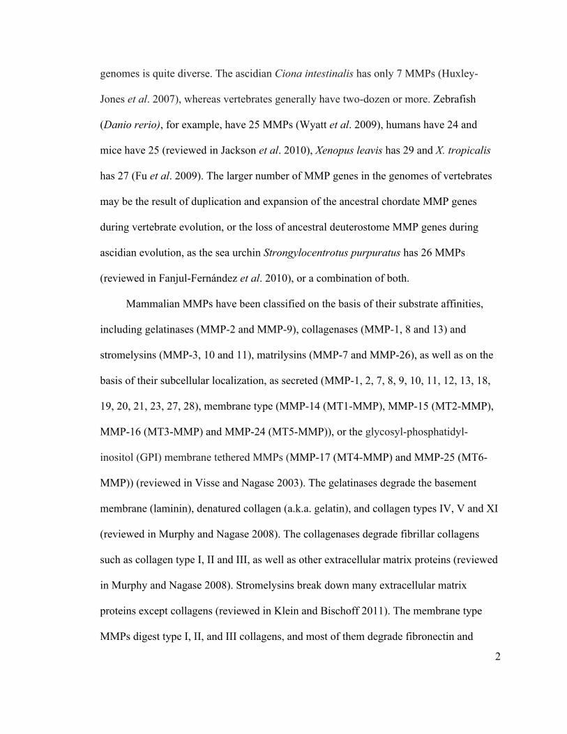
Most MMPs share four identifiable sequence hallmarks, and can be distinguished
according to the presence or absence of specific structures. From amino to carboxyl,
these consist of a secretory signal peptide motif, a pro-peptide domain, a catalytic
domain, a hinge region, and finally a hemopexin-like domain (reviewed in Nagase et al.
2006) (Figure 1.1). All MMPs have an amino-terminal signal peptide domain, which is
responsible for delivering the nascent protein to the endoplasmic reticulum (ER) during
translation for export (or anchoring into the plasma membrane through the glycosyl-
phosphatidyl-inositol (GPI) linkage or transmembrane domain) (reviewed in Murphy and
Nagase 2010).
The pro-peptide domain, which consists of about 80 to 100 moderately-conserved
amino acids (reviewed in Nagase et al. 2006), contains a cystine switch motif!
(PRCGxPD) (where ‘x’ indicates a non-conserved residue). The pro-peptide plays an
important role in controlling the activity of MMPs by folding into the catalytic site,
positioning the thiol of the cysteine switch in such a way as to interact with the catalytic
zinc ion, thereby preventing it from activating water molecules and thus inhibiting
proteolytic activity (Van Wart and Birkedal-Hansen 1990; reviewed in Cauwe and
4
Opdenakker 2010). Activation of MMPs therefore requires disruption of the interaction
between the pro-peptide and the catalytic site; this is typically accomplished by
proteolytic removal of the N-terminal pro-peptide domain, but can also result from
Figure 1.1: MMP Structure. All MMPs share an architecture consisting of a signal peptide (S), pro-peptide (Pro) and catalytic domain. Some other structures are present in some MMPs but not all; for example type II fibronectin repeats (Fn), cysteine-rich domains (Ca), immunoglobulin-like domains (Ig), type I or II transmembrane domains (I or II), vitronectin inserts (V), glycophosphatidylinistitol linkages (G), and cytoplasmic tails (Cp) (from Visse and Nagase 2003).
chemical modification of the cysteine in the switch motif (Okamoto et al. 2001).
The catalytic domain, common to all MMPs, binds two zinc ions, one for structure
and the other as a catalyst, and three calcium ions that provide stability to the structure.
5
This zinc-binding motif of the catalytic domain contains the consensus sequence
HExGHxxGxxH, in which the three histidines coordinate the catalytic Zn+2 ion and the
glutamic acid side chain serves as the general base, which buffers the protons released by
the activated water molecules (reviewed in Visse and Nagase 2003). Additionally, most
MMPs exhibit a conserved methionine residue (Met-turn) in the catalytic domain for
maintaining the structure of the S1-specificity pocket (reviewed in Visse and Nagase
2003). Carboxyl to the catalytic domain, a hinge region works as a link between the
catalytic domain and the hemopexin-like domain. The hemopexin-like domain is
responsible for interaction between the MMP enzymes and their substrates as well as
with endogenous tissue inhibitors of metalloproteinases (TIMPs) (reviewed in Amălinei
et al. 2007). However, some MMPs lack the hinge region and hemopexin-like domains,
for example MMP-7 (matrilysin-1), MMP-26 (matrilysin-2) and MMP-23 (reviewed in
Cauwe and Opdenakker 2010). In addition, there are three fibronectin type II repeats
within the catalytic domains of MMP-2 (a.k.a. Gelatinase-A) and MMP-9 (a.k.a.
Gelatinase-B), which also facilitate interaction with their substrates (reviewed in
Amălinei et al. 2007).
There is a significant level of complexity in the regulation of MMPs and the control
of their activity, including at the level of transcription, translation, and post-translational
processes including secretion, activation of the pro-MMPs (zymogen), and inhibition the
MMPs' activity by TIMPs (reviewed in Page-McCaw et al. 2007; Mannello and Medda
2012). Most MMPs are transcriptionally regulated by hormones, cytokines, growth
factors, and cell-cell and cell-matrix interactions (reviewed in Amălinei et al. 2007).
6
Post-translationally, MMP-11 and MT-MMPs can be activated intracellularly by furin
pro-protein convertase, as these MMPs bear conserved Rx(R/K)R paired basic amino
acid cleavage sites for this serine protease between their pro-peptides and catalytic
domains (Pei and Weiss 1995; reviewed in Nagase et al. 2006). Other MMPs are
activated extracellularly by collaborating proteases including other MMPs (reviewed in
Klein and Bischoff 2011).!
Non-proteolytic chemical agents, especially in pathological contexts, can cause
activation of full-length MMPs. For instance peroxynitrite (ONOO-), which occurs in
cells under oxidative stress, can nitrosylate the thiol of the cysteine switch rendering it
ineffective, and thereby giving rise to an activated full-length MMP (reviewed in Schulz
2007). Once MMPs are activated, TIMPs and other endogenous inhibitors are responsible
for regulating their activity and protecting substrates from uncontrolled degradation and
associated diseases (reviewed in Loffek et al. 2011). Of particular interest is the recent
discovery that phosphorylation also modulates MMP activity (Sariahmetoglu et al. 2007).
This is notable because, while phosphorylation is a common post-translational
modification that is known to regulate the activity of many enzymes, it occurs as the
result of the activity of kinases and relatively high local concentrations of adenosine 5’-
triphosphate (ATP), and is therefore generally characteristic of intracellular, rather than
extracellular proteins. However it is worth noting that the biological importance of
‘exokinases’ has recently begun to emerge (Yalak et al. 2014) (discussed in Chapter 3),
making it inaccurate to consider phosphorylation as a strictly intracellular phenomenon.
7
1.1.2 The function of Matrix Metalloproteinases
1.1.2.1 Extracellular matrix remodelling
The extracellular matrix (ECM) is a cross-linked matrix of insoluble glycoproteins
and protoglycans secreted by the cells of all multicellular organisms, which give tissues
structural integrity and many of their biological functions. In animals, the ECM is
composed of collagen, elastin, fibronectin, laminin, nidogen and other large insoluble
glycoproteins that are ubiquitous constituents of almost all tissues (reviewed in Frantz et
al. 2010). Although considered by some as ‘packing material’ and uninteresting, the
many dynamic and pleiotropic roles of the ECM are continuing to emerge and are
becoming central in understanding the development, function and evolution of animal
tissues. The matrix influences cell differentiation, proliferation, and tissue reorganization
by binding directly to receptors such as integrins (reviewed in Harburger and Calderwood
2009), and by modulating the presentation, stability and distribution of signalling
molecules such as growth factors and cytokines (reviewed in Venkatasubramanian 2012).
Thus the ECM plays many essential roles in cellular processes and embryonic
development; providing permissive and non-permissive substrates for cell migration,
permitting and resisting changes in cell-shape, and promoting or inhibiting cell
proliferation and survival. Further, the composition and even mechanical load on the
matrix influences cell fate decisions and differentiation, and works as a bio-scaffold for
rebuilding injured cells and tissues (reviewed in Lu et al. 2011).
Remodelling of the ECM during or after embryonic development is clearly crucial
to the formation new tissue and structures in vivo. However, it is worth noting that under
8
normal conditions, the level of MMP expression is generally low in adult tissues, and its
induction depends on several factors, many of which are associated with inflammatory or
other pathological processes (reviewed in Phatharajaree et al. 2007).
1.1.2.2 Matrix Metalloproteinase in development and disease
MMPs are strongly implicated in many physiological and pathological processes.
The physiological processes include those associated with embryonic development, such
as angiogenesis, normal tissue remodeling, and in adults MMPs are required for normal
inflammatory response, wound healing and tissue homeostasis (reviewed in Visse and
Nagase 2003). MMPs are also implicated in a wide range of devastating diseases.
Examples include rheumatoid arthritis and osteoarthritis, in which collagenases (MMP-1,
-8, -13, and -18) are responsible for breaking down connective tissues (reviewed in
Vartak et al. 2007). MMP-3 expression in adults is associated with neurodegenerative
diseases such as Parkinson’s disease (reviewed in Kim and Hwang 2011) and many more.
The most relevant with respect to the work discussed in this thesis are the roles of MMP-
2 in muscle disorders. MMP-2 has a significant impact on muscle atrophy (Liu 2011)
and, intracellular MMP-2 is implicated in the degradation of intracellular sarcomeric
proteins in cardiomyocytes under oxidative stress (reviewed in Schulz 2007).
1.2 Intracellular localization of MMPs
For most of the past four decades, research on MMPs concentrated on their
canonical biological functions proteolyzing and remodeling extracellular proteins.
However, more recently evidence has demonstrated that MMPs can degrade non-
extracellular matrix proteins inside and outside the cells (reviewed in Cauwe and
9
Opdenakker 2010). Several MMPs are detected at significant concentrations inside cells,
such as in the nucleus (MMP-1, -2, -3, -9, -13, -26 and -14) (Limb et al. 2005; Kwan et
al. 2004; Eguchi et al. 2008; Yang et al. 2010; Cuadrado et al. 2009; Zhang et al. 2002;
Yang et al. 2010), mitochondria (MMP-1, -2 and -9) (Limb et al. 2005; Wang et al. 2002;
Moshal et al. 2008; reviewed in Cauwe and Opdenakker 2010), and within sarcomeres in
cardiac myocytes (Sawicki et al. 2005). In the context of cardiac sarcomeres,
pathologically activated MMP-2 has been shown to degrade troponin I, myosin light
chain, α-actinin and titin (Wang et al. 2002; Sawicki et al. 2005; Sung et al. 2007; Ali et
al. 2010; reviewed in Ali et al. 2011a), all of which are essential sarcomeric proteins,
making the pathological activation of intracellular MMP-2 a direct cause of the loss of
muscle cell contractility in ischemia/reperfusion injury.
MMP-2 is the most abundant member of the MMP family. It is constitutively
expressed in most tissues and it is ubiquitous in cardiomyocytes (reviewed in Schulz
2007). The intracellular localization of MMP-2 and its pathological role in cardiac
muscle was reported a decade ago (Sawicki et al. 2005). Recently, Ali and colleagues
(2011b) reported the mechanisms that result in this unexpected distribution of this
ostensibly secreted enzyme. Firstly, the N-terminal signal sequence of the canonical
MMP-2- MEALMARGALTGPLRALCLLGCLLSHAAA - (the sequence of amino acids
that direct the newly synthesized peptide to the ER via signal recognition particles (SRP))
appears to interact only weakly with the SRP. The SRP binds secretory signals in nascent
polypeptides as they emerge from the large subunit of the ribosome, and simultaneously
binds the incoming tRNA binding site (the A-site in the large ribosomal subunit) causing
10
a pause in translation. The paused translation complex is then bound by protein
translocator complexes embedded in the endoplasmic reticulum membrane. The SRP is
then released and translation continues, with the nascent protein being elongated into the
lumen of the endoplasmic reticulum (Cooper and Hausman 2009). From the endoplasmic
reticulum, secreted proteins proceed through the Golgi apparatus and into the constitutive
secretory pathway. Therefore, the efficiency with which a protein’s secretory signal is
recognized by the SRP determines what proportion of that protein is translocated into the
ER and thence into the secretory pathway. At least in the case of human MMP-2, a
significant proportion of the translated protein is not recognized by the SRP, and remains
cytosolic (Ali et al. 2011b). As will be discussed further in Chapter 2, this appears to be
an evolutionarily conserved feature of Gelatinase-A. Furthermore, alternatively spliced
mmp-2 transcripts expressed in human cardiomyocytes encode an MMP-2 protein
completely lacking an N-terminal secretory signal, contributing more of this protease to
the cytosolic pool (Ali et al. 2011b). To understand potential consequences of
metalloproteinases within muscle cells we must recall some basics of sarcomere structure
and function.
1.3 Muscle structure and function
Muscle cells are a highly specialized, essential cell type in most metazoans. In
addition to allowing voluntary movement under the control of the nervous system, the
contractions of muscles participate in maintaining body homeostasis by moving blood
through vessels, air or oxygenated water across gas-exchange surfaces, and food through
the digestive system (Arms & Camp 1997). All muscle cells contain specialized
11
complexes of cytoskeletal proteins (primarily actin and myosin, with an array of
accessory proteins discussed below), which exert contractile force fuelled by the
regulated hydrolysis of ATP.
Vertebrates have both striated and smooth muscle. Much of the involuntary
musculature associated with the gut and vascular system is smooth muscle, in which the
contractile apparatus of the cells is not organized into repeating units. In contrast, the
contractile apparatus of cardiac and skeletal muscle is arranged in repeating units called
sarcomeres, which give these cells their characteristic striated appearance.
Cardiac and smooth muscle forms about 10% of the muscle mass of typical
vertebrates, in contrast to skeletal muscle, which is large and highly complex tissue
forming about 40% of the body weight in humans, and around 60 % of the total body
weight in fish (Sänger and Stoiber 2001). Muscle consists one or more fibres, each of
which consist of groups of myofibrils covered by sarcolemma (a.k.a. cell membrane).
The myofibrils are made up of linearly repeated sarcomeres. The sarcomere is a highly
organized structural unit of cytoskeletal proteins (see Figure 1.2). Thus, the muscle
consists of hierarchically organized groups of contractile fibres.
The contractile function of the sarcomere results from the interaction of two kinds
of myofilament proteins; actin that forms the main component of thin filaments, and
myosin that forms the thick filaments. Thin and thick filaments overlap and the myosin
protein converts the chemical energy of ATP into mechanical energy to drive the
filaments past one another, which causes muscular contraction. Inside the sarcomere, the
ends of the actin filaments are attached to the terminal Z-discs by α-actinin which extends
from the Z-disc into the light I-band.
12
!!
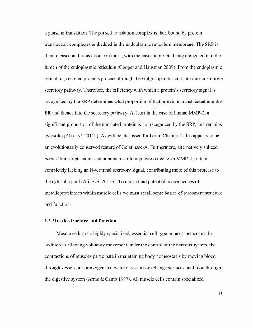
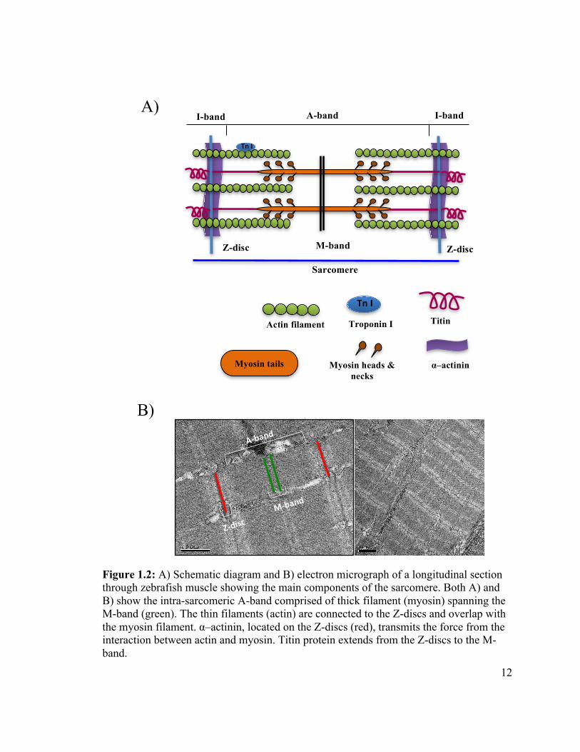
!
Figure 1.2: A) Schematic diagram and B) electron micrograph of a longitudinal section through zebrafish muscle showing the main components of the sarcomere. Both A) and B) show the intra-sarcomeric A-band comprised of thick filament (myosin) spanning the M-band (green). The thin filaments (actin) are connected to the Z-discs and overlap with the myosin filament. α–actinin, located on the Z-discs (red), transmits the force from the interaction between actin and myosin. Titin protein extends from the Z-discs to the M-band.
Sarcomere
M-band Z-disc
A-band
I-band I-band
Z-disc
Actin filament
Titin
α–actinin
Myosin heads &
necks Myosin tails
A)
Tn#I
Troponin I Tn#I
B)
A&band
M&band
Z&disc
13
At their free ends, actin filaments are stabilized by trompomodulin caps. Along their
length, which is determined by nebulin, the actin filaments are bound by a complex of
proteins including tropomyosin (Tm), troponin T (tropomyosin-binding), troponin C
(Ca+2 binding), and troponin I (inhibitory proteins), which regulate the exposure of actin
binding sites in a calcium-dependent manner.
Myosin thick filaments are composed of tail, neck and P-loop ATP-ase head
domains; the tails of multiple myosin heavy chains interact to form the thick filament,
and are oriented towards the middle of the sarcomere (M-band), while the heads are
oriented away from the M-band, such that they can interact with the actin filaments
(Sarantitis et al. 2012). In addition, there are titin and nebulin macromolecule proteins
that form a part of the sarcomere structure. Titin is the largest protein in nature and
extends from Z-band of the sarcomere to the M-band in the middle (reviewed in Kruger
and Linke 2011). It works as a molecular spring and scaffold protein that keeps the
myosin filaments in the middle of the sarcomere. Finally, nebulin is connected to the Z-
discs and regulates the growth of actin filaments, acting as a molecular ruler to adjust the
length of thin filaments in sarcomeres (reviewed in Fowler et al. 2006).
The sliding filament model of contraction in striated muscle was first proposed in
1954 by two independent groups (Huxley & Niedergerke 1954; Huxley & Hanson 1954).
Contraction is triggered by the release of acetylcholine by motor neuron synapses at the
neuromuscular junction, causing depolarization of the muscle cell membrane. This gives
rise to the liberation of calcium from the sarcoplasmic reticulum into the cytoplasm of the
muscle cell. Ca+2 binds troponin C causing a conformational change which exposes
myosin binding sites on the actin filaments. In each cycle of contraction, the myosin
14
heads bind ATP, hydrolyze the ATP to ADP+Pi which triggers a dramatic change in
conformation typical of P-loop ATPases, and while in an ADP-bound state they gain
affinity for actin and bind exposed sites on the thin filaments. This binding triggers the
relaxation of the lever arm of the head group which exerts contractile force on the actin
filament, and the expulsion of the ADP. The myosin molecule is then free to bind ATP
which triggers the release of the thin filament and the cycle repeats, causing the myosin
thick filament to pull its way along the actin filaments in ratchet-like steps.
During relaxation of the muscle, the sarcoplasmic reticulum Ca+2 - ATPase
(SERCA)- dependent calcium transporters present in the membrane of the sarcoplasmic
reticulum pump calcium ions back into storage. This reverses the conformational change
in tropomyosin, eliminating the interaction between myosin head groups and actin thin
filaments, and allowing the thick and thin filaments to slide past each other back to their
original configuration.
1.4 MMPs in ischemia-reperfusion injury
Ischemia occurs when the flow of blood to tissues is temporarily blocked. Ischemia
and reperfusion injury (IRI) is a pathological situation arising when oxygenated blood
flow is restored after a period of ischemia. This leads to the production of reactive
oxygen species (ROS) as built-up reducing power from electron transport is transferred to
oxygen faster than endogenous mechanisms can manage (reviewed in Chow et al. 2007).
Prolonged ischemia causes an alteration in cellular function and metabolism by reducing
oxidative phosphorylation and depleting high-energy phosphate molecules, such as ATP.
Furthermore, ATP catabolism during ischemia-altered sodium/potassium ATPase activity
15
leads to the influx of sodium, calcium and water molecules into cells.
Reperfusion after ischemia leads to inflammation of cells due to ionic imbalance
and production of ROS. The inability of the cell to eliminate ROS such as superoxide
(·O2-), and nitric oxide (NO·) results in the production of highly reactive molecules such
as peroxynitrite (ONOO-). The latter in particular stimulates the activity of MMPs due to
the nitrosylation and/or glutathiolation of the auto-inhibitory cystine switch (Viappiani et
al. 2009). As a result, the intracellular MMP-2 becomes activated and destroys the
contractile proteins within the sarcomere (reviewed in Ali et al. 2011a). Making matters
worse, peroxynitrite additionally inhibits the action of TIMPs in vitro (Donnini et al.
2008), which could exacerbate the activity of MMPs in ischemia-reperfusion injury.
1.5 Zebrafish as model system for the study of muscle
For decades now, the zebrafish has been described as a tractable vertebrate model
system for research of many types. It has been extensively utilized for studying genetics
and human diseases (reviewed in Lieschke and Currie 2007). The advantage of zebrafish
over insect or other invertebrate model systems is primarily that, as a vertebrate, it is less
evolutionarily divergent from mammals, making it easier to generalize findings of
biomedical significance. The advantages zebrafish have over mammalian models such as
mice include the optical clarity of the externally fertilized embryos that simplify
observation of biological processes and their rapid, external development at room
temperature, requiring only 72 hours to become feeding larvae, compared to almost a
month for rats or mice. Furthermore, the cost to obtain and raise zebrafish is less that a
thousandth of the cost associated with mice. Many of the attractive features of
16
mammalian systems, such as the availability of many mutant and transgenic lines, a
sequenced genome, amenability to genome editing, and reverse genetic approaches, also
apply to zebrafish. An additional advantage is the availability and amenability of the
zebrafish to high throughput and transgenic approaches; thousands of embryos can be
arrayed in plates for treatment with libraries of compounds or other combinatorial
experiments, and we can order various types of transgenic fish from stock centres or
engineer our own transgenic fish as necessary. What is more crucial is their fundamental
similarity to human and other vertebrates, particularly with respect to muscular
development and function (reviewed in Squire et al. 2008). Thus, using zebrafish gives us
a good opportunity to study the action and activity of MMPs (Wyatt et al. 2009) and
conduct other biological research.
1.6 Objectives
The common ancestor of humans and zebrafish lived approximately 460 to 360
Mya (Campbell et al. 2008). Although many of the characteristics of this organism are
now obscure, it is clear that it had a gelatinase-A gene. What is less clear is whether the
protease encoded by that gene was inefficiently secreted (as is the case with modern
mammalian orthologues), whether it accumulated within the sarcomeres of ancestral
striated muscle, whether its activity was regulated by phosphorylation, what proteins it
degraded, and what, if any, roles it might have played in normal muscle cell physiology.
In this thesis I hope to shed some light on these questions by determining if zebrafish
Mmp2 is present within skeletal muscle sarcomeres and if it is phosphorylated.
17
1.7 Overall hypothesis
My core hypothesis is that gelatinase-A has ancient conserved functions within the
sarcomere of striated muscle. My predictions are therefore: 1) gelatinase-A will be
observed to localize intracellularly in most, if not all, striated muscle cells rather than
this being an idiosyncratic characteristic of mammalian cardiac myocytes. 2) The
secretory signals of gelatinase-A proteins from phylogenetically diverse vertebrates will
be consistently poorly recognized as such, and therefore indicative of evolutionary
selection in favour of intracellular accumulation of this protease. Finally, 3) the
phosphorylation phenomenon and its role in modulating the activity of gelatinase-A will
be conserved across vertebrate species. The specific approaches I undertook to test these
predictions, which are presented in this thesis are as follows: 1) I unequivocally localized
zebrafish Mmp2 within the sarcomeres of zebrafish skeletal muscle using
immunofluorescence in both optical and physical sections of embryonic and adult
muscle. 2) I examined the predicted amino acid sequences encoding gelatinase-A genes
and compared their secretory signals to those of two efficiently ER-targeted proteins
(gelatinase-B and BiP) for all species for which good sequence data are available for all
three genes. I found that the N-terminal sequence of gelatinase-A proteins is significantly
less well recognized as such than the secretory signals of either gelatinase-Bs or BiPs.
Finally, 3) while I was unable to complete the analysis of its phosphorylation status
biochemically, I have developed a protocol to purify zebrafish Mmp2 protein from
skeletal muscle using gelatin-affinity chromatography, and the determination of its
phosphorylation status will only require small changes in the technique. In addition, I
analyzed the conservation of phosphorylation sites in available gelatinase-A sequence

18
data, and found that several phosphorylation sites identified in human MMP-2 are also
conserved in zebrafish as well as in other species.
These findings support my core hypothesis, and I conclude this thesis with some
speculation regarding the conserved physiological function gelatinase-A may have within
the sarcomeres of striated muscle.
19
1.8 References
Ali MA, Cho WJ, Hudson B, Kassiri Z, Granzier H, Schulz R. 2010. Titin is a target of matrix metalloproteinase-2: implications in myocardial ischemia/reperfusion injury. Circulation. 122(20): 2039-47.
Arms K, Camp PS. 1997. Biology. Fourth edition. Orlando (Fl): Saunders College Publishing. p. 844-862.
Ali MA, Fan X, Schulz R. 2011a. Cardiac sarcomeric proteins: novel intracellular targets of matrixmetalloproteinase-2 in heart disease. Trends Cardiovasc Med. 21(4): 112-118. Ali MA, Chow AK, Kandasamy AD, Fan X, West LJ, Crawford BD, Simmen T, Schulz R. 2011b. Mechanisms of cytosolic targeting of matrix metalloproteinase-2. J Cell Physiol. 227: 3397-3404. Amalinei C, Caruntu ID, Balan RA. 2007. Biology of metalloproteinases. Rom J Morphol Embryol. 48(4): 323–334. Campbell NA, Reece JB, Urry LA, Cain ML, Wasserman SA, Minorsky PV, Jackson RB. 2008. Biology. Eighth edition. San Francisco (CA): Pearson Education Inc. p. 654-665. Cauwe B, Opdenakker G. 2010. Intracellular substrate cleavage: a novel dimension in the biochemistry, biology and pathology of matrix metalloproteinases. Crit Rev Biochem Mol Biol. 45(5): 351–423.
Chow AK, Cena J, Schulz R. 2007. Acute actions and novel targets of matrix metalloproteinases in the heart and vasculature. Br J Pharmacol. 152(2): 189–205.
Cooper GM, Hausman RE. 2009. The cell: a molecular approach. Fifth edition. Washington (DC): The American Society of Microbiology Press. p. 383-432.
Cuadrado E, Rosell A, Borrell-Pages M, Garcia-Bonilla L, Hernandez- Guillamon M, Ortega-Aznar A, Montaner J. 2009. Matrix metalloproteinase-13 is activated and is found in the nucleus of neural cells after cerebral ischemia. J Cereb Blood Flow Metab. 29(2): 398–410.
Donnini S, Monti M, Roncon R, Morbidelli L, Rocchigiani M, Oliviero S, Casella L, Giachetti A, Schulz R, Ziche M. 2008. Peroxynitrite inactivates human-tissue inhibitor of metalloproteinase-4. FEBS Lett. 582(7): 1135–1140.
Eguchi T, Kubota S, Kawata K, Mukudai Y, Uehara J, Ohgawara T, Ibaragi S, Sasaki A, Kuboki T, Takigawa M. 2008. Novel transcription-factor-like function of human matrix metalloproteinase 3 regulating the CTGF/CCN2 gene. Mol Cell Biol. 28(7): 2391–413.
20
Fanjul-Fernández M, Folgueras AR, Cabrera S, López-Otín C. 2010. Matrix metalloproteinases: evolution, gene regulation and functional analysis in mouse models. Biochim Biophys Acta.1803(1): 3-19.
Fowler VM, McKeown CR, Fischer RS. 2006. Nebulin: does it measure up as a ruler? Curr Biol. 16(1): R18–20.
Frantz C, Stewart KM, Weaver VM. 2010. The extracellular matrix at a glance. J Cell Sci. 123(24): 4195-4200.
Fu L, Das B, Mathew S, Shi YB. 2009. Genome-wide identification of Xenopus matrix metalloproteinases: conservation and unique duplications in amphibians. BMC Genomics. 10:81.
Gomis-Ruth FX. 2009. Catalytic domain architecture of metzincin metalloproteases. J Biol Chem. 284(23): 15353-7.
Gross J, Lapiere CM. 1962. Collagenolytic activity in amphibian tissues: a tissue culture assay. Proc Natl Acad Sci U S A. 48(6): 1014-1022.
Harburger DS, Calderwood DA. 2009. Integrin signalling at a glance. J Cell Sci. 122(2): 159-163.
Huxley-Jones J, Clarke TK, Beck C, Toubaris G, Robertson DL, Boot-Handford RP. 2007. The evolution of the vertebrate metzincins; insights from Ciona intestinalis and Danio rerio. BMC Evol Biol. 7:63.
Huxley AF, Niedergerke R. 1954. Structural changes in muscle during contraction; interference microscopy of living muscle fibres. Nature.173(4412): 971-973.
Huxley H, Hanson J. 1954. Changes in the cross-striations of muscle during contraction and stretch and their structural interpretation. Nature.173(4412): 973-976.
Jackson BC, Nebert DW, Vasiliou V. 2010. Update of human and mouse matrix metalloproteinase families. Hum Genomics. 4(3): 194–201.
Kessenbrock K, Plaks V, Werb Z. 2010. Matrix metalloproteinases: regulators of the tumor microenvironment. Cell. 141(1): 52-67.
Kim EM, Hwang O. 2011. Role of matrix metalloproteinase-3 in neurodegeneration. J Neurochem. 116(1): 22-32.
Klein T, Bischoff R. 2011. Physiology and pathophysiology of matrix metalloproteases. Amino Acids. 41(2): 271-290.
Kruger M, Linke WA. 2011. The Giant Protein Titin: a regulatory node that integrates myocyte signaling pathways. J Biol Chem. 286(12): 9905-9912.
21
Kwan JA, Schulze CJ, Wang W, Leon H, Sariahmetoglu M, Sung M, Sawicka J, Sims DE, Sawicki G, Schulz R. 2004. Matrix metalloproteinase-2 (MMP-2) is present in the nucleus of cardiac myocytes and is capable of cleaving poly (ADP-ribose) polymerase (PARP) in vitro. FASEB J. 18(6): 690-2.
Lapière ChM. 2005. Tadpole collagenase, the single parent of such a large family. Biochimie. 87(3-4): 243–247.
Lieschke GJ, Currie PD. 2007. Animal models of human disease: zebrafish swim into view. Nat Rev Genet. 8(5): 353-67.
Limb GA, Matter K, Murphy G, Cambrey AD, Bishop PN, Morris GE, Khaw PT. 2005. Matrix metalloproteinase-1 associates with intracellular organelles and confers resistance to lamin A/C deg- radation during apoptosis. Am J Pathol. 166(5): 1555–1563.
Liu X. 2011. Emerging ideas: Matrix Metalloproteinase-2 in muscle atrophy. Clin Orthop Relat Res. 469(6): 1797–1799.
Loffek S, Schilling O, Franzke CW. 2011. Biological role of matrix metalloproteinases: a critical balance. Eur Respir J. 38(1): 191–208.
Lu P, Takai K, Weaver VM, Werb Z. 2011. Extracellular matrix degradation and remodeling in development and disease. Cold Spring Harb Perspect Biol. 3(12): a005058.
Mannello F, Medda V. 2012. Nuclear localization of Matrix metalloproteinases. Prog Histochem Cytochem. 47(1): 27–58.
Moshal KS, Tipparaju SM, Vacek TP, Kumar M, Singh M, Frank IE, Patibandla PK, Tyagi N, Rai J, Metreveli N, et al. 2008. Mitochondrial matrix metalloproteinase activation decreases myocyte contractility in hyperhomocysteinemia. Am J Physiol Heart Circ Physiol. 295(2): 890–897.
Murphy G, Nagase H. 2008. Progress in matrix metalloproteinase research. Mol Aspects Med. 29(5): 290–308.
Murphy G, Nagase H. 2010. Localizing matrix metalloproteinase activities in the pericellular environment. FEBS J. 278(1): 2 –15.
Nagase H, Visse R, Murphy G. 2006. Structure and function of matrix metalloproteinases and TIMPs. Cardiovasc Res. 69(3): 562–573.
Okamoto T, Akaike T, Sawa T, Miyamoto Y, Van der Vliet A, Maeda H. 2001. Activation of matrix metalloproteinases by peroxynitrite-induced protein S-glutathiolation via disulfide S-oxide formation. J Biol Chem. 276 (31): 29596–602.
Page-McCaw A, Ewald AJ, Werb Z. 2007. Matrix metalloproteinases and the regulation
22
of tissue remodeling. Nat Rev Mol Cell Biol. 8(3): 221-233.
Pei D, Weiss SJ. 1995. Furin-dependent intracellular activation of the human stromelysin-3 zymogen. Nature. 375(6528): 244-247.
Phatharajaree W, Phrommintikul A, Chattipakorn N. 2007. Matrix metalloproteinases and myocardial infarction. Can J Cardiol. 23(9): 727-733.
Sänger AM, Stoiber W. Muscle fibre diversity and plasticity. In: Johnston IA, editor. Muscle development and growth. San Diego (CA): Academic Press; 2001. p. 187-250. Sarantitis I, Papanastasopoulos P, Manousi M, Baikoussis NG, Apostolakis E. 2012. The cytoskeleton of the cardiac muscle cell. Hellenic J Cardiol. 53(5): 367-79.
Sariahmctoglu M, Crawford BD, Leon H, Sawicka J, Li L, Ballermann BJ, Holmes C, Berthiaume LG, Holt A, Sawicki G, et al. 2007. Regulation of matrix metalloproteinase-2 (MMP-2) activity by phosphorylation. FASEB J. 21(10): 2486-2495.
Sawicki G, Leon H, Sawicka J, Sariahmctoglu M, Schulze CJ, Scott PG, Szczesna-Cordary D, Schulz R. 2005. Degradation of myosin light chain in isolated rat hearts subjected to ischemia-reperfusion injury: a new intracellular target for matrix metalloproteinase-2. Circulation. 112(4): 544-552.
Schulz R. 2007. Intracellular targets of matrix metalloproteinase-2 in cardiac disease: rationale and therapeutic approaches. Annu Rev Pharmacol Toxicol. 47: 211–242.
Squire JM, Knupp C, Luther PK. 2008. Zebrafish topical, transparent, and tractable for ultrastructural studies. J Gen Physiol. 131(5): 439–443.
Sung MM, Schulz CG, Wang W, Sawicki G, Bautista-Lopez NL, Schulz R. 2007. Matrix metalloproteinase-2 degrades the cytoskeletal protein α- actinin in peroxynitrite mediated myocardial injury. J Mol Cell Cardiol. 43(4): 429-436.
Tallant C, Marrero A, Gomis-Rüth FX. 2010. Matrix metalloproteinases: fold and function of their catalytic domains. Biochim Biophys Acta. 1803(1): 20-8.
Vartak DG, Gemeinhart RA. 2007. Matrix metalloproteases: underutilized targets for drug delivery. J Drug Targeting. 15(1): 1-20.
Van-Wart HE, Birkedal-Hansen H. 1990. The cysteine switch: a principle of regulation of metalloproteinase activity with potential applicability to the entire matrix metalloproteinase gene family. Proc Natl Acad Sci USA. 87(14): 5578-5582.
Venkatasubramanian PN. Imaging the pancreatic ECM. In: Grippo PJ, Munshi HG, editors. Pancreatic cancer and tumor microenvironment. Trivandrum (India): Transworld Research Network; 2012. p. 11-27.
23
Viappiani S, Nicolescu AC, Holt A, Sawicki G, Crawford BD, León H, van Mulligen T, Schulz R. 2009. Activation and modulation of 72kDa matrix metalloproteinase-2 by peroxynitrite and glutathione. Biochem Pharmacol. 77(5): 826–34.
Visse R, Nagase H. 2003. Matrix metalloproteinases and tissue inhibitors of metalloproteinases structure, function, and biochemistry. Circ Res. 92(8): 827-839.
Wang W, Schulze CJ, Suarez-Pinzon WL, Dyck JR, Sawicki G, Schulz R. 2002. Intracellular action of matrix metalloproteinase-2 accounts for acute myocardial ischemia and reperfusion injury. Circulation.106(12): 1543-1549.
Wyatt RA, Keow JY, Harris ND, Hache CA, Li DH, Crawford BD. 2009. The zebrafish embryo: a powerful model system for investigating matrix remodeling. Zebrafish. 6(4): 347-354.
Yalak G, Ehrlich YH, Olsen BR. 2014. Ecto-protein kinases and phosphatases: an emerging field for translational medicine. J Transl Med. 12:165.
Yang Y, Candelario-Jalil E, Thompson JF, Cuadrado E, Estrada EY, Rosell A, Montaner J, Rosenberg GA. 2010. Increased intranuclear matrix metalloproteinase activity in neurons interferes with oxidative DNA repair in focal cerebral ischemia. J Neurochem 112(1): 134–149.
Zhang J, Cao YJ, Zhao YG, Sang QX, Duan EK. 2002. Expression of matrix metalloproteinase-26 and tissue inhibitor of metalloproteinase-4 in human normal cytotrophoblast cells and a choriocarcinoma cell line, JEG-3. Mol Hum Reprod. 8(7): 659–666.

24
Chapter 2: Intracellular localization of gelatinase-A (Mmp2) in zebrafish
skeletal muscle
2.1 Introduction
As discussed in the previous chapter, MMPs are known as secreted proteases, and
collectively, they degrade all of the proteinacious components of the ECM. However,
recent evidence has emerged that indicates some MMPs are not secreted from the cell,
and that these proteases may have significant roles in the pathology of
ischemia/reperfusion injury (reviewed in Chow et al. 2007). For instance, MMP-2 has
been detected intracellularly in rat cardiac myocytes, and is implicated in the degradation
of sarcomeric proteins under conditions of oxidative stress injury (Wang et al. 2002;
Sawicki et al. 2005; Sung et al. 2007; Ali et al. 2010; reviewed in Schulz 2007 and!Ali et
al. 2011a). One obvious question that arises from these observations is ‘how does this
secreted protein accumulate in the cytoplasm of myocytes?’ Gelatinase-A is a class-I
secreted protein, which means that its N-terminus consists of a signal sequence that is
bound by the signal recognition particle (SRP) as the nascent polypeptide emerges from
the ribosome. The SRP facilitates binding to the endoplasmic reticulum (ER)
translocation complex, such that the protein is translocated into the ER, and from there to
the Golgi apparatus and secretory pathway (Cooper and Hausman 2009, and discussed
further in Chapter 4). However, it has now become clear that the signal sequence of the
human MMP-2 protein - MEALMARGALTGPLRALCLLGCLLSHAAA – is not bound
efficiently by the SRP, and consequently approximately 40% of the MMP-2 protein
produced in cells remains cytosolic (Ali et al. 2011b). Furthermore, in human cardiac
25
myocytes, a splice variant of mmp-2 is expressed in which an alternative first exon
encodes a slightly truncated form of the protein lacking any secretory signal (Ali et al.
2011b).
Intuitively, it would seem unlikely that cardiac myocytes accumulate intracellular
stores of dangerous proteases simply to make themselves more vulnerable to oxidative
stress. If this pathological activity of gelatinase-A were not balanced by some valuable
physiological role, one would expect natural selection to have driven the evolution of a
more efficient secretory signal for this protease, as is found in other secreted proteins.
Furthermore, MMP-2 is phosphorylated in vivo, and its proteolytic activity is modulated
by this post-translational modification (Sariahmetoglu et al. 2007). This implies the
existence of kinases and phosphatases that regulate the activity of this intracellular pool
of MMP-2, and furthermore that such regulatory pathways likely evolved to modulate a
normal, physiological role for this protease. What might be the function of this
intracellular gelatinase-A?
In order to address these questions, I used zebrafish to determine if the intracellular
localization of gelatinase-A is unique to mammalian cardiac myocytes, or alternatively, if
it may be a common feature of striated muscle, implying a more ancient and
evolutionarily-conserved function for this protease in muscle physiology. The zebrafish is
an excellent vertebrate model for the study of ultrastructure and muscle physiology
(Squire et al. 2008), and this is enhanced by the availability of anti-zebrafish Mmp2
antibody (Harris 2010). In zebrafish mmp2 expression at the mRNA level is essentially
constant and homogeneous in all tissues throughout development (Zhang et al. 2003).
26
However, immunoblots of whole embryo homogenates reveal Mmp2 protein
accumulation beginning during gastrulation, and immunostaining reveals the protein
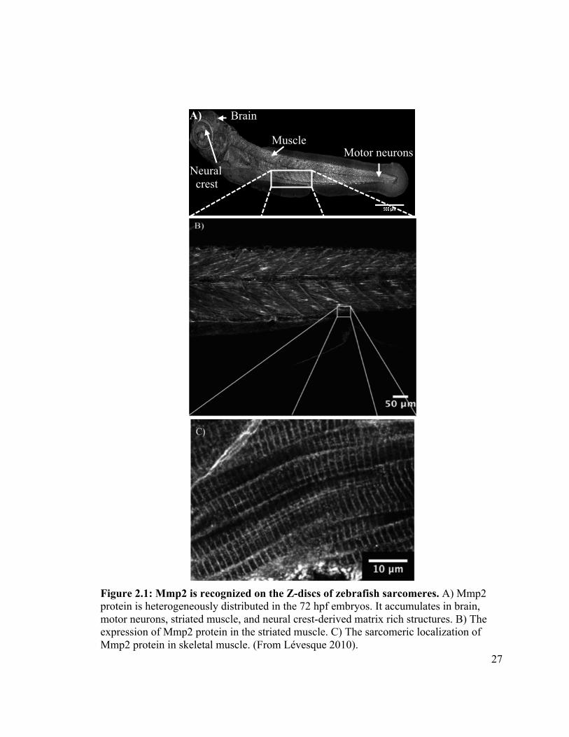
accumulates heterogeneously, with significant concentrations occurring along motor
axons, in the developing central nervous system, at myotome boundaries, within the
epidermal folds of developing fins, and within the muscle tissue itself (Lévesque 2010).
Detailed examination of the distribution of Mmp2 protein in the skeletal muscle of the
trunk reveals a distinctly striated pattern, consistent with sarcomeric localization of the
protease (Figure 2.1) (Lévesque 2010).
In order to determine if this protein is actually located within the sarcomere of
zebrafish muscle, I used double immunostaining of tissues from embryos and adult fish,
using anti-Mmp2 to characterize its distribution relative to α–actinin, a sarcomeric
protein known to be a component of the Z-disc (reviewed in Luther 2009). These data
suggest that Mmp2 accumulates in M-bands within the sarcomere of zebrafish skeletal
muscle. However, because the axial resolution of confocal microscopy (Murray 2006)
and the thickness of myofibrils (reviewed in Recher et al. 2009) are nearly the same (~1
µm) it is impossible to distinguish periodic cell surface staining from periodic
intracellular staining within the sarcomeres. Thus my interpretation of immunostaining
results could be contested. By using cryo-sectioning to cut the muscle samples at around
a half micron in thickness, I was able to prove the intracellular localization of Mmp2 in
zebrafish skeletal muscle. Furthermore, using a bioinformatic approach, I show that the
poorly-recognized secretory signal described for human MMP-2 is typical of gelatinase-
A orthologues across a phylogenetically diverse sample of vertebrates. This is consistent
27
Figure 2.1: Mmp2 is recognized on the Z-discs of zebrafish sarcomeres. A) Mmp2 protein is heterogeneously distributed in the 72 hpf embryos. It accumulates in brain, motor neurons, striated muscle, and neural crest-derived matrix rich structures. B) The expression of Mmp2 protein in the striated muscle. C) The sarcomeric localization of Mmp2 protein in skeletal muscle. (From Lévesque 2010).
Muscle
Brain
Motor neurons Neural crest
A)
28
with the existence of an unknown but selectively advantageous function for gelatinase-A
within the sarcomeres of striated muscle.
Despite the fact that the expression of Mmp2 is very high in skeletal muscle, the
role(s) of intracellular Mmp2 in zebrafish skeletal muscle remains unknown. I speculated
that Mmp2 may play a role in sarcomere development, which occurs in an anterior-to-
posterior wave in the 24 hpf embryo (Kimmel et al. 1995), but immunostaining shows
Mmp2 does not accumulate in developing myofibrils until after sarcomere establishment
is complete, suggesting a role in maintenance, rather than formation of the contractile
apparatus.
29
2.2 Materials and methods
2.2.1 Spawning zebrafish and collecting embryos
Wild-type adult zebrafish were maintained on a 14-hour light and 10-hour dark
cycle as described in Westerfield (1995) and fed brine shrimp twice a day and fish pellets
(Adult zebrafish diet, Zeigler) three to four times a day. The embryos were collected after
natural spawning over trays filled with marbles. Embryos were grown in embryo rearing
medium (ERM) (13 mM NaCl, 0.5 mM KCl, 0.02 mM Na2HPO4, 0.04 mM KH2PO4, 1.3
mM CaCl2, 1.0 mM MgSO4, and 4.2 mM NaHCO3, pH 7.4) at 28.5 °C. 0.001-0.002%
methylene blue was added to minimize fungal growth. For pre-hatching stages, embryos
were manually dechronionated using fine forceps under the microscope prior to fixation.
2.2.2 Immunostaining and confocal microscopy
To prepare for immunostaining, 10 embryos at 24 and 72 hour post fertilization
(hpf) were collected, dechorionated, and chilled. Three adult zebrafish were terminally
anaesthetized in 0.3 mg/ml tricaine methanesulfonate (Sigma) in ERM, after which the
fish tail muscles were dissected from the fish using forceps. Subsequently, embryos and
adult muscle tissue samples were fixed in Dent’s solution (20% dimethyl sulfoxide
(DMSO), 80% methanol) overnight at 4°C. Samples were washed with PBSTx (0.1%
Triton X-100 in PBS (20 mM phosphate pH 7.3, 137 mM NaCl, 2.7 mM KCl)) five times
for five minutes to remove fixative, blocked in blocking buffer (5% bovine serum
albumin (BSA) in PBSTx) overnight at 4°C, and incubated with primary antibodies,
rabbit anti-MMP2 (Anaspec catalogue #55111) and mouse anti α-actinin (Sigma,
catalogue #A7811), diluted (1:1000) in blocking buffer overnight at 4°C. Samples were
30
washed another five times for five minutes with PBSTx and then incubated with
fluorescent conjugated secondary antibodies, goat anti-rabbit Alexa-488 and goat anti-
mouse Alexa-633 (Invitrogen) diluted (1:1000) in blocking buffer overnight at 4°C. After
the final incubation, they were again washed with PBSTx, five times for five minutes
each, and imaged using a Leica SP2 laser scanning confocal microscope with 63x 1.4 NA
lens.
2.2.3 Cryo-sectioning
A dozen 72 hpf zebrafish embryos were immunostained as described above and
then washed in PBSTx and embedded in 2.3 M sucrose dissolved in PBS overnight at 4
°C. The following day, the embedded embryos were frozen with liquid nitrogen and cut
into 500 nm ultrathin sections using a Leica Ultracut T ultramicrotome. Sections were
mounted on poly-L-Lysine coated glass slides and imaged as described above.
2.2.4 Prediction the signal sequence cleavage sites in Gelatinase-A, Gelatinase-B and
BiP (Binding immunoglobulin protein) using SignalP
SignalP is a program that uses a neural network that has been trained using known
secreted and non-secreted proteins. Analysis of eukaryotic protein sequences using
SignalP generates a high discrimination score (D-score) (i.e., above 0.450) for proteins
that are secreted, allowing SignalP to identify secreted proteins with 90-91% accuracy
(Klee and Ellis 2005).
The sequences of gelatinase-A, gelatinase-B, and BiP orthologues from 73
vertebrates were selected from the PubMed protein database
(http:/www.ncbi.nlm.nih.gov/protein/), on the basis of the existence of high quality
31
sequence data for all three proteins for each organism. SignalP 4.1 software (Nielsen et
al., 1997) (http://www.cbs.dtu.dk/services/SignalP/) was used to score the probability of
secretion for each sequence (See appendix A).
2.2.5 Statistical analysis
The Mann-Whitney test (for data that are not normally distributed) was performed
using http://www.socscistatistics.com/tests/mannwhitney/, to determine if there were
significant differences among the D scores generated by SignalP for each of the proteins.
Significance was determined at the p < 0.01 level. Data are expressed as mean ± standard
error of the mean (S.E.M).
32
2.3 Results
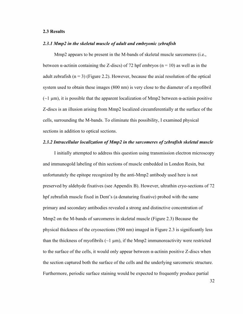
2.3.1 Mmp2 in the skeletal muscle of adult and embryonic zebrafish
Mmp2 appears to be present in the M-bands of skeletal muscle sarcomeres (i.e.,
between α-actinin containing the Z-discs) of 72 hpf embryos (n = 10) as well as in the
adult zebrafish (n = 3) (Figure 2.2). However, because the axial resolution of the optical
system used to obtain these images (800 nm) is very close to the diameter of a myofibril
(~1 µm), it is possible that the apparent localization of Mmp2 between α-actinin positive
Z-discs is an illusion arising from Mmp2 localized circumferentially at the surface of the
cells, surrounding the M-bands. To eliminate this possibility, I examined physical
sections in addition to optical sections.
2.3.2 Intracellular localization of Mmp2 in the sarcomeres of zebrafish skeletal muscle
I initially attempted to address this question using transmission electron microscopy
and immunogold labeling of thin sections of muscle embedded in London Resin, but
unfortunately the epitope recognized by the anti-Mmp2 antibody used here is not
preserved by aldehyde fixatives (see Appendix B). However, ultrathin cryo-sections of 72
hpf zebrafish muscle fixed in Dent’s (a denaturing fixative) probed with the same
primary and secondary antibodies revealed a strong and distinctive concentration of
Mmp2 on the M-bands of sarcomeres in skeletal muscle (Figure 2.3) Because the
physical thickness of the cryosections (500 nm) imaged in Figure 2.3 is significantly less
than the thickness of myofibrils (~1 µm), if the Mmp2 immunoreactivity were restricted
to the surface of the cells, it would only appear between α-actinin positive Z-discs when
the section captured both the surface of the cells and the underlying sarcomeric structure.
Furthermore, periodic surface staining would be expected to frequently produce partial
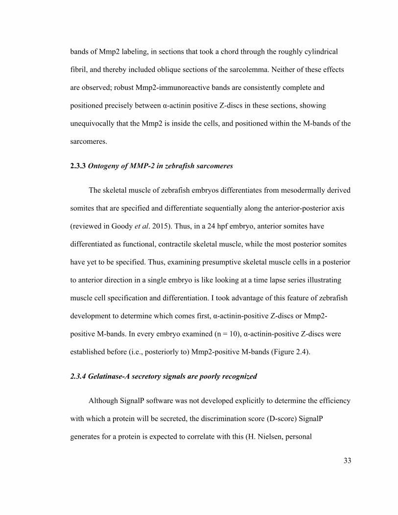
33
bands of Mmp2 labeling, in sections that took a chord through the roughly cylindrical
fibril, and thereby included oblique sections of the sarcolemma. Neither of these effects
are observed; robust Mmp2-immunoreactive bands are consistently complete and
positioned precisely between α-actinin positive Z-discs in these sections, showing
unequivocally that the Mmp2 is inside the cells, and positioned within the M-bands of the
sarcomeres.
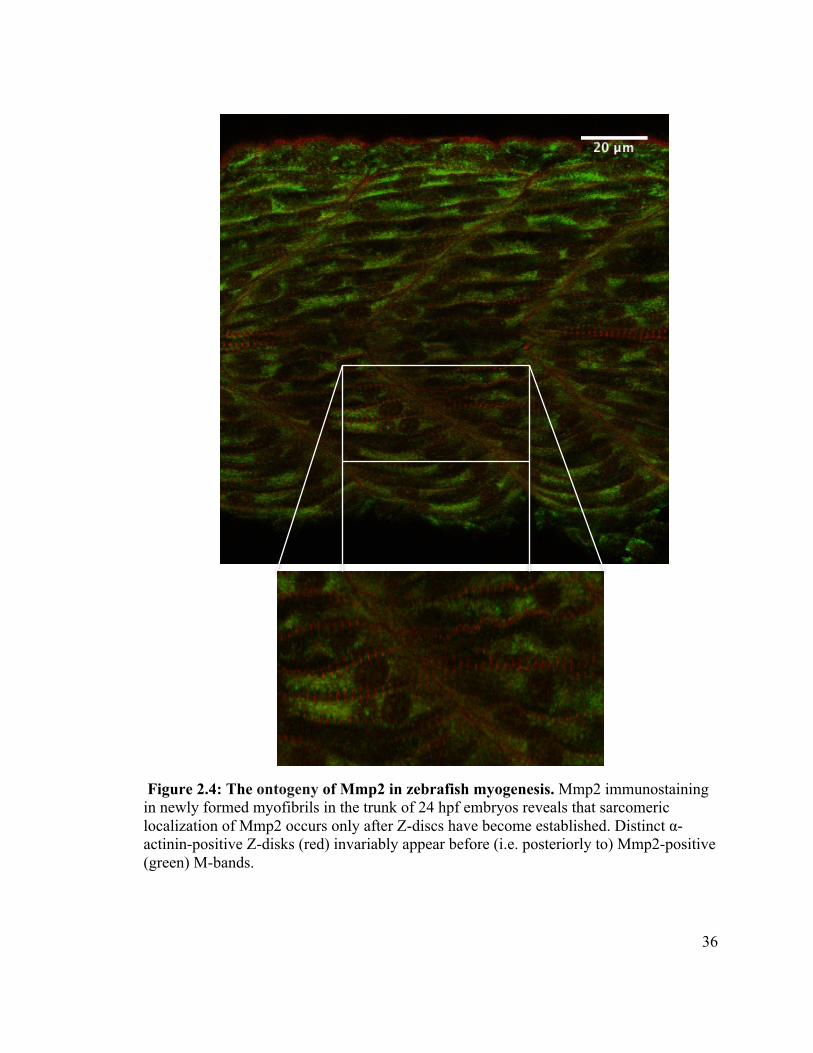
2.3.3 Ontogeny of MMP-2 in zebrafish sarcomeres
The skeletal muscle of zebrafish embryos differentiates from mesodermally derived
somites that are specified and differentiate sequentially along the anterior-posterior axis
(reviewed in Goody et al. 2015). Thus, in a 24 hpf embryo, anterior somites have
differentiated as functional, contractile skeletal muscle, while the most posterior somites
have yet to be specified. Thus, examining presumptive skeletal muscle cells in a posterior
to anterior direction in a single embryo is like looking at a time lapse series illustrating
muscle cell specification and differentiation. I took advantage of this feature of zebrafish
development to determine which comes first, α-actinin-positive Z-discs or Mmp2-
positive M-bands. In every embryo examined (n = 10), α-actinin-positive Z-discs were
established before (i.e., posteriorly to) Mmp2-positive M-bands (Figure 2.4).
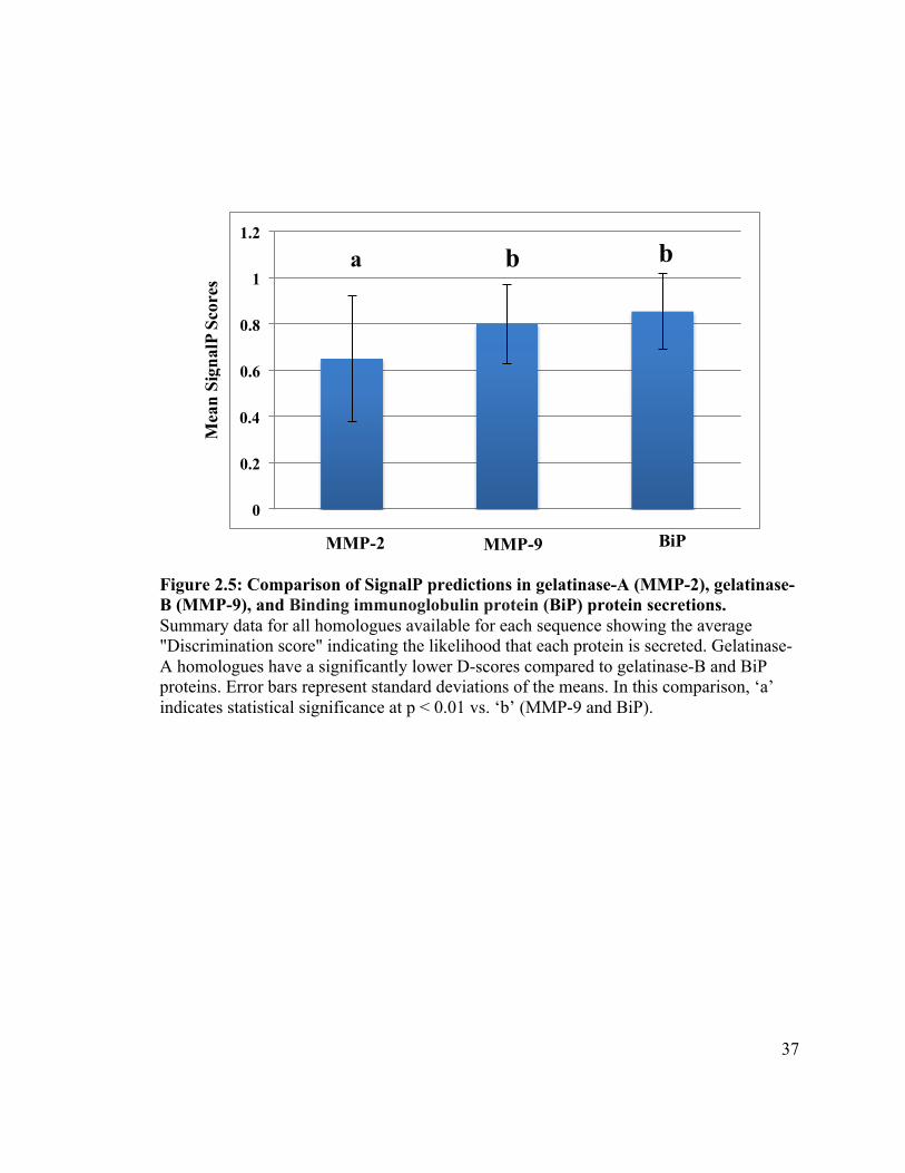
2.3.4 Gelatinase-A secretory signals are poorly recognized
Although SignalP software was not developed explicitly to determine the efficiency
with which a protein will be secreted, the discrimination score (D-score) SignalP
generates for a protein is expected to correlate with this (H. Nielsen, personal
34
communication).
I assembled a database of all gelatinase-A, gelatinase-B and BiP homologues from
vertebrate species for which all three were available as complete sequences, and
subjected them to analysis using SignalP (Appendix A). In this analysis, BiP is a positive
control, in that it is a well-known ER-resident protein with a strong signal sequence.
Gelatinase-B (MMP-9) was also included in this analysis, as a closely related MMP to
gelatinase-A.
Gelatinase-A homologues have a mean D-score of 0.651 (± 0.273) (Figure 2.5).
The default SignalP threshold above which a protein is considered ‘secreted’ is 0.450,
making Mmp2 homologues clearly identifiable as secreted proteins as expected.
However, both gelatinase-B and BiP had much higher mean D-scores (0.801 ± 0.170 and
0.854 ± 0.164, respectively; Figure 2.5), consistent with the hypothesis that the inefficient
secretion of gelatinase-A homologues observed in mammals and zebrafish may be a
conserved feature of this protein. Because the D-scores generated for these sequences
were not normally distributed, I used a Mann-Whitney U-test to determine if the
differences in the scores were statistically significant, and found that the D-scores of
gelatinase-A was significantly lower than the D-scores of gelatinase-B and BiP (p <
0.01).
35
Figure 2.2: Apparent Mmp2 localization at M-bands in zebrafish muscles. The longitudinal images of 72 hpf embryo (left) and adult (right) zebrafish skeletal musculature showing the distribution of Mmp2 protein (green) with respect to α–actinin (red) imaged by confocal microscopy.
Figure 2.3: Evidence of M-band localization of Mmp2 within 72 hpf zebrafish sarcomeres. Representative image of ultra-thin (500 nm) cryosection of 72 hpf zebrafish skeletal muscle with Mmp2 (green) clearly localized precisely between the Z-discs that contain α-actinin (red).
36
Figure 2.4: The ontogeny of Mmp2 in zebrafish myogenesis. Mmp2 immunostaining in newly formed myofibrils in the trunk of 24 hpf embryos reveals that sarcomeric localization of Mmp2 occurs only after Z-discs have become established. Distinct α-actinin-positive Z-disks (red) invariably appear before (i.e. posteriorly to) Mmp2-positive (green) M-bands.
37
Figure 2.5: Comparison of SignalP predictions in gelatinase-A (MMP-2), gelatinase-B (MMP-9), and Binding immunoglobulin protein (BiP) protein secretions. Summary data for all homologues available for each sequence showing the average "Discrimination score" indicating the likelihood that each protein is secreted. Gelatinase-A homologues have a significantly lower D-scores compared to gelatinase-B and BiP proteins. Error bars represent standard deviations of the means. In this comparison, ‘a’ indicates statistical significance at p < 0.01 vs. ‘b’ (MMP-9 and BiP).
0
0.2
0.4
0.6
0.8
1
1.2
BiP MMP-9 MMP-2
b b a
Mea
n Si
gnal
P Sc
ores
38
2.4 Discussion
In this chapter I show by using immunofluorescence, cryosectioning, and confocal
microscopy that Mmp2 has distinct intracellular localization in skeletal muscle in
zebrafish. Furthermore, the poorly recognized secretory signal characterized in human
MMP-2 appears typical of gelatinase-A orthologues, suggesting a conserved function of
this protease within the sarcomeres of vertebrate striated muscle.
I found Mmp2 accumulated on the M-bands of zebrafish skeletal muscle sarcomeres.
Interestingly, Mmp2 is not detected in Z-discs in zebrafish muscle, in contrast to what has
been reported regarding mammalian MMP-2 (Wang et al. 2002; Sawicki et al. 2005; Ali
et al. 2010). By using immunohistochemistry, Ali and co workers showed strong co-
localization of MMP2 with the T12 anti-titin antibody, which binds close to the Z-disc in
human and rat cardiomyocytes (Ali et al. 2010). On the other hand, the immunoreactivity
of MMP2 did not co-localize with the M8 anti-titin antibody, which targets the M-bands
(reviewed in Müller et al. 2012). Thus, while it is clear that in zebrafish Mmp2 is present
within the sarcomere of skeletal muscle, its distribution within the contractile apparatus
appears to be different than that in mammalian cardiomyocytes. This difference may have
important functional implications, and is worthy of further investigation.
The sarcomeric localization of Mmp2 in zebrafish skeletal muscle occurs
subsequently to the establishment of Z-discs, and therefore, while I cannot rule out a
developmental function for this protease in the development of the contractile apparatus,
it seems more likely that Mmp2 functions in the maintenance of the sarcomere.
39
Others have shown empirically that mammalian MMP-2 is inefficiently secreted (Ali
et al. 2011b), and my analysis of signal sequences in all vertebrate gelatinase-A
homologues for which sequence data are available is consistent with this. This
conservation of inefficient signal sequences suggests a selective pressure to maintain an
intracellular pool of this protease. This intracellular pool is clearly involved in
pathological activities under conditions of oxidative stress (reviewed in Kandasamy et al.
2010; Ali et al. 2011a), but its physiological function remains obscure.
40
2.5 References
Ali MA, Cho WJ, Hudson B, Kassiri Z, Granzier H, Schulz R. 2010. Titin is a target of matrix metalloproteinase-2: implications in myocardial ischemia/reperfusion injury. Circulation. 122(20): 2039-47.
Ali MA, Fan X, Schulz R. 2011a. Cardiac sarcomeric proteins: novel intracellular targets of matrixmetalloproteinase-2 in heart disease. Trends Cardiovasc Med. 21(4): 112-118.
Ali MA, Chow AK, Kandasamy AD, Fan X, West LJ, Crawford BD, Simmen T, Schulz R. 2011b. Mechanisms of cytosolic targeting of matrix metalloproteinase-2. J Cell Physiol. 227: 3397-3404. !
Chow AK, Cena J, Schulz R. 2007. Acute actions and novel targets of matrix metalloproteinases in the heart and vasculature. Br J Pharmacol. 152(2): 189–205.
Cooper GM, Hausman RE. 2009. The cell: a molecular approach. Fifth edition. Washington (DC): The American Society of Microbiology Press. p. 383-432.
Goody MF, Sher RB, Henry CA. 2015. Hanging on for the ride: adhesion to the extracellular matrix mediates cellular responses in skeletal muscle morphogenesis and disease. Dev Biol.401(1): 75–91.
Harris ND. 2010. Molecular ontogeny of extracellular matrix remodeling proteases during development of the zebrafish, and the effects of simulated micro-gravity on their expression. MSc thesis. University of New Brunswick [Fredericton (NB)].
Kandasamy AD, Chow AK, Ali MA, Schulz R. 2010. Matrix metalloproteinase-2 and myocardial oxidative stress injury: beyond the matrix. Cardiovasc Res. 85(3): 413–423.
Kimmel CB, Ballard WW, Kimmel SR, Ullmann B, Schilling TF. 1995. Stages of embryonic development of the zebrafish. Dev Dyn. 203(3): 253-310.
Klee EW, Ellis LB. 2005. Evaluating eukaryotic secreted protein prediction. BMC Bioinf. 14(6): 256.
Lévesque JM. 2010. The localization of MMP2 in zebrafish embryos implies unexpected roles in neurogenesis and muscle function. Honours thesis. University of New Brunswick [Fredericton (NB)].
Luther PK. 2009. The vertebrate muscle Z-disc: sarcomere anchor for structure and signalling. J Muscle Res Cell Motil. 30(5-6): 171-85.
Müller AL, Hryshko LV, Dhalla NS. 2012. Extracellular and intracellular proteases in cardiac dysfunction due to ischemia–reperfusion injury. Int J Cardiol. 164(1): 39-47.
Murray JM. Confocal microscopy, deconvolution, and structured illumination methods.
41
In: Spector DL, Goldman RD, editors. Basic methods in microscopy, protocols and concepts from cell: a laboratory manual. New York (NY): Cold Spring Harbor Laboratory Press; 2006. p. 43-81.
Nielsen H, Engelbrecht J, Brunak S, von Heijne G.1997. Identification of prokaryotic and eukaryotic signal peptides and prediction of their cleavage sites. Protein Eng. 10(1): 1-6.
Recher G, Rouede D, Richard P, Simon A, Bellanger JJ, Tiaho F. 2009. Three distinct sarcomeric patterns of skeletal muscle revealed by SHG and TPEF microscopy. Opt Express.17(22): 19763-77.
Sariahmetoglu M, Crawford BD, Leon H, Sawicka J, Li L, Ballermann BJ, Holmes C, Berthiaume LG, Holt A, Sawicki G, et al. 2007. Regulation of matrix metalloproteinase-2 (MMP-2) activity by phosphorylation. FASEB J. 21(10): 2486-2495.
Sawicki G, Leon H, Sawicka J, Sariahmctoglu M, Schulze CJ, Scott PG, Szczesna-Cordary D, Schulz R. 2005. Degradation of myosin light chain in isolated rat hearts subjected to ischemia-reperfusion injury: a new intracellular target for matrix metalloproteinase-2. Circulation. 112(4): 544-552.
Schulz R. 2007. Intracellular targets of matrix metalloproteinase-2 in cardiac disease: rationale and therapeutic approaches. Annu Rev Pharmacol Toxicol. 47: 211–242.!
Squire JM, Knupp C, Luther PK. 2008. Zebrafish topical, transparent, and tractable for ultrastructural studies. J Gen Physiol. 131(5): 439–443.
Sung MM, Schulz CG, Wang W, Sawicki G, Bautista-Lopez NL, Schulz R. 2007. Matrix metalloproteinase-2 degrades the cytoskeletal protein α- actinin in peroxynitrite mediated myocardial injury. J Mol Cell Cardiol. 43(4): 429-436.
Wang W, Schulze CJ, Suarez-Pinzon WL, Dyck JR, Sawicki G, Schulz R. 2002. Intracellular action of matrix metalloproteinase-2 accounts for acute myocardial ischemia and reperfusion injury. Circulation.106(12): 1543-1549.
Westerfield M. 1995. The Zebrafish Book: A guide for the laboratory use of zebrafish (Danio rerio). Eugene (OR): University of Oregon Press.
Zhang J, Bai S, Zhang X, Nagase H, Sarras MP. 2003. The expression of gelatinase A (MMP-2) is required for normal development of zebrafish embryos. Dev Genes Evol. 231(9): 456–463.
42
Chapter 3: Phosphorylation status of Mmp2 in zebrafish myocytes
3.1 Introduction
After 40 years of study MMPs were viewed as functioning exclusively as extracellular
proteases (reviewed in Brinckerhoff and Matrisian 2002). However, in the last decade
and a half, work primarily from the Schulz lab at the University of Alberta revealed the
unexpected presence of MMP2 within cardiomyocytes of mammals (Wang et al. 2002;
Sawicki et al. 2005), and implicated it in the pathology of ischemia/reperfusion injury
(IRI) (reviewed in Schulz 2007). Other labs have now demonstrated the presence of
MMP2 within the skeletal muscle fibres of mouse (Hadler-Olsen et al. 2014) as well as
within neurons (Yang et al. 2010) and hepatocytes (Kwan et al. 2004). In 2007,
Sariahmetoglu and her colleagues reported that human MMP-2 is phosphorylated at
several serine and threonine residues in vivo, and that this post-translational modification
dramatically changes its enzymatic activity (Sariahmetoglu et al. 2007; reviewed in
Schulz 2007). Regulation by phosphorylation is generally a characteristic of intracellular
proteins, although there are exceptions (Yalak et al. 2014). Moreover, the specificity of
protein kinases suggests that this may be an evolutionarily conserved regulatory
mechanism.
Subsequently, several studies addressed the roles of this post-transitional modification
on the structure and activity of gelatinase-A in the presence of peroxynitrite (ONOO-).
Sariahmetoglu and her colleagues (2012) reported the importance of phosphorylation
status of MMP2 in myocardial injury. They noticed that increasing the proportion of
43
phosphorylated (inactive) MMP2 using phosphatase inhibitors helps protect the
contractile proteins from degradation through the deleterious action of MMP2 activated
by ONOO- (Sariahmetoglu et al. 2012; reviewed in Sawicki 2013). Moreover, the
combined effect of both ONOO- and dephosphorylation of MMP2 generated a greater
increase in proteolytic activity than ONOO- alone (Jacob-Ferreira et al. 2013), suggesting
that mis-regulation of MMP2 phosphorylation may be more important in the pathology of
IRI than the direct effect of activation by reactive oxygen species (ROS).
Protein kinases are one of the biggest families of eukaryotic genes (reviewed in
Ubersax and Ferrell 2007). They can be roughly divided into two major classes: tyrosine
kinases and serine/threonine kinases. Tyrosine, serine and threonine are the only amino
acids that are normally phosphorylated by kinases in proteins. Kinases catalyze the
nucleophilic attack of the deprotonated hydroxyl group in the amino acid side chain on
the gamma phosphate of an adenosine triphosphate (ATP) molecule, yielding adenosine
diphosphate and a phosphorylated protein. Conversely, phosphatases remove a phosphate
group from phosphoproteins, yielding inorganic phosphate (Pi) and a dephosphorylated
protein (reviewed in Cheng et al. 2011).
Although the post-translational modification by phosphorylation is generally
associated with regulation of intracellular proteins, a variety of proteins in the
extracellular milieu appeared to be phosphorylated by exokinases (Yalak et al. 2014). For
example, Fam20C is a kinase that resides in the Golgi apparatus and is responsible for the
phosphorylation of proteins destined for secretion, including caseins and bone proteins
that are important in biomineralization. Fam20C phosphorylates the serine residue within
44
S-x-E motifs (where x is any amino acid) (Tagliabracci et al. 2012).
The finding that human gelatinase-A (MMP-2) is phosphorylated in living cells, and
that this phosphorylation changes its enzymatic activity, made me want to explore
whether this mechanism applies outside of mammalian cells, and therefore may represent
an ancient regulatory mechanism. The presence and activity of such a system may shed
some light on the normal physiological roles of MMP-2 inside cells, and may also
indicate possible strategies for reducing undesirable MMP-2 activity associated with
pathology.
Here I present an approach for the detection of phospho-Mmp2 in tissue samples using
homogenized muscle cells of adult zebrafish. This approach involves affinity
chromatography, in which gelatin sepharose is bound by the fibronectin type II repeats of
the collagen binding domain of Mmp2 (zebrafish gelatinase-A) and Mmp9 (zebrafish
gelatinase-B). I used Pro-Q Diamond stain that is specific for phosphoproteins on SDS-
PAGE gels. By comparing the fluorescent intensity of protein bands stained with Pro-Q-
Diamond to the intensity obtained using SyproRuby, which is a general protein stain, and
then comparing that to a series of known standards, it is possible to quantitatively
determine the proportion of any given protein that is phosphorylated in a standard SDS-
PAGE gel (reviewed in Delom and Chevet 2006). Unfortunately, technical limitations
prevented me from purifying sufficient Mmp2 from zebrafish muscle to use this
approach. I therefore turned to a bioinformatic analysis of the conservation of predicted
phosphorylation sites in the gelatinase-A sequences across as many species as possible, in
order to look for evidence that phosphorylation of this protease may be significant in its
45
physiological regulation.
NetPhos is an artificial neural network that predicts potential phosphorylation sites by
examining the sequences flanking empirically demonstrated sites of phosphorylation, and
using a heuristic based on these, determines the probability that a given serine, threonine
or tyrosine in an unknown sequence will be phosphorylated (Blom et al. 1999). I
collected and made an alignment of all available gelatinase-A sequences, and analyzed
the conservation of motifs that were identified as high-probability sites of
phosphorylation by using NetPhos. While I was unable to empirically verify Mmp2
phosphorylation in zebrafish, I determined that it is highly probable that zebrafish Mmp2
is subject to phosphorylation and that this post-translational modification is important in
regulating gelatinase-A activity throughout the animal kingdom.
46
3.2 Materials and methods
3.2.1 Tissue preparation
In the pilot attempt of the gelatin affinity chromatography preparation, six adult
zebrafish were terminally anesthetized in 0.3 mg/ml tricaine methanesulfonate (Sigma)
solution for 10 minutes. The tail muscles were dissected from the fish, frozen in liquid
nitrogen and crushed with a cell crusher (Stratech Scientific Limited). Following several
attempts working with this method, I increased the number of fish in order to increase the
amount of muscle homogenate, using 57 adult zebrafish in the final chromatography
experiment.
3.2.2 Isolation of Gelatinases using gelatin-affinity chromatography
Protein was purified by pulverizing tail muscle dissected from adult zebrafish after
freezing in liquid nitrogen. The frozen pulverized muscle was transferred to cold
homogenization buffer (150 mM NaCl, 50 mM Tris pH 7.5, 0.1% Brij-35, 1% NP-40,
0.02 M sodium deoxycholate, 5 mM EDTA, 1x protease inhibitor cocktail), vortexed and
placed on ice. Homogenized muscle was centrifuged at 16,000x g for 25 minutes at 4°C
to pellet the insoluble debris. The resulting supernatant was collected and stored at -20°C
for later use. Gelatin affinity columns were prepared using either commercially available
gelatin sepharose 4B (GE Healthcare Bio-Sciences AB, Bjorkgatan, Sweden) or gelatin
affinity media prepared using cyanogen bromide activated sepharose 4B (Sigma), with a
final bed volume of 25 ml. For the cyanogen bromide preparation, 1 g of dry resin was
washed with 100 ml of 1 mM HCl at room temperature for 10 minutes four times and left
47
for five minutes to allow the resin to settle down in order to pour off the supernatant. The
resin was washed with distilled H2O and then with two volumes of coupling buffer (0.5
M NaCl, 0.1 M NaHCO3, pH 8.5). While preparing the resin, 10 mg/ml of ligand gelatin
protein (Fisher) was dissolved in coupling buffer, with gentle warming. The washing
coupling buffer was poured off and ligand solution was added immediately, and
incubated overnight at 4°C with gentle agitation. Supernatant was removed, the resin
washed with 100 ml of coupling buffer for 10 minutes and left to settle. Buffer was
poured off and the unreacted sites were blocked by incubating with 0.2 M glycine in
coupling buffer overnight at 4°C with gentle agitation. Finally, the prepared resin was
poured into the column and washed extensively, alternating with two column volumes of
acetate buffer (0.5 M NaCl, 0.1 M sodium acetate pH 4), and two column volumes of
coupling buffer, five times. The resin was equilibrated with binding buffer before use, as
described below.
Prior to loading in the column, the thawed muscle homogenate was centrifuged at
16,000x g for 25 minutes at 4°C in order to pellet any precipitate. Gelatin sepharose
medium was equilibrated with at least 5 column volumes of binding buffer (150 mM
NaCl, 50 mM Tris pH 7.5, 0.1% Brij-35, 5 mM EDTA), and roughly 170 ml of muscle
homogenate was allowed to run into the column at 2 ml per minute. Unbound protein was
washed from the column using 10 column volumes of binding buffer at 2 ml per minute.
Finally, bound protein was eluted with elution buffer (4M urea, 50 mM Tris pH 7.5, 5
mM EDTA, and 0.1% Brij-35). Roughly 30 ml of eluted fractions were concentrated
down to 200 µl using an Amicon Ultra 4 mL Filter (Millipore) centrifuged at 894.4x g at
4°C.
48
3.2.3 SDS-polyacrylaminde gel electrophoresis and phospho-protein identification
Samples of total homogenate and gelatin-affinity purified proteins were diluted 1:1 in
2x reducing sample buffer (250 mM Tris-HCl pH 6.8, 4% sodium dodecyl sulfate, 0.2%
bromophenol blue, 20% glycerol, 200 mM β-mercaptoethanol). 70 µl of each sample, as
well as 6 µl of EZ-run (Thermo) protein molecular weight standards, were resolved on a
10% SDS-PAGE gel at 50V for four hours. The gel was fixed in 100 ml of 50% methanol
and 10% acetic acid for 30 minutes, twice, and washed three times with 100 ml of
deionized water for 10 minutes. The gel was incubated in 60 ml of Pro-Q Diamond
Phosphoprotein Gel Stain solution (Invitrogen) for 60-90 minutes according to the
manufacturer’s instructions. After incubation, the gel was destained three times in 100 ml
of 20% acetonitrile and 50 mM sodium acetate, pH 4, for 30 minutes. Finally, the gel was
washed twice with 100 ml of deionized water for five minutes, and imaged using the
ChemiDoc MP imaging system.
After documenting the abundance of phosphoproteins, the gel was re-stained in 60 ml
of Sypro Ruby (Invitrogen) gel stain overnight for total protein detection. After washing
in 10% methanol and 7% acetic acid for 30 minutes the gel was re-imaged with the
ChemiDoc MP imaging system, and intensity quantified using Image Lab version 4.0.1
(Bio-Rad).
3.2.4 Immunoblots
Purified protein samples (from 3.2.2) were diluted 1:1 in 2x reducing sample buffer
(250 mM Tris-HCl pH6.8, 4% sodium dodecyl sulfate, 0.2% bromophenol blue, 20%
glycerol, 200 mM β-mercaptoethanol) and separated using SDS-PAGE on 10% gels, as
49
described in 3.2.3. The proteins were transferred to polyvinylidene difluoride (PVDF)
membranes at 50 volts for four hours in Tobin buffer (25 mM Tris base, 192 mM glycine
and 20% methanol, pH 8.3). The membrane’s remaining protein-binding capacity was
saturated by incubating overnight in blocking buffer (5% skim milk powder in PBSTw
(0.1% Tween-20 in PBS (20 mM phosphate pH 7.3, 137 mM NaCl, 2.7 mM KCl)) at 4°C
with gentle agitation. The blocked membrane was incubated with primary antibody
(rabbit anti-Mmp2) (Anaspec catalogue #55111) diluted (1:1000) in blocking buffer and
gently agitated overnight at 4°C. The membrane was then washed with PBSTw three
times for five minutes each time. The membrane was re-incubated in horse-radish
peroxidase (HRP) conjugated goat anti-rabbit secondary antibody diluted in blocking
buffer (1:1000) overnight at 4°C with constant agitation. After washing the membrane
three times for five minutes each time with PBSTw, 2 ml of enhanced
chemiluminescence (ECL plus) (GE Healthcare Life Sciences) was applied to the
membrane and luminescent bands imaged using the ChemiDoc MP imaging system
3.2.5 Gelatin zymography
Samples of muscle homogenate and gelatin affinity purified proteins were diluted 1:1
in 2x non-reducing SDS-PAGE sample buffer (250 mM Tris-HCl pH 6.8, 4% sodium
dodecyl sulfate, 0.2% bromophenol blue, 20% glycerol) and resolved on a 10%
acrylamide zymogram containing 4 µg/ml gelatin (Fisher) at 50 V for four hours. The
zymogram was washed twice for 30 minutes at room temperature in 1x zymogram
renaturing buffer (2.5% Triton X-100 in 50 mM TrisHCl pH 7.5). The renaturing buffer
was removed and replaced with 1x zymogram developing buffer (50 mM TrisHCl pH
50
7.5, 5 mM CaCl2, 1 mM ZnSO4, 2.5% Triton X-100) and gently agitated for 30 minutes
at room temperature. This buffer was replaced with a fresh solution of 1x zymogram
developing buffer and incubated for 48 h at 28°C. The gel was then stained in 0.5%
Coomassie brilliant blue dye G-250, dissolved in 25% methanol and 10% acetic acid at
room temperature overnight. Destaining was conducted the following day using a
solution of methanol, acetic acid, and water (40:10:50). Images of the gelatinolytic
activity detected as clear bands against the Coomassie brilliant blue stained background
were taken using the ChemiDoc MP imaging system.
3.2.6 Silver staining
Samples of whole muscle homogenate and affinity purified eluate in 2x reducing
sample buffer, as well 6 µl of Precision Plus Protein All Blue standards (BioRad), were
resolved on a 10% SDS-PAGE gel as described above. The gel was fixed in a solution of
50% methanol, 12% glacial acetic acid, 38% water with 200 µl of 37% formaldehyde,
with heating for one minute in the microwave. The gel was washed in 100 ml of 1:1 95%
ethanol: water in the microwave for one minute and rehydrated in distilled water for 2-3
minutes at room temperature. The proteins in the gel were then reduced in a solution of
0.04% sodium thiosulphate in the microwave for one minute. This was followed by
washing in distilled water for 30 seconds at room temperature and staining in 100 ml of
water, 200 mg of silver nitrate, and 150 µl 37% formaldehyde for one minute in the
microwave. Finally, the gel was washed for 30 seconds in distilled water, and developed
in a solution of 100 ml water, 6 g sodium carbonate, 10 µl 4% sodium thiosulfate, and
100 µl 37% formaldehyde at room temperature. 3% acetic acid was used to stop the
51
developing reaction, and the results photographed using the ChemiDoc MP imaging
system.
3.2.7 Prediction of phosphorylation sites within vertebrate Gelatinase-A homologues
The sequences of vertebrate MMP-2 genes (see Appendix A) were obtained from
PubMed protein database (http://www.ncbi.nlm.nih.gov/protein/?term=MMP-2). NetPhos
2.0 software (http://www.cbs.dtu.dk) was used to predict the phosphorylation sites. This
program is based on a neural network method that predicts tyrosine, threonine and serine
phosphorylations domains located in eukaryote proteins (Blom et al. 1999).

52
3.3 Results
3.3.1 The purification of Mmp2 using gelatin affinity chromatography
In order to determine the phosphorylation status of Mmp2 in zebrafish muscle, I
attempted to purify this protease from skeletal muscle using gelatin affinity
chromatography. I was able to purify small amounts of Mmp2 from adult zebrafish
muscle and the identity of the eluted protein was confirmed by probing western blots of
eluted fractions with anti-Mmp2 (Figure 3.1). Further, when I modified the protocols and
used urea in the elution buffer in some experiments, I was able to detect gelatinolytic
activity in the eluate (Figure 3.2), although the residual urea in these samples caused
extensive distortion of the bands during electrophoresis. I conclude that gelatin affinity
chromatography can be an effective method for extracting Mmp2 from fish muscle tissue.
However, replicating these results proved very challenging, presumably due to low yields
and/or degradation of the protease during or after chromatographic purification.
In my final set of experiments, I dramatically increased the amount of starting
material, using the muscle dissected from 57 adult fish. Unfortunately, this resulted in the
highly contaminated eluate shown in Figure 3.3. I was not able to detect any gelatinolytic
activity in zymograms of this material (Figure 3.4), nor was there any Mmp2
immunoreactivity (Figure 3.5). It seems likely that the Mmp2 broke down during
homogenate preparation or during storage, as both homogenate and eluate were stored for
one week while phosphorylation analysis was conducted, before immunobloting and
zymography was done in order to verify the presence of Mmp2.
53
3.3.2 Mmp2 phosphorylation status remains undetermined
Analysis of phosphorylation was conducted prior to confirmation of the presence of
Mmp2 in the eluate. In hindsight, this was an error. However, a small amount of 62 kD
protein appears to be present in the eluate, which may represent Mmp2 (Figure 3.3).
Moreover, abundant proteins in the homogenate muscle sample were clearly detectable in
both SYPRO Ruby and Pro-Q Diamond stains, indicating the workability of this
technique to analyze the phosphorylation status of zebrafish proteins (Figure 3.6 A).
Using SYPRO Ruby stain, the 62 kD protein present in the eluate is just detectable
(Figure 3.6 A and B), but it is not detectable using Pro-Q Diamond (Figure 3.7),
suggesting that Mmp2 either is not phosphorylated, or its abundance is below the limit of
detection for Pro-Q Diamond. The lower limit of detection for Pro-Q Diamond is in the
range of 1-16 ng of phosphoprotein (Invitrogen product information sheet), depending on
the extent of phosphorylation, while the limit of detection for SYPRO Ruby is
approximately 1 ng of protein (Invitrogen product information sheet). Thus, if the circled
band in Figure 3.6 represents a few nanograms of Mmp2, it may be that the
phosphorylation is below the limit of Pro-Q Diamond sensitivity. Therefore, it is
impossible to determine what the phosphorylation status of Mmp2 is from this result.
3.3.3 Prediction of putative MMP-2 phosphorylation sites
Using NetPhos software, human MMP-2 phosphorylation sites have been predicted,
and several of these sites have been empirically verified by mass spectrometry
(Sariahmetoglu et al. 2007; Jacob-Ferreira et al. 2013). One of the major questions in my
54
thesis is whether this post-translational regulatory mechanism might apply outside of
humans, and may therefore represent an evolutionarily ancient characteristic of
gelatinases. Because I was unable to establish the phosphorylation status of Mmp2
empirically, I used NetPhos to look for putative phosphorylation sites in the sequences of
gelatinase-A orthologues from as many vertebrates for which sequence data are available.
The domain structure of gelatinase-A orthologues is highly conserved, with a
stereotypical pattern of cysteines that form disulfide bridges that constrain protein
folding. The position of these cysteine residues makes the aligning of MMP-2 sequences
from different species relatively straightforward even when there are short insertions or
deletions. This also makes the conservation, or lack of conservation, of putative
phosphorylation sites quite obvious. NetPhos predicted that there are 14 high-probability
phosphorylation sites common to all or nearly all gelatinase-A sequences (out of an
average of 32 high-probability phosphorylation sites per protein) (Figure 3.8). Three of
these sites are conserved in all 73 sequences available, seven of them are conserved in
72/73, and four are conserved in 71/73 sequences. I did not consider sites that showed
less conservation.
Seven of the conserved high-probability phosphorylation sites are located in the
collagen-binding domain, and six predicted sites in the collagnase-like domain. In the C-
terminal hemopexin-like domain, there is one predicted site, on a tyrosine residue.
Interestingly, there are no conserved phosphorylation sites predicted in the pro-peptide
domain.
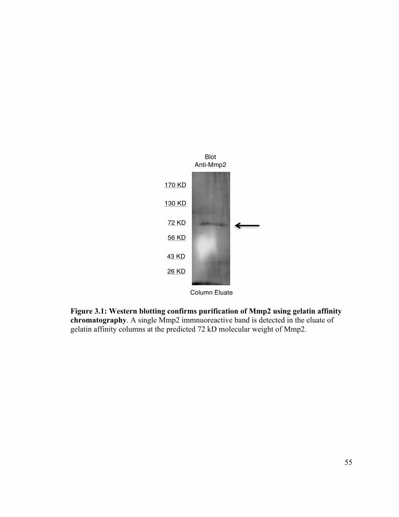
55
Figure 3.1: Western blotting confirms purification of Mmp2 using gelatin affinity chromatography. A single Mmp2 immnuoreactive band is detected in the eluate of gelatin affinity columns at the predicted 72 kD molecular weight of Mmp2.
72 KD
56 KD
43 KD
26 KD
170 KD
130 KD
Blot Anti-Mmp2
Column Eluate
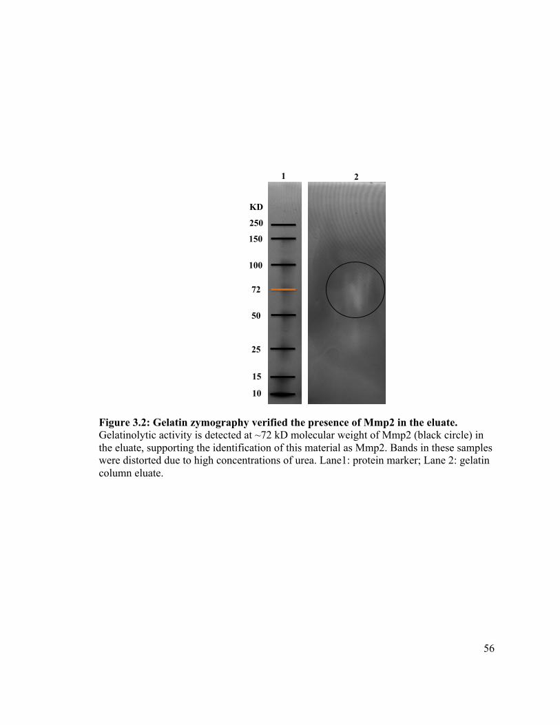
56
Figure 3.2: Gelatin zymography verified the presence of Mmp2 in the eluate. Gelatinolytic activity is detected at ~72 kD molecular weight of Mmp2 (black circle) in the eluate, supporting the identification of this material as Mmp2. Bands in these samples were distorted due to high concentrations of urea. Lane1: protein marker; Lane 2: gelatin column eluate.
2 2
72
50
25
10
15
100
150
250
KD
1
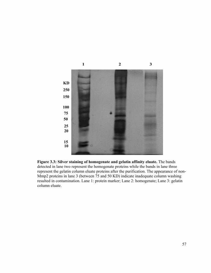
57
Figure 3.3: Silver staining of homogenate and gelatin affinity eluate. The bands detected in lane two represent the homogenate proteins while the bands in lane three represent the gelatin column eluate proteins after the purification. The appearance of non-Mmp2 proteins in lane 3 (between 75 and 50 KD) indicate inadequate column washing resulted in contamination. Lane 1: protein marker; Lane 2: homogenate; Lane 3: gelatin column eluate.
1 2 3
10 15
20 25
50
75
100
150
250
KD
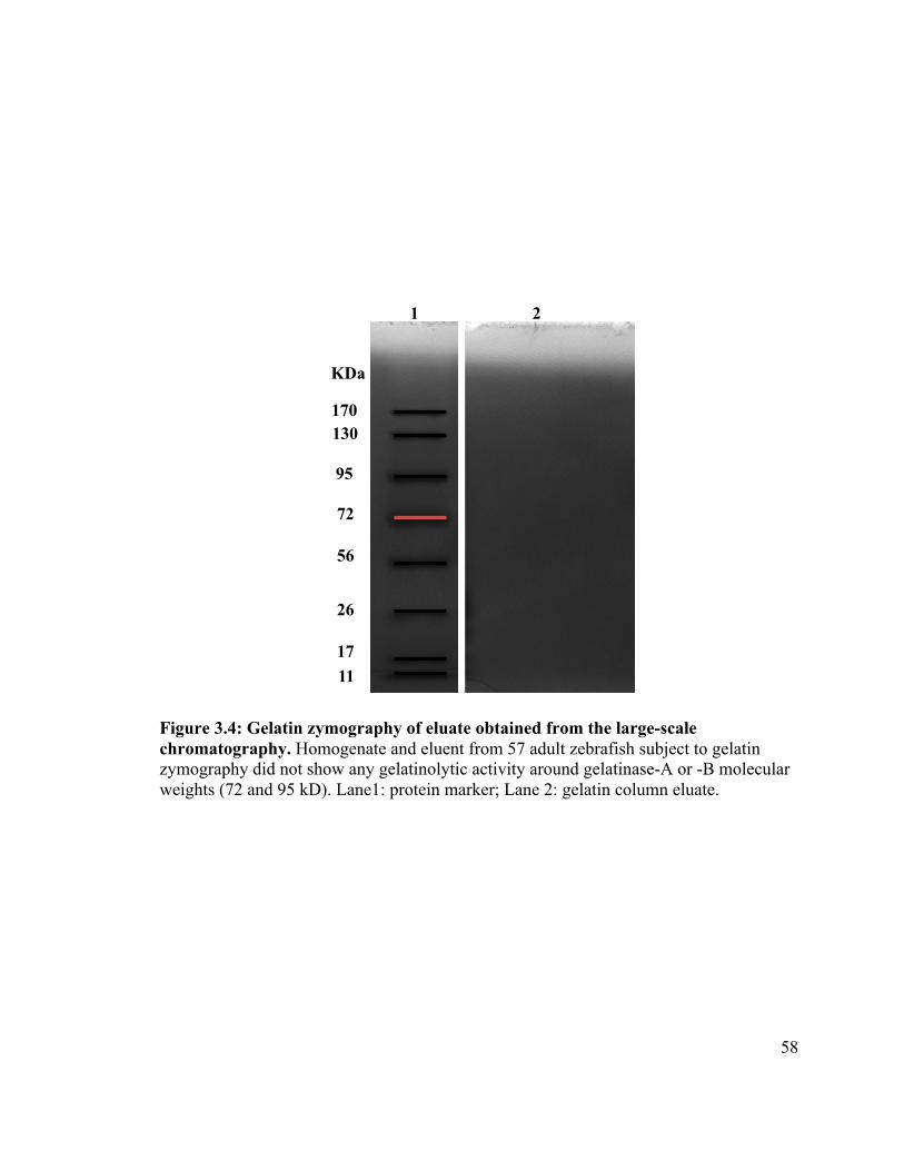
58
Figure 3.4: Gelatin zymography of eluate obtained from the large-scale chromatography. Homogenate and eluent from 57 adult zebrafish subject to gelatin zymography did not show any gelatinolytic activity around gelatinase-A or -B molecular weights (72 and 95 kD). Lane1: protein marker; Lane 2: gelatin column eluate.
170 130
95
72
56
11 17
26
KDa
1 2
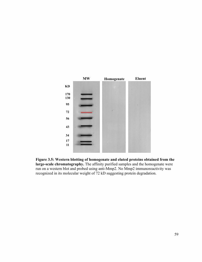
59
Figure 3.5: Western blotting of homogenate and eluted proteins obtained from the large-scale chromatography. The affinity purified samples and the homogenate were run on a western blot and probed using anti-Mmp2. No Mmp2 immunoreactivity was recognized in its molecular weight of 72 kD suggesting protein degradation.
170 130
95
56
72!
43
17 11
KD
34
MW Homogenate Eluent
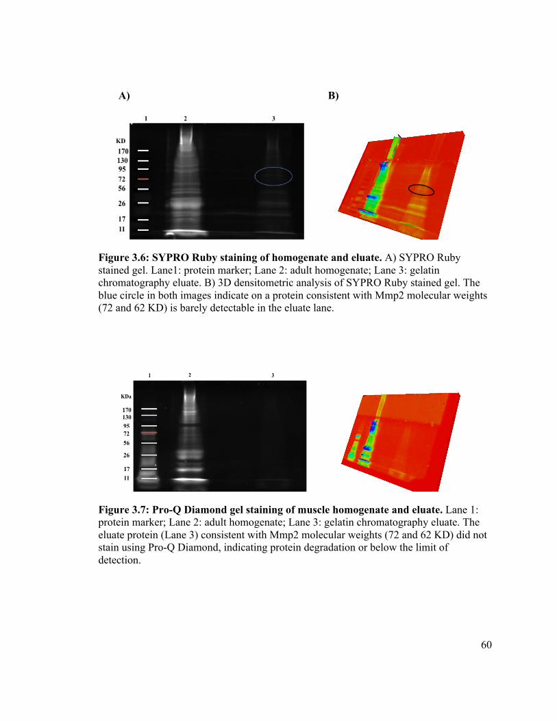
60
A) B)
Figure 3.6: SYPRO Ruby staining of homogenate and eluate. A) SYPRO Ruby stained gel. Lane1: protein marker; Lane 2: adult homogenate; Lane 3: gelatin chromatography eluate. B) 3D densitometric analysis of SYPRO Ruby stained gel. The blue circle in both images indicate on a protein consistent with Mmp2 molecular weights (72 and 62 KD) is barely detectable in the eluate lane.
Figure 3.7: Pro-Q Diamond gel staining of muscle homogenate and eluate. Lane 1: protein marker; Lane 2: adult homogenate; Lane 3: gelatin chromatography eluate. The eluate protein (Lane 3) consistent with Mmp2 molecular weights (72 and 62 KD) did not stain using Pro-Q Diamond, indicating protein degradation or below the limit of detection.
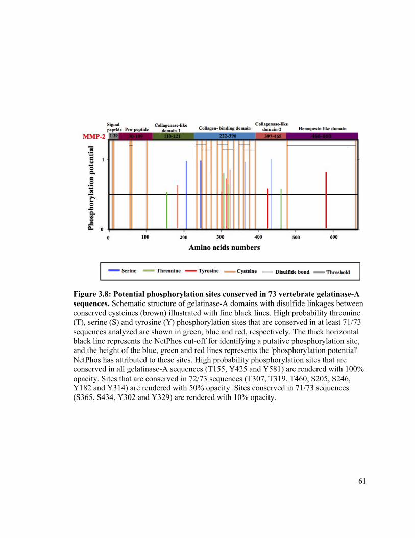
61
Figure 3.8: Potential phosphorylation sites conserved in 73 vertebrate gelatinase-A sequences. Schematic structure of gelatinase-A domains with disulfide linkages between conserved cysteines (brown) illustrated with fine black lines. High probability threonine (T), serine (S) and tyrosine (Y) phosphorylation sites that are conserved in at least 71/73 sequences analyzed are shown in green, blue and red, respectively. The thick horizontal black line represents the NetPhos cut-off for identifying a putative phosphorylation site, and the height of the blue, green and red lines represents the 'phosphorylation potential' NetPhos has attributed to these sites. High probability phosphorylation sites that are conserved in all gelatinase-A sequences (T155, Y425 and Y581) are rendered with 100% opacity. Sites that are conserved in 72/73 sequences (T307, T319, T460, S205, S246, Y182 and Y314) are rendered with 50% opacity. Sites conserved in 71/73 sequences (S365, S434, Y302 and Y329) are rendered with 10% opacity.
62
3.4 Discussion
The complex regulation of MMPs, which is affected by several factors including
regulation by post-translational processes, has been the centre of attention for some time.
The action of phosphorylation in controlling MMP activity had not been considered until
2007. Sariahmetoglu et al. (2007) reported for the first time that human MMP-2 is
phosphorylated on several serine and thronine residues, and that it dramatically regulates
the enzymatic activity of this protease. Elucidating whether this phenomenon may apply
in a broad spectrum of vertebrates including zebrafish is the focus of this chapter of my
thesis. This effort may reveal an evolutionarily ancient characteristic of gelatinases.
In this exploration, several efforts were made, both empirically and in silico, to
determine if Mmp2 from zebrafish muscle may be phosphorylated in vivo. Despite some
success using gelatin chromatography to purify gelatinase-A from zebrafish muscle, I
was not able to determine whether Mmp2 is phosphorylated in zebrafish. This may be
due to the limit of detection of Pro-Q Diamond in combination with poor protein yields
and/or degradation of the protein during or after purification.
In order to continue the work, I tested my hypothesis in silico, using software to look
for phosphorylation sites in the sequences of gelatinases from taxa for which sequence
data are available. I used NetPhos, a bioinformatic analysis method, to predict specific
motifs within protein sequences that are liable to phosphorylation. Although
phosphorylation sites have been predicted and empirically verified at several serine and
threonine residues in human and rat MMP-2 (Sariahmetoglu et al. 2007; Sariahmetoglu et
al. 2012), nothing has been done to verify if these sites are evolutionarily conserved. If
63
they are of functional significance, they should be conserved in many species. This would
support the hypothesis that gelatinases have important role(s) within the cells, and
suggest that these functions may be regulated by phosphorylation. I found that there are a
number of predicted phosphorylation sites, in particular within the collagen-binding
domain, conserved in zebrafish as well as in all other vertebrates for which sequence data
are available. One of these (S365) has already been empirically verified by mass
spectrometry as being phosphorylated in humans (Sariahmetoglu et al. 2007), suggesting
that phosphorylation within this domain affects substrate binding.
As a result of this work, I conclude that gelatinase-A proteins are likely to be
phosphorylated in vertebrates. This implies the existence of one or more kinases and one
or more phosphatases that act on gelatinase-A orthologues, either extracellularly or
intracellularly. Because the putative phosphorylation sites identified do not conform to
the SxE motif recognized by known exokinases (Yalak et al. 2014), I favour the former
hypothesis. Thus it seems likely that some intracellular mechanism involving a specific
kinase has evolved to regulate the activity of this protease within the sarcomere.
Due to the technical difficulties experienced in purifying Mmp2, work must be done
to refine this process in order to analyze the phosphorylation status of Mmp2 empirically.
Although zebrafish is an ideal model to analyze the regulation of MMP activity, the
difficulties in obtaining sufficient quantities of purified Mmp2 from zebrafish make it
worthwhile to attempt this approach using muscle tissue from larger fish, such as salmon
or trout.
64
3.5 References
Blom N, Gammeltoft S, Brunak S. 1999. Sequence and structure-based prediction of eukaryotic protein phosphorylation sites. J Mol Biol. 294(5): 1351- 62.
Brinckerhoff CE, Matrisian LM. 2002. Matrix metalloproteinases: a tail of a frog that became a prince. Nat Rev Mol Cell Biol. 3(3): 207-14.
Cheng HC, Qi RZ, Paudel H, Zhu HJ. 2011. Regulation and function of protein kinases and phosphatases. Enzyme Res. 2011: 1-3. Delom F, Chevet E. 2006. Phosphoprotein analysis: from proteins to proteomes. Proteome Sci. 4: 15.
Hadler-Olsen E, Solli AI, Hafstad A, Winberg JO, Uhlin- Hansen L. 2014. Intracellular MMP-2 activity in skeletal muscle is associated with type II fibers. J Cell Physiol. 230(1): 160-9.
Jacob-Ferreira AL, Kondo MY, Baral PK, James MN, Holt A, Fan X, Schulz R. 2013. Phosphorylation status of 72 kDa MMP-2 determines its structure and activity in response to peroxynitrite. PLoS One. 8(8): e71794.
Kwan JA, Schulze CJ, Wang W, Leon H, Sariahmetoglu M, Sung M, Sawicka J, Sims DE, Sawicki G, Schulz R. 2004. Matrix met- alloproteinase-2 (MMP-2) is present in the nucleus of cardiac myocytes and is capable of cleaving poly (ADP-ribose) polymerase (PARP) in vitro. FASEB J. 18(6): 690–692.
Sariahmetoglu M, Crawford BD, Leon H, Sawicka J, Li L, Ballermann BJ, Holmes C, Berthiaume LG, Holt A, Sawicki G, et al. 2007. Regulation of matrix metalloproteinase-2 (MMP-2) activity by phosphorylation. FASEB J. 21(10): 2486-2495.
Sariahmetoglu MI, Skrzypiec-Spring M, Yossef N, Jacob-Ferreira AL, Sawicka J, Holmes C, Sawicki G, Schulz R. 2012. Phosphorylation status of matrix metalloproteinase 2 in myocardial ischaemia-reperfusion injury. Heart. 98(8): 656-62.
Sawicki G, Leon H, Sawicka J, Sariahmctoglu M, Schulze CJ, Scott PG, Szczesna-Cordary D, Schulz R. 2005. Degradation of myosin light chain in isolated rat hearts subjected to ischemia-reperfusion injury: a new intracellular target for matrix metalloproteinase-2. Circulation. 112(4): 544-552.
Sawicki G. 2013. Intracellular regulation of matrix metalloproteinase-2 activity: new strategies in treatment and protection of heart subjected to oxidative stress. Scientifica. 2013:1-12. doi:10.1155/2013/130451.
Schulz R. 2007. Intracellular targets of matrix metalloproteinase-2 in cardiac disease: rationale and therapeutic approaches. Annu Rev Pharmacol Toxicol. 47: 211–242.
65
Tagliabracci VS,Engel JL, Wen J, Wiley SE, Worby CA, Kinch LN, Xiao J, Grishin NV, Dixon JE. 2012. Secreted kinase phosphorylates extracellular proteins that regulate biomineralization. Science. 336 (6085): 1150-1153.
Ubersax JA, Ferrell JE Jr. 2007. Mechanisms of specificity in protein phosphorylation. Nat Rev Mol Cell Biol. 8(7): 530-41.
Wang W, Schulze CJ, Suarez-Pinzon WL, Dyck JR, Sawicki G, Schulz R. 2002. Intracellular action of matrix metalloproteinase-2 accounts for acute myocardial ischemia and reperfusion injury. Circulation.106(12):1543-1549.
Yalak G, Ehrlich YH, Olsen BR. 2014. Ectoprotein kinases and phosphatases: an emerging field for translational medicine. J Transl Med. 12:165.
Yang Y, Candelario-Jalil E, Thompson JF, Cuadrado E, Estrada EY, Rosell A, Montaner J, Rosenberg GA. 2010. Increased intranuclear matrix metalloproteinase activity in neurons interferes with oxidative DNA repair in focal cerebral ischemia. J Neurochem. 112(1): 134–149.
66
Chapter 4: General Discussion and Conclusions
The discovery of matrix metalloproteinases (MMPs) in the 1960s has had enormous
impact on our understanding of organismal development, evolution, and physiology, as
well as many pathologies, such as the metastasis of cancer cells and cardiac disease. New
biological roles of these enzymes are still being uncovered. Originally they were
characterized as class-1 secreted enzymes that work to hydrolyze and remodel a variety
of extracellular matrix proteins under both pathological and physiological conditions
(reviewed in Lenz et al. 2000; Bellayr et al. 2009). However, this assessment, and even
the name "matrix metalloproteinases”, does not encompass their biological roles. In
addition to matrix proteins, MMPs degrade many non-matrix substrates, both
extracellularly and intracellularly. When researchers first detected MMPs within cells,
little attention was given to their potential roles there. However, the pathological and/or
physiological roles that intracellular MMPs may play are now being explored as
researchers recognize the significance of their activities (reviewed in Schulz 2007;
Cauwe and Opdenakker 2010). The vast majority of this work has been conducted using
in vitro studies and, to my knowledge, all of it has been focused on mammalian cells
rather than non-mammalian vertebrate or invertebrate organisms. This, and the emerging
popularity of the zebrafish as a model system for the study of MMPs (Wyatt et al. 2009),
led me to investigate whether gelatinase-A accumulates within the sarcomeres of non-
mammalian striated muscle, and whether the activity of Mmp2 in zebrafish is regulated
by phosphorylation, as has been shown in mammals. This investigation adds to the pool
of current knowledge by looking at the question from a different evolutionary
67
perspective, and by assessing the localization and phosphorylation status of Mmp2 in
skeletal muscle rather than in tissue culture or cardiac muscle. All of my data support the
theory that gelatinase-A is present within the sarcomeres of striated (i.e., both cardiac and
skeletal) muscle and likely has physiological role(s) that may be regulated by
phosphorylation.
4.1 Mmp2 is unequivocally localized intracellularly within skeletal muscle in zebrafish
As outlined in Chapter 2, I demonstrate that Mmp2 is unequivocally localized
intracellularly in the skeletal myofibrils of zebrafish. My result is partially consistent with
the findings of other researchers who looked at MMP-2 inside cardiac myocytes (Wang
et al. 2002; Sawicki et al. 2005; Sung et al. 2007; Ali et al. 2010). However, I found that
Mmp2 protein accumulates exclusively in the M-band of the sarcomeres in zebrafish
skeletal muscle, in contrast to the localization of MMP2 in the Z-disc region in
mammalian cardiac myocytes. However, even in the context of mammalian cardiac
myocytes, the data are not entirely consistent with Z-disc localization of MMP-2. Using
immunogold electron microscopy, MMP2 immunoreactivity has been seen scattered
throughout sarcomeres of rat cardiac myocytes (Wang et al. 2002). Sawicki et al. (2005)
discovered that in rat hearts subjected to ischemia/reperfusion injury (IRI), myosin light
chain-1 (MLC-1), a component of thick filaments found in the A-band of sarcomeres, is
an intracellular substrate that is degraded by MMP2. In addition, immunogold labeling
shows that MMP2 is localized along the A band/thick filaments (reviewed in Chow et al.
2007; Cauwe and Opdenakker 2010).
More consistent with the purported Z-disc localization, in human and rat
68
cardiomyocytes MMP2 is found close to the T12 epitope of titin, which is bound to the
Z-disc, while it is not co-localized with the M8 epitope that is associated with the M-band
(Ali et al. 2010; reviewed in Müller et al. 2012). Furthermore, Sung et al. (2007) report
the co-localization of MMP2 with the cytoskeletal protein α-actinin at the Z-disc and that
α-actinin and another cytoskeletal protein, desmin, are subject to breakdown by MMP2
following oxidative stress stimulus in isolated rat hearts (reviewed in Kandasamy et al.
2010; Ali et al. 2011a).
Thus, although some data are consistent with MMP2 being present at the Z-discs of
mammalian cardiac myocytes, different studies localize it scattered throughout the
sarcomere and acting on M-band proteins. In my experimental work, Mmp2 has been
clearly localized in the M-band. This raises the possibility that the localization of this
protease within the sarcomere may not be static; its location may be modulated by the
physiological/pathological state of the cells. Alternatively, this discrepancy between my
data and those of previous researchers may reflect differences between cardiac and
skeletal, or mammalian and teleost muscle, or both.
Recently, dystrophin, which links the actin cytoskeleton of the muscle cell to the
membrane complexes that bind laminin in the extracellular matrix, has been identified as
a novel substrate for MMP2 in rabbit hearts with ischemic injury (Buchholz et al. 2014).
Ischemic preconditioning prevents breakdown of dystrophin by inhibiting the activity of
MMP2 (reviewed in Bozzi et al. 2015). Interestingly, MMP2 is found not only in cardiac
myocytes, but is also localized in skeletal muscles of mouse (Hadler-Olsen et al. 2014).
In situ gelatin zymography on sections of vastus lateralis and bicep muscles stained with
69
ATPase reveals that type II fast-twitch muscle fibres have more gelatinolytic activity
compared to the type I slow-twitch fibres, and immunostaining reveals MMP2 in the Z-
discs of fast-twitch type II fibres, their mitochondria, and associated with their nuclear
membranes.
Skeletal and cardiac muscle are similar in their sarcomereic protein components but
they have differences, mainly regarding the mechanism triggering contraction. However,
we now know that gelatinase-A is located within the sarcomeres of both of these types of
muscles. The intracellular localization of Mmp2 in zebrafish skeletal muscle suggests this
is an ancestral characteristic in striated muscle types.
Metalloproteinases are an ancient family of proteases, with representatives in the
genomes of both prokaryotes and eukaryotes (reviewed in Massova et al. 1998; Das et al.
2003). The subset of these enzymes classified as matrix metalloproteinases (MMPs) are
generally thought to have arisen as a result of gene duplication events associated with the
emergence of chordates, although there are examples of MMP-like proteases in
invertebrates, plants and even some bacteria, suggesting that this group may even have
been around longer than multicellular organisms (reviewed in Massova et al. 1998).
Consistent with this, MMP-like genes in plants and invertebrates are more similar to each
other than they are to vertebrate MMPs (reviewed in Fanjul-Fernandez et al. 2010).
Gelatinases, which are different from other MMPs due to the presence of fibronectin-
type II (FnII) repeats in their catalytic domains (reviewed in Nagase et al. 2006), are
absent among the MMPs of Cnidarians (reviewed in Sarras et al. 2002) and were thought
70
to be unique to vertebrates (Das et al. 2003). However, the urochordate Ciona intestinlis
MMPe sequence (Huxley-Jones et al. 2007) clearly has FnII repeats within the catalytic
domain, showing that the gelatinases arose earlier in the deuterostome lineage than
previously thought. It would seem the first gelatinases appeared during the expansion of
the ancestral chordata MMP genes during evolution.
With regard to the relationship of gelatinases to striated muscle, based on the
observation that Cnidarians such as hydra and jellyfish have both smooth and striated
muscles (reviewed in Seipel and Schmid 2005), it appears that muscle developed initially,
and then gelatinases either took on their unknown function within the muscle, or
gelatinase-A evolved from an unknown MMP (which was initially lacking FnII repeats)
that functioned within the muscle prior to the divergence of chordates. It would therefore
be interesting to determine whether any of the MMP-like proteases found in the hydra
genome accumulate within the sarcomeres of these animals.
4.2 Evolutionary argument for an important physiological function of intracellular
gelatinase-A
While some alternatively spliced forms of MMP-2 genes do not share the presence of
an N-terminal signal peptide with other MMPs, canonical MMP-2 sequence domains
clearly do (reviewed in Kandasamy et al. 2010; Sawicki 2013). Proteins with N-terminal
secretory signals are subjected to a cascade of interactions that are generally thought to
culminate in their export from the cell. These processes involve moving the nascent
secretory proteins into the lumen of endoplasmic reticulum (ER), then to the Golgi
apparatus, and finally targeting them to vesicles bound for fusion with the plasma

71
membrane for secretion. The first step in this process is the recognition of the secretory
signal (SS) near the amino (N) terminus of the nascent polypeptide as it emerges from the
ribosome by the signal recognition particle (SRP). The SS consists of a stretch of
approximately 20 primarily hydrophobic amino acids with a core of at least 10
hydrophobics in a row. Binding of the SRP complex blocks entry of transfer ribonucleic
acids (tRNAs) to the A-site on the large ribosomal subunit, causing a pause in translation,
until the whole complex is bound to the rough ER through binding to the signal
recognition particle receptor (SRPR) on the ER membrane. The SRPR releases the SRP
and translation resumes. Subsequently, the SS is positioned to enter the ER through a
pore in the protein translocation complex, called Sec61. As translation proceeds, the
nascent protein moves through the translocon into the ER, and the signal peptidase cuts
off the SS. This is an important point to mention, because it means that mutations in the
SS will not have any effect on the function of the mature protein; it will only affect its
localization. Completion of translation releases the polypeptide into the lumen, where an
enormous group of molecular chaperones assists in folding; one of these chaperones is
called Binding immunoglobulin Protein (BiP), which comes to reside in the ER by virtue
of its own very effective SS (Cooper and Hausman 2009).
In 2011b, Ali and coworkers elucidated two mechanisms underlying the surprising
intracellular localization of MMP-2 in mammalian cardiac myocytes. An alternative
splice variant of MMP-2 lacking the SS motif is expressed in human cardiomyocytes,
giving a simple mechanism that results in some cytoplasmic MMP-2. There is no
evidence for the expression of such a splice variant in zebrafish. However, by applying a
72
chimeric protein approach Ali and his colleagues showed that the SS of the canonical
MMP-2 does not result in efficient sorting of the nascent MMP-2 into the secretory
pathway, leading to a significant portion of this protease being retained in the cytoplasm.
The SS of BiP is known to be efficiently recognized by the SRP, and replacing the SS of
MMP-2 with that of BiP significantly increases the amount of MMP-2 protein secretion
compared to the canonical MMP-2. On the other hand, replacing the BiP SS with that of
MMP-2 dramatically reduces the proportion of BiP entering into the ER. Thus, it would
seem the SRP does not recognize the SS of canonical MMP-2 efficiently, and this gives
rise to a significant intracellular pool of this protease. Expression of the splice variant
form that completely lacks any SS would therefore simply increase the abundance of
MMP-2 in the cytoplasmic pool (reviewed in Jacob-Ferreira and Schulz 2013a; DeCoux
et al. 2014).
This is consistent with my observation that zebrafish Mmp2 has a signal sequence
that is poorly recognized as such by software designed to detect secreted proteins.
Empirically, others in our lab have observed that expressing epitope tagged proteins
bearing the zebrafish Mmp2 SS often remain cytoplasmic (E. Chaston, personal
communication). Moreover, all the gelatinase-A orthologues for which I was able to get
good sequence data generally share this poorly recognized SS. Mutations in the SS do not
normally have deleterious consequences on the function of mature proteins. Therefore, if
efficient secretion of gelatinase-A proteins were selectively advantageous, mutations that
improve the efficiency of the SS would become fixed in the population and inefficient
SSs would be uncommon, as is the case for most secreted proteins. The fact that I observe

73
the opposite implies that selective pressure favours inefficient secretion of gelatinase-A;
this suggests an advantageous, i.e., physiological rather than pathological, intracellular
function for this protease.
The conservation of the phosphorylation sites in gelatinase-A orthologues provides
another basis from which to follow the same line of thinking. Although extracellular
kinases have recently been discovered (reviewed in Yalak and Vogel 2012), the action of
kinases on their protein substrates requires a relatively high concentration of ATP, a
common feature within an intracellular milieu, but not common extracellularly. As
mentioned previously, human MMP-2 is phosphorylated, and this phosphorylation
dramatically alters its enzymatic activity, altering both KM and kcat (Sariahmetoglu et al.
2007). I show in Chapter 3 that several high-probability phosphorylation sites are
conserved among the gelatinase-A orthologues for which sequence data are available.
Moreover, the majority of these phosphorylation sites are situated in the collagen-binding
domain of the MMP-2 structure. Unlike phosphatases, which are notably promiscuous
regarding their substrates, kinases are generally very specific. The conservation of
putative kinase recognition motifs across such a broad phylogenetic sample of gelatinase-
A orthologues suggests functional constraints of these sites, implying the physiological
importance of the action of these kinases on gelatinase-A. This is consistent with reports
regarding the effect of phosphorylation on the activity of MMP-2 under the pathological
condition of IRI. Sariahmetoglu et al. (2012) found that isolated rat hearts were protected
from IRI, and degradation of troponin I was reduced by treatment with phosphatase
inhibitors, suggesting that keeping MMP2 in a phosphorylated (inactive) state, may be an
74
effective approach to protecting heart muscle from the damage caused by ischemic
events. Another study, by Jacob-Ferreira et al. (2013b), reports the influence of the
phosphorylation on the conformation of MMP-2, as well as its reaction with ONOO-. The
combined effect of both ONOO- and dephosphorylation of MMP-2 dramatically increases
its proteolytic activity over the effect of ONOO- alone, suggesting that mis-regulation of
MMP-2 phosphorylation may be more important in the pathology of IRI than the direct
effect of activation by reactive oxygen species (ROS) (all reviewed in Sawicki 2013;
Jacob-Ferreira and Schulz 2013a).
It is also possible that phosphorylation modulates the interaction of gelatinase-A with
other protein partners, and thereby alters the distribution of the protein within the
sarcomere, which might explain the discrepancy between my observations of Mmp2 in
the M-band of zebrafish muscle and other reports of MMP2 at the Z-discs.
4.3 What role(s) does gelatinase-A play in the sarcomere?
What are the possible novel action(s) of Mmp2 in the sarcomere? Might there be
beneficial role(s) that makes keeping an intracellular pool of this protease worth the risk
of its pathological effects? All the facts that I have discussed in this thesis support the
theory that this protease has some conserved function(s) within the contractile apparatus,
but these function(s), and how they may be modulated by phosphorylation, remain
obscure. To date, there have been no reports of muscle myopathies in MMP2 knock-out
in mice, but like the phenotypes of other MMP knock-outs, these effects may not be
obvious in mammals in which there are numerous redundant members of this family of
proteases. Significant negative effects of the loss of MMP2 genes are not obvious in

75
animals tested under lab conditions. This may not be the case for those animals living in
normal, wild conditions, in which survival and daily activities such as finding food and
reproduction may require more demanding physical activity than regularly required of
laboratory mice.
Mechanical and chemical stresses have an impact on proteins within and outside of
all cells, and this is particularly true of the contractile apparatus. Therefore, it is not
surprising that proteolytic recycling of damaged proteins is essential to maintain muscle
function (reviewed in Lyon et al. 2013). For example, calpain 3 is a muscle-specific
cystein protease thought to degrade specific isoforms of titin, and mutations in this
protease result in limb girdle muscular dystrophy (reviewed in Clark et al. 2002).
As it appears that Mmp2 accumulates in the M-bands only after the sarcomeres have
become established (Chapter 2), it does not seem likely that this protease plays an
important role in the initial establishment of the contractile apparatus. According to this I
speculate that it may participate in providing homeostasis to the sarcomere, either during
normal muscle activity or during recovery from intense exercise, through proteolysis of
unneeded or damaged proteins inside the contractile system, thereby encouraging further
regeneration of new protein molecules.
We know that there are a significant number of proteins associated with the M-band
of the sarcomeres. Several of these, such as myomesin and M-protein, are thought to
function primarily in maintaining the structure of the thick filaments by cross linking the
the anti-parallel tails of the myosin heavy chains and the carboxyl termini of the giant
76
titin proteins that keep the thick filaments centred in the M-band (reviewed in Hu et al.
2015). More notably in the context of this thesis are the large collection of kinases and
phosphatases present in the M-band.
Protein Kinase A (PKA) and Protein Kinase C (PKC) are both present and active
within the M-band (Ackermann and Kontrogianni-Konstantopoulos 2011). Protein kinase
C is particularly noteworthy because it has been shown to phosphorylate MMP-2 in vitro
(Sariahmetoglu et al. 2007). Unc-83, a serine/threonine protein kinase associated with the
M-band of striated muscle in C. elegans, plays a role in thick filament assembly and
maintenance (Hoppe et al. 2010). Obscurin is an essential protein in the margin of the
sarcomeric M-band that contributes to myofibrillogenesis, cytoskeleton organization, and
cell adhesion. It has catalytically active kinase domains at its C-terminus (Hu and
Kontrogianni-Konstantopoulos 2013), making this region of the sarcomere inherently a
site of protein phosphorylation. Finally, titin itself possesses an active kinase domain,
which interestingly is present near its carboxyl terminus which is the end that is found in
the M-band. Of particular note, the kinase activity of titin is regulated by mechanical load
(Puchner et al. 2008), and is thought to regulate muscle-specific RING finger domain
protein (MuRF)-dependent protein turnover in response to exercise (Lange et al. 2005).
With respect to phosphatases, calcineurin (a.k.a. protein phosphatase 2B, (PP2B)), a
calmodulin-dependent serine/threonine phosphatase localized in the M-band of skeletal
muscle sarcomeres, is thought to regulate the function of striated muscles and their
development (Torgan and Daniels 2006). Protein phosphatase 2A and small C-terminal
domain (CTD) phosphatase-like 1 (SCPL-1) are also found in the M-band, but their
77
functions remain uncharacterized (reviewed in Hu et al. 2015).
One prominent role of the kinases and phosphatases in the M-band is the regulation
of proteasomal recycling of sarcomeric proteins (reviewed in Hu et al. 2015). This is
entirely consistent with the role that I suggest gelatinase-A plays in the homeostasis of
the contractile apparatus. The continued contraction of muscles under mechanical stress
leads to damage to the protein contractile apparatus, which in turn is recognized and
degraded through proteasomal degradation systems to maintain homeostasis in the
muscles. The MuRFs are members of the E3 ubiquitin ligase family, localized in striated
muscles, which attach ubiquitin to damaged proteins and target them to the 26S
proteasomal degradation system for recycling (Attaix et al. 2005). Any protease that
hydrolyzes denatured or otherwise damaged proteins in the sarcomere, making them
substrates for ubiquitinylation by MuRFs and subsequent recycling, could function as a
component of this system. Pathologically, this occurs in the muscles of patients suffering
from chronic kidney disease; activation of caspase-3 results in cleavage of sarcomeric
proteins and their subsequent ubiqutinylation by MuRF-1 (Thomas and Mitch 2013).
Thus, the enzymatic machinery necessary to activate and inactivate gelatinase-A is
known to be present in the M-band, and the activity of these kinases and phosphatases
has already been linked to the regulated protein turnover of sarcomeric components. I
speculate that Mmp2 is a previously unrecognized component of this system.
In addition to the work presented here, there are some unpublished data to support
this hypothesis. Terminal Amine Isotopic Labeling of Substrates (TAILS) has allowed
the identification of some novel Mmp2 substrates by comparing the 'degradome' of
78
control zebrafish embryos to that of siblings developing in the presence of an irreversible
gelatinase-specific inhibitor. Cleavage of a substrate protein by a given protease
obviously generates a novel amino (as well as carboxyl) terminus. A TAILS experiment
involves making two proteomes, one from tissue in which a protease of interest is active
and one from tissue in which that protease is inactive. The amino termini present in each
proteome are then labeled by reacting with isotopically distinct formaldehyde, and the
proteomes are then analyzed by mass spectrometry. N-termini present, either exclusively
or enriched, in the proteome in which the protease was active can then be identified and
validated as bona fide targets of the protease in question (Kleifeld et al. 2010). A TAILS
assay comparing the proteomes made from 1000 48 hpf zebrafish embryos exposed to
either a gelatinase-specific inhibitor (SB-3CT (Lee et al. 2005)) or vehicle control
identified skeletal muscle specific myosin heavy chain as a highly significant target of
gelatinases (B. Crawford, personal communication). Given that at this stage of
development the other gelatinase (Mmp9) is not significantly expressed (Harris, 2010),
this suggests that Mmp2 is likely degrading myosin heavy chain molecules in the thick
filaments of skeletal muscle. It would be very interesting to replicate this experiment in
the presence and absence of phosphatase inhibitors to see if it has the predicted effect on
the presence of this putative Mmp2 degradation target.
4.4 Conclusion
In this thesis, I set out initially to establish if Mmp2 accumulates within skeletal
muscle cells of zebrafish, and if so, where could it be? Is it similar to the myocytes of
mammals? Secondly, I wanted to determine if Mmp2 is phosphorylated in zebrafish, as it

79
is in mammals, thereby supporting the hypothesis that its activity is subject to regulation
by kinases and phosphatases.
I was completely successful in the first objective by demonstrating the intracellular
localization of Mmp2 within the M-bands of sarcomeres of zebrafish skeletal muscle.
Moreover, I determined that inefficient secretion appears to be an evolutionarily
conserved feature of gelatinase-A orthologues among vertebrates. Regarding the second
objective, I was able to partially purify some of the protease using gelatin affinity
chromatography and use western blots and zymography to verify its identity. However, I
was unable to determine its phosphorylation status biochemically. On the other hand,
analysis of predicted phosphorylation sites in silico reveals there are several conserved
sites in known gelatinase-A sequences, implying this post-translational modification is of
physiological importance. To empirically verify that Mmp2 is phosphorylated in fish, as
it is in mammals, I am confident that using different materials, such as muscle tissue from
larger fish (e.g., salmon or trout) will provide enough starting material to facilitate the
experimental approach I have demonstrated here.
In the end, the intracellular localization of Mmp2, in combination with the predicted
conservation of phosphorylation sites in gelatinase-A orthologues, along with the
emerging role of M-band kinases in regulating protein turnover in the sarcomeres, expose
exciting possibilities for uncovering previously unknown roles of gelatinase-A in the
homeostasis of the contractile apparatus.
80
4.5 References
Ackermann MA, Kontrogianni-Konstantopoulos A. 2011. Myosin binding protein-C slow is a novel substrate for protein kinase A (PKA) and C (PKC) in skeletal muscle. J Proteome Res.10(10): 4547-4555.
Ali MA, Cho WJ, Hudson B, Kassiri Z, Granzier H, Schulz R. 2010. Titin is a target of matrix metalloproteinase-2: implications in myocardial ischemia/reperfusion injury. Circulation. 122(20): 2039-47.
Ali MA, Fan X, Schulz R. 2011a. Cardiac sarcomeric proteins: novel intracellular targets of matrixmetalloproteinase-2 in heart disease. Trends Cardiovasc Med. 21(4): 112-118.
Ali MA, Chow AK, Kandasamy AD, Fan X, West LJ, Crawford BD, Simmen T, Schulz R. 2011b. Mechanisms of cytosolic targeting of matrix metalloproteinase-2. J Cell Physiol. 227: 3397-3404.
Attaix D, Ventadour S, Codran A, Béchet D, Taillandier D, Combaret L. 2005. The ubiquitin-proteasome system and skeletal muscle wasting. Essays Biochem. 41: 173–186.
Bellayr IH, Mu X, Li Y. 2009. Biochemical insights into the role of matrix metalloproteinases in regeneration: challenges and recent developments. Future Med Chem. 1(6): 1095-1111.
Bozzi M, Sciandra F, Brancaccio A. 2015. Role of gelatinases in pathological and physiological processes involving the dystrophin–glycoprotein complex. Matrix Biol. http://dx.doi.org/10.1016/j.matbio.2015.02.005.
Buchholz B, Perez V, Siachoque N, Miksztowicz V, Berg G, Rodríguez M, Donato M, Gelpi RJ. 2014. Dystrophin proteolysis: a potential target for MMP-2 and its prevention by ischemic preconditioning. Am J Physiol Heart Circ Physiol. 307(1): H88-96.
Cauwe B, Opdenakker G. 2010. Intracellular substrate cleavage: a novel dimension in the biochemistry, biology and pathology of matrix metalloproteinases. Crit Rev Biochem Mol Biol. 45(5): 351–423.
Chow AK, Cena J, Schulz R. 2007. Acute actions and novel targets of matrix metalloproteinases in the heart and vasculature. Br J Pharmacol. 152(2): 189–205.
Clark KA, McElhinny AS, Beckerle MC, Gregorio CC. 2002. Striated muscle cytoarchitecture: an intricate web of form and function. Annu Rev Cell Dev Biol. 18:637-706.
Cooper GM, Hausman RE. 2009. The cell: a molecular approach. Fifth edition. Washington (DC): The American Society of Microbiology Press. p. 383-432.
Das S, Mandal M, Chakraborti T, Mandal A, Chakraborti S. 2003. Structure and
81
evolutionary aspects of matrix metalloproteinases: a brief overview. Mol Cell Biochem. 253(1-2): 31-40.
DeCoux A, Lindsey ML,Villarreal F, Garcia RA, Schulz R. 2014. Myocardial matrix metalloproteinase-2: inside out and upside down. J Mol Cell Cardiol. 77: 64-72.
Fanjul-Fernández M, Folgueras AR, Cabrera S, López-Otín C. 2010. Matrix metalloproteinases: evolution, gene regulation and functional analysis in mouse models. Biochim Biophys Acta. 1803(1): 3-19.
Hadler-Olsen E, Solli AI, Hafstad A, Winberg JO, Uhlin- Hansen L. 2014. Intracellular MMP-2 activity in skeletal muscle is associated with type II fibers. J Cell Physiol. 230(1): 160-9.
Harris ND. 2010. Molecular ontogeny of extracellular matrix remodeling proteases during development of the zebrafish, and the effects of simulated micro-gravity on their expression [master’s thesis]. [Fredericton (NB)]: University of New Brunswick.
Hoppe PE, Chau J, Flanagan KA, Reedy AR, Schriefer LA. 2010. Caenorhabditis elegans unc-82 encodes a Serine/Threonine kinase important for myosin filament organization in muscle during growth. Genetics. 184(1): 79-90. Hu LY, Ackermann MA, Kontrogianni-Konstantopoulos A. 2015. The sarcomeric M-region: a molecular command center for diverse cellular processes. BioMed Res Int. vol. 2015, Article ID 714197, 25 pages. doi:10.1155/2015/714197.
Hu LY, Kontrogianni-Konstantopoulos A. 2013. The kinase domains of obscurin interact with intercellular adhesion proteins. FASEB J. 27(5): 2001-2012.
Huxley-Jones J, Clarke TK, Beck C, Toubaris G, Robertson DL, Boot-Hanford RP. 2007.The evolution of the vetebrates metzinicins; insights from Ciona intestinalis and Danio rerio. BMC Evol Biol. 7: 63-83.
Jacob-Ferreira AL, Schulz R. 2013a. Activation of intracellular matrix metalloproteinase-2 by reactive oxygen–nitrogen species: Consequences and therapeutic strategies in the heart. Arch Biochem Biophys. 540(1-2): 82-93.
Jacob-Ferreira AL, Kondo MY, Baral PK, James MN, Holt A, Fan X, Schulz R. 2013b. Phosphorylation status of 72 kDa MMP-2 determines its structure and activity in response to peroxynitrite. PLoS One. 8(8): e71794.
Kandasamy AD, Chow AK, Ali MA, Schulz R. 2010. Matrix metalloproteinase-2 and myocardial oxidative stress injury: beyond the matrix. Cardiovasc Res. 85(3): 413–423.
Kleifeld O, Doucet A, auf dem Keller U, Prudova A, Schilling O, Kainthan RK, Starr AE, Foster LJ, Kizhakkedathu JN, Overall CM. 2010. Isotopic labeling of terminal
82
amines in complex samples identifies protein N-termini and protease cleavage products. Nat Biotechnol. 28(3): 281-288.
Lange S, Xiang F, Yakovenko A, Vihola A, Hackman P, Rostkova E, Kristensen J, Brandmeier B, Franzen G, Hedberg B, et al. 2005. The kinase domain of Titin controls muscle gene expression and protein turnover. Science. 308(5728): 1599-603.
Lee M, Bernardo MM, Meroueh SO, Brown S, Fridman R, Mobashery S. 2005. Synthesis of Chiral 2-(4-Phenoxyphenylsulfonylmethyl) thiiranes as selective gelatinase inhibitors. Org Lett. 7(20): 4463-4465.
Lenz O, Elliot SJ, Stetler-Stevenson WG. 2000. Matrix metalloproteinases in renal development and disease. J Am Soc Nephrol. 11(3): 574-81.
Lyon RC, Lange S, Sheikh F. 2013. Breaking down protein degradation mechanisms in cardiac muscle. Trends Mol Med. 19(4): 239-249.
Massova I, Kotra LP, Fridman R, Mobashery S. 1998. Matrix metalloproteinases: structures, evolution, and diversification. FASEB J. 12(12): 1075-1095.
Müller AL, Hryshko LV, Dhalla NS. 2012. Extracellular and intracellular proteases in cardiac dysfunction due to ischemia–reperfusion injury. Int J Cardiol. 164(1): 39-47.
Nagase H, Visse R, Murphy G. 2006. Structure and function of matrix metalloproteinases and TIMPs. Cardiovasc Res. 69(3): 562 – 573.
Puchner EM, Alexandrovich A, Kho AL, Hensen U, Scha¨fer LV, Brandmeier B, Grater F, Grubmuller H, Gaub HE, Gautel M. 2008. Mechanoenzymatics of Titin kinase. PNAS. 105(36):13385–13390.
Sarras MP JR, Yan L, Leontovich A, Zhang JS. 2002. Structure, expression, and developmental function of early divergent forms of metalloproteinases in hydra. Cell Res.12(3-4): 163-176.
Sariahmetoglu M, Crawford BD, Leon H, Sawicka J, Li L, Ballermann BJ, Holmes C, Berthiaume LG, Holt A, Sawicki G, et al. 2007. Regulation of matrix metalloproteinase-2 (MMP-2) activity by phosphorylation. FASEB J. 21(10): 2486-2495.
Sariahmetoglu MI, Skrzypiec-Spring M, Yossef N, Jacob-Ferreira AL, Sawicka J, Holmes C, Sawicki G, Schulz R. 2012. Phosphorylation status of matrix metalloproteinase 2 in myocardial ischaemia-reperfusion injury. Heart. 98(8): 656-62.
Sawicki G, Leon H, Sawicka J, Sariahmctoglu M, Schulze CJ, Scott PG, Szczesna-Cordary D, Schulz R. 2005. Degradation of myosin light chain in isolated rat hearts subjected to ischemia-reperfusion injury: a new intracellular target for matrix metalloproteinase-2. Circulation. 112(4): 544-552.
83
Sawicki G.2013. Intracellular regulation of Matrix Metalloproteinase-2 activity: New strategies in treatment and protection of heart subjected to oxidative stress. Scientifica. 2013:1-12. doi:10.1155/2013/130451.
Schulz R. 2007. Intracellular targets of matrix metalloproteinase-2 in cardiac disease: rationale and therapeutic approaches. Annu Rev Pharmacol Toxicol. 47: 211–242.
Seipel K, Schmid V. 2005. Evolution of striated muscle: Jellyfish and the origin of triploblasty. Dev Biol. 282(1): 14-26.
Sung MM, Schulz CG, Wang W, Sawicki G, Bautista-Lopez NL, Schulz R. 2007. Matrix metalloproteinase-2 degrades the cytoskeletal protein α- actinin in peroxynitrite mediated myocardial injury. J Mol Cell Cardiol. 43(4): 429-436. Thomas SS, Mitch WE. 2013. Mechanisms stimulating muscle wasting in chronic kidney disease: the roles of the Ubiquitin-Proteasome system and myostatin. Clin Exp Nephrol. 17(2): 174-182.
Torgan CE, Daniels MP. 2006. Calcineurin localization in skeletal muscle offers insights into potential new targets. J Histochem Cytochem. 54(1): 119–128.
Wang W, Schulze CJ, Suarez-Pinzon WL, Dyck JR, Sawicki G, Schulz R. 2002. Intracellular action of matrix metalloproteinase-2 accounts for acute myocardial ischemia and reperfusion injury. Circulation.106(12): 1543-1549.
Wyatt RA, Keow JY, Harris ND, Hache CA, Li DH, Crawford BD. 2009. The zebrafish embryo: a powerful model system for investigating matrix remodeling. Zebrafish. 6(4): 347-354.
Yalak G, Vogel V. 2012. Extracellular phosphorylation and phosphorylated proteins: not just curiosities but physiologically important. Sci Signaling. 5(225): re7.
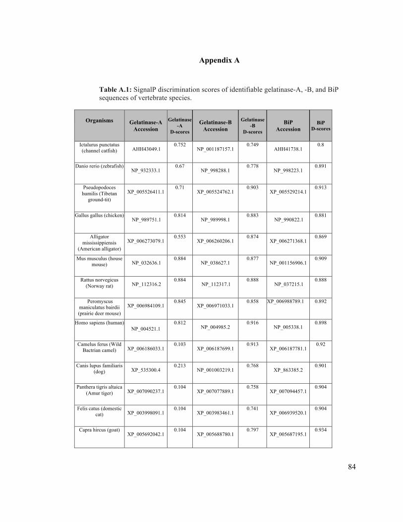
84
Appendix A
Table A.1: SignalP discrimination scores of identifiable gelatinase-A, -B, and BiP sequences of vertebrate species.
Organisms !
Gelatinase-A Accession
Gelatinase -A
D-scores
Gelatinase-B
Accession
Gelatinase -B
D-scores
BiP
Accession BiP
D-scores
Ictalurus punctatus (channel catfish) AHH43049.1
0.752 NP_001187157.1
0.749 AHH41738.1
0.8
Danio rerio (zebrafish) NP_932333.1
0.67 NP_998288.1
0.778 NP_998223.1
0.891
Pseudopodoces humilis (Tibetan
ground-tit) XP_005526411.1
0.71 XP_005524762.1
0.903 XP_005529214.1 0.913
Gallus gallus (chicken) NP_989751.1
0.814 NP_989998.1
0.883 NP_990822.1
0.881
Alligator mississippiensis
(American alligator) XP_006273079.1
0.553 XP_006260206.1
0.874 XP_006271368.1
0.869
Mus musculus (house mouse) NP_032636.1
0.884 NP_038627.1
0.877 NP_001156906.1
0.909
Rattus norvegicus (Norway rat) NP_112316.2
0.884 NP_112317.1
0.888 NP_037215.1
0.888
Peromyscus maniculatus bairdii (prairie deer mouse)
XP_006984109.1
0.845 XP_006971033.1 0.858 XP_006988789.1 0.892
Homo sapiens (human) NP_004521.1
0.812 NP_004985.2
0.916 NP_005338.1
0.898
Camelus ferus (Wild Bactrian camel) XP_006186033.1
0.103 XP_006187699.1
0.913 XP_006187781.1
0.92
Canis lupus familiaris (dog) XP_535300.4
0.213 NP_001003219.1
0.768 XP_863385.2
0.901
Panthera tigris altaica (Amur tiger) XP_007090237.1
0.104 XP_007077889.1
0.758 XP_007094457.1
0.904
Felis catus (domestic cat) XP_003998091.1
0.104 XP_003983461.1
0.741 XP_006939520.1
0.904
Capra hircus (goat) XP_005692042.1
0.104 XP_005688780.1
0.797 XP_005687195.1
0.934
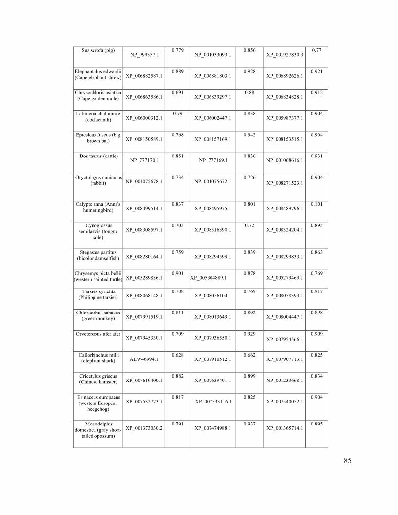
85
Sus scrofa (pig) NP_999357.1
0.779 NP_001033093.1
0.856 XP_001927830.3
0.77
Elephantulus edwardii (Cape elephant shrew) XP_006882587.1
0.889 XP_006881803.1
0.928 XP_006892626.1
0.921
Chrysochloris asiatica (Cape golden mole) XP_006863586.1
0.691 XP_006839297.1
0.88 XP_006834828.1
0.912
Latimeria chalumnae (coelacanth) XP_006000312.1
0.79 XP_006002447.1
0.838 XP_005987377.1
0.904
Eptesicus fuscus (big brown bat) XP_008150589.1
0.768 XP_008157169.1
0.942 XP_008153515.1
0.904
Bos taurus (cattle) NP_777170.1
0.851 NP_777169.1
0.836 NP_001068616.1
0.931
Oryctolagus cuniculus (rabbit) NP_001075678.1 0.734
NP_001075672.1
0.726 XP_008271523.1
0.904
Calypte anna (Anna's hummingbird)
XP_008499514.1
0.837 XP_008495975.1
0.801 XP_008489796.1
0.101
Cynoglossus semilaevis (tongue
sole)
XP_008308597.1
0.703 XP_008316390.1
0.72 XP_008324204.1
0.893
Stegastes partitus (bicolor damselfish)
XP_008280164.1
0.759 XP_008294599.1
0.839 XP_008299833.1
0.863
Chrysemys picta bellii (western painted turtle) XP_005289836.1 0.901
XP_005304889.1 0.878
XP_005279469.1 0.769
Tarsius syrichta (Philippine tarsier)
XP_008068148.1
0.788 XP_008056104.1
0.769 XP_008058393.1
0.917
Chlorocebus sabaeus (green monkey)
XP_007991519.1
0.811 XP_008013649.1
0.892 XP_008004447.1
0.898
Orycteropus afer afer XP_007945330.1
0.709 XP_007936550.1
0.929 XP_007954566.1
0.909
Callorhinchus milii (elephant shark)
AEW46994.1
0.628 XP_007910512.1
0.662 XP_007907713.1 0.825
Cricetulus griseus (Chinese hamster)
XP_007619400.1
0.882 XP_007639491.1 0.899
NP_001233668.1 0.834
Erinaceus europaeus (western European
hedgehog)
XP_007532773.1
0.817 XP_007533116.1
0.825 XP_007540052.1
0.904
Monodelphis domestica (gray short-
tailed opossum)
XP_001373030.2
0.791 XP_007474988.1
0.937 XP_001365714.1
0.895
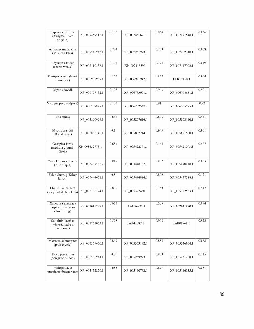
86
Lipotes vexillifer (Yangtze River
dolphin)
XP_007459512.1
0.103 XP_007451691.1
0.864 XP_007471548.1
0.826
Astyanax mexicanus (Mexican tetra)
XP_007246942.1
0.724 XP_007231993.1
0.759 XP_007252148.1 0.868
Physeter catodon (sperm whale)
XP_007110336.1
0.104 XP_007115590.1
0.775 XP_007117702.1
0.849
Pteropus alecto (black flying fox)
XP_006908907.1
0.165 XP_006921942.1
0.878 ELK07198.1
0.904
Myotis davidii XP_006777132.1
0.103 XP_006773601.1
0.943 XP_006768631.1 0.901
Vicugna pacos (alpaca) XP_006207898.1
0.103 XP_006202537.1
0.911 XP_006205575.1 0.92
Bos mutus XP_005890996.1
0.883 XP_005897616.1
0.836 XP_005893110.1
0.931
Myotis brandtii (Brandt's bat)
XP_005865346.1
0.1 XP_005862214.1
0.943 XP_005881560.1
0.901
Geospiza fortis (medium ground-
finch)
XP_005422778.1
0.684 XP_005422371.1
0.164 XP_005421393.1
0.527
Oreochromis niloticus (Nile tilapia)
XP_003437582.2
0.819 XP_003448187.1
0.802 XP_005470418.1
0.865
Falco cherrug (Saker falcon)
XP_005444651.1
0.8 XP_005444884.1
0.809 XP_005437288.1 0.121
Chinchilla lanigera (long-tailed chinchilla)
XP_005388374.1
0.839 XP_005392450.1
0.759 XP_005382523.1
0.917
Xenopus (Silurana) tropicalis (western
clawed frog)
NP_001015789.1
0.653 AAH76927.1
0.535 XP_002941690.1
0.894
Callithrix jacchus (white-tufted-ear
marmoset) XP_002761063.1
0.598 JAB41082.1
0.908 JAB09769.1 0.923
Microtus ochrogaster (prairie vole)
XP_005369650.1
0.847 XP_005363192.1
0.885 XP_005346064.1
0.888
Falco peregrinus (peregrine falcon)
XP_005238944.1
0.8 XP_005239973.1
0.809 XP_005231480.1
0.115
Melopsittacus undulatus (budgerigar)
XP_005152279.1
0.683 XP_005140762.1
0.877 XP_005146335.1
0.881
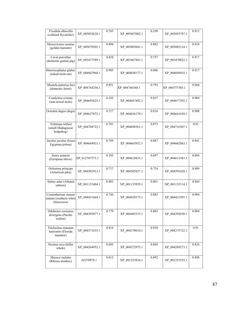
87
Ficedula albicollis (collared flycatcher)
XP_005052630.1 0.765
XP_005057082.1
0.109 XP_005055797.1
0.913
Mesocricetus auratus (golden hamster)
XP_005079205.1
0.894 XP_005085041.1
0.882 XP_005082124.1
0.924
Cavia porcellus (domestic guinea pig)
XP_003477589.1
0.838 XP_003467841.1
0.757 XP_003470822.1
0.917
Heterocephalus glaber (naked mole-rat)
XP_004847968.1
0.905 XP_004858177.1
0.806 XP_004849435.1
0.917
Mustela putorius furo (domestic ferret)
XP_004744256.1
0.851 XP_004746360.1
0.793 XP_004757385.1
0.904
Condylura cristata (star-nosed mole)
XP_004693625.1
0.103 XP_004687492.1 0.837
XP_004677392.1
0.904
Octodon degus (degu) XP_004637072.1
0.717 XP_004636179.1
0.816 XP_004641630.1
0.908
Echinops telfairi (small Madagascar
hedgehog)
XP_004704722.1
0.781 XP_004698361.1
0.873 XP_004716507.1
0.91
Jaculus jaculus (lesser Egyptian jerboa)
XP_004664921.1
0.789 XP_004663952.1
0.887 XP_004662863.1
0.941
Sorex araneus (European shrew)
XP_012787573.1
0.103 XP_004618833.1
0.697 XP_004613383.1
0.884
Ochotona princeps (American pika)
XP_004583913.1
0.717 XP_004585927.1
0.724 XP_004593628.1
0.889
Salmo salar (Atlantic salmon)
NP_001133404.1
0.803 NP_001133929.1
0.861 NP_001135114.1
0.844
Ceratotherium simum simum (southern white
rhinoceros)
XP_004431664.1
0.744 XP_004430375.1 0.885
XP_004423507.1
0.904
Odobenus rosmarus divergens (Pacific
walrus)
XP_004395877.1
0.779 XP_004405315.1 0.803
XP_004392650.1
0.904
Trichechus manatus latirostris (Florida
manatee)
XP_004371635.1
0.816 XP_004370610.1 0.929
XP_004375722.1
0.91
Orcinus orca (killer whale)
XP_004264952.1
0.695 XP_004272975.1 0.869
XP_004269273.1
0.826
Macaca mulatta (Rhesus monkey)
AFJ70978.1
0.812 NP_001253834.1
0.892 NP_001253525.1 0.898
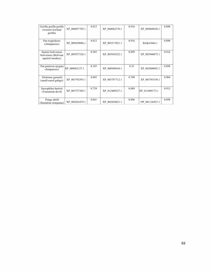
88
Gorilla gorilla gorilla (western lowland
gorilla)
XP_004057705.1 0.812 XP_004062370.1 0.916
XP_004048650.1
0.898
Pan troglodytes (chimpanzee)
XP_009429086.1
0.812 XP_003317021.1
0.916 BAK63666.1
0.898
Saimiri boliviensis boliviensis (Bolivian
squirrel monkey)
XP_003937326.1
0.585 XP_003936522.1
0.899 XP_003940675.1 0.916
Pan paniscus (pygmy chimpanzee)
XP_008962127.1
0.103 XP_008949630.1 0.33
XP_003808092.1
0.898
Otolemur garnettii (small-eared galago)
XP_003792593.1
0.895 XP_003787712.1
0.709 XP_003785350.1
0.904
Sarcophilus harrisii (Tasmanian devil)
XP_003757305.1
0.724 XP_012409327.1
0.889 XP_012409173.1
0.915
Pongo abelii (Sumatran orangutan) XP_002826479.1 0.841
XP_002830421.1 0.896 NP_001126927.1 0.898
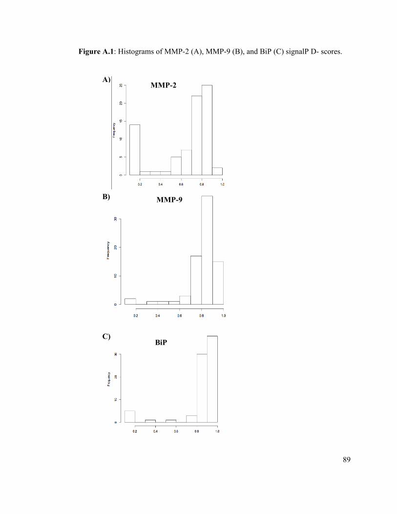
89
Figure A.1: Histograms of MMP-2 (A), MMP-9 (B), and BiP (C) signalP D- scores.
MMP-2 A)
MMP-9 B)
BiP C)

90
Table A.2: The NetPhos prediction scores of gelatinase-A sequences of vertebrate species.
(in order to save space in this thesis, the raw data from the NetPhos analysis of
gelatinase-A sequences is available online at this URL)
https://unbscholar.lib.unb.ca/islandora/object/unbscholar%3A7073
91
Appendix B
I. Transmission Electron Microscopy (TEM)
The purpose of this experiment was to demonstrate the intracellular localization of
Mmp2 in zebrafish skeletal muscle. Although the immunostaining data imaged by
confocal microscopy strongly suggested that Mmp2 was present within the sarcomeres
(chapter 2), due to the limited axial resolution of this optical microscope, it was possible
that this important result could be contested. Before resorting to cryosectioning, I tried
immunogold detection of Mmp2 by transmission electron microscopy, hoping that I
would be able to unequivocally localize Mmp2 within the sarcomere has had been done
with mammalian cardiomyocytes (Wang et al. 2002; Sawicki et al. 2005).
Although I was able to get good sections of skeletal muscle tissue, the results were
disappointing because the immunogold labeling of Mmp2 was not successful (Figure A.1).
This is likely due to the necessity of using aldehyde-fixatives in the preparation of tissue
for TEM, which we now know destroy the epitope recognized by the rabbit anti-Mmp2
antibody I have used throughout this thesis.
Material and methods
Transmission Electron Microscopy (TEM)
Seventy-two hour post fertilization embryos were incubated in a primary fixative that
contained 3% paraformaldehyde in 0.1 M sodium cacodylate buffer, pH 7.4, overnight at
4°C. Samples were rinsed in sodium cacodylate buffer, with at least three changes of
92
buffer, for two hours; no secondary fixative was used. The embryos were dehydrated via
increasing concentrations of ethanol (30, 50, 70, 80 to 90%) for 30 minutes each,
culminating in two 30 minute incubations in anhydrous ethanol. Ethanol was replaced
with a 1:2 ratio of LR (London Resin) white resin to ethanol and set for four hours, then
replaced with a 2:1ratio of LR white resin to ethanol and left overnight. The final change
with 100% fresh resin was made and the sample left for four hours.
Twenty-one embryos were then embedded in gelatin capsules with fresh resin and
cured in an oven at 50-55°C for 24 hours. The following day, the samples were cut into
80-100 nm ultrathin sections using a diamond knife (Leica Ultracut T ultramicrotome),
and collected on 400 mesh nickel grids with formvar support films. The grids with
ultrathin sections were floated on blocking buffer (5% Bovine serum albumin (BSA) in
PBS (20 mM phosphate pH 7.3, 137 mM NaCl, 2.7 mM KCl)) overnight in a humid
chamber at room temperature. The sections were then incubated overnight with either
anti-Mmp2 or anti-α-actinin, or blocking buffer alone (as a negative control) at room
temperature in humid chamber. They were then rinsed with PBS for15 minutes, and
incubated with protein A/G conjugated to10-nm gold particles (British Biocell
International) (diluted 1:20 in PBS) for 30 minutes at room temperature in humid
chamber. The sections were washed with a PBS 3x5 minutes, rinsed with dH2O, dried,
stained with 2% uranyl acetate for five minutes, rinsed with dH2O, stained with lead
citrate for one minute and finally rinsed in dH2O. The stained sections on grids were
dried in desiccator before being examined using a JEOL-2011 (Scanning) TEM (at 200
kV) with a Gatan Ultrascan digital camera and Digital Micrograph software.

93
!
Figure I.1. Transmission Electron Micrograph of longitudinal section of zebrafish muscle processed for immunogold detection of Mmp2. A) Representative micrograph of negative control. B) The sections of sarcomeres were probed with anti-Mmp2 and while a few 10 nm gold particles were retained in some sections, these were rare (comparable to negative controls treated with no primary antibody) and exhibited no spatially distinct pattern. !
A)
94
II. Investigation of Mmp2’s potential role in muscle hypertrophy
The aim of this experiment was to address whether the intracellular Mmp2 is
involved in muscle cell growth. To conduct this experiment I exercised zebrafish and
treated them with testosterone to induce muscle hypertrophy, as has previously been
reported (Piferrer and Donaldson, 1994; McClelland, et al. 2006). I hoped to detect
changes in Mmp2 distribution, phosphorylation and/or expression that might correlate
with hypertrophy.
Materials and methods
Testosterone hormone treatments
A total of 62 age-matched male and female adult zebrafish were distributed into
three litre containers (five individuals in each of 10 containers, and six in two containers).
The containers had continuous aeration as well as artificial plants for cover. The fish
were fed pellets and brine shrimp 4-5 times per day. 10% of the water was changed per
day, and the temperature was maintained between 26 and 28°C with a 14/10 hour
light/dark cycle (as described in Westerfield, 1995). Each treatment group was
maintained for 4 weeks.
Treatments consisted of exercise (see below) (n=16); hormone (400 µg/l
testosterone) plus exercise (n=15); hormone alone (n=16); or no treatment (n=15).
Exercise treatment was modelled after McClelland, et al. (2006); the fish were
exercised for three hours twice daily, for six days each week for four weeks, with a two-
hour rest period when the fish were fed. Fish were required to swim at 2 body lengths per
95
second (BL/s), equivalent to 6.0 cm/s, and each week the speed was increased 1 BL/s to
the final speed of 5 BL/s (15.0 cm/s). After the fourth week the speed of the water was
maintained at 3 cm/s (∼1 BL/s), including overnight and in non-exercising conditions.
At the end of the experiment the fish were removed from the buckets and anesthetized
(see 3.2.2) as previously described, with the length and weight being recorded. After the
tail muscles were dissected from the fish, the hypertrophy of muscle tissue was
determined using quantitative PCR, with MyoD and Myf5 transcription factors as
positive controls.
RNA purification, cDNA synthesis, and qPCR
RNA was extracted using a Norgen Total RNA purification kit following the
manufacturer’s instructions. cDNA was synthesized using Enhanced Avian Reverse
Transcriptase (Sigma) according to the manufacturer’s protocol. cDNAs from each
treatment group were then subjected to qPCR in order to determine if there were
significant changes in myogenic (MyoD and Myf5) genes (with respect to housekeeping
controls GAPDH and EF1), and also to characterize any changes in the expression of
mmp2. Primers are listed in Table II.1 below. All reactions were done in triplicate for
each primer set. The PCR efficiencies for all primers and the values of critical threshold
(Ct) were verified using LinRegPCR software.
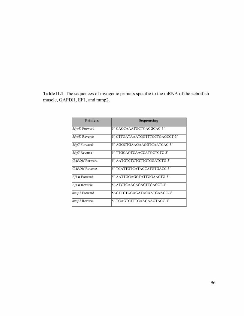
96
Table II.1. The sequences of myogenic primers specific to the mRNA of the zebrafish muscle, GAPDH, EF1, and mmp2.
Primers Sequencing
MyoD Forward 5’-CACCAAATGCTGACGCAC-3’
MyoD Reverse 5’-CTTGATAAATGGTTTCCTGAGCCT-3’
Myf5 Forward 5’-AGGCTGAAGAAGGTCAATCAC-3’
Myf5 Reverse 5’-TTGCAGTCAACCATGCTCTC-3’
GAPDH Forward 5’-AATGTCTCTGTTGTGGATCTG-3’
GAPDH Reverse 5’-TCATTGTCATACCATGTGACC-3’
Ef1!α Forward 5’-AATTGGAGGTATTGGAACTG-3’
Ef1!α Reverse 5’-ATCTCAACAGACTTGACCT-3’
mmp2 Forward 5’-GTTCTGGAGATACAATGAAGC-3’
mmp2 Reverse 5’-TGAGTCTTTGAAGAAGTAGC-3’
97
Results
The hormone/exercise zebrafish treatment did not result in hypertrophy
Disappointingly, none of the treatments resulted in significant increases in mass
(Figure II.1), body size, expression of myogenic genes, or expression of mmp2 (data not
shown). Indeed, although these changes were not significant, none of the treated fish
(exercise alone, hormone alone, or combination of exercise and hormone) grew as well as
the untreated controls. This suggests that the experimental conditions were somewhat
stressful to the fish, reducing their growth rates.
Because of the failure of any treatments to yield evidence of muscle hypertrophy,
interpretation of any changes in the distribution or phosphorylation of Mmp2 protein
would not have been possible, so the experiment was abandoned.
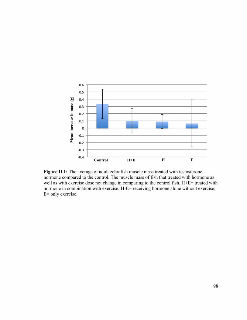
98
Figure II.1: The average of adult zebrafish muscle mass treated with testosterone hormone compared to the control. The muscle mass of fish that treated with hormone as well as with exercise dose not change in comparing to the control fish. H+E= treated with hormone in combination with exercise; H-E= receiving hormone alone without exercise; E= only exercise.!
!0.4%
!0.3%
!0.2%
!0.1%
0%
0.1%
0.2%
0.3%
0.4%
0.5%
0.6%
Control H+E H E
Mea
n in
crea
se in
mas
s (g)
99
References
McClelland GB, Craig PM, Dhekney K, Dipardo S. 2006. Temperature- and exercise-induced gene expression and metabolic enzyme changes in skeletal muscle of adult zebrafish (Danio rerio). J Physiol. 577(2): 739-51.
Piferrer F, Donaldson EM. 1994. Uptake and clearance of exogenous estradiol-1713 and testosterone during the early development of coho salmon (Oncorhynchus kisutch), including eggs, alevins and fry. Fish Physiol Biochem. 13(3): 219-232.
Sawicki G, Leon H, Sawicka J, Sariahmctoglu M, Schulze CJ, Scott PG, Szczesna-Cordary D, Schulz R. 2005. Degradation of myosin light chain in isolated rat hearts subjected to ischemia-reperfusion injury: a new intracellular target for matrix metalloproteinase-2. Circulation. 112(4): 544-552.
Wang W, Schulze CJ, Suarez-Pinzon WL, Dyck JR, Sawicki G, Schulz R. 2002. Intracellular action of matrix metalloproteinase-2 accounts for acute myocardial ischemia and reperfusion injury. Circulation.106(12): 1543-1549.
Westerfield M. 1995. The zebrafish book. A guide for the laboratory use of zebrafish (Danio rerio). 3rd edition. Eugene (OR): University of Oregon Press. 385 p.

Curriculum Vitae
Candidate’s full name: Amina Mohammed Ahmed fallata
Universities attended (with dates and degrees obtained):
King Abdul-Aziz University (2009, B.Sc).
Conference Presentations:
1) Amina M. Fallata and Bryan D. Crawford, April 2014. Intracellular localization and regulation of Gelatinase A in zebrafish skeletal muscle. Graduate Research Conference (GRC), University of New Brunswick, Fredericton, NB, Canada. (Oral presentation).
2) Amina M. Fallata and Bryan D. Crawford, March 2014. Intracellular localization and regulation of Gelatinase A in zebrafish skeletal muscle. Canadian Zebrafish Conference, Mont-Temblant, QC, Canada. (Poster presentation).
3) Amina M. Fallata and Bryan D. Crawford, March 2014. Intracellular localization and regulation of Gelatinase A in zebrafish skeletal muscle. The 7th Canadian Developmental Biology, Mont-Temblant, QC, Canada. (Poster presentation).
4) Amina M. Fallata and Bryan D. Crawford, June 2013. Intracellular localization and regulation of Gelatinase A in zebrafish skeletal muscle. The 3rd North Atlantic Zebrafish Research Symposium, University of Maine, Orono, Maine, USA. (Poster presentation).
5) Amina M. Fallata and Bryan D. Crawford, July 2012. Intracellular localization and regulation of Gelatinase A in zebrafish skeletal muscle. Society for Developmental Biology 71st annual meeting, Montreal, QC, Canada. (Poster presentation).
6) Amina M. Fallata and Bryan D. Crawford, May 2012. Intracellular localization and regulation of Gelatinase A in zebrafish skeletal muscle. 51st Canadian Society of Zoology meeting, Sackville, NB, Canada. (Poster presentation).
Related Documents