Human Reproduction Update 1996, Vol. 2, No. 2 pp. 103–117 E European Society for Human Reproduction and Embryology Gonadal cell apoptosis: hormone-regulated cell demise Håkan Billig 1 , Sang-Young Chun 2 , Karen Eisenhauer 2 and Aaron J.W.Hsueh 2,3 1 Department of Physiology, University of Göteborg, Medicinaregatan 11, 413 90 Göteborg, Sweden and 2 Division of Reproductive Biology, Department of Gynecology and Obstetrics, Stanford University School of Medicine, Stanford, CA 94305–5317, USA TABLE OF CONTENTS Introduction 103 Molecular mechanism of apoptosis 104 Cell depletion in the gonads 105 Detection and localization of apoptosis in the gonads 105 Hormonal control of gonadal cell apoptosis 106 Comparison of ovarian and testicular cell apoptosis 112 Conclusion 113 Acknowledgement 114 References 114 It has become evident that apoptosis, an active form of cell ‘suicide’, plays an important role in the normal function of all tissues. A balance of cell proliferation and apoptosis is maintained in a healthy individual and any imbalance of the two processes could lead to pathological changes. In both sexes, massive apoptosis accounts for the demise of a majority of gonadal cells (ovarian granulosa cells and male germ cells) during reproductive life. Recent studies have indicated the important role of gonadotrophins as survival factors in both the ovary and the testis. Furthermore, intra- gonadal survival factors in the ovary (oestrogens, insulin-like growth factor I, epidermal growth factor, basic fibroblast growth factor, interleukin-1β, nitric oxide, etc.) and testis (androgens) have been shown to act in concert with the gonadotrophins. In contrast, several apoptotic factors (androgens, gonadotrophin- releasing hormone-like peptide and interleukin-6) may be important in inducing the demise of ovarian follicles. Understanding of the hormonal and cellular mechanisms responsible for gonadal cell apoptosis will provide new approaches for the treatment of gonadal degenerative conditions such as premature ovarian failure and cryptorchidism, as well as for the design of new contraceptive approaches. Key words: apoptosis/cell demise/gonadal cells Introduction Increasing attention has been focused on the physiological removal of cells through apoptosis or programmed cell death during fetal tissue remodelling and during normal development in adults. The capacity for cell ‘suicide’ seems to be present in most, if not all, tissues to maintain a homeostatic state of the individual (Raff, 1992). During the fertile phase of the life span of mammals, the function of the gonads is hormonally controlled. In addi- tion to the proliferation of somatic and germ cells in the ovary and in the testis during normal gonadal development, degeneration of gonadal cells plays an important physio- logical role and results in the depletion of a majority of germ cells in both sexes. In the testis, morphological signs of germ cell degeneration during spermatogenesis were recognized almost a century ago (Regaud, 1900). Even earlier, the phenomenon of cell removal associated with the presence of small or fragmented cells was noted in the study of regression of Graafian follicles (Flemming, 1885). The process, called chromolysis, resembles follicular atre- sia and the morphological changes during apoptosis seen in other cell types. Apoptosis in gonads has been recently characterized biochemically, and its role in gonadal physi- ology is now gaining increasing attention. Apoptotic cell death is morphologically characterized by disruption of the cell skeleton, cell shrinkage, membrane blebbing, nuclear condensation, cell disruption into small membrane-enclosed fragments called apoptotic bodies and phagocytosis by neighbouring cells to complete the degrada- tion of the cell. One of the most striking biochemical features of apoptosis is the activation of a calcium/magnesium- dependent endonuclease which specifically cleaves cellular 3 To whom correspondence should be addressed. Phone: 1 415 725 6802; Fax: 1 415 725 7102; Email: [email protected].

Welcome message from author
This document is posted to help you gain knowledge. Please leave a comment to let me know what you think about it! Share it to your friends and learn new things together.
Transcript

Human Reproduction Update 1996, Vol. 2, No. 2 pp. 103–117 � European Society for Human Reproduction and Embryology
Gonadal cell apoptosis: hormone-regulatedcell demise
Håkan Billig 1, Sang-Young Chun2, Karen Eisenhauer2 and Aaron J.W.Hsueh2,3
1Department of Physiology, University of Göteborg, Medicinaregatan 11, 413 90 Göteborg, Sweden and 2Division ofReproductive Biology, Department of Gynecology and Obstetrics, Stanford University School of Medicine, Stanford, CA94305–5317, USA
TABLE OF CONTENTS
Introduction 103Molecular mechanism of apoptosis 104Cell depletion in the gonads 105Detection and localization of apoptosis in the
gonads 105Hormonal control of gonadal cell apoptosis 106Comparison of ovarian and testicular cell
apoptosis 112Conclusion 113Acknowledgement 114References 114
It has become evident that apoptosis, an active form ofcell ‘suicide’, plays an important role in the normalfunction of all tissues. A balance of cell proliferationand apoptosis is maintained in a healthy individualand any imbalance of the two processes could lead topathological changes. In both sexes, massive apoptosisaccounts for the demise of a majority of gonadal cells(ovarian granulosa cells and male germ cells) duringreproductive life. Recent studies have indicated theimportant role of gonadotrophins as survival factorsin both the ovary and the testis. Furthermore, intra-gonadal survival factors in the ovary (oestrogens,insulin-like growth factor I, epidermal growth factor,basic fibroblast growth factor, interleukin-1β, nitricoxide, etc.) and testis (androgens) have been shown toact in concert with the gonadotrophins. In contrast,several apoptotic factors (androgens, gonadotrophin-releasing hormone-like peptide and interleukin-6)may be important in inducing the demise of ovarianfollicles. Understanding of the hormonal and cellularmechanisms responsible for gonadal cell apoptosiswill provide new approaches for the treatment ofgonadal degenerative conditions such as premature
ovarian failure and cryptorchidism, as well as for thedesign of new contraceptive approaches.
Key words: apoptosis/cell demise/gonadal cells
Introduction
Increasing attention has been focused on the physiologicalremoval of cells through apoptosis or programmed celldeath during fetal tissue remodelling and during normaldevelopment in adults. The capacity for cell ‘suicide’seems to be present in most, if not all, tissues to maintain ahomeostatic state of the individual (Raff, 1992).
During the fertile phase of the life span of mammals, thefunction of the gonads is hormonally controlled. In addi-tion to the proliferation of somatic and germ cells in theovary and in the testis during normal gonadal development,degeneration of gonadal cells plays an important physio-logical role and results in the depletion of a majority ofgerm cells in both sexes. In the testis, morphological signsof germ cell degeneration during spermatogenesis wererecognized almost a century ago (Regaud, 1900). Evenearlier, the phenomenon of cell removal associated with thepresence of small or fragmented cells was noted in thestudy of regression of Graafian follicles (Flemming, 1885).The process, called chromolysis, resembles follicular atre-sia and the morphological changes during apoptosis seen inother cell types. Apoptosis in gonads has been recentlycharacterized biochemically, and its role in gonadal physi-ology is now gaining increasing attention.
Apoptotic cell death is morphologically characterized bydisruption of the cell skeleton, cell shrinkage, membraneblebbing, nuclear condensation, cell disruption into smallmembrane-enclosed fragments called apoptotic bodies andphagocytosis by neighbouring cells to complete the degrada-tion of the cell. One of the most striking biochemical featuresof apoptosis is the activation of a calcium/magnesium-dependent endonuclease which specifically cleaves cellular
3To whom correspondence should be addressed. Phone: 1 415 725 6802; Fax: 1 415 725 7102; Email: [email protected].

104 H.Billig et al.
DNA between regularly spaced nucleosomal units that canbe visualized as a distinct ladder of DNA bands followingagarose gel electrophoresis and ethidium bromide staining(Wyllie et al., 1980; Arends et al., 1990). TheCa2+/Mg2+-dependent endonuclease is present in diversecell types (Arends et al., 1990; Gaido and Cidlowski, 1991),including the ovarian granulosa and luteal cells (Zeleznik etal., 1989; Boone et al., 1995). Once activated, the apoptoticdegradation of DNA is irreversible. Indeed, recent studieshave indicated that rat testis (Tapanainen et al., 1993; Shi-kone et al., 1994; Billig et al., 1995; Henriksen et al., 1995),as well as atretic rat, avian, porcine, bovine and human fol-licles, contains DNA fragments resembling oligonucleo-somes of different sizes (Hughes and Gorospe, 1991; Tilly etal., 1991, 1992a; Jolly et al., 1994; Quirk et al., 1995).
Molecular mechanism of apoptosis
In most cells apoptosis requires active transcription of newgenes. For instance, glucocorticoid-induced apoptosis inthymocytes is suppressed by inhibitors of transcription ortranslation (Cohen and Duke, 1984). To date, a number ofgenes have been identified as being involved in the activationof apoptosis in different tissues and species. Some of thesegenes are essential for the activation of the apoptosis pro-gramme, and loss of their function prevents cell death. In thenematode Caenorhabditis elegans, an excellent model forthe study of gene regulation during apoptosis, products of theced-3 and ced-4 genes initiate apoptosis (Yuan and Horvitz,1990, 1992). In contrast, the ced-9 gene product blocks
apoptosis (Hengartner et al., 1992). The gene bcl-2 has beenshown to be the mammalian analogue of ced-9 and, whenover-expressed, blocks apoptosis in many cells. Indeed,over-expression of bcl-2 leads to suppression of lymphocytecell death, prolonged cell survival and tumour formation(Liu et al., 1991; Reed, 1994). The mammalian homologueto the gene product ced-3 is the interleukin-1β-convertingenzyme (ICE) (Miura et al., 1993) and several other cysteineproteases involved in apoptosis (Martin and Green, 1995)(Table I). Over-expression of ICE induces apoptosis in somecell types. It is becoming apparent that the genes responsiblefor the synthesis of bcl-2 and ICE each represent only onemember of two separate gene families. One could envisionthat different members of these two gene families are in-volved in the regulation of apoptosis in specific mammaliancell types (Table I).
Only limited data on genes involved in the activation orinhibition of apoptosis in gonads have been reported. Al-though bcl-2 protein was not found in the human ovary(Hockenbery et al., 1991; Reed et al., 1991), a recent reporthas suggested that bcl-2 mRNA is expressed in rat ovariesbut its level does not vary after gonadotrophin treatment. Theexpression of another member of the bcl-2 gene family, bax,that antagonizes bcl-2 activity is, however, reduced aftergonadotrophin treatment (Tilly et al., 1995). Due to the poss-ible involvement of multiple genes of the bcl-2 family anddue to the potential contamination of ovarian preparations byblood cells expressing bcl-2 and bax, more comprehensiveanalyses are needed to establish a cause–effect relationshipbetween these genes and ovarian cell apoptosis.
Table I. Genes involved in the suppression or initiation of apoptosis in different tissues
Gene Function Method of identification Origin Reference
ced-9 prevents apoptosis genetic analysis (Caenorhabditis elegans) nematode Hengartner et al. (1992)
bcl-2 prevents apoptosis over-expression in B cell lymphomachromosomal translocation (mammalianhomologue to ced-9)
mammalian Hengartner & Horvitz (1994)
bax prevents bcl-2 action co-precipitates with bcl-2 protein andincreases apoptosis
mammalian Oltavi et al. (1993)
bcl-x
Long form prevents apoptosis homologous screening of cDNA library mammalian Boise et al. (1993)
Short form promotes apoptosis
A1 prevents apoptosis differential screening (haematopoieticcells) (similarities to bcl-2)
mammalian Lin et al. (1993)
bak promotes apoptosis homologous screening of cDNA mammalian Kiefer et al. (1995), Chittenden et al. (1995)
ced-3 promotes apoptosis genetic analysis (C. elegans) nematode Yuan & Horvitz (1990)
ced-4 promotes apoptosis genetic analysis (C. elegans) nematode Yuan & Horvitz (1990, 1992)
ICE promotes apoptosis protease homologue to ced-3 mammalian Miura et al. (1993)
nedd-2/Ich-1 promotes apoptosis protease homologue to ced-3 mammalian Kumar et al. (1992), Wang et al. (1994)
Apopain/cpp32 promotes apoptosis protease homologue to ced-3 mammalian Fernadez-Alnemri et al. (1994), Nicholsonet al. (1995)

Gonadal cell apoptosis 105
Cell depletion in the gonads
In the human ovary, there are two million oocytes at birthand 400 000 follicles are present at the onset of puberty.However, only 400 follicles can possibly be ovulated dur-ing the reproductive phase of a woman’s life (Baker, 1963).At the time of menopause, only a few follicles can be foundin the ovary. More than 99.9% of follicles, includingoocytes and granulosa and theca cells, are deleted. Indeed,it is the ‘norm’ for a follicle to die rather than to ovulate.The degenerative process by which 99.9% of follicles areirrevocably committed to undergo cell death is termedatresia. Despite its critical role during the recruitment offollicles for ovulation, the mechanisms underlying theonset and progression of atresia remain poorly understood(Hirshfield, 1991; Hsueh et al., 1994).
In mammals, there are at least four degenerative stagesduring ovarian development to account for the massive lossof ovarian cells. During migration of the primordial germcells from the yolk sac to the genital ridge, these cellsundergo degeneration unless rescued by stem cell factorand other related factors (Dolci et al., 1991; Godin et al.,1991). Coincident with their entry into meiosis, germ cellsundergo attrition before formation of the follicles (Beau-mont and Mandl, 1962). At the penultimate stage of devel-opment, early antral follicles either differentiate or undergoatresia. If ovulatory signals are absent, the mature folliclesalso may undergo degeneration. After ovulation, the cor-pora lutea have a finite life span and undergo luteolysis.
The morphological features of atresia have been de-scribed as progressive changes in all follicular cell types.An early sign of atresia is the presence of scattered pykno-tic nuclei in the granulosa cell layer (Hirshfield, 1989).Further evidence of follicular deterioration in follicles ofmore advanced stages of atresia includes detachment of thegranulosa cell layer from the basement membrane (Jun-quiera et al., 1989), fragmentation of the basal lamina (Ba-gavandoss et al., 1983) and the presence of cell debris in thefollicular antrum (Hay et al., 1976). A reduction of protein(Byskov, 1979) and DNA (Greenwald, 1989; Hirshfield,1989) synthesis within granulosa cells of atretic follicleshas also been reported. In the oocyte, meiosis-like changes(germinal vesicle breakdown) occur, followed by oocytefragmentation. These changes are accompanied by disrup-tion of the oocyte–cumulus connection (Tsafriri and Braw,1984). In ovine atretic follicles (O’shea et al., 1978), thecacells undergo degeneration. In contrast, human, rat andrabbit theca cells undergo extensive hypertrophy duringfollicle atresia (Braw et al., 1976; Erickson et al., 1985).The morphological changes seen during atresia resemblethose of cells undergoing apoptosis.
Germ cell depletion during normal spermatogenesis re-sults in the loss of up to 75% of potential numbers ofmature sperm cells in the testis of adults (Oakland, 1956;Huckins, 1978; De Rooij and Lok, 1987). Morphologicalanalysis indicated that germ cell demise is present through-out testis development (Huckins, 1978). Peaks of germ cellloss have been found in three distinct stages of spermato-genesis: (i) during mitotic division of type A spermato-gonia, (ii) during meiotic division of spermatocytes and(iii) during spermiogenesis (Huckins, 1978 and referencestherein). In neonatal and pubertal rodents, the incidence oftestis germ cell degeneration also varies (Russel et al.,1987). This degeneration shows distinct morphologicalsigns, including margination and condensation of nuclearchromatin and phagocytosis by Sertoli cells without in-flammation (Allan et al., 1992), that are consistent with thecell morphology found during apoptotic cell death (Cohen,1993; Schwartz and Osborne, 1993).
Although individual seminiferous tubules in the testis donot undergo an ‘all-or-none’ degenerative process likeatresia in ovarian follicles, it is clear that germ cell deathplays an important role in testis physiology and patho-physiology. In addition, the seminiferous tubules arehighly sensitive to damage by ionizing radiation, chemo-therapeutic agents and hyperthermia, highlighting the roleof testis cell death in pathological conditions.
Detection and localization of apoptosis in thegonads
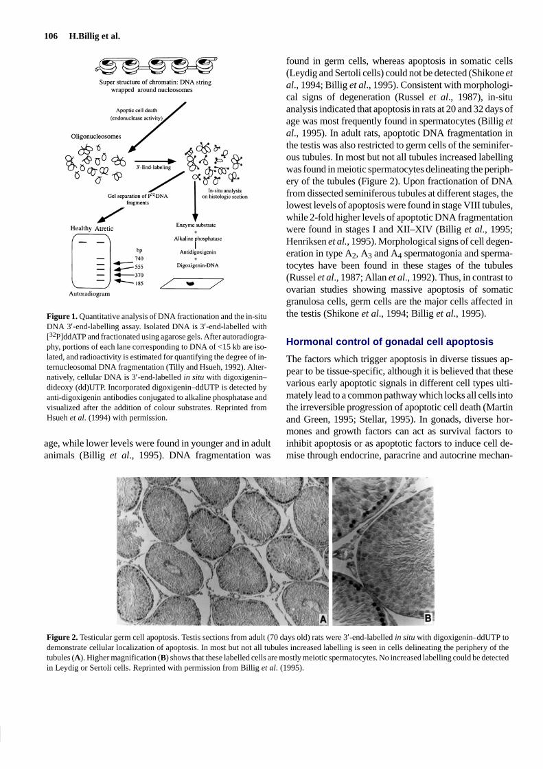
Studies of atresia and apoptosis in ovarian follicles havebecome more accessible with the development of sensitiveDNA fractionation and in-situ DNA end-labelling assays.In the quantitative DNA fractionation assay, isolated DNAis labelled at its 3′-ends with [32P]dideoxy (dd)ATP. Elec-trophoretic separation of the labelled DNA enables bothquantitative estimation of [32P]ddATP incorporation andautoradiographic visualization of apoptotic DNA frag-ments (Figure 1; Tilly and Hsueh, 1992). In contrast, spe-cific cell types undergoing apoptosis can be studied onhistological sections after in-situ 3′-end-labelling of DNAwith a modified nucleotide, digoxigenin–ddUTP (Figure1; Billig et al., 1993). With the recent application of thesemethods to the analysis of ovarian cell death, it is clear thatgranulosa cells are the major cell type undergoing apopto-sis during follicle atresia (Billig et al., 1993; Palumbo andYeh, 1994; Guthrie et al., 1995).
DNA 3′-end-labelling and in-situ analyses have alsobeen used to investigate testis cell apoptosis. In the normaldeveloping testis, increased levels of apoptotic DNA frag-mentation were detected in rats between 16 and 32 days of
106 H.Billig et al.
Figure 1. Quantitative analysis of DNA fractionation and the in-situDNA 3′-end-labelling assay. Isolated DNA is 3′-end-labelled with[32P]ddATP and fractionated using agarose gels. After autoradiogra-phy, portions of each lane corresponding to DNA of <15 kb are iso-lated, and radioactivity is estimated for quantifying the degree of in-ternucleosomal DNA fragmentation (Tilly and Hsueh, 1992). Alter-natively, cellular DNA is 3′-end-labelled in situ with digoxigenin–dideoxy (dd)UTP. Incorporated digoxigenin–ddUTP is detected byanti-digoxigenin antibodies conjugated to alkaline phosphatase andvisualized after the addition of colour substrates. Reprinted fromHsueh et al. (1994) with permission.
age, while lower levels were found in younger and in adultanimals (Billig et al., 1995). DNA fragmentation was
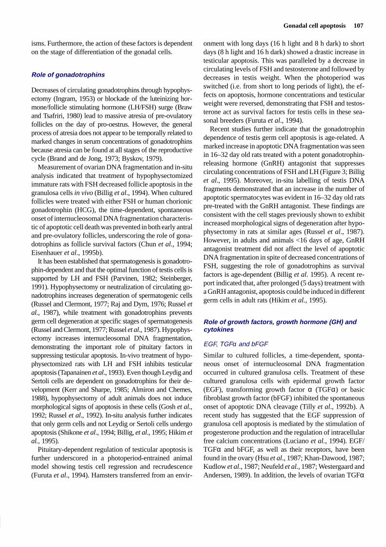
found in germ cells, whereas apoptosis in somatic cells(Leydig and Sertoli cells) could not be detected (Shikone etal., 1994; Billig et al., 1995). Consistent with morphologi-cal signs of degeneration (Russel et al., 1987), in-situanalysis indicated that apoptosis in rats at 20 and 32 days ofage was most frequently found in spermatocytes (Billig etal., 1995). In adult rats, apoptotic DNA fragmentation inthe testis was also restricted to germ cells of the seminifer-ous tubules. In most but not all tubules increased labellingwas found in meiotic spermatocytes delineating the periph-ery of the tubules (Figure 2). Upon fractionation of DNAfrom dissected seminiferous tubules at different stages, thelowest levels of apoptosis were found in stage VIII tubules,while 2-fold higher levels of apoptotic DNA fragmentationwere found in stages I and XII–XIV (Billig et al., 1995;Henriksen et al., 1995). Morphological signs of cell degen-eration in type A2, A3 and A4 spermatogonia and sperma-tocytes have been found in these stages of the tubules(Russel et al., 1987; Allan et al., 1992). Thus, in contrast toovarian studies showing massive apoptosis of somaticgranulosa cells, germ cells are the major cells affected inthe testis (Shikone et al., 1994; Billig et al., 1995).
Hormonal control of gonadal cell apoptosis
The factors which trigger apoptosis in diverse tissues ap-pear to be tissue-specific, although it is believed that thesevarious early apoptotic signals in different cell types ulti-mately lead to a common pathway which locks all cells intothe irreversible progression of apoptotic cell death (Martinand Green, 1995; Stellar, 1995). In gonads, diverse hor-mones and growth factors can act as survival factors toinhibit apoptosis or as apoptotic factors to induce cell de-mise through endocrine, paracrine and autocrine mechan-
Figure 2. Testicular germ cell apoptosis. Testis sections from adult (70 days old) rats were 3′-end-labelled in situ with digoxigenin–ddUTP todemonstrate cellular localization of apoptosis. In most but not all tubules increased labelling is seen in cells delineating the periphery of thetubules (A). Higher magnification (B) shows that these labelled cells are mostly meiotic spermatocytes. No increased labelling could be detectedin Leydig or Sertoli cells. Reprinted with permission from Billig et al. (1995).
Gonadal cell apoptosis 107
isms. Furthermore, the action of these factors is dependenton the stage of differentiation of the gonadal cells.
Role of gonadotrophins
Decreases of circulating gonadotrophins through hypophys-ectomy (Ingram, 1953) or blockade of the luteinizing hor-mone/follicle stimulating hormone (LH/FSH) surge (Brawand Tsafriri, 1980) lead to massive atresia of pre-ovulatoryfollicles on the day of pro-oestrus. However, the generalprocess of atresia does not appear to be temporally related tomarked changes in serum concentrations of gonadotrophinsbecause atresia can be found at all stages of the reproductivecycle (Brand and de Jong, 1973; Byskov, 1979).
Measurement of ovarian DNA fragmentation and in-situanalysis indicated that treatment of hypophysectomizedimmature rats with FSH decreased follicle apoptosis in thegranulosa cells in vivo (Billig et al., 1994). When culturedfollicles were treated with either FSH or human chorionicgonadotrophin (HCG), the time-dependent, spontaneousonset of internucleosomal DNA fragmentation characteris-tic of apoptotic cell death was prevented in both early antraland pre-ovulatory follicles, underscoring the role of gona-dotrophins as follicle survival factors (Chun et al., 1994;Eisenhauer et al., 1995b).
It has been established that spermatogenesis is gonadotro-phin-dependent and that the optimal function of testis cells issupported by LH and FSH (Parvinen, 1982; Steinberger,1991). Hypophysectomy or neutralization of circulating go-nadotrophins increases degeneration of spermatogenic cells(Russel and Clermont, 1977; Raj and Dym, 1976; Russel etal., 1987), while treatment with gonadotrophins preventsgerm cell degeneration at specific stages of spermatogenesis(Russel and Clermont, 1977; Russel et al., 1987). Hypophys-ectomy increases internucleosomal DNA fragmentation,demonstrating the important role of pituitary factors insuppressing testicular apoptosis. In-vivo treatment of hypo-physectomized rats with LH and FSH inhibits testicularapoptosis (Tapanainen et al., 1993). Even though Leydig andSertoli cells are dependent on gonadotrophins for their de-velopment (Kerr and Sharpe, 1985; Almiron and Chemes,1988), hypophysectomy of adult animals does not inducemorphological signs of apoptosis in these cells (Gosh et al.,1992; Russel et al., 1992). In-situ analysis further indicatesthat only germ cells and not Leydig or Sertoli cells undergoapoptosis (Shikone et al., 1994; Billig, et al., 1995; Hikim etal., 1995).
Pituitary-dependent regulation of testicular apoptosis isfurther underscored in a photoperiod-entrained animalmodel showing testis cell regression and recrudescence(Furuta et al., 1994). Hamsters transferred from an envir-
onment with long days (16 h light and 8 h dark) to shortdays (8 h light and 16 h dark) showed a drastic increase intesticular apoptosis. This was paralleled by a decrease incirculating levels of FSH and testosterone and followed bydecreases in testis weight. When the photoperiod wasswitched (i.e. from short to long periods of light), the ef-fects on apoptosis, hormone concentrations and testicularweight were reversed, demonstrating that FSH and testos-terone act as survival factors for testis cells in these sea-sonal breeders (Furuta et al., 1994).
Recent studies further indicate that the gonadotrophindependence of testis germ cell apoptosis is age-related. Amarked increase in apoptotic DNA fragmentation was seenin 16–32 day old rats treated with a potent gonadotrophin-releasing hormone (GnRH) antagonist that suppressescirculating concentrations of FSH and LH (Figure 3; Billiget al., 1995). Moreover, in-situ labelling of testis DNAfragments demonstrated that an increase in the number ofapoptotic spermatocytes was evident in 16–32 day old ratspre-treated with the GnRH antagonist. These findings areconsistent with the cell stages previously shown to exhibitincreased morphological signs of degeneration after hypo-physectomy in rats at similar ages (Russel et al., 1987).However, in adults and animals <16 days of age, GnRHantagonist treatment did not affect the level of apoptoticDNA fragmentation in spite of decreased concentrations ofFSH, suggesting the role of gonadotrophins as survivalfactors is age-dependent (Billig et al. 1995). A recent re-port indicated that, after prolonged (5 days) treatment witha GnRH antagonist, apoptosis could be induced in differentgerm cells in adult rats (Hikim et al., 1995).
Role of growth factors, growth hormone (GH) andcytokines
EGF, TGFα and bFGF
Similar to cultured follicles, a time-dependent, sponta-neous onset of internucleosomal DNA fragmentationoccurred in cultured granulosa cells. Treatment of thesecultured granulosa cells with epidermal growth factor(EGF), transforming growth factor α (TGFα) or basicfibroblast growth factor (bFGF) inhibited the spontaneousonset of apoptotic DNA cleavage (Tilly et al., 1992b). Arecent study has suggested that the EGF suppression ofgranulosa cell apoptosis is mediated by the stimulation ofprogesterone production and the regulation of intracellularfree calcium concentrations (Luciano et al., 1994). EGF/TGFα and bFGF, as well as their receptors, have beenfound in the ovary (Hsu et al., 1987; Khan-Dawood, 1987;Kudlow et al., 1987; Neufeld et al., 1987; Westergaard andAndersen, 1989). In addition, the levels of ovarian TGFα

108 H.Billig et al.
Figure 3. Gonadotrophin regulation of testicular cell apoptosis: age-related changes in gonadotrophin dependence. Pre-treatment of ratswith a potent gonadotrophin-releasing hormone (GnRH) antagonistthat suppressed circulating concentrations of follicle stimulatinghormone and luteinizing hormone caused marked increases in apop-totic DNA fragmentation in 16–32 day old animals. Testicular DNAwas 3′-end-labelled with [32P]ddATP and fractionated through 2%agarose gels followed by exposure on films to visualize the incorpor-ation of [32P]ddATP into DNA 3′-ends (V = vehicle-treated, A =GnRH antagonist-treated). Reprinted from Billig et al. (1995) withpermission.
message are up-regulated following FSH stimulation invivo (Kudlow et al., 1987). Although immunocytochemicalanalysis has revealed the localization of TGFα protein tothe theca–interstitial layer of follicles in several species(Kudlow et al., 1987), high-affinity receptors for EGF/TGFα (Jones et al., 1982; St-Arnaud et al., 1983; Feng etal., 1987) and bFGF (Shikone et al., 1992) have been local-ized to granulosa cells of ovarian follicles. Therefore, it ispossible that apoptotic cell death in granulosa cells of pre-ovulatory follicles is prevented by the paracrine actions ofEGF/TGFα secreted by theca–interstitial cells, or the auto-crine actions of bFGF synthesized by granulosa cells.
IGF and GH
The ovary expresses insulin-like growth factor (IGF)-I re-ceptor, IGF-I, IGF-binding proteins (IGFBP), and pro-teases degrading the binding protein. Local production ofIGF-I plays an important intra-ovarian role in the aug-mentation of gonadotrophin stimulation of follicle differ-entiation (Adashi et al., 1985; Giudice, 1992). In rats, theovarian mRNA for IGF-I is abundant (fourth only to liver,oviduct and uterus; Murphy et al., 1987; Carlsson et al.,1993b), underscoring the importance of local controlmechanisms. Early studies also demonstrated the presenceof IGF-I receptors in the granulosa cells (Davoren et al.,1986) and the stimulation of an increase in ovarian IGF-Iconcentrations by both FSH (Hsu and Hammond, 1987;Hernandez et al., 1989) and GH (Davoren and Hsueh,
1986), although gonadotrophins are probably the primarystimulator of granulosa cell IGF-I production (Carlsson etal., 1989).
IGF in body fluids are bound to IGFBP, and six IGFBPhave been cloned and sequenced (Shimasaki and Ling,1991). Although circulating IGFBP prolong the half-life ofIGF, these proteins mostly inhibit IGF actions (Ui et al.,1989; Bicsak et al., 1990; Adashi et al., 1992). IGFBP-4and -5 are produced by rat granulosa cells (Erickson et al.,1992a,b), and FSH treatment decreases the secretion ofthese proteins in rat ovaries (Adashi et al., 1990). In atretichuman follicles of both normal and polycystic ovarian syn-drome patients, high concentrations of IGFBP have beendetected (Cataldo and Giudice, 1992a,b). In-situ mRNAanalysis has further demonstrated the presence of IGFBP inatretic but not in healthy follicles (Erickson et al., 1992b).In addition, the binding of endogenously produced IGF-Iby exogenously added IGFBP-3 suppresses gonadotrophinstimulation of follicle growth and subsequent ovulation (Uiet al., 1989; Bicsak et al., 1990), suggesting that some ofthe physiological effects of FSH are mediated by endogen-ously produced IGF-I.
The spontaneous onset of apoptosis in pre-ovulatory fol-licles in vitro is prevented by IGF-I, FSH and HCG (Chunet al., 1994). However, the same hormones do not preventdevelopment of apoptosis in isolated granulosa cells, inspite of the presence of their receptors on granulosa cells(Tilly et al., 1992b), suggesting that theca cells are import-ant for mediating the suppressive effect of IGF-I and gona-dotrophins on apoptosis. Furthermore, the suppressiveeffect of both IGF-I and gonadotrophins on follicle DNAfragmentation was prevented by co-treatment withIGFBP-3 (Figure 4). Indeed, treatment with HCG inducesan increase in IGF-I mRNA concentrations in culturedfollicles (Chun et al., 1994). The apoptosis-suppressiveaction of gonadotrophins is probably manifested by de-creased production of IGFBP (Adashi et al., 1990), in-creased IGFBP proteolysis (Fielder et al., 1993; Liu et al.,1993) as well as the stimulation of local IGF-I production(Davoren and Hsueh, 1986; Hsu and Hammond, 1987;Carlsson et al., 1989; Hernandez et al., 1989).
In many tissues, IGF-I production is stimulated by GH,and IGF-I mediates many of the GH effects (D’Ercole etal., 1984). In several species, including rat and human, GHreceptor/binding protein has been demonstrated in granu-losa and theca cells and in the corpus luteum (Lobie et al.,1990; Carlsson et al., 1992, 1993a). In the ovary, GH alsoincreases the production of IGF-I (Davoren et al., 1986).Furthermore, treatment with GH inhibits apoptosis in cul-tured rat follicles (Eisenhauer et al., 1995a). This effect ismost likely mediated through local production of IGF-I

Gonadal cell apoptosis 109
Figure 4. Gonadotrophin and insulin-like growth factor (IGF)-I regulation of ovarian apoptosis: inhibition of apoptosis in cultured ovarian fol-licles by human chorionic gonadotrophin (HCG) and IGF-I and reversal by IGF-binding protein-3 (IGFBP-3). Pre-ovulatory follicles were incu-bated for 24 h with HCG or IGF-I with or without IGFBP-3. DNA extracted was 3′-end-labelled and gel-fractionated as described in Figure 1.Reprinted with permission from Chun et al. (1994).
because GH treatment increases IGF-I mRNA concentra-tions in cultured follicles. Also, IGFBP-3 treatment re-verses the inhibitory effect of GH on apoptosis (Eisenhaueret al., 1995a). These data suggest that IGF-I and GH aresurvival factors for ovarian follicles and the ability of gona-dotrophins and GH to suppress apoptosis is partially me-diated by endogenously produced IGF-I.
Cytokines
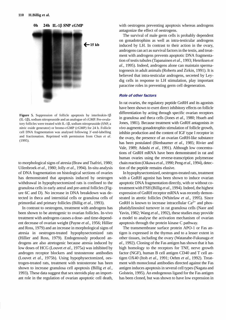
Recent studies have indicated that the pleiotropic cytokineinterleukin-6 (IL-6) is produced by granulosa cells (Go-rospe et al., 1992) under the control of FSH, IL-1α, IL-1βand lipopolysaccharide but not tumour necrosis factor α(TNF-α). Using cultured granulosa cells from rats primedwith pregnant mare’s serum gonadotrophin, IL-6 wasshown to stimulate apoptotic DNA fragmentation in granu-losa cells, suggesting that the cytokine may be an intra-fol-licular atretogenic factor (Gorospe and Spangelo, 1993).IL-1β has also been shown to be cytotoxic to dispersedovarian cells in vitro (Hurwitz et al., 1992). This effectmight be mediated via nitric oxide (NO)-induced cGMPproduction (Ellman et al., 1993). However, preliminarystudies suggest that apoptosis is inhibited by NO-inducedguanylate cyclase activation and by the addition of cGMPanalogue in isolated granulosa cells (Assarsson et al.,1994). In cultured pre-ovulatory follicles, IL-1β treatmentincreases NO production and inhibits apoptosis (Figure 5;Chun et al., 1995). Interestingly, HCG also increases NO
production in cultured follicles, and the suppressive effectof gonadotrophins could in part be reversed by IL-1 recep-tor antagonist, suggesting mediation of gonadotrophin ac-tion by endogenous IL-1β (Chun et al., 1995).Furthermore, follicle apoptosis could be prevented bytreatment with an NO generator (sodium nitroprusside) or amembrane-permeable analogue of cGMP (Figure 5).
In contrast to the ovary, regulation of testis cell apoptosisby growth factors, GH and cytokines has not been studied.
Role of sex steroids
In the ovary, sex steroids are important intra-ovarian regu-lators of follicle atresia and, when oestrogen is given tohypophysectomized rats, fewer follicles become atretic(Ingram, 1959). Furthermore, the profile of sex steroidproduction in healthy follicles differs from that in atreticfollicles. In rat and hamster ovaries, the production of bothoestrogens and androgens decreases in atretic follicles(Braw and Tsafriri, 1980; Uilenbroek et al., 1980; Braw etal., 1981; Terranova, 1981). In human, ovine and porcineovaries the oestradiol production by atretic follicles is alsodecreased but the production of androgens is increased(Carson et al., 1981; Maxson et al., 1985; Moor et al.,1978). The common denominator for all these species is adecreased oestrogen production in atretic follicles (Moor etal., 1978; Braw and Tsafriri, 1980; Braw et al., 1981; Car-son et al., 1981; Terranova, 1981; Maxson et al., 1985). Ingeneral, changes in steroidogenesis can be observed prior
110 H.Billig et al.
Figure 5. Suppression of follicle apoptosis by interleukin-1β(IL-1β), sodium nitroprusside and an analogue of cGMP. Pre-ovula-tory follicles were treated with IL-1β, sodium nitroprusside (SNP, anitric oxide generator) or bromo-cGMP (cGMP) for 24 h. Folliclecell DNA fragmentation was analysed following 3′-end-labellingand fractionation. Reprinted with permission from Chun et al.(1995).
to morphological signs of atresia (Braw and Tsafriri, 1980;Uilenbroek et al., 1980; Jolly et al., 1994). In-situ analysisof DNA fragmentation on histological sections of ovarieshas demonstrated that apoptosis induced by oestrogenwithdrawal in hypophysectomized rats is confined to thegranulosa cells in early antral and pre-antral follicles (Fig-ure 6C and D). No increase in DNA breakdown was de-tected in theca and interstitial cells or granulosa cells ofprimordial and primary follicles (Billig et al., 1993).
In contrast to oestrogens, treatment with androgens hasbeen shown to be atretogenic to ovarian follicles. In-vivotreatment with androgens causes a dose- and time-depend-ent decrease of ovarian weight (Payne et al., 1956; Hillierand Ross, 1979) and an increase in morphological signs ofatresia in oestrogen-treated hypophysectomized rats(Hillier and Ross, 1979). Endogenously produced an-drogens are also atretogenic because atresia induced bylow doses of HCG (Louvet et al., 1975a) was inhibited byandrogen receptor blockers and testosterone antibodies(Louvet et al., 1975b). Using hypophysectomized, oes-trogen-treated rats, treatment with testosterone has beenshown to increase granulosa cell apoptosis (Billig et al.,1993). These data suggest that sex steroids play an import-ant role in the regulation of ovarian apoptotic cell death,
with oestrogens preventing apoptosis whereas androgensantagonize the effect of oestrogens.
The survival of male germ cells is probably dependenton gonadotrophins as well as intra-testicular androgensinduced by LH. In contrast to their action in the ovary,androgens can act as survival factors in the testis, and treat-ment with androgens prevents apoptotic DNA fragmenta-tion of testis tubules (Tapanainen et al., 1993; Henriksen etal., 1995). Indeed, androgens alone can maintain sperma-togenesis in adult animals (Roberts and Zirkin, 1991). It isbelieved that intra-testicular androgens, secreted by Ley-dig cells in response to LH stimulation, play importantparacrine roles in preventing germ cell degeneration.
Role of other factors
In rat ovaries, the regulatory peptide GnRH and its agonistshave been shown to exert direct inhibitory effects on follicledifferentiation by acting through specific ovarian receptorsin granulosa and theca cells (Jones et al., 1980; Hsueh andJones, 1981). Because treatment with GnRH antagonists invivo augments gonadotrophin stimulation of follicle growth,inhibin production and the content of IGF type I receptor inthe ovary, the presence of an ovarian GnRH-like substancehas been postulated (Birnbaumer et al., 1985; Rivier andVale, 1989; Adashi et al., 1991). Although low concentra-tions of GnRH mRNA have been demonstrated in rat andhuman ovaries using the reverse-transcription polymerasechain reaction (Oikawa et al., 1990; Peng et al., 1994), detec-tion of the peptide remains elusive.
In hypophysectomized, oestrogen-treated rats, treatmentwith a GnRH agonist has been shown to induce ovarianapoptotic DNA fragmentation directly, with or without co-treatment with FSH (Billig et al., 1994). Indeed, the highestexpression of GnRH receptor mRNA was recently demon-strated in atretic follicles (Whitelaw et al., 1995). SinceGnRH is known to increase intracellular Ca2+ and phos-phatidylinositol turnover in rat granulosa cells (Naor andYavin, 1982; Wang et al., 1992), these studies may providea model to analyse the activation mechanism of ovarianapoptosis through the protein kinase C pathway.
The transmembrane surface protein APO-1 or Fas an-tigen is expressed in the thymus and to a lesser extent inother tissues, including the ovary (Watanabe-Fukunaga etal., 1992). Cloning of the Fas antigen has shown that it hashigh homology to the receptors for TNF, nerve growthfactor (NGF), human B cell antigen CD40 and T cell an-tigen OX40 (Itoh et al., 1991; Oehm et al., 1992). Treat-ment with monoclonal antibodies directed against the Fasantigen induces apoptosis in several cell types (Nagata andGolstein, 1995). An endogenous ligand for the Fas antigenhas been cloned, but was shown to have low expression in

Gonadal cell apoptosis 111
Figure 6. Sex steroid regulation of ovarian apoptosis: in-situ 3′-end-labelling of ovarian DNA from rats with or without oestrogen treatment.Immature hypophysectomized rats were treated with or without oestrogen implants followed by in-situ 3′-end-labelling with digoxigenin–ddUTP (Billig et al., 1993). (A and B) Ovary after 48 h of oestrogen treatment. (C and D) Ovary 48 h after termination of oestrogen treatmentshowing labelling of granulosa cells (gc) but not theca cells (tc) in pre-antral follicles (arrow). Also indicated is the lack of labelling in primordialfollicles (arrowhead) and interstitial tissue. Bars = 250 µm. Reprinted from Billig et al. (1993) with permission.
the gonads (Suda et al., 1993). However, recent studiesdemonstrated that activation of ovarian Fas antigen inhuman granulosa/luteal cells induces apoptosis (Quirk etal., 1995).
In the testis, four studies have been performed to studythe direct role of GnRH or Fas ligand as regulators ofapoptosis. However, testis cells are highly responsive totemperature elevation. Experimentally induced bilateralcryptorchidism (re-position of the testis inside the abdo-men) induces germ cell apoptosis prior to decreases in theconcentration of LH receptors (Shikone et al., 1994). Incryptorchid testes, apoptosis is not due to a lack of gonado-trophin stimulation since the procedure causes significantincreases in serum concentrations of LH and FSH (Ama-tayakul et al., 1971; Swedloff et al., 1971; Kerr et al., 1978;Au et al., 1983). Furthermore, in unilaterally cryptorchidrats, the abdominal testis showed apoptotic DNA degrada-tion while the contralateral sham-operated testis in the
same animal remained viable despite exposure to the samecirculating hormone concentrations (Shikone et al., 1994).
These studies demonstrate that elevation of testis tem-perature or other changes associated with cryptorchidismmay regulate male germ cell apoptosis. Indeed, morpho-logical signs of apoptosis in spermatocytes have been dem-onstrated in testis exposed to high temperatures (Allan etal., 1987). During apoptotic cell death in the ventral pros-tate, mRNA levels of a 70 kDa heat shock protein increase(Buttyan et al., 1988). In contrast, inactivation of heatshock proteins in fibroblasts increases their sensitivity toheat-induced apoptosis (Riabowol et al., 1987). Becausethe mRNA and gene products of several heat shock pro-teins have been detected in both normal and heat-treatedtestis (Lemaire and Heinlein, 1990; Zakeri et al., 1990; Itohand Tashima, 1991; Rosario et al., 1992), both activationand inactivation of different heat shock proteins may play arole in the apoptosis of testis germ cells.

112 H.Billig et al.
Table II. Comparison of ovarian and testicular apoptosis
Ovary Ref. Testis Ref.
Reduction of potential germ cell pool 75–99.9% 1 50–75% 2
Primary cell type affected granulosa cells 3 germ cells 4
Survival factors
Gonadotrophins LH, FSH 5, 7 LH, FSH 4, 6
Steroids oestrogens 3 androgens 6
Growth factors bFGF, TGFα, EGF 8 ?
IGF-I 7
Others NO, IL-1β 9, 10
Dependence on the stage of differentiationin target cell
yes 3, 11 yes 4, 12
Apoptotic factors androgens 3 temperature (mediated by local factors?) 13
GnRH 5
IGFBPs 7
Fas ligand 14
Cell types responsive to survival andapoptotic factors
granulosa cells, theca cells 15 Leydig cells, Sertoli cells, germ cells 16
LH = luteinizing hormone; FSH = follicle stimulating hormone; bFGF = basic fibroblast growth factor; EGF = epidermal growthfactor; IGF-I = insulin-like growth factor I; NO = nitric oxide; IL-1β = interleukin 1β; GnRH = gonadotrophin-releasing hormone;IGFBP = insulin-like growth factor-binding protein.
References: (1) Baker, 1963; (2) Huckins, 1978; (3) Billig et al., 1993; (4) Billig et al., 1995; (5) Billig et al., 1994; (6) Tapanainenet al., 1993; (7) Chun et al., 1994; (8) Tilly et al., 1992b; (9) Assarsson et al., 1994; (10) Chun et al., 1995; (11) Eisenhauer et al.,1995b; (12) Henriksen et al., 1995; (13) Shikone et al., 1994; (14) Quirk et al., 1995; (15) Hsueh et al., 1984; (16) Vornberger etal., 1994.
Comparison of ovarian and testicular cellapoptosis
In the ovary, most of the follicles endowed during neonataldevelopment degenerate during life. In all species, granu-losa cells are the primary cell type undergoing apoptosis,whereas the fate of the theca cells varies in differentspecies. During later stages of follicle atresia, the oocytesundergo germinal vesicle breakdown and eventually dis-integrate into fragments (Table II). In the testis, 50–75% ofthe germ cells from the potential germ cell pool undergoapoptosis at different stages of spermatogenesis. In con-trast, the testis somatic cells (Leydig and Sertoli cells) donot show any signs of degeneration (Table II).
In addition to their trophic actions on the ovary andtestis, gonadotrophins are essential survival factors for go-nadal cells in both sexes (Table II). It is also clear thatdifferent peptides and steroid hormones play importantroles as survival and apoptotic factors in the regulation ofovarian and testicular apoptosis (Table II). The main prod-ucts of gonadal cells, the sex steroids, are regulators ofapoptosis. In addition, the less studied IL-6 and GnRH-likepeptides may also play important intra-ovarian regulatoryroles. However, it is also clear that specific developmental
stages of gonadal cells determine their susceptibility tosurvival or apoptotic factors. For instance, granulosa cellsin primordial follicles do not undergo apoptosis after oes-trogen withdrawal or androgen stimulation, while granulosacells in more differentiated follicles do (Billig et al., 1993).Furthermore, GH, IL-1β and EGF inhibit apoptosis in pre-ovulatory follicles in vitro (Tilly et al., 1992b; Eisenhaueret al., 1995a; Chun et al., 1995) but do not affect apoptosisin early antral follicles in vitro (Eisenhauer et al., 1995b).In testis, germ cells at different developmental stages ofspermatogenesis demonstrate varying, but stage-specificapoptosis (Kerr, 1992; Billig et al., 1995; Henriksen et al.,1995).
Based mainly on studies using pre-ovulatory follicles ofthe rat, one can postulate a model for the regulation offollicle apoptosis by intra-ovarian hormonal mechanismsinvolving several growth factors (Hsueh et al., 1994). Be-cause granulosa cells are the exclusive site of apoptotic celldeath in rats, endocrine and paracrine signals converge onthis cell type to regulate apoptosis. However, studies usingcultured granulosa cells indicated that treatments withFSH, LH/HCG and IGF-I are ineffective in the preventionof spontaneous apoptosis, despite their apoptosis-suppres-sing action in cultured follicles. These findings suggest an

Gonadal cell apoptosis 113
Figure 7. Intra-ovarian mechanisms involved in follicle atresia:growth factors and their control by gonadotrophins. Based on stud-ies of cultured rat follicles, gonadotrophins and growth hormone(GH) act on granulosa cells to stimulate insulin-like growth factor I(IGF-I) production and suppress IGF-binding protein (IGFBP). Theunbound IGF-I may act on the theca cells to stimulate the productionof survival factors like epidermal growth factor/transforminggrowth factor α (EGF/TGFα), which in turn acts on granulosa cellsto suppress apoptosis. In addition, gonadotrophins may stimulate therelease of unknown survival factors (X) to stimulate the release ofthecal EGF/TGFα, which is responsible for suppression of apopto-sis. Furthermore, basic fibroblast growth factor (bFGF) and TGFαmay be produced in the granulosa cells and suppress granulosa cellapoptosis through a paracrine or autocrine action. In parallel, lutein-izing hormone (LH) may increase interleukin (IL)-1β production bytheca cells. The cytokine then stimulates granulosa cells to releaseNO, which in turn increases the activity of a soluble guanylatecyclase, leading to suppression of apoptosis.
important role of the neighbouring theca cells. FSH or LHmay act at the granulosa cells to produce a theca cell stimu-lator which, in turn, increases the secretion of EGF or TGF-α by the theca cells. Subsequently, these growth factorscould diffuse back to granulosa cells to inhibit apoptosis.Because bFGF is produced by granulosa cells (Neufeld etal., 1987), this peptide could also play an autocrine role inthe regulation of follicle apoptosis (Figure 7).
Because granulosa cells are the main site of IGF-I syn-thesis in rat ovaries, one can further postulate that gonado-trophins stimulate the production of IGF-I production bygranulosa cells and the secreted IGF-I acts on theca cells tostimulate the production of EGF/TGFα. Again, theca cellEGF/TGFα or other factors may diffuse back to granulosacells to inhibit apoptosis. To ensure the survival of ‘se-lected’ follicles, several redundant cellular pathways are
involved to prevent follicle cell apoptosis. In addition tostimulating IGF-I production and inhibiting IGFBP secre-tion, gonadotrophins also increase follicle production ofIL-1β which, in turn, enhances NO production. Elevationof intracellular NO activates a soluble form of guanylatecyclase, leading to cGMP generation and apoptosis sup-pression (Figure 7).
In the testis, increases in germ cell apoptosis after GnRHantagonist treatment in rats between 16 and 32 days of ageare correlated with decreases in serum FSH but not LHconcentrations. It is therefore postulated that FSH is moreimportant than LH as a survival factor for these cells. Inaddition, photoperiod-entrained changes in testis cell apop-tosis in seasonally breeding hamsters are correlated withcirculating FSH concentrations. Because FSH receptors arefound exclusively in the Sertoli cells of the seminiferoustubules but not in germ cells themselves (Heckert et al.,1991), it is likely that the FSH stimulation of Sertoli cellsresults in the stimulation of an intra-tubular factor essentialfor the survival of the male germ cells (Bardin et al., 1988).In addition, in adult animals, treatment with androgen alonecan maintain spermatogenesis. Thus, both androgen and LH,the stimulator of androgen production, are important sur-vival factors for testis cells (Roberts and Zirkin, 1991).
Conclusion
Although early morphological analyses have clearlydocumented that the majority of ovarian follicles andtesticular germ cells undergo degeneration during repro-ductive life, only recent studies have demonstrated theinvolvement of the apoptosis pathway. Availability of aquantitative method to analyse apoptotic DNA fragmenta-tion and in-situ methods to localize the specific cell typesinvolved in DNA degradation opens new experimentalpossibilities for increasing our understanding of thehormonal control of gonadal cell apoptosis. Analysis offollicle cell apoptosis confirms and extends earlier findingsshowing the role of gonadotrophins and oestrogens as fol-licle survival factors as well as the role of androgens as anatretogenic factor. In addition, several ovarian growth fac-tors, regulatory peptides and a signalling gas have recentlybeen identified as either survival factors (EGF/TGFα,basic FGF, IGF-I, IL-1β and nitric oxide) or atretogenicfactors (IL-6 and GnRH). Likewise, analysis of testicularcell apoptosis confirms and extends earlier findings show-ing the role of gonadotrophins and androgens as testicularsurvival factors. It is also becoming evident in both gonadsthat the susceptibility to both apoptotic and survival factorsis dependent on the stage of differentiation of the targetcells. Even though the primary target cell types undergoing
114 H.Billig et al.
apoptosis in the gonads differ (germ cells in the testis andgranulosa cells in the ovary), apoptosis is a physiologicalprocess in the reduction of the potential germ cell pool inboth sexes. Future analysis of the hormonal control ofgonadal cell apoptosis should provide new understandingof the molecular process underlying cell demise as well asbetter treatment protocols for the management of patho-logical conditions involving excessive gonadal cell degen-eration such as premature ovarian failure and polycysticovary syndrome in the female and oligospermia andcryptorchidism in the male. Furthermore, defects in thephysiological process of gonadal cell apoptosis may beassociated with tumorigenesis of ovarian and testis cells.
Acknowledgement
This work was supported by NIH Grant HD-3/566 and SwedishMRC Grant 10380.
References
Adashi, E.Y., Resnick, C.E., D’Ercole, A.J., Svoboda, M.E. and Van Wyk,J.J. (1985) Insulin-like growth factors as intraovarian regulators ofgranulosa cell growth and function. Endocr. Rev., 6, 400–420.
Adashi, E.Y., Resnick, C.E., Hernandez, E.R., Hurwitz, A. and Rosenfeld,R.G. (1990) Follicle-stimulating hormone inhibits the constitutiverelease of insulin-like growth factor binding proteins by cultured ratovarian granulosa cells. Endocrinology, 126, 1305–1307.
Adashi, E.Y., Resnick, C.E., Vera, A. and Hernandez, E.R. (1991) In vivoregulation of granulosa cell type I IGF receptors: evidence for aninhibitory role for the putative endogenous ligand(s) of the ovarianGnRH receptor. Endocrinology, 128, 3130–3137.
Adashi, E.Y., Resnick, C.E., Ricciarelli, E. et al. (1992) Granulosacell-derived insulin-like growth factor (IGF) binding proteins. J. Clin.Invest., 90, 1593–1599.
Allan, D.J., Harmon, B.V. and Kerr, J.F.R. (1987) Cell death inspermatogenesis. In Potten, C.S. (ed.), Perspective on MammalianCell Death. Oxford University Press, London, pp. 229–258.
Allan, D.J., Harmon, B.V. and Roberts, S.A. (1992) Spermatogonialapoptosis has three morphologically recognizable phases and showsno circadian rhythm during normal spermatogenesis in the rat. CellProliferation, 25, 241–250.
Almiron, I. and Chemes, H. (1988) Spermatogenic onset. II FSHmodulates mitotic activity of germ and Sertoli cells in immature rats.Int. J. Androl., 11, 235–246.
Amatayakul, K., Ryan, R., Uozumi, T. and Albert, A. (1971) A reinvestigationof testicular-anterior pituitary relationships in the rat: I. Effects ofcastration and cryptorchidism. Endocrinology, 88, 872–880.
Arends, M.J., Morris, R.G. and Wyllie, A.H. (1990) Apoptosis: the role ofthe endonuclease. Am. J. Pathol., 136, 593–608.
Assarsson, B., Björnheden, D. and Billig, H. (1994) Guanosine 3′,5′-cyclicmonophosphate (cGMP) mediated suppression of apoptosis in culturedovarian granulosa cells. Biol. Reprod., 50 (Suppl. 1), Abstr. 155.
Au, C.L., Robertson, D.M. and de Kretser, D.M. (1983) In vitro bioassay ofinhibin in testes of normal and cryptorchid rats. Endocrinology, 112,239–244.
Bagavandoss, P., Midgley, A.R. Jr and Wicha, M. (1983) Developmentalchanges in the ovarian follicular basal lamina detected byimmunofluorescence and electron microscopy. J. Histochem.Cytochem., 31, 633–640.
Baker, T.G. (1963) A quantitative and cytological study of germ cells inhuman ovaries. Proc. R. Soc. Lond., Ser. B, 158, 417–433.
Bardin, C.W., Cheng, C.Y., Musto, N.A. and Gunsalus, G.L. (1988) TheSertoli cell. In Knobil, E. and Neil, J. (eds), The Physiology ofReproduction. Raven Press, New York, pp. 933–974.
Beaumont, H.M. and Mandl, A.M. (1962) A quantitative and cytologicalstudy of oogonia and oocytes in foetal and neonatal rat. Proc. R. Soc.Lond., Ser. B, 155, 557–579.
Bicsak, T.A., Shimonaka, M., Malkowski, M. and Ling, N. (1990)Insulin-like growth factor binding protein inhibition of granulosa cellfunction: effect on cAMP, DNA synthesis and comparison with theeffect of an IGF-I antibody. Endocrinology, 126, 2184–2189.
Billig, H., Furuta, I. and Hsueh, A.J.W. (1993) Estrogens inhibit andandrogens enhance ovarian granulosa cell apoptosis. Endocrinology,133, 2204–2212.
Billig, H., Furuta, I. and Hsueh, A.J.W. (1994) Gonadotropin releasinghormone (GnRH) directly induces apoptotic cell death in the rat ovary:biochemical and in situ detection of DNA fragmentation in granulosacells. Endocrinology, 134, 245–252.
Billig, H., Furuta, I., Rivier, C., Tapanainen, J., Parvinen, M. and Hsueh,A.J.W. (1995) Apoptosis in testis germ cells: developmental changesin gonadotropin dependence and localization to selective tubulestages. Endocrinology, 136, 5–12.
Birnbaumer, L., Shahabi, N., Rivier, J. and Vale, W. (1985) Evidence for aphysiological role of gonadotropin-releasing hormone (GnRH) orGnRH-like material in the ovary. Endocrinology, 116, 1367–1370.
Boise, L.H., Gonzalez-Garcia, M., Postema, C.E., Ding, L. et al. (1993)Bcl-x, a bcl-2-related gene that functions as a dominant regulator ofapoptotic cell death. Cell, 74, 5597–5608.
Boone, D.L., Yan, W. and Tsang, B.K. (1995) Identification of adeoxyribonuclease I-like endonuclease in rat granulosa and luteal cellnuclei. Biol. Reprod., 53, 1057–1065.
Brand, A. and de Jong, W.H.R. (1973) Qualitative and quantitativemicromorphological investigations of the tertiary follicle populationduring the oestrous cycle of the sheep. J. Reprod. Fertil., 33, 431–439.
Braw, R.H. and Tsafriri, A. (1980) Follicles explanted frompentobarbitone-treated rats provide a model for atresia. J. Reprod.Fertil., 59, 259–265.
Braw, R.H., Byskov, A.G., Peters, H. and Faber, M. (1976) Follicularatresia in the human infant ovary. J. Reprod. Fertil., 46, 55–59.
Braw, R.H., Bar-Ami, S. and Tsafriri, A. (1981) Effect of hypophysectomyon atresia of rat preovulatory follicles. Biol. Reprod., 25, 989–996.
Buttyan, R., Zakeri, Z., Lockshin, R. and Wolgemuth, D. (1988) Cascadeinduction of c-fos, c-myc, and heat shock 70 K transcripts duringregression of the rat ventral prostate gland. Mol. Endocrinol., 2,650–657.
Byskov, A.G.S. (1979) Atresia. In Midgley, A.R. and Sadler, W.A. (eds),Ovarian Follicular Development and Function. Raven Press, NewYork, pp. 41–57.
Carlsson, B., Carlsson, L. and Billig, H. (1989) Estrus cycle-dependentco-variations of the level of insulin-like growth factor-I (IGF-I)messenger ribonucleic acid and protein in the rat ovary. Mol. Cell.Endocrinol., 64, 271–275.
Carlsson, B., Bergh, C., Bentham, J., Olsson, J.-H. et al. (1992) Expressionof functional growth hormone receptors in human granulosa cells.Hum. Reprod., 7, 1205–1209.
Carlsson, B., Nilsson, A., Isaksson, O.G.P. and Billig, H. (1993a) Growthhormone-receptor messenger RNA in the rat ovary: regulation andlocalization. Mol. Cell. Endocrinol., 95, 59–66.
Carlsson, B., Hillensjö, T., Nilsson, A., Törnell, J. and Billig, H. (1993b)Expression of insulin-like growth factor-I (IGF-I) in the rat fallopiantube: possible autocrine and paracrine action of Fallopiantube-derived IGF-I on the Fallopian tube and on the preimplantationembryo. Endocrinology, 133, 2031–2039.
Carson, R.S., Findlay, J.K., Clarke, I.J. and Burger, H.F. (1981) Estradiol,testosterone, and androstenedione in ovine follicular fluid duringgrowth and atresia of ovarian follicles. Biol. Reprod., 24, 105–113.
Cataldo, N.A. and Giudice, L.C. (1992a) Follicular fluid IGF bindingprotein profiles in polycystic ovary syndrome. J. Clin. Endocrinol.Metab., 74, 695–697.
Cataldo, N.A. and Giudice, L.C. (1992b) IGF binding protein profiles inhuman ovarian follicular fluid correlate with follicular functionalstatus. J. Clin. Endocrinol. Metab., 74, 821–829.

Gonadal cell apoptosis 115
Chittenden, T., Harrington, E.A., O’Connor, R. et al.(1995) Induction ofapoptosis by the Bcl-2 homologue Bak. Nature, 374, 733–736.
Chun, S.Y., Billig, H., Tilly, J.L., Furuta, I., Tsafriri, A. and Hsueh, A.J.W.(1994) Gonadotropin suppression of apoptosis in culturedpreovulatory follicles: mediatory role of endogenous IGF-I.Endocrinology, 135, 1845–1853.
Chun, S.Y., Eisenhauer, K.M., Kubo, M. and Hsueh, A.J.W. (1995)Interleukin-1β suppresses apoptosis in rat ovarian follicles byincreasing nitric oxide production. Endocrinology, 136, 3120–3127.
Cohen, J.J. (1993) Apoptosis. Immunol. Today, 14, 126–130.Cohen, J.J. and Duke, R.C. (1984) Glucocorticoid activation of a
calcium-dependent endonuclease in thymocyte nuclei leads to celldeath. J. Immunol., 132, 38–42.
Davoren, J.B. and Hsueh, A.J.W. (1986) Growth hormone increasesovarian levels of immunoreactive somatomedin C/insulin-like growthfactor I in vivo. Endocrinology, 118, 888–890.
Davoren, J.B., Kasson, B.G., Li, C.H. and Hsueh, A.J.W. (1986) Specificinsulin-like growth factor I and II binding sites on rat granulosa cells:relation to IGF action. Endocrinology, 119, 2155–2162.
D’Ercole, A.J., Stiles, A.D. and Underwood, L.E. (1984) Tissueconcentrations of somatomedin-C: further evidence for multiple sitesof synthesis and paracrine or autocrine mechanisms of action. Proc.Natl. Acad. Sci. USA, 81, 935–939.
De Rooij, D.G. and Lok, D. (1987) Regulation of the density ofspermatogonia in the seminiferous epithelium of the Chinese hamster:II Differentiating spermatogonia. Anat. Rec., 217, 131–136.
Dolci, S., Williams, D.E., Ernst, M.K. et al. (1991) Requirement for mastcell growth factor for primordial germ cell survival in culture. Nature,352, 809–811.
Eisenhauer, K., Chun, S.Y., Billig, H. and Hsueh, A.J.W. (1995a) Growthhormone suppression of apoptosis in preovulatory follicles and partialneutralization by insulin-like growth factor binding protein (IGFBP).Biol. Reprod., 53, 13–20.
Eisenhauer, K., Chun, S.Y., Minami, S. and Hsueh, A.J.W. (1995b)Hormonal regulation of apoptosis is dependent on the stage ofdifferentiation: FSH, LH and IGF-I as survival factors for early antralfollicles. Biol. Reprod., 52 (Suppl. 1), 369.
Ellman, C., Corbett, J.A., Misko, T.P., McDaniel, M. and Beckerman, K.P.(1993) Nitric oxide mediates interleukin-1-induced cellularcytotoxicity in the rat ovary. J. Clin. Invest., 93, 3053–3056.
Erickson, G.F., Magoffin, D.A., Dyer, C.A. and Hofeditz, C. (1985) Theovarian androgen producing cells: a review of structure/functionrelationships. Endocr. Rev., 6, 371–399.
Erickson, G.F., Nakatani, A., Ling, N. and Shimasaki, S. (1992a)Localization of insulin-like growth factor-binding protein-5messenger ribonucleic acid in rat ovaries during the estrous cycle.Endocrinology, 130, 1867–1878.
Erickson, G.F., Nakatani, A., Ling, N. and Shimasaki, S. (1992b) Cyclicchanges in insulin-like growth factor-binding protein-4 messengerribonucleic acid in the rat ovary. Endocrinology, 130, 625–636.
Feng, P., Knecht, M. and Catt, K.J. (1987) Hormonal control of epidermalgrowth factor receptors by gonadotropins during granulosa celldifferentiation. Endocrinology, 120, 1121–1126.
Fernadez-Alnemri, T., Litwack, G. and Alnemri, E.S. (1994) CPP32, anovel human apoptotic protein with homology to Caenorhabditiselegans cell death protein Ced-3 and mammalian interleukin-1beta-converting enzyme. J. Biol. Chem., 269, 30761–30764.
Fielder, P., Pham H., Adashi, E.Y. and Rosenfeld, R.G. (1993) Insulin-likegrowth factors (IGFBPs) block FSH-induced proteolysis ofIGF-binding protein-5 (BP-5) in cultured rat granulosa cells.Endocrinology, 133, 415–418.
Flemming, W. (1885) Uber die Bildung von Richtunsfifuren inSaugethiereiern beim Untergang Graafscher Follikel. Arch. Anat.Entw. Gesch., 221–244.
Furuta, I., Porkka-Heiskanen, T., Scarbrough, K. et al. (1994) Photoperiodregulates testis cell apoptosis in Djungarian hamsters. Biol. Reprod.,51, 1315–1321.
Gaido, M.L. and Cidlowski, J.A. (1991) Identification, purification, andcharacterization of calcium-dependent endonuclease (NUC18) fromapoptotic rat thymocytes. J. Biol. Chem., 266, 18580–18585.
Giudice, L. (1992) Insulin-like growth factors and ovarian folliculardevelopment. Endocr. Rev., 13, 641–669.
Godin, I., Deed, R., Cooke, J., Zsebo, K., Dexter, M. and Wylie, C.C.(1991) Effects of the Steel gene product on mouse primordial germcell culture. Nature, 352, 807–809.
Gorospe, W.C. and Spangelo, B.L. (1993) Interleukin-6; potential roles inneuroendocrine and ovarian function. Endocr. J., 1, 3–9.
Gorospe, W.C., Hughes, F.M. and Spangelo, B.L. (1992) Interleukin-6:effects on and production by rat granulosa cells in vitro.Endocrinology, 130, 1750–1752.
Gosh, S., Bartke, A., Grasso, P., Reichert, L.E. and Russel, L.D. (1992)Structural manifestations of the rat Sertoli cell to hypophysectomy: acorrelative morphometric and endocrine study. Endocrinology, 131,485–497.
Greenwald, G.S. (1989) Temporal and topographic changes in DNAsynthesis after induced follicular atresia. Biol. Reprod., 40, 175–181.
Guthrie, H.G., Cooper, B.S., Welch, G.R., Zakaria, A.D. and Johnson, L.A.(1995) Atresia in follicles grown after ovulation in the pig:measurement of increased apoptosis in granulosa cells and reducedfollicular fluid estradiol. Biol. Reprod., 52, 920–927.
Hay, M.F., Cran, D.G. and Moor, R.M. (1976) Structural changesoccurring during atresia in sheep ovarian follicles. Cell Tissue Res.,169, 515–529.
Heckert, L.L. and Griswald, M.D. (1991) Expression offollicle-stimulating hormone receptor mRNA in rat testes and Sertolicells. Mol. Endocrinol., 5, 670–677.
Hengartner, M.O. and Horvitz, R.H. (1994) C. elegans cell survival geneced-9 encodes a functional homolog of the mammalianproto-oncogene bcl-2. Cell, 76, 665–676.
Hengartner, M.O., Ellis, R.E. and Horvitz, H.R. (1992) Caenorhabditiselegans gene ced-9 protects cells from programmed cell death. Nature,356, 494–499.
Henriksen, K., Hakovirta, H. and Parvinen, M. (1995) Testosteroneinhibits and induces apoptosis in rat seminiferous tubules in astage-specific manner: in situ quantification in squash preparationsafter administration of ethane dimethane sulfonate. Endocrinology,136, 3285–3291.
Hernandez, E.R., Roberts, C.T.J., LeRoith, D. and Adashi, E.Y. (1989) Ratovarian insulin-like growth factor I (IGF-I) gene expression isgranulosa cell-selective: 5′-untranslated mRNA variantrepresentation and hormonal regulation. Endocrinology, 125,572–574.
Hikim, A.P.S., Wang, C., Leung, A. and Swerdloff, R.S. (1995)Involvement of apoptosis in the induction of germ cell degeneration inadult rats after GnRH antagonist treatment. Endocrinology, 136,2770–2775.
Hillier, S.G. and Ross, G.T. (1979) Effects of testosterone on ovarianweight, follicular morphology and intraovarian progesteroneconcentration in estrogen-primed hypophysectomized immaturefemale rats. Biol. Reprod., 20, 261–268.
Hirshfield, A.N. (1989) Rescue of atretic follicles in vitro and in vivo. Biol.Reprod., 40, 181–190.
Hirshfield, A.N. (1991) Development of follicles in the mammalian ovary.Int. Rev. Cytol., 124, 43–101.
Hockenbery, D.M., Zutter, M., Hickey, W., Nahm, M. and Korsmeyer, S.J.(1991) Bcl-2 protein is topographically restricted in tissuescharacterized by apoptotic cell death. Proc. Natl. Acad. Sci. USA, 88,6961–6965.
Hsu, C.J. and Hammond, J.M. (1987) Gonadotropins and estradiolstimulate immunoreactive insulin-like growth factor-I production byporcine granulosa cells in vitro. Endocrinology, 120, 198–207.
Hsu, C., Holmes, C.D. and Hammond, J. (1987) Ovarian epidermal growthfactor-like activity. Concentrations in porcine follicular fluid duringfollicular enlargement. Biochem. Biophys. Res. Commun., 147,242–247.
Hsueh, A.J.W. and Jones, P.B. (1981) Extrapituitary actions ofgonadotropin-releasing hormone. Endocr. Rev., 2, 437–461.
Hsueh, A.J.W., Adashi, E.Y., Jones, P.B.C. and Welsh, T.H. Jr (1984)Hormonal regulation of the differentiation of cultured ovariangranulosa cells. Endocr. Rev., 5, 76–127.
116 H.Billig et al.
Hsueh, A.J.W., Billig, H. and Tsafriri, A. (1994) Ovarian follicle atresia:hormonally controlled apoptotic process. Endocr. Rev., 15, 707–724.
Huckins, C. (1978) The morphology and kinetics of spermatogonialdegeneration in normal adult rats. An analysis using a simplifiedclassification of germinal epithelium. Anat. Rec., 190, 905–926.
Hughes, F.M. Jr and Gorospe, W.C. (1991) Biochemical identification ofapoptosis (programmed cell death) in granulosa cells: evidence for apotential mechanism underlying follicular atresia. Endocrinology,129, 2415–2422.
Hurwitz, A., Hernandez, E.R., Payne, D.W., Dharmarajan, A.M. andAdashi, E.Y. (1992) Interleukin-1 is both morphogenic and cytotoxicto cultured rat ovarian cells: obligatory role for heterologous,contact-independent cell-cell interaction. Endocrinology, 131,1643–1649.
Ingram, D.L. (1953) The effect of hypophysectomy on the number ofoocytes in the adult albino rat. J. Endocrinol., 9, 307–311.
Ingram, D.L. (1959) The effect of oestrogen on the atresia of ovarianfollicles. J. Endocrinol., 19, 123–125.
Itoh, N. and Tashima, Y. (1991) Different expression time of the 105-kDaprotein and 90-kDa heat-shock protein in rat testis. FEBS, 289,110–112.
Itoh, N., Yonehara, S., Ishii, A. et al. (1991) The polypeptide encoded bythe cDNA for human cell surface antigen Fas can mediate apoptosis.Cell, 66, 233–243.
Jolly, P.D., Tisdall, D.J., Heath, D.A., Lun, S. and McNatty, S. (1994)Apoptosis in bovine granulosa cells in relation to steroid synthesis,cyclic adenosine 3′,5′-monophosphate response tofollicle-stimulating hormone and luteinizing hormone, and follicleatresia. Biol. Reprod., 51, 934–944.
Jones, P.B.C., Conn, P.M., Marian, J. and Hsueh, A.J.W. (1980) Binding ofgonadotropin releasing hormone agonist to rat ovarian granulosa cells.Life Sci., 27, 2125–2132.
Jones, P.B.C., Welch, T.H. Jr. and Hsueh, A.J.W. (1982) Regulation ofovarian progestin production by epidermal growth factor in culturedrat granulosa cells. J. Biol. Chem., 257, 11268–11273.
Junquiera, L.C., Carneiro, J. and Kelly, R.O. (1989) Basic Histology.Appleton and Lange, Norwalk, pp. 443–444.
Kerr, J.B. (1992) Spontaneous degeneration of germ cells in the normal rattestis: assessment of cell types and frequency during thespermatogenic cycle. J. Reprod. Fertil., 95, 825–830.
Kerr, J.B. and Sharpe, R.M. (1985) Follicle-stimulating hormoneinduction of Leydig cell maturation. Endocrinology, 116, 2592–2604.
Kerr, J.B., Risbridger, G.P., Murray, P.J. and Knell, C.M. (1978) Effect ofunilateral cryptorchidism on the intertubular tissue of the adult testis:evidence for intracellular changes within the Leydig cells. Int. J.Androl., 11, 209–223.
Khan-Dawood, F.S. (1987) Human corpus luteum: immunocytochemicallocalization of epidermal growth factor. Fertil. Steril., 47, 916–919.
Kiefer, M.C., Brauer, M.J., Powers, V.C. et al. (1995) Modulation ofapoptosis by the widely distributed Bcl-2 homologue Bak. Nature,374, 736–739.
Kudlow, J.E., Kobrin, M.S., Purchio, A.F. et al. E.Y. (1987) Ovariantransforming growth factor-alpha gene expression:immunohistochemical localization to the theca-interstitial cells.Endocrinology, 121, 1577–1579.
Kumar, S., Tomooka, Y. and Noda, M. (1992) Identification of a set ofgenes with developmentally down-regulated expression in the mousebrain. Biochem. Biophys. Res. Commun., 185, 1155–1161.
Lemaire, L. and Heinlein, U.A.O. (1990) Detection of secreted andtemporarily inducible heat shock responsive proteins in mousetesticular tissue. Life Sci., 48, 365–372.
Lin, E.Y., Orlofsky, A., Berger, M.S. and Prystowsky, M.B. (1993)Characterization of A1, a novel hematopoietic-specificearly-response gene with sequence similarity to bcl-2. J. Immunol.,151, 1979–1988.
Liu, X.J., Malkowski, M., Guo, Y., Erickson, G., Shimasaki, S. and Ling,N.C. (1993) Development of specific antibodies to rat insulin-likegrowth factor-binding proteins (IGFBP-2 to -6): analysis of IGFBPproduction by rat granulosa cells. Endocrinology, 132, 1176–1183.
Liu, Y.J., Mason, D.Y., Johnson, G.D. et al. (1991) Germinal center cellsexpress bcl-2 protein after activation by signals which prevent theirentry into apoptosis. Eur. J. Immunol., 21, 1905–1910.
Lobie, P.E., Breigphol, W., Aragon, J.G. and Watres, M.J. (1990) Cellularlocalization of the growth hormone receptor/binding protein in maleand female reproductive systems. Endocrinology, 126, 2214–2221.
Louvet, J.P., Harman, S.M. and Ross, G.T. (1975a) Effects of humanchorionic gonadotropin, human intestinal cell stimulating hormoneand human follicle-stimulating hormone on ovarian weights inestrogen-primed hypophysectomized immature female rats.Endocrinology, 96, 1179–1186.
Louvet, J.P., Harman, S.M., Schreiber, J.R. and Ross, G.T. (1975b)Evidence for a role of androgens in follicular maturation.Endocrinology, 97, 366–372.
Luciano, A.M., Pappalardo, A., Ray C., and Peluso, J.J. (1994) Epidermalgrowth factor inhibits large granulosa cell apoptosis by stimulatingprogesterone synthesis and regulating the distribution of intracellularfree calcium. Biol. Reprod., 51, 646–654.
Martin, J.S. and Green, D.R. (1995) Protease activation during apoptosis:death by a thousand cuts? Cell, 82, 349–352.
Maxson, W.S., Haney, A.F. and Schomberg, D.W. (1985) Steroidogenesisin porcine atretic follicles: loss of aromatase activity in isolatedgranulosa and theca. Biol. Reprod., 33, 495–501.
Miura, M., Zhu, H., Rotello, R., Hartwieg, E.A. and Yan, J. (1993)Induction of apoptosis in fibroblasts by IL-1β-converting enzyme, amammalian homolog of the C. elegans cell death gene ced-3. Cell, 75,653–660.
Moor, R.M., Dott, H.M. and Cran, D.G. (1978) Macroscopic identificationand steroidogenic function of atretic follicles in sheep. J. Endocrinol.,77, 309–318.
Murphy, L.J., Bell, G.I. and Friesen, H.G. (1987) Tissue distribution ofinsulin-like growth factor I and II messenger ribonucleic acid in theadult rat. Endocrinology, 120, 1279–1282.
Nagata, S. and Golstein, P. (1995) The FAS death factor. Science, 267,1449–1456.
Naor, Z. and Yavin, E. (1982) Gonadotropin-releasing hormone stimulatesphospholipid labeling in cultured granulosa cells. Endocrinology, 111,1615–1619.
Neufeld, G., Ferrara, N., Schweigerer, L., Mitchell, R. andGospodarowicz, D. (1987) Bovine granulosa cells produce basicfibroblast growth factor. Endocrinology, 121, 597–603.
Nicholson, D.W., All, A., Thornberry, N.A., Valliancourt, J.P., Ding, C.K.and Galiant, M. (1995) Identification and inhibition of the ICE/CED-3protease necessary for mammalian apoptosis. Nature, 376, 37–43.
Oakland, E. (1956) A description of spermatogenesis in the mouse and itsuse in analysis of the cycle of seminiferous epithelium and germ cellrenewal. Am. J. Anat., 99, 391–413.
Oehm, A., Behrmann, I., Falk, W. et al. (1992) Purification and molecularcloning of the APO-1 cell surface antigen: a member of the tumornecrosis factor/nerve growth factor receptor superfamily: sequenceidentity with the Fas antigen. J. Biol. Chem., 267, 10709–10715.
Oikawa, M., Dargan, C.M., Ny, T. and Hsueh, A.J.W. (1990) Expression ofgonadotropin-releasing hormone and prothymosin-alpha messengerribonucleic acid in the ovary. Endocrinology, 127, 2350–2356.
Oltavi, Z.N., Milliam, C.L. and Korsmeyer, S.J. (1993) Bcl-2heterodimerizes in vivo with a conserved homolog, Bax, thataccelerates programmed cell death. Cell, 74, 609–619.
O’shea, J.D., Hay, M.F. and Cran, D.G. (1978) Ultrastructural changes inthe theca interna during follicular atresia in sheep. J. Reprod. Fertil.,54, 183–187.
Palumbo, A. and Yeh, J. (1994) In situ localization of apoptosis in the ratovary during follicle atresia. Biol. Reprod., 51, 888–895.
Parvinen, M. (1982) Regulation of the seminiferous epithelium. Endocr.Rev., 3, 404–417.
Payne, R.W., Hellbaum, A.A. and Owens, J.N. (1956) The effect ofandrogen on the ovaries and uterus of the estrogen treatedhypophysectomized immature rat. Endocrinology, 59, 306–316.
Peng, C., Fan, N.C., Ligier, M., Väänänen, J. and Leung, P.C.K. (1994)Expression and regulation of gonadotropin-releasing hormone

Gonadal cell apoptosis 117
(GnRH) and GnRH receptor messenger ribonucleic acids in humangranulosa-luteal cells. Endocrinology, 135, 1740–1746.
Quirk, S.M., Cowan, R.G., Joshi, S,G. and Henrikson, K.P. (1995) Fasantigen-mediated apoptosis in human granulosa/luteal cells. Biol.Reprod., 52, 279–287.
Raff, M.C. (1992) Social controls on cell survival and cell death. Nature,356, 397–400.
Raj, H.G.M. and Dym, M. (1976) The effects of selective withdrawal ofFSH and LH on spermiogenesis in the immature rat. Biol. Reprod., 14,489–494.
Reed, J.C. (1994) Bcl-2 and the regulation of programmed cell death. J.Cell. Biol., 124, 1–6.
Reed, J.C., Meister, L., Tanaka, S., Cuddy, M., Yum, S., Geyer, C. andPleasure, D. (1991) Differential expression of bcl2 protooncogene inneuroblastoma and other human tumor cell lines of neural origin.Cancer Res., 51, 6529–6538.
Regaud, C.P. (1900) Degenerescence des cellules seminales chezmammiferes en l’abscence de tout etat pathologique. C.R. SéancesSoc. Biol., 52, 268–270.
Riabowol, K.T., Mizzen, L.A. and Welch, W.J. (1987) Heat shock is lethalto fibroblast microinjected with antibodies against hsp70. Science,242, 433–436.
Rivier, C. and Vale, W. (1989) Immunoreactive inhibin secretion by thehypophysectomized female rat: demonstration of the modulatingeffect of gonadotropin-releasing hormone and estrogen through adirect ovarian site of action. Endocrinology, 124, 195–198.
Roberts, K.P. and Zirkin, B.R. (1991) Androgen regulation ofspermatogenesis in the rat. Ann. N.Y. Acad. Sci., 637, 90–106.
Rosario, M.O., Perkins, S.L., O’Brien, D.A., Allen, R.L. and Eddy, E.M.(1992) Identification of the gene for developmentally expressed 70kDA heat-shock protein (P70) of mouse spermatocytogenic cells. Dev.Biol., 150, 1–11.
Russel, L.D. and Clermont, Y. (1977) Degeneration of germ cells innormal, hypophysectomized and hormone treated hypo-physectomized rats. Anat. Rec., 187, 347–366.
Russel, L.D., Alger, L.E. and Nequin, L.G. (1987) Hormonal control ofpubertal spermatogenesis. Endocrinology, 120, 1615–1632.
Russel, L.D., Corbin, T.J., Ren, H.P., Arador, A., Bartke, A. and Gosh, S.(1992) Structural changes in rat Leydig cells posthypophysectomy: amorphometric and endocrine study. Endocrinology, 131, 498–508.
Schwartz, L.M. and Osborne, B.A. (1993) Programmed cell death,apoptosis and killer genes. Immunol. Today, 14, 582–590.
Shikone, T., Yamoto, M. and Nakano, R. (1992) Follicle-stimulatinghormone induces functional receptors for basic fibroblast growthfactor in rat granulosa cells. Endocrinology, 131, 1063–1068.
Shikone, T., Billig, H. and Hsueh, A.J.W. (1994) Experimentally-inducedcryptorchidism increases apoptosis in rat testis. Biol. Reprod., 51,865–872.
Shimasaki, S. and Ling, N. (1991) Identification and molecularcharacterization of insulin-like growth factor binding proteins(IGFBP-1, -2, -3, -4, -5 and -6). Prog. Growth Factor Res., 3, 243–266.
St-Arnaud, R., Walker, P., Kelly, P.A. and Labrie, F. (1983) Rat ovarianepidermal growth factor receptors: characterization and hormonalregulation. Mol. Cell. Endocrinol., 31, 43–52.
Steinberger, E. (1991) Hormonal control of mammalian spermatogenesis.Physiol. Rev., 51, 1–22.
Stellar, M. (1995) Mechanisms and genes of cellular suicide. Science, 267,1445–1449.
Suda, T., Takahashi, T., Golstein, P. and Nagata, S. (1993) Molecularcloning and expression of the Fas ligand, a novel member of the tumornecrosis factor family. Cell, 75, 1169–1178.
Swedloff, R.S., Walsh, P.C., Jacobs, H.S. and O’Dell, W.D. (1971) SerumLH and FSH during sexual maturation in the male rat: effect ofcastration and cryptorchidism. Endocrinology, 88, 120–128.
Tapanainen, J.S., Tilly, J.L., Vihko, K.K. and Hsueh, A.J.W. (1993)Hormonal control of apoptotic cell death in the testis: gonadotropinsand androgens as testicular cell survival factors. Mol. Endocrinol., 7,643–650.
Terranova, P.F. (1981) Steroidogenesis in experimentally induced atreticfollicles of the hamster: a shift from estradiol to progesteronesynthesis. Endocrinology, 108, 1855–1890.
Tilly, J.L. and Hsueh, A.J.W. (1992) A microscale autoradiographicanalysis of apoptotic DNA fragmentation. J. Cell. Physiol., 131,1063–1068.
Tilly, J.L., Kowalski, K.I., Johnson, A.L. and Hsueh, A.J.W. (1991)Involvement of apoptosis in ovarian follicular atresia andpostovulatory regression. Endocrinology, 129, 2799–2801.
Tilly, J.L., Kowalski, K.I., Schomberg, D.W. and Hsueh, A.J.W. (1992a)Apoptosis in atretic ovarian follicles is associated with selectivedecreases in messenger RNA transcripts for gonadotropin receptorsand cytochrome P450 aromatase. Endocrinology, 131, 1670–1676.
Tilly, J.L., Billig, H., Kowalski, K.I. and Hsueh, A.J.W. (1992b) Epidermalgrowth factor and basic fibroblast growth factor suppress thespontaneous onset of apoptosis in cultured rat ovarian granulosa cellsand follicles by a tyrosine kinase-dependent mechanism. Mol.Endocrinol., 6, 1942–1950.
Tilly, J.L., Tilly, K.I., Kenton, M.L. and Johnson, A.L. (1995) Expressionof members of the Bcl-2 gene family in the immature rat ovary: equinechorionic gonadotropin-mediated inhibition of granulosa cellapoptosis is associated with decreased Bax and constitutive bcl-2 andbcl-x long messenger ribonucleic acid levels. Endocrinology, 136,232–241.
Tsafriri, A. and Braw, R.H. (1984) Experimental approaches to atresia inmammals. Oxford Rev. Reprod. Biol., 6, 226–265.
Ui, M., Shimonaka, M., Shimasaki, S. and Ling, N. (1989) An insulin-likegrowth factor-binding protein in ovarian follicular fluid blocksfollicle-stimulating hormone-stimulated steroid production byovarian granulosa cells. Endocrinology, 125, 912–916.
Uilenbroek, J.T.J., Woutersen, P.J.A. and van der Schoot, P. (1980) Atresiaof preovulatory follicles: gonadotropin binding and steroidogenicactivity. Biol. Reprod., 23, 219–229.
Vornberger, W., Prins, G., Musto, N.A. and Suarez-Quian, C.A. (1994)Androgen receptor distribution in the rat testis: new implications forandrogen regulation of spermatogenesis. Endocrinology, 134,2307–2316.
Wang, J., Steele, G.L., Baimbridge, K.G., Rodway, M.R. and Leung, P.C.(1992) Intracellular calcium and the signaling mechanism ofluteinizing hormone-releasing hormone in rat granulosa cells. Am. J.Obstet. Gynecol., 167, 541–547.
Wang, L., Miura, M., Bergeron, L., Zhu, H. and Yuan, J. (1994) Ich-1, anIce/ced-3-related gene, encodes both positive and negative regulatorsof programmed cell death. Cell, 78, 739–750.
Watanabe-Fukunaga, R., Brannan, C.I., Itoh, N. et al. (1992) The cDNAstructure, expression and chromosomal assignment of the Fas antigen.J. Immunol, 148, 1274–1279.
Westergaard, L.G. and Andersen, C.Y. (1989) Epidermal growth factor(EGF) in human preovulatory follicles. Hum. Reprod., 4, 257–260.
Whitelaw, P.F., Eidne, K.A., Seller, R., Smyth, C.D. and Hillier, S.G.(1995) Gonadotropin-releasing hormone receptor messengerribonucleic acid expression in rat ovary. Endocrinology, 136,172–179.
Wyllie, A.H., Kerr, J.F.R. and Currie, A.R. (1980) Cell death: thesignificance of apoptosis. Int. Rev. Cytol., 68, 251–306.
Yuan, J. and Horvitz, H.R. (1990) The Caenorhabditis elegans genes ced-3and ced-4 act cell autonomously to cause programmed cell death. Dev.Biol., 138, 33–41.
Yuan, J. and Horvitz, H.R. (1992) The Caenorhabditis elegans cell deathgene ced-4 encodes a novel protein and is expressed during the periodof extensive programmed cell death. Development, 116, 309–320.
Zakeri, S.F., Welch, W.J. and Wolgemuth, D.J. (1990) Characterizationand inducibility of hsp 70 proteins in the male mouse germ line. J. CellBiol., 111, 1785–1792.
Zeleznik, A.J., Ihrig, L. and Bassett, S.G. (1989) Developmentalexpression of Ca++/Mg++-dependent endonuclease activity in ratgranulosa and luteal cells. Endocrinology, 125, 2218–2220.
Received on August 21, 1995; accepted on January 12, 1996
Related Documents


