Glycoinositolphospholipids from Trypanosomatids Subvert Nitric Oxide Production in Rhodnius prolixus Salivary Glands Felipe Gazos-Lopes 1,8 , Rafael Dias Mesquita 2,8¤ , Lı ´via Silva-Cardoso 1,8 , Raquel Senna 1,8 , Alan Barbosa Silveira 1,8 , Willy Jablonka 1,8 , Cecı´lia Oliveira Cudischevitch 1,8 , Alan Brito Carneiro 1,8 , Ednildo Alcantara Machado 3 , Luize G. Lima 1 , Robson Queiroz Monteiro 1 , Roberto Henrique Nussenzveig 4 , Evelize Folly 5,8 , Alexandre Romeiro 3 , Jorick Vanbeselaere 6 , Lucia Mendonc ¸ a-Previato 3 , Jose ´ Osvaldo Previato 3 , Jesus G. Valenzuela 7 , Jose ´ Marcos Chaves Ribeiro 7 , Georgia Correa Atella 1,8 , Ma ´ rio Alberto Cardoso Silva-Neto 1,8 * 1 Instituto de Bioquı ´mica Me ´dica, Universidade Federal do Rio de Janeiro, Rio de Janeiro, Brazil, 2 Instituto Federal de Educac ¸a ˜o, Cie ˆncia e Tecnologia do Rio de Janeiro, Rio de Janeiro, Brazil, 3 Instituto de Biofı ´sica Carlos Chagas Filho, Universidade Federal do Rio de Janeiro, Rio de Janeiro, Brazil, 4 Department of Leukemia, M.D. Anderson Cancer Center, Houston, Texas, United States of America, 5 Universidade Federal Fluminense, Instituto de Biologia. Campus Valonguinho, Pre ´dio do Instituto de Biologia, Departamento de Biologia Celular e Molecular, Centro, Nitero ´ i, Rio de Janeiro, Brasil, 6 Universite ´ de Lille 1, Unite ´ de Glycobiologie Structurale et Fonctionnelle, Villeneuve d’Ascq, France, 7 Vector Molecular Biology Section, Laboratory of Malaria and Vector Research, National Institute of Allergy and Infectious Diseases, National Institutes of Health, Rockville, Maryland, United States of America, 8 Instituto Nacional de Cie ˆncia e Tecnologia em Entomologia Molecular (INCT-EM), Rio de Janeiro, Brazil Abstract Background: Rhodnius prolixus is a blood-sucking bug vector of Trypanosoma cruzi and T. rangeli. T. cruzi is transmitted by vector feces deposited close to the wound produced by insect mouthparts, whereas T. rangeli invades salivary glands and is inoculated into the host skin. Bug saliva contains a set of nitric oxide-binding proteins, called nitrophorins, which deliver NO to host vessels and ensure vasodilation and blood feeding. NO is generated by nitric oxide synthases (NOS) present in the epithelium of bug salivary glands. Thus, T. rangeli is in close contact with NO while in the salivary glands. Methodology/Principal Findings: Here we show by immunohistochemical, biochemical and molecular techniques that inositolphosphate-containing glycolipids from trypanosomatids downregulate NO synthesis in the salivary glands of R. prolixus. Injecting insects with T. rangeli-derived glycoinositolphospholipids (Tr GIPL) or T. cruzi-derived glycoinositolpho- spholipids (Tc GIPL) specifically decreased NO production. Salivary gland treatment with Tc GIPL blocks NO production without greatly affecting NOS mRNA levels. NOS protein is virtually absent from either Tr GIPL- or Tc GIPL-treated salivary glands. Evaluation of NO synthesis by using a fluorescent NO probe showed that T. rangeli-infected or Tc GIPL-treated glands do not show extensive labeling. The same effect is readily obtained by treatment of salivary glands with the classical protein tyrosine phosphatase (PTP) inhibitor, sodium orthovanadate (SO). This suggests that parasite GIPLs induce the inhibition of a salivary gland PTP. GIPLs specifically suppressed NO production and did not affect other anti-hemostatic properties of saliva, such as the anti-clotting and anti-platelet activities. Conclusions/Significance: Taken together, these data suggest that trypanosomatids have overcome NO generation using their surface GIPLs. Therefore, these molecules ensure parasite survival and may ultimately enhance parasite transmission. Citation: Gazos-Lopes F, Mesquita RD, Silva-Cardoso L, Senna R, Silveira AB, et al. (2012) Glycoinositolphospholipids from Trypanosomatids Subvert Nitric Oxide Production in Rhodnius prolixus Salivary Glands. PLoS ONE 7(10): e47285. doi:10.1371/journal.pone.0047285 Editor: Mauricio Martins Rodrigues, Federal University of Sa ˜o Paulo, Brazil Received January 6, 2012; Accepted September 14, 2012; Published October 15, 2012 Copyright: ß 2012 Gazos-Lopes et al. This is an open-access article distributed under the terms of the Creative Commons Attribution License, which permits unrestricted use, distribution, and reproduction in any medium, provided the original author and source are credited. Funding: This work was supported by grants from Conselho Nacional de Desenvolvimento Cientı ´fico e Tecnolo ´ gico (CNPq), Coordenac ¸a ˜o de Aperfeic ¸oamento de Pessoal de Nı ´vel Superior (CAPES), Fundac ¸a ˜o de Amparo a Pesquisa de Estado do Rio de Janeiro (FAPERJ) and Instituto Nacional de Cie ˆncia e Tecnologia em Entomologia Molecular (INCT-Entomologia Molecular). This work was also supported by two grants provided by the International Foundation for Science (IFS- Sweden) to Dr. G. C. Atella (F/3619-1) and to Dr. M. A. C. Silva-Neto (F/2887-3). The funders had no role in study design, data collection and analysis, decision to publish, or preparation of the manuscript. Competing Interests: The authors have declared that no competing interests exist. * E-mail: [email protected] ¤ Current address: Departamento de Bioquı ´mica, Instituto de Quı ´mica, Universidade Federal do Rio de Janeiro, Rio de Janeiro, Brazil Introduction Invasion of vector salivary glands is a mandatory step in the life cycle of several pathogens. This event ensures pathogen transmis- sion to a vertebrate host in the next feeding cycle. However, by infecting the salivary glands, pathogens come into direct contact with vector saliva and with the anti-hemostatic factors it contains [1]. Thus, their survival in this new environment relies on their ability to avoid the action of potentially harmful anti-hemostatic PLOS ONE | www.plosone.org 1 October 2012 | Volume 7 | Issue 10 | e47285

Welcome message from author
This document is posted to help you gain knowledge. Please leave a comment to let me know what you think about it! Share it to your friends and learn new things together.
Transcript
Glycoinositolphospholipids from TrypanosomatidsSubvert Nitric Oxide Production in Rhodnius prolixusSalivary GlandsFelipe Gazos-Lopes1,8, Rafael Dias Mesquita2,8¤, Lıvia Silva-Cardoso1,8, Raquel Senna1,8, Alan
Barbosa Silveira1,8, Willy Jablonka1,8, Cecılia Oliveira Cudischevitch1,8, Alan Brito Carneiro1,8, Ednildo
Alcantara Machado3, Luize G. Lima1, Robson Queiroz Monteiro1, Roberto Henrique Nussenzveig4,
Evelize Folly5,8, Alexandre Romeiro3, Jorick Vanbeselaere6, Lucia Mendonca-Previato3, Jose
Osvaldo Previato3, Jesus G. Valenzuela7, Jose Marcos Chaves Ribeiro7, Georgia Correa Atella1,8, Mario
Alberto Cardoso Silva-Neto1,8*
1 Instituto de Bioquımica Medica, Universidade Federal do Rio de Janeiro, Rio de Janeiro, Brazil, 2 Instituto Federal de Educacao, Ciencia e Tecnologia do Rio de Janeiro,
Rio de Janeiro, Brazil, 3 Instituto de Biofısica Carlos Chagas Filho, Universidade Federal do Rio de Janeiro, Rio de Janeiro, Brazil, 4 Department of Leukemia, M.D. Anderson
Cancer Center, Houston, Texas, United States of America, 5 Universidade Federal Fluminense, Instituto de Biologia. Campus Valonguinho, Predio do Instituto de Biologia,
Departamento de Biologia Celular e Molecular, Centro, Niteroi, Rio de Janeiro, Brasil, 6 Universite de Lille 1, Unite de Glycobiologie Structurale et Fonctionnelle, Villeneuve
d’Ascq, France, 7 Vector Molecular Biology Section, Laboratory of Malaria and Vector Research, National Institute of Allergy and Infectious Diseases, National Institutes of
Health, Rockville, Maryland, United States of America, 8 Instituto Nacional de Ciencia e Tecnologia em Entomologia Molecular (INCT-EM), Rio de Janeiro, Brazil
Abstract
Background: Rhodnius prolixus is a blood-sucking bug vector of Trypanosoma cruzi and T. rangeli. T. cruzi is transmitted byvector feces deposited close to the wound produced by insect mouthparts, whereas T. rangeli invades salivary glands and isinoculated into the host skin. Bug saliva contains a set of nitric oxide-binding proteins, called nitrophorins, which deliver NOto host vessels and ensure vasodilation and blood feeding. NO is generated by nitric oxide synthases (NOS) present in theepithelium of bug salivary glands. Thus, T. rangeli is in close contact with NO while in the salivary glands.
Methodology/Principal Findings: Here we show by immunohistochemical, biochemical and molecular techniques thatinositolphosphate-containing glycolipids from trypanosomatids downregulate NO synthesis in the salivary glands of R.prolixus. Injecting insects with T. rangeli-derived glycoinositolphospholipids (Tr GIPL) or T. cruzi-derived glycoinositolpho-spholipids (Tc GIPL) specifically decreased NO production. Salivary gland treatment with Tc GIPL blocks NO productionwithout greatly affecting NOS mRNA levels. NOS protein is virtually absent from either Tr GIPL- or Tc GIPL-treated salivaryglands. Evaluation of NO synthesis by using a fluorescent NO probe showed that T. rangeli-infected or Tc GIPL-treatedglands do not show extensive labeling. The same effect is readily obtained by treatment of salivary glands with the classicalprotein tyrosine phosphatase (PTP) inhibitor, sodium orthovanadate (SO). This suggests that parasite GIPLs induce theinhibition of a salivary gland PTP. GIPLs specifically suppressed NO production and did not affect other anti-hemostaticproperties of saliva, such as the anti-clotting and anti-platelet activities.
Conclusions/Significance: Taken together, these data suggest that trypanosomatids have overcome NO generation usingtheir surface GIPLs. Therefore, these molecules ensure parasite survival and may ultimately enhance parasite transmission.
Citation: Gazos-Lopes F, Mesquita RD, Silva-Cardoso L, Senna R, Silveira AB, et al. (2012) Glycoinositolphospholipids from Trypanosomatids Subvert Nitric OxideProduction in Rhodnius prolixus Salivary Glands. PLoS ONE 7(10): e47285. doi:10.1371/journal.pone.0047285
Editor: Mauricio Martins Rodrigues, Federal University of Sao Paulo, Brazil
Received January 6, 2012; Accepted September 14, 2012; Published October 15, 2012
Copyright: � 2012 Gazos-Lopes et al. This is an open-access article distributed under the terms of the Creative Commons Attribution License, which permitsunrestricted use, distribution, and reproduction in any medium, provided the original author and source are credited.
Funding: This work was supported by grants from Conselho Nacional de Desenvolvimento Cientıfico e Tecnologico (CNPq), Coordenacao de Aperfeicoamentode Pessoal de Nıvel Superior (CAPES), Fundacao de Amparo a Pesquisa de Estado do Rio de Janeiro (FAPERJ) and Instituto Nacional de Ciencia e Tecnologia emEntomologia Molecular (INCT-Entomologia Molecular). This work was also supported by two grants provided by the International Foundation for Science (IFS-Sweden) to Dr. G. C. Atella (F/3619-1) and to Dr. M. A. C. Silva-Neto (F/2887-3). The funders had no role in study design, data collection and analysis, decision topublish, or preparation of the manuscript.
Competing Interests: The authors have declared that no competing interests exist.
* E-mail: [email protected]
¤ Current address: Departamento de Bioquımica, Instituto de Quımica, Universidade Federal do Rio de Janeiro, Rio de Janeiro, Brazil
Introduction
Invasion of vector salivary glands is a mandatory step in the life
cycle of several pathogens. This event ensures pathogen transmis-
sion to a vertebrate host in the next feeding cycle. However, by
infecting the salivary glands, pathogens come into direct contact
with vector saliva and with the anti-hemostatic factors it contains
[1]. Thus, their survival in this new environment relies on their
ability to avoid the action of potentially harmful anti-hemostatic
PLOS ONE | www.plosone.org 1 October 2012 | Volume 7 | Issue 10 | e47285
factors. This may be achieved by the manipulation of the
production of some of these factors by parasite-derived molecules.
These processes will enhance the transfer of saliva (and parasites)
to the vertebrate host skin. In fact, we can state that the outcome
of vertebrate host infection is pre-determined by pathogen
interactions that occur while they are still within the invertebrate
host saliva. Nitric oxide synthase (NOS) is an enzyme that
catalyzes the generation of nitric oxide (NO), NADP and L-
citrulline, using L-arginine as substrate, NADPH as a cofactor and
O2 as oxygen donor [2]. There are three known isoforms of this
enzyme in vertebrates: a neuronal (nNOS or type 1), an inducible
(iNOS or type 2) and an endothelial (eNOS or type 3) [3]. The
type 1 and 3 isoforms are collectively known as constitutive NOS.
NOS activity in an invertebrate animal was first described for the
salivary glands of Rhodnius prolixus, a triatominae bug that is a
vector of Trypanosoma cruzi, the etiological agent of Chagas disease
[4], and in the brain of Schistocerca gregaria [5]. R. prolixus is also the
vector for T. rangeli, another trypanosomatid that is considered to
be harmless to humans. T. rangeli is transmitted by bug saliva and is
in close contact with an NO source until its transmission [6].
The role of NO in invertebrate immune response has been
observed in several models [3,7]. Early evidence that insects
generate this molecule when infected with protozoan parasites was
obtained in Anophelinae mosquitoes infected with Plasmodium
berghei [8]. The NOS of the midgut of S. stephensi was monitored by
semi-quantitative PCR at different days after infection. There was
a large increase in gene expression in infected animals [8]. When
the generation of NO was blocked using a NOS inhibitor, L-
NAME, there was an increase in the number of parasites [8]. The
ability of NO to eliminate parasites was also observed in gastropod
hemocytes resistant to Schistosoma mansoni [9]. Harmful effects of
NO in biological systems include the oxidation of heme groups
and nitrosylation of some amino-acid residues, causing unsched-
uled changes in the secondary structure of proteins, and thus
blocking parasite viability [10]. Finally, NO also acts as a signaling
molecule, having a key role in the insect immune system [11].
Infection with T. rangeli inhibited the generation of NO in the
salivary glands of R. prolixus [12]. However, the techniques used by
those authors did not evaluate NOS activity, and its expression
and inhibitory mechanisms were not described. It was later
demonstrated that the administration of NOS inhibitors leads to
an increase in T. rangeli parasitemia [12]. When NOS expression
and NO generation were measured in different tissues of R. prolixus
infected either with T. rangeli, T. cruzi or with lipopolysaccharide
(LPS) [13,14], each tissue responded differently. In general, LPS
and T. cruzi infection increased the expression of NOS and no
changes were detected with T. rangeli infection. However, salivary
glands were not evaluated in that study. Since salivary glands are
ultimately involved with parasite transmission and are the last
tissue to be infected, a specific study of the effects of T. rangeli
infection is still needed.
Besides its activities in the insect immune system, NO also
facilitates the feeding of R. prolixus. This occurs when NO binds to
the heme of the host soluble guanylate cyclase enzyme, which
converts magnesium guanosine 59-triphosphate into cGMP. The
elevation of intracellular cGMP levels causes the smooth-muscle
layer surrounding the vascular endothelium to relax, increasing
the caliber of blood vessels. NO is also an inhibitor of platelet
aggregation [15,16]. Since the description of the presence of NO
in the salivary glands of R. prolixus, it has been suggested that a
group of proteins present in the saliva could bind to NO [15,17].
Such proteins were found to reversibly bind NO, being named
nitrophorins (NPs) [15,18]. So far seven different NPs have been
identified in R. prolixus, and are responsible for NO delivery to the
vertebrate host bloodstream [19]. Therefore, the interaction
between triatominae and trypanosomatids provides a unique host
versus parasite model system where salivary NO plays a dual role,
as both an anti-hemostatic molecule and as an immune-activated
molecule.
Glycosylphosphatidylinositol (GPI)-anchored glycoproteins
from the surface of trypanosomatids are key mediators of host-
parasite interactions. This idea was first established through studies
in T. brucei [20]. The surface of this parasite is covered by a dense
layer of glycoproteins, and these molecules display high antigenic
variation. These data were correlated with early work describing
the ability of this parasite to avoid the immune response of their
vertebrate hosts. Since then, many studies have described the
effects of different glycoconjugates from trypanosomatids on the
immune system of vertebrates, with emphasis on mammals
[21,22,23,24]. A common feature to almost all of these glycocon-
jugates is their attachment to the outer membrane of parasites
through a GPI-anchor. The GPI-anchor consists of an inositolpho-
spholipid skeleton bound to a conserved carbohydrate chain
(Mana1-2Mana1-6Mana1-4GlcNa1-6myoinositol1-PO4-lipid).
The lipid portion remains immersed in the cell membrane and can
consist of either a glycerolipid or a ceramide [21]. Proteins may be
attached to such anchors through a peptide bound between the
protein’s C-terminus and either an ethanolaminephosphate or
aminoethylphosphonate substituent branching out of the third
mannose of the aforementioned conserved glycan chain.
GPI-anchors that are not linked to proteins or polysaccharides
are known as glycoinositolphospholipids (GIPLs). These molecules
are found in large quantities on the surface of trypanosomatids
and, in general, there may be more than 107 GIPL copies per cell
in T. cruzi [21]. They can also be found on the surface of other
parasites, such as Plasmodium [22,23]. Studies have shown that
parasite infection relies on bioactive GIPLs or GPI-anchors that
modulate the mammalian immune system. These effects include
the modulation of NO synthesis in macrophages, regulation of the
levels of cytokines and cell-adhesion molecules and the manipu-
lation of cell-signaling pathways, among many others [21–24].
Very little is known about what effects parasite GIPLs have on the
cell signaling of insect vectors. Thus, it is clear that more studies on
the effects of surface molecules from parasites on insect vectors are
needed. The use of purified molecules allows experiments to be
done in the absence of living parasites and provides new
information on the relationship between parasites and vectors.
Here, we provide for the first time evidence for the role of surface
inositolphosphate-containing glycolipids (GIPLs) as molecules
responsible for subverting NO synthesis in a blood-sucking
arthropod.
Results
T. rangeli Blocks NO Production in Salivary Glandsthrough a Mechanism that Involves its Surface GIPLs
Following a blood meal, Rhodnius salivary glands are continu-
ously refilled by the secretion of anti-hemostatic molecules
synthesized from salivary-gland epithelia. In R. prolixus the nitric
oxide-binding proteins, nitrophorins (NP), are synthesized in the
first part of the feeding cycle in the 4th instar, and later on after
moulting are readily loaded with NO [25]. NO synthase (NOS)
activity in Rhodnius salivary glands can be reliably evaluated by
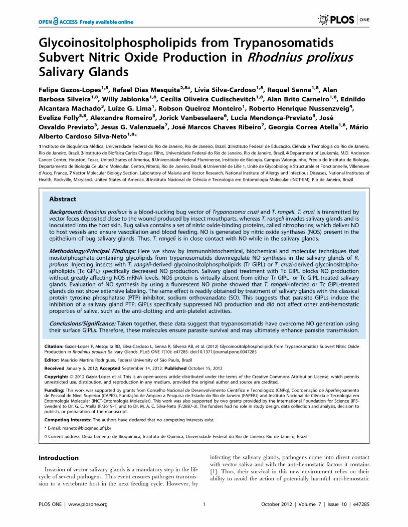
NADPH-diaphorase activity [25]. The surge on NADPH-diaph-
orase activity as a function of NOS synthase activity was due to an
increase in the expression of the NO synthase gene itself, as
evaluated by enzyme activity and immunoblotting against this
enzyme (Figures 1A and 1B). Semi-quantitative PCR experiments
Subversion of Nitric Oxide Synthesis by GIPLs
PLOS ONE | www.plosone.org 2 October 2012 | Volume 7 | Issue 10 | e47285
suggested that the increase in NOS expression occurred following
an increase in the levels of mRNA coding for this enzyme
(Figure 1C). In order to verify the role of parasite infection in this
NO-NOS model, in the next experiments we have separately
evaluated the levels of NOS and NO synthesis in isolated salivary
glands obtained from both control and T. rangeli-infected insects as
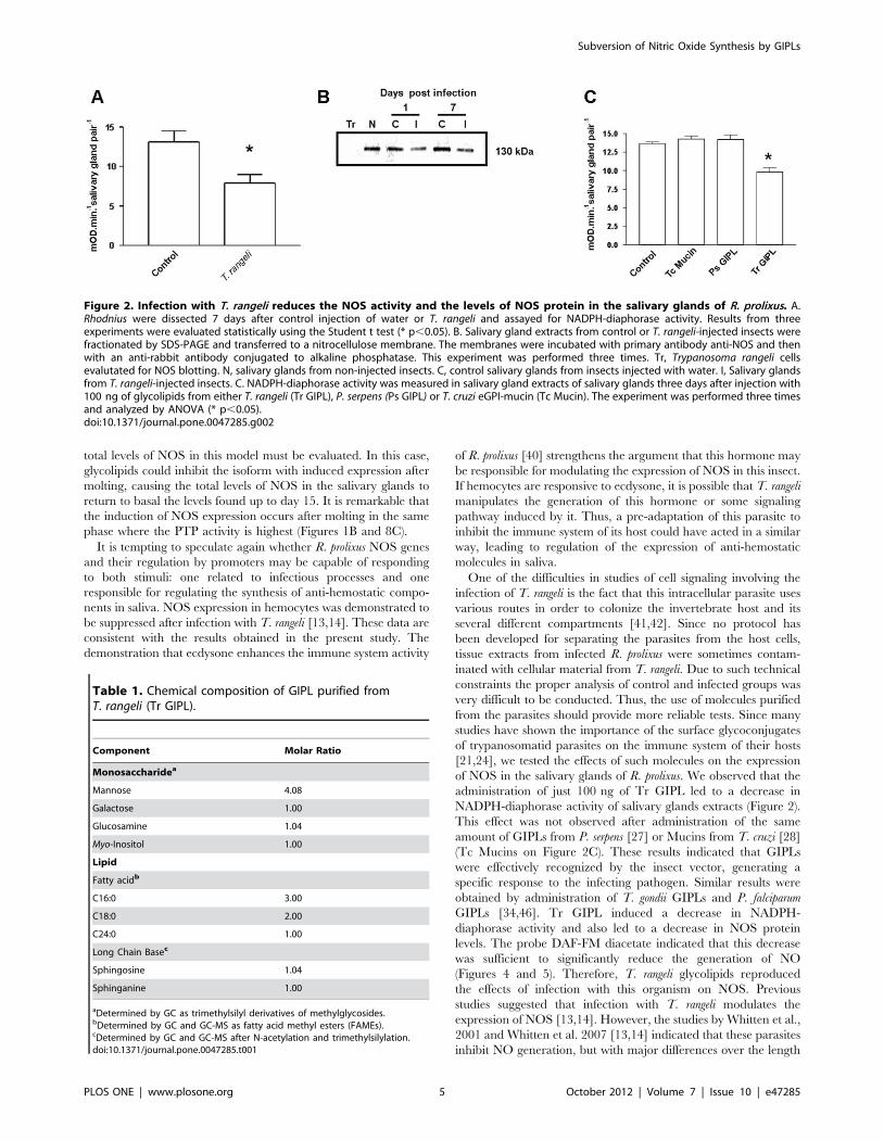
shown on Figure 2. Figures 2A and 2B show a decrease in
NADPH-diaphorase activity and NOS protein in infected salivary
glands, as evaluated by western blotting and enzyme assay data.
To test for the role of parasite-derived surface molecules in the
suppression of NADPH-diaphorase activity we have next evalu-
ated the effect of T. cruzi-derived GIPLs, a GPI-anchored mucin-
like glycoprotein isolated from epimastigote forms (eGPI-mucin)
from T. cruzi surface glycoconjugate, GIPLs isolated from T. rangeli
surface (Tr GIPL) and a GIPL preparation isolated from
Phytomonas serpens, a trypanosomatid that infects plant [26,27,28].
Figure 2C shows that only (Tr GIPL) suppressed NADPH-
diaphorase activity. Tr GIPL chemical composition was then
determined (Table 1). Figure 3 shows that the levels of NOS are
decreased in infected salivary glands specifically in the neighboring
epithelial cells around the gland lumen, as evaluated through
immunocytochemistry (Figure 3). This confirmed that T. rangeli-
mediated suppression of NADPH-diaphorase activity (Figure 1A)
and NOS expression (Figure 2B) specifically occurred on salivary
gland epithelium, which is the major site of NOS localization in
this tissue. So far, NO synthesis was indirectly evaluated by the
NADPH-diaphorase assay, and a direct evaluation assay of NO
production was not conducted. Thus, isolated salivary glands from
control and infected insects were loaded with the NO-sensor DAF-
FM and evaluated for tissue integrity (Figures 4A and 4C).
T. rangeli infection leads to a large decrease in NO production
(Figures 4B and 4D).
In most pathogen x host cell interaction models parasite
interaction with host cells and tissues occurs before invasion.
Surface molecules are usually involved in such interaction and
mediate the first signaling events leading to the suppression of
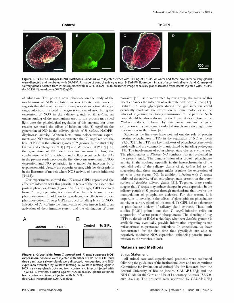
host-mounted defenses against pathogens. As shown in the
previous experiments, Tr GIPLs were able to decrease NADPH-
diaphorase activity from salivary glands (Figure 2C). We next
tested whether Tr GIPL could affect NO synthesis. Figure 5 shows
that NO synthesis was suppressed in glands injected with Tr GIPL.
Also, such effect is associated with the suppression of the level of
NOS protein, since this protein is markedly reduced by treatment
of salivary glands with Tr GIPL (Figure 6A). This effect was also
seen in glands injected with T. cruzi-derived GIPLs (Tc GIPL)
(Figure 6B). The amount of Tr GIPL obtained in our experiments
was very low. Besides that, the same effects on NO biology in
Rhodnius observed with Tr GIPL were also observed with Tc
GIPL. Therefore, in the following experiments Tc-GIPL was used
in order to detail GIPL effects on NO production in Rhodnius.
Tc GIPL-induced Manipulation of NO Synthesis is SpecificTowards this Anti-hemostatic Molecule and is Mediatedby the Suppression of a Protein Tyrosine PhosphataseActivity
The following experiments were designed to obtain more
detailed information regarding the effect of Tc GIPL on Rhodnius
salivary glands. We first evaluated the effect of Tc GIPL of the
production of overall anti-hemostatic molecules of Rhodnius, and in
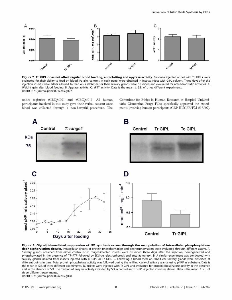
the ability of the insects to feed on blood. Figure 7A shows that Tc
GIPL did not affect insect regular blood-feeding following days
after treatment. Also, there was no change in total apyrase activity
or clotting time for saliva from Tc GIPL-treated glands (Figures 7B
and 7C). This indicates that the parasites manipulation of salivary
gland anti-hemostatic components occurred by a specific intracel-
lular signaling pathway that only disrupted NO biological circuits.
Protein phosphorylation-dephosphorylation is a major mechanism
of regulation of NO production in several models [2]. In order to
obtain evidence that components of this signaling pathway are
being manipulated, the following experiments were conducted.
Figure 8A shows that in-vitro exposure of T. rangeli-infected glands
extracts to 32P-ATP induced the phosphorylation of a major set of
,75-kDa bands. Also, glands of insects treated with Tr GIPL or
Tc GIPL showed a similar phosphorylated set of bands (Figure 8B).
This result suggested that the phosphorylation circuits in both
infected and glycolipid-treated glands were very similar. We next
evaluated the role of protein dephosphorylation in GIPL-mediated
NO suppression in salivary glands. We first tested whether the
activity of protein tyrosine phosphatases (PTPs) are involved in the
regulation of NO production [29]. Figure 8C shows that total PTP
activity increased during the refilling cycle, being the highest at 30
days, the time for maximal loading with NO (as demonstrated in
Figure 1 for NOS). Salivary gland PTP activity is highly inhibited
by sodium orthovanadate (SO), which is a classical inhibitor of
PTPs [29,30,31,32,33]. Furthermore, injection of the insects with
Tc GIPL decreased PTP activity in glands (Figure 8D).
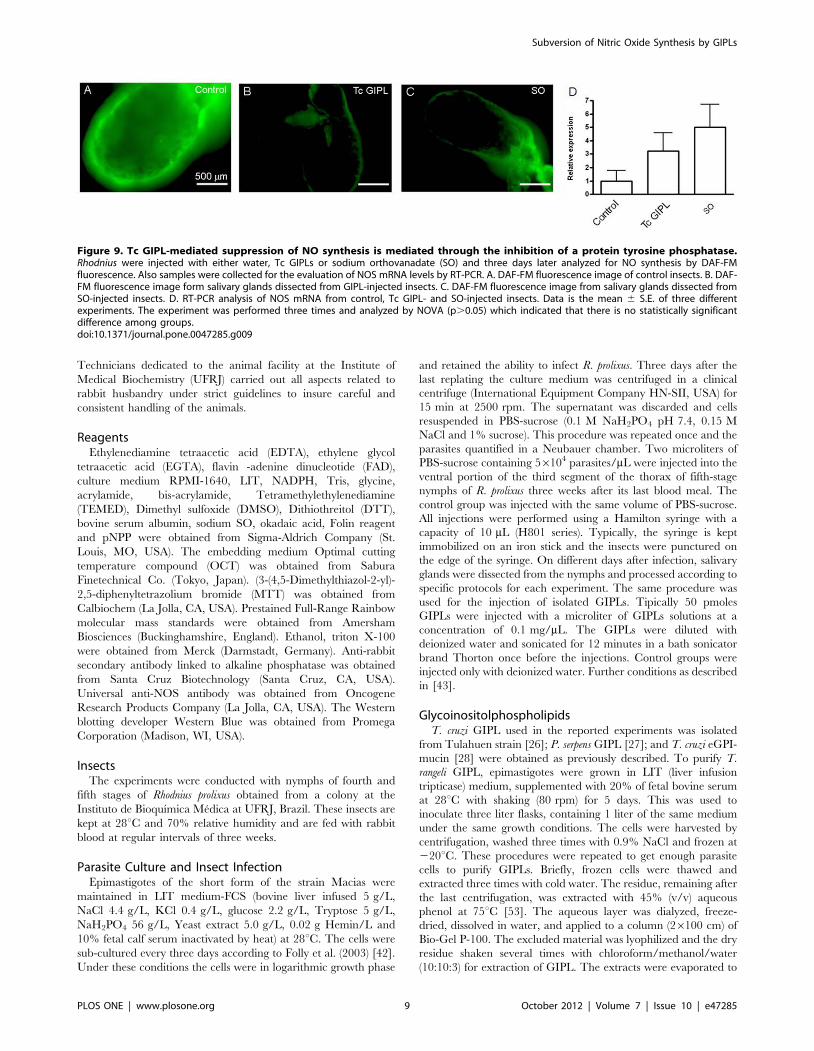
Next, we evaluated whether SO could induce a decrease in NO
synthesis as compared to control glands (Figure 9). Figure 9B and
9C show that Tc GIPL and SO treatment each reduced NO
production to a similar extent. However, both interventions (Tc
GIPL and SO) did not statistically affect the levels of mRNA for
NOS (Figure 9D). Thus, it is likely that upon invasion of the
salivary gland, parasite surface GIPLs inhibited a vector PTP that
was essential to keep NOS mRNA continuously under translation
and ultimately led to inhibition of NO synthesis.
Discussion
The first published study addressing the role of glycolipids in
NO biology during vector 6 parasite interaction used the P.
falciparum 6 An. stephensi model [34]. In that study, it was
demonstrated that GIPL concentrations between 0.25 and
2.5 mM were able to induce NOS expression at the same intensity
as infection with live parasites. Nogueira et al. (2007), working in
another model showed that administration of 100 nM of Tc GIPL
inhibited the adhesion of these microorganisms to the digestive
tract of bugs by 95% [35]. Possibly, parasite glycoconjugates
interact with vector gut through lectins present in the peritrophic
matrix found in most insects and in Rhodnius perimicrovilllar
membrane [34,36]. Another work also observed an interaction
between T. rangeli and the salivary glands of R. prolixus, showing
that T. rangeli incubation with certain types of carbohydrates
inhibited parasite-salivary glands interaction. Incubation with
salivary glands glycoconjugates also resulted in the same effect
[37]. Thus, it may suggest that lectins and other receptors on the
surface of T. rangeli and salivary glands of R. prolixus may be
involved in the interaction between these two organisms.
In this study, the sugar, fatty acid and long-chain base
compositions of GIPLs purified from T. rangeli have been
elucidated (Table 1). It is known that trypanosomatids are
phylogenetically close groups, and tend to have overall similar
surface molecules. The species closest to T. rangeli that has had its
surface glycoconjugates characterized is T. cruzi [21,38]. Indeed,
the Tr GIPL is a glycoinositolphosphosphingolipid whose com-
position is similar to Tc GIPL [39]. The comparison of the effects
caused by administration of GIPLs from T. cruzi or T. rangeli on
invertebrate host might bring new information on the biology of
Subversion of Nitric Oxide Synthesis by GIPLs
PLOS ONE | www.plosone.org 3 October 2012 | Volume 7 | Issue 10 | e47285
these two species. Since both species coexist in the same host, it is
possible that in some stages of their life cycles, they may use similar
strategies to suppress vector immune responses. Also, the
interference of parasite species on one another resulting from
the co-existence on the same host must be evaluated.
Our experiments suggested that the effects of live parasites or
their glycolipids were due to the down regulation in NOS
expression. Tr GIPL largely affected NOS protein expression and
NO synthesis (Figures 2, 5). Furthermore, one may consider that
there may be more than one isoform of NOS in the salivary glands
of R. prolixus. In this situation, one NOS isoform might be
constitutively expressed throughout the period of saliva replace-
ment, while a second NOS isoform might be expressed only after
insect molting. In this case, NP expression would be higher in the
first days after blood feeding and dramatically lower following
insect molting [18,25]. Thus, there may be specific mechanisms
that adjust NP synthesis to NO availability. The information
regarding the presence of multiple NOS isoforms in R. prolixus is
not available. However, if this is the case, potential effects of
T. rangeli GIPLs on each isoform, resulting in partial inhibition of
Figure 1. NADPH-diaphorase activity of NOS in Rhodnius prolixus salivary glands after a blood meal and the expression of NOS. A.Salivary glands were dissected in different days after blood feeding and evaluated for NOS NAPDH-diaphorase activity. Salivary glands were assayedin 10 mM Tris-HCl pH 8,0, 0,05 M NaCl, 0,1%, Triton X-100, 1 mM CaCl2, 5 mM FAD, 1 mM NADPH and 0,5 mg/mL MTT. MTT reduction was followed at540 nm for 30 min at 37uC. Also samples were obtained and NOS content evaluated by Western blotting. Each point is the average and SE of 05different experiments. B. Immunoblotting using an anti-NOS antibody. Blottings were developed with the use of a secondary antibody conjugated toalkaline phosphatase in the presence of the substrate Western Blue. Molecular mass markers are indicated at the left. C. Upper panel, total RNA fromthe salivary glands at different days after feeding was isolated and cDNA was synthesized. Samples were then analyzed by semi-quantitative PCR withtemperatures of 55, 72 and 94uC for 27 cycles with primers for NOS. Lower panel, analysis of 18 S RNA levels. In this case reaction occurred for 25cycles. The products of reactions shown on panels C were separated on agarose gel 1.4% stained with ethidium bromide and photographed underultraviolet light. Molecular mass standards are indicated at the left.doi:10.1371/journal.pone.0047285.g001
Subversion of Nitric Oxide Synthesis by GIPLs
PLOS ONE | www.plosone.org 4 October 2012 | Volume 7 | Issue 10 | e47285
total levels of NOS in this model must be evaluated. In this case,
glycolipids could inhibit the isoform with induced expression after
molting, causing the total levels of NOS in the salivary glands to
return to basal the levels found up to day 15. It is remarkable that
the induction of NOS expression occurs after molting in the same
phase where the PTP activity is highest (Figures 1B and 8C).
It is tempting to speculate again whether R. prolixus NOS genes
and their regulation by promoters may be capable of responding
to both stimuli: one related to infectious processes and one
responsible for regulating the synthesis of anti-hemostatic compo-
nents in saliva. NOS expression in hemocytes was demonstrated to
be suppressed after infection with T. rangeli [13,14]. These data are
consistent with the results obtained in the present study. The
demonstration that ecdysone enhances the immune system activity
of R. prolixus [40] strengthens the argument that this hormone may
be responsible for modulating the expression of NOS in this insect.
If hemocytes are responsive to ecdysone, it is possible that T. rangeli
manipulates the generation of this hormone or some signaling
pathway induced by it. Thus, a pre-adaptation of this parasite to
inhibit the immune system of its host could have acted in a similar
way, leading to regulation of the expression of anti-hemostatic
molecules in saliva.
One of the difficulties in studies of cell signaling involving the
infection of T. rangeli is the fact that this intracellular parasite uses
various routes in order to colonize the invertebrate host and its
several different compartments [41,42]. Since no protocol has
been developed for separating the parasites from the host cells,
tissue extracts from infected R. prolixus were sometimes contam-
inated with cellular material from T. rangeli. Due to such technical
constraints the proper analysis of control and infected groups was
very difficult to be conducted. Thus, the use of molecules purified
from the parasites should provide more reliable tests. Since many
studies have shown the importance of the surface glycoconjugates
of trypanosomatid parasites on the immune system of their hosts
[21,24], we tested the effects of such molecules on the expression
of NOS in the salivary glands of R. prolixus. We observed that the
administration of just 100 ng of Tr GIPL led to a decrease in
NADPH-diaphorase activity of salivary glands extracts (Figure 2).
This effect was not observed after administration of the same
amount of GIPLs from P. serpens [27] or Mucins from T. cruzi [28]
(Tc Mucins on Figure 2C). These results indicated that GIPLs
were effectively recognized by the insect vector, generating a
specific response to the infecting pathogen. Similar results were
obtained by administration of T. gondii GIPLs and P. falciparum
GIPLs [34,46]. Tr GIPL induced a decrease in NADPH-
diaphorase activity and also led to a decrease in NOS protein
levels. The probe DAF-FM diacetate indicated that this decrease
was sufficient to significantly reduce the generation of NO
(Figures 4 and 5). Therefore, T. rangeli glycolipids reproduced
the effects of infection with this organism on NOS. Previous
studies suggested that infection with T. rangeli modulates the
expression of NOS [13,14]. However, the studies by Whitten et al.,
2001 and Whitten et al. 2007 [13,14] indicated that these parasites
inhibit NO generation, but with major differences over the length
Figure 2. Infection with T. rangeli reduces the NOS activity and the levels of NOS protein in the salivary glands of R. prolixus. A.Rhodnius were dissected 7 days after control injection of water or T. rangeli and assayed for NADPH-diaphorase activity. Results from threeexperiments were evaluated statistically using the Student t test (* p,0.05). B. Salivary gland extracts from control or T. rangeli-injected insects werefractionated by SDS-PAGE and transferred to a nitrocellulose membrane. The membranes were incubated with primary antibody anti-NOS and thenwith an anti-rabbit antibody conjugated to alkaline phosphatase. This experiment was performed three times. Tr, Trypanosoma rangeli cellsevalutated for NOS blotting. N, salivary glands from non-injected insects. C, control salivary glands from insects injected with water. I, Salivary glandsfrom T. rangeli-injected insects. C. NADPH-diaphorase activity was measured in salivary gland extracts of salivary glands three days after injection with100 ng of glycolipids from either T. rangeli (Tr GIPL), P. serpens (Ps GIPL) or T. cruzi eGPI-mucin (Tc Mucin). The experiment was performed three timesand analyzed by ANOVA (* p,0.05).doi:10.1371/journal.pone.0047285.g002
Table 1. Chemical composition of GIPL purified fromT. rangeli (Tr GIPL).
Component Molar Ratio
Monosaccharidea
Mannose 4.08
Galactose 1.00
Glucosamine 1.04
Myo-Inositol 1.00
Lipid
Fatty acidb
C16:0 3.00
C18:0 2.00
C24:0 1.00
Long Chain Basec
Sphingosine 1.04
Sphinganine 1.00
aDetermined by GC as trimethylsilyl derivatives of methylglycosides.bDetermined by GC and GC-MS as fatty acid methyl esters (FAMEs).cDetermined by GC and GC-MS after N-acetylation and trimethylsilylation.doi:10.1371/journal.pone.0047285.t001
Subversion of Nitric Oxide Synthesis by GIPLs
PLOS ONE | www.plosone.org 5 October 2012 | Volume 7 | Issue 10 | e47285
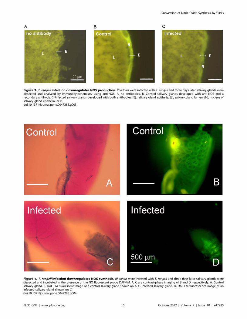
Figure 4. T. rangeli infection downregulates NOS synthesis. Rhodnius were infected with T. rangeli and three days later salivary glands weredissected and incubated in the presence of the NO fluorescent probe DAF-FM. A, C are contrast-phase imaging of B and D, respectively. A. Controlsalivary gland. B. DAF-FM fluorescent image of a control salivary gland shown on A. C. Infected salivary gland. D. DAF-FM fluorescence image of aninfected salivary gland shown on C.doi:10.1371/journal.pone.0047285.g004
Figure 3. T. rangeli infection downregulates NOS production. Rhodnius were infected with T. rangeli and three days later salivary glands weredissected and analyzed by immunocytochemistry using anti-NOS. A. no antibodies. B. Control salivary glands developed with anti-NOS and asecondary antibody. C. Infected salivary glands developed with both antibodies. (E), salivary gland epithelia, (L), salivary gland lumen, (N), nucleus ofsalivary gland epithelial cells.doi:10.1371/journal.pone.0047285.g003
Subversion of Nitric Oxide Synthesis by GIPLs
PLOS ONE | www.plosone.org 6 October 2012 | Volume 7 | Issue 10 | e47285
of inhibition. This poses a novel challenge on the study of the
mechanisms of NOS inhibition in invertebrate hosts, once it
suggests that different mechanisms may operate over time during a
single infection. If indeed T. rangeli is capable of modulating the
expression of NOS in the salivary glands of R. prolixus, an
understanding of the mechanisms used in this process may shed
light onto the physiological regulation of this enzyme. For these
reasons we tested the effects of infection with T. rangeli on the
generation of NO in the salivary glands of R. prolixus. NADPH-
diaphorase activity, Western-blots, immunolocalization experi-
ments and NO imaging all demonstrated that T. rangeli reduces the
level of NOS in the salivary glands of R. prolixus. In the studies by
Garcia and colleagues (1994) [12] and Whitten et al (2007) [14],
the generation of NO itself was not measured. Thus, the
combination of NOS antibody and a fluorescent probe for NO
in the present study provides the first direct measurement of NOS
expression and NO generation in a model for infection by a
trypanosomatid. Usually the opposite occurs, with few descriptions
in the literature of models where NOS activity of hosts is inhibited
[44,45].
Our experiments showed that T. rangeli GIPLs reproduced the
effects of infection with this parasite in terms of the increased total
protein phosphorylation (Figure 8A). Surprisingly, GIPLs derived
from T. cruzi epimastigotes induced similar effects on protein
phosphorylation. In addition to reproducing the effects on protein
phosphorylation, T. cruzi GIPLs also led to falling levels of NOS.
Injection of T. cruzi into the hemolymph of these insects leads to an
activation of their immune system and the elimination of these
parasites [46]. As demonstrated by our group, the saliva of this
insect enhances the infection of vertebrate hosts with T. cruzi [47].
Perhaps, T. cruzi glycolipids during the gut infection could
eventually modulate the expression of some molecules in the
saliva of R. prolixus, facilitating transmission of the parasite. Such
point should be also addressed in the future. A description of the
Rhodnius sialome followed by microarray analysis of gene
expression in trypanosomatid-infected insects may shed light onto
this question in the future [48].
Studies in the literature have pointed out the role of protein
tyrosine phosphatases (PTPs) in the regulation of NO synthesis
[29,30,32]. The PTPs are key mediators of phosphotyrosine levels
inside cells and are commonly manipulated by invading pathogens
[49]. The involvement of other phosphatase classes, such as Ser/
Thr phosphatases in Rhodnius NO synthesis was not evaluated in
the present study. The demonstration of a protein phosphatase
activity in the nucleus, especially in the heterochromatin of the
epithelial cells of the salivary glands of R. prolixus led to the
suggestion that these enzymes might regulate the expression of
genes in these organs [50]. In addition, infection with T. rangeli
inhibited the activity of an ecto-phosphatase present on the outer
surface of Rhodnius salivary glands [51]. Together, these results
suggest that T. rangeli may induce changes in gene expression in the
salivary glands of R. prolixus through mechanisms that involve the
manipulation of phosphatase activities. For this reason, it is
important to investigate the effects of glycolipids on phosphatase
activity in salivary glands of this model. Tr GIPL led to a decrease
in phosphatase activity of salivary gland extracts. Thus, both
studies [50,51] pointed out that T. rangeli infection relies on
suppression of vector protein phosphatases. The silencing of bug
PTPs by the aid of RNAi technology whenever Rhodnius genome is
available may eventually provide information regarding vector
refractoriness to protozoan infections. In conclusion, we have
demonstrated for the first time that glycolipids are able to
negatively modulate NOS expression, enhancing parasite trans-
mission to the vertebrate host.
Materials and Methods
Ethics StatementAll animal care and experimental protocols were conducted
following the guidelines of the institutional care and use committee
(Committee for Evaluation of Animal Use for Research from the
Federal University of Rio de Janeiro, CAUAP-UFRJ) and the
NIH Guide for the Care and Use of Laboratory Animals (ISBN 0-
309-05377-3). The protocols were approved by CAUAP-UFRJ
Figure 5. Tr GIPLs suppress NO synthesis. Rhodnius were injected either with 100 ng of Tr GIPL or water and three days later salivary glandswere dissected and incubated with DAF-FM. A. Image of control salivary glands. B. DAF-FM fluorescent image of a control salivary gland. C. Image ofsalivary glands isolated from insects injected with Tr GIPL. D. DAF-FM fluorescence image of salivary glands isolated from insects injected with Tr GIPL.doi:10.1371/journal.pone.0047285.g005
Figure 6. Glycolipids from T. rangeli and T. cruzi suppress NOSexpression. Rhodnius were injected with either Tr GIPL or Tc GIPL andthree days later salivary glands were dissected, homogenized and NOSexpression evaluated by Western blotting. A. Western blotting againstNOS in salivary glands obtained from control and insects injected withTr GIPLs. B. Western blotting against NOS in salivary glands obtainedfrom control and insects injected with Tc GIPLs.doi:10.1371/journal.pone.0047285.g006
Subversion of Nitric Oxide Synthesis by GIPLs
PLOS ONE | www.plosone.org 7 October 2012 | Volume 7 | Issue 10 | e47285
under registries #IBQM001 and #IBQM011. All human
participants involved in this study gave their verbal consent once
blood was collected through a non-harmful procedure. The
Committee for Ethics in Human Research at Hospital Universi-
tario Clementino Fraga Filho specifically approved the experi-
ments involving human participants (CEP-HUCFF/FM 213/07).
Figure 7. Tc GIPL does not affect regular blood feeding, anti-clotting and apyrase activity. Rhodnius injected or not with Tc GIPLs wereevaluated for their ability to feed on blood. Parallel controls in each panel were obtained in insects inject with GIPL solvent. Three days after theinjection insects were either allowed to feed on a rabbit ear or their salivary glands were dissected and evaluated for anti-hemostatic activities. A.Weight gain after blood feeding. B. Apyrase activity. C. aPTT activity. Data is the mean 6 S.E. of three different experiments.doi:10.1371/journal.pone.0047285.g007
Figure 8. Glycolipid-mediated suppression of NO synthesis occurs through the manipulation of intracellular phosphorylation-dephosphorylation circuits. Intracellular circuits of protein-phosphorylation and dephosphorylation were evaluated through different assays. A.Salivary glands obtained from either control or T. rangeli-infected insects were dissected three days after the injection, homogeneized andphosphorylated in the presence of 32P-ATP followed by SDS-gel electrophoresis and autoradiograph. B. A similar experiment was conducted withsalivary glands isolated from insects injected with Tr GIPL or Tc GIPL. C. Following a blood meal on rabbit ear salivary glands were dissected atdifferent points in time. Total protein phosphatase activity was followed during the refilling cycle of salivary glands using pNPP as substrate. Data isthe mean 6 S.E. of three different experiments. D. Insects were injected with Tr GIPL and evaluated for protein phosphatase activity in the presenceand in the absence of SO. The fraction of enzyme activity inhibited by SO in control and Tr GIPL-injected insects is shown. Data is the mean 6 S.E. ofthree different experiments.doi:10.1371/journal.pone.0047285.g008
Subversion of Nitric Oxide Synthesis by GIPLs
PLOS ONE | www.plosone.org 8 October 2012 | Volume 7 | Issue 10 | e47285
Technicians dedicated to the animal facility at the Institute of
Medical Biochemistry (UFRJ) carried out all aspects related to
rabbit husbandry under strict guidelines to insure careful and
consistent handling of the animals.
ReagentsEthylenediamine tetraacetic acid (EDTA), ethylene glycol
tetraacetic acid (EGTA), flavin -adenine dinucleotide (FAD),
culture medium RPMI-1640, LIT, NADPH, Tris, glycine,
acrylamide, bis-acrylamide, Tetramethylethylenediamine
(TEMED), Dimethyl sulfoxide (DMSO), Dithiothreitol (DTT),
bovine serum albumin, sodium SO, okadaic acid, Folin reagent
and pNPP were obtained from Sigma-Aldrich Company (St.
Louis, MO, USA). The embedding medium Optimal cutting
temperature compound (OCT) was obtained from Sabura
Finetechnical Co. (Tokyo, Japan). (3-(4,5-Dimethylthiazol-2-yl)-
2,5-diphenyltetrazolium bromide (MTT) was obtained from
Calbiochem (La Jolla, CA, USA). Prestained Full-Range Rainbow
molecular mass standards were obtained from Amersham
Biosciences (Buckinghamshire, England). Ethanol, triton X-100
were obtained from Merck (Darmstadt, Germany). Anti-rabbit
secondary antibody linked to alkaline phosphatase was obtained
from Santa Cruz Biotechnology (Santa Cruz, CA, USA).
Universal anti-NOS antibody was obtained from Oncogene
Research Products Company (La Jolla, CA, USA). The Western
blotting developer Western Blue was obtained from Promega
Corporation (Madison, WI, USA).
InsectsThe experiments were conducted with nymphs of fourth and
fifth stages of Rhodnius prolixus obtained from a colony at the
Instituto de Bioquımica Medica at UFRJ, Brazil. These insects are
kept at 28uC and 70% relative humidity and are fed with rabbit
blood at regular intervals of three weeks.
Parasite Culture and Insect InfectionEpimastigotes of the short form of the strain Macias were
maintained in LIT medium-FCS (bovine liver infused 5 g/L,
NaCl 4.4 g/L, KCl 0.4 g/L, glucose 2.2 g/L, Tryptose 5 g/L,
NaH2PO4 56 g/L, Yeast extract 5.0 g/L, 0.02 g Hemin/L and
10% fetal calf serum inactivated by heat) at 28uC. The cells were
sub-cultured every three days according to Folly et al. (2003) [42].
Under these conditions the cells were in logarithmic growth phase
and retained the ability to infect R. prolixus. Three days after the
last replating the culture medium was centrifuged in a clinical
centrifuge (International Equipment Company HN-SII, USA) for
15 min at 2500 rpm. The supernatant was discarded and cells
resuspended in PBS-sucrose (0.1 M NaH2PO4 pH 7.4, 0.15 M
NaCl and 1% sucrose). This procedure was repeated once and the
parasites quantified in a Neubauer chamber. Two microliters of
PBS-sucrose containing 56104 parasites/mL were injected into the
ventral portion of the third segment of the thorax of fifth-stage
nymphs of R. prolixus three weeks after its last blood meal. The
control group was injected with the same volume of PBS-sucrose.
All injections were performed using a Hamilton syringe with a
capacity of 10 mL (H801 series). Typically, the syringe is kept
immobilized on an iron stick and the insects were punctured on
the edge of the syringe. On different days after infection, salivary
glands were dissected from the nymphs and processed according to
specific protocols for each experiment. The same procedure was
used for the injection of isolated GIPLs. Tipically 50 pmoles
GIPLs were injected with a microliter of GIPLs solutions at a
concentration of 0.1 mg/mL. The GIPLs were diluted with
deionized water and sonicated for 12 minutes in a bath sonicator
brand Thorton once before the injections. Control groups were
injected only with deionized water. Further conditions as described
in [43].
GlycoinositolphospholipidsT. cruzi GIPL used in the reported experiments was isolated
from Tulahuen strain [26]; P. serpens GIPL [27]; and T. cruzi eGPI-
mucin [28] were obtained as previously described. To purify T.
rangeli GIPL, epimastigotes were grown in LIT (liver infusion
tripticase) medium, supplemented with 20% of fetal bovine serum
at 28uC with shaking (80 rpm) for 5 days. This was used to
inoculate three liter flasks, containing 1 liter of the same medium
under the same growth conditions. The cells were harvested by
centrifugation, washed three times with 0.9% NaCl and frozen at
220uC. These procedures were repeated to get enough parasite
cells to purify GIPLs. Briefly, frozen cells were thawed and
extracted three times with cold water. The residue, remaining after
the last centrifugation, was extracted with 45% (v/v) aqueous
phenol at 75uC [53]. The aqueous layer was dialyzed, freeze-
dried, dissolved in water, and applied to a column (26100 cm) of
Bio-Gel P-100. The excluded material was lyophilized and the dry
residue shaken several times with chloroform/methanol/water
(10:10:3) for extraction of GIPL. The extracts were evaporated to
Figure 9. Tc GIPL-mediated suppression of NO synthesis is mediated through the inhibition of a protein tyrosine phosphatase.Rhodnius were injected with either water, Tc GIPLs or sodium orthovanadate (SO) and three days later analyzed for NO synthesis by DAF-FMfluorescence. Also samples were collected for the evaluation of NOS mRNA levels by RT-PCR. A. DAF-FM fluorescence image of control insects. B. DAF-FM fluorescence image form salivary glands dissected from GIPL-injected insects. C. DAF-FM fluorescence image from salivary glands dissected fromSO-injected insects. D. RT-PCR analysis of NOS mRNA from control, Tc GIPL- and SO-injected insects. Data is the mean 6 S.E. of three differentexperiments. The experiment was performed three times and analyzed by NOVA (p.0.05) which indicated that there is no statistically significantdifference among groups.doi:10.1371/journal.pone.0047285.g009
Subversion of Nitric Oxide Synthesis by GIPLs
PLOS ONE | www.plosone.org 9 October 2012 | Volume 7 | Issue 10 | e47285
dryness, under reduced pressure; the residue was dissolved in
water and precipitated overnight at 220uC by addition of 5
volumes of methanol.
Carbohydrate Analysis of Tr GIPLsThe monosaccharide composition of Tr GIPLs was determined
according to Sweeley et al. [54]. After methanolysis with 0.5 M
HCl in methanol (18 h, 80uC), the mixture was extracted with
hexane and the methanolic phase neutralized with Ag2CO3. The
products were N-acetylated with acetic anhydride, dried, and
treated with bis-(trimethylsilyl)trifluoroacetamide (BSTFA)/pyri-
dine (1:1, v/v, 1 h, room temperature).
Trimethylsilyl derivatives were analyzed by gas chromatogra-
phy (GC) using a DB-5 fused silica capillary column
(25 m60.25 mm i.d.) with hydrogen (10 psi) as the carrier gas.
The column temperature was programmed from 120uC to 240uCat 2uC/min.
Analysis of Inositol and Glucosamine of Tr GIPLsThe Tr GIPLs were treated with 3 M HCl in methanol for 18 h
at 80uC. The methanolysates were dried under a stream of N2, the
resulting residue was dissolved in 1.0 ml of aqueous 6 M HCl and
heated for 18 h at 105uC. After hexane extraction, to remove fatty
acids, the aqueous layer was lyophilized. The products were N-
acetylated with acetic anhydride, dried, and treated with bis-
(trimethylsilyl)trifluoroacetamide (BSTFA)/pyridine (1:1, v/v, 1 h,
room temperature). Trimethylsilyl derivatives were analyzed by
gas chromatography (GC) using a DB-5 fused silica capillary
column (25 m60.25 mm i.d.) with hydrogen (10 psi) as the carrier
gas. The column temperature was programmed from 120uC to
240uC at 2uC/min.
Lipid Analysis of Tr GIPLsAfter methanolysis of the Tr GIPL with methanolic-HCl (18 h
at 80uC), fatty acid methyl esters (FAMEs) were extracted with
hexane. The extracts were concentrated and analyzed by GC after
derivatization with BSTFA/pyridine as described in carbohydrate
analysis. The column temperature was programmed from 180uCto 280uC at 3uC/min. Peaks were identified by their retention time
compared with authentic standards and by gas chromatography
coupled mass spectrometry (GC-MS). For the analysis of long-
chain bases, GIPCs were methanolyzed (1 M methanol-HCl made
10 M with respect to water) [55]) for 18 h at 80uC. After adjusting
the pH to about 11 with aqueous NaOH, long-chain bases were
extracted with diethyl ether. The extracts were washed with water,
dried with anhydrous sodium sulfate, evaporated to dryness,
dissolved in methanol, and N-acetylated with acetic anhydride
(18 h, room temperature in the dark). The product was treated
with BSTFA/pyridine and analyzed by GC and GC-MS as
described for the FAMEs.
Dissection and Homogenization of Salivary GlandsNymphs of R. prolixus previously anesthetized in ice had their
salivary glands dissected in TBS buffer (10 mM Tris-HCl pH 8.0,
150 mM NaCl). The homogenization of salivary glands was
performed using a 2 mL buffer for each pair of glands. These
samples were frozen in liquid N2 three times and sonicated twice,
in cycles of 12 min in a Thornton sonicator bath. Subsequently,
samples were centrifuged for 5 min at 11000 g. The precipitated
material was discarded and the supernatant referred to as salivary
gland extract. The protein content of these samples was measured
using a colorimetric method [56]. Extracts routinely contained 15
pairs of salivary glands. This material was designed to test several
enzyme activities such as NADPH-diaphorase, phosphotyrosine
phosphatase, and also for Western blots. Using this method both
the major salivary glands and the accessory gland were homog-
enized together. In order to simplify the reading of the protocols
below, the term ‘‘salivary gland’’ includes the main and accessory
glands.
NADPH-diaphorase Activity of Salivary-gland ExtractsSalivary-gland homogenates were assayed for NADPH-diaph-
orase activity according to Ribeiro and Nussenzveig (1993) [4].
Briefly, the extracts were adjusted to a final concentration of 0.5
pairs/mL in TBS buffer. The tests were performed in buffer
containing 10 mM Tris-HCl pH 8.0, 0.05 M NaCl, Triton X-100
0.1%, 1 mM CaCl2, 5 mM FAD, NADPH 1 mM and MTT at
0.5 mg/mL. The MTT reduction was monitored colorimetrically
at 540 nm for 30 min at 37uC. Readings were taken on a Thermo
Max ELISA reader (Molecular Devices) using Thermo Max
software for the measurement of enzyme kinetics. Each test was
performed in triplicate, using a pair of salivary glands (150 mg
protein) per experimental point.
Western BlottingSalivary-gland extracts (100 mg protein) were diluted in sample
buffer (2% SDS, 5% b-mercaptoethanol, 62.5 mM Tris-HCl
pH 6.8, 1% glycerol and 12.5 mg/mL bromophenol blue). After
boiling for 5 min, samples and prestained Full-Range Rainbow
molecular mass standards (250, 160, 105, 75, 50, 35, 30, 25, 15,
10 kDa) were used in a 7.5% bis-acrylamide gel (1561560.1 cm)
with 0.1% SDS. Protein extracts were fractionated using a
constant current of 20 mA [57], then electrotransferred from the
gel to a nitrocellulose membrane at 200 mA for 120 min at 4uC in
Tris-HCl 25 mM pH 8.3, 192 mM glycine and 20% methanol (v/
v) [58]. The membrane was blocked with 10 mM Tris-HCl
pH 8.0 containing 0.15 M NaCl, 0.05% Tween-20 and 2%
bovine serum albumin fraction V (TBSTA) for 18 h at 4uC. The
next day, the blocking buffer was discarded and the membrane
incubated with polyclonal anti-universal NOS (anti-NOS, Onco-
gene) diluted 1:1000 in TBSTA for 1 h at room temperature.
Then, the membrane was washed three times for 5 min with
15 mL of TBSTA and incubated for 30 min at room temperature
with anti-rabbit IgG (conjugated with alkaline phosphatase)
diluted 1:10,000 in TBSTA. The membrane was washed three
times for 5 min each with 15 mL of TBS. Finally, the antigen-
antibody complex was revealed by adding 7 mL of the Western
Blue developer (Pierce). After development, the reaction was
stopped with two washes of 50 mL of distilled water.
Immunolocalization of NOSTwenty days after parasite infection or injection with PBS-
sucrose, nymphs had their salivary glands dissected as described
above. After dissection, glands were fixed whole in 50 mL of PBS-
4% paraformaldehyde for 2 h. After fixation the material was
washed three times with 500 mL of PBS for 2 min and then
incubated in 500 mL of PBS-20% sucrose (0.1 M NaH2PO4
pH 7.4, 0.15 M NaCl and 20% sucrose) at 40uC for 24 h.
Samples were taken from the buffer and placed in trays of 0.5 cm2,
containing half the final volume of the embedding medium OCT.
The glands were carefully positioned and covered with the rest of
the embedding medium. This material was then frozen in liquid
N2 for later use. The samples were processed in a cryostat at
2180uC, generating slices 10 mm thick, which were placed in
quadruplicate on glass slides. These slides had been previously
soaked in alcohol, dried, soaked in 1% gelatin solution and dried
again to allow the cryosectioning. Slides with cryosections were
Subversion of Nitric Oxide Synthesis by GIPLs
PLOS ONE | www.plosone.org 10 October 2012 | Volume 7 | Issue 10 | e47285
subsequently thawed and the OCT removed by incubating the
same slides with TBS for 10 min. Then these sections were
permeabilized with 0.1% Triton-TBS for 10 min. After these
procedures, cryosections were blocked in TBSTA solution for 1 h
at 4uC and immunolocalization was performed using anti-NOS
AB-1 (Oncogene) diluted 1:100 in TBSTA for 1 h. After one hour
the samples were incubated in TBSTA for 30 min, then incubated
with anti-rabbit secondary antibody conjugated with fluorescein
for 2 h. Next 3 washes of 5 min each with TBS-T and three
washes with TBS were conducted. After the last wash a drop of n-
propylgalactate was added to each blade which was then closed
with a coverslip and kept in the dark at 6uC until image
acquisition. The images were acquired on an Axioplan epifluor-
escence microscope (Zeiss) at a magnification of 63x. Images were
then treated equally in X and Adobe Photoshop 5.5 (Adobe).
Controls were performed with uninfected glands or incubated in
medium devoid of primary antibody. All incubations and washes
mentioned above were performed in a final volume of 200 mL.
NO ImagingDAF-FM diacetate (Molecular Probes) is a readily permeable
probe for NO which diffuses through the cytoplasmic membrane.
Upon reaching the cytoplasm of cells, DAF-FM diacetate is
deacetylated by esterases, becoming DAF-FM, which is slightly
fluorescent. Once NO reacts with DAF-FM it is converted into a
derivative that fluoresces around 160 times more than the original
molecule. The wavelengths for excitation/emission maxima are
495/515 nm respectively. Salivary glands (freshly dissected and
intact) were incubated with Rhodnius Ringer (130 mM NaCl,
8 mM KCl, 2 mM CaCl2, 8.6 mM MgCl2, 10 mM NaHCO3,
4 mM NaH2PO4, 30 mM glucose). The DAF-FM diacetate was
added in the dark at a final concentration of 25 mM and the
glands were incubated for one hour at 25uC and then washed with
1 mL of Ringer’s to remove excess DAF-FM diacetate. Finally, the
material was taken to the epifluorescence microscope for visual
inspection at the above-mentioned wavelengths. Each experiment
was performed with 10 pairs of glands per experimental group.
RNA Extraction and PCR AnalysisEighteen pairs of salivary glands of R. prolixus (per experimental
point) were homogenized in TRIzol H (Invitrogen). Total RNA
was isolated from these samples by following the manufacturer’s
instructions. One microgram of RNA from each sample was
digested with DNAse according to the manufacturer’s protocol
(Fermentas) and cDNA was generated using MultiScribeTM
Reverse Transcriptase kit (Applied Biosystems). Quantitative real
time PCR was performed using Sybr Green PCR Master Mix
(Applied Biosystems). Rhodnius prolixus primers sequences used
were: NOS: Forward 59 GCTTTCCATTCCCAGGTGTTAT
39; Reverse 59 TTGCCAAACGTTGAAGCTACA 39; endoge-
nous control used was ribosomal protein 18S: Forward 59
TGTCGGTGTAACTGGCATGT 39; Reverse 59 TCGGCCAA-
CAAAAGTACACA 39 [60,61]. Data were analyzed by compar-
ative Ct Method (Applied Biosystems user bulletin #2). Also in
some experiments we have conducted semi-quantitative PCR
assays were also conducted as indicated in Results and Figure
legends. In this case primers sequences used were: NOS: Forward
59CCT TCA GTA GGC GTT CTT C 39, Reverse 59
TACGACGGCTACAGTCAAAT 39; endogenous control used
was R. prolixus ribosomal protein 18S : Forward 59 GTT
GGTATTGATGTACGCTGGA 39, Reverse 59 CCTACG-
GAAACCTTGTTACGA 39.
Anticoagulant Activity Measured by Activated PartialThromboplastin Time (aPTT) Assay
The ability of R. prolixus salivary gland extracts to inhibit plasma
coagulation was assessed by aPTT assay on an Amelung KC4A
coagulometer (Labcon, Heppenheim, Germany). Human blood
samples were collected from healthy donors in 3.8% trisodium
citrate (9:1, v/v), and platelet-poor plasma were obtained by
further centrifugation at 2,0006g for 15 min. Plasma (50 mL) was
incubated with 10 mL of salivary glands at various concentrations
(suspension in PBS buffer) for 2 min at 37uC, followed by addition
of the aPTT reagent (cephalin plus kaolin, 50 ml). After 1 min at
37uC, plasma clotting was initiated by the addition of 100 mL of
25 mM CaCl2 and the time for clot formation was then recorded.
The anticoagulant activity of extracts of salivary gland was
calculated as the fold increase in coagulation time of plasma
incubated with 1 mg of salivary gland extract relative to control
plasma [61].
Endogenous Phosphorylation of Proteins of SalivaryGland Extracts
Salivary gland extracts were incubated in reaction medium for
kinases (20 mM MgCl2, 150 mM NaCl, 50 mM Tris-HCl pH 8.0
and 100 mM 32P-ATP [1,000 cpm/pmol]) for an hour at 37uC.
The reaction was stopped by adding sample buffer (2% SDS, 5%
b-mercaptoethanol, 62.5 mM Tris-HCl pH 6.8, 1% glycerol and
12.5 mg/mL bromophenol blue). The samples were boiled for
5 min and then fractionated on a 10% Bis-acrylamide gel
(1561560.1 cm) using a constant current of 20 mA in the
presence of SDS [57]. After this procedure the gels were stained
with a dye solution (Coomassie Blue G-250 1g/L, methanol 20%
v/v, acetic acid 10% v/v) for one hour. Then the gels were
bleached (methanol 40% v/v, acetic acid 10% v/v). The gels were
coated with 5% glycerol solution (v/v) and dried in cellophane at
room temperature, then covered with an X-ray film in the dark
and stored at –80uC for 30 days. At the end of this time the films
were developed, scanned and fixed. The gels were also scanned
and their images were worked together, making use of Adobe
Photoshop 5.5 (Adobe).
Apyrase Activity of Salivary Gland ExtractsApyrase activity of 0.1 mg of total salivary gland extract was
determined by measuring the generation of Pi after addition of
ATP (final concentration 2 mM) to the reaction medium (50 mM
Tris-HCl pH 7.5, 0.15 M NaCl, 5 mM CaCl2). The medium was
incubated for 15 min at room temperature and the reaction was
stopped by the addition of buffer containing 1.25% ammonium
molybdate, 2.5 N sulfuric acid, 90 mM Na2SO3, 110 mM
Na2S2O5 and 1-amino-2-naphthol 4–8 mM. Pi generated was
quantified by according to Fiske and Subbarow (1925) [62].
Readings were taken at 660 nm in a Thermo Max ELISA reader
(Molecular Devices) using Thermo Max software and a standard
curve of NaH2PO4. A curve with salivary-gland protein samples of
0.1, 0.2, 0.3, 0.4, 0.5 and 1.0 mg was performed to test whether
there was depletion of substrate in 15 min. The reaction remained
linear up to 0.5 mg of protein.
Phosphotyrosine Phosphatase ActivityHomogenates with the equivalent of a pair of salivary glands of
the fifth-stage nymphs were diluted in 100 mM Tris, 100 mM
glycine, 100 mM citrate and 100 mM acetate pH 5.0. pNPP was
added to a final concentration of 0.1 mM and the samples were
incubated at 37uC for one hour in the presence or absence of
1 mM SO, a classic inhibitor of phosphotyrosine phosphatases. At
Subversion of Nitric Oxide Synthesis by GIPLs
PLOS ONE | www.plosone.org 11 October 2012 | Volume 7 | Issue 10 | e47285
the end of the reaction the medium was alkalinized with
2 N NaOH. The dephosphorylation reaction was measured
colorimetrically at 405 nm on a Thermomax Elisa reader. The
SO-sensitive activity was calculated as the fraction of total enzyme
activity that was inhibited in the presence of 1 mM SO [63].
Statistical AnalysisAll experiments were performed in triplicate. The results are
presented as the mean and standard error of the mean SE.
Normalized data were analyzed by one-way analysis of variance
(ANOVA) or Student t-test as indicated in Figure legends using the
software GraphPad Prism.
Acknowledgments
This work is dedicated to Dr. Mar-tha Meriwether Sorenson who has
reviewed with outstanding scientific criticism hundreds of manuscripts
produced by the researchers of IBqM-UFRJ, Rio de Janeiro, Brazil in the
last 28 years.
Author Contributions
Conceived and designed the experiments: FGL GCA MACSN. Performed
the experiments: FGL RDM LSC RS ABS COC ABC EAM RQM LGL
RHN EF AR WJ. Analyzed the data: FGL RDM EF GCA LMP WJ
MACSN. Contributed reagents/materials/analysis tools: EAM RQM
GCA LMP JOP JGV JMCR. Wrote the paper: FGL MACSN.
References
1. Ribeiro JM (1989) Vector saliva and its role in parasite transmission. Exp
Parasitol 69: 104–106.
2. Alderton WK, Cooper CE, Knowles RG (2001) Nitric oxide synthases:
structure, function and inhibition. Biochem J 357: 593–615.
3. Muller U (1997) The nitric oxide system in insects. Prog Neurobiol 51: 363–381.
4. Ribeiro JMC, Nussenzveig RH (1993) Nitric oxide synthase activity from a
hematophagous insect salivary gland. FEBS Letters 330: 165–168.
5. Elphick MR, Green IC, O’Shea M (1993) Nitric oxide synthesis and action in a
invertebrate brain. Brain Res 619: 344–346.
6. Vallejo GA, Guhl F, Schaub GA (2009) Triatominae-Trypanosoma cruzi/T. rangeli:
Vector-parasite interactions. Acta Trop 110: 137–47.
7. Stefano GB, Ottaviani E (2002) The biochemical substrate of nitric oxide
signaling is present in primitive non-cognitive organisms. Brain Res 924: 82–9.
8. Luckhart S, Vodovotz Y, Cui L, Rosenberg R (1998) The mosquito Anopheles
stephensi limits malaria parasite development with inducible synthesis of nitric
oxide. Proc Natl Acad Sci USA 95: 5700–5705.
9. Hahn UK, Bender RC, Bayne CJ (2001) Involvement of nitric oxide in killing of
Schistosoma mansoni sporocysts by hemocytes from resistant Biomphalaria
glabrata. J Parasitol 87(4): 778–85.
10. Radi R (2004) Nitric oxide, oxidants and protein tyrosine nitration. Proc Natl
Acad Sci USA 101: 4003–4008.
11. Dimopoulos G, Seeley D, Wolf A, Kafatos FC (1998) Malaria infection of the
mosquito Anopheles gambiae activates immune-responsive genes during critical
transition stages of the parasite life cycle. EMBO Journal 21: 6115–6123.
12. Garcia ES, Mello CB, Azambuja P, Ribeiro JM (1994) Rhodnius prolixus: salivary
antihemostatic components decrease with Trypanosoma rangeli infection. Exp
Parasitol 78: 287–293.
13. Whitten MM, Mello CB, Gomes SA, Nigam Y, Azambuja P, et al. (2001) Role
of superoxide and reactive nitrogen intermediates in Rhodnius prolixus
(Reduviidae)/Trypanosoma rangeli interactions. Exp Parasitol 98: 44–57.
14. Whitten MSF, Tew I, Shaub G, Soukou C, Nappi A, et al. (2007) Differential
modulation of Rhodnius prolixus nitric oxide activities following challenge with
Trypanosoma rangeli, T. cruzi, and bacterial cell wall components. Insect Biochem
Mol Biol 37: 440–445.
15. Ribeiro JM, Hazzard JM, Nussenzveig RH, Champagne DE, Walker FA (1993)
Reversible binding of nitric oxide by a salivary heme protein from a
bloodsucking insect. Science 260: 539–541.
16. Golodne DM, Monteiro RQ, Graca-Souza AV, Silva-Neto MA, Atella GC
(2003) Lysophosphatidylcholine acts as an anti-hemostatic molecule in the saliva
of the blood-sucking bug Rhodnius prolixus. J Biol Chem 278: 27766–27771.
17. Ribeiro JM, Marinotti O, Gonzales R (1990) A salivary vasodilator in the blood-
sucking bug, Rhodnius prolixus. Br J Pharmacol 101: 932–936.
18. Champagne DE, Nussenzveig RH, Ribeiro JM (1995) Purification, partial
characterization, and cloning of nitric oxide-carrying heme proteins (nitrophor-
ins) from salivary glands of the blood-sucking insect Rhodnius prolixus. J Biol
Chem. 270(15): 8691–5.
19. Andersen JF (2010) Structure and mechanism in salivary proteins from blood-
feeding arthropods. Toxicon 56: 1120–9.
20. Cross GA (1975) Identification, purification and properties of clone-specific
glycoprotein antigens constituting the surface coat of Trypanosoma brucei.
Parasitology 71(3): 393–417.
21. Ferguson MAJ (1999) The structures, biosynthesis and function of glycosylpho-
sphatidylinositol anchors, and the contributions of trypanosome research. J Cell
Sci 112: 2799–2809.
22. Tachado SD, Gerold P, Schwarz R, Novakovic S, McConville M (1997) Signal
transduction in macrophages by glycosylphosphatidylinositols of Plasmodium,
Trypanosoma, and Leishmania: activation of protein tyrosine kinases and protein
kinase C by inositolglycan and diacylglycerol moieties. Proc Natl Acad Sci U S A
94: 4022–4027.
23. Medeiros MM, Peixoto JR, Oliveira AC, Cardilo-Reis L, Koatz VL, et al. (2007)
Toll-like receptor 4 (TLR4)-dependent proinflammatory and immunomodula-
tory properties of the glycoinositolphospholipid (GIPL) from Trypanosoma
cruzi. J Leukoc Biol 82: 488–96. Epub 2007 May 31.
24. Almeida IC, Gazzinelli RT (2001) Proinflammatory activity of glycosylpho-
sphatidylinositol anchors derived from Trypanosoma cruzi: structural andfunctional analyses. J Leukoc Biol 70: 467–477.
25. Nussenzveig RH, Bentley DL, Ribeiro JMC (1995) Nitric oxide loading of thesalivary nitric-oxide-carrying hemoproteins (nitrophorins) in the blood-sucking
bug Rhodnius prolixus. J Exp Biol 198: 1093–1098.
26. Previato JO, Wait R, Jones C, DosReis GA, Todeschini AR, et al. (2004)Glycoinositolphospholipid from Trypanosoma cruzi: structure, biosynthesis and
immunobiology. Adv Parasitol 56: 1–41.
27. Redman CA, Schneider P, Mehlert A, Ferguson MA (1995) The glycoinositol-phospholipids of Phytomonas. Biochem J 311: 495–503.
28. Previato JO, Jones C, Xavier MT, Wait R, Travassos LR, et al. (1995) Structural
characterization of the major glycosylphosphatidylinositol membrane-anchoredglycoprotein from epimastigote forms of Trypanosoma cruzi Y-strain. J Biol Chem
270: 7241–7250.
29. Patel B, Sharifi M, Milward AD, Oberprieler NG, Gibbins JM, et al. (2006)Platelet nitric oxide synthase is activated by tyrosine dephosphorylation: possible
role for SHP-1 phosphatase. J Thromb Haemost 11: 2423–2432.
30. Milward AD, Riba R, Patel B, Oberprieler NG, Naseem KM (2006) Sodiumorthovanadate induced tyrosine phosphorylation of platelet nitric oxide synthase
negatively regulates enzyme activity. Biochim Biophys Acta 1760: 1411–1417.
31. Silveira AB, Castro-Santos J, Senna R, Logullo C, Fialho E, et al. (2006) Tickvitellin is dephosphorylated by a protein tyrosine phosphatase during egg
development: effect of dephosphorylation on VT proteolysis. Insect BiochemMol Biol 36: 200–209.
32. Heneberg P (2009) Use of protein tyrosine phosphatase inhibitors as promising
targeted therapeutic drugs. Curr Med Chem 16: 706–733.
33. Jablonka W, Senna R, Nahu T, Ventura G, Menezes L, et al. (2011) A transientincrease in total head phosphotyrosine levels is observed upon the emergence of
Aedes aegypti from the pupal stage. Mem Inst Oswaldo Cruz 106: 546–552.
34. Lim J, Gowda DC, Krishnegowda G, Luckhart S (2005) Induction of nitricoxide synthase in Anopheles stephensi by Plasmodium falciparum: mechanism of
signaling and the role of parasite glycosylphosphatidylinositols. Infect Immun 73:2778–2789.
35. Nogueira NF, Gonzalez MS, Gomes JE, de Souza W, Garcia ES, et al. (2007)
Trypanosoma cruzi: involvement of glycoinositolphospholipids in the attachment tothe luminal midgut surface of Rhodnius prolixus. J Exp Biol 116: 120–128.
36. Dinglasan RR, Jacobs-Lorena M (2005) Insight into a conserved lifestyle:
protein-carbohydrate adhesion strategies of vector-borne pathogens. InfectImmun 73: 7797–7807.
37. Basseri HR, Tew IF, Ratcliffe NA (2002) Identification and distribution of
carbohydrate moieties on the salivary glands of Rhodnius prolixus and theirpossible involvement in attachment/invasion by Trypanosoma rangeli. Exp
Parasitol 100: 226–234.
38. Mendonca-Previato L, Todeschini AR, Heise N, Previato JO (2005) Protozoanparasite-specific carbohydrate structures. Curr Opin Struct Biol 15: 499–505.
39. Previato JO, Gorin PA, Mazurek M, Xavier MT, Fournet B, et al. (1990)
Primary structure of the oligosaccharide chain of lipopeptidophosphoglycan ofepimastigote forms of Trypanosoma cruzi. J Biol Chem 265: 2518–2526.
40. Azambuja P, Garcia ES, Mello CB, Feder D (1997) Immune responses in
Rhodnius prolixus: influence of nutrition and ecdysone. J Insect Physiol 43: 513–519.
41. Hecker H, Schwarzenbach M, Rudin W (1990) Development and interactions ofTrypanosoma rangeli in and with the reduviid bug Rhodnius prolixus. Parasitol Res
76: 311–318.
42. Folly E, Cunha e Silva NL, Lopes AH, Silva-Neto MA, Atella GC (2003)Trypanosoma rangeli uptakes the main lipoprotein from the hemolymph of its
invertebrate host. Biochem Biophys Res Commun 310: 555–561.
43. Ferreira LL, Lorenzo MG, Elliot SL, Guarneri AA (2010) A standardizableprotocol for infection of Rhodnius prolixus with Trypanosoma rangeli, which
mimics natural infections and reveals physiological effects of infection upon the
insect. J Invertebr Pathol 105: 91–7.44. Arrighi RBG, Debierre-Grockiego F, Schwarz RT, Faye I (2008) The
immunogenic properties of protozoan glycosylphosphatidylinositols in the
mosquito Anopheles gambiae. Dev Comp Immun 33: 216–223.
Subversion of Nitric Oxide Synthesis by GIPLs
PLOS ONE | www.plosone.org 12 October 2012 | Volume 7 | Issue 10 | e47285
45. Bian Z, Yan ZQ, Hansson GK, Thoren P, Normark S (2001) Activation of
inducible nitric oxide synthase/nitric oxide by curli fibers leads to a fall in blood
pressure during systemic Escherichia coli infection in mice. J Infect Dis 183:
612–619.
46. Luder CG, Algner M, Lang C, Bleicher N, Gross U (2003) Reduced expression
of the inducible nitric oxide synthase after infection with Toxoplasma gondii
facilitates parasite replication in activated murine macrophages. Int J Parasitol
33: 833–844.
47. Mello CB, Garcia ES, Ratcliffe NA, Azambuja P (1995) Trypanosoma cruzi and
Trypanosoma rangeli: interplay with hemolymph components of Rhodnius prolixus.
J Invertebr Pathol 65: 261–268.
48. Mesquita RD, Carneiro AB, Bafica A, Gazos-Lopes F, Takiaya CM, et al. (2008)
Trypanosoma cruzi infection is enhanced by vector saliva through immunosup-
pressant mechanisms mediated by lysophosphatidylcholine. Infect Immun 76:
5543–5552.
49. Ribeiro JMC, Andersen J, Silva-Neto MA, Pham VM, Garfield MK (2004)
Exploring the sialome of the blood-sucking bug Rhodnius prolixus. Insect Biochem
Mol Biol 34: 61–67.
50. Gregory DJ, Olivier M (2005) Subversion of host cell signalling by heprotozoan
parasite Leishmania. Parasitology 130 (Suppl): S27–35.
51. Anhe AC, Lima-Oliveira AP, Azeredo-Oliveira MT (2007) Acid phosphatase
activity distribution in salivary glands of triatomines (Heteroptera, Reduviidae,
Triatominae. Genetics and Mol Res 6: 197–205.
52. Gomes SA, Fonseca de Souza AL, Kiffer-Moreira T, Dick CF, Santos AL, et al.
(2008) Ecto-phosphatase activity on the external surface of Rhodnius prolixus
salivary glands: modulation by carbohydrates and Trypanosoma rangeli. Acta Trop
106: 137–142.
53. Osborn MG (1966) Preparation of lipopolysaccharide from mutant strains of
Salmonella. Methods Enzymol VIII: 161–164.54. Sweeley CC, Benthley R, Makita M, Wells WW (1963) Gas-liquid chromatog-
raphy of trimethylsilyl derivates of sugars and related substances. J Am Chem
Soc 85: 2497.55. Carter HE, Gaver RC (1967) Improved reagent for trimethylsilylation of
sphingolipid bases. J Lipid Res 8: 391–395.56. Lowry OH, Rosebrough NJ, Farr AL, Randall RJ (1951) Protein measurement
with the Folin phenol reagent. J Biol Chem 193: 265–275.
57. Laemmli UK (1970) Cleavage of structural proteins during the assembly of thehead of bacteriophage T4. Nature 2270: 680–685.
58. Towbin H, Staehelin T, Gordon J (1979) Electrophoretic transfer of proteinsfrom polyacrylamide gels to nitrocellulose sheets: procedure and some
applications. Proc Nat Acad Sci USA 76: 4350–4354.59. Yuda M, Hirai M, Miura K, Matsumura H, Ando K, et al. (1996) cDNA
cloning, expression and characterization of nitric-oxide synthase from the
salivary glands of the blood-sucking insect Rhodnius prolixus. Eur J Biochem 242:807–812.
60. Rozen S, Skaletsky H (2000) Primer3 on the WWW for general users and forbiologist programmers. Bioinformatics, methods and protocols: Methods in
molecular biology. S Krawetz and S Misener. Totowa, NJ, Humana Press: 365–
386.61. Gulliani GL, Hyun BH, Litten MB (1976) Blood recalcification time. A simple
and reliable test to monitor heparin therapy. Am J Clin Pathol 65: 390–396.62. Fiske CH, Subbarow Y (1925) The colorimetric determination of phosphorus.
J Biol Chem 66: 375–400.63. Montalibet J, Skorey KI, Kennedy BP (2005) Protein tyrosine phosphatase:
enzymatic assays. Methods 35: 2–8.
Subversion of Nitric Oxide Synthesis by GIPLs
PLOS ONE | www.plosone.org 13 October 2012 | Volume 7 | Issue 10 | e47285
Related Documents


![THE CUTICULAR PATTERN IN AN INSECT, RHODNIUS ...[ 45 ]9 THE CUTICULAR PATTERN IN AN INSECT,RHODNIUS PROLIXUS STAL BY M. LOCKE Department of Zoology, University College of the West](https://static.cupdf.com/doc/110x72/60d8dfdd6bafa25aa5444dad/the-cuticular-pattern-in-an-insect-rhodnius-45-9-the-cuticular-pattern-in.jpg)


