Advanced Cancer Course Glomus Jugulare Tumors: Certain clinical and radiological aspects observed following Gamma Knife Radiosurgery By Khaled Abdel Karim, MD,PhD Lecturer of Clinical Oncology, Ain Shams University Lecturer of Clinical Oncology, Ain Shams University South & East Mediterranean College of Oncology 26 – 28 March 2008 Cairo - Egypt

Welcome message from author
This document is posted to help you gain knowledge. Please leave a comment to let me know what you think about it! Share it to your friends and learn new things together.
Transcript

Advanced Cancer Course
Glomus Jugulare Tumors: Certain clinical and radiological
aspects observed following Gamma Knife Radiosurgery
ByKhaled Abdel Karim, MD,PhD
Lecturer of Clinical Oncology, Ain Shams UniversityLecturer of Clinical Oncology, Ain Shams University
South & East MediterraneanCollege of Oncology
26 – 28 March 2008Cairo - Egypt

Advanced Cancer Course
South & East MediterraneanCollege of Oncology
26 – 28 March 2008Cairo - Egypt

Advanced Cancer Course
Background:These tumors are rare, slow growing tumors that arise from the paraganglion cells of glossopharyngeal or vagusthe paraganglion cells of glossopharyngeal or vagus nerves, the cells contain chromaffin and may secrete catecholamines. They have an estimated incidence of one per 1.3 million population.p p pLower cranial nerves may be involved with deficit caused by the mass effect and the most common symptoms are loss of hearing, pulsatile tinnitus, facial palsies, and larger
i f b i itumors gives symptoms of brainstem compression.The classical treatment has been surgery with or without radiotherapy. Because of the location, local anatomy and
l it f th t t ti li ti ivascularity of these tumors, post-operative complications in the form of new cranial neuropathy are not uncommon. In addition, total removal is not always possible.
South & East MediterraneanCollege of Oncology
26 – 28 March 2008Cairo - Egypt
Advanced Cancer Course
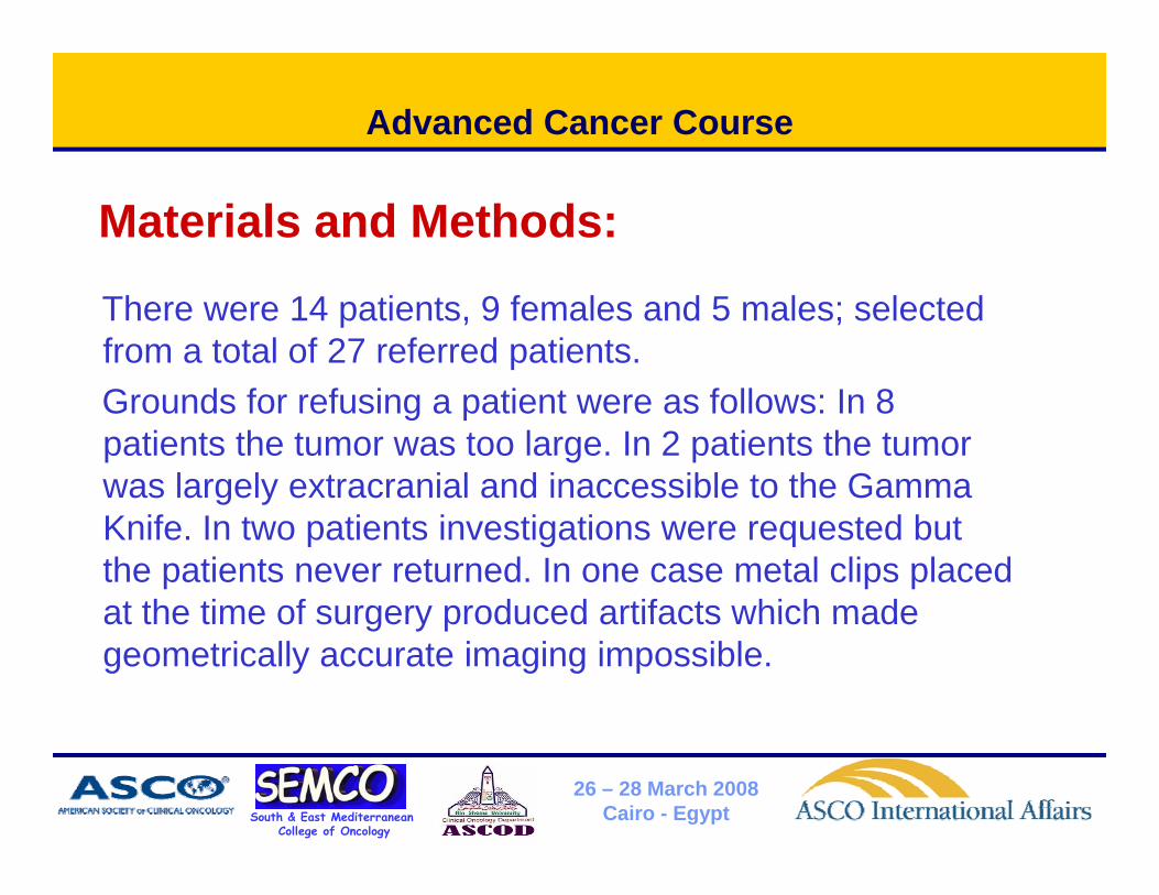
Materials and Methods:
There were 14 patients, 9 females and 5 males; selected from a total of 27 referred patients.G d f f i ti t f ll I 8Grounds for refusing a patient were as follows: In 8 patients the tumor was too large. In 2 patients the tumor was largely extracranial and inaccessible to the Gamma g yKnife. In two patients investigations were requested but the patients never returned. In one case metal clips placed at the time of surgery produced artifacts which madeat the time of surgery produced artifacts which made geometrically accurate imaging impossible.
South & East MediterraneanCollege of Oncology
26 – 28 March 2008Cairo - Egypt

Advanced Cancer Course
South & East MediterraneanCollege of Oncology
26 – 28 March 2008Cairo - Egypt

Advanced Cancer Course
• Glomus tumors are well demarcated on MRdemarcated on MR images and rarely invade the brain, which makes them ideal candidates for treatment with radiosurgery because itradiosurgery because it allows steep dose decrease at the marginsdecrease at the margins.(Ringer et al., Minim Invasive Neurosurg.2001)
South & East MediterraneanCollege of Oncology
26 – 28 March 2008Cairo - Egypt

Advanced Cancer Course
South & East MediterraneanCollege of Oncology
26 – 28 March 2008Cairo - Egypt

Advanced Cancer Course
South & East MediterraneanCollege of Oncology
26 – 28 March 2008Cairo - Egypt

Advanced Cancer Course
South & East MediterraneanCollege of Oncology
26 – 28 March 2008Cairo - Egypt

Advanced Cancer Course
South & East MediterraneanCollege of Oncology
26 – 28 March 2008Cairo - Egypt
Advanced Cancer Course
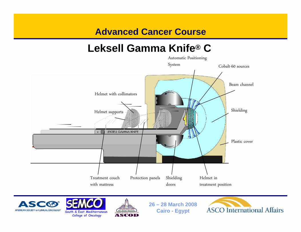
Leksell Gamma Knife® CAutomatic Positioning System Cobalt-60 sources
Beam channel
Helmet with collimators
Helmet supports Shielding
Plastic cover
Treatment couch with mattress
Protection panels Shielding doors
Helmet in treatment position
South & East MediterraneanCollege of Oncology
26 – 28 March 2008Cairo - Egypt

Advanced Cancer Course
Results:
The mean follow up period was 28 months (range 6 to 60 months). All the tumors except one were Fisch Type D and the mean volume was 14 2 cm3 (rangeType D and the mean volume was 14.2 cm3 (range 3.7 cm3 to 28.4 cm3). The mean prescription dose was 13.6 Gy (range 12 to 16 Gy).was 13.6 Gy (range 12 to 16 Gy).In three patients previous surgery had confirmed the diagnosis. In the remainder the diagnosis was based g gon MR findings and a typical angiogram with blood supply mainly by the ascending pharyngeal artery.
South & East MediterraneanCollege of Oncology
26 – 28 March 2008Cairo - Egypt

Advanced Cancer Course
Results: (cont.)N h i d Ei h llNo tumor has continued to grow. Eight are smaller and 6 unchanged in volume. Two patients with bruit have no improvement in symptoms. All the p y pother 12 patients have symptomatic improvementof dysphagia in 5, dysphonia in 4, facial numbness in 3 ataxia in 3 and tinnitus in 2in 3, ataxia in 3, and tinnitus in 2. Single patients have experienced improvement of vomiting, vertigo, tongue fasciculation, hearing, h d h f i l l d iheadache, facial palsy, and an accessory paresis.
South & East MediterraneanCollege of Oncology
26 – 28 March 2008Cairo - Egypt
Advanced Cancer Course
Results: (cont.)
One patient developed a transient facial palsy. Symptomatic improvement began commonlySymptomatic improvement began commonly before any reduction in tumor volume could be detected radiologicaly. The mean time to clinical improvement was 6 5The mean time to clinical improvement was 6.5 months whereas the mean time to shrinkage was 13.5 months.
South & East MediterraneanCollege of Oncology
26 – 28 March 2008Cairo - Egypt
Advanced Cancer Course
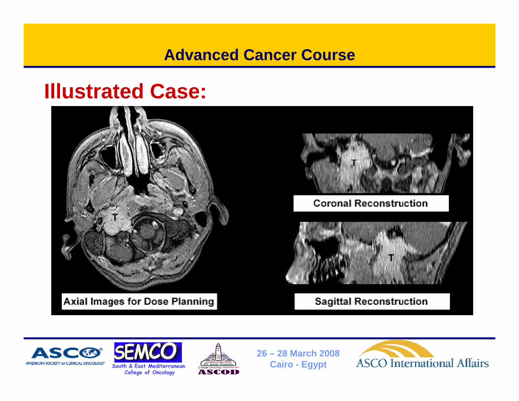
Illustrated Case:
South & East MediterraneanCollege of Oncology
26 – 28 March 2008Cairo - Egypt

Advanced Cancer Course
South & East MediterraneanCollege of Oncology
26 – 28 March 2008Cairo - Egypt

Advanced Cancer Course
Conclusions:
Gamma Knife treatment of glomus jugulare tumors is associated with a high incidence of clinical gimprovement with few complications. Clinical improvement would seem to be a more sensitive
l i di t f th ti thearly indicator of therapeutic success than radiological volume reduction. F th f ll ill b d dFurther follow up will be needed.
South & East MediterraneanCollege of Oncology
26 – 28 March 2008Cairo - Egypt

Advanced Cancer Course
Thank You
South & East MediterraneanCollege of Oncology
26 – 28 March 2008Cairo - Egypt
Related Documents