THE ANTIGENIC STRUCTURE OF THE POLYPEPTIDE CHAINS OF HUMAN ~/-GLOBULIN BY D. E. OLINS mtvDG. M. EDELMAN, M.D. (From The Rockefeller Institute) (Received for publication, July 2, 1952) One of the goals of chemical and immunological studies of T-globulins is to relate the biological activities of the whole molecules to structural features of their constituent polypeptide chains. 7S ~'-globulin molecules consist of multi- ple polypeptide chains linked by disulfide bonds and non-covalent interactions (1). Two types of chains have been described (2, 3): light (L) chains, having molecular weights of approximately 20,000; and heavy (H) chains, having molecular weights greater than 50,000. Certain physicochemical characterisitics of L chains have been associated with differences in the specificity of antibodies (4), with the individuality of myeloma proteins (5), and with the unique thermo- solubility properties of Bence-Jones proteins (3). The current evidence and hypotheses relating the properties of normal and pathological "y-globulins to properties of their L and H chains have been reviewed (6). The present study is concerned with an analysis of the antigenic characteris- tics of the polypeptide chains of normal human 7S %globulin and of a human myeloma protein. The antigenic determinants of L and H chains have been correlated with the previously described S and F determinants (7), which com- prise two major non-cross-reacting antigenic groups of the whole "},-globulin molecule. Antigenic identity has been found between the L chains of a myeloma protein and the Bence-Jones protein from the same patient, thus providing further evidence of their structural similarity, and corroborating the results of previous chemical studies (3). These results furnish a basis for relating the polypeptide chains of qt-globu- lin molecules to the fragments produced by hydrolysis with papain. Methods and Materials 'y-Globul~ns.--Lyophillzed human 7S T-globulln was obtained as fraction II of Cohn (Lot C-679) from Lederle Laboratories (Pearl River, New York). A portion was further purified by zone electrophoresis on starch (8). The preparation of a "y-myeloma protein (patient Haw) and Bence-Jonesprotein from the same patient has been describedpreviously (3). Protein concentrations were determined by measurement of the optical density of solu- tions at 280 m# and by a modification of the Folin-Ciocalteu method (9). Dissociation of Normal Human T-Globulin and Mydoma Protein, and Separation of the L and H Chains.--Reduction and alkylationin the absence of urea (2) was necessaryto retain 635

Welcome message from author
This document is posted to help you gain knowledge. Please leave a comment to let me know what you think about it! Share it to your friends and learn new things together.
Transcript

THE ANTIGENIC STRUCTURE OF THE POLYPEPTIDE CHAINS OF HUMAN ~/-GLOBULIN
BY D. E. OLINS mtvD G. M. EDELMAN, M.D.
(From The Rockefeller Institute)
(Received for publication, July 2, 1952)
One of the goals of chemical and immunological studies of T-globulins is to relate the biological activities of the whole molecules to structural features of their constituent polypeptide chains. 7S ~'-globulin molecules consist of multi- ple polypeptide chains linked by disulfide bonds and non-covalent interactions (1). Two types of chains have been described (2, 3): light (L) chains, having molecular weights of approximately 20,000; and heavy (H) chains, having molecular weights greater than 50,000. Certain physicochemical characterisitics of L chains have been associated with differences in the specificity of antibodies (4), with the individuality of myeloma proteins (5), and with the unique thermo- solubility properties of Bence-Jones proteins (3). The current evidence and hypotheses relating the properties of normal and pathological "y-globulins to properties of their L and H chains have been reviewed (6).
The present study is concerned with an analysis of the antigenic characteris- tics of the polypeptide chains of normal human 7S %globulin and of a human myeloma protein. The antigenic determinants of L and H chains have been correlated with the previously described S and F determinants (7), which com- prise two major non-cross-reacting antigenic groups of the whole "},-globulin molecule. Antigenic identity has been found between the L chains of a myeloma protein and the Bence-Jones protein from the same patient, thus providing further evidence of their structural similarity, and corroborating the results of previous chemical studies (3).
These results furnish a basis for relating the polypeptide chains of qt-globu- lin molecules to the fragments produced by hydrolysis with papain.
Methods and Materials
'y-Globul~ns.--Lyophillzed human 7S T-globulln was obtained as fraction II of Cohn (Lot C-679) from Lederle Laboratories (Pearl River, New York). A portion was further purified by zone electrophoresis on starch (8). The preparation of a "y-myeloma protein (patient Haw) and Bence-Jones protein from the same patient has been described previously (3). Protein concentrations were determined by measurement of the optical density of solu- tions at 280 m# and by a modification of the Folin-Ciocalteu method (9).
Dissociation of Normal Human T-Globulin and Mydoma Protein, and Separation of the L and H Chains.--Reduction and alkylation in the absence of urea (2) was necessary to retain
635

636 ANTIGENIC STRUCTURE OF It-GLOBULIN CHAINS
antigenicity (10) of the separated components. A 0.15 ~ sodium chloride solution brought to pH 8.0 by the dropwise addition of 1 ~ tris-(hydroxymethyl)aminomethane and made 0.1 x+ in mercaptoethanol was used for the reduction and alkylation of normal human "y- globulin at a protein concentration of 10 rag/m1. Reduction proceeded for 16 hours at room temperature under nitrogen. For the myeloma protein, the buffer used was sodium phosphate, pH 7.0, 0.1 M, made 0.1 g in mercaptoethanol, and reduction proceeded for 1 hour at 37°C. The reduced and alkylated normal 7-globutin was exhaustively dialyzed with distilled water at 4°C and lyophilized. The reduced and alkylated myeloma protein was dialyzed against distilled water, and then against 0.5 N propiouic add.
The chromatography of reduced and alkylated v-globulins on carboxymethylcellulose (CMC) in buffers containing 6 ~ urea at 4°C has been previously described (2). The reduced and alkylated 7-giobulin and myeloma protein were chromatographed on sephadex G-75 (Pharmacia, Uppsala, Sweden) in 05 N propiouic acid, as devised by Porter (11). The column dimensions and other pertinent parameters are described in the text. The recovery of protein from sephadex G-75 was 100 per cent. The chromatographic fractions were dialyzed against distilled water at 4°C. and subsequently against 0.05 x+ barbital buffer, pH 8.2, 0.15 ~ sodium chloride. These equilibrated fractions were concentrated to approximately 0.5 per cent solu- tions by negative pressure ultrafiltration in collodion bags (Schleicher and Schue11, Keene, New Hampshire) prior to use in the immunologic studies. There were no losses of protein through the bags.
The procedure of starch gel electrophoresis in 8 ~ urea has been described previously (2). In order to study the antigenic properties of the highly resolved components obtained
after starch gel electrophoresis in urea, efforts were made to isolate these components from the gel. a-amylase was found to be effective for this purpose since it is immunologically un- related to the system under investigation. Regions corresponding to the L and H chain bands on the stained half were removed from the unstained half of a starch urea gel Amounts of wet gel ranging from 2.5 to 7 gm were homogenized and suspended in 25 ml of 0.05 M sodium phosphate buffer, pH 6.0. To this suspension was added approximately 0.4 mg of crystal|inc a-amylase (Worthington Biochemical Corp., Preehold, New Jersey) for each gram of wet starch gal. The mixture was incubated at 37°C for 4 hours and was then centrifuged at 1800 g for 1 hour. Hydrolysis of the starch gel by a-amylase could be confirmed by the decrease in turbidity of the reaction mixture and the decrease in precipitate upon centrifugation, com- pared to suspensions of gel incubated in the absence of amylase. The supernate was dialyzed against 0.15 x~ sodium chloride and concentrated by ultrafiltration. During dialysis and con- centration, a precipitate of starch derivatives formed. This was removed by centrifugation prior to the immunologic analyses.
Isolation of the F and S Fragments from Papain-Treated Normal Human T-Globulin.- Hydrolysis of T-globulin with papain was carried out as described (7); in one case the diges- tion mixture was incubated for 1 hour at 37°C. F and S fragments were isolated by starch zone electrophoresis and also by chromatography of the papain-treated ")'-globulin on diethyl- aminoethylcellulose (DEAE-cellulose) (7). The conditions for chromatography were: mixing chamber vohime, 500 mi; column size, 2.0 × 60.0 cm; buffers, sodium phosphate, pH 7.8; ionic strength gradient, 0.005 to 0.5 x~. The electrophoretic and chromatographic fractions were dialyzed exhaustively against distilled water at 4°C and lyophilized. They were dissolved to a final concentration of 0.5 per cent in 0.05 ~r barbital buffer, pH 8.2, 0.15 ~ sodium chloride for use in the immunological procedures.
Antisera.--Seven different antisera were obtained by immunizing rabbits with antigens in Freund's adjuvant. One antiserum (501) was produced by immunization with normal human fraction I I T-globulin. Three antisera (503, 504, 505) were obtained by immunizing with electrophoretically isolated T-globulin from a single individual. One antiserum (506)
D. E. OLINS AND G. M. EDEL]~IAN 637
was obtained by immunizing with chromatographically isolated S component. The serum of this animal showed the presence of anti-S antibodies, with no detectable anti-F antibodies, and this serum will be denoted (506S). One antiserum (509) was obtained by immunizing with chromatographically isolated F component. All of the above antisera, with the exception of (506S), showed the presence of both anti-F and anti-S antibodies, as tested by immuno- electrophoresis of papain-treated human T-globulin. These sera are denoted (F,S) antisera.
Different aliquots from three of the above (F,S) antisera (501, 505, 509) were absorbed separately with F or S fragments isolated by starch zone electrophoresis. The six absorbed antisera are referred to by their residual spedficities. For example, the original antiserum (501) is denoted (501F,S); when absorbed with F component, (501S); and with S component, (S0iF).
Immunologic Methods.--Immunoelectrophoresis was done by the Scheidegger micro- method (12), except that 1 per cent agar in 0.05 ~ barbital buffer, pi t 8.2 was used. The electrode vessels were filled with 0.05 M barbital buffer, pH 8.2. Electrophoresis was con- ducted for 3 hours at 10 v/cm. Other conditions were similar to those described previously (7). The slides were photographed by darkfield illumination before washing and staining. Ouchterlony plates were adapted to microscale, using 1 per cent agar in 0.05 M barbital buffer, pH 8.2, on 25 X 75 mm microscope slides. Wells holding 10/~1 of the solution of antigen were placed at the corners of a hexagon surrounding the central well, which held 125 #1 of the antiserum. The distance between central and peripheral wells was 5 mm. The precipita- tion lines and relationships of fusion could be seen with a single filling of the antiserum well. Clearest visualization was obtained, however, by filling the wells containing the antiserum a second time, just before they had emptied. The conditions for development of the patterns and photography were the same as those used for the immunoelectrophoretic experiments.
RESULTS
Separation of L and H Chains and F and S Fragments.from Human %Globu- lin.--L chains and H chains of reduced and a lkyla ted 1'-globulin were sepa- ra ted b y two chromatographic methods. Separat ion on carboxymethylcel lulose yie lded two peaks, the first of which contained L chains and the second of which contained H chains and par t i a l ly reduced material , as found in previous studies (2). Chromatography of reduced and a lkyla ted normal "Y-globulin on sephadex G-75 also yielded two peaks (Fig. 1 a). In Fig. 1 b is a comparable separat ion of the chains of a reduced a lky la ted myeloma protein. As indicated b y s tarch gel electrophoresis in urea, the first peaks in both cases consisted of H chains and par t i a l ly reduced material , and the second peaks consisted of L chains. Slightly improved resolution of the L chains and the H chain fraction of normal human T-globulin was obtained b y employing 1.0 X 50.0 cm columns of sephadex G-100 in 0.5 N propionic acid. I n the studies repor ted here, however, only sephadex G-75 was uti l ized in the prepara t ion of L and H chain fractions.
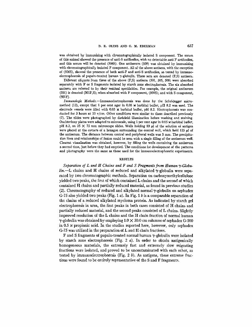
F and S fragments of papain- txeated normal human ~'-globulin were isolated b y s tarch zone electrophoresis (Fig. 2 a). In order to obtain ant igenical ly homogeneous materials , the extremely fast and extremely slow migrat ing fractions were isolated, and proved to be uncontamina ted with each other, as tested b y immunoelectrophoresis (Fig. 2 b). As antigens, these extreme frac- tions were found to be ent i rely representat ive of the S and F fragments.

1.5
ANTIGENIC STRUCTURE OF ")'-GLOBULIN CHAINS
1.0
I0 80
=L E 0 coO oJ a
o 1.5
l.O
0
Cl
v
30 4 0
638
b
I = - I
30 40 50 60
Ef f luent v o l u m e - m l
FIGS. 1 a and 1 b. Chromatography on sephadex G-75 in 0.5 N propionic acid of partially reduced alkylated human fraction I I "},-globulin and myeloma protein (patient Haw).
(a) Partially reduced alkylated human fraction I I ')'-globulin. Column dimensions: 100 X 1.0 cm; column load: 19 mg protein.
(b) Partially reduced alkylated myeloma protein. Column dimensions: 100 X 1.5 cm; column load: 30 mg protein; H and L-fractions taken for further analysis; OD~m~--optical density at 280 m#. The OD values do not reflect the absolute amounts of protein loaded, since different fraction volumes and dilutions were used in a and b.
639
0.4
0.1
=L E O.3 0 0
C) 0 0.2
0 43.O
(+)
I
~ ' F j i I I i J I I t I , , S j +2.0 +1.0 ¢ -I .0 -2.0 -3.0 -4.0
Inches from origin ( _ )
D. E. 0LINS AND G. M. EDELMAN
FI6S. 2 a and 2 b. Isolation of F and S fragments from papain-treated, electrophoretically purified human fraction I1 "y-globulin.
(a) Starch zone electrophoresis of human fraction II "y-globulin digested with papain for 1 hour. Load: 100 mg of papain-digested 3~-globulin; 1" --sample origin; F,S--fractions pooled for further analysis; ODz00mu--optical density of Folin reaction at 700 m/z.
(b) Immunoelectrophoretic comparison of isolated F and S fragments and unfractionated papain-treated human fraction II "y-globulin. F - - F fragments; S--S fragments obtained by starch zone electrophoresis; total unfractlonated papain-treated 7-globulin; antiserum: 503F,S; (+) , anode; and ( - ) , cathode.

640 A N T I G E N I C S T R U C T U R E O F " y - G L O B U L I N C H A I N S
I 0
o
~ 0
~ . N
o o . ~
g~L"
~ ° ~
~ °
J o ~
-~-- o~ ~'~ ~
Qo to ~r ca - ~ d d d o o -.
o

D. E. OLINS AND G. M. ~EDELMAN 641
A chromatographic pattern of papain-treated human fraction I I 3'-globulin is shown in Fig. 3. The five fractions taken were tested by immunoelectrophore- sis. Fraction 1 contained S fragments only. Fractions 2 to 5 contained both S and F fragments, although the later fractions consisted largely of F fragments.
Immunologic Analyses of lhe L and H Chain Fractions from Partially Reduced and Alkylated Normal Human y-Globulin.--A comparison by inmmne diffusion of the L and H chains of normal human 7S q,-globulin with undissociated "r-glob- ulin is shown in Fig. 4 a. Using antiserum 509F,S both L and H chains showed reactions of partial identity with whole 3'-globulin. Similar comparisons were made using antisera 501S and 501F. In these experiments the precipitation line
FIG. 4. Comparisons by immune diffusion of: (a) L and H chain fractions and whole human fraction II "y-globulin. Antiserum: 509F,S. (b) L and H chain fractions. Antiserum: 503F,S. The L chain fraction was obtained by chromatography. The H chain fraction was isolated from starch urea gel.
of the L chains with anti-S antiserum showed a reaction of partial identity with that of whole T-globulin, demonstrating that certain of the determinants of the whole molecule are not present in the isolated L chains. H chains showed reac- tions of complete identity with whole %globulin, when anti-F antiserum was employed. Using anti-S antiserum, the H chain fraction often revealed the presence of two faint lines, one showing a reaction of partial identity with whole T-globulin, the other showing a reaction of complete identity with whole "},-globulin. H chain fractions isolated from starch urea gel by amylase digestion showed less contamination by these components, as tested by immune diffusion.
Fig. 4 a also contains an immunologic comparison between the L and H chains. Using antiserum 509F,S the precipitation lines of L and H chains were seen to mutually penetrate each other, representing reactions of non-identity.

642 ANTIGENIC STRUCTURE OF V-GLOBULIN CHAINS
In addition, the precipitation line of the L chain preparation was seen to fuse with that of a minor component in the H chain fraction. Clearer visualization of these reactions resulted, when antiserum 503F,S was employed (Fig. 4 b).
Immunologic Comparisons of the L and H Chain Fractions to the S and F Fragments of Papain- Treated "y-Globulin.--Comparisons by immune diffusion of the L chains of normal human 7S 3'-globulin and the S and F fragments are shown in Figs. 5 a and 5 b. The L chains precipitated with antiserum 509S and the precipitin line showed a reaction of partial identity with that of the S frag- ment. Antiserum 509F failed to precipitate the L chains and the S fragment. Similar results were obtained with all of the anti-S and anti-F antisera. Anti- serum 506S, completely absorbed with L chains, still precipitated the S frag-
FIO. 5. Comparisons by immune diffusion of L chain fraction and F and S fragments using (a) antiserum 509S, (b) antiserum 509F.
ment, confirming the antigenic deficiency of L chains with respect to S frag- ments.
A faint, additional precipitation line of the S fragment with antiserum 509S was seen (Fig. 5 a), regardless of whether the S fragments were obtained by zone electrophoresis on starch or by chromatography. This second line was also seen with antiserum 509F,S but not with any of the other anti-S, anti-F, or anti-F, S antisera. Using antiserum 509F,S it was demonstrated that this second line did not cross-react with the F precipitation line. This second S line possibly repre- sents additional specificities of this one antiserum to a second S determinant. (13). The faint precipitation lines near the F antigen well, in Fig. 5 a, and near the S antigen well, Fig. 5 b, were produced by the cross-diffusion of F and S fragments, reacting with the homologous antiserum on the opposite end of the slide. These lines could be eliminated by shifting the positions of the well slightly, or by performing only one diffusion experiment on each microscope slide.

D. E . O L I N S A N D G. M. E D E L M A N 643
Fractions containing H chains were also compared with the F and S frag- ments (Figs. 6 a, b, c). The enrichment of the H chains in the fractions isolated from starch urea gels by amylase digestion permitted a clear demonstration of the reaction of identity between H chains and F fragments, and of the reaction
FIGS. 6 a, 6 b, and 6 c. Comparisons by immune diffusion of H chain fraction and F and S fragments.
(a) Antiserum 509F,S. H chain fraction obtained from starch urea gel. (b) Antiserum 509S. H chain fraction from sephadex chromatography. (c) Antiserum 509F. H chain fraction from sephadex chromatography. In all instances the F and S fragments were isolated by starch zone electrophoresis.
of non-identity between H chains and S fragments, using antiserum 509F,S (Fig. 6 a). Antiserum 509F developed a single precipitation line with the H chain fraction, and this line showed a reaction of identity with the F fragment (Fig. 6 c). Antiserum 509S yielded two precipitation lines against the H chain fractions obtained by CMC or sephadex chromatography (Fig. 6 b). One line

644 A N T I G E N I C STRUCTURE OF "}'-GLOBULIN CHAINS
showed a reaction of identity with the precipitation line of the S fragment, and the other showed a reaction of partial identity with that of the S fragment. H chain fractions isolated from starch urea gels showed very faint lines corre- sponding to these minor components. That these extra components resulted from contamination of the H chain fractions with partially dissociated 3'-globu- lin and free light chains was substantiated by starch gel electrophoresis in urea.
Similar comparisons between L and H chain fractions and F and S fragments were done in conventional Ouchterlony plates. The patterns obtained were identical to those described above.
{+) o.~.~ (-)
O
O
O
O
o ~ ~ . . FIG. 7. Immunoelectrophoretic comparisons of whole human fraction lI 33-globulin, F and
S fragments, L chain fraction, and H chain fraction. Composite diagram of experiments using antisera: 501F,S; 503F,S; 509F,S; 506S; 501F. Only the major precipitin line of the H chain fraction is depicted. Faint lines with mobilities similar to L chains and 33-globulin were also present. (+) anode, and ( - ) cathode.
Immunoelectrophoretic patterns of L and H chain fractions and F and S fragments are shown in Fig. 7, which represents a summary of many experi- ments. L chains and whole 7-globulin showed similar mobilities. H chains showed a mobility between those of F fragments and whole T-globulin.
Immunologic Relations between the Chains of a Myeloma Protein and the Bence- Jones Protein from the Same Patient.--Chemical and physical similarities among Bence-Jones proteins and the L chains of myeloma proteins and normal T-globu- lins, have led to the conclusion that Bence-Jones proteins are composed of polypeptide chains of the L type (3).
A myeloma protein, the L chains from this myeloma protein, and the Bence- Jones protein from the same patient (Haw) were compared by means of ira-

D. E. OLINS AND G. M. EDELMAN 645
mune diffusion (Figs. 8 a, b, c). The L chains from the myeloma protein and the Bence-Jones protein used in these studies have been shown previously to be identical by several chemical criteria (3). Using antiserum 506S the precipita- tion lines of the Bence-Jones protein and the L chains of the myeloma protein
FIGS. 8 a, 8 b, and 8 c. Comparisons by immune diffusion of a myeloma protein (M), a Bence-Jones protein (B J) from the same patient, and L chain fraction from the myeloma protein (LM). (a) Antiserum 506S, (b) antiserum 505F, (c) antiserum 503F,S.
showed a reaction of identity (Fig. 8 a). In addition, both the Bence-Jones pro- tein and the L chains of the myeloma protein showed reactions of partial iden- tity with the whole myeloma protein. Antiserum 501F gave a strong precipita- tion line with the myeloma protein and a faint line with both Bence-Jones protein and the L chain fraction, suggesting the presence in both cases of small amounts of contaminating protein (Fig. 8 b). This was confirmed using anti-
646 ANTIGENIC STRUCTURE OF ')'-GLOBULIN CtIAINS
serum 503F,S which distinguished the precipitation lines of the Bence-Jones protein and L chains from the faint lines developed by the anti-F antibodies, which showed reactions of identity with the myeloma protein (Fig. 8 c). These findings suggested that the trace lines were produced by slight contamination of the L chain fraction with the H chain fraction, and of the Bence-Jones pro- tein with the myeloma protein.
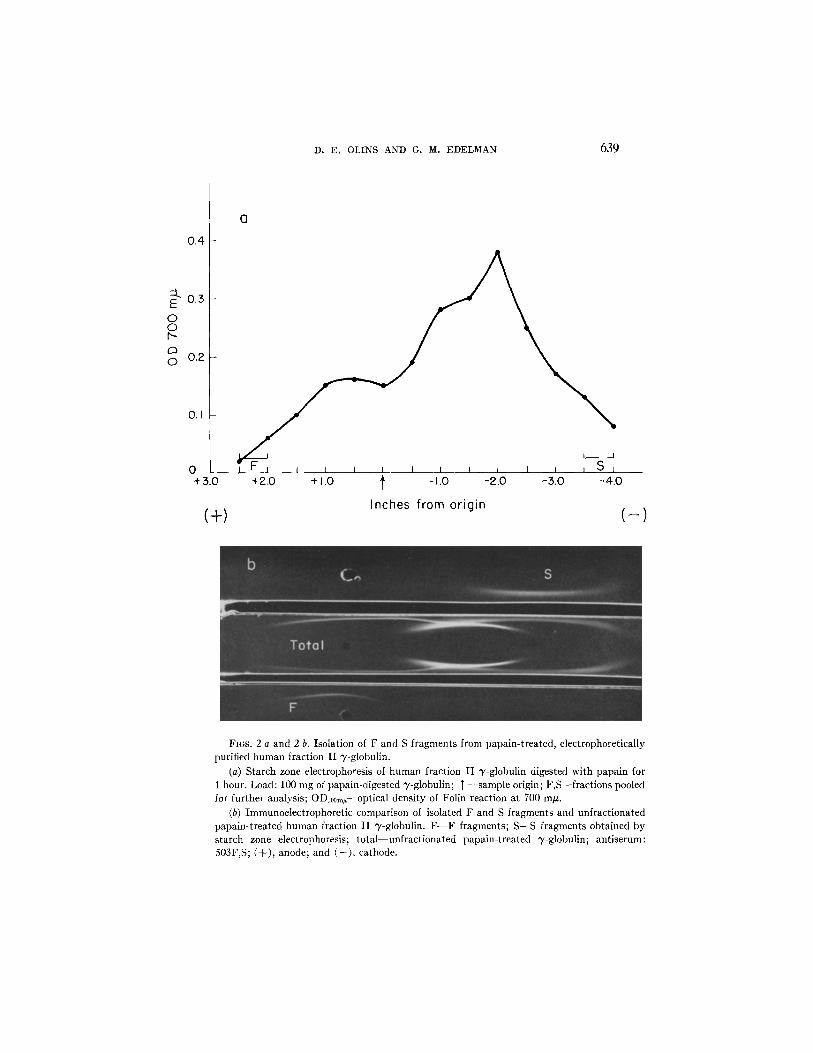
An immunologic comparison between the Bence-Jones protein and L chains obtained from the myeloma protein and from normal q,-globulin is shown in Fig. 9 a. There was complete fusion between the precipitin lines developed by antiserum 506S with these preparations. In addition, normal L chains showed a reaction of partial identity with the myeloma protein (Fig. 9 b).
FIGS. 9 a and 9 b. Comparisons by immune diffusion of (a) Bence-Jones protein (B J), L chain fraction of normal human fraction II v-globulin (L,), and L chain fraction of a myeloma protein (LM). (b) whole myeloma protein (M) and L chain fraction of normal human fraction II v-globulin. Antiserum: 506S.
DISCUSSION
The results of the present study indicate that L and H chains of 7S 3,-globulin are antigenically unrelated to each other, and that they are antigenically de- ficient with respect to the whole molecule. A number of relationships have been demonstrated between the antigenic properties of the polypeptide chains and the two major antigenic determinants carried by fragments produced by treat- ment of the whole molecule with papain. L chains showed reactions of partial identity with S fragments, and did not react with antisera directed specifically against the F fragment. H chains, on the other hand, shared antigenic determinants with the F fragments, and appeared to contain no determinants in common with the S fragments.
These findings suggest that the S fragment is composed in part of L chains or portions of L chains. In addition, they imply that the F fragment is composed in

D. E . OLINS AND G. M. ED]ELMAN 647
part of portions of H chains. If has not been possible, however, to decide whether the determinants of S fragments not present on L chains repre- sent remaining portions of the H chains, or whether these determinants arise from conformations of L chains that are destroyed in the isolation procedure. The fact that the major component of the H chain fractions did not cross-react with the S fragment does not exclude the former possibility, since antigenic groups may have been destroyed in dissociation and separation of the chains.
These relationships between the antigenic determinants of the polypeptide chains and those of the fragments produced by papain thus reveal certain structural relationships among the chains of the whole molecule. The S frag- ment may consist of one L chain bound by disulfide bonds to a portion of an H chain. The F fragment would then consist of the residual portions of the H chains. This model provides a basis for explaining the cleavage of y-globulin by papain and cysteine into fragments of different antigenic structure and biological activity (7, 14-16). Certain peptide bonds (17) of H chains may be hydrolyzed more rapidly by papain than any of the peptide bonds of L chains, thus releasing large fragments of different properties, particularly after cleavage of a labile disulfide bond holding the S fragments together (18).
That portion of the ~'-globulin molecule represented by the S fragment has been shown to retain the antibody combining site (14, 7, 15). The present find- ings indicate that this fragment is made in part of L chains, and point to a possible role of L chains in determining the specificity of antibodies. Recent studies (4) of specifically purified guinea pig antibodies of different specificity have shown differences in their L chains, a finding consistent with the above interpretation.
The number of L chains in the whole molecule and in each S fragment is not established. The molecular weight of L chains is about 20,000 (2, 5), whereas the molecular weight of S fragments is about 50,000 (14). Thus the S fragment might contain one L chain, and a portion of an H chain with a molecular weight of 30,000. L chains in aqueous solution appear to exist as dimers that behave as Bence-Jones proteins (3). This suggests the possibility that the S fragment might consist of two L chains and a portion of the H chain of molecular weight of 10,000. Amino acid analysis of the chains of a 7S myeloma protein (3) would favor the former possibility, but present evidence does not permit a definite choice. In any case, the bivalency of 7S antibodies is explicable by a model in which there is duplication in the molecule of those polypeptide chains in- volved in the combining regions.
Preliminary experiments (19) have indicated that partial reduction of puri- fied S fragments of human y-globulin followed by chromatography on sephadex G-75 in 0.5 N propionic acid releases a fragment that behaves identically with L chains when examined in Ouchterlony plates and by immunoelectrophoresis. Furthermore, partial reduction of S fragments releases components resembling L chains from partially reduced whole ~'-globulin, when examined by starch

6~8 ANTIGENIC STRUCTURE OF 'y-GLOBULIN CHAINS
urea gel electrophoresis. Additional experiments to establish the yield of these components may permit a decision on the number of L chains in the S fragment.
The investigation of the antigenic properties of H chains was made ditticult by the fact that preparations containing H chains also contained small amounts of free L chains in addition to partially degraded -y-globulin. Reduction and alkylation in urea has been found to diminish the amounts of partially degraded material, but also destroys the antigenicity of the derivatives (2). The fact that anti-F antibodies reacted with H chain preparations and not with L chain preparations, combined with the fact that the major component of the H chain fraction showed a reaction of identity with F fragments and a reaction of non- identity with S fragments, permits the conclusion that the F fragments are derived in part from H chains.
Considerable immunologic evidence has accumulated showing that Bence- Jones proteins cross-react with, but are antigenically deficient to, myeloma proteins and normal 7S "y-globulins (20, 21). It was not possible, however, to conclude from such studies that Bence-Jones proteins were identical with sub- units of the homologous myeloma proteins. Recent structural studies (3) have strongly suggested that Bence-Jones proteins are composed of polypeptide chains of the L type, in some cases identical with those of the myeloma protein from the same patient. The antigenic studies of a Bence-Jones protein and a -y-mydoma protein from the same patient that are reported here confirm and extend these conclusions. Similar studies using anfisera directed against myeloma proteins and Bence-Jones proteins should further clarify the rela- tionships amongst these congeners of normal -y-globulins. It is clear from the present experiments thatL chains of normal -y-globulin, which have been shown to behave physically like Bence-Jones proteins, are more closely related anti- genically to Bence-Jones proteins and L chains of myeloma proteins than to whole myeloma proteins. Although the present experiments showed reactions of identity between the L chains of the -r-myeloma protein and the L chains of normal -y-globulin, it would not he surprising if more extensive studies re- vealed that L chains of various myeloma proteins were antigenically deficient to those of normal -y-globulin (21).
The foregoing antigenic map provides substantiation of a proposed scheme (6) assigning the different functional properties of -y-globulin molecules to the structural features of the different types of polypeptide chains of which they are composed. In view of recent evidence (22-24) that the S determinant is respon- sible for the antigenic cross-reactivity of -y-globulins of different claSses (% -Y1A, -Y1M), it seems likely that the L chains of these different -y-globulins have struc- tural features in common. 1 Of particular interest is the recent finding (25, 26)
: Recent experiments (19) have indicated that the L chains of a pathological macroglobu!in cross-reacted with, and were antigenically deficient with respect to L chains of normal 7S v-globulin, when tested with rabbit antiserum to L chains of 7S "y-globulin.

D. E. 0LINS AND G. M. EDELMAN 649
that the S and F fragments of myeloma protein molecules contain different Grubb types, controlled by independent loci. The mapping of S and F deter- minants onto L and H chains, and the results of previous chemical studies (3), taken in conjunction with the findings of Harboe, Osterla~d, and Kunkel (25) indicate that the Gm and Inv factors are on the H and L chains respectively, and thus that the structure of these chains is controlled at different genetic loci.
SVM~RY
The antigenic properties of the polypepfide chains of human 7S 3'-globulin have been related to two major non-cross-reacting antigenic determinants of the whole molecule. These determinants, called S and F, were obtained by hydrolysis of 3"-globulin with papain. Anfisera against whole 3'-globulin and against S and F fragments were used in techniques of immune diffusion. Light (L) chains of 3"-globulin showed reactions of partial identity with S fragments, and thus are antigenically deficient with respect to these fragments. Antlsera directed against F determinants did not react with L chains but did react with heavy (H) polypeptide chain preparations. In addition, the major component of t t chain fractions did not appear to contain determinants in common with the S fragments.
L chains of a 3"-myeloma protein were shown to be antigenicaUy deficient with respect to the whole myeloma molecule, and antigenically identical with the Bence-Jones protein of the same patient.
Correlation of these results with those of previous investigations have led to the conclusions that the S fragment which is known to contain the combining region of antibody molecules, consists in part of L chains or portions of L chains, and that the F fragment, which mediates several other functions of the whole molecule, is composed in part of portions of H chains.
The authors wish to thank Dr. C. K. Osterland and Dr. H. G. Kunkel for providing samples of serum and urine from patients with multiple myeloma. This work was supported by PHS grant A-4256 from the National Institute of Arthritis and Metabolic Diseases, Public Health Service.
BIBLIOGRAPHY
1. Edelman, G. M., Dissociation of 3"-globulin, J. Am. Chem. Soc., 1959, 81, 3155. 2. Edelman, G. M., and Poulik, M. D., Studies on structural units of the 3,-globulins,
J. Exp. Med., 1961, 113, 861. 3. Edelman, G. M., and Gaily, J. A., The nature of Bence-Jones proteins. Chemical
similarities of polypeptide chains of myeloma globulins and normal 7-globu- lins, J. Exp. Med., 1962, 116, 207.
4. Edelman, G. 1~I., Benacerraf, B., Ovary, Z., and Poulik, M. D., Structural differ- ences among antibodies of different specificities, Proc. Nat. Acad. So., 1961, 47, 1751.

650 ANTIGENIC STRUCTURE OF "y-GLOBULIN CHAINS
5. Poulik, M. D., and Edelman, G. M., Comparison of reduced alkylated deriva- tives of some myeloma globulins and Bence-Jones proteins, Nature, 1961, 191, 1274.
6. Edelman, G. M., and Benacerraf, B., On structural and functional relations between antibodies and proteins of the gamma-system, Proc. Nat. Acad. Sc., 1962, 48, 1035.
7. Edelman, G. M., Heremans, J. F., Heremans, M.-Th., and Kunkel, H. G., Immu- nological studies of human T-globulin. Relation of the precipitin lines of whole T-globulin to those of the fragments produced by papain, J. Exp. Med., 1960, 112, 203.
8. Kunkel, H. G., Zone electrophoresis, Methods Biochem. Anal., 1954, 1, 141. 9. Lowry, O. H., Rosebrough, N. J., Farr, A. L., and Randall, R. J., Protein meas-
urement with the Folin phenol reagent, J. Biol. Chem., 1951, 193, 265. 10. Edelman, G. M., and Poulik, M. D., Characterization of subunits of the ?-globu-
lins, Fed. Proc., 1961, 9.0, 387. 11. Porter, R. R., The structure of gamma-globulins and antibodies, in Basic Prob-
lems in Neoplastic Disease, (A. Gellhorn and E. Hirschberg, editors), New York, Columbia University Press, in press.
12. Scheidegger, J. J., Une micro-m~thode d'immuno-electrophordse, Internat. Arch. Allergy and Appl. Immunol., 1955, 7, 103.
13. Mannik, M., Kunkd, H. G., Biro, C., and Edelman, G. M., The occurrence of antigenically deficient 7~-globulins in normal human serum, Fed. Proc., 1962, 21, 11.
14. Franklin, E. C., Structural units of human 7S gamma globulin, J. Clin. lay., 1960, 39, 1933.
15. Porter, R. R., The hydrolysis of rabbit ?-globulin and antibodies with crystalline papain, Biochem. J., 1959, 73, 119.
16. Ovary, Z., and Karush, F., Studies on the immunologic mechanism of anaphylaxis. II. Sensitizing and combining capacity in vivo of fractions separated from papain digests of antihapten antibody, J. Immunol., 1961, 86, 146.
17. Putnam, F. W., Easley, C. W., and Lynn, L. T., Site of cleavage of -/-globulins by papain, Biochim. et Biophyslca Acta, 1962, 58, 279.
18. Nisonoff, A., Markus, G., and Wissler, F. C., Separation of univalent fragments of rabbit antibody by reduction of a single, labile disulphide bond, Nature, 1961, 189, 293.
19. Olins, D. E., and Edelman, G. M., unpublished observations. 20. Slater, R. J., Ward, S. M., and Kunkel, H. G., Immunological relationships among
the myeloma proteins, J. Exp. Med., 1955, 101, 85. 21. Korngold, L., and Lipari, R., Multiple-myeloma proteins III, The antigenic
relationship of Bence-]ones proteins to normal gamma-globulin and multiple- myeloma serum proteins, Cancer, 1956, 9, 262.
22. Heremans, J. F., and Heremans, M.-Th., Immunoelectrophoresis, Acta Med. Scand., 1961, suppl. 367, 27.
23. Franklin, E. C., and Stanworth, D. R., Antigenic relationships between immune globulins and certain rdated paraproteins in man, J. Exp. Med., 1961, 114, 521.

D. E . OLINS AND G. M. E D E L M A N 651
24. Askonas, B. A., and Fahey, J. L., Enzymatically produced subunits of protein formed by plasma cells in mice. II. /~2A-myeloma protein and Bence-Jones protein, Y. Exp. Med., 1962, 115, 641.
25. Harboe, M., Osterland, C. K., and Kunkel, H. G., Localization of two genetic factors to different areas of 7-globulin molecules, Science, 1962, 136, 979.
26. Franklin, E. C., Fudenberg, H., Meltzer, M., and Stanworth, D., The structural basis for genetic variations of normal human -},-globulins, Proc. Nat. Acad. Sc. 1962, 48, 914.
Related Documents