Global distribution of conformational states derived from redundant models in the PDB points to non-uniqueness of the protein structure Prasad V. Burra a , Ying Zhang b , Adam Godzik b , and Boguslaw Stec 1 a Department of Bioengineering, University of California at San Diego, La Jolla, CA 92037, and b Burnham Institute for Medical Research, La Jolla, CA 92037 Edited by Peter G. Wolynes, University of California at San Diego, La Jolla, CA, and approved May 8, 2009 (received for review December 2, 2008) It is commonly accepted that proteins have evolutionarily conserved 3-dimensional structures, uniquely defined by their amino acid se- quence. Here, we question the direct association of structure to sequence by comparing multiple models of identical proteins. Rapidly growing structural databases contain models of proteins determined independently multiple times. We have collected these models in the database of the redundant sets of protein structures and then derived their conformational states by clustering the models with low root- mean-square deviations (RMSDs). The distribution of conformational states represented in these sets is wider than commonly believed, in fact exceeding the possible range of structure determination errors, by at least an order of magnitude. We argue that differences among the models represent the natural distribution of conformational states. Our results suggest that we should change the common notion of a protein structure by augmenting a single 3-dimensional model by the width of the ensemble distribution. This width must become an indispensible attribute of the protein description. We show that every protein contains regions of high rigidity (solid-like) and regions of high mobility (liquid-like) in different and characteristic contribution. We also show that the extent of local flexibility is correlated with the functional class of the protein. This study suggests that the protein- folding problem has no unique solution and should be limited to defining the folding class of the solid-like fragments even though they may constitute only a small part of the protein. These results limit the capability of modeling protein structures with multiple confor- mational states. conformational ensemble conformational states protein folding O ur basic understanding of the structure of a protein has been radically changing with time (1, 2). The initial notion that proteins are basically unstructured has been replaced by the notion of uniquely and beautifully folded rigid structures (3, 4). Currently, even this notion is being gradually modified to include elements of mobility necessary for protein function (5, 6). Understanding the protein function at the molecular level is an extremely daunting problem in biology (7, 8). Structural studies of proteins (rigid models) provided important results leading to a better understanding of many biological pro- cesses. However, proteins must undergo significant energy and volume fluctuations (9). Despite the mounting evidence that changes in protein structures are necessary to produce a desired function (10), the dominant paradigm of protein structure-function relationship is still based on the concept of a rigid protein with a unique structure. Pictures of solid, apparently rigid structures dominate textbooks, scientific magazines, and even the popular press. Although crystallographers at large are well aware of con- formational variability of proteins, a change of a paradigm is gradual as expressed in a series of recent papers (11, 12). Moreover, recent discoveries of intrinsically disordered proteins have in- creased our awareness of the flexibility of protein structures (13). One of the main reasons proteins are represented by solid, rigid bodies is that the dominating experimental technique of structural biology is X-ray crystallography. This technique pro- duces a single structure. Here, we argue that because of the rapid growth of the Protein Data Bank (PDB) (14), we now have a valuable and unanticipated window into the wide range of protein conformational flexibility. A systematic review of the accumulated protein structures has allowed us to gain insight into the structural differences between independently obtained models of identical proteins. Availability of multiple models of the same or very closely related proteins was studied earlier to establish the principles of structure/sequence co-conservation. In a classical paper, Cho- thia and Lesk (15) explored the divergence of structures with reduced homology. Brian Matthews (16) investigated the resil- ience of structure to sequence changes and Martin Karplus (17, 18) investigated conformational variability of the crystal struc- tures of a single protein. Only recently the availability of multiple structures allowed us to explore this subject in a more quanti- tative manner (19). However, none of these studies addressed the question of the structural uniqueness and identity of the indi- vidual protein nor performed a comprehensive review of the available structures of identical proteins present in the PDB. Although some experimental techniques, such as NMR, pro- vide direct measures of the flexibility of a protein (20), X-ray crystallography provides only limited information about protein mobility. There is, however, an additional source of information about a protein’s mobility that only recently has come into focus (21). When 2 (or more) models of the same protein are deposited into the PDB, they can vary by as much as 0.1– 0.4 Å (14). When the conditions vary the changes can reach tens of angstroms (22). Such ‘‘redundant’’ depositions now dominate the PDB (50,000 depositions represents 15,000 proteins) (14). This redundancy is largely ignored in most large-scale analyses of protein structures, and a given analysis is usually performed on non-redundant (or ‘‘culled’’) sets of PDB proteins (23, 24). Non- redundant sets are very useful for some purposes, such as classifying proteins into fold groups but, as we argue here, not for other purposes, such as analyzing the conformational ensemble for a single protein (25, 26). To investigate the latter, we have prepared a redundant set of proteins by specifically selecting clusters of independently solved models of the same protein. This particular database will be the focus of most of our analyses, as described in the Research and Methods of this paper. A recent paper explored this database of ‘‘redundant’’ protein structure and reported the discovery of dual-personality (DP) fragments that can be found in either an ordered or a disordered state among different members of the same cluster (21). The DP fragments have unique features that differentiate them from both Author contributions: B.S. designed research; P.V.B., Y.Z., and B.S. performed research; A.G. and B.S. contributed new reagents/analytic tools; P.V.B., A.G., and B.S. analyzed data; and P.V.B., A.G., and B.S. wrote the paper. The authors declare no conflict of interest. This article is a PNAS Direct Submission. 1 To whom correspondence should be addressed. E-mail: [email protected]. This article contains supporting information online at www.pnas.org/cgi/content/full/ 0812152106/DCSupplemental. www.pnas.orgcgidoi10.1073pnas.0812152106 PNAS June 30, 2009 vol. 106 no. 26 10505–10510 APPLIED PHYSICAL SCIENCES BIOPHYSICS AND COMPUTATIONAL BIOLOGY

Welcome message from author
This document is posted to help you gain knowledge. Please leave a comment to let me know what you think about it! Share it to your friends and learn new things together.
Transcript
Global distribution of conformational states derivedfrom redundant models in the PDB points tonon-uniqueness of the protein structurePrasad V. Burraa, Ying Zhangb, Adam Godzikb, and Boguslaw Stec1
aDepartment of Bioengineering, University of California at San Diego, La Jolla, CA 92037, and bBurnham Institute for Medical Research, La Jolla, CA 92037
Edited by Peter G. Wolynes, University of California at San Diego, La Jolla, CA, and approved May 8, 2009 (received for review December 2, 2008)
It is commonly accepted that proteins have evolutionarily conserved3-dimensional structures, uniquely defined by their amino acid se-quence. Here, we question the direct association of structure tosequence by comparing multiple models of identical proteins. Rapidlygrowing structural databases contain models of proteins determinedindependently multiple times. We have collected these models in thedatabase of the redundant sets of protein structures and then derivedtheir conformational states by clustering the models with low root-mean-square deviations (RMSDs). The distribution of conformationalstates represented in these sets is wider than commonly believed, infact exceeding the possible range of structure determination errors,by at least an order of magnitude. We argue that differences amongthe models represent the natural distribution of conformationalstates. Our results suggest that we should change the common notionof a protein structure by augmenting a single 3-dimensional model bythe width of the ensemble distribution. This width must become anindispensible attribute of the protein description. We show that everyprotein contains regions of high rigidity (solid-like) and regions ofhigh mobility (liquid-like) in different and characteristic contribution.We also show that the extent of local flexibility is correlated with thefunctional class of the protein. This study suggests that the protein-folding problem has no unique solution and should be limited todefining the folding class of the solid-like fragments even thoughthey may constitute only a small part of the protein. These results limitthe capability of modeling protein structures with multiple confor-mational states.
conformational ensemble � conformational states � protein folding
Our basic understanding of the structure of a protein hasbeen radically changing with time (1, 2). The initial notion
that proteins are basically unstructured has been replaced by thenotion of uniquely and beautifully folded rigid structures (3, 4).Currently, even this notion is being gradually modified to includeelements of mobility necessary for protein function (5, 6).Understanding the protein function at the molecular level is anextremely daunting problem in biology (7, 8).
Structural studies of proteins (rigid models) provided importantresults leading to a better understanding of many biological pro-cesses. However, proteins must undergo significant energy andvolume fluctuations (9). Despite the mounting evidence thatchanges in protein structures are necessary to produce a desiredfunction (10), the dominant paradigm of protein structure-functionrelationship is still based on the concept of a rigid protein with aunique structure. Pictures of solid, apparently rigid structuresdominate textbooks, scientific magazines, and even the popularpress. Although crystallographers at large are well aware of con-formational variability of proteins, a change of a paradigm isgradual as expressed in a series of recent papers (11, 12). Moreover,recent discoveries of intrinsically disordered proteins have in-creased our awareness of the flexibility of protein structures (13).
One of the main reasons proteins are represented by solid,rigid bodies is that the dominating experimental technique ofstructural biology is X-ray crystallography. This technique pro-duces a single structure. Here, we argue that because of the rapid
growth of the Protein Data Bank (PDB) (14), we now have avaluable and unanticipated window into the wide range ofprotein conformational f lexibility. A systematic review of theaccumulated protein structures has allowed us to gain insightinto the structural differences between independently obtainedmodels of identical proteins.
Availability of multiple models of the same or very closelyrelated proteins was studied earlier to establish the principles ofstructure/sequence co-conservation. In a classical paper, Cho-thia and Lesk (15) explored the divergence of structures withreduced homology. Brian Matthews (16) investigated the resil-ience of structure to sequence changes and Martin Karplus (17,18) investigated conformational variability of the crystal struc-tures of a single protein. Only recently the availability of multiplestructures allowed us to explore this subject in a more quanti-tative manner (19). However, none of these studies addressed thequestion of the structural uniqueness and identity of the indi-vidual protein nor performed a comprehensive review of theavailable structures of identical proteins present in the PDB.
Although some experimental techniques, such as NMR, pro-vide direct measures of the flexibility of a protein (20), X-raycrystallography provides only limited information about proteinmobility. There is, however, an additional source of informationabout a protein’s mobility that only recently has come into focus(21). When 2 (or more) models of the same protein are depositedinto the PDB, they can vary by as much as 0.1–0.4 Å (14). Whenthe conditions vary the changes can reach tens of angstroms (22).Such ‘‘redundant’’ depositions now dominate the PDB (�50,000depositions represents �15,000 proteins) (14).
This redundancy is largely ignored in most large-scale analyses ofprotein structures, and a given analysis is usually performed onnon-redundant (or ‘‘culled’’) sets of PDB proteins (23, 24). Non-redundant sets are very useful for some purposes, such as classifyingproteins into fold groups but, as we argue here, not for otherpurposes, such as analyzing the conformational ensemble for asingle protein (25, 26). To investigate the latter, we have prepareda redundant set of proteins by specifically selecting clusters ofindependently solved models of the same protein. This particulardatabase will be the focus of most of our analyses, as described inthe Research and Methods of this paper.
A recent paper explored this database of ‘‘redundant’’ proteinstructure and reported the discovery of dual-personality (DP)fragments that can be found in either an ordered or a disorderedstate among different members of the same cluster (21). The DPfragments have unique features that differentiate them from both
Author contributions: B.S. designed research; P.V.B., Y.Z., and B.S. performed research; A.G.and B.S. contributed new reagents/analytic tools; P.V.B., A.G., and B.S. analyzed data; andP.V.B., A.G., and B.S. wrote the paper.
The authors declare no conflict of interest.
This article is a PNAS Direct Submission.
1To whom correspondence should be addressed. E-mail: [email protected].
This article contains supporting information online at www.pnas.org/cgi/content/full/0812152106/DCSupplemental.
www.pnas.org�cgi�doi�10.1073�pnas.0812152106 PNAS � June 30, 2009 � vol. 106 � no. 26 � 10505–10510
APP
LIED
PHYS
ICA
LSC
IEN
CES
BIO
PHYS
ICS
AN
DCO
MPU
TATI
ON
AL
BIO
LOG
Y
regularly folded and intrinsically unstructured/disordered frag-ments. An analysis of these differences among redundant structurescan provide insights into intrinsic proteins’ variability and into howa protein structure reacts to small changes in its environment.
In this paper, we study the structural differences in ‘‘redundant’’structures. For many types of analyses, a general description of anensemble for a single protein would be needed (27). It is needed notonly for assessing proteins’ similarities, or in more general taskssuch as classifying proteins or building databases of distantlyhomologous domains, but also in practical tasks like solving proteinstructures by molecular replacement. Finally, in anticipation of thenext Critical Assessment of Structure Prediction (CASP) compe-tition the description of the structural ensemble could serve as avaluable tool for properly assessing the modeling results (28).
ResultsI. The Database of Redundant Protein Structures. The database ofredundant protein structures we used was generated from thecollection of X-ray structure files deposited in the PDB beforeJuly 2007. The PDB is highly redundant because an averageprotein is represented more than 4 times. Different chains in oneprotein structure (the same PDB ID) were treated as separateentries, which further increased the redundancy. A total of68,881 entries (independent protein chains) were collected forprocessing. After removing the data in accordance with theprocedures specified in the Methods section (deposited before1990; resolution � 2.5; R-value � 0.25), we reduced this numberby 37% to 43,525 individual entries.
The clustering at the 100% level sequence identity resulted in12,406 clusters (individual proteins). Out of these 7,206 (54%) wererepresented by more than 1 entry and were therefore included inour further analysis. Out of 7,206 multiple representative clustersabout 50% had a single pair of structures to compare. The size ofindividual clusters varied from approximately 220 to 2 structures inthe cluster. Approximately 600 clusters contained more than 10structures in the cluster; the majority of the clusters in the databasecontained less than 10 structures in the cluster.
The total number of non-redundant pairs used in the analysiswas 220,345. The size of the proteins varied from less than 100
residues to approximately 1,500 residues. The largest number ofstructures and the resulting pairs was in the range of 100–400amino acids in length, representing 5,298 clusters (individualproteins) that constituted 73% of the clusters.
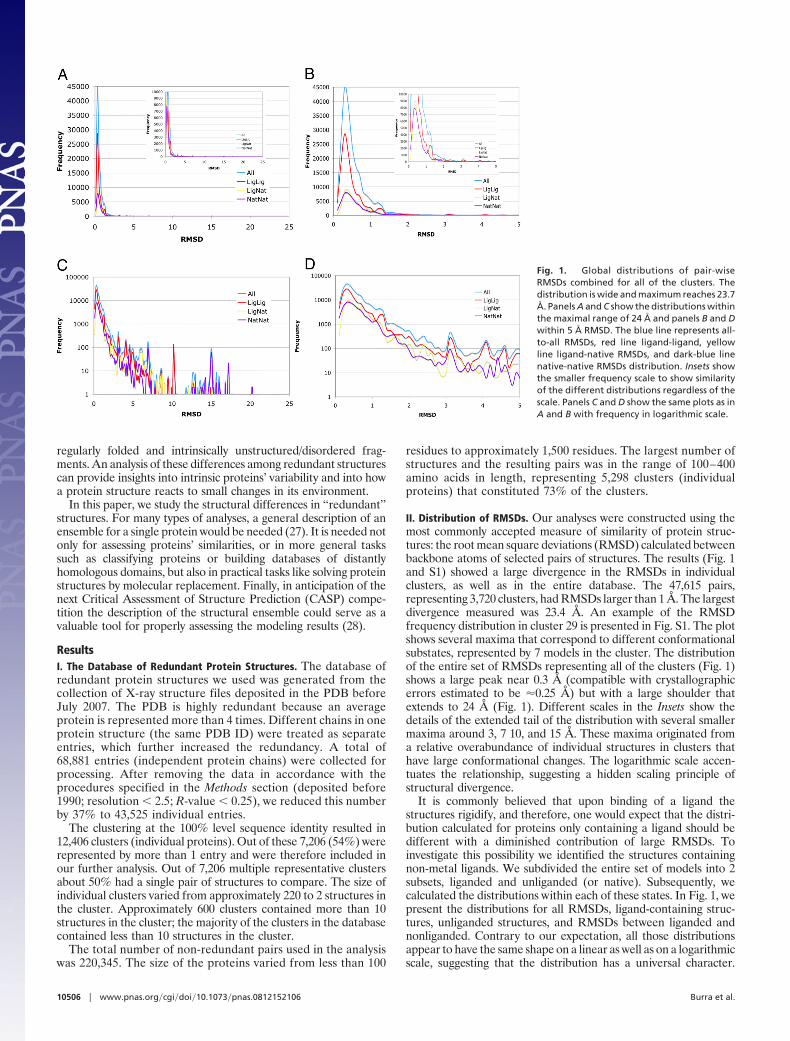
II. Distribution of RMSDs. Our analyses were constructed using themost commonly accepted measure of similarity of protein struc-tures: the root mean square deviations (RMSD) calculated betweenbackbone atoms of selected pairs of structures. The results (Fig. 1and S1) showed a large divergence in the RMSDs in individualclusters, as well as in the entire database. The 47,615 pairs,representing 3,720 clusters, had RMSDs larger than 1 Å. The largestdivergence measured was 23.4 Å. An example of the RMSDfrequency distribution in cluster 29 is presented in Fig. S1. The plotshows several maxima that correspond to different conformationalsubstates, represented by 7 models in the cluster. The distributionof the entire set of RMSDs representing all of the clusters (Fig. 1)shows a large peak near 0.3 Å (compatible with crystallographicerrors estimated to be �0.25 Å) but with a large shoulder thatextends to 24 Å (Fig. 1). Different scales in the Insets show thedetails of the extended tail of the distribution with several smallermaxima around 3, 7 10, and 15 Å. These maxima originated froma relative overabundance of individual structures in clusters thathave large conformational changes. The logarithmic scale accen-tuates the relationship, suggesting a hidden scaling principle ofstructural divergence.
It is commonly believed that upon binding of a ligand thestructures rigidify, and therefore, one would expect that the distri-bution calculated for proteins only containing a ligand should bedifferent with a diminished contribution of large RMSDs. Toinvestigate this possibility we identified the structures containingnon-metal ligands. We subdivided the entire set of models into 2subsets, liganded and unliganded (or native). Subsequently, wecalculated the distributions within each of these states. In Fig. 1, wepresent the distributions for all RMSDs, ligand-containing struc-tures, unliganded structures, and RMSDs between liganded andnonliganded. Contrary to our expectation, all those distributionsappear to have the same shape on a linear as well as on a logarithmicscale, suggesting that the distribution has a universal character.
Fig. 1. Global distributions of pair-wiseRMSDs combined for all of the clusters. Thedistribution is wide and maximum reaches 23.7Å. Panels A and C show the distributions withinthe maximal range of 24 Å and panels B and Dwithin 5 Å RMSD. The blue line represents all-to-all RMSDs, red line ligand-ligand, yellowline ligand-native RMSDs, and dark-blue linenative-native RMSDs distribution. Insets showthe smaller frequency scale to show similarityof the different distributions regardless of thescale. Panels C and D show the same plots as inA and B with frequency in logarithmic scale.
10506 � www.pnas.org�cgi�doi�10.1073�pnas.0812152106 Burra et al.
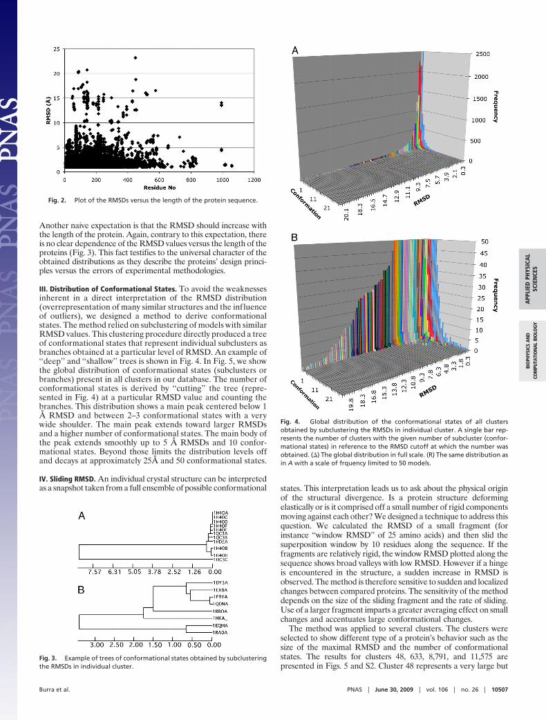
Another naive expectation is that the RMSD should increase withthe length of the protein. Again, contrary to this expectation, thereis no clear dependence of the RMSD values versus the length of theproteins (Fig. 3). This fact testifies to the universal character of theobtained distributions as they describe the proteins’ design princi-ples versus the errors of experimental methodologies.
III. Distribution of Conformational States. To avoid the weaknessesinherent in a direct interpretation of the RMSD distribution(overrepresentation of many similar structures and the influenceof outliers), we designed a method to derive conformationalstates. The method relied on subclustering of models with similarRMSD values. This clustering procedure directly produced a treeof conformational states that represent individual subclusters asbranches obtained at a particular level of RMSD. An example of‘‘deep’’ and ‘‘shallow’’ trees is shown in Fig. 4. In Fig. 5, we showthe global distribution of conformational states (subclusters orbranches) present in all clusters in our database. The number ofconformational states is derived by ‘‘cutting’’ the tree (repre-sented in Fig. 4) at a particular RMSD value and counting thebranches. This distribution shows a main peak centered below 1Å RMSD and between 2–3 conformational states with a verywide shoulder. The main peak extends toward larger RMSDsand a higher number of conformational states. The main body ofthe peak extends smoothly up to 5 Å RMSDs and 10 confor-mational states. Beyond those limits the distribution levels offand decays at approximately 25Å and 50 conformational states.
IV. Sliding RMSD. An individual crystal structure can be interpretedas a snapshot taken from a full ensemble of possible conformational states. This interpretation leads us to ask about the physical origin
of the structural divergence. Is a protein structure deformingelastically or is it comprised off a small number of rigid componentsmoving against each other? We designed a technique to address thisquestion. We calculated the RMSD of a small fragment (forinstance ‘‘window RMSD’’ of 25 amino acids) and then slid thesuperposition window by 10 residues along the sequence. If thefragments are relatively rigid, the window RMSD plotted along thesequence shows broad valleys with low RMSD. However if a hingeis encountered in the structure, a sudden increase in RMSD isobserved. The method is therefore sensitive to sudden and localizedchanges between compared proteins. The sensitivity of the methoddepends on the size of the sliding fragment and the rate of sliding.Use of a larger fragment imparts a greater averaging effect on smallchanges and accentuates large conformational changes.
The method was applied to several clusters. The clusters wereselected to show different type of a protein’s behavior such as thesize of the maximal RMSD and the number of conformationalstates. The results for clusters 48, 633, 8,791, and 11,575 arepresented in Figs. 5 and S2. Cluster 48 represents a very large but
Fig. 2. Plot of the RMSDs versus the length of the protein sequence.
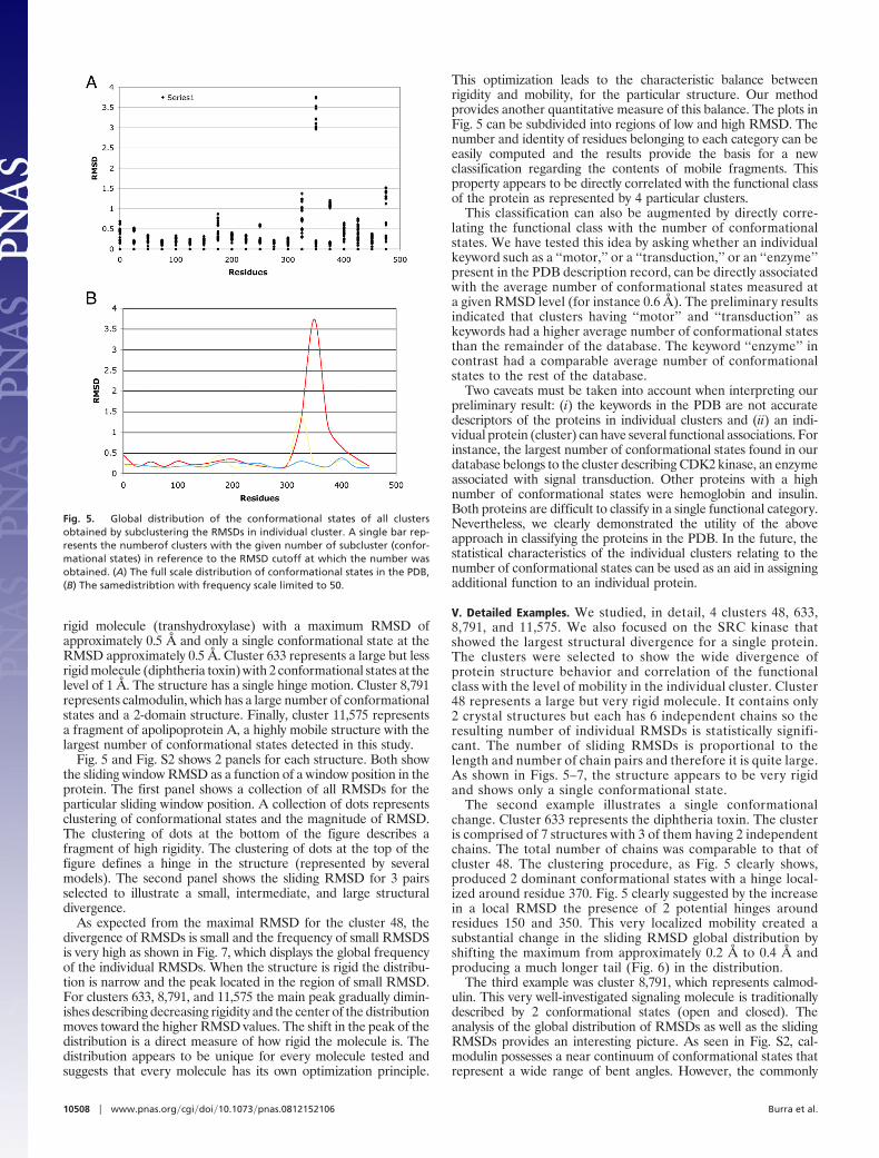
Fig. 3. Example of trees of conformational states obtained by subclusteringthe RMSDs in individual cluster.
Fig. 4. Global distribution of the conformational states of all clustersobtained by subclustering the RMSDs in individual cluster. A single bar rep-resents the number of clusters with the given number of subcluster (confor-mational states) in reference to the RMSD cutoff at which the number wasobtained. (�) The global distribution in full scale. (R) The same distribution asin A with a scale of frquency limited to 50 models.
Burra et al. PNAS � June 30, 2009 � vol. 106 � no. 26 � 10507
APP
LIED
PHYS
ICA
LSC
IEN
CES
BIO
PHYS
ICS
AN
DCO
MPU
TATI
ON
AL
BIO
LOG
Y
rigid molecule (transhydroxylase) with a maximum RMSD ofapproximately 0.5 Å and only a single conformational state at theRMSD approximately 0.5 Å. Cluster 633 represents a large but lessrigid molecule (diphtheria toxin) with 2 conformational states at thelevel of 1 Å. The structure has a single hinge motion. Cluster 8,791represents calmodulin, which has a large number of conformationalstates and a 2-domain structure. Finally, cluster 11,575 representsa fragment of apolipoprotein A, a highly mobile structure with thelargest number of conformational states detected in this study.
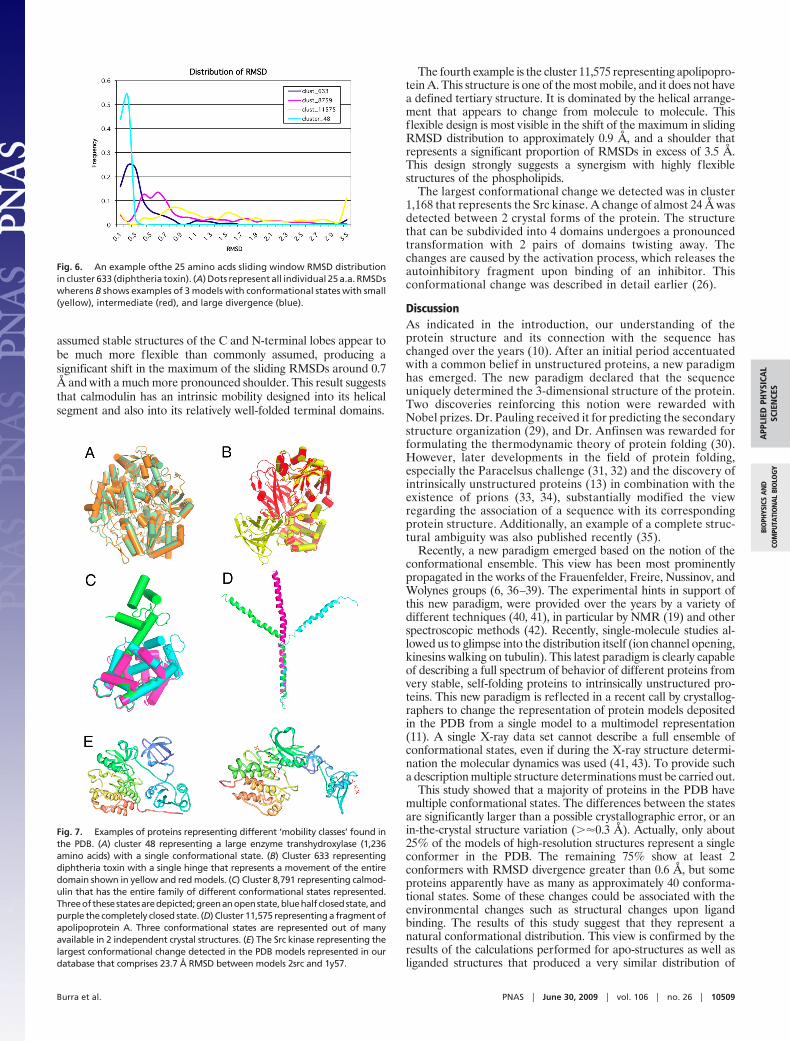
Fig. 5 and Fig. S2 shows 2 panels for each structure. Both showthe sliding window RMSD as a function of a window position in theprotein. The first panel shows a collection of all RMSDs for theparticular sliding window position. A collection of dots representsclustering of conformational states and the magnitude of RMSD.The clustering of dots at the bottom of the figure describes afragment of high rigidity. The clustering of dots at the top of thefigure defines a hinge in the structure (represented by severalmodels). The second panel shows the sliding RMSD for 3 pairsselected to illustrate a small, intermediate, and large structuraldivergence.
As expected from the maximal RMSD for the cluster 48, thedivergence of RMSDs is small and the frequency of small RMSDSis very high as shown in Fig. 7, which displays the global frequencyof the individual RMSDs. When the structure is rigid the distribu-tion is narrow and the peak located in the region of small RMSD.For clusters 633, 8,791, and 11,575 the main peak gradually dimin-ishes describing decreasing rigidity and the center of the distributionmoves toward the higher RMSD values. The shift in the peak of thedistribution is a direct measure of how rigid the molecule is. Thedistribution appears to be unique for every molecule tested andsuggests that every molecule has its own optimization principle.
This optimization leads to the characteristic balance betweenrigidity and mobility, for the particular structure. Our methodprovides another quantitative measure of this balance. The plots inFig. 5 can be subdivided into regions of low and high RMSD. Thenumber and identity of residues belonging to each category can beeasily computed and the results provide the basis for a newclassification regarding the contents of mobile fragments. Thisproperty appears to be directly correlated with the functional classof the protein as represented by 4 particular clusters.
This classification can also be augmented by directly corre-lating the functional class with the number of conformationalstates. We have tested this idea by asking whether an individualkeyword such as a ‘‘motor,’’ or a ‘‘transduction,’’ or an ‘‘enzyme’’present in the PDB description record, can be directly associatedwith the average number of conformational states measured ata given RMSD level (for instance 0.6 Å). The preliminary resultsindicated that clusters having ‘‘motor’’ and ‘‘transduction’’ askeywords had a higher average number of conformational statesthan the remainder of the database. The keyword ‘‘enzyme’’ incontrast had a comparable average number of conformationalstates to the rest of the database.
Two caveats must be taken into account when interpreting ourpreliminary result: (i) the keywords in the PDB are not accuratedescriptors of the proteins in individual clusters and (ii) an indi-vidual protein (cluster) can have several functional associations. Forinstance, the largest number of conformational states found in ourdatabase belongs to the cluster describing CDK2 kinase, an enzymeassociated with signal transduction. Other proteins with a highnumber of conformational states were hemoglobin and insulin.Both proteins are difficult to classify in a single functional category.Nevertheless, we clearly demonstrated the utility of the aboveapproach in classifying the proteins in the PDB. In the future, thestatistical characteristics of the individual clusters relating to thenumber of conformational states can be used as an aid in assigningadditional function to an individual protein.
V. Detailed Examples. We studied, in detail, 4 clusters 48, 633,8,791, and 11,575. We also focused on the SRC kinase thatshowed the largest structural divergence for a single protein.The clusters were selected to show the wide divergence ofprotein structure behavior and correlation of the functionalclass with the level of mobility in the individual cluster. Cluster48 represents a large but very rigid molecule. It contains only2 crystal structures but each has 6 independent chains so theresulting number of individual RMSDs is statistically signifi-cant. The number of sliding RMSDs is proportional to thelength and number of chain pairs and therefore it is quite large.As shown in Figs. 5–7, the structure appears to be very rigidand shows only a single conformational state.
The second example illustrates a single conformationalchange. Cluster 633 represents the diphtheria toxin. The clusteris comprised of 7 structures with 3 of them having 2 independentchains. The total number of chains was comparable to that ofcluster 48. The clustering procedure, as Fig. 5 clearly shows,produced 2 dominant conformational states with a hinge local-ized around residue 370. Fig. 5 clearly suggested by the increasein a local RMSD the presence of 2 potential hinges aroundresidues 150 and 350. This very localized mobility created asubstantial change in the sliding RMSD global distribution byshifting the maximum from approximately 0.2 Å to 0.4 Å andproducing a much longer tail (Fig. 6) in the distribution.
The third example was cluster 8,791, which represents calmod-ulin. This very well-investigated signaling molecule is traditionallydescribed by 2 conformational states (open and closed). Theanalysis of the global distribution of RMSDs as well as the slidingRMSDs provides an interesting picture. As seen in Fig. S2, cal-modulin possesses a near continuum of conformational states thatrepresent a wide range of bent angles. However, the commonly
Fig. 5. Global distribution of the conformational states of all clustersobtained by subclustering the RMSDs in individual cluster. A single bar rep-resents the numberof clusters with the given number of subcluster (confor-mational states) in reference to the RMSD cutoff at which the number wasobtained. (A) The full scale distribution of conformational states in the PDB,(B) The samedistribtion with frequency scale limited to 50.
10508 � www.pnas.org�cgi�doi�10.1073�pnas.0812152106 Burra et al.
assumed stable structures of the C and N-terminal lobes appear tobe much more flexible than commonly assumed, producing asignificant shift in the maximum of the sliding RMSDs around 0.7Å and with a much more pronounced shoulder. This result suggeststhat calmodulin has an intrinsic mobility designed into its helicalsegment and also into its relatively well-folded terminal domains.
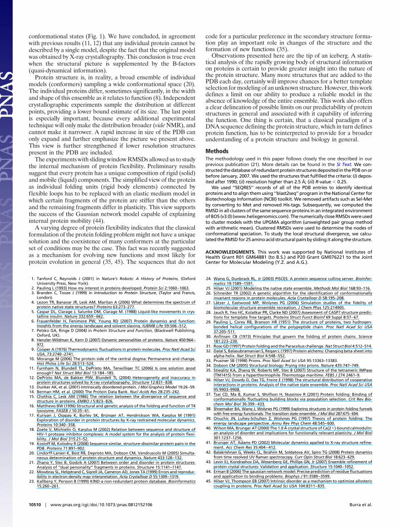
The fourth example is the cluster 11,575 representing apolipopro-tein A. This structure is one of the most mobile, and it does not havea defined tertiary structure. It is dominated by the helical arrange-ment that appears to change from molecule to molecule. Thisflexible design is most visible in the shift of the maximum in slidingRMSD distribution to approximately 0.9 Å, and a shoulder thatrepresents a significant proportion of RMSDs in excess of 3.5 Å.This design strongly suggests a synergism with highly flexiblestructures of the phospholipids.
The largest conformational change we detected was in cluster1,168 that represents the Src kinase. A change of almost 24 Å wasdetected between 2 crystal forms of the protein. The structurethat can be subdivided into 4 domains undergoes a pronouncedtransformation with 2 pairs of domains twisting away. Thechanges are caused by the activation process, which releases theautoinhibitory fragment upon binding of an inhibitor. Thisconformational change was described in detail earlier (26).
DiscussionAs indicated in the introduction, our understanding of theprotein structure and its connection with the sequence haschanged over the years (10). After an initial period accentuatedwith a common belief in unstructured proteins, a new paradigmhas emerged. The new paradigm declared that the sequenceuniquely determined the 3-dimensional structure of the protein.Two discoveries reinforcing this notion were rewarded withNobel prizes. Dr. Pauling received it for predicting the secondarystructure organization (29), and Dr. Anfinsen was rewarded forformulating the thermodynamic theory of protein folding (30).However, later developments in the field of protein folding,especially the Paracelsus challenge (31, 32) and the discovery ofintrinsically unstructured proteins (13) in combination with theexistence of prions (33, 34), substantially modified the viewregarding the association of a sequence with its correspondingprotein structure. Additionally, an example of a complete struc-tural ambiguity was also published recently (35).
Recently, a new paradigm emerged based on the notion of theconformational ensemble. This view has been most prominentlypropagated in the works of the Frauenfelder, Freire, Nussinov, andWolynes groups (6, 36–39). The experimental hints in support ofthis new paradigm, were provided over the years by a variety ofdifferent techniques (40, 41), in particular by NMR (19) and otherspectroscopic methods (42). Recently, single-molecule studies al-lowed us to glimpse into the distribution itself (ion channel opening,kinesins walking on tubulin). This latest paradigm is clearly capableof describing a full spectrum of behavior of different proteins fromvery stable, self-folding proteins to intrinsically unstructured pro-teins. This new paradigm is reflected in a recent call by crystallog-raphers to change the representation of protein models depositedin the PDB from a single model to a multimodel representation(11). A single X-ray data set cannot describe a full ensemble ofconformational states, even if during the X-ray structure determi-nation the molecular dynamics was used (41, 43). To provide sucha description multiple structure determinations must be carried out.
This study showed that a majority of proteins in the PDB havemultiple conformational states. The differences between the statesare significantly larger than a possible crystallographic error, or anin-the-crystal structure variation (��0.3 Å). Actually, only about25% of the models of high-resolution structures represent a singleconformer in the PDB. The remaining 75% show at least 2conformers with RMSD divergence greater than 0.6 Å, but someproteins apparently have as many as approximately 40 conforma-tional states. Some of these changes could be associated with theenvironmental changes such as structural changes upon ligandbinding. The results of this study suggest that they represent anatural conformational distribution. This view is confirmed by theresults of the calculations performed for apo-structures as well asliganded structures that produced a very similar distribution of
Fig. 6. An example ofthe 25 amino acds sliding window RMSD distributionin cluster 633 (diphtheria toxin). (A) Dots represent all individual 25 a.a. RMSDswherens B shows examples of 3 models with conformational states with small(yellow), intermediate (red), and large divergence (blue).
Fig. 7. Examples of proteins representing different ‘mobility classes’ found inthe PDB. (A) cluster 48 representing a large enzyme transhydroxylase (1,236amino acids) with a single conformational state. (B) Cluster 633 representingdiphtheria toxin with a single hinge that represents a movement of the entiredomain shown in yellow and red models. (C) Cluster 8,791 representing calmod-ulin that has the entire family of different conformational states represented.Threeofthesestatesaredepicted;greenanopenstate,bluehalf closedstate,andpurple the completely closed state. (D) Cluster 11,575 representing a fragment ofapolipoprotein A. Three conformational states are represented out of manyavailable in 2 independent crystal structures. (E) The Src kinase representing thelargest conformational change detected in the PDB models represented in ourdatabase that comprises 23.7 Å RMSD between models 2src and 1y57.
Burra et al. PNAS � June 30, 2009 � vol. 106 � no. 26 � 10509
APP
LIED
PHYS
ICA
LSC
IEN
CES
BIO
PHYS
ICS
AN
DCO
MPU
TATI
ON
AL
BIO
LOG
Y
conformational states (Fig. 1). We have concluded, in agreementwith previous results (11, 12) that any individual protein cannot bedescribed by a single model, despite the fact that the original modelwas obtained by X-ray crystallography. This conclusion is true evenwhen the structural picture is supplemented by the B-factors(quasi-dynamical information).
Protein structure is, in reality, a broad ensemble of individualmodels (conformers) sampling a wide conformational space (20).The individual proteins differ, sometimes significantly, in the widthand shape of this ensemble as it relates to function (8). Independentcrystallographic experiments sample the distribution at differentpoints, providing a lower bound estimate of its size. The last pointis especially important, because every additional experimentaltechnique will only make the distribution broader (vide NMR), andcannot make it narrower. A rapid increase in size of the PDB canonly expand and further emphasize the picture we present above.This view is further strengthened if lower resolution structurespresent in the PDB are included.
The experiments with sliding window RMSDs allowed us to studythe internal mechanism of protein flexibility. Preliminary resultssuggest that every protein has a unique composition of rigid (solid)and mobile (liquid) components. The simplified view of the proteinas individual folding units (rigid body elements) connected byflexible loops has to be replaced with an elastic medium model inwhich certain fragments of the protein are stiffer than the othersand the remaining fragments differ in plasticity. This view supportsthe success of the Gaussian network model capable of explaininginternal protein mobility (44).
A varying degree of protein flexibility indicates that the classicalformulation of the protein folding problem might not have a uniquesolution and the coexistence of many conformers at the particularset of conditions may be the case. This fact was recently suggestedas a mechanism for evolving new functions and most likely forprotein evolution in general (35, 45). The sequences that do not
code for a particular preference in the secondary structure forma-tion play an important role in changes of the structure and theformation of new functions (35).
Observations presented here are the tip of an iceberg. A statis-tical analysis of the rapidly growing body of structural informationon proteins is certain to provide greater insight into the nature ofthe protein structure. Many more structures that are added to thePDB each day, certainly will improve chances for a better templateselection for modeling of an unknown structure. However, this workdefines a limit on our ability to produce a reliable model in theabsence of knowledge of the entire ensemble. This work also offersa clear delineation of possible limits on our predictability of proteinstructures in general and associated with it capability of inferringthe function. One thing is certain, that a classical paradigm of aDNA sequence defining the protein structure, which in turn definesprotein function, has to be reinterpreted to provide for a broaderunderstanding of a protein structure and biology in general.
MethodsThe methodology used in this paper follows closely the one described in ourprevious publication (21). More details can be found in the SI Text. We con-structed the database of redundant protein structures deposited in the PDB on orbefore January, 2007. We used the structures that fulfilled the criteria: (i) depos-ited after 1990; (ii) resolution higher than 2.5 Å; (iii) R-value � 0.25.
We used ‘‘SEQRES’’ records of all of the PDB entries to identify identicalproteins and to align them using ‘‘blast2seq’’ program in the National Center forBiotechnology Information (NCBI) toolkit. We removed artifacts such as Sel-Metby converting to Met and removed His-tags. Subsequently, we computed theRMSD in all clusters of the same sequence proteins in an integrated environmentof BOS (v3.0) (www.helixgenomics.com). The numerically close RMSDs were usedto cluster models with the UPGMA algorithm (unweighted pair group methodwith arithmetic mean). Clustered RMSDs were used to determine the nodes ofconformational speciation. To study the local structural divergence, we calcu-lated the RMSD for 25 amino acid structural pairs by sliding it along the structure.
ACKNOWLEDGMENTS. This work was supported by National Institutes ofHealth Grant R01 GM64881 (to B.S.) and P20 Grant GM076221 to the JointCenter for Molecular Modeling (Y.Z. and A.G.).
1. Tanford C, Reynolds J (2001) in Nature’s Robots: A History of Proteins, (OxfordUniversity Press, New York).
2. Pauling L (1993) How my interest in proteins developed. Protein Sci 2:1060–1063.3. Branden C, Tooze J (1999) in Introduction to Protein Structure, (Taylor and Francis,
London).4. Lezon TR, Banavar JR, Lesk AM, Maritan A (2006) What determines the spectrum of
protein native state structures? Proteins 63:273–277.5. Caspar DL, Clarage J, Salunke DM, Clarage M. (1988) Liquid-like movements in crys-
talline insulin. Nature 332:659–662.6. Frauenfelder H, Fenimore PW, Young RD (2007) Protein dynamics and function:
Insights from the energy landscape and solvent slaving. IUBMB Life 59:506–512.7. Petsko GA, Ringe D (2004) in Protein Structure and Function, (Blackwell Publishing,
Oxford, UK).8. Henzler-Wildman K, Kern D (2007) Dynamic personalities of proteins. Nature 450:964–
972.9. Cooper A (1976) Thermodynamic fluctuations in protein molecules. Proc Natl Acad Sci
USA, 73:2740–2741.10. Morange M (2006) The protein side of the central dogma: Permanence and change.
Hist Philos Life Sci 28:513–524.11. Furnham N, Blundell TL, DePristo MA, Terwilliger TC (2006) Is one solution good
enough? Nat Struct Mol Biol 13:184–185.12. DePristo MA, de Bakker PIW, Blundell TL (2004) Heterogeneity and inaccuracy in
protein structures solved by X-ray crystallography. Structure 12:831–838.13. Dunker AK, et al. (2001) Intrinsically disordered protein. J Mol Graphics Model 19:26–59.14. Berman HM, et al. (2000) The Protein Data Bank. Nucl Acids Res 28:235–242.15. Chothia C, Lesk AM (1986) The relation between the divergence of sequence and
structure in proteins. EMBO J 5:823–826.16. Matthews BW (1996) Structural and genetic analysis of the folding and function of T4
lysozyme. FASEB J 10:35–41.17. Kuriyan J, Osapay K, Burley SK, Brunger AT, Hendrickson WA, Karplus M (1991)
Exploration of disorder in protein structures by X-ray restrained molecular dynamics.Proteins 10:340–358.
18. Zoete V, Michielin O, Karplus M (2002) Relation between sequence and structure ofHIV-1 protease inhibitor complexes: A model system for the analysis of protein flexi-bility. J Mol Biol 315:21–52.
19. Kosloff M, Kolodny R (2008) Sequence-similar, structure-dissimilar protein pairs in thePDB. Proteins 71:891–902.
20. Lindorff-Larsen K, Best RB, Depristo MA, Dobson CM, Vendruscolo M (2005) Simulta-neous determination of protein structure and dynamics. Nature 433:128–132.
21. Zhang Y, Stec B, Godzik A (2007) Between order and disorder in protein structures:Analysis of ‘‘dual personality’’ fragments in proteins. Structure 15:1141–1147.
22. Mowbray SL, Helgstrand C, Sigrell JA, Cameron AD, Jones TA (1999) Errors and reproduc-ibility in electron-density map interpretation. Acta Crystallogr D 55:1309–1319.
23. Kallberg Y, Persson B (1999) KIND-a non-redundant protein database. Bioinformatics15:260–261.
24. Wang G, Dunbrack RL, Jr (2003) PISCES: A protein sequence culling server. Bioinfor-matics 19:1589–1591.
25. Hilser VJ (2001) Modeling the native state ensemble. Methods Mol Biol 168:93–116.26. Schneider TR (2002) A genetic algorithm for the identification of conformationally
invariant regions in protein molecules. Acta Crystallogr D 58:195–208.27. Latzer J, Eastwood MP, Wolynes PG (2006) Simulation studies of the fidelity of
biomolecular structure ensemble recreation. J Chem Phys 125:214905.28. Jauch R, Yeo HC, Kolatkar PR, Clarke ND (2007) Assessment of CASP7 structure predic-
tions for template free targets. Proteins Struct Funct Bioinf 69 Suppl 8:57–67.29. Pauling L, Corey RB, Branson HR (1951) The structure of proteins, two hydrogen-
bonded helical configurations of the polypeptide chain. Proc Natl Acad Sci USA37:205–511.
30. Anfinsen CB (1973) Principles that govern the folding of protein chains. Science181:223–230.
31. Rose GD (1997) Protein folding and the Paracelsus challenge. Nat Struct Biol 4:512–514.32. Dalal S, Balasubramanian S, Regan L (1997) Protein alchemy: Changing beta-sheet into
alpha-helix. Nat Struct Biol 4:548–552.33. Prusiner SB (1998) Prions. Proc Natl Acad Sci USA 95:13363–13383.34. Dobson CM (2005) Structural biology: Prying into prions. Nature 435:747–749.35. Stieglitz KA, Zhang W, Roberts MF, Stec B (2007) Structure of the tetrameric IMPase
(TM1415) from a hyperthermophile Thermotoga maritime. FEBS J 274 2461–9.36. Hilser VJ, Dowdy D, Oas TG, Freire E (1998) The structural distribution of cooperative
interactions in proteins: Analysis of the native state ensemble. Proc Natl Acad Sci USA95:9903–9908.
37. Tsai CD, Ma B, Kumar S, Wolfson H, Nussinov R (2001) Protein folding: Binding ofconformationally fluctuating building blocks via population selection. Crit Rev Bio-chem Mol Biol 36:399–433.
38. Shoemaker BA, Wang J, Wolynes PG (1999) Exploring structures in protein folding funnelswith free energy functionals: The transition state ensemble. J Mol Biol 287:675–694.
39. Onuchic JN, Luhey-Schulten Z, Wolynes PG (1997) Theory of protein folding: Theenergy landscape perspective. Annu Rev Phys Chem 48:545–600.
40. Wilson MA, Brunger AT (2000) The 1.0 Å crystal structure of Ca(2�)-bound calmodulin:an analysis of disorder and implications for functionally relevant plasticity. J Mol Biol301:1237–1256.
41. Brunger AT, Adams PD (2002) Molecular dynamics applied to X-ray structure refine-ment. Acc Chem Res 35:404–412.
42. Balakrishnan G, Weeks CL, Ibrahim M, Soldatova AV, Spiro TG (2008) Protein dynamicsfrom time resolved UV Raman spectroscopy. Curr Opin Struct Biol 18:623–629.
43. Levin EJ, Kondrashov DA, Wesenberg GE, Phillips GN, Jr (2007) Ensemble refinement ofprotein crystal structures: Validation and application. Structure 15:1040–1052.
44. Erman B (2006) The gaussian network model: Precise prediction of residue fluctuationsand application to binding problems. Biophys J 91:3589–3599.
45. Hilser VJ, Thompson EB (2007) Intrinsic disorder as a mechanism to optimize allostericcoupling in proteins. Proc Natl Acad Sci USA 104:8311–835.
10510 � www.pnas.org�cgi�doi�10.1073�pnas.0812152106 Burra et al.
Related Documents