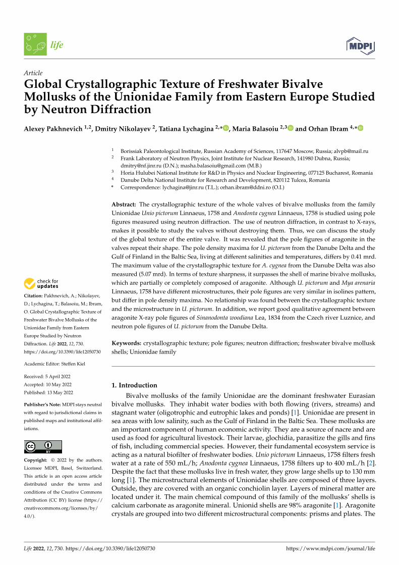
Citation: Pakhnevich, A.; Nikolayev, D.; Lychagina, T.; Balasoiu, M.; Ibram, O. Global Crystallographic Texture of Freshwater Bivalve Mollusks of the Unionidae Family from Eastern Europe Studied by Neutron Diffraction. Life 2022, 12, 730. https://doi.org/10.3390/life12050730 Academic Editor: Steffen Kiel Received: 5 April 2022 Accepted: 10 May 2022 Published: 13 May 2022 Publisher’s Note: MDPI stays neutral with regard to jurisdictional claims in published maps and institutional affil- iations. Copyright: © 2022 by the authors. Licensee MDPI, Basel, Switzerland. This article is an open access article distributed under the terms and conditions of the Creative Commons Attribution (CC BY) license (https:// creativecommons.org/licenses/by/ 4.0/). life Article Global Crystallographic Texture of Freshwater Bivalve Mollusks of the Unionidae Family from Eastern Europe Studied by Neutron Diffraction Alexey Pakhnevich 1,2 , Dmitry Nikolayev 2 , Tatiana Lychagina 2, * , Maria Balasoiu 2,3 and Orhan Ibram 4, * 1 Borissiak Paleontological Institute, Russian Academy of Sciences, 117647 Moscow, Russia; [email protected] 2 Frank Laboratory of Neutron Physics, Joint Institute for Nuclear Research, 141980 Dubna, Russia; [email protected] (D.N.); [email protected] (M.B.) 3 Horia Hulubei National Institute for R&D in Physics and Nuclear Engineering, 077125 Bucharest, Romania 4 Danube Delta National Institute for Research and Development, 820112 Tulcea, Romania * Correspondence: [email protected] (T.L.); [email protected] (O.I.) Abstract: The crystallographic texture of the whole valves of bivalve mollusks from the family Unionidae Unio pictorum Linnaeus, 1758 and Anodonta cygnea Linnaeus, 1758 is studied using pole figures measured using neutron diffraction. The use of neutron diffraction, in contrast to X-rays, makes it possible to study the valves without destroying them. Thus, we can discuss the study of the global texture of the entire valve. It was revealed that the pole figures of aragonite in the valves repeat their shape. The pole density maxima for U. pictorum from the Danube Delta and the Gulf of Finland in the Baltic Sea, living at different salinities and temperatures, differs by 0.41 mrd. The maximum value of the crystallographic texture for A. cygnea from the Danube Delta was also measured (5.07 mrd). In terms of texture sharpness, it surpasses the shell of marine bivalve mollusks, which are partially or completely composed of aragonite. Although U. pictorum and Mya arenaria Linnaeus, 1758 have different microstructures, their pole figures are very similar in isolines pattern, but differ in pole density maxima. No relationship was found between the crystallographic texture and the microstructure in U. pictorum. In addition, we report good qualitative agreement between aragonite X-ray pole figures of Sinanodonta woodiana Lea, 1834 from the Czech river Luznice, and neutron pole figures of U. pictorum from the Danube Delta. Keywords: crystallographic texture; pole figures; neutron diffraction; freshwater bivalve mollusk shells; Unionidae family 1. Introduction Bivalve mollusks of the family Unionidae are the dominant freshwater Eurasian bivalve mollusks. They inhabit water bodies with both flowing (rivers, streams) and stagnant water (oligotrophic and eutrophic lakes and ponds) [1]. Unionidae are present in sea areas with low salinity, such as the Gulf of Finland in the Baltic Sea. These mollusks are an important component of human economic activity. They are a source of nacre and are used as food for agricultural livestock. Their larvae, glochidia, parasitize the gills and fins of fish, including commercial species. However, their fundamental ecosystem service is acting as a natural biofilter of freshwater bodies. Unio pictorum Linnaeus, 1758 filters fresh water at a rate of 550 mL/h; Anodonta cygnea Linnaeus, 1758 filters up to 400 mL/h [2]. Despite the fact that these mollusks live in fresh water, they grow large shells up to 130 mm long [1]. The microstructural elements of Unionidae shells are composed of three layers. Outside, they are covered with an organic conchiolin layer. Layers of mineral matter are located under it. The main chemical compound of this family of the mollusks’ shells is calcium carbonate as aragonite mineral. Unionid shells are 98% aragonite [1]. Aragonite crystals are grouped into two different microstructural components: prisms and plates. The Life 2022, 12, 730. https://doi.org/10.3390/life12050730 https://www.mdpi.com/journal/life

Welcome message from author
This document is posted to help you gain knowledge. Please leave a comment to let me know what you think about it! Share it to your friends and learn new things together.
Transcript
Citation: Pakhnevich, A.; Nikolayev,
D.; Lychagina, T.; Balasoiu, M.; Ibram,
O. Global Crystallographic Texture of
Freshwater Bivalve Mollusks of the
Unionidae Family from Eastern
Europe Studied by Neutron
Diffraction. Life 2022, 12, 730.
https://doi.org/10.3390/life12050730
Academic Editor: Steffen Kiel
Received: 5 April 2022
Accepted: 10 May 2022
Published: 13 May 2022
Publisher’s Note: MDPI stays neutral
with regard to jurisdictional claims in
published maps and institutional affil-
iations.
Copyright: © 2022 by the authors.
Licensee MDPI, Basel, Switzerland.
This article is an open access article
distributed under the terms and
conditions of the Creative Commons
Attribution (CC BY) license (https://
creativecommons.org/licenses/by/
4.0/).
life
Article
Global Crystallographic Texture of Freshwater BivalveMollusks of the Unionidae Family from Eastern Europe Studiedby Neutron DiffractionAlexey Pakhnevich 1,2, Dmitry Nikolayev 2, Tatiana Lychagina 2,* , Maria Balasoiu 2,3 and Orhan Ibram 4,*
1 Borissiak Paleontological Institute, Russian Academy of Sciences, 117647 Moscow, Russia; [email protected] Frank Laboratory of Neutron Physics, Joint Institute for Nuclear Research, 141980 Dubna, Russia;
[email protected] (D.N.); [email protected] (M.B.)3 Horia Hulubei National Institute for R&D in Physics and Nuclear Engineering, 077125 Bucharest, Romania4 Danube Delta National Institute for Research and Development, 820112 Tulcea, Romania* Correspondence: [email protected] (T.L.); [email protected] (O.I.)
Abstract: The crystallographic texture of the whole valves of bivalve mollusks from the familyUnionidae Unio pictorum Linnaeus, 1758 and Anodonta cygnea Linnaeus, 1758 is studied using polefigures measured using neutron diffraction. The use of neutron diffraction, in contrast to X-rays,makes it possible to study the valves without destroying them. Thus, we can discuss the studyof the global texture of the entire valve. It was revealed that the pole figures of aragonite in thevalves repeat their shape. The pole density maxima for U. pictorum from the Danube Delta and theGulf of Finland in the Baltic Sea, living at different salinities and temperatures, differs by 0.41 mrd.The maximum value of the crystallographic texture for A. cygnea from the Danube Delta was alsomeasured (5.07 mrd). In terms of texture sharpness, it surpasses the shell of marine bivalve mollusks,which are partially or completely composed of aragonite. Although U. pictorum and Mya arenariaLinnaeus, 1758 have different microstructures, their pole figures are very similar in isolines pattern,but differ in pole density maxima. No relationship was found between the crystallographic textureand the microstructure in U. pictorum. In addition, we report good qualitative agreement betweenaragonite X-ray pole figures of Sinanodonta woodiana Lea, 1834 from the Czech river Luznice, andneutron pole figures of U. pictorum from the Danube Delta.
Keywords: crystallographic texture; pole figures; neutron diffraction; freshwater bivalve molluskshells; Unionidae family
1. Introduction
Bivalve mollusks of the family Unionidae are the dominant freshwater Eurasianbivalve mollusks. They inhabit water bodies with both flowing (rivers, streams) andstagnant water (oligotrophic and eutrophic lakes and ponds) [1]. Unionidae are present insea areas with low salinity, such as the Gulf of Finland in the Baltic Sea. These mollusks arean important component of human economic activity. They are a source of nacre and areused as food for agricultural livestock. Their larvae, glochidia, parasitize the gills and finsof fish, including commercial species. However, their fundamental ecosystem service isacting as a natural biofilter of freshwater bodies. Unio pictorum Linnaeus, 1758 filters freshwater at a rate of 550 mL/h; Anodonta cygnea Linnaeus, 1758 filters up to 400 mL/h [2].Despite the fact that these mollusks live in fresh water, they grow large shells up to 130 mmlong [1]. The microstructural elements of Unionidae shells are composed of three layers.Outside, they are covered with an organic conchiolin layer. Layers of mineral matter arelocated under it. The main chemical compound of this family of the mollusks’ shells iscalcium carbonate as aragonite mineral. Unionid shells are 98% aragonite [1]. Aragonitecrystals are grouped into two different microstructural components: prisms and plates. The
Life 2022, 12, 730. https://doi.org/10.3390/life12050730 https://www.mdpi.com/journal/life
Life 2022, 12, 730 2 of 11
prisms are oriented perpendicularly to the surface of the valves; the lamellar elements, theplates forming the nacre layer, are parallel to the same surface.
Relatively recently, crystallographic texture has been used to characterize molluskshells. Crystallographic texture is a collection of orientations of a polycrystalline sample.It is formed during sample growth or formation, and influences the anisotropy of thesample’s physical properties. The shells are usually composed of calcite and aragonitecrystallites which have trigonal and orthorhombic symmetry, respectively. These crystal-lites have anisotropic thermal and mechanical properties. The anisotropy of individualcrystallites will cause anisotropy of a polycrystalline sample if the crystallographic textureis not uniform. The need to characterize crystallographic textures has been recognizedprimarily in materials science; quantitative texture analysis has been developed to a sophis-ticated level [3–12]. Quantitative information about crystallographic texture is containedin measured pole figures that are two dimensional distributions of relative volumes forspecific crystallographic directions on a unit sphere. Values on a pole figure are given inmrd (multiple random distribution) units. The value 1 corresponds to uniform isotropicdistribution of crystalline orientations.
Most shell pole figure measurements are carried out using X-rays or electron backscat-ter diffraction (EBSD) [13–18]. These techniques are very local, i.e., they allow measurementof only a small portion of a valve, and do not provide a complete picture of the distributionof crystal orientations in a shell. Due to the large neutron penetration depth, this type ofdiffraction makes it possible to measure the entire shell.
The aim of this work is to reveal the features of the global crystallographic texture ofthe valves of some representative bivalve mollusks of the Unionidae family, to establishwhether they change depending on the temperature and salinity of the water, and tocompare results of texture measurement using neutrons and X-rays.
2. Materials and Methods2.1. Climatic Conditions and Salinity in the Habitats of the Studied Mollusks
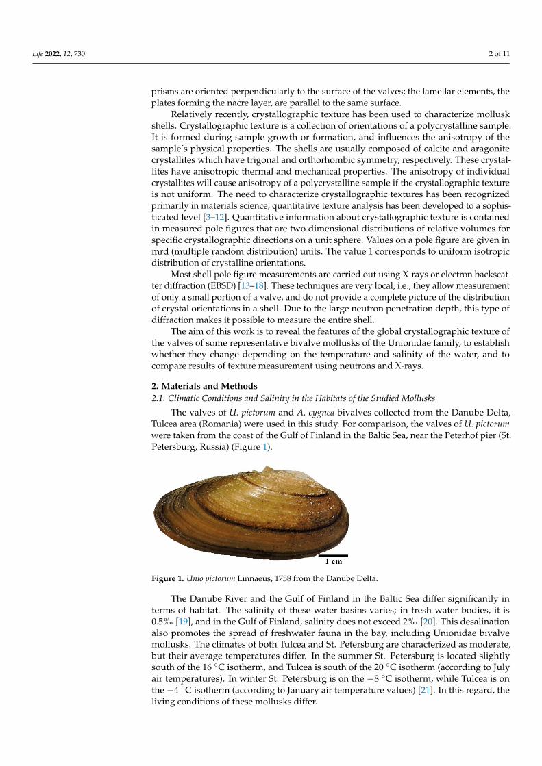
The valves of U. pictorum and A. cygnea bivalves collected from the Danube Delta,Tulcea area (Romania) were used in this study. For comparison, the valves of U. pictorumwere taken from the coast of the Gulf of Finland in the Baltic Sea, near the Peterhof pier (St.Petersburg, Russia) (Figure 1).
Figure 1. Unio pictorum Linnaeus, 1758 from the Danube Delta.
The Danube River and the Gulf of Finland in the Baltic Sea differ significantly interms of habitat. The salinity of these water basins varies; in fresh water bodies, it is0.5‰ [19], and in the Gulf of Finland, salinity does not exceed 2‰ [20]. This desalinationalso promotes the spread of freshwater fauna in the bay, including Unionidae bivalvemollusks. The climates of both Tulcea and St. Petersburg are characterized as moderate,but their average temperatures differ. In the summer St. Petersburg is located slightlysouth of the 16 ◦C isotherm, and Tulcea is south of the 20 ◦C isotherm (according to Julyair temperatures). In winter St. Petersburg is on the −8 ◦C isotherm, while Tulcea is onthe −4 ◦C isotherm (according to January air temperature values) [21]. In this regard, theliving conditions of these mollusks differ.
Life 2022, 12, 730 3 of 11
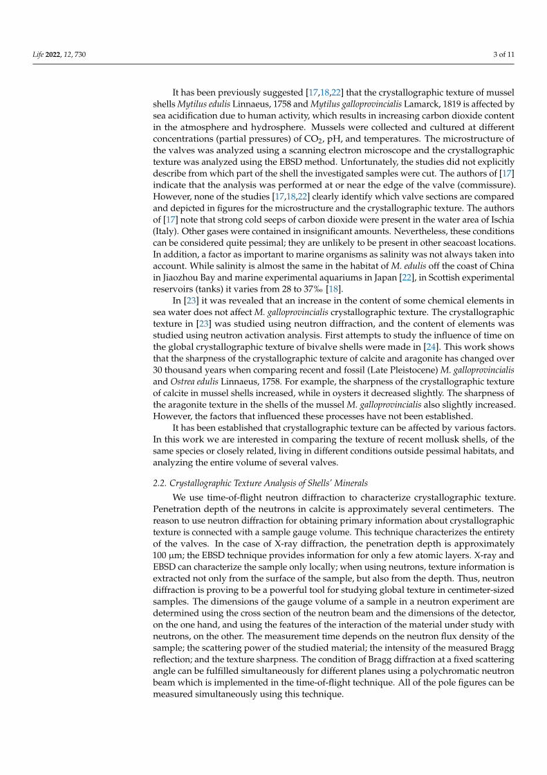
It has been previously suggested [17,18,22] that the crystallographic texture of musselshells Mytilus edulis Linnaeus, 1758 and Mytilus galloprovincialis Lamarck, 1819 is affected bysea acidification due to human activity, which results in increasing carbon dioxide contentin the atmosphere and hydrosphere. Mussels were collected and cultured at differentconcentrations (partial pressures) of CO2, pH, and temperatures. The microstructure ofthe valves was analyzed using a scanning electron microscope and the crystallographictexture was analyzed using the EBSD method. Unfortunately, the studies did not explicitlydescribe from which part of the shell the investigated samples were cut. The authors of [17]indicate that the analysis was performed at or near the edge of the valve (commissure).However, none of the studies [17,18,22] clearly identify which valve sections are comparedand depicted in figures for the microstructure and the crystallographic texture. The authorsof [17] note that strong cold seeps of carbon dioxide were present in the water area of Ischia(Italy). Other gases were contained in insignificant amounts. Nevertheless, these conditionscan be considered quite pessimal; they are unlikely to be present in other seacoast locations.In addition, a factor as important to marine organisms as salinity was not always taken intoaccount. While salinity is almost the same in the habitat of M. edulis off the coast of Chinain Jiaozhou Bay and marine experimental aquariums in Japan [22], in Scottish experimentalreservoirs (tanks) it varies from 28 to 37‰ [18].
In [23] it was revealed that an increase in the content of some chemical elements insea water does not affect M. galloprovincialis crystallographic texture. The crystallographictexture in [23] was studied using neutron diffraction, and the content of elements wasstudied using neutron activation analysis. First attempts to study the influence of time onthe global crystallographic texture of bivalve shells were made in [24]. This work showsthat the sharpness of the crystallographic texture of calcite and aragonite has changed over30 thousand years when comparing recent and fossil (Late Pleistocene) M. galloprovincialisand Ostrea edulis Linnaeus, 1758. For example, the sharpness of the crystallographic textureof calcite in mussel shells increased, while in oysters it decreased slightly. The sharpness ofthe aragonite texture in the shells of the mussel M. galloprovincialis also slightly increased.However, the factors that influenced these processes have not been established.
It has been established that crystallographic texture can be affected by various factors.In this work we are interested in comparing the texture of recent mollusk shells, of thesame species or closely related, living in different conditions outside pessimal habitats, andanalyzing the entire volume of several valves.
2.2. Crystallographic Texture Analysis of Shells’ Minerals
We use time-of-flight neutron diffraction to characterize crystallographic texture.Penetration depth of the neutrons in calcite is approximately several centimeters. Thereason to use neutron diffraction for obtaining primary information about crystallographictexture is connected with a sample gauge volume. This technique characterizes the entiretyof the valves. In the case of X-ray diffraction, the penetration depth is approximately100 µm; the EBSD technique provides information for only a few atomic layers. X-ray andEBSD can characterize the sample only locally; when using neutrons, texture information isextracted not only from the surface of the sample, but also from the depth. Thus, neutrondiffraction is proving to be a powerful tool for studying global texture in centimeter-sizedsamples. The dimensions of the gauge volume of a sample in a neutron experiment aredetermined using the cross section of the neutron beam and the dimensions of the detector,on the one hand, and using the features of the interaction of the material under study withneutrons, on the other. The measurement time depends on the neutron flux density of thesample; the scattering power of the studied material; the intensity of the measured Braggreflection; and the texture sharpness. The condition of Bragg diffraction at a fixed scatteringangle can be fulfilled simultaneously for different planes using a polychromatic neutronbeam which is implemented in the time-of-flight technique. All of the pole figures can bemeasured simultaneously using this technique.
Life 2022, 12, 730 4 of 11
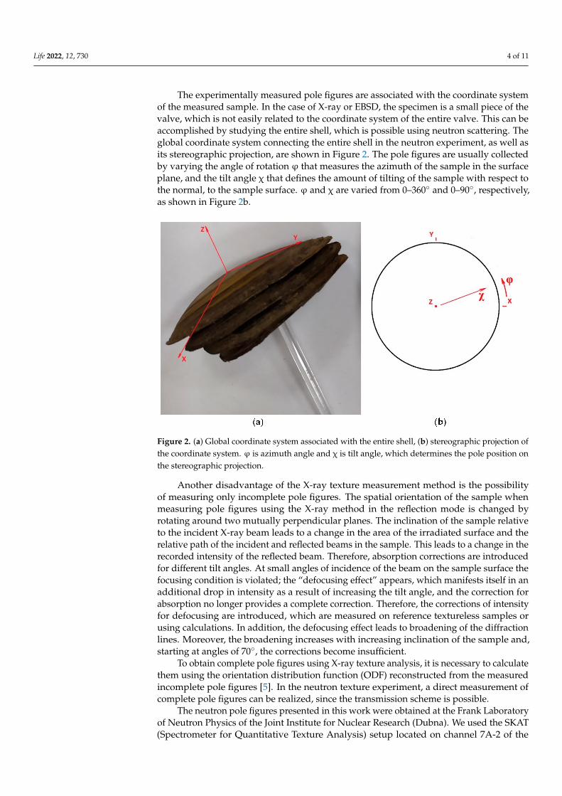
The experimentally measured pole figures are associated with the coordinate systemof the measured sample. In the case of X-ray or EBSD, the specimen is a small piece of thevalve, which is not easily related to the coordinate system of the entire valve. This can beaccomplished by studying the entire shell, which is possible using neutron scattering. Theglobal coordinate system connecting the entire shell in the neutron experiment, as well asits stereographic projection, are shown in Figure 2. The pole figures are usually collectedby varying the angle of rotation ϕ that measures the azimuth of the sample in the surfaceplane, and the tilt angle χ that defines the amount of tilting of the sample with respect tothe normal, to the sample surface. ϕ and χ are varied from 0–360◦ and 0–90◦, respectively,as shown in Figure 2b.
Figure 2. (a) Global coordinate system associated with the entire shell, (b) stereographic projection ofthe coordinate system. ϕ is azimuth angle and χ is tilt angle, which determines the pole position onthe stereographic projection.
Another disadvantage of the X-ray texture measurement method is the possibilityof measuring only incomplete pole figures. The spatial orientation of the sample whenmeasuring pole figures using the X-ray method in the reflection mode is changed byrotating around two mutually perpendicular planes. The inclination of the sample relativeto the incident X-ray beam leads to a change in the area of the irradiated surface and therelative path of the incident and reflected beams in the sample. This leads to a change in therecorded intensity of the reflected beam. Therefore, absorption corrections are introducedfor different tilt angles. At small angles of incidence of the beam on the sample surface thefocusing condition is violated; the “defocusing effect” appears, which manifests itself in anadditional drop in intensity as a result of increasing the tilt angle, and the correction forabsorption no longer provides a complete correction. Therefore, the corrections of intensityfor defocusing are introduced, which are measured on reference textureless samples orusing calculations. In addition, the defocusing effect leads to broadening of the diffractionlines. Moreover, the broadening increases with increasing inclination of the sample and,starting at angles of 70◦, the corrections become insufficient.
To obtain complete pole figures using X-ray texture analysis, it is necessary to calculatethem using the orientation distribution function (ODF) reconstructed from the measuredincomplete pole figures [5]. In the neutron texture experiment, a direct measurement ofcomplete pole figures can be realized, since the transmission scheme is possible.
The neutron pole figures presented in this work were obtained at the Frank Laboratoryof Neutron Physics of the Joint Institute for Nuclear Research (Dubna). We used the SKAT(Spectrometer for Quantitative Texture Analysis) setup located on channel 7A-2 of the
Life 2022, 12, 730 5 of 11
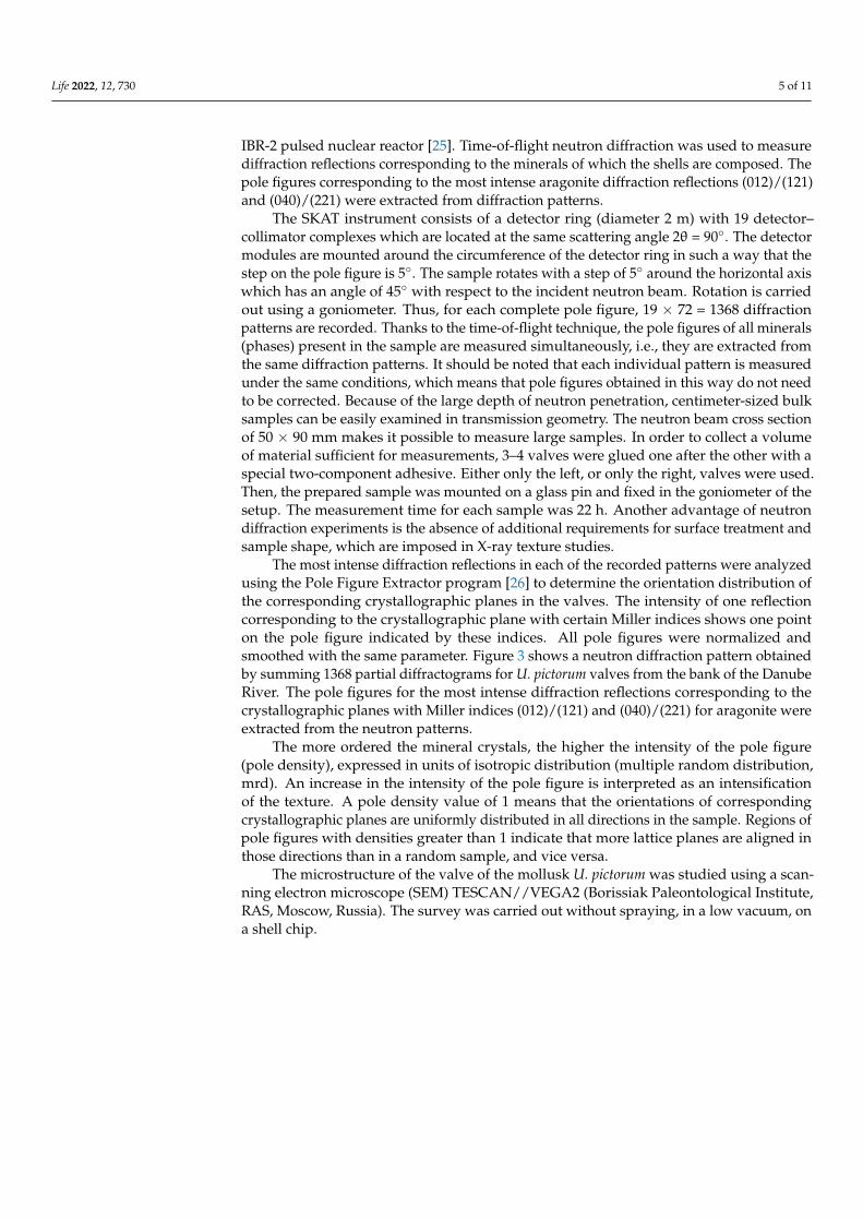
IBR-2 pulsed nuclear reactor [25]. Time-of-flight neutron diffraction was used to measurediffraction reflections corresponding to the minerals of which the shells are composed. Thepole figures corresponding to the most intense aragonite diffraction reflections (012)/(121)and (040)/(221) were extracted from diffraction patterns.
The SKAT instrument consists of a detector ring (diameter 2 m) with 19 detector–collimator complexes which are located at the same scattering angle 2θ = 90◦. The detectormodules are mounted around the circumference of the detector ring in such a way that thestep on the pole figure is 5◦. The sample rotates with a step of 5◦ around the horizontal axiswhich has an angle of 45◦ with respect to the incident neutron beam. Rotation is carriedout using a goniometer. Thus, for each complete pole figure, 19 × 72 = 1368 diffractionpatterns are recorded. Thanks to the time-of-flight technique, the pole figures of all minerals(phases) present in the sample are measured simultaneously, i.e., they are extracted fromthe same diffraction patterns. It should be noted that each individual pattern is measuredunder the same conditions, which means that pole figures obtained in this way do not needto be corrected. Because of the large depth of neutron penetration, centimeter-sized bulksamples can be easily examined in transmission geometry. The neutron beam cross sectionof 50 × 90 mm makes it possible to measure large samples. In order to collect a volumeof material sufficient for measurements, 3–4 valves were glued one after the other with aspecial two-component adhesive. Either only the left, or only the right, valves were used.Then, the prepared sample was mounted on a glass pin and fixed in the goniometer of thesetup. The measurement time for each sample was 22 h. Another advantage of neutrondiffraction experiments is the absence of additional requirements for surface treatment andsample shape, which are imposed in X-ray texture studies.
The most intense diffraction reflections in each of the recorded patterns were analyzedusing the Pole Figure Extractor program [26] to determine the orientation distribution ofthe corresponding crystallographic planes in the valves. The intensity of one reflectioncorresponding to the crystallographic plane with certain Miller indices shows one pointon the pole figure indicated by these indices. All pole figures were normalized andsmoothed with the same parameter. Figure 3 shows a neutron diffraction pattern obtainedby summing 1368 partial diffractograms for U. pictorum valves from the bank of the DanubeRiver. The pole figures for the most intense diffraction reflections corresponding to thecrystallographic planes with Miller indices (012)/(121) and (040)/(221) for aragonite wereextracted from the neutron patterns.
The more ordered the mineral crystals, the higher the intensity of the pole figure(pole density), expressed in units of isotropic distribution (multiple random distribution,mrd). An increase in the intensity of the pole figure is interpreted as an intensificationof the texture. A pole density value of 1 means that the orientations of correspondingcrystallographic planes are uniformly distributed in all directions in the sample. Regions ofpole figures with densities greater than 1 indicate that more lattice planes are aligned inthose directions than in a random sample, and vice versa.
The microstructure of the valve of the mollusk U. pictorum was studied using a scan-ning electron microscope (SEM) TESCAN//VEGA2 (Borissiak Paleontological Institute,RAS, Moscow, Russia). The survey was carried out without spraying, in a low vacuum, ona shell chip.
Life 2022, 12, 730 6 of 11
Figure 3. Neutron diffraction pattern obtained by summing 1368 partial diffractograms for Uniopictorum valves from the bank of the Danube River. The most intense diffraction reflections for whichpole figures have been extracted are highlighted in bold type.
3. Results
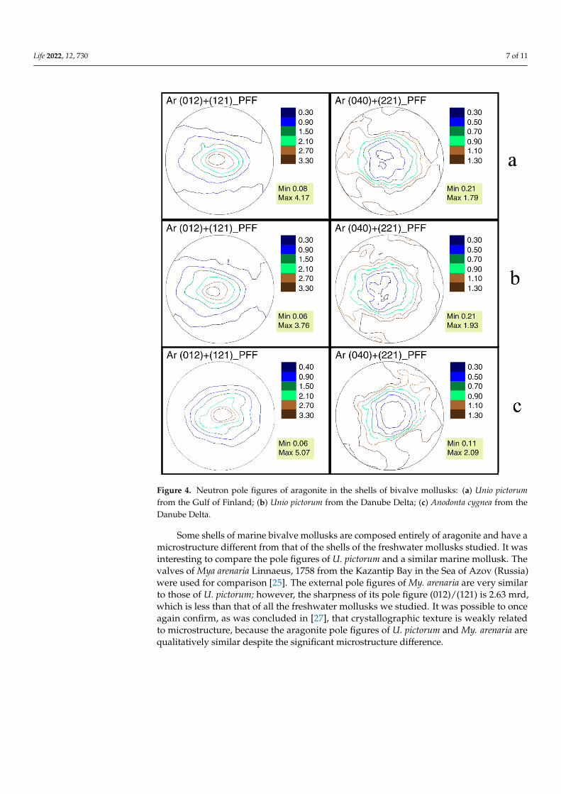
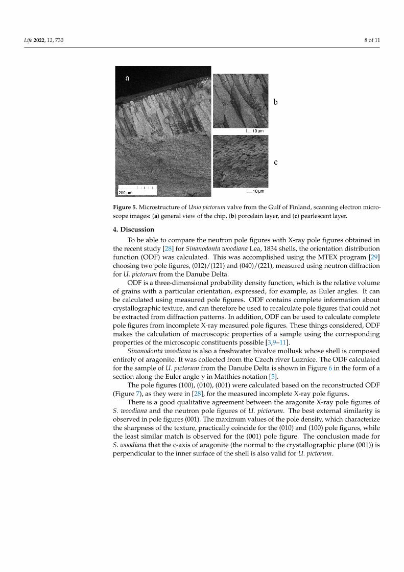
In all studied valves, only aragonite (CaCO3) was found. For each of the collectedsamples, neutron pole figures were obtained. For the crystallographic direction (012)/(121),it is noticeable that the crystals are oriented in a wide range. The contours of the polefigures are elongated horizontally, i.e., the aragonite crystals repeat the shape of the shell(Figure 4). The SEM image (Figure 5) shows that the crystals are organized into variousmicrostructural units such as prisms and plates, positioned at different angles to the shellsurface. As a result, one can expect several sharp peaks on the pole figures. However, thedifferent arrangement of microstructural elements relative to the surface of the mollusk’sshells does not affect the measured texture in any way. That is, the microstructure andtexture of the shell are weakly related. Similar conclusions for different mollusk specieswere reported in [27].
The appearance of the U. pictorum pole figures from Tulcea and Peterhof is very similarfor A. cygnea. The sharpness of the crystallographic texture of the valves of U. pictorumfrom the Danube delta is 4.17 mrd, and from the Gulf of Finland 3.76 mrd, respectively. Weestimate the texture sharpness using the maxima of the aragonite pole figure (012)/(121).The difference in texture sharpness of the same species in different habitat conditions is0.41 mrd. This is a very important value, since there are almost no data regarding thedifference in crystallographic texture in organisms living in different conditions.
The isolines of the pole figures of A. cygnea are also horizontally elongated. However,the pole figure is more rounded than that of the U. pictorum. Aragonite crystals are alsooriented in the shape of the valves. An important feature of the shell texture of this speciesis its sharpness of 5.07 mrd. Here, bivalve, whose shells also include aragonite, should becompared. The inner layers of mussel shells are composed of nacre. Earlier we studied [25]the crystallographic texture of the shells of three species of the genus Mytilus Linnaeus,1758. The arrangement of their aragonite crystals is also similar to the shape of the valves.However, the sharpness of the aragonite pole figures does not exceed 3.36 mrd for M. edulis.
Life 2022, 12, 730 7 of 11
Figure 4. Neutron pole figures of aragonite in the shells of bivalve mollusks: (a) Unio pictorumfrom the Gulf of Finland; (b) Unio pictorum from the Danube Delta; (c) Anodonta cygnea from theDanube Delta.
Some shells of marine bivalve mollusks are composed entirely of aragonite and have amicrostructure different from that of the shells of the freshwater mollusks studied. It wasinteresting to compare the pole figures of U. pictorum and a similar marine mollusk. Thevalves of Mya arenaria Linnaeus, 1758 from the Kazantip Bay in the Sea of Azov (Russia)were used for comparison [25]. The external pole figures of My. arenaria are very similarto those of U. pictorum; however, the sharpness of its pole figure (012)/(121) is 2.63 mrd,which is less than that of all the freshwater mollusks we studied. It was possible to onceagain confirm, as was concluded in [27], that crystallographic texture is weakly relatedto microstructure, because the aragonite pole figures of U. pictorum and My. arenaria arequalitatively similar despite the significant microstructure difference.
Life 2022, 12, 730 8 of 11
Figure 5. Microstructure of Unio pictorum valve from the Gulf of Finland, scanning electron micro-scope images: (a) general view of the chip, (b) porcelain layer, and (c) pearlescent layer.
4. Discussion
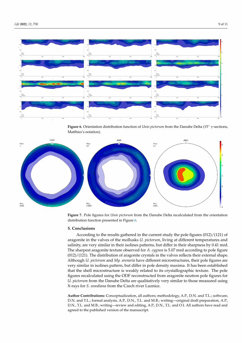
To be able to compare the neutron pole figures with X-ray pole figures obtained inthe recent study [28] for Sinanodonta woodiana Lea, 1834 shells, the orientation distributionfunction (ODF) was calculated. This was accomplished using the MTEX program [29]choosing two pole figures, (012)/(121) and (040)/(221), measured using neutron diffractionfor U. pictorum from the Danube Delta.
ODF is a three-dimensional probability density function, which is the relative volumeof grains with a particular orientation, expressed, for example, as Euler angles. It canbe calculated using measured pole figures. ODF contains complete information aboutcrystallographic texture, and can therefore be used to recalculate pole figures that could notbe extracted from diffraction patterns. In addition, ODF can be used to calculate completepole figures from incomplete X-ray measured pole figures. These things considered, ODFmakes the calculation of macroscopic properties of a sample using the correspondingproperties of the microscopic constituents possible [3,9–11].
Sinanodonta woodiana is also a freshwater bivalve mollusk whose shell is composedentirely of aragonite. It was collected from the Czech river Luznice. The ODF calculatedfor the sample of U. pictorum from the Danube Delta is shown in Figure 6 in the form of asection along the Euler angle γ in Matthies notation [5].
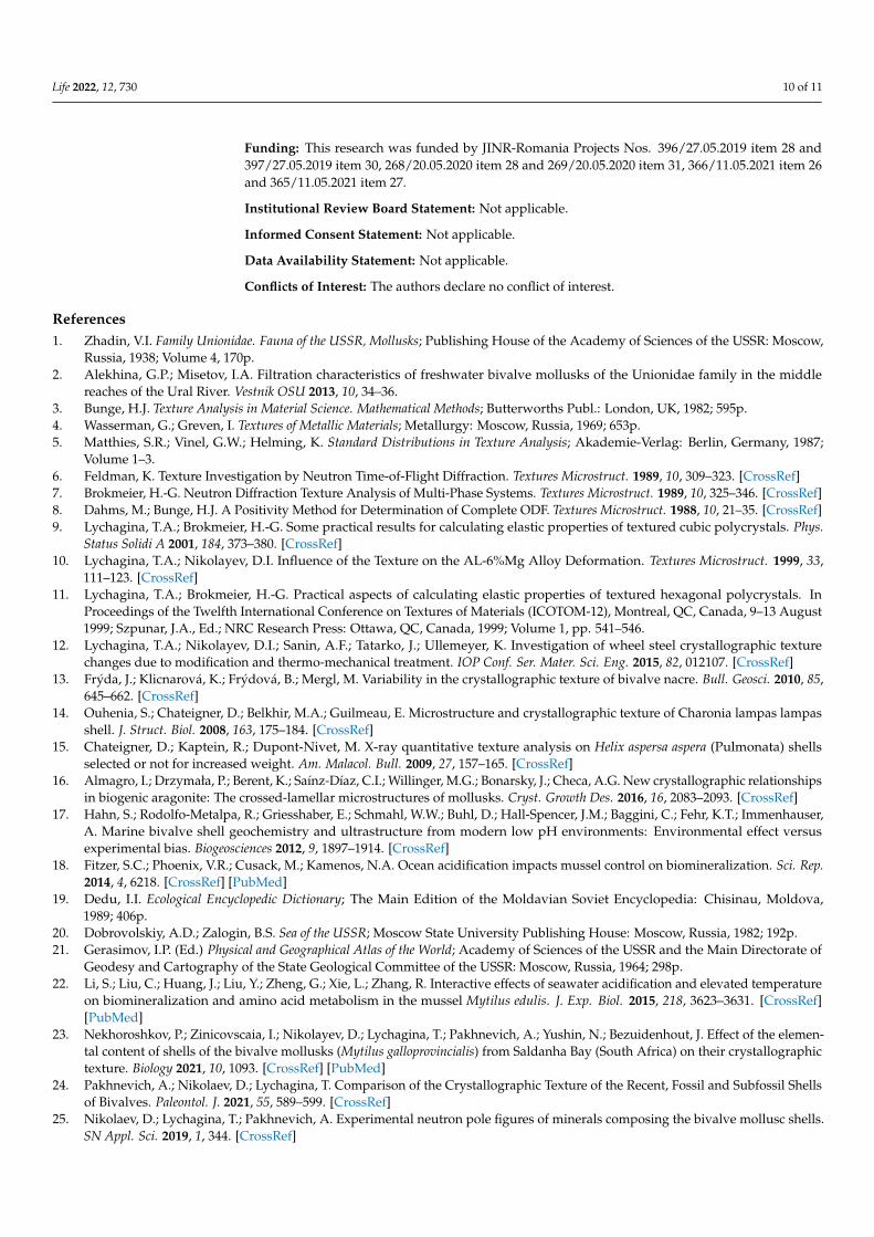
The pole figures (100), (010), (001) were calculated based on the reconstructed ODF(Figure 7), as they were in [28], for the measured incomplete X-ray pole figures.
There is a good qualitative agreement between the aragonite X-ray pole figures ofS. woodiana and the neutron pole figures of U. pictorum. The best external similarity isobserved in pole figures (001). The maximum values of the pole density, which characterizethe sharpness of the texture, practically coincide for the (010) and (100) pole figures, whilethe least similar match is observed for the (001) pole figure. The conclusion made forS. woodiana that the c-axis of aragonite (the normal to the crystallographic plane (001)) isperpendicular to the inner surface of the shell is also valid for U. pictorum.
Life 2022, 12, 730 9 of 11
0 120 240 360
0
30
60
900 120 240 360
0
30
60
900 120 240 360
0
30
60
90
0 120 240 360
0
30
60
900 120 240 360
0
30
60
900 120 240 360
0
30
60
90
0 120 240 360
0
30
60
900 120 240 360
0
30
60
900 120 240 360
0
30
60
90
0 120 240 360
0
30
60
900 120 240 360
0
30
60
900 120 240 360
0
30
60
90 0
2
4
6
8
10
12
14
Figure 6. Orientation distribution function of Unio pictorum from the Danube Delta (15◦ γ-sections,Matthies’s notation).
Figure 7. Pole figures for Unio pictorum from the Danube Delta recalculated from the orientationdistribution function presented in Figure 6.
5. Conclusions
According to the results gathered in the current study the pole figures (012)/(121) ofaragonite in the valves of the mollusks U. pictorum, living at different temperatures andsalinity, are very similar in their isolines patterns, but differ in their sharpness by 0.41 mrd.The sharpest aragonite texture observed for A. cygnea is 5.07 mrd according to pole figure(012)/(121). The distribution of aragonite crystals in the valves reflects their external shape.Although U. pictorum and My. arenaria have different microstructures, their pole figures arevery similar in isolines pattern, but differ in pole density maxima. It has been establishedthat the shell microstructure is weakly related to its crystallographic texture. The polefigures recalculated using the ODF reconstructed from aragonite neutron pole figures forU. pictorum from the Danube Delta are qualitatively very similar to those measured usingX-rays for S. woodiana from the Czech river Luznice.
Author Contributions: Conceptualization, all authors; methodology, A.P., D.N. and T.L.; software,D.N. and T.L.; formal analysis, A.P., D.N., T.L. and M.B.; writing—original draft preparation, A.P.,D.N., T.L. and M.B.; writing—review and editing, A.P., D.N., T.L. and O.I. All authors have read andagreed to the published version of the manuscript.
Life 2022, 12, 730 10 of 11
Funding: This research was funded by JINR-Romania Projects Nos. 396/27.05.2019 item 28 and397/27.05.2019 item 30, 268/20.05.2020 item 28 and 269/20.05.2020 item 31, 366/11.05.2021 item 26and 365/11.05.2021 item 27.
Institutional Review Board Statement: Not applicable.
Informed Consent Statement: Not applicable.
Data Availability Statement: Not applicable.
Conflicts of Interest: The authors declare no conflict of interest.
References1. Zhadin, V.I. Family Unionidae. Fauna of the USSR, Mollusks; Publishing House of the Academy of Sciences of the USSR: Moscow,
Russia, 1938; Volume 4, 170p.2. Alekhina, G.P.; Misetov, I.A. Filtration characteristics of freshwater bivalve mollusks of the Unionidae family in the middle
reaches of the Ural River. Vestnik OSU 2013, 10, 34–36.3. Bunge, H.J. Texture Analysis in Material Science. Mathematical Methods; Butterworths Publ.: London, UK, 1982; 595p.4. Wasserman, G.; Greven, I. Textures of Metallic Materials; Metallurgy: Moscow, Russia, 1969; 653p.5. Matthies, S.R.; Vinel, G.W.; Helming, K. Standard Distributions in Texture Analysis; Akademie-Verlag: Berlin, Germany, 1987;
Volume 1–3.6. Feldman, K. Texture Investigation by Neutron Time-of-Flight Diffraction. Textures Microstruct. 1989, 10, 309–323. [CrossRef]7. Brokmeier, H.-G. Neutron Diffraction Texture Analysis of Multi-Phase Systems. Textures Microstruct. 1989, 10, 325–346. [CrossRef]8. Dahms, M.; Bunge, H.J. A Positivity Method for Determination of Complete ODF. Textures Microstruct. 1988, 10, 21–35. [CrossRef]9. Lychagina, T.A.; Brokmeier, H.-G. Some practical results for calculating elastic properties of textured cubic polycrystals. Phys.
Status Solidi A 2001, 184, 373–380. [CrossRef]10. Lychagina, T.A.; Nikolayev, D.I. Influence of the Texture on the AL-6%Mg Alloy Deformation. Textures Microstruct. 1999, 33,
111–123. [CrossRef]11. Lychagina, T.A.; Brokmeier, H.-G. Practical aspects of calculating elastic properties of textured hexagonal polycrystals. In
Proceedings of the Twelfth International Conference on Textures of Materials (ICOTOM-12), Montreal, QC, Canada, 9–13 August1999; Szpunar, J.A., Ed.; NRC Research Press: Ottawa, QC, Canada, 1999; Volume 1, pp. 541–546.
12. Lychagina, T.A.; Nikolayev, D.I.; Sanin, A.F.; Tatarko, J.; Ullemeyer, K. Investigation of wheel steel crystallographic texturechanges due to modification and thermo-mechanical treatment. IOP Conf. Ser. Mater. Sci. Eng. 2015, 82, 012107. [CrossRef]
13. Frýda, J.; Klicnarová, K.; Frýdová, B.; Mergl, M. Variability in the crystallographic texture of bivalve nacre. Bull. Geosci. 2010, 85,645–662. [CrossRef]
14. Ouhenia, S.; Chateigner, D.; Belkhir, M.A.; Guilmeau, E. Microstructure and crystallographic texture of Charonia lampas lampasshell. J. Struct. Biol. 2008, 163, 175–184. [CrossRef]
15. Chateigner, D.; Kaptein, R.; Dupont-Nivet, M. X-ray quantitative texture analysis on Helix aspersa aspera (Pulmonata) shellsselected or not for increased weight. Am. Malacol. Bull. 2009, 27, 157–165. [CrossRef]
16. Almagro, I.; Drzymała, P.; Berent, K.; Saínz-Díaz, C.I.; Willinger, M.G.; Bonarsky, J.; Checa, A.G. New crystallographic relationshipsin biogenic aragonite: The crossed-lamellar microstructures of mollusks. Cryst. Growth Des. 2016, 16, 2083–2093. [CrossRef]
17. Hahn, S.; Rodolfo-Metalpa, R.; Griesshaber, E.; Schmahl, W.W.; Buhl, D.; Hall-Spencer, J.M.; Baggini, C.; Fehr, K.T.; Immenhauser,A. Marine bivalve shell geochemistry and ultrastructure from modern low pH environments: Environmental effect versusexperimental bias. Biogeosciences 2012, 9, 1897–1914. [CrossRef]
18. Fitzer, S.C.; Phoenix, V.R.; Cusack, M.; Kamenos, N.A. Ocean acidification impacts mussel control on biomineralization. Sci. Rep.2014, 4, 6218. [CrossRef] [PubMed]
19. Dedu, I.I. Ecological Encyclopedic Dictionary; The Main Edition of the Moldavian Soviet Encyclopedia: Chisinau, Moldova,1989; 406p.
20. Dobrovolskiy, A.D.; Zalogin, B.S. Sea of the USSR; Moscow State University Publishing House: Moscow, Russia, 1982; 192p.21. Gerasimov, I.P. (Ed.) Physical and Geographical Atlas of the World; Academy of Sciences of the USSR and the Main Directorate of
Geodesy and Cartography of the State Geological Committee of the USSR: Moscow, Russia, 1964; 298p.22. Li, S.; Liu, C.; Huang, J.; Liu, Y.; Zheng, G.; Xie, L.; Zhang, R. Interactive effects of seawater acidification and elevated temperature
on biomineralization and amino acid metabolism in the mussel Mytilus edulis. J. Exp. Biol. 2015, 218, 3623–3631. [CrossRef][PubMed]
23. Nekhoroshkov, P.; Zinicovscaia, I.; Nikolayev, D.; Lychagina, T.; Pakhnevich, A.; Yushin, N.; Bezuidenhout, J. Effect of the elemen-tal content of shells of the bivalve mollusks (Mytilus galloprovincialis) from Saldanha Bay (South Africa) on their crystallographictexture. Biology 2021, 10, 1093. [CrossRef] [PubMed]
24. Pakhnevich, A.; Nikolaev, D.; Lychagina, T. Comparison of the Crystallographic Texture of the Recent, Fossil and Subfossil Shellsof Bivalves. Paleontol. J. 2021, 55, 589–599. [CrossRef]
25. Nikolaev, D.; Lychagina, T.; Pakhnevich, A. Experimental neutron pole figures of minerals composing the bivalve mollusc shells.SN Appl. Sci. 2019, 1, 344. [CrossRef]

Life 2022, 12, 730 11 of 11
26. Nikolayev, D.I.; Lychagina, T.A.; Nikishin, A.V.; Yudin, V.V. Investigation of measured pole figures errors. Mater. Sci. Forum 2005,495–497, 307–312. [CrossRef]
27. Chateigner, D.; Hedegaard, C.; Wenk, H.-R. Mollusc shell microstructures and crystallographic textures. J. Struct. Geol. 2000, 22,1723–1735. [CrossRef]
28. Kucerakova, M.; Rohlicek, J.; Vratislav, S.; Nikolayev, D.; Lychagina, T.; Kalvoda, L.; Douda, K. Texture Study of Sinanodontawoodiana Shells by X-ray Diffraction. J. Surf. Investig. X-ray Synchrotron Neutron Tech. 2021, 15, 640–643. [CrossRef]
29. Bachmann, F.; Hielscher, R.; Schaeben, H. Texture Analysis with MTEX-Free and Open Source Software Toolbox. Solid State Phenom.2010, 160, 63–68. [CrossRef]
Related Documents