Research Article Ginkgo biloba Extract Improves Insulin Signaling and Attenuates Inflammation in Retroperitoneal Adipose Tissue Depot of Obese Rats Bruna Kelly Sousa Hirata, 1 Renata Mancini Banin, 1 Ana Paula Segantine Dornellas, 2 Iracema Senna de Andrade, 2 Juliane Costa Silva Zemdegs, 2 Luciana Chagas Caperuto, 1 Lila Missae Oyama, 2 Eliane Beraldi Ribeiro, 2 and Monica Marques Telles 1 1 Departamento de Ciˆ encias Biol´ ogicas, Universidade Federal de S˜ ao Paulo (UNIFESP), Rua Arthur Riedel, 275 Eldorado, 09972-270 Diadema, SP, Brazil 2 Departamento de Fisiologia, Disciplina de Fisiologia da Nutric ¸˜ ao, Universidade Federal de S˜ ao Paulo (UNIFESP), Rua Botucatu, 862 Vila Clementino, S˜ ao Paulo, SP, Brazil Correspondence should be addressed to Monica Marques Telles; [email protected] Received 21 October 2014; Revised 16 December 2014; Accepted 17 December 2014 Academic Editor: Yi Fu Yang Copyright © 2015 Bruna Kelly Sousa Hirata et al. is is an open access article distributed under the Creative Commons Attribution License, which permits unrestricted use, distribution, and reproduction in any medium, provided the original work is properly cited. Due to the high incidence and severity of obesity and its related disorders, it is highly desirable to develop new strategies to treat or even to prevent its development. We have previously described that Ginkgo biloba extract (GbE) improved insulin resistance and reduced body weight gain of obese rats. In the present study we aimed to evaluate the effect of GbE on both inflammatory cascade and insulin signaling in retroperitoneal fat depot of diet-induced obese rats. Rats were fed with high fat diet for 2 months and thereaſter treated for 14 days with 500mg/kg of GbE. Rats were then euthanized and samples from retroperitoneal fat depot were used for western blotting, RT-PCR, and ELISA experiments. e GbE treatment promoted a significant reduction on both food/energy intake and body weight gain in comparison to the nontreated obese rats. In addition, a significant increase of both Adipo R1 and IL-10 gene expressions and IR and Akt phosphorylation was also observed, while NF-B p65 phosphorylation and TNF- levels were significantly reduced. Our data suggest that GbE might have potential as a therapy to treat obesity-related metabolic diseases, with special interest to treat obese subjects resistant to adhere to a nutritional education program. 1. Introduction e incidence of both obesity and overweight has been dramatically increasing around the world. In 2008, 35% of worldwide population was overweight while 11% was obese [1]. In addition, it has been estimated that obesity will achieve one-third of the population in 2030 [2]. is perspective is particularly worrying since obesity is related to chronic comorbidities, such as insulin resistance, type 2 diabetes (T2D), subclinical inflammation, and others [3]. It has been suggested that consumption of high-fat diet is directly involved in the obesity pathogenesis since it affects either central control of food intake and peripheral metabolism, resulting in increased body weight gain, insulin resistance, and other metabolic disturbances [4, 5]. us, we have previously demonstrated that prolonged hyperlipidic diet ingestion promoted in rats a significant increase of body adiposity, triacylglycerol, and glucose plasma levels with a concomitant loss of insulin sensitivity [6]. Insulin resistance is a chronic condition in which the hormone insulin fails to activate its own signaling cascade, resulting in hyperglycemia. It has been highly correlated to visceral adiposity excess and increased white adipose tissue (WAT) expression of cytokines, such as tumor necrosis factor-alpha (TNF-) and interleukin-6 (IL-6) [3, 7]. Further- more, high-fat diet intake has been pointed as an important Hindawi Publishing Corporation Mediators of Inflammation Volume 2015, Article ID 419106, 9 pages http://dx.doi.org/10.1155/2015/419106

Welcome message from author
This document is posted to help you gain knowledge. Please leave a comment to let me know what you think about it! Share it to your friends and learn new things together.
Transcript

Research ArticleGinkgo biloba Extract Improves Insulin Signaling andAttenuates Inflammation in Retroperitoneal Adipose TissueDepot of Obese Rats
Bruna Kelly Sousa Hirata,1 Renata Mancini Banin,1 Ana Paula Segantine Dornellas,2
Iracema Senna de Andrade,2 Juliane Costa Silva Zemdegs,2 Luciana Chagas Caperuto,1
Lila Missae Oyama,2 Eliane Beraldi Ribeiro,2 and Monica Marques Telles1
1Departamento de Ciencias Biologicas, Universidade Federal de Sao Paulo (UNIFESP), Rua Arthur Riedel, 275 Eldorado,09972-270 Diadema, SP, Brazil2Departamento de Fisiologia, Disciplina de Fisiologia da Nutricao, Universidade Federal de Sao Paulo (UNIFESP),Rua Botucatu, 862 Vila Clementino, Sao Paulo, SP, Brazil
Correspondence should be addressed to Monica Marques Telles; [email protected]
Received 21 October 2014; Revised 16 December 2014; Accepted 17 December 2014
Academic Editor: Yi Fu Yang
Copyright © 2015 Bruna Kelly Sousa Hirata et al. This is an open access article distributed under the Creative CommonsAttribution License, which permits unrestricted use, distribution, and reproduction in any medium, provided the original work isproperly cited.
Due to the high incidence and severity of obesity and its related disorders, it is highly desirable to develop new strategies to treator even to prevent its development. We have previously described that Ginkgo biloba extract (GbE) improved insulin resistanceand reduced body weight gain of obese rats. In the present study we aimed to evaluate the effect of GbE on both inflammatorycascade and insulin signaling in retroperitoneal fat depot of diet-induced obese rats. Rats were fed with high fat diet for 2 monthsand thereafter treated for 14 days with 500mg/kg of GbE. Rats were then euthanized and samples from retroperitoneal fat depotwere used for western blotting, RT-PCR, and ELISA experiments. The GbE treatment promoted a significant reduction on bothfood/energy intake and body weight gain in comparison to the nontreated obese rats. In addition, a significant increase of bothAdipo R1 and IL-10 gene expressions and IR and Akt phosphorylation was also observed, while NF-𝜅B p65 phosphorylation andTNF-𝛼 levels were significantly reduced. Our data suggest that GbE might have potential as a therapy to treat obesity-relatedmetabolic diseases, with special interest to treat obese subjects resistant to adhere to a nutritional education program.
1. Introduction
The incidence of both obesity and overweight has beendramatically increasing around the world. In 2008, 35% ofworldwide population was overweight while 11% was obese[1]. In addition, it has been estimated that obesity will achieveone-third of the population in 2030 [2]. This perspectiveis particularly worrying since obesity is related to chroniccomorbidities, such as insulin resistance, type 2 diabetes(T2D), subclinical inflammation, and others [3].
It has been suggested that consumption of high-fat dietis directly involved in the obesity pathogenesis since itaffects either central control of food intake and peripheral
metabolism, resulting in increased body weight gain, insulinresistance, and other metabolic disturbances [4, 5]. Thus, wehave previously demonstrated that prolonged hyperlipidicdiet ingestion promoted in rats a significant increase of bodyadiposity, triacylglycerol, and glucose plasma levels with aconcomitant loss of insulin sensitivity [6].
Insulin resistance is a chronic condition in which thehormone insulin fails to activate its own signaling cascade,resulting in hyperglycemia. It has been highly correlatedto visceral adiposity excess and increased white adiposetissue (WAT) expression of cytokines, such as tumor necrosisfactor-alpha (TNF-𝛼) and interleukin-6 (IL-6) [3, 7]. Further-more, high-fat diet intake has been pointed as an important
Hindawi Publishing CorporationMediators of InflammationVolume 2015, Article ID 419106, 9 pageshttp://dx.doi.org/10.1155/2015/419106
2 Mediators of Inflammation
risk factor for insulin resistance, since it both impairs insulinsignaling pathway and stimulates inflammation, via Toll-likereceptors signaling cascade [5, 7, 8].
Taking into account thatmost of hypoglycemiants presentundesirable side effects [9–12] and due to the severity ofinsulin resistance progression it is highly desirable to discovernew drugs and treatment methods. It has been proposed thatGinkgo biloba extract (GbE) might have positive effects onhyperglycemia. This plant extract mainly contains around24% flavonoid glycosides and 6% terpenoids, including A, B,C, M, J, P, and Q ginkgolides [13].
We have previously described that prolonged GbE treat-ment significantly reduced food intake and body adiposity,prevented against hyperglycemia and dyslipidemia, whileit increased insulin sensitivity evaluated by ITT (insulintolerance test) in obese rats fed with lard-enriched hyper-lipidic diet [6]. In agreement to our previous findings,other studies proposed that GbE intake improved glycaemicprofile of both healthy and T2D patients [14, 15]. In addi-tion, a reduction on glucose elevation stimulated by oraladministration of saccharin agents in rats was demonstrated[16].
Thedata above suggest beneficial effects ofGbEon insulinresistance and obesity-related disorders. However, it is highlyimportant to better describe the mechanisms by which GbEimproves insulin action. In this context, the present studyaimed to evaluate if a 14-day oral GbE treatment altersretroperitoneal WAT depot insulin and Toll-like receptorssignaling cascades of diet-induced obese rats, a model ofinsulin resistance.
2. Materials and Methods
2.1. Animals. The Committee on Animal Research Ethicsof the Universidade Federal de Sao Paulo approved allprocedures for the care of the animals used in this study(Process number: 271359). All efforts were made to minimizesuffering. Male Wistar rats from CEDEME (Sao Paulo,Brazil) were housed 4 per cage and maintained in controlledconditions of light (12 : 12-h light/dark, lights on at 6 am) andtemperature (23∘C ± 1∘C), with free access to food and water.
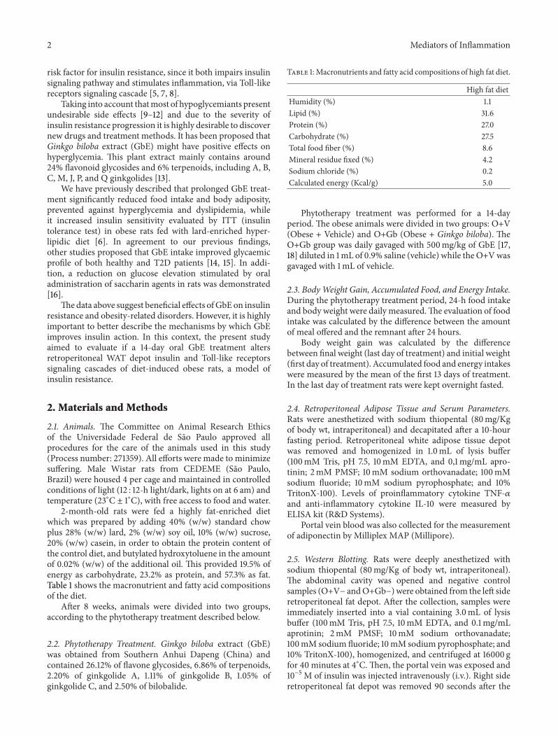
2-month-old rats were fed a highly fat-enriched dietwhich was prepared by adding 40% (w/w) standard chowplus 28% (w/w) lard, 2% (w/w) soy oil, 10% (w/w) sucrose,20% (w/w) casein, in order to obtain the protein content ofthe control diet, and butylated hydroxytoluene in the amountof 0.02% (w/w) of the additional oil. This provided 19.5% ofenergy as carbohydrate, 23.2% as protein, and 57.3% as fat.Table 1 shows the macronutrient and fatty acid compositionsof the diet.
After 8 weeks, animals were divided into two groups,according to the phytotherapy treatment described below.
2.2. Phytotherapy Treatment. Ginkgo biloba extract (GbE)was obtained from Southern Anhui Dapeng (China) andcontained 26.12% of flavone glycosides, 6.86% of terpenoids,2.20% of ginkgolide A, 1.11% of ginkgolide B, 1.05% ofginkgolide C, and 2.50% of bilobalide.
Table 1:Macronutrients and fatty acid compositions of high fat diet.
High fat dietHumidity (%) 1.1Lipid (%) 31.6Protein (%) 27.0Carbohydrate (%) 27.5Total food fiber (%) 8.6Mineral residue fixed (%) 4.2Sodium chloride (%) 0.2Calculated energy (Kcal/g) 5.0
Phytotherapy treatment was performed for a 14-dayperiod. The obese animals were divided in two groups: O+V(Obese + Vehicle) and O+Gb (Obese + Ginkgo biloba). TheO+Gb group was daily gavaged with 500mg/kg of GbE [17,18] diluted in 1mL of 0.9% saline (vehicle) while theO+Vwasgavaged with 1mL of vehicle.
2.3. BodyWeight Gain, Accumulated Food, and Energy Intake.During the phytotherapy treatment period, 24-h food intakeand body weight were daily measured.The evaluation of foodintake was calculated by the difference between the amountof meal offered and the remnant after 24 hours.
Body weight gain was calculated by the differencebetween final weight (last day of treatment) and initial weight(first day of treatment). Accumulated food and energy intakeswere measured by the mean of the first 13 days of treatment.In the last day of treatment rats were kept overnight fasted.
2.4. Retroperitoneal Adipose Tissue and Serum Parameters.Rats were anesthetized with sodium thiopental (80mg/Kgof body wt, intraperitoneal) and decapitated after a 10-hourfasting period. Retroperitoneal white adipose tissue depotwas removed and homogenized in 1.0mL of lysis buffer(100mM Tris, pH 7.5, 10mM EDTA, and 0,1mg/mL apro-tinin; 2mM PMSF; 10mM sodium orthovanadate; 100mMsodium fluoride; 10mM sodium pyrophosphate; and 10%TritonX-100). Levels of proinflammatory cytokine TNF-𝛼and anti-inflammatory cytokine IL-10 were measured byELISA kit (R&D Systems).
Portal vein blood was also collected for the measurementof adiponectin by Milliplex MAP (Millipore).
2.5. Western Blotting. Rats were deeply anesthetized withsodium thiopental (80mg/Kg of body wt, intraperitoneal).The abdominal cavity was opened and negative controlsamples (O+V− andO+Gb−) were obtained from the left sideretroperitoneal fat depot. After the collection, samples wereimmediately inserted into a vial containing 3.0mL of lysisbuffer (100mM Tris, pH 7.5, 10mM EDTA, and 0.1mg/mLaprotinin; 2mM PMSF; 10mM sodium orthovanadate;100mM sodium fluoride; 10mM sodium pyrophosphate; and10% TritonX-100), homogenized, and centrifuged at 16000 gfor 40 minutes at 4∘C.Then, the portal vein was exposed and10−5M of insulin was injected intravenously (i.v.). Right sideretroperitoneal fat depot was removed 90 seconds after the

Mediators of Inflammation 3
i.v. insulin injection (positive samples: O+V+ and O+Gb+)following the same protocol described above [19, 20]. Totalprotein was quantified by BCA kit (BioRad) and sampleswere used for both immunoprecipitation and total extractevaluations.
To reduce the risk of nonspecific antibody binding, weevaluated the IR phosphorylation levels after immunopre-cipitation with antibody against IR. To perform immuno-precipitation experiments, samples were overnight incubatedwith 10 𝜇L primary antibody anti-IR (insulin R𝛽 sc-711) andproteinswere precipitated by ProteinA Sepharose (GE). Afterall, proteins were separated on 10% SDS-PAGE. Proteins werethen transferred to nitrocellulose membranes by wet transferapparatus (Bio-Rad). The membranes were preincubated for1 hour in blocking buffer (5% bovine serum albumin [BSA],1MTris, pH 7.5, 5MNaCl, and 0.02%Tween-20).Membraneswere overnight incubated at 4∘C with the primary antibodyagainst p-Tyr (Cell Signaling 8954). Allmembranes were thenincubated with specific horseradish peroxidase-conjugatedanti-rabbit IgG antibody (Cell Signaling 7074) followedby chemiluminescence detection (Amersham Biosciences).Since all samples were immunoprecipitated with IR antibody,we considered that bands with molecular weight of 95 kDawere related to the phosphorylated form of IR. In addition,IR levels were used as internal standards since all the otherproteins were removed by the immunoprecipitation method.
To perform the total extract experiments, after the proteinquantification, total proteins were then separated on 8%SDS-PAGE. Proteins were transferred by semidry transferapparatus (Bio-Rad).
All membranes were overnight incubated at 4∘C with theprimary antibody against phospho-Akt (Cell Signaling Ser473–9271); Akt (Cell Signaling 9272), phospho-NF-𝜅B p65(Cell Signaling Ser 536–3033), NF-𝜅B p65 (Cell Signaling6956), MyD88 (Cell Signaling 4283), TLR4 (SC 293072), and𝛽-tubulin (Cell Signaling 2146). All membranes were thenincubated with specific horseradish peroxidase-conjugatedantimouse/rabbit IgG antibody (Cell Signaling 7076;Cell Sig-naling 7074, resp.) followed by chemiluminescence detection(Amersham Biosciences). 𝛽-tubulin (Cell Signaling 2146)level was used as an internal standard.
Quantitative analysis was performed with Scion Imagesoftware (Scion Corporation, Frederick, MD, USA). In allexperiments, at least one sample from each group wasanalyzed simultaneously and the results were expressed aspercentage change relative to the basal levels.
2.6. RNA Extraction and Quantitative Real-Time PolymeraseChain Reaction (qPCR). In order to evaluate the gene expres-sion of Adipo R1, Adipo R2, and IL-10, additional groups(O+V and O+Gb) of five rats each were performed. For totalRNA extraction, two hundred mg of frozen retroperitonealadipose tissue from each sample were homogenized byadding 1mL of Trizol reagent (Invitrogen, USA).The sampleswere centrifuged at 16.000 g for 15min at 4∘C and the aqueousphase was removed and mixed with 0.5mL of isopropylalcohol. After centrifugation at 16.000 g for 10min at 4∘C,the pellet was washed with 1mL of 75% ethanol and thendissolved in 20𝜇L DEPC-Treated water (Ambion, USA).
Table 2: Retroperitoneal fat depot cytokine levels (𝜌g/𝜇g of pro-tein).
Cytokine O + V O + GbIL-10 0.47 ± 0.09 0.33 ± 0.03IL-6 0.57 ± 0.09 0.55 ± 0.08TNF-𝛼 0.47 ± 0.07 0.30 ± 0.02∗∗
𝑃 < 0.05 versus O + V.
One microgram of RNA was reverse transcribed tocDNA using the High-Capacity cDNA kit (Applied Biosys-tems). Gene expression was evaluated by real-time qPCRusing the Taqman PCR Assays. Primers and probe cat-alog numbers were Adipo R1 (Rn01483784 m1), AdipoR2 (Rn01463173 m1), IL-10 (Rn00563409 m1), and Actin b(Rn00667869 m1).
Reactions were performed in 96-well plates and carriedout in triplicate. Amplification conditions consisted of 40cycles of 50∘C/2min, 95∘C/10min, 95∘C/15 s, and 60∘C/1min.The method 2−ΔΔCt was used to evaluate the relative quantifi-cation of amplification products.
2.7. Statistics. Statistical analysis was performed using PASWStatistics version 19 (SPSS Inc, Chicago, IL, USA) with thelevel of statistical significance set at 𝑃 < 0.05. Comparisonsamong two groups were performed by Student’s 𝑡 test.
3. Results
3.1. Food Intake and Body Adiposity in Response to Phytother-apy Treatment. Accumulated food intake during the first 13days of phytotherapy treatment is illustrated in Figure 1(a). Itis interesting to note thatO+Gb group ingested 6.3% less thanO+V group (𝑃 = 0.031). In relation to energy intake, it can beobserved at Figure 1(b) thatO+Gb also presented a significantreduction of 6.3% in comparison to O+V (𝑃 = 0.031).
The effect of GbE on body weight gain is presentedin Figure 1(c). It can be seen that the O+Gb group had asignificant reduction of 62% (𝑃 = 0.013) in comparison toO+V group.
3.2. Cytokine Levels andGene Expression. Table 2 presents theresults of retroperitoneal fat depot cytokine levels. A decreaseof 36% (𝑃 = 0.014) on TNF-𝛼 was observed in the O+Gb incomparison to the O+V group. The levels of IL-10 and IL-6were similar in both groups.
Figure 2 depicts the effect of GbE on retroperitoneal fatdepot gene expression of Adipo R1, Adipo R2, and IL-10.It can be observed in Figures 2(a) and 2(c) that the GbEtreatment promoted a significant increase on gene expressionof both Adipo R1 (33%; 𝑃 = 0.013) and IL-10 (70%;𝑃 = 0.040), in comparison to the O+V group. However, nodifferences were observed in gene expression of Adipo R2 inresponse to GbE treatment (Figure 2(b)).
3.3. Fasting Serum Adiponectin Levels. In relation to adipn-ectin serum levels, no differences were observed among

4 Mediators of Inflammation
0
1
2
3
4Ac
cum
ulat
ed fo
od in
take
∗
(g/100
g/24
h)
O+V O+Gb
(a)
0
5
10
15
20
∗
Ener
gy in
take
(kca
l/100
g/24
h)
O+V O+Gb
(b)
0
5
10
15
20
Body
wei
ght g
ain
(g)
∗
O+V O+Gb
(c)
Figure 1: Food intake and body weight gain in response to EGb treatment. (a) Accumulated food intake (g/100 g/24 h), (b) energy intake(Kcal/100 g/24 h), and (c) body weight gain (g) of O+V (𝑛 = 17) and O+Gb (𝑛 = 15) groups during the phytotherapy treatment. ∗𝑃 < 0.05versus O+V.
O+V group (14.74 ± 0.92 𝜇g/mL) and O+Gb group (12.96 ±1.16 𝜇g/mL).
3.4. IR and AKT Phosphorylation Levels. In Figure 3(a) itcan be observed that insulin-induced IR phosphorylation(O+V+) was impaired by the ingestion of high-fat diet,since no differences were observed in relation to basallevels (O+V−). However, it can be seen in Figure 3(b) thatprolonged administration ofGbE promoted a significant 2.81-fold increase (𝑃 = 0.004) on insulin-induced IR phosphory-lation (O+Gb+) in relation to basal levels (O+Gb−).
Figure 4 illustrates that Akt phosphorylation was alsostimulated by the GbE treatment. The GbE treatment pro-moted a significant 0.67-fold increase (𝑃 = 0.039) on Aktphosphorylation levels in comparison to basal levels (O+Gb+versus O+Gb−) (Figure 4(b)) whilst no effect was observedin nontreated obese rats after insulin infusion (O+V+ versusO+V−) (Figure 4(a)).
3.5. Inflammatory Signaling Pathway. It can be seen inFigure 5 that GbE treatment did not modify the total proteinlevels of TLR4, MyD88, and NF-𝜅B p65 (𝑃 = 0.900; 𝑃 =0.982; 𝑃 = 0.163, resp.) in retroperitoneal fat depot. Yet, theGbE treatment did significantly reduce the phosphorylation
of NF-𝜅B p65 by 60% in comparison to the nontreated obeserats (𝑃 = 0.004).
4. Discussion
It has been considered that prolonged fat intake is the mainpredisposing risk factor for the development of obesity [21,22]. High fat intake also impairs insulin action by reducingglucose uptake and both IR and Akt phosphorylation inbrown and white adipose tissues [5, 6, 23]. Due to the risksinvolved in the obesity and insulin resistance establishment,it is highly desirable to develop new strategies to treat obesityand its related disorders.
In our previous study it was demonstrated that pro-longed treatment with GbE promoted a significant visceraladiposity loss, improvement of insulin sensitivity, reductionof dyslipidemia, and stimulation of insulin signaling cascadein gastrocnemius muscle [6]. Taking into consideration thepromising results obtained in our previous study, the presentone was aimed to further evaluate the beneficial effects ofGbE on obesity-related insulin resistance, focusing now onboth insulin and inflammatory cascades of retroperitoneal fatdepot, an insulin-dependent tissue.
Similar to our previous study [6], the present data hasdemonstrated that GbE treatment significantly has decreased

Mediators of Inflammation 5
0
0.5
1
1.5
Adip
o R1
relat
ive m
RNA
expr
essio
n
∗
O+V O+Gb
(a)
0
0.5
1
1.5
Adip
o R2
relat
ive m
RNA
expr
essio
n
O+V O+Gb
(b)
0
0.5
1
1.5
2
2.5
IL-1
0 re
lativ
e mRN
A ex
pres
sion
∗
O+V O+Gb
(c)
Figure 2: Effect of GbE on retroperitoneal fat depot gene expression of Adipo R1, Adipo R2, and IL-10. Gene expression in retroperitonealWAT depot of O+V (𝑛 = 5) and O+Gb (𝑛 = 5) groups evaluated by Real Time PCR. ∗𝑃 < 0.05 versus O+V.
0
50
100
150
p-IR
/IR (%
relat
ion
to b
asal
leve
ls)
O+V− O+V+
p-IR
IR
(a)
0
100
200
300
400
500
p-IR
/IR (%
relat
ion
to b
asal
leve
ls)
O+Gb− O+Gb+
∗
p-IR
IR
(b)
Figure 3: Effect ofGbE on IR phosphorylation levels of retroperitoneal fat depot: insulin-induced IR phosphorylation levels in retroperitonealWATdepot of groups: (a) O+V− (𝑛 = 10) andO+V+ (𝑛 = 9); (b) O+Gb− (𝑛 = 9) andO+Gb+ (𝑛 = 9) evaluated by western blotting. ∗𝑃 < 0.05versus basal levels.

6 Mediators of Inflammation
0
50
100
150
p-A
kt/A
kt (%
relat
ion
to b
asal
leve
ls)
O+V− O+V+
p-Akt
Akt
(a)
0
50
100
150
200
250
p-A
kt/A
kt (%
relat
ion
to b
asal
leve
ls)
O+Gb− O+Gb+
∗
p-Akt
Akt
(b)
Figure 4: Effect of GbE on Akt phosphorylation levels of retroperitoneal fat depot: insulin-induced Akt phosphorylation levels inretroperitoneal WAT depot of groups: (a) O+V− (𝑛 = 8) and O+V+ (𝑛 = 9); (b) O+Gb− (𝑛 = 8) and O+Gb+ (𝑛 = 7) evaluated by westernblotting. ∗𝑃 < 0.05 versus basal levels.
0
50
100
150
200
TLR
4 (%
relat
ion
to O+
V)
TLR 4
Tubulin
O+V O+Gb
(a)
0
50
100
150
MyD
88 (%
relat
ion
to O+
V)
MyD88
Tubulin
O+V O+Gb
(b)
0
50
100
150
200
(% re
latio
n to
O+
V)
Tubulin
O+V O+Gb
NF-𝜅b p65
NF-𝜅
b p6
5
(c)
0
50
100
150
(% re
latio
n to
O+
V)
O+V O+Gb
∗
p-N
F-𝜅
b p6
5/N
F-𝜅
b p6
5
p-NF-𝜅b p65
NF-𝜅b p65
(d)
Figure 5: Effect of GbE on inflammatory signaling pathway: total protein levels of TLR4 (O+V 𝑛 = 6; O+Gb 𝑛 = 6), MyD88 (O+V 𝑛 = 13;O+Gb 𝑛 = 9), NF-𝜅B p65 (O+V 𝑛 = 5; O+Gb 𝑛 = 4), and phosphorylation of NF-𝜅B p65 (O+V 𝑛 = 10; O+Gb 𝑛 = 8) in retroperitoneal WATdepot evaluated by western blotting. ∗𝑃 < 0.05 versus O+V.

Mediators of Inflammation 7
food/energy intake and, in addition, it has also reducedthe body weight gain of diet-induced obese rats. Data onliterature are scarce to demonstrate such effect. However,some studies demonstrated a potent anti-inflammatory effectof GbE (24–26) especially via reduction of LPS-inducedinflammatory cytokines or inhibition of the Toll-like recep-tors pathway (27–30). Since obesity is related to hypothalamicinflammation (31–35), it is possible that the treatment withGbEmight have promoted a positive anti-inflammatory effecton hypothalamus, increasing anorexigenic peptides levelsand/or reducing the orexigenic ones resulting in appetitesuppression and weight loss. Additional studies are necessaryto better comprehend the mechanisms involved in the GbE-induced appetite suppression of obese rats.
In the nontreated obese group insulin failed to stimulatethe phosphorylation of both IR and Akt in retroperitonealfat depot indicating that high fat intake impairs insulinsignaling. Interestingly, in the obese group treated with GbE,the phosphorylation of both IR and Akt was significantlyincreased by 281% and 67%, respectively. It is noteworthythat the beneficial effects of GbE were observed in rats thatremained fed with high fat diet, suggesting that it might beefficient to treat the development of obesity-related insulinresistance.
Previous study of our laboratory showed that GbEimproved insulin sensitivity evaluated by the insulin toler-ance test while it did significantly improve insulin-inducedAkt phosphorylation and IRS-1 levels with a concomitantreduction on PTP-1B levels in gastrocnemius muscle [6].In addition, other studies have shown that GbE reducesglycaemia and improves glucose intolerance [16, 24]. Besides,GbE stimulated both pancreatic beta-cells function andinsulin production in healthy subjects with normal glu-cose tolerance, while it significantly reduced the glycatedhemoglobin levels of T2D patients after a 3-month period oftreatment [14, 15].
It is well described that adiponectin—an adipokyneexpressed inversely to body adiposity—improves insulinsignaling and reduces inflammation especially via Adipo R1receptor [25, 26]. We failed to demonstrate an effect of GbEon the adiponectin serum levels. However, the present studyhas demonstrated a significant increase on the adiponectinreceptor, Adipo R1, gene expression in retroperitoneal fatdepot while no effect was observed on the Adipo R2, indicat-ing that GbE might improve the signaling of adiponectin. Inagreement with our data, Liu et al. [27] revealed that the GbEfraction isoginkgetin enhances adiponectin secretion in vitro,suggesting a positive effect of GbE on the adiponectin antidi-abetic action. In addition, Rasmussen et al. [25] describedthat the weight loss observed in obese subjects submitted toa hypocaloric diet was associated with an increase in AdipoR1 mRNA levels. Yamaguchi et al. [26] demonstrated that thebinding of adiponectin to the Adipo R1 receptor, but not toAdipo R2, in macrophages was responsible for the inhibitionof TLR signaling pathwaymediated by the suppression ofNF-𝜅B. In view of the above considerations, it is possible that theincreased expression of Adipo R1 herein demonstrated mighthave contributed for the stimulatory effect of GbE on insulinsignaling.
Another important factor involved in the pathogenesis ofinsulin resistance is the low grade inflammation present inobese subjects [28]. It has been shown that, in this condition,the proinflammatory adipokine TNF-𝛼 is increased whilea reduction can be observed in the levels of the anti-inflammatory IL-10, resulting in the impairment of insulinsensitivity and glucose uptake [29].
Despite the fact that, in the present study GbE failedto alter TLR4, MyD88, and NF-𝜅B p65 proteins expression,it has significantly reduced the phosphorylation of NF-𝜅Bp65 in retroperitoneal fat depot, indicating an inhibitoryeffect on this inflammatory pathway. In fact, Yoshikawa et al.[30] described GbE as a potent anti-inflammatory agent. Themajority of GbE anti-inflammatory effects were observed byLPS induction while the effect of GbE on the obesity-relatedinflammation has remained unclear. Thus, the present studyis the first to demonstrate a beneficial role of GbE in suchcondition.
The present data have also shown that GbE reducedTNF-𝛼 levels while IL-10 and IL-6 levels were not modifiedin retroperitoneal adipose tissue. Besides, our results havealso demonstrated an increase on the anti-inflammatorycytokine IL-10 gene expression in retroperitoneal fat depot. Itis possible that the GbE treatment duration was not sufficientto affect the other cytokine levels rather than TNF-𝛼. Inaddition, it is well known that the white adipose tissuepresents a depot-specific response to different stimuli [31, 32].It allows to speculate that other fat depots rather than theretroperitoneal one might present altered levels of IL-6 andIL-10 in response to GbE treatment.
It is known that increased plasma IL-10 levels areassociated with visceral reduction [33]. Furthermore, IL-10improves insulin sensitivity and glucose transport, therebyhaving a protective role against obesity-induced insulinresistance [29, 34]. In addition, the low IL-10 productioncapacity presented in pathological conditions such as obesityis associated with the development of metabolic syndromeand T2D [35]. In this context, it is possible that, in a moreprolonged treatment period, the stimulatory effect of GbEon IL-10 gene expression herein demonstrated might alsolead to an increase on IL-10 tissue levels, contributing to thebeneficial effects of GbE on insulin signaling cascade alreadyobserved after 14 days of treatment.
An inhibitory effect of GbE on TNF-𝛼 levels on othertissues, such as brain and lungs, has been described [36, 37].We consider that the anti-inflammatory effect of GbE viareduction of TNF-𝛼 retroperitoneal fat depot levels mightsoften the harmful effects of the prolonged consumptionof high fat diets, resulting in the stimulation of the insulinsignaling pathway.
5. Conclusions
The data presented above showed that GbE markedly stim-ulated the insulin signaling cascade, since it promoted theinsulin-induced phosphorylation of both IR and Akt inretroperitoneal fat depot. Nevertheless, our results indicatethat the inhibitory effect of GbE on both NF-𝜅B p65 phos-phorylation and TNF-𝛼 levels might have contributed to

8 Mediators of Inflammation
the stimulation of the insulin signaling. Summing up, theresults herein presented suggest a potential use ofGbE to treatobesity-related insulin resistance. These results are especiallyinteresting taking into consideration the high number ofobese people resistant to perform diet therapy. However,additional studies are necessary to better comprehend theeffects of GbE on obesity-related disorders.
Conflict of Interests
The authors declare that there is no conflict of interestsregarding the publication of this paper.
Acknowledgments
The authors gratefully acknowledge the valuable supportgiven by Janilda de Pina Pereira, Mauro Cardoso Pereira,Valter Tadeu Boldarine, and Viviane da Silva Julio. Thisresearch was supported by grants from the Brazilian agencies:FAPESP (Fundacao de Amparo a Pesquisa do Estado de SaoPaulo) and CAPES (Coordenacao de Aperfeicoamento dePessoal de Nıvel Superior).
References
[1] “World Health Organization database,” http://www.who.int/gho/ncd/risk factors/overweight/en/.
[2] M. Knaapen, R. S. Kootte, E. G. Zoetendal et al., “Obesity, non-alcoholic fatty liver disease, and atherothrombosis: a role for theintestinal microbiota?” Clinical Microbiology and Infection, vol.19, no. 4, pp. 331–337, 2013.
[3] A. Federico, E. D’Aiuto, F. Borriello et al., “Fat: a matter ofdisturbance for the immune system,”World Journal of Gastroen-terology, vol. 16, no. 38, pp. 4762–4772, 2010.
[4] T. Jiang, Z. Wang, G. Proctor et al., “Diet-induced obesityin C57BL/6J mice causes increased renal lipid accumulationand glomerulosclerosis via a sterol regulatory element-bindingprotein-1c-dependent pathway,” Journal of Biological Chemistry,vol. 280, no. 37, pp. 32317–32325, 2005.
[5] R. Buettner, J. Scholmerich, and L. C. Bollheimer, “High-fatdiets: modeling the metabolic disorders of human obesity inrodents,” Obesity, vol. 15, no. 4, pp. 798–808, 2007.
[6] R. M. Banin, B. K. S. Hirata, I. S. Andrade et al., “Beneficialeffects of Ginkgo biloba extract on insulin signaling cascade,dyslipidemia, and body adiposity of diet-induced obese rats,”Brazilian Journal of Medical and Biological Research, vol. 47, no.9, pp. 780–788, 2014.
[7] V. Z. Rocha and E. J. Folco, “Inflammatory concepts of obesity,”International Journal of Inflammation, vol. 2011, Article ID529061, 14 pages, 2011.
[8] J.-F. Tanti and J. Jager, “Cellular mechanisms of insulin resis-tance: role of stress-regulated serine kinases and insulin recep-tor substrates (IRS) serine phosphorylation,” Current Opinionin Pharmacology, vol. 9, no. 6, pp. 753–762, 2009.
[9] L. Azoulay, V. Schneider-Lindner, S. Dell’Aniello, K. B. Filion,and S. Suissa, “Thiazolidinediones and the risk of incidentstrokes in patients with type 2 diabetes: a nested case-controlstudy,” Pharmacoepidemiology andDrug Safety, vol. 19, no. 4, pp.343–350, 2010.
[10] A. Vanasse, A. C. Carpentier, J. Courteau, and S. Asghari,“Stroke and cardiovascular morbidity and mortality associatedwith rosiglitazone use in elderly diabetic patients,”Diabetes andVascular Disease Research, vol. 6, no. 2, pp. 87–93, 2009.
[11] D. J. Graham, R. Ouellet-Hellstrom, T. E. Macurdy et al.,“Risk of acute myocardial infarction, stroke, heart failure, anddeath in elderly medicare patients treated with rosiglitazone orpioglitazone,” Journal of the American Medical Association, vol.304, no. 4, pp. 411–418, 2010.
[12] J. Cuypers, C. Mathieu, and K. Benhalima, “SGLT2-inhibitors:a novel class for the treatment of type 2 diabetes introduction ofSGLT2-inhibitors in clinical practice,” Acta Clinica Belgica, vol.68, no. 4, pp. 287–293, 2013.
[13] Z. Zeng, J. Zhu, L. Chen, W. Wen, and R. Yu, “Biosynthesispathways of ginkgolides,” Pharmacognosy Reviews, vol. 7, no. 13,pp. 47–52, 2013.
[14] G. B. Kudolo, “The effect of 3-month ingestion of Ginkgo bilobaextract on pancreatic 𝛽-cell function in response to glucoseloading in normal glucose tolerant individuals,” Journal ofClinical Pharmacology, vol. 40, no. 6, pp. 647–654, 2000.
[15] G. B. Kudolo, W. Wang, M. Javors, and J. Blodgett, “The effectof the ingestion of Ginkgo biloba extract (EGb 761) on thepharmacokinetics of metformin in non-diabetic and type 2diabetic subjects—a double blind placebo-controlled, crossoverstudy,” Clinical Nutrition, vol. 25, no. 4, pp. 606–616, 2006.
[16] S. Tanaka, L.-K. Han, Y.-N. Zheng, and H. Okuda, “Effectsof the flavonoid fraction from Ginkgo biloba extract on thepostprandial blood glucose elevation in rats,” Yakugaku Zasshi,vol. 124, no. 9, pp. 605–611, 2004.
[17] D. R. Oliveira, P. F. Sanada, F. A. C. Saragossa et al., “Neuromod-ulatory property of standardized extract Ginkgo biloba L. (EGb761) on memory: behavioral and molecular evidence,” BrainResearch, vol. 1269, pp. 68–89, 2009.
[18] D. R. Oliveira, P. F. Sanada, A. C. S. Filho, G. M. S. Conceicao, J.M. Cerutti, and S. M. Cerutti, “Long-term treatment with stan-dardized extract of Ginkgo biloba L. enhances the conditionedsuppression of licking in rats by the modulation of neuronaland glial cell function in the dorsal hippocampus and centralamygdala,” Neuroscience, vol. 235, pp. 70–86, 2013.
[19] R. Zanuto, M. A. Siqueira-Filho, L. C. Caperuto et al., “Mela-tonin improves insulin sensitivity independently of weight lossin old obese rats,” Journal of Pineal Research, vol. 55, no. 2, pp.156–165, 2013.
[20] L. C. Caperuto, G. F. Anhe, A. M. Amanso et al., “Distinctregulation of IRS proteins in adipose tissue fromobese aged anddexamethasone-treated rats,” Endocrine, vol. 29, no. 3, pp. 391–398, 2006.
[21] A. L. Hoefel, F. Hansen, P. D. Rosa et al., “The effects ofhypercaloric diets on glucose homeostasis in the rat: influenceof saturated andmonounsaturated dietary lipids,”Cell Biochem-istry and Function, vol. 29, no. 7, pp. 569–576, 2011.
[22] Y. Yang, L. Zhou, Y. Gu et al., “Diet chickpeas reverse visceraladiposity, dyslipidaemia and insulin resistance in rats inducedby a chronic high-fat diet,” British Journal of Nutrition, vol. 98,no. 4, pp. 720–726, 2007.
[23] M. Stumvoll, B. J. Goldstein, and T. W. van Haeften, “Type 2diabetes: principles of pathogenesis and therapy,” The Lancet,vol. 365, no. 9467, pp. 1333–1346, 2005.
[24] L. Zhou, Q. Meng, T. Qian, and Z. Yang, “Ginkgo biloba extractenhances glucose tolerance in hyperinsulinism-induced hepaticcells,” Journal of NaturalMedicines, vol. 65, no. 1, pp. 50–56, 2011.

Mediators of Inflammation 9
[25] M. S. Rasmussen, A. S. Lihn, S. B. Pedersen, J. M. Bruun, andB. Richelsen, “Adiponectin receptors in human adipose tissue:effects of obesity, weight loss, and fat depots,” Obesity, vol. 14,no. 1, pp. 28–35, 2006.
[26] N. Yamaguchi, J. G.M.Argueta, Y.Masuhiro et al., “Adiponectininhibits Toll-like receptor family-induced signaling,” FEBS Let-ters, vol. 579, no. 30, pp. 6821–6826, 2005.
[27] G. Liu, M. Grifman, J. Macdonald, P. Moller, F.Wong-Staal, andQ.-X. Li, “Isoginkgetin enhances adiponectin secretion fromdifferentiated adiposarcoma cells via a novel pathway involvingAMP-activated protein kinase,” Journal of Endocrinology, vol.194, no. 3, pp. 569–578, 2007.
[28] U. J. Jung and M. S. Choi, “Obesity and its metabolic compli-cations: the role of adipokines and the relationship betweenobesity, inflammation, insulin resistance, dyslipidemia andnonalcoholic fatty liver disease,” International Journal of Molec-ular Sciences, vol. 15, no. 4, pp. 6184–6223, 2014.
[29] K. Makki, P. Froguel, and I. Wolowczuk, “Adipose tissuein obesity-related inflammation and insulin resistance: cells,cytokines, and chemokines,” ISRN Inflammation, vol. 2013,Article ID 139239, 12 pages, 2013.
[30] T. Yoshikawa, Y. Naito, and M. Kondo, “Ginkgo biloba leafextract: review of biological actions and clinical applications,”Antioxidants and Redox Signaling, vol. 1, no. 4, pp. 469–480,1999.
[31] M. M. Telles, T. G. da Silva, R. L. H. Watanabe et al., “Lateralhypothalamic serotonin is not stimulated during central leptinhypophagia,” Regulatory Peptides, vol. 184, pp. 75–80, 2013.
[32] A. S. Yamashita, F. S. Lira, J. C. Rosa et al., “Depot-specificmodulation of adipokine levels in rat adipose tissue by diet-induced obesity: the effect of aerobic training and energyrestriction,” Cytokine, vol. 52, no. 3, pp. 168–174, 2010.
[33] G. Formoso, M. Taraborrelli, M. T. Guagnano et al., “Cor-rection: magnetic resonance imaging determined visceral fatreduction associates with enhanced IL-10 plasma levels incalorie restricted obese subjects,” PLoS ONE, vol. 8, no. 9, 2013.
[34] S. Tateya, F. Kim, and Y. Tamori, “Recent advances in obesity-induced inflammation and insulin resistance,” Frontiers inEndocrinology, vol. 4, 2013.
[35] E. van Exel, J. Gussekloo, A. J. M. de Craen, M. Frolich, A. B.-V.D. Wiel, and R. G. J. Westendorp, “Low production capacity ofinterleukin-10 associates with themetabolic syndrome and type2 diabetes: the Leiden 85-plus study,”Diabetes, vol. 51, no. 4, pp.1088–1092, 2002.
[36] Y.-Y. Hu, M. Huang, X.-Q. Dong, Q.-P. Xu, W.-H. Yu, andZ.-Y. Zhang, “Ginkgolide B reduces neuronal cell apoptosisin the hemorrhagic rat brain: possible involvement of Toll-like receptor 4/nuclear factor-kappa B pathway,” Journal ofEthnopharmacology, vol. 137, no. 3, pp. 1462–1468, 2011.
[37] N. A. E. Boghdady, “Antioxidant and antiapoptotic effectsof proanthocyanidin and Ginkgo biloba extract againstdoxorubicin-induced cardiac injury in rats,” Cell Biochemistryand Function, vol. 31, no. 4, pp. 344–351, 2013.
Related Documents