GI/Hepatobiliary Step 1 Review UMMSM Board Review Series Thursday, February 23 rd , 2012 Graham Ingalsbe [email protected]

GI/Hepatobiliary Step 1 Review UMMSM Board Review Series Thursday, February 23 rd, 2012 Graham Ingalsbe [email protected].
Dec 28, 2015
Welcome message from author
This document is posted to help you gain knowledge. Please leave a comment to let me know what you think about it! Share it to your friends and learn new things together.
Transcript

GI/Hepatobiliary Step 1 Review
UMMSM Board Review SeriesThursday, February 23rd, 2012 Graham [email protected]

First Aid GI Content
• Anatomy – Peritoneal structures, ligaments, vasculature, histology, inguinal canal
• Physiology – Hormones, GI cell bio, digestion/absorption, bilirubin
• GI Pathology – Esophageal, absorption, stomach, IBD, intestinal/colonic, cancers
• Hepatobiliary Pathology – Cirrhosis, liver disease, jaundice, hereditary conditions, biliary disease, gallstones, pancreatitis
• Pharmacology – PUD, IBD, anti-emetics
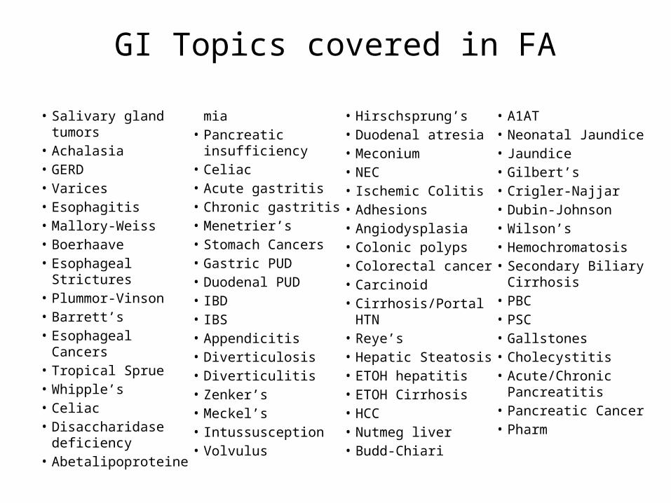
GI Topics covered in FA
• Salivary gland tumors• Achalasia• GERD• Varices• Esophagitis• Mallory-Weiss• Boerhaave• Esophageal Strictures• Plummor-Vinson• Barrett’s• Esophageal Cancers• Tropical Sprue• Whipple’s• Celiac• Disaccharidase
deficiency• Abetalipoproteinemi
a• Pancreatic
insufficiency• Celiac • Acute gastritis• Chronic gastritis• Menetrier’s• Stomach Cancers• Gastric PUD• Duodenal PUD• IBD• IBS• Appendicitis• Diverticulosis• Diverticulitis• Zenker’s• Meckel’s• Intussusception• Volvulus• Hirschsprung’s
• Duodenal atresia• Meconium• NEC• Ischemic Colitis• Adhesions• Angiodysplasia• Colonic polyps• Colorectal cancer• Carcinoid• Cirrhosis/Portal HTN• Reye’s• Hepatic Steatosis• ETOH hepatitis• ETOH Cirrhosis• HCC• Nutmeg liver• Budd-Chiari• A1AT
• Neonatal Jaundice• Jaundice• Gilbert’s• Crigler-Najjar• Dubin-Johnson• Wilson’s• Hemochromatosis• Secondary Biliary
Cirrhosis• PBC• PSC• Gallstones• Cholecystitis• Acute/Chronic
Pancreatitis• Pancreatic Cancer• Pharm

• Jaundice• Hepatitis Serologies• Inflammatory Bowel Disease• The C Word• Questions
Jaundice
• Due to a disruption in normal heme metabolism– RBC breakdown, transport, hepatic uptake,
excretion

Jaundice
Macrophages:RBC -> Heme -> Unconjugated bili
In the bloodstream:Unconj bili binds albumin (Indirect bilirubin)
To the Liver:Uptake, then UDP glucoronyl transferase conjugates with glucuronic acid = Direct Bilirubin (now water soluble)
From the hepatocytes to the gut in bile:Urobilinogen in the gut either is excreted in feces (80%, ‘stercobilin’) or reabsorbed (20%, most sent back to the liver, trace amount excreted in urine, ‘urobilin’)


Jaundice
• Problems with overproduction, absorption, conjugation, or excretion
• Often presented in review books as tables of either hepatocellular, obstructive, hemolytic, or mixed
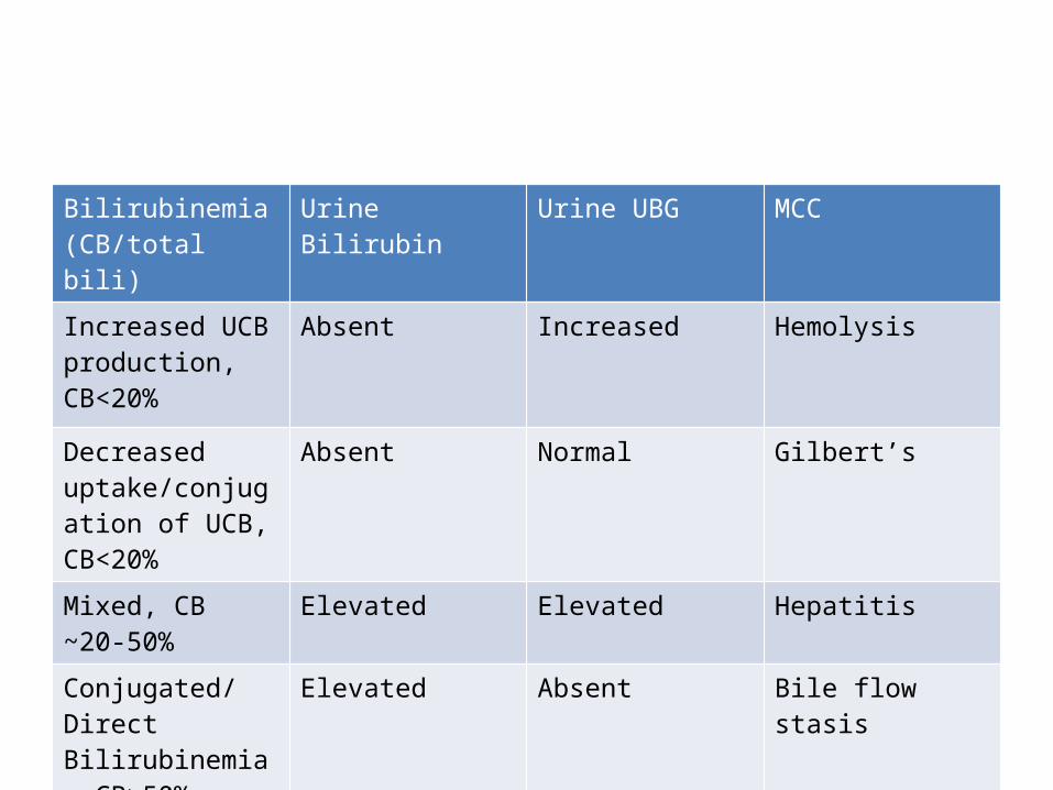
Bilirubinemia(CB/total bili)
Urine Bilirubin Urine UBG MCC
Increased UCB production, CB<20%
Absent Increased Hemolysis
Decreased uptake/conjugation of UCB, CB<20%
Absent Normal Gilbert’s
Mixed, CB ~20-50% Elevated Elevated Hepatitis
Conjugated/Direct Bilirubinemia. CB>50%
Elevated Absent Bile flow stasis

Jaundice

Jaundice
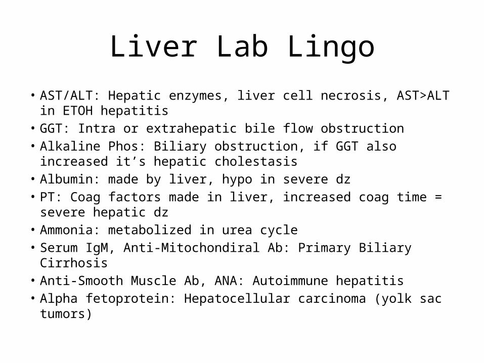
Liver Lab Lingo• AST/ALT: Hepatic enzymes, liver cell necrosis, AST>ALT in ETOH
hepatitis• GGT: Intra or extrahepatic bile flow obstruction• Alkaline Phos: Biliary obstruction, if GGT also increased it’s
hepatic cholestasis• Albumin: made by liver, hypo in severe dz• PT: Coag factors made in liver, increased coag time = severe
hepatic dz• Ammonia: metabolized in urea cycle• Serum IgM, Anti-Mitochondiral Ab: Primary Biliary Cirrhosis• Anti-Smooth Muscle Ab, ANA: Autoimmune hepatitis• Alpha fetoprotein: Hepatocellular carcinoma (yolk sac tumors)

UC Crohn’s
Extent Mucosal, submucosa Transmural
Location Rectum, continuous into colon
‘Mouth to anus’, rectal sparing, terminal ileum
Gross Friable residual mucosa, ulcers, hemorrhage, pseudopolyps
Skip lesions, strictures, fistulas, cobblestone pattern, ‘string sign’
Micro Ulcers, crypt abscess, CA/dysplasia
Noncaseating granulomas
Clinical Recurrent cramping LLQ, bloody diarrhea; PSC, HLA B27 positive arthritis, p-ANCA, pyoderma gangrenosum
RLQ colicky pain, diarrhea +/- bleeding, aphthous ulcers, iritis, ank spond, erythema nodosum
Complications adenoCA, toxic megacolon, iatrogenic
Fistulas, obstruction, cancer, iatrogenic
Treatment 5-ASA, steroids, nicotine patch, immunosupps, surgery
5-ASA, Steroids, immunosups, anti-TNF, surgery

GI Cancers
• Esophageal– ABCDEFGH (Adeno in US, squamous worldwide)– Alcohol/Achalasia, Barrett’s, Cigarettes, Diverticuli,
Esophageal web, Familial, GERD, Hot Dogs
• Stomach– Adeno, Signet ring cells, Virchow’s node,
Krukenberg met to ovaries, Linitis plastica
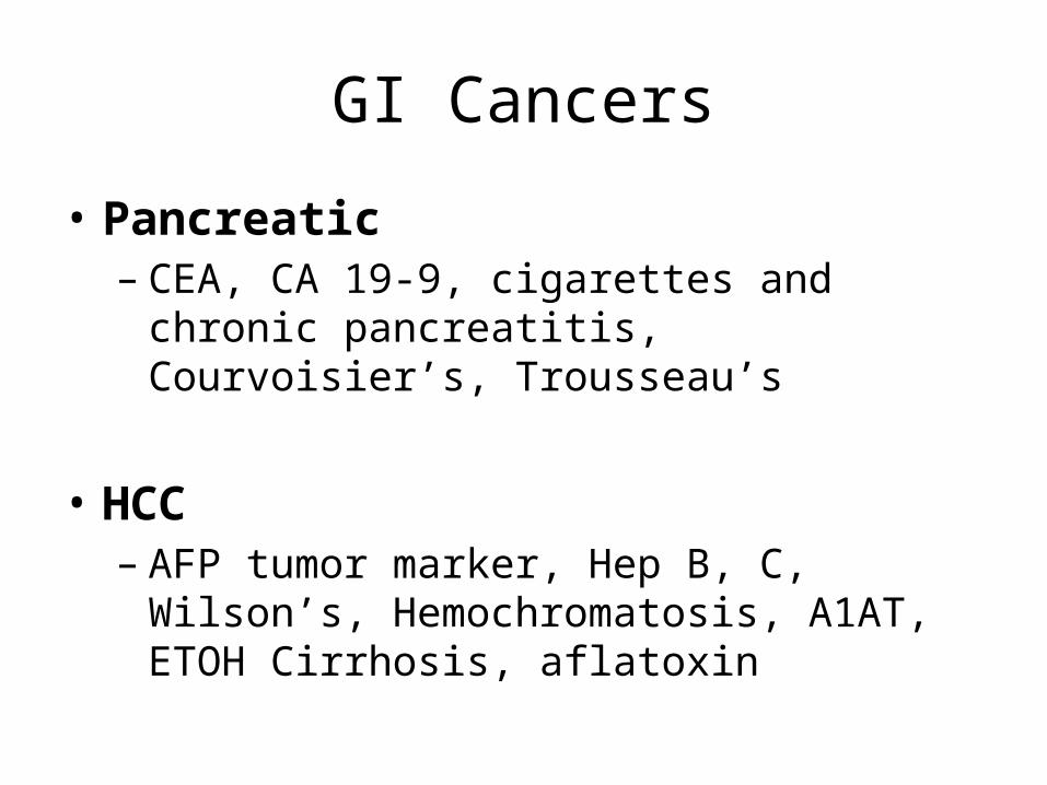
GI Cancers
• Pancreatic– CEA, CA 19-9, cigarettes and chronic pancreatitis,
Courvoisier’s, Trousseau’s
• HCC– AFP tumor marker, Hep B, C, Wilson’s,
Hemochromatosis, A1AT, ETOH Cirrhosis, aflatoxin

GI Cancers
• Colon– Pathogensis:
• Normal -> lose APC -> Kras mutation -> lose p53 -> CA
– 3rd MC Cancer, 3rd most deadly. – FAP (APC gene), Gardner’s, Turcot, HNPCC– IBD, Strep Bovis, tobacco, Peutz-Jeghers– Screen all >50 YO
• Carcinoid– Neuroendocrine tumor, produce 5-HT, can cause classic
syndrome of wheezing, murmurs, diarrhea, flushing
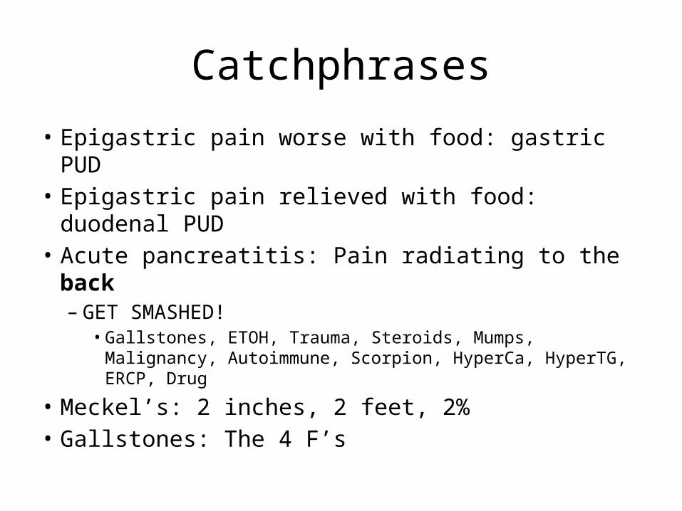
Catchphrases
• Epigastric pain worse with food: gastric PUD• Epigastric pain relieved with food: duodenal PUD• Acute pancreatitis: Pain radiating to the back– GET SMASHED!
• Gallstones, ETOH, Trauma, Steroids, Mumps, Malignancy, Autoimmune, Scorpion, HyperCa, HyperTG, ERCP, Drug
• Meckel’s: 2 inches, 2 feet, 2%• Gallstones: The 4 F’s


A 52-year-old man comes to the emergency department because he has had vomiting, nausea, and abdominal pain for the past 12 hours. He says he attempted suicide 3 days ago by "taking everything in the medicine cabinet." He was stuporous for approximately 12 hours after the overdose but felt better the following day. At this time, he has jaundice and pain in the right upper quadrant. Which of the following drugs is most likely to have caused the pain, vomiting, and jaundice?
(A) Acetaminophen(B) Aspirin(C) Cimetidine(D) Diphenhydramine(E) Triazolam

A 25-year-old man comes to the physician because of progressive weakness and an increasingly protuberant abdomen during the past 3 years. Physical examination shows splenomegaly. His hematocrit is 28%, and platelet count is 20,000/mm3. A biopsy specimen of bone marrow shows accumulation of lipidladen macrophages. Glucocerebroside has accumulated in the patient's reticuloendothelial cells. Inheritance of mutant alleles most likely caused impairment of which of the following enzyme activities in this patient?
(A) Ceramidase(B) α-Galactosidase(C) β-Glucosidase(D) Hexosaminidase(E) α-L-Iduronidase(F) Sphingomyelinase
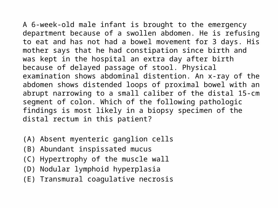
A 6-week-old male infant is brought to the emergency department because of a swollen abdomen. He is refusing to eat and has not had a bowel movement for 3 days. His mother says that he had constipation since birth and was kept in the hospital an extra day after birth because of delayed passage of stool. Physical examination shows abdominal distention. An x-ray of the abdomen shows distended loops of proximal bowel with an abrupt narrowing to a small caliber of the distal 15-cm segment of colon. Which of the following pathologic findings is most likely in a biopsy specimen of the distal rectum in this patient?
(A) Absent myenteric ganglion cells(B) Abundant inspissated mucus(C) Hypertrophy of the muscle wall(D) Nodular lymphoid hyperplasia(E) Transmural coagulative necrosis
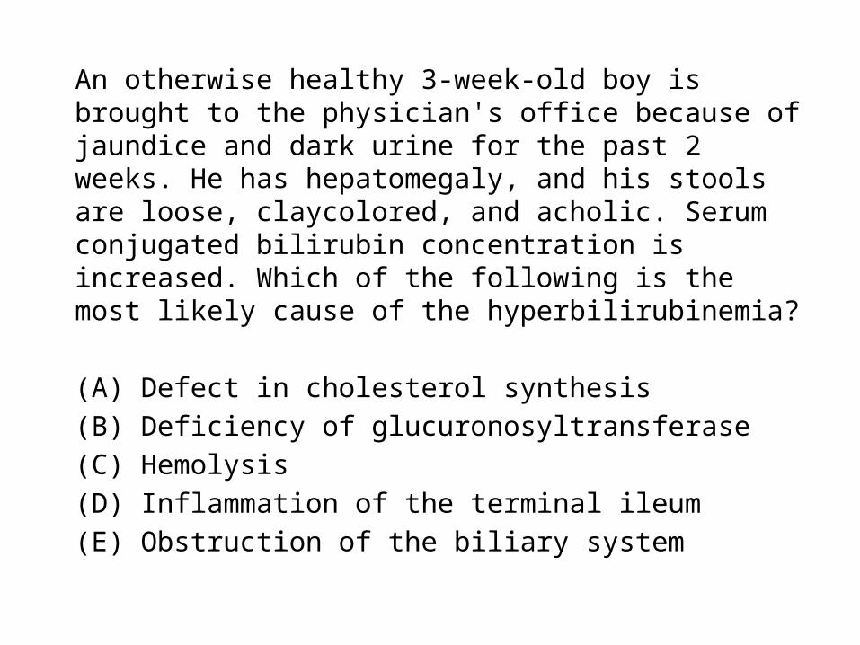
An otherwise healthy 3-week-old boy is brought to the physician's office because of jaundice and dark urine for the past 2 weeks. He has hepatomegaly, and his stools are loose, claycolored, and acholic. Serum conjugated bilirubin concentration is increased. Which of the following is the most likely cause of the hyperbilirubinemia?
(A) Defect in cholesterol synthesis(B) Deficiency of glucuronosyltransferase(C) Hemolysis(D) Inflammation of the terminal ileum(E) Obstruction of the biliary system
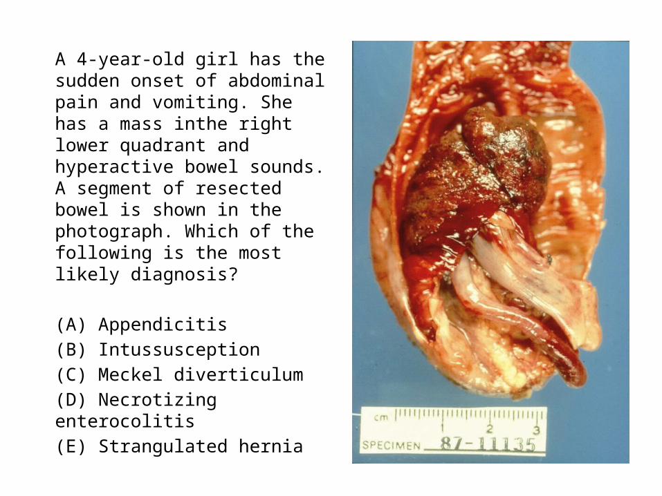
A 4-year-old girl has the sudden onset of abdominal pain and vomiting. She has a mass inthe right lower quadrant and hyperactive bowel sounds. A segment of resected bowel is shown in the photograph. Which of the following is the most likely diagnosis?
(A) Appendicitis(B) Intussusception(C) Meckel diverticulum(D) Necrotizing enterocolitis(E) Strangulated hernia

A 76-year-old man comes to the emergency department because of a 12-hour history of fever and left lower quadrant abdominal pain. He has not passed a stool for the past 36 hours. His temperature is 38.3°C (100.9°F). A tender mass is palpable in the left lower quadrant of the abdomen. Stool is negative for occult blood.
Laboratory studies show:Hemoglobin 13 g/dL Leukocyte count 17,000/mm3 (84% neutrophils)Platelet count 200,000/mm3 Serum amylase 115 U/LUrinalysis 0 to 1 WBC/hpf
An x-ray of the abdomen shows no abnormalities. The most likely diagnosis is anacute episode of which of the following disorders?
(A) Cystitis(B) Diverticulitis(C) Infectious colitis(D) Ischemic colitis(E) Pyelonephritis
A 5-year-old girl is brought to the emergencydepartment because of fever and severeabdominal pain. Acute appendicitis is diagnosed.In the examination room, she keeps her right hipflexed and resists active extension of the hip. Theinflamed structure associated with thesesymptoms is most likely in contact with which ofthe following structures?
(A) Abdominal wall and the external oblique muscle(B) Obturator internus muscle(C) Psoas major muscle(D) Quadratus lumborum muscle(E) Transversus abdominis muscle

A 55-year-old man who has alcoholic cirrhosis is brought to the emergency department because he has been vomiting blood for 2 hours. He has a 2-month history of abdominal distention, dilated veins over the anterior abdominal wall, and internal hemorrhoids. Which of the following veins is the most likely origin of the hematemesis?
(A) Inferior mesenteric veins(B) Left gastric vein(C) Periumbilical veins(D) Superior rectal vein(E) Superior vena cava

A 30-year-old woman has anxiety about episodes of abdominal pain that have alternated with diarrhea and constipation over the past year. She often has these episodes when she is stressed or tired. Physical examination and laboratory studies are within normal limits during these episodes. Which of the following is the most likely diagnosis?
(A) Gastroenteritis(B) Generalized anxiety disorder(C) Hypochondriasis(D) Irritable bowel syndrome(E) Major depressive disorder(F) Somatization disorder
Related Documents