Send Orders for Reprints to [email protected] Current Pharmaceutical Design, 2013, 19, 6773-6790 6773 Ghrelin as a Neuroprotective and Palliative Agent in Alzheimer's and Parkinson's Disease Vanessa V. dos Santos a , Ana Lúcia S. Rodrigues a,b , Thereza C. De Lima c , Susana R. de Barioglio d , Rita Raisman-Vozari e and Rui D. Prediger a,c, * a Programa de Pós-Graduação em Neurociências, Centro de Ciências Biológicas, Universidade Federal de Santa Catarina, UFSC, Florianópolis-SC, Brazil; b Departamento de Bioquímica, Centro de Ciências Biológicas, Universidade Federal de Santa Catarina, UFSC, Florianópolis, SC, Brazil; c Departamento de Farmacologia, Centro de Ciências Biológicas, Universidade Federal de Santa Catarina, UFSC, Florianópolis-SC, Brazil; d Departamento de Farmacología Facultad de Ciencias Químicas, Universidad Nacional de Córdoba and IFEC-CONICET, Córdoba, Argentina; e UMR 975 INSERM - Université Pierre et Marie Curie. Centre de Recherche de l'Institut du Cerveau et de la Moelle Epinière – CRICM Thérapeutique Expérimentale de la Neurodégénérescence. Hôpital de la Salpêtrière, Paris, France Abstract: Ghrelin is a gastric hormone that stimulates growth hormone (GH) secretion and food intake to regulate energy homeostasis and body weight by binding to its receptor, GH secretagogue receptor (GHSR1a), which is most highly expressed in the pituitary and hy- pothalamus. Nowadays there is considerable evidence showing that the GHSR1a is also expressed in numerous extra-hypothalamic neu- ronal populations and the physiological role of ghrelin is by far wider than considered before including learning and memory, anxiety, depression and neuroprotection. The present review attempts to provide a comprehensive picture of the role of ghrelin in the central nervous system and to highlight recent findings showing its potential as an innovative therapeutic agent in neurodegenerative diseases in- cluding Alzheimer’s disease and Parkinson’s disease. Keywords: Ghrelin, neuroprotection, Alzheimer’s disease, Parkinson’s disease, learning and memory, anxiety, depression. INTRODUCTION Alzheimer’s disease (AD) is the most common form of demen- tia, accounting for 50–60% of all cases [1]. The prevalence of AD is below 1% in individuals aged less than 65 years, but it increases to approximately 25% of the individuals aged 85 years or older in the Western world [2]. This neurodegenerative disease is associated with progressive and permanent decline in memory and overall cognitive abilities, reducing the post-diagnosis lifespan to nearly half the duration of a nondemented elderly person [3]. The first cognitive function affected is the episodic memory [4], but during the progression of the disease, attention, executive functions, se- mantic memory, spatial orientation and even language are deterio- rated [5]. The examination of post-mortem brains of AD patients indicated the main histopathological hallmarks of the disease: the formation of senile plaques and neurofibrillary tangles, which are mainly formed due to deposition of -amyloid (A) peptides and the hyperphosphorylated tau protein, respectively [6]. Parkinson’s disease (PD) is the second most common neurode- generative disorder that affects approximately 1% of the population older than 50 years [7], and it is characterized by a slow and pro- gressive degeneration of neuromelanin-containing dopaminergic neurons in the substantia nigra pars compacta (SNpc) with presence of eosinophillic, intracytoplasmic, proteinaceous inclusions termed as Lewy bodies and dystrophic Lewy neurites in surviving neurons [8]. At the time of diagnosis, patients typically display an array of motor impairments including bradykinesia, resting tremor, rigidity, and postural instability. Although most of the typical motor im- pairments are due to the loss of nigrostriatal dopaminergic neurons, PD affects multiple neuronal systems both centrally and peripher- ally, leading to a constellation of non-motor symptoms including olfactory deficits, affective disorders (e.g., depression and anxiety), *Address correspondence to this author at the Departamento de Farmacolo- gia, Universidade Federal de Santa Catarina, Campus Trindade, 88049-900, Florianópolis, SC, Brazil; Tel: 55 48 3721 9491; Fax: 55 48 3721 9813; E-mail: [email protected] memory impairments, as well as autonomic and digestive dysfunc- tion [9]. These non-motor features of PD do not meaningfully re- spond to dopaminergic medication and are a challenge to the clini- cal management of PD [9]. The limitations of the current pharmacological treatments of AD and PD have led to extensive investigation of novel drugs that may provide alternative or adjunctive treatment for the relief of symptoms with a reduced profile of side effects, as well as to the discovery of compounds to modify the course of these neurodegen- erative diseases. The definition of neuroprotection is complex and involves the potential for preventing cell death and restoring func- tion to damaged neurons, as well as increasing neuronal number. The development of drugs to slow or prevent the progression of AD and PD might logically evolve from an improved understanding of the etiology and pathogenesis of such diseases. There have certainly been major advances in these areas over the past few years and the prospect for the introduction of "neuroprotective" therapies is much improved. However, despite extensive efforts and research, to date, there is no proven therapy to prevent cell death or to restore af- fected neurons to a normal state in AD and PD. Preclinical studies in laboratory animals have provided several candidate neuroprotec- tive drugs, but clinical endpoints are readily confounded by any symptomatic effect of the study intervention and thus do not pro- vide an unequivocal measure of disease progression that can be used to determine if a drug has a neuroprotective effect. In this context, ghrelin emerges a gastric hormone that stimu- lates growth hormone (GH) secretion and food intake to regulate energy homeostasis and body weight by binding to its receptor, GH secretagogue receptor (GHS-R1a), which is most highly expressed in the pituitary and hypothalamus. Nowadays there is considerable evidence showing that the GHS-R1a is also expressed in numerous extra-hypothalamic neuronal populations and the physiological role of ghrelin is by far wider than considered before including learning and memory, anxiety, depression and neuroprotection. The present review attempts to provide a comprehensive picture of the role of ghrelin in the central nervous system (CNS) and to highlight recent 1873-4286/13 $58.00+.00 © 2013 Bentham Science Publishers

Welcome message from author
This document is posted to help you gain knowledge. Please leave a comment to let me know what you think about it! Share it to your friends and learn new things together.
Transcript
Send Orders for Reprints to [email protected]
Current Pharmaceutical Design, 2013, 19, 6773-6790 6773
Ghrelin as a Neuroprotective and Palliative Agent in Alzheimer's and Parkinson's Disease
Vanessa V. dos Santosa, Ana Lúcia S. Rodriguesa,b, Thereza C. De Limac, Susana R. de Bariogliod, Rita Raisman-Vozarie and Rui D. Predigera,c,*
aPrograma de Pós-Graduação em Neurociências, Centro de Ciências Biológicas, Universidade Federal de Santa Catarina, UFSC, Florianópolis-SC, Brazil; bDepartamento de Bioquímica, Centro de Ciências Biológicas, Universidade Federal de Santa Catarina, UFSC, Florianópolis, SC, Brazil; cDepartamento de Farmacologia, Centro de Ciências Biológicas, Universidade Federal de Santa Catarina, UFSC, Florianópolis-SC, Brazil; dDepartamento de Farmacología Facultad de Ciencias Químicas, Universidad Nacional de Córdoba and IFEC-CONICET, Córdoba, Argentina; eUMR 975 INSERM - Université Pierre et Marie Curie. Centre de Recherche de l'Institut du Cerveau et de la Moelle Epinière – CRICM Thérapeutique Expérimentale de la Neurodégénérescence. Hôpital de la Salpêtrière, Paris, France
Abstract: Ghrelin is a gastric hormone that stimulates growth hormone (GH) secretion and food intake to regulate energy homeostasis and body weight by binding to its receptor, GH secretagogue receptor (GHSR1a), which is most highly expressed in the pituitary and hy-pothalamus. Nowadays there is considerable evidence showing that the GHSR1a is also expressed in numerous extra-hypothalamic neu-ronal populations and the physiological role of ghrelin is by far wider than considered before including learning and memory, anxiety, depression and neuroprotection. The present review attempts to provide a comprehensive picture of the role of ghrelin in the central nervous system and to highlight recent findings showing its potential as an innovative therapeutic agent in neurodegenerative diseases in-cluding Alzheimer’s disease and Parkinson’s disease.
Keywords: Ghrelin, neuroprotection, Alzheimer’s disease, Parkinson’s disease, learning and memory, anxiety, depression.
INTRODUCTION Alzheimer’s disease (AD) is the most common form of demen-tia, accounting for 50–60% of all cases [1]. The prevalence of AD is below 1% in individuals aged less than 65 years, but it increases to approximately 25% of the individuals aged 85 years or older in the Western world [2]. This neurodegenerative disease is associated with progressive and permanent decline in memory and overall cognitive abilities, reducing the post-diagnosis lifespan to nearly half the duration of a nondemented elderly person [3]. The first cognitive function affected is the episodic memory [4], but during the progression of the disease, attention, executive functions, se-mantic memory, spatial orientation and even language are deterio-rated [5]. The examination of post-mortem brains of AD patients indicated the main histopathological hallmarks of the disease: the formation of senile plaques and neurofibrillary tangles, which are mainly formed due to deposition of �-amyloid (A�) peptides and the hyperphosphorylated tau protein, respectively [6]. Parkinson’s disease (PD) is the second most common neurode-generative disorder that affects approximately 1% of the population older than 50 years [7], and it is characterized by a slow and pro-gressive degeneration of neuromelanin-containing dopaminergic neurons in the substantia nigra pars compacta (SNpc) with presence of eosinophillic, intracytoplasmic, proteinaceous inclusions termed as Lewy bodies and dystrophic Lewy neurites in surviving neurons [8]. At the time of diagnosis, patients typically display an array of motor impairments including bradykinesia, resting tremor, rigidity, and postural instability. Although most of the typical motor im-pairments are due to the loss of nigrostriatal dopaminergic neurons, PD affects multiple neuronal systems both centrally and peripher-ally, leading to a constellation of non-motor symptoms including olfactory deficits, affective disorders (e.g., depression and anxiety),
*Address correspondence to this author at the Departamento de Farmacolo-gia, Universidade Federal de Santa Catarina, Campus Trindade, 88049-900, Florianópolis, SC, Brazil; Tel: 55 48 3721 9491; Fax: 55 48 3721 9813; E-mail: [email protected]
memory impairments, as well as autonomic and digestive dysfunc-tion [9]. These non-motor features of PD do not meaningfully re-spond to dopaminergic medication and are a challenge to the clini-cal management of PD [9]. The limitations of the current pharmacological treatments of AD and PD have led to extensive investigation of novel drugs that may provide alternative or adjunctive treatment for the relief of symptoms with a reduced profile of side effects, as well as to the discovery of compounds to modify the course of these neurodegen-erative diseases. The definition of neuroprotection is complex and involves the potential for preventing cell death and restoring func-tion to damaged neurons, as well as increasing neuronal number. The development of drugs to slow or prevent the progression of AD and PD might logically evolve from an improved understanding of the etiology and pathogenesis of such diseases. There have certainly been major advances in these areas over the past few years and the prospect for the introduction of "neuroprotective" therapies is much improved. However, despite extensive efforts and research, to date, there is no proven therapy to prevent cell death or to restore af-fected neurons to a normal state in AD and PD. Preclinical studies in laboratory animals have provided several candidate neuroprotec-tive drugs, but clinical endpoints are readily confounded by any symptomatic effect of the study intervention and thus do not pro-vide an unequivocal measure of disease progression that can be used to determine if a drug has a neuroprotective effect. In this context, ghrelin emerges a gastric hormone that stimu-lates growth hormone (GH) secretion and food intake to regulate energy homeostasis and body weight by binding to its receptor, GH secretagogue receptor (GHS-R1a), which is most highly expressed in the pituitary and hypothalamus. Nowadays there is considerable evidence showing that the GHS-R1a is also expressed in numerous extra-hypothalamic neuronal populations and the physiological role of ghrelin is by far wider than considered before including learning and memory, anxiety, depression and neuroprotection. The present review attempts to provide a comprehensive picture of the role of ghrelin in the central nervous system (CNS) and to highlight recent
1873-4286/13 $58.00+.00 © 2013 Bentham Science Publishers
6774 Current Pharmaceutical Design, 2013, Vol. 19, No. 38 Santos et al.
findings showing its potential as a new palliative and neuroprotec-tive agent in neurodegenerative diseases.
GHRELIN: HISTORICAL BACKGROUND, RECEPTOR AND FUNCTIONS The ghrelin history began in 1977, when Frank Momany and Cyril Bowers from Tulane University (New Orleans, USA) reported a series of synthetic peptide analogs of Leu- and Met-enkephalins that specifically released growth hormone from pituitary without any opioid activity [10-12]. Surprisingly, it was found that these molecules stimulate and amplify pulsatile GH secretion, independ-ently from GH releasing hormone (GHRH) [13]. Then, some other peptidyl derivatives with similar properties were characterized. The family of these molecules, both peptidyl and non-peptidyl com-pounds, has been named growth hormone secretagogues (GHSs). GHSs are synthetic compounds that are potent stimulators of GH release, working through a G-protein-coupled receptor (GPCR), the GHS-receptor (GHS-R) [14]. Because GHSs are a group of artificial compounds and do not exist naturally, it was postulated that there must exist an endogenous ligand that binds to GHS-R and carries out similar functions to GHSs in situ. This makes the dis-covery of ghrelin an example of reverse pharmacology, starting with the synthesis of analogs and ending with the discovery of an endogenous ligand and its receptor. A cultured cell line expressing the GHS-R was established and used to identify tissue extracts that could stimulate the GHS-R, as monitored by increases in intracellular Ca2+ levels. After screening
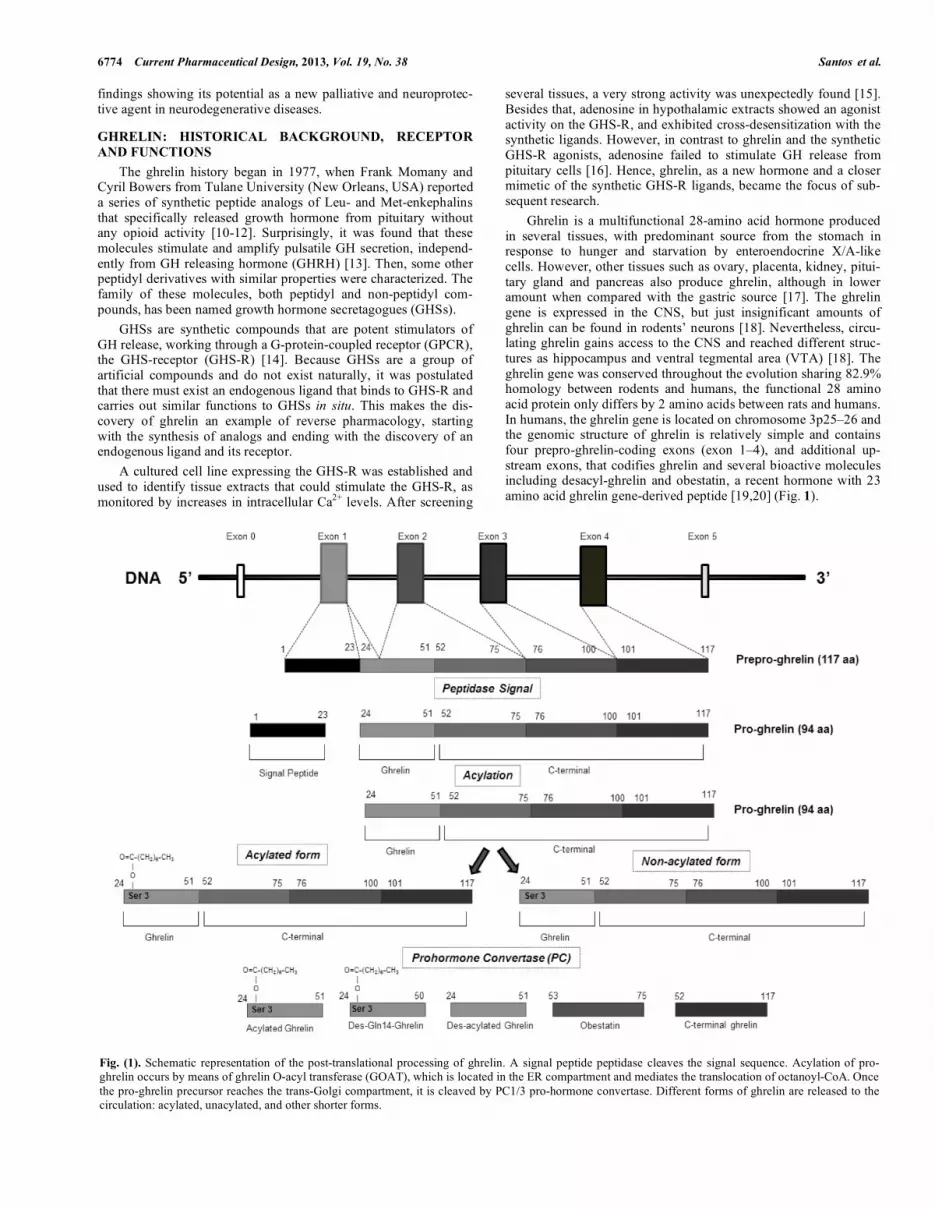
several tissues, a very strong activity was unexpectedly found [15]. Besides that, adenosine in hypothalamic extracts showed an agonist activity on the GHS-R, and exhibited cross-desensitization with the synthetic ligands. However, in contrast to ghrelin and the synthetic GHS-R agonists, adenosine failed to stimulate GH release from pituitary cells [16]. Hence, ghrelin, as a new hormone and a closer mimetic of the synthetic GHS-R ligands, became the focus of sub-sequent research. Ghrelin is a multifunctional 28-amino acid hormone produced in several tissues, with predominant source from the stomach in response to hunger and starvation by enteroendocrine X/A-like cells. However, other tissues such as ovary, placenta, kidney, pitui-tary gland and pancreas also produce ghrelin, although in lower amount when compared with the gastric source [17]. The ghrelin gene is expressed in the CNS, but just insignificant amounts of ghrelin can be found in rodents’ neurons [18]. Nevertheless, circu-lating ghrelin gains access to the CNS and reached different struc-tures as hippocampus and ventral tegmental area (VTA) [18]. The ghrelin gene was conserved throughout the evolution sharing 82.9% homology between rodents and humans, the functional 28 amino acid protein only differs by 2 amino acids between rats and humans. In humans, the ghrelin gene is located on chromosome 3p25–26 and the genomic structure of ghrelin is relatively simple and contains four prepro-ghrelin-coding exons (exon 1–4), and additional up-stream exons, that codifies ghrelin and several bioactive molecules including desacyl-ghrelin and obestatin, a recent hormone with 23 amino acid ghrelin gene-derived peptide [19,20] (Fig. 1).
Fig. (1). Schematic representation of the post-translational processing of ghrelin. A signal peptide peptidase cleaves the signal sequence. Acylation of pro-ghrelin occurs by means of ghrelin O-acyl transferase (GOAT), which is located in the ER compartment and mediates the translocation of octanoyl-CoA. Once the pro-ghrelin precursor reaches the trans-Golgi compartment, it is cleaved by PC1/3 pro-hormone convertase. Different forms of ghrelin are released to the circulation: acylated, unacylated, and other shorter forms.
Role of Ghrelin in Neurodegenerative Diseases Current Pharmaceutical Design, 2013, Vol. 19, No. 38 6775
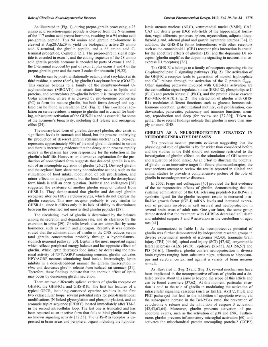
As illustrated in (Fig. 1), during prepro-ghrelin processing, a 23 amino acid secretion-signal peptide is cleaved from the N-terminus of the 117 amino acid prepro-hormone, resulting in a 94 amino acid pro-ghrelin peptide. This 94 amino acid ghrelin pro-hormone is cleaved at Arg28/Ala29 to yield the biologically active 28 amino acid N-terminal, the ghrelin peptide, and a 66 amino acid C-terminal propeptide, C-ghrelin [21]. The prepro-ghrelin signal pep-tide is encoded in exon 1, and the coding sequence of the 28 amino acid ghrelin peptide hormone is encoded by parts of exons 1 and 2, the C-terminal encoded by part of exon 2, plus exons 3 and 4 of the prepro-ghrelin gene and the exon 3 codes for obestatin [19,22]. Ghrelin can be post-translationally octanoylated (acylated) at its third residue, a serine (Ser3), by ghrelin O-acyltransferase (GOAT). This enzyme belongs to a family of the membrane-bound O-acyltransferases (MBOATs) that attach fatty acids to lipids and proteins, and octanoylates pro-ghrelin before it is transported to the Golgi apparatus, where it is cleaved by pro-hormone convertase (PC) to form the mature ghrelin, but both forms desacyl and acy-lated can be found in circulation [23] (Fig. 1). This n-octanoyl acy-lation on serine residues is unique to ghrelin and is crucial for bind-ing, subsequent activation of the GHS-R1a and is essential for some of the hormone’s bioactivity, including GH release and orexigenic effect [24]. The nonacylated form of ghrelin, des-acyl ghrelin, also exists at significant levels in stomach and blood, but the process underlying the production of des-acyl ghrelin remains unclear [25]. Des-acyl represents approximately 90% of the total ghrelin detected in serum and there is increasing evidence that the deacylation process rapidly occurs in the plasma has been responsible for the reduction in the ghrelin’s half-life. However, an alternative explanation for the pro-duction of nonacylated form suggests that des-acyl ghrelin is a re-sult of an incomplete acylation of ghrelin [26,27]. Des-acyl ghrelin and the acylated form share many nonendocrine actions, such as the stimulation of food intake, modulation of cell proliferation, and minor effects on adipogenesis, but the local where the desacylated form binds is still an open question. Baldanzi and colleagues [28] suggested the existence of another ghrelin receptor distinct from GHSR-1a. They demonstrated that ghrelin and des-acyl ghrelin recognize sites on H9c2 cardiomyocytes, which do not express the ghrelin receptor. This new receptor probably is very similar to GHSR-1a, since it differs only in its lack of ability to discriminate between the esterified and unesterified ghrelin peptides [28]. The circulating level of ghrelin is determined by the balance among its secretion and degradation rate, and its clearance by the excretion in urine [29]. Ghrelin levels also are controlled by some hormones, such as insulin and glucagon. Recently it was demon-strated that the administration of insulin in the CNS reduces serum total ghrelin concentration probably through a hypothalamus–stomach neuronal pathway [30]. Leptin is the most important signal which reflects peripheral energy balance and has opposite effects of ghrelin. While leptin decreases food intake by decreasing the neu-ronal activity of NPY/AGRP-containing neurons, ghrelin activates NPY/AGRP neurons stimulating food intake. Interestingly, leptin inhibits in a dose-dependent manner the ghrelin transcription in vitro and decreases ghrelin release from isolated rat stomach [31]. Therefore, these findings indicate that the anorexic effect of leptin may occur by decreasing ghrelin secretion. There are two differently spliced variants of ghrelin receptor or GHS-R; the GHS-R1a and GHS-R1b. The first has features of a typical GPCR, including conserved cysteine residues in the first two extracellular loops, several potential sites for post-translational modifications (N-linked glycosylation and phosphorylation), and an aromatic triplet sequence (E/DRY) located immediately after TM-3 in the second intracellular loop. The last one is truncated and has been reported as an inactive form that fails to bind ghrelin and has no known signaling activity [32,33]. The GHS-R1a receptor is ex-pressed in brain areas and peripheral organs including the hypotha-
lamic arcuate nucleus (ARC), ventromedial nuclei (VMN), CA2, CA3 and detate gyrus (DG) sub-fields of the hippocampal forma-tion, vagal afferents, pancreas, spleen, myocardium, adipose tissue, thyroid gland, adrenal gland and gastric myenteric neurons [34]. In addition, the GHS-R1a forms heterodimers with other receptors such as the cannabinoid 1 (CB1) receptor (this interaction is crucial for the appetitive effects of ghrelin) [35] and the dopamine D1 re-ceptor (ghrelin amplifies the dopamine signaling in neurons that co-express D1 receptors) [36]. The GHS-R1a belongs to a family of receptors operating via the Gq-phospholipase C signaling pathways (Fig. 2). The activation of the GHS-R1a receptor leads to generation of inositol triphosphate and Ca2+ release through the activation of the G protein G�q/11.Other signaling pathways involved with GHS-R1a activation are the extracellular signal-regulated kinase (ERK1/2), phospholipase C (PLC) and protein kinase C (PKC), and the protein kinase cascade Raf–MEK–MAPK (Fig. 2). The interaction of ghrelin with GHS-R1a modulates different functions such as glucose homeostasis, hormone secretion, gastrointestinal motility, cell proliferation, car-diovascular, pancreatic, pulmonary and immune functions, mem-ory, reproduction and sleep (for review see [37-39]). Taken to-gether, these recent findings indicate that ghrelin is more than sim-ply a natural GHS.
GHRELIN AS A NEUROPROTECTIVE STRATEGY IN NEURODEGENERATIVE DISEASES The previous section presents evidence suggesting that the physiological role of ghrelin is by far wider than considered before and the studies in the field should not continue restricted to the investigation of ghrelin effects on the stimulation of GH secretion and regulation of food intake. As an effort to illustrate the potential of ghrelin as an innovative target for future pharmacotherapies, the next sections attempt to review the results reported in clinical and animal studies to provide a comprehensive picture of the role of ghrelin in neurodegenerative diseases. In 2002, Frago and colleagues [40] provided the first evidence of the neuroprotective effects of ghrelin, demonstrating that the systemic administration of the GH releasing peptide-6 (GHRP-6), a synthetic ligand for the ghrelin receptor, results in increased insu-lin-like growth factor (IGF-I) mRNA levels and increased expres-sion of proteins involved in cell survival and neuroprotection in several brain areas of adult rats. One year later, the same group demonstrated that the treatment with GHRP-6 decreased cell death and inhibited caspase 3 and 9 activation in the cerebellum of aged rats [41]. As summarized in Table 1, the neuroprotective potential of ghrelin was further demonstrated by independent research groups in diverse experimental models of ischemia [42,43], traumatic brain injury (TBI) [44-46], spinal cord injury (SCI) [47,48], amyotrophic lateral sclerosis (ALS) [49,50], epilepsy [51-55], AD [56,57] and PD [58-61]. Therefore, ghrelin confers neuroprotection in diverse brain regions ranging from substantia nigra, striatum to hippocam-pus and cerebral cortex, and against a variety of brain noxious stimuli. As illustrated in (Fig. 2) and (Fig. 3), several mechanisms have been implicated in the neuroprotective effects of ghrelin and a de-tailed review about this issue is beyond the scope of this article and can be found elsewhere [37,62]. At this moment, particular atten-tion is paid to the role of ghrelin in modulating the activation of intracellular signaling cascades (such as Erk1/2, Akt1/2, PI3K and PKC pathways) that lead to the inhibition of apoptotic events, via the subsequent increase in the Bcl-2:Bax ratio, the prevention of cytochrome c release and the inhibition of caspase 3 activation [42,43,63,64]. Moreover, ghrelin prevents activation of pro-apoptotic events, such as the activation of p38 and JNK. Further-more, ghrelin prevents inflammatory microglial activation [60] and activates the mitochondrial protein uncoupling protein-2 (UCP2)
6776 Current Pharmaceutical Design, 2013, Vol. 19, No. 38 Santos et al.
[59]. This protein enhances neuroprotection by decreasing the gen-eration of reactive oxygen species (ROS) and promoting mitochon-drial biogenesis [59,63] (Fig. 2). Therefore, the common neuropro-tective or neuromodulatory role of ghrelin in the brain could in-volve UCP2-dependent mitochondrial adaptation. Finally, these neuroprotective effects of ghrelin appear to be mediated through activation of GHSR-1a, as they were abolished by the pharmacol-ogical blockage or genetic deletion of this receptor. These recent findings demonstrating that ghrelin is involved in neuroprotection, together with the wide distribution of ghrelin re-ceptors in many brain areas, reinforce the idea that changes in this system could be involved in the development and/or progression of AD and PD.
Role of ghrelin in Alzheimer’s disease AD is the most prevalent age-related neurodegenerative disease that leads to cognitive impairments and dementia. The neuropa-thological hallmarks of AD are diffuse and neuritic plaques, which are predominantly composed of amyloid-� (A�) peptides, and neu-rofibrillary tangles composed of filamentous aggregates of hyper-phosphorylated tau protein [65]. The classical A� cascade hypothe-sis in AD pathogenesis postulates that the deposition of A� peptides and the activation of glial cells surrounding senile plaques in brain areas involved in cognitive functions trigger marked neuronal al-terations such as synaptic dysfunction, synaptic loss and neuronal death finally leading to cognitive impairments [65,66].
The first evidence showing a direct effect of ghrelin on AD-like alterations was reported in a mouse model widely used to examine the pathophysiology of early defects seen in AD. The senescence-accelerated mouse prone8 or SAMP8 mice develop early learning and memory impairments related to abnormalities in septo-hippocampal function, which are due to overproduction of �-amyloid peptides. Diano and colleagues [56] demonstrated that ghrelin improved retention of T-maze footshock avoidance in 12 and 14 month-old SAMP8 mice. Of interest, ghrelin was found to promote both long-term potentiation (LTP) generation in hippo-campal slices and the formation of spine synapses in the stratum radiatum of the hippocampal CA1 subregion, which are considered to be basic mechanisms involved in spatial learning and memory [56] (Table 2). More recently, Moon et al. [57] investigated the effects of ghre-lin on memory impairments and neuropathological alterations in-duced by intra-hippocampal injection of A�1-42 peptide in mice. The authors reported that repeated systemic injection of ghrelin rescued A�1-42-induced memory deficits evaluated in two behavioral para-digms (Y-maze and passive avoidance tasks). Moreover, ghrelin attenuated hippocampal microgliosis and neuronal loss induced by A�1-42 administration [57] (Table 2). Corroborating these findings, unpublished results from our laboratory have indicated that the acute intracerebroventricular (i.c.v.) injection of ghrelin (3 nmol), 15 min before the infusion of A�1-40 (400 pmol, i.c.v.), prevented the A�1-40-induced spatial
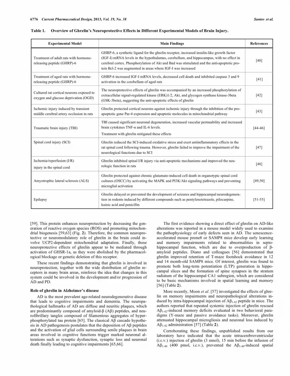
Table 1. Overview of Ghrelin’s Neuroprotective Effects in Different Experimental Models of Brain Injury.
Experimental Model Main Findings References
Treatment of adult rats with hormone-releasing peptide (GHRP)-6
GHRP-6, a synthetic ligand for the ghrelin receptor, increased insulin-like growth factor (IGF-I) mRNA levels in the hypothalamus, cerebellum, and hippocampus, with no effect in cerebral cortex. Phosphorylation of Akt and Bad was stimulated and the anti-apoptotic pro-tein Bcl-2 was augmented in areas where IGF-I was increased
[40]
Treatment of aged rats with hormone-releasing peptide (GHRP)-6
GHRP-6 increased IGF-I mRNA levels, decreased cell death and inhibited caspase 3 and 9 activation in the cerebellum of aged rats
[41]
Cultured rat cortical neurons exposed to oxygen and glucose deprivation (OGD)
The neuroprotective effects of ghrelin was accompanied by an increased phosphorylation of extracellular signal-regulated kinase (ERK)1/2, Akt, and glycogen synthase kinase-3beta (GSK-3beta), suggesting the anti-apoptotic effects of ghrelin
[42]
Ischemic injury induced by transient middle cerebral artery occlusion in rats
Ghrelin protected cortical neurons against ischemic injury through the inhibition of the pro-apoptotic gene Par-4 expression and apoptotic molecules in mitochondrial pathway
[43]
Traumatic brain injury (TBI) TBI caused significant neuronal degeneration, increased vascular permeability and increased brain cytokines TNF-� and IL-6 levels.
Treatment with ghrelin mitigated these effects [44-46]
Spinal cord injury (SCI) Ghrelin reduced the SCI-induced oxidative stress and exert antiinflammatory effects in the rat spinal cord following trauma. However, ghrelin failed to improve the impairment of the neurological functions due to SCI
[47]
Ischemia/reperfusion (I/R)
injury in the spinal cord
Ghrelin inhibited spinal I/R injury via anti-apoptotic mechanisms and improved the neu-rologic function in rats [48]
Amyotrophic lateral sclerosis (ALS) Ghrelin protected against chronic glutamate-induced cell death in organotypic spinal cord cultures (OSCC) by activating the MAPK and PI3K/Akt signaling pathways and preventing microglial activation
[49,50]
Epilepsy Ghrelin delayed or prevented the development of seizures and hippocampal neurodegenera-tion in rodents induced by different compounds such as pentylenetetrazole, pilocarpine, kainic acid and penicillin
[51-55]
Role of Ghrelin in Neurodegenerative Diseases Current Pharmaceutical Design, 2013, Vol. 19, No. 38 6777
memory impairments and depressive-like behaviors in adult Swiss mice evaluated in the object location and forced swimming task, respectively. Moreover, ghrelin mitigated a series of neurochemical changes induced by i.c.v. infusion of A�1-40, including the increase of oxidative stress biomarkers and acetylcholinesterase (AChE) activity and the decrease of glutamate uptake in the hippocampus and frontal cortex of mice (Table 2). Finally, ghrelin (1 nM) was found to prevent the impairments on LTP generation induced by A�1-40 (200 nM) in the CA1 subregion of hippocampal slices of mice (Santos et al., unpublished data) (Table 2). Altogether, these results suggest that ghrelin may counteract neurotoxic effects of A� peptides by reducing excitotoxicity, neu-roinflammation, oxidative stress and activation of apoptotic cell death mechanims. Moreover, the ghrelin’s effects on AChE activity and LTP generation may represent potential mechanisms responsi-ble for its cognitive enhancing properties (Fig. 3). A better under-standing of how the multiple actions of ghrelin influence survival of neurons might further consolidate ghrelin as a potential neuropro-tective agent for the treatment of AD. At this moment, few clinical studies have attempted to compre-hend the potential implication of the ghrelin system in human AD (Table 2). In 2002, it was reported that mean plasma ghrelin con-centrations in older normal weight subjects were significantly lower than those observed in young normal weight subjects, providing the first evidence for an age-related decline of peripheral ghrelin con-
centrations [67]. Nevertheless, Proto et al. [68] reported that ghrelin levels do not vary in the cerebrospinal fluid of AD patients when compared with age-matched controls. In a recent study, Castaño’s group (University of Córdoba, Spain) analyzed the mRNA expres-sion of the ghrelin system in three different regions of the temporal gyrus (inferior, medial and superior) of control and AD human brains, since it is one of the most affected memory-related regions in AD. This study showed, for the first time, that AD patients have a reduction in local brain ghrelin production, as compared with age-matched controls [69]. In addition, Shibata et al. [70] investigated whether single nu-cleotide polymorphisms (SNPs) of the ghrelin gene are associated with AD in a Japanese population. A total of 182 AD patients and 143 age-matched controls were included in this study and the SNPs were genotyped using TaqMan technology and were analyzed using a case-control study design. The authors observed that one SNP, rs4684677 (Leu90Gln), showed a marginal association with age of AD onset, but no additional association between other SNPs of the ghrelin gene and AD were detected [70] (Table 2). Moreover, Theodoropoulou et al. [71] investigated recently the potential rela-tionship between serum ghrelin levels and weight loss in patients with AD. The authors reported that the area-under-the-curve (AUC) for serum ghrelin levels after 75 g of glucose load is lower in male patients with AD compared to control males, while no difference was observed between females AD and controls. Therefore, the
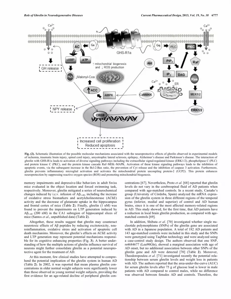
Fig. (2). Schematic illustration of the possible molecular mechanisms associated with the neuroprotective effects of ghrelin observed in experimental models of ischemia, traumatic brain injury, spinal cord injury, amyotrophic lateral sclerosis, epilepsy, Alzheimer’s disease and Parkinson’s disease. The interaction of ghrelin with GHS-R1a leads to activation of diverse signaling pathways including the extracellular signal-regulated kinase (ERK1/2), phospholipase C (PLC) and protein kinase C (PKC), and the protein kinase cascade Raf–MEK–MAPK. Activation of these kinase signaling pathways leads to the inhibition of apoptotic events, via the subsequent increase in the Bcl-2:Bax ratio, the prevention of Cyt release and the inhibition of caspase 3 activation. Furthermore, ghrelin prevents inflammatory microglial activation and activates the mitochondrial protein uncoupling protein-2 (UCP2). This protein enhances neuroprotection by suppressing reactive oxygen species (ROS) and promoting mitochondrial biogenesis.

6778 Current Pharmaceutical Design, 2013, Vol. 19, No. 38 Santos et al.
disruption of the normal compensatory modulation of ghrelin secre-tion might contribute to the metabolic changes (e.g., lower lean mass content) observed in male patients with AD [71] (Table 2).However, it must be conceded that further multifactorial studies are needed to clarify the relationship between ghrelin and AD.
Role of Ghrelin in Parkinson’s Disease Classically, PD is considered to be a motor system disease and its diagnosis is based on the presence of a set of cardinal motor signs (e.g. rigidity, bradykinesia, rest tremor and postural reflex disturbance). These symptoms of PD mainly result from the pro-gressive degeneration of dopamine neurons of the SNpc, which causes a consequent reduction of dopamine levels in the striatum [8]. Dopamine-replacement therapy has dominated the treatment of PD since the early 1960s and although the currently approved anti-parkinsonian agents offer effective relief of the motor deficits, es-pecially in the early-moderate stages of the disease, they have not be found to alleviate the underlying dopaminergic neuron degenera-tion and drug efficacy is gradually lost [72]. Moreover, another major limitation of chronic dopaminergic therapy is the numerous adverse effects such as the development of abnormal involuntary
movements (namely dyskinesia), psychosis and behavioral distur-bance (e.g., compulsive gambling, hypersexuality) [73]. Dopamine replacement therapy is based on the importance of nigral dopaminergic cell loss, the ensuing striatal dopamine deple-tion, and onset of motor symptoms. However, the neurodegenera-tive processes that lead to sporadic PD begin many years before the appearance of the characteristic motor symptoms and additional neuronal fields and neurotransmitter systems are also involved in PD, including the anterior olfactory structures, dorsal motor nucleus of vagus, caudal raphe nuclei, locus coeruleus, the autonomic nerv-ous system, hippocampus and the cerebral cortex [74]. Accordingly, cholinergic, adrenergic and serotoninergic neurons are also lost which seems to be responsible for the non-motor symptoms of PD encompassing olfactory and memory impairments, sleep abnormali-ties and depression, as well as gastrointestinal disturbance, which precede the classical motor symptoms [9]. Non-motor features of PD invariably do not respond to dopaminergic medication and probably form the major current challenge faced in the clinical management of PD [9]. Therefore, the limitations of the current pharmacological treat-ment of PD have led to extensive investigation of novel non-
Table 2. Overview of the Role of Ghrelin in Experimental Models and Human Alzheimer’s Disease (AD).
Experimental Model Main Findings References
Senescence-accelerated mouse prone8 (SAMP8 mice)
Ghrelin improved retention of T-maze footshock avoidance in 12 and 14 month-old SAMP8 mice. Moreover, ghrelin promoted dendritic spine synapse formation and generation of long-term potentiation (LTP) in the hippocampus of mice. Disruption of the gene that encodes ghrelin resulted in decreased numbers of spine synapses in the CA1 region and impaired performance of mice in the T-maze foot shock avoidance task. Ghrelin administration reversed these alterations
[56]
Intra-hippocampal injection of A�1-42
peptide in ICR mice
Intraperitoneal injection of ghrelin (80 �g/kg) improved A�1-42 (10 �M, 3 �l)-induced memory deficits evaluated in the Y-maze and passive avoidance tasks. Moreover, ghrelin attenuated hippocampal microgliosis and neuronal loss induced by A�1-42 ad-ministration
[57]
Intracerebroventricular injection of A�1-40 peptide in Swiss mice
Ghrelin (3 nmol, i.c.v.) prevented the A�1-40 (400 pmol, i.c.v.)-induced spatial memory impairments and depressive-like behaviors evaluated in the object location and forced swimming tasks. Moreover, ghrelin mitigated the increase of oxidative stress biomark-ers and acetylcholinesterase (AChE) activity and the decrease of glutamate uptake in the hippocampus and frontal cortex of mice
Santos et al., unpub-lished data
Hippocampal slices of Swiss adult mice
Ghrelin (1 nM) prevented the impairments on LTP generation induced by A�1-40 (200 nM) in the CA1 subfield of hippocampal slices of adult mice
Santos et al., unpub-lished data
12 young and 7 old normal weight subjects
Mean plasma ghrelin concentrations in older normal weight subjects were significantly lower than those present in young normal weight subjects
[67]
14 AD patients Ghrelin levels in the cerebrospinal fluid of AD patients were similar to those of age-matched controls
[68]
Analysis of temporal lobe of 6 patients with AD and 6 non-demented controls obtained from the Netherlands Brain Bank
mRNA levels for ghrelin, ghrelin-O-acyltransferase (enzyme responsible for ghrelin acylation), and its receptor GHS-R1a were reduced, while expression of GHS-R1b increased, in the temporal lobe of AD patients
[69]
182 AD patients and 143 age-matched controls
One single nucleotide polymorphisms (SNP), rs4684677 (Leu90Gln), showed a mar-ginal association with age of AD onset, but no additional association between other SNPs of the ghrelin gene and AD were detected
[70]
27 AD patients and 23 age-matched controls
The area-under-the-curve (AUC) for serum ghrelin levels after 75 g of glucose load was lower in male patients with AD compared to control males, while no difference was observed between females AD and controls
[71]

Role of Ghrelin in Neurodegenerative Diseases Current Pharmaceutical Design, 2013, Vol. 19, No. 38 6779
dopaminergic drugs that may provide alternative or adjunctive treatment for both motor and non-motor symptoms relief with a reduced side-effect profile as well as the discovery of compounds to modify the course of PD. Over the last years, several lines of evi-dence have suggested the potential of ghrelin in the treatment of PD and an increasing number of studies have investigated the effects of ghrelin in different animal models and PD patients (Table 3). Experimental models of PD have attempted to reproduce the pathogenic process and to involve areas of the brain pathologically affected in humans. Pathogenic modeling has been attempted using a range of toxins, as well as through the use of transgenic models of gene defects in familial PD and mutant rodent strains. However, there are still no accepted progressive models of PD that mimic the processes known to occur during cell death and that result in the motor and non-motor deficits, pathology and biochemistry features, and drug responsiveness as seen in humans (for recent review see [75]). Despite these limitations, over the past couple of decades, the proneurotoxin 1-methyl-4-phenyl-1,2,3,6-tetrahydro-pyridine (MPTP) has become a widely used approach for modeling PD. In humans and non-human primates, MPTP causes a severe and irreversible PD-like syndrome [76]. Although rodents are less sensitive to MPTP toxicity, largely because of the economic, logis-tic, and ethical constraints related to experimental research in pri-mates, the MPTP mouse model has become the most commonly used animal model of PD [77]. The MPTP toxicokinetics is complex and has several stages, and the pathogenic mechanisms involved in the neurodegeneration induced by MPTP include mitochondrial dysfunction, oxidative
stress, activation of apoptotic cell death mechanisms and glutama-tergic excitotoxicity (for review see [78]). Jiang and co-workers [58] published a pioner study investigating whether ghrelin protects the dopaminergic neurons from MPTP insult in vivo. C57BL6 mice were pretreated with different doses of ghrelin (50, 100, 200 or 400 ng/mouse, i.c.v.) once per day for 8 consecutive days, and received MPTP (30 mg/kg, i.p.) for the last 5 days. The authors described that ghrelin, acting through GHS-R1a, inhibited MPTP-induced dopaminergic neuronal loss in the SNpc as well as dopamine deple-tion in the striatum [58]. Further in vivo [59,60] and in vitro [61] studies have confirmed the capability of ghrelin to protect dopa-minergic neurons against the toxicity induced by MPTP (Table 3), suggesting its potential as a new neuroprotective agent in PD. Although the sequence of events leading to the protective ef-fects of ghrelin against the loss of dopaminergic neurons has not been fully elucidated, several mechanisms have been implicated in these effects. Ghrelin attenuates MPTP-induced caspase 3 activity by regulating intracellular apoptotic signaling molecules, such as Bcl-2 and Bax [58,61] (Table 3). Consistent with the known antiin-flammatory effects of ghrelin in the periphery, inflammatory mark-ers such as activated microglia, tumor necrosis factor-� (TNF-�)and interleukin 1� (IL-1�) were significantly inhibited by ghrelin in the substantia nigra and striatum of MPTP-treated mice [60] (Table 3). In addition, Andrews’ group (Monash University, Australia) published a highlight study demonstrating that the systemic admini-stration of ghrelin could reduce the MPTP-induced dopamine cell loss in the substantia nigra of mice by increasing mitochondrial
Fig. (3). Schematic illustration of the possible molecular mechanisms associated with the neuroprotective and cognitive enhancing properties of ghrelin ob-served in experimental models of Alzheimers’s disease. Ghrelin mitigates a series of neurochemical changes induced by infusion of amyloid-beta (A�) pep-tides including microgliosis, neuronal loss, increase of oxidative stress biomarkers, increase of acetylcholinesterase (AChE) activity, decrease of glutamate uptake and impairments on long-term potentiation (LTP) generation in the hippocampus and frontal cortex of mice.
6780 Current Pharmaceutical Design, 2013, Vol. 19, No. 38 Santos et al.
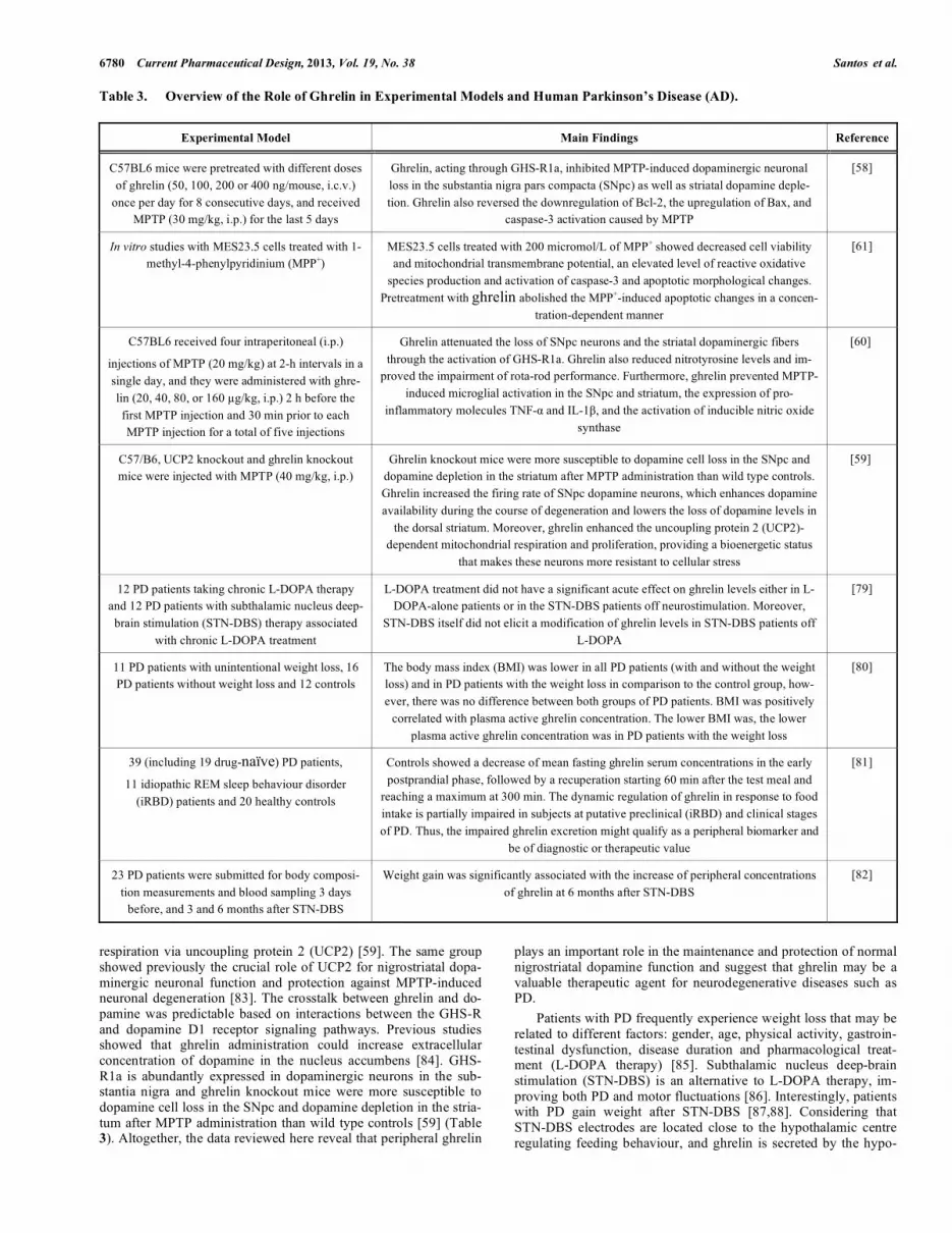
Table 3. Overview of the Role of Ghrelin in Experimental Models and Human Parkinson’s Disease (AD).
Experimental Model Main Findings Reference
C57BL6 mice were pretreated with different doses of ghrelin (50, 100, 200 or 400 ng/mouse, i.c.v.)
once per day for 8 consecutive days, and received MPTP (30 mg/kg, i.p.) for the last 5 days
Ghrelin, acting through GHS-R1a, inhibited MPTP-induced dopaminergic neuronal loss in the substantia nigra pars compacta (SNpc) as well as striatal dopamine deple-tion. Ghrelin also reversed the downregulation of Bcl-2, the upregulation of Bax, and
caspase-3 activation caused by MPTP
[58]
In vitro studies with MES23.5 cells treated with 1-methyl-4-phenylpyridinium (MPP+)
MES23.5 cells treated with 200 micromol/L of MPP+ showed decreased cell viability and mitochondrial transmembrane potential, an elevated level of reactive oxidative
species production and activation of caspase-3 and apoptotic morphological changes. Pretreatment with ghrelin abolished the MPP+-induced apoptotic changes in a concen-
tration-dependent manner
[61]
C57BL6 received four intraperitoneal (i.p.)
injections of MPTP (20 mg/kg) at 2-h intervals in a single day, and they were administered with ghre-lin (20, 40, 80, or 160 �g/kg, i.p.) 2 h before the first MPTP injection and 30 min prior to each MPTP injection for a total of five injections
Ghrelin attenuated the loss of SNpc neurons and the striatal dopaminergic fibers through the activation of GHS-R1a. Ghrelin also reduced nitrotyrosine levels and im-
proved the impairment of rota-rod performance. Furthermore, ghrelin prevented MPTP-induced microglial activation in the SNpc and striatum, the expression of pro-
inflammatory molecules TNF-� and IL-1�, and the activation of inducible nitric oxide synthase
[60]
C57/B6, UCP2 knockout and ghrelin knockout mice were injected with MPTP (40 mg/kg, i.p.)
Ghrelin knockout mice were more susceptible to dopamine cell loss in the SNpc and dopamine depletion in the striatum after MPTP administration than wild type controls. Ghrelin increased the firing rate of SNpc dopamine neurons, which enhances dopamine availability during the course of degeneration and lowers the loss of dopamine levels in
the dorsal striatum. Moreover, ghrelin enhanced the uncoupling protein 2 (UCP2)-dependent mitochondrial respiration and proliferation, providing a bioenergetic status
that makes these neurons more resistant to cellular stress
[59]
12 PD patients taking chronic L-DOPA therapy and 12 PD patients with subthalamic nucleus deep-brain stimulation (STN-DBS) therapy associated
with chronic L-DOPA treatment
L-DOPA treatment did not have a significant acute effect on ghrelin levels either in L-DOPA-alone patients or in the STN-DBS patients off neurostimulation. Moreover,
STN-DBS itself did not elicit a modification of ghrelin levels in STN-DBS patients off L-DOPA
[79]
11 PD patients with unintentional weight loss, 16 PD patients without weight loss and 12 controls
The body mass index (BMI) was lower in all PD patients (with and without the weight loss) and in PD patients with the weight loss in comparison to the control group, how-ever, there was no difference between both groups of PD patients. BMI was positively
correlated with plasma active ghrelin concentration. The lower BMI was, the lower plasma active ghrelin concentration was in PD patients with the weight loss
[80]
39 (including 19 drug-naïve) PD patients,
11 idiopathic REM sleep behaviour disorder (iRBD) patients and 20 healthy controls
Controls showed a decrease of mean fasting ghrelin serum concentrations in the early postprandial phase, followed by a recuperation starting 60 min after the test meal and
reaching a maximum at 300 min. The dynamic regulation of ghrelin in response to food intake is partially impaired in subjects at putative preclinical (iRBD) and clinical stages of PD. Thus, the impaired ghrelin excretion might qualify as a peripheral biomarker and
be of diagnostic or therapeutic value
[81]
23 PD patients were submitted for body composi-tion measurements and blood sampling 3 days
before, and 3 and 6 months after STN-DBS
Weight gain was significantly associated with the increase of peripheral concentrations of ghrelin at 6 months after STN-DBS
[82]
respiration via uncoupling protein 2 (UCP2) [59]. The same group showed previously the crucial role of UCP2 for nigrostriatal dopa-minergic neuronal function and protection against MPTP-induced neuronal degeneration [83]. The crosstalk between ghrelin and do-pamine was predictable based on interactions between the GHS-R and dopamine D1 receptor signaling pathways. Previous studies showed that ghrelin administration could increase extracellular concentration of dopamine in the nucleus accumbens [84]. GHS-R1a is abundantly expressed in dopaminergic neurons in the sub-stantia nigra and ghrelin knockout mice were more susceptible to dopamine cell loss in the SNpc and dopamine depletion in the stria-tum after MPTP administration than wild type controls [59] (Table 3). Altogether, the data reviewed here reveal that peripheral ghrelin
plays an important role in the maintenance and protection of normal nigrostriatal dopamine function and suggest that ghrelin may be a valuable therapeutic agent for neurodegenerative diseases such as PD. Patients with PD frequently experience weight loss that may be related to different factors: gender, age, physical activity, gastroin-testinal dysfunction, disease duration and pharmacological treat-ment (L-DOPA therapy) [85]. Subthalamic nucleus deep-brain stimulation (STN-DBS) is an alternative to L-DOPA therapy, im-proving both PD and motor fluctuations [86]. Interestingly, patients with PD gain weight after STN-DBS [87,88]. Considering that STN-DBS electrodes are located close to the hypothalamic centre regulating feeding behaviour, and ghrelin is secreted by the hypo-
Role of Ghrelin in Neurodegenerative Diseases Current Pharmaceutical Design, 2013, Vol. 19, No. 38 6781
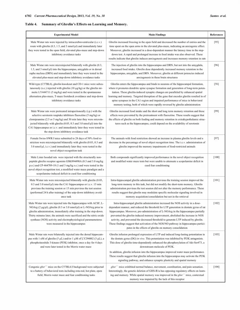
thalamic neurones [89], and among other functions, ghrelin is in-volved in the homeostatic regulation of appetite and energy bal-ance, Corcuff et al. [79] investigated possible changes on serum ghrelin levels in PD patients treated with STN-DBS and/or L-DOPA. In this study, all patients were investigated before and after receiving dopamine treatment, and the group of patients with an implanted neurostimulator was investigated with and without ongo-ing neurostimulation. The results indicated that L-DOPA treatment did not have a significant acute effect on ghrelin levels either in L-DOPA-alone patients or in the STN-DBS patients off neurostimula-tion. Moreover, STN-DBS itself did not elicit a modification of ghrelin levels in STN-DBS patients off L-DOPA. Therefore, the authors concluded that total circulating ghrelin does not play an important role in the modification of weight homeostasis in PD patients treated with STN-DBS [79] (Table 3). More recently, Markaki et al. [82] investigated a possible in-volvement of ghrelin in the weight gain of PD patients after STN-DBS. Twenty-three PD patients were submitted for body composi-tion measurements and blood sampling 3 days before, and 3 and 6 months after STN-DBS. Weight gain was significantly associated with the increase of peripheral concentrations of ghrelin at 6 months after STN-DBS. Therefore, contrasting with the previous observations by Corcuff et al. [79], STN-DBS seems to temporarily dysregulate the hypothalamic secretion of ghrelin that may be re-sponsible for the weight gain of PD patients after STN-DBS [82]. Moreover, the authors of this study emphasize that a possible neu-roprotective role of DBS, exerted through the increase of ghrelin levels, should be further investigated. In another study, Fiszer et al. [80] measured the plasma active ghrelin concentration in 11 PD patients with unintentional weight loss, 16 PD patients without weight loss and 12 controls. The body mass index (BMI) was lower in all PD patients investigated (with and without the weight loss) and in PD patients with the weight loss in comparison to the control group. However, there was no differ-ence between both groups of PD patients. BMI was positively cor-related with plasma active ghrelin concentration. Interestingly, the lower BMI was, the lower plasma active ghrelin concentration was in PD patients with the weight loss [80] (Table 3). As stated before, besides motor symptoms, PD patients fre-quently exhibit non-motor symptoms such as hyposmia, REM sleep behaviour disorder (RBD) and disturbed gastrointestinal motility [90-92] very early in the course of the disease. In relation to the gastrointestinal tract, the stomach has been proposed as one possi-ble ignition point of PD related neuropathology. In respect to the sleep disorder iRBD, about two-thirds of patients with idiopathic RBD (iRBD) develop the alpha-synucleinopathy PD over time [93,94]. Therefore, iRBD is considered a putative pre-motor stage of PD. In this context, Unger et al. [81] measured fasting and post-prandial total ghrelin serum concentrations in 20 healthy controls, 39 (including 19 drug-naïve) PD patients and 11 iRBD patients. Controls showed a decrease of mean fasting ghrelin serum concen-trations in the early postprandial phase, followed by a recuperation starting 60 min after the test meal and reaching a maximum at 300 min. The dynamic regulation of ghrelin in response to food intake is partially impaired in subjects at putative preclinical (iRBD) and clinical stages of PD (Table 3). The authors speculate that reduced ghrelin excretion might increase the vulnerability of nigrostriatal dopaminergic neurons in PD patients as suggested by animal stud-ies. Finally, the impaired ghrelin excretion might qualify as a pe-ripheral biomarker and be of diagnostic or therapeutic value [81].
GHRELIN AS A PALLIATIVE TREATMENT FOR THE MEMORY IMPAIRMENTS IN NEURODEGENERATIVE DISEASES The increasing incidence of neurodegenerative diseases that involve the deterioration of cognitive function has led the scientific community to explore the underlying mechanisms of memory proc-
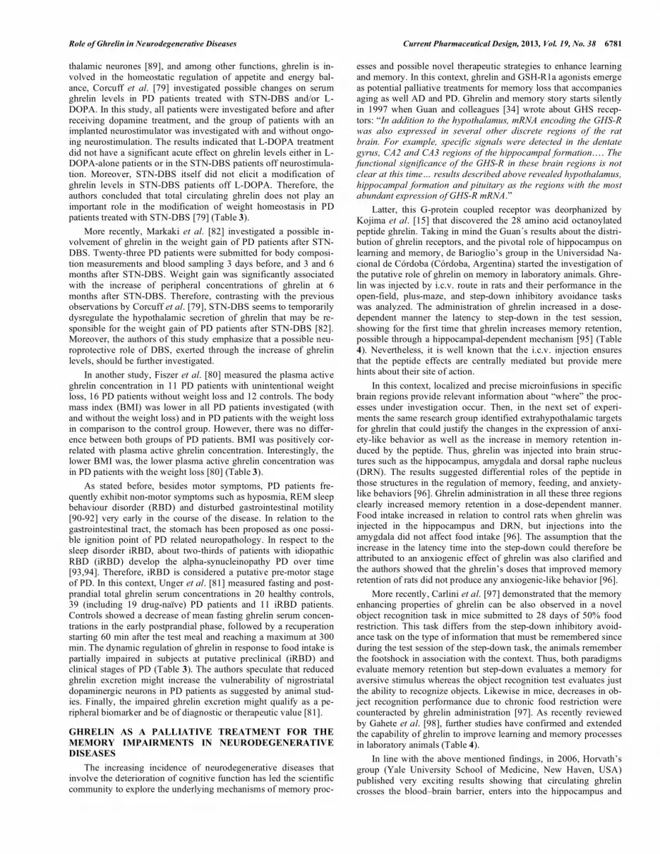
esses and possible novel therapeutic strategies to enhance learning and memory. In this context, ghrelin and GSH-R1a agonists emerge as potential palliative treatments for memory loss that accompanies aging as well AD and PD. Ghrelin and memory story starts silently in 1997 when Guan and colleagues [34] wrote about GHS recep-tors: “In addition to the hypothalamus, mRNA encoding the GHS-R was also expressed in several other discrete regions of the rat brain. For example, specific signals were detected in the dentate gyrus, CA2 and CA3 regions of the hippocampal formation…. Thefunctional significance of the GHS-R in these brain regions is not clear at this time… results described above revealed hypothalamus, hippocampal formation and pituitary as the regions with the most abundant expression of GHS-R mRNA.” Latter, this G-protein coupled receptor was deorphanized by Kojima et al. [15] that discovered the 28 amino acid octanoylated peptide ghrelin. Taking in mind the Guan´s results about the distri-bution of ghrelin receptors, and the pivotal role of hippocampus on learning and memory, de Barioglio’s group in the Universidad Na-cional de Córdoba (Córdoba, Argentina) started the investigation of the putative role of ghrelin on memory in laboratory animals. Ghre-lin was injected by i.c.v. route in rats and their performance in the open-field, plus-maze, and step-down inhibitory avoidance tasks was analyzed. The administration of ghrelin increased in a dose-dependent manner the latency to step-down in the test session, showing for the first time that ghrelin increases memory retention, possible through a hippocampal-dependent mechanism [95] (Table 4). Nevertheless, it is well known that the i.c.v. injection ensures that the peptide effects are centrally mediated but provide mere hints about their site of action. In this context, localized and precise microinfusions in specific brain regions provide relevant information about “where” the proc-esses under investigation occur. Then, in the next set of experi-ments the same research group identified extrahypothalamic targets for ghrelin that could justify the changes in the expression of anxi-ety-like behavior as well as the increase in memory retention in-duced by the peptide. Thus, ghrelin was injected into brain struc-tures such as the hippocampus, amygdala and dorsal raphe nucleus (DRN). The results suggested differential roles of the peptide in those structures in the regulation of memory, feeding, and anxiety-like behaviors [96]. Ghrelin administration in all these three regions clearly increased memory retention in a dose-dependent manner. Food intake increased in relation to control rats when ghrelin was injected in the hippocampus and DRN, but injections into the amygdala did not affect food intake [96]. The assumption that the increase in the latency time into the step-down could therefore be attributed to an anxiogenic effect of ghrelin was also clarified and the authors showed that the ghrelin’s doses that improved memory retention of rats did not produce any anxiogenic-like behavior [96]. More recently, Carlini et al. [97] demonstrated that the memory enhancing properties of ghrelin can be also observed in a novel object recognition task in mice submitted to 28 days of 50% food restriction. This task differs from the step-down inhibitory avoid-ance task on the type of information that must be remembered since during the test session of the step-down task, the animals remember the footshock in association with the context. Thus, both paradigms evaluate memory retention but step-down evaluates a memory for aversive stimulus whereas the object recognition test evaluates just the ability to recognize objects. Likewise in mice, decreases in ob-ject recognition performance due to chronic food restriction were counteracted by ghrelin administration [97]. As recently reviewed by Gahete et al. [98], further studies have confirmed and extended the capability of ghrelin to improve learning and memory processes in laboratory animals (Table 4). In line with the above mentioned findings, in 2006, Horvath’s group (Yale University School of Medicine, New Haven, USA) published very exciting results showing that circulating ghrelin crosses the blood–brain barrier, enters into the hippocampus and
6782 Current Pharmaceutical Design, 2013, Vol. 19, No. 38 Santos et al.
Table 4. Summary of Ghrelin’s Effects on Learning and Memory.
Experimental Model Main Findings References
Male Wistar rats were injected by intracerebroventricular (i.c.v.) route with ghrelin (0.3, 1.5, and 3 nmol/�l) and immediately later
they were tested in the open field, elevated plus-maze and step-down inhibitory avoidance tasks
Ghrelin increased freezing in the open field and decreased the number of entries and the time spent on the open arms in the elevated plus-maze, indicating an anxiogenic effect. Moreover, ghrelin increased in a dose-dependent manner the latency time in the step-down test. A rapid and prolonged increase in food intake was also observed. These
results indicate that ghrelin induces anxiogenesis and increases memory retention in rats
[95]
Male Wistar rats were microinjected bilaterally with ghrelin (0.3, 1.5, and 3 nmol/�l) into the hippocampus, amygdala or in dorsal
raphe nucleus (DRN) and immediately later they were tested in the elevated plus-maze and step-down inhibitory avoidance tasks
The injection of ghrelin into the hippocampus and DRN, but not into the amygdala, increased food intake. Ghrelin dose dependently increased memory retention in the
hippocampus, amygdala, and DRN. Moreover, ghrelin at different potencies induced anxiogenesis in these brain structures
[96]
Wild-type (C57BL6), ghrelin knockout and CD-1 mice were subcu-taneously (s.c.) injected with ghrelin (10 �g/kg) or the ghrelin mi-
metic LY444711 (5 mg/kg) and were tested in the spontaneous alternation plus-maze, T-maze footshock avoidance and step-down
inhibitory avoidance tasks
Ghrelin enters the hippocampus and binds to neurons of the hippocampal formation, where it promotes dendritic spine synapse formation and generation of long-term poten-
tiation. These ghrelin-induced synaptic changes are paralleled by enhanced spatial learning and memory. Targeted disruption of the gene that encodes ghrelin resulted in of
spine synapses in the CA1 region and impaired performance of mice in behavioral memory testing, both of which were rapidly reversed by ghrelin administration
[56]
Male Wistar rats were pretreated intraperitoneally (i.p.) with the selective serotonin reuptake inhibitors fluoxetine (5 mg/kg) or
clomipramine (2.5 or 5 mg/kg) and 30 min later they were microin-jected bilaterally with ghrelin (0.03, 0.3 and 3.0 nmol/�l) into the
CA1 hippocampus or i.c.v. and immediately later they were tested in the step-down inhibitory avoidance task
Ghrelin increased food intake and the short and long term memory retention and these effects were prevented by the pretreatment with fluoxetine. These results suggest that
the effects of ghrelin on both feeding and memory retention in extrahypothalamic struc-tures such as the hippocampus, could depend on the availability of serotonin
[99]
Female Swiss-SWR/J mice submitted to 28 days of 50% food re-striction were microinjected bilaterally with ghrelin (0.03, 0.3 and 3.0 nmol/�l, i.c.v.) and immediately later they were tested in the
novel object recognition task
The animals with food restriction showed an increase in plasma ghrelin levels and a decrease in the percentage of novel object recognition time. The i.c.v. administration of
ghrelin improved the memory impairments of food-restricted animals
[97]
Male Lister hooded rats were injected with the structurally non-peptide ghrelin receptor agonists GSK894490A (0.3 and 3.0 mg/kg p.o.) and CP-464709-18 (1 and 3 mg/kg s.c.) and were tested in the
novel object recognition test, a modified water maze paradigm and a scopolamine-induced deficit in cued fear conditioning
Both compounds significantly improved performance in the novel object recognition and modified water maze tests but were unable to attenuate a scopolamine deficit in
cued fear conditioning
[100]
Male Wistar rats were microinjected bilaterally with ghrelin (0.03, 0.3 and 3.0 nmol/�l) into the CA1 hippocampus or i.c.v. 15 min previous the training session or 15 min previous the test session
(performed 24 h after training) of the setp-down inhibitory avoid-ance task
Intra-hippocampal ghrelin administration previous the training session improved the long-term memory in this task, but did not modify the short-term memory. Ghrelin
administration previous the test session did not alter the memory performance. These results suggest that ghrelin may modulate specific molecular signaling involved in
memory acquisition/consolidation but not in the retrieval
[101]
Male Wistar rats were injected into the hippocampus with ACSF, L-NOArg (2 �g/�l), ghrelin (0.3 or 3.0 nmol/�l) or L-NOArg prior to ghrelin administration, immediately after training in the step-down. Thirty minutes later, the animals were sacrificed and the nitric oxide
synthase (NOS) activity and electrophysiological paramameteres were measured in the hippocampus
Intra-hippocampal ghrelin administration increased the NOS activity in a dose-dependent manner, and reduced the threshold for LTP generation in dentate gyrus of rat hippocampus. Moreover, pre-administration of L-NOArg in the hippocampus partially prevented the ghrelin-induced memory improvement, abolished the increase in NOS activity, and prevented the decreased threshold to generate LTP induced by ghrelin.
These findings suggest that activation of the NOS/NO pathway in hippocampus partici-pates in the effects of ghrelin on memory consolidation
[102]
Male Wistar rats were bilaterally injected into the dorsal hippocam-pus with 1 nM of ghrelin (5 �L) and/or 1 �M of LY294002 (3 �L), a
phosphoinositide 3-kinase (PI3K) inhibitor, once a day for 4 days and were later tested in the Morris water maze
Ghrelin infusion prolonged expression of LTP and induced long-lasting potentiation in the dentate gyrus (DG) in vivo. This potentiation was inhibited by PI3K antagonists.
This dose of ghrelin time-dependently enhanced the phosphorylation of Akt-Ser473, a downstream molecule of PI3K.
In addition, ghrelin infusion into the hippocampus improved water maze performance. These results suggest that ghrelin infusion into the hippocampus may activate the PI3K
signaling pathway, and enhance synaptic plasticity and spatial memory
[103]
Congenic ghsr�/� mice on the C57BL6/J background were subjected to a battery of behavioral tests including rota-rod, hot plate, open
field, Morris water maze and fear conditioning tasks
ghsr�/� mice exhibited normal balance, movement, coordination, and pain sensation. Interestingly, the genetic deletion of GHS-R1a has opposing regulatory effects on learn-
ing and memory. While spatial memory was improved in the ghsr�/� mice, contextual memory was impaired by the lack of this receptor
[104]
Role of Ghrelin in Neurodegenerative Diseases Current Pharmaceutical Design, 2013, Vol. 19, No. 38 6783
binds to neurons of the hippocampal formation, promoting dendritic spine synapse formation and generating LTP [56]. In this same study, the authors demonstrated that the subcutaneous (s.c.) admini-stration of ghrelin or the ghrelin mimetic LY444711 led to a marked improvement in spatial memory retention in mice. In addi-tion, ghrelin knockout mice presented a reduced number of spine synapses in the hippocampal brain region as well as displayed im-paired performance in learning and memory paradigms [56] (Table 4). Moreover, Atcha et al. [100] demonstrated that the oral or s.c. administration of two structurally non-peptide ghrelin receptor ago-nists (GSK894490A and CP-464709-18) readily cross the blood/brain barrier and elicit pro-cognitive effects in recognition and spatial learning and memory tasks in rats (Table 4). Interestingly, Carlini et al. [101] showed that intra-hippocampal ghrelin administration prior the training session, but not prior the test session (performed 24 h after training), improved the long-term memory of rats in the step-down inhibitory avoidance task (Table 4). These findings suggest that ghrelin modulates molecular and/or cellular signaling events involved in memory acquisition and/or consolidation, but not in memory retrieval [101]. Moreover, recent electrophysiological studies provided evidence showing that in vivoghrelin microinjection into the CA1 subregion of hippocampus of rats reduced the threshold values to generate LTP in the dentate gyrus, which is the first synaptic input arising the hippocampus from entorrinal cortex. Moreover, a significant negative correlation was established between this electrophysiological phenomena and the ghrelin effect on the step-down inhibitory avoidance task [102] (Table 4). In relation to the signaling pathway, it was demonstrated that ghrelin increases nitric oxide synthase (NOS) activity in a dose-dependent manner in trained animals, suggesting the participation of the NOS/NO pathway in the ghrelin’s effects on memory [102]. Moreover, it has been also postulated that GHS-R1a likely serves as a modifier of key neurotransmitters required for memory formation such as glutamate, dopamine and serotonin [104]. For instance, Albarran- Zeckler and co-workers [104] demonstrated that genetic deletion of GHS-R1a has opposing regulatory effects on learning and memory. While spatial memory was improved in the ghsr�/�
mice, contextual memory was impaired by the lack of this receptor (Table 4). One plausible explanation for these results is that ghrelin acts as a neuromodulator of other neurotransmitters such as dopa-mine. Of particular relevance, studies have shown that antagonism of dopamine D1 receptors in the hippocampus blocks formation of long-term memory [105] and dopamine D1 receptor knockout mice show deficits in contextual fear conditioning [106]. Interestingly, it was recently demonstrated by Smith’s group (The Scripps Research Institute, Florida, USA) that the ghrelin receptor (GHS-R1a) is co-expressed in neurons that express dopamine D1 and D2 receptors, and that a subset of GHS-R1a, which are not occupied by the ago-nist (apo-GHS-R1a), heterodimerize with these two receptors to regulate dopamine-induced feeding suppression in mice [107]. It is thus very likely that there is similar importance of the GHS-R1a for effects of dopamine signaling on learning and memory. In addition, it was demonstrated that the selective serotonin reuptake inhibitor (SSRI) fluoxetine, given i.p. 30 min prior to in-tra-hippocampal ghrelin injection, prevented the ghrelin-induced increase in food intake and short- and long-term memory retention in rats [99]. These findings suggest that the effects of ghrelin on both feeding and memory retention could depend on the availability of serotonin. Experiments using hipocampal slices demonstrated that few minutes after addition of ghrelin in the superfusion me-dium, serotonin release was inhibited [108]. In another set of ex-periments, ghrelin was injected into the hippocampus of rats and the animals were killed 24 h later for the measurement of serotonin release. Remarkably, ghrelin significantly inhibited the serotonin release 24 h after its in vivo administration, indicating that ghrelin-induced inhibitory effects on serotonin release starts immediately
after injection and can persist for at least 24 h after its central ad-ministration [108]. Certainly additional brain systems and molecular mechanisms need to be studied to further clarify the role of ghrelin on learning and memory processes. However, from recent findings demonstrat-ing the ability of ghrelin to improve the cognitive dysfunction in rats submitted to models of sepsis-associated encephalopathy [109] and diabetic encephalopathy [110], it appears that ghrelin might be particularly useful to restore impaired learning and memory proc-esses associated to neurodegenerative diseases.
GHRELIN AS A PALLIATIVE TREATMENT FOR DE-PRESSION IN NEURODEGENERATIVE DISEASES Depression is a prevalent disease; 10-20% of people in the world’s population will develop depression at least once in their lifetime, causing impairment in functioning and quality of life, with high medical and social costs [111,112]. According to World Health Organization major depression will be the world’s second most debilitating disease by 2020, eclipsed only by heart disease [113]. This disease is characterized by apathy, indifference and anhedonia, behavioral sluggishness and increasing inactivity, feel-ings of guilt, pessimism, regret and low self-esteem, psychophysi-ological disturbances of sleep and appetite [111]. Major depression is frequently found coexisting with long-standing chronic medical conditions such as cardiovascular disease, diabetes mellitus, obesity and neurodegenerative diseases [114]. Depression is common and a clinically important feature of PD and can precede the onset of the motor symptoms. The prevalence of depression in patients with PD is approximately 40% [115,116]. Concomitant depression in PD is associated with greater healthcare system use, including medical hospitalizations [117]. Moreover, major depressive disorder is considered a risk factor for developing AD later in life [118]. Depressive symptoms are frequent and affect nearly 40% of AD patients [119]. Noteworthy, brains of patients with AD with comorbid depression showed higher levels of cortical neurofibrillary tangles than brains of patients with AD without co-morbid depression, suggesting an interaction between depression and the neuropathologic processes in AD [120]. Drugs used in the treatment of depression cause several side effects and generally influence weight gain. Among hormones that act on weight regulation, ghrelin has been suggested to exert an antidepressant action. Regarding the preclinical studies, there are several pieces of evidence supporting a role for ghrelin in the modulation of mood (Table 5). The administration of antisense DNA for ghrelin into the lateral ventricle was reported to reduce the immobility time in the forced swimming test (FST) in rats, which is a result indicative of an antidepressant-like effect [121]. Further evidence of the possible antidepressant activity of ghrelin, it is a study by Lutter et al. [122] showing that the subcutaneous admini-stration of ghrelin produced antidepressant-like responses in the FST. In addition, increasing ghrelin levels through a diet containing 60% of normal calories resulted in an antidepressant-like response in the FST. Moreover, mice submitted to chronic social defeat stress (CSDS) procedure, which induces behavioral deficits remi-niscent of depression including social avoidance, had significantly elevated levels of acylated ghrelin that persisted for at least 4 weeks after the procedure [122]. Moreover, genetic deletion of GHSRs exacerbated depression-like behavior induced by CSDS, a finding also described by Chuang et al. [123]. The study by Lutter et al.[122] demonstrated that ghrelin’s antidepressant-like effects in the FST were blocked in mice lacking orexin, suggesting that the anti-depressant-like actions of this peptide may be dependent on a direct and/or indirect activation of orexin-containing neurons. A recent evidence provided by our group [124] reinforced the notion of the ghrelin’s antidepressant action, since it was shown that the acute administration of ghrelin by i.c.v. route produced antidepressant-like effect in a predictive test of antidepressant activ-
6784 Current Pharmaceutical Design, 2013, Vol. 19, No. 38 Santos et al.
ity, the tail suspension test (TST) in mice. In addition, this study also showed that ghrelin, administered acutely to mice by i.c.v. route, was able to abolish the depressive-like behavior induced by olfactory bulbectomy (OB) [124], an animal model of depression which produces behavioral, neurochemical and neuroendocri-nological changes that resemble some of the symptoms observed in depressed patients [125]. Of note, these behavioral and neuro-chemical changes in OB rodents are reported to be normalized only by the chronic administration of antidepressants. Interestingly, there are few studies reporting that the acute administration of agents that inhibit the glutamatergic transmission such as zinc and riluzole produce a rapid reversal of the hyperlocomotion activity induced by OB [126,127], as opposed to the conventional antidepressants. Therefore, it remains to be established if an antiglutamatergic mechanism could be responsible for the antidepressant-like effect of ghrelin. Clinical studies have also supported the idea that ghrelin exerts a possible beneficial role in depressive disorders. The serum ghrelin levels were lower before and after treatment in depressive patients as compared with non-depressed individuals [128,129]. Moreover, it was demonstrated that electroconvulsive therapy (ECT), an effec-tive treatment for depression, decreased serum ghrelin levels in depressive patients as compared with the levels of this peptide be-fore ECT [130]. Taking into account that ghrelin inhibits serotonin release [108,131], one of the hypothesis raised to explain these results is that the decreased circulating ghrelin levels may be a compensatory response to depression, potentially elevating sero-tonin levels [129]. Moreover, after a pulsatile administration of ghrelin to depressive patients, an improvement in the depressive symptoms assessed by a validated self-rating scale (‘Be-findlichkeits-Skala’), at trend level (p=0.093) in men, but not in women, was observed [132]. Furthermore, ghrelin gene polymor-phism was previously associated with depression [133]. However, is must be conceded that other studies failed to show a correlation between ghrelin and depression [134,135]. For instance, nocturnal plasma ghrelin of depressed patients and matched healthy subjects did not differ when stratified for sex [134] and plasma ghrelin was not different between 83 depressed patients and 46 healthy controls [135]. Several studies have linked ghrelin with stress, which, in turn, is a risk factor for the development of depression. Humans sub-jected acutely to psychosocial stress exhibit increased plasma ghre-lin levels [136], and similar results were described in rats after acute psychological stress [137]. The increase in ghrelin levels could contribute to the mechanisms responsible for the develop-ment of stress-induced depression or may represent a protective mechanism to minimize manifestations of depression following stress [37,138,139]. This protective mechanism may be related to the activation of hedonic signalling pathway and stimulation of the
intake of palatable caloric foods that, in turn, elicits central reward pathways and increases dopamine signaling [139]. However, the stress�induced food rewards behavior and hyperphagia of palatable caloric dense foods increases body weight. Following prolonged exposure to stress and palatable foods, a desensitization of reward signalling may be linked to the risk of depression and co�morbid obesity [139]. Another possible mechanism that links ghrelin to depressive disorders is that a ghrelin’s action on mood may be mediated through the modulation of neuroinflammatory mechanisms [138], which has been demonstrated to play a role in the pathophysiology of depression [114,140,141]. Therefore, it remains to be established if ghrelin administration is able to ameliorate the depressive-like behavior elicited by neuroinflammatory conditions in rodents, tak-ing into account that ghrelin or ghrelin mimetics are able to sup-press the synthesis and release of pro-inflammatory cytokines [142].
GHRELIN AS A PALLIATIVE TREATMENT FOR OF STRESS AND ANXIETY DISORDERS IN NEURODEGEN-ERATIVE DISEASES The gut-brain connection has been implicated in brain disorders ranging from anxiety to depression and schizophrenia [143-145]. This connection between gastrointestinal tract and the hypotha-lamic-pituitary axis (HPA) is bidirectional and it is mediated through the release of peptides that exert responses within the brain as well as via neuroendocrine and sensory inputs from the gut. The exact mechanisms governing such communication are unclear and most studies focus on the impact of altered signaling from the brain to the gut although the reverse is now being studied (for review see [144,145]). Even though a complete discussion of the interrelation-ships between the gut and the brain in the control of stress and anxiety disorders is beyond the intended scope of this article, focus will be placed on briefly reviewing the current literature on ghrelin and its role in the mediation/modulation of stress/anxiety states. There is increasing evidence that ghrelin has several physio-logical functions, especially in CNS, including a role in neuropro-tection, learning and memory, reward and motivation, and depres-sion as aforementioned, besides being important in anxiety and stressful conditions (for review see [37]), since plasmatic ghrelin is able to cross the blood–brain barrier, and to accumulate and bind to neurons in several brain areas underlying stress and anxiety re-sponses. Actually, the orexigenic and pro-obesity targets of ghre-lin’s actions are located in hypothalamic and mesolimbic circuits involved not only in energy balance, appetite and reward but also in regulating mood and cognition (for review see [146]). Animal and human studies suggest that stressful conditions can result in low mood and increased energy intake, particularly from fatty acids and sugars, and potential changes in body weight, lead-
Table 5. Summary of the Ghrelin’s Antidepressant-Like Effects Observed in Preclinical Studies.
Animal Model Main Findings References
Forced swimming test Antidepressant-like effect following the administration of antisense DNA for ghrelin into the lateral ventricle, or subcutaneous administration of ghrelin in rats. Also, increasing ghrelin
levels through a diet containing 60% of normal calories caused antidepressant profile.
Antidepressant-like effects of ghrelin in the FST were blocked in mice lacking orexin
[122]
Tail suspension test Acute administration of ghrelin by i.c.v. route caused antidepressant-like effect in mice [124]
Olfactory bulbectomy Acute administration of ghrelin by i.c.v. route abolished the depressive-like behavior induced by olfactory bulbectomy in mice
[124]
Chronic social defeat stress (CSDS) Genetic deletion of GHSR exacerbated depression-like behaviors induced by CSDS [122,123]
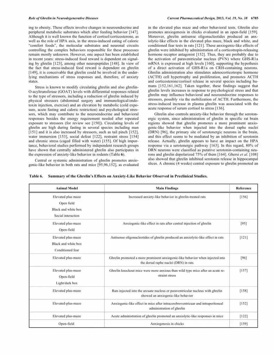
Role of Ghrelin in Neurodegenerative Diseases Current Pharmaceutical Design, 2013, Vol. 19, No. 38 6785
ing to obesity. These effects involve changes in neuroendocrine and peripheral metabolic substrates which alter feeding behavior [147]. Although it is well known the function of cortisol/corticosterone, as well as the role of HPA axis, in the stress-induced eating of caloric “comfort foods”, the molecular substrates and neuronal circuits controlling the complex behaviors responsible for these processes remain mostly unknown. However, one aspect has been established in recent years: stress-induced food reward is dependent on signal-ing by ghrelin [123], among other neuropeptides [148]. In view of the fact that stress-induced food reward is dependent on ghrelin [149], it is conceivable that ghrelin could be involved in the under-lying mechanisms of stress responses and, therefore, of anxiety states. Stress is known to modify circulating ghrelin and also ghrelin-O-acyltransferase (GOAT) levels with differential responses related to the type of stressors, including a reduction of ghrelin induced by physical stressors (abdominal surgery and immunological/endo-toxin injection, exercise) and an elevation by metabolic (cold expo-sure, acute fasting and caloric restriction) and psychological stres-sors, which may contribute to the neuroendocrine and behavioral responses besides the energy requirement needed after repeated exposure to stressors (for review see [150]). Circulating levels of ghrelin are high during fasting in several species including man [151] and it is also increased by stressors, such as tail pinch [152], water immersion [153], social defeat [122], restraint stress [154] and chronic stress (caged filled with water) [155]. Of high impor-tance, behavioral studies performed by independent research groups have shown that centrally administered ghrelin also participates in the expression of anxiety-like behavior in rodents (Table 6). Central or systemic administration of ghrelin promotes anxio-genic-like behavior in both rats and mice [95,96,152], as evaluated
in the elevated plus maze and other behavioral tests. Ghrelin also promotes anxiogenesis in chicks evaluated in an open-field [159]. Moreover, ghrelin antisense oligonucleotides produced an anx-iolytic-like effects in the elevated plus maze, black and white, and conditioned fear tests in rats [121]. These anxiogenic-like effects of ghrelin were inhibited by administration of a corticotropin-releasing hormone receptor antagonist [152]. Thus, they are probably due to the activation of paraventricular nucleus (PVN) where GHS-R1a mRNA is expressed at high levels [160], supporting the hypothesis of a direct activation of GHS-R1a on CRH-containing neurons. Ghrelin administration also stimulates adenocorticotropic hormone (ACTH) cell hypertrophy and proliferation, and promotes ACTH and corticosterone/cortisol release in several species including hu-mans [152,161,162]. Taken together, these findings suggest that ghrelin levels increases in response to psychological stress and that ghrelin may influence behavioral and neuroendocrine responses to stressors, possibly via the mobilization of ACTH. Furthermore, the stress-induced increase in plasma ghrelin was associated with the acute response of serum cortisol to stress [136]. Ghrelin also controls anxiety-like behavior through the seroton-ergic system, since administration of ghrelin in specific rat brain regions showed that ghrelin promotes a more prominent anxio-genic-like behavior when injected into the dorsal raphe nuclei (DRN) [96], the primary site of serotonergic neurons in the brain, and this effect seems to be mediated by an inhibition of serotonin release. Actually, ghrelin appears to have an impact on the HPA response via a serotonergic pathway [163]. In this regard, 80% of DRN neurons were classified as putative serotonin-containing neu-rons and ghrelin depolarized 75% of them [164]. Ghersi et al. [108] also showed that ghrelin inhibited serotonin release in hippocampal slices. A chronic (4 weeks) central exposure to ghrelin promoted an
Table 6. Summary of the Ghrelin’s Effects on Anxiety-Like Behavior Observed in Preclinical Studies.
Animal Model Main Findings Reference
Elevated plus maze
Open field
Black and white box
Social interaction
Increased anxiety-like behavior in ghrelin-treated rats [156]
Elevated plus-maze
Open-field
Anxiogenic-like effect in rats after central injection of ghrelin [95]
Elevated plus-maze
Black and white box
Conditioned fear
Antisense oligonucleotides of ghrelin produced an anxiolytic-like effect in rats [121]
Elevated plus-maze Ghrelin promoted a more prominent anxiogenic-like behavior when injected into the dorsal raphe nuclei (DRN) in rats
[96]
Elevated plus-maze
Open-field
Light/dark box
Ghrelin knockout mice were more anxious than wild type mice after an acute re-straint stress
[157]
Elevated plus-maze Rats injected into the arcuate nucleus or paraventricular nucleus with ghrelin showed an anxiogenic-like behavior
[158]
Elevated-plus-maze Anxiogenic-like effect in mice after intracerebroventricuar and intraperitoneal administration of ghrelin
[152]
Elevated plus-maze Acute administration of ghrelin promoted an anxiolytic-like responses in mice [122]
Open-field Anxiogenesis in chicks [159]

6786 Current Pharmaceutical Design, 2013, Vol. 19, No. 38 Santos et al.
increase in anxiety- and depression-like behavior in rats. Changes in expression of a number of genes representing key systems impli-cated in these behavioral effects were found as well as an inhibition of the electrophysiological response of DRN after a ghrelin chal-lenge [156]. Additionally, anxiogenic and orexigenic effects of ghrelin seems to be mediated, at least in part, via endocannabinoid signal-ing since PVN injections of the ghrelin promoted anxiogenic-like profile and a significantly increase in food intake and these effects were blocked by AM251, a cannabinoid CB1 receptor antagonist [158]. On the other hand, caloric restriction for 10 days or acute ad-ministration of ghrelin promoted an increase in circulating ghrelin levels in ad libitum-fed C57BL6/J mice, producing anxiolytic-like responses in the elevated plus maze. However, when GHSR-null mice were calorie-restricted, these anxiolytic-like behavioral re-sponses were no longer observed indicating a specific role of GHS-R1a receptors in this effect [122]. In addition, recently, it was shown that ghrelin knockout mice are more anxious after an acute restraint stress, compared with wild-type mice, as evaluated in three behavioral tests (elevated plus-maze, open-field and light/dark box). Acute restraint stress exacerbated neuronal activation in the hypo-thalamic PVN and medial nucleus of the amygdala in knockout mice compared with wild type mice, and the administration of ex-ogenous ghrelin was able to reverse this effect. Spencer et al. [157] proposed that ghrelin is able to reduce anxiety after acute stress by stimulating the HPA axis at the anterior pituitary level, by involving urocortin 1 receptor on this effect. These observations showing an anxiolytic-like effect of ghrelin seem to be supported by findings that ghrelin levels were higher in the Sprague–Dawley rats (low-anxiety strain) than in the Wistar Kyoto rats (high-anxiety strain) after acute stress, whereas ACTH are equally enhanced in both strains [137,165]. Moreover, although psychological stress induces an increase in plasma ghrelin levels in humans, the post-stress in-duced urge for uncontrolled eating is not acutely modulated by stress related elevations in ghrelin levels [136]. In this regard, a recent study described that isolation stress resulted in a reduction of plasma ghrelin that seems to be dependent on CRF1-R, and MC4 receptor in PVN and 5-HT1B/2C receptors in the arcuate nucleus (ARN) [166]. The number of immunoreactive neurons in PVN of the hypothalamus was significantly increased after peripheral ad-ministration of ghrelin, an effect that seems to occur via NPY-positive projections from the ARN [167]. As the PVN is involved in a neuronal network mediating the autonomic, neuroendocrine, and skeletal-motor responses of fear and anxiety [168], an increased density of immunoreactive neurons in the PVN, after peripheral ghrelin administration, could also be the result of ghrelin transport into the brain and/or resultant of an activation of the hippocampal–amygdalic–hypothalamic network involved in the regulation of fear and anxiety rather than an activation of hunger/satiety pathways along the ARN-PVN axis [167]. In addition, rates of anxiety and cognitive impairment were higher in the hypertensive elders, which were negatively correlated with plasma ghrelin levels, resulted from chronic cortisol response to long-term anxiety [169]. The reason for these contradictory findings are presently un-known, but it could be related in animals to the timing of the behav-ioral tests after ghrelin administration since the anxiogenic-like effect of ghrelin was seen when the behavioral evaluation was per-formed within 5-10 min [95,96,121,152,158], whereas the latter study conducted the evaluation after 45 min of the injection [122]. As ghrelin has a plasmatic half-life of about 30 min, it is conceiv-able that the dose which had significant effects on food intake in all previous studies, underlined the anxiogenic responses in the 5-10 min studies. These issues have to be clarified since not all studies show changes in ghrelin levels [134,135]. Several findings suggest that ghrelin was secreted as a result of alarm signals concerning physiological changes such as severe
weight loss, which are potentially life threatening [155]. Altogether, evidence shows that ghrelin is involved in emotional reactivity in rodents, although no differences were found in patients with obses-sive compulsive disorder [170]. Thus, ghrelin is a peptide hormone implicated in diverse biological functions such as the modulation of centrally controlled behaviors ranging from energy balance (food intake, body-weight regulation and glucose homeostasis) to stress, anxiety and memory processes. Some regions of the hypothalamus appear to be differentially sensitive and responsive to the feeding-stimulant, metabolic, and anxiogenic actions of ghrelin and that the ARN and PVN, in particular, exert a primary role in mediating these effects [158]. Therefore, the bidirectional effects of ghrelin on stress and anxiety behaviors seem to be stressor, test- and time-dependent, and may be partly mediated by ghrelin via the CRF, serotonin and the endocanabinoid systems.
CONCLUSION This article reviews the recent evidence that the gastric hor-mone ghrelin plays an important role not only as a modulator of GH secretion and food intake, but fundamentally as an important factor for neuronal plasticity, growth and survival in the CNS. Possible therapeutic opportunities for neurological disorders through the modulation of ghrelin system are pointed out, in the expectancy that this review may inspire clinical researchers and foster experimental approaches using ghrelin as the therapeutic target. Two major conclusions should be drawn: first, the physiologi-cal role of ghrelin is by far wider than considered before including learning and memory, anxiety, depression and neuroprotection; second, changes in the GHS-R1a receptors seem to underlie these central actions of ghrelin. Establishing the function of ghrelin in physiological stress responses and whether control of its activity would be useful for prevention and/or treatment of stress-induced diseases, such as anxiety and mood disorders as well as psychiatric symptoms associated to neurodegenerative diseases, remain impor-tant research aims. Since apoptotic cell death, mitochondrial dysfunction, oxidative stress and neuroinflammation have been identified as molecular processes associated with the neurological disorders here presented, manipulation of the ghrelin system beckons as an opportunity for neuroprotection, improving neuronal plasticity and counteracting the lost of brain functions in patients with neurodegenerative dis-eases, including AD and PD. Future research aiming the develop-ment of novel non-peptide ghrelin receptor antagonists, a growing number of ghrelin receptor selective ligands, and also inverse ago-nists, will help to elucidate the neurobiology and physiological role of ghrelin as well as its potential as novel palliative and neuropro-tective agent in neurodegenerative diseases.
CONFLICT OF INTEREST The authors confirm that this article content has no conflicts of interest.
ACKNOWLEDGEMENTS The authors gratefully acknowledge Dr. Aderbal S. Aguiar Jr. and Dr. Daniel Rial for their editorial assistance. Some of the re-search reviewed in this article was supported by the Brazilian agen-cies Conselho Nacional de Desenvolvimento Científico e Tec-nológico (CNPq), Coordenação de Aperfeiçoamento de Pessoal de Nível Superior (CAPES-COFECUB France/Brazil 681/2010), CAFP (Programa de Fortalecimiento de Centros Asociados de Postgrado Brasil-Argentina, CAPES –SPU), Programa de Apoio aos Núcleos de Excelência (PRONEX - Project NENASC), Funda-ção de Apoio à Pesquisa do Estado de Santa Catarina (FAPESC), FINEP (Financiadora de Estudos e Projetos – IBN-Net #01.06.0842-00) and INCT (Instituto Nacional de Ciência e Tec-nologia) for Excitotoxicity and Neuroprotection. VVS receive a

Role of Ghrelin in Neurodegenerative Diseases Current Pharmaceutical Design, 2013, Vol. 19, No. 38 6787
scholarship from CAPES. ALR, TCML and RDP are supported by research fellowships from CNPq. The authors have no financial or personal conflicts of interest related to this work.
REFERENCES[1] Blennow K, de Leon MJ, Zetterberg H. Alzheimer's disease. Lancet
2006; 368: 387-403. [2] Ferri CP, Prince M, Brayne C, et al. Global prevalence of
dementia: a Delphi consensus study. Lancet 2005; 366: 2112-7. [3] Larson EB, Shadlen MF, Wang L, et al. Survival after initial
diagnosis of Alzheimer disease. Ann Intern Med 2004; 140: 501-9. [4] Artero S, Tierney MC, Touchon J, Ritchie K. Prediction of
transition from cognitive impairment to senile dementia: a prospective, longitudinal study. Acta Psychiatr Scand 2003; 107: 390-3.
[5] Lambon Ralph MA, Patterson K, Graham N, Dawson K, Hodges JR. Homogeneity and heterogeneity in mild cognitive impairment and Alzheimer's disease: a cross-sectional and longitudinal study of 55 cases. Brain 2003; 126: 2350-62.
[6] Cotman CW, Su JH. Mechanisms of neuronal death in Alzheimer's disease. Brain Pathol 1996; 6: 493-506.
[7] Mayeux R. Epidemiology of neurodegeneration. Annu Rev Neurosci 2003; 26: 81-104.
[8] Hirsch E, Graybiel AM, Agid YA. Melanized dopaminergic neu-rons are differentially susceptible to degeneration in Parkinson's disease. Nature 1988; 28; 345-8.
[9] Chaudhuri KR, Healy DG, Schapira AH, Excellence NIfC. Non-motor symptoms of Parkinson's disease: diagnosis and management. Lancet Neurol 2006; 5: 235-45.
[10] Momany FA. Conformational analysis of methionine-enkephalin and some analogs. Biochem Biophys Res Commun 1977; 75: 1098-103.
[11] Momany FA, Bowers CY, Reynolds GA, Chang D, Hong A, Newlander K. Design, synthesis, and biological activity of peptides which release growth hormone in vitro. Endocrinology 1981; 108: 31-9.
[12] Bowers CY, Momany FA, Reynolds GA, Hong A. On the in vitroand in vivo activity of a new synthetic hexapeptide that acts on the pituitary to specifically release growth hormone. Endocrinology 1984; 114: 1537-45.
[13] Smith RG, Van der Ploeg LH, Howard AD, et al. Peptidomimetic regulation of growth hormone secretion. Endocr Rev 1997; 18: 621-45.
[14] Korbonits M, Ciccarelli E, Ghigo E, Grossman AB. The growth hormone secretagogue receptor. Growth Horm IGF Res 1999; 9 Suppl A: 93-9.
[15] Kojima M, Hosoda H, Date Y, Nakazato M, Matsuo H, Kangawa K. Ghrelin is a growth-hormone-releasing acylated peptide from stomach. Nature 1999; 402: 656-60.
[16] Smith RG, Griffin PR, Xu Y, et al. Adenosine: A partial agonist of the growth hormone secretagogue receptor. Biochem Biophys Res Commun 2000; 276: 1306-13.
[17] Stengel A, Taché Y. Yin and Yang - the Gastric X/A-like Cell as Possible Dual Regulator of Food Intake. J Neurogastroenterol Motil 2012; 18: 138-49.
[18] Furness JB, Hunne B, Matsuda N, et al. Investigation of the presence of ghrelin in the central nervous system of the rat and mouse. Neuroscience 2011; 193: 1-9.
[19] Zhang JV, Ren PG, Avsian-Kretchmer O, et al. Obestatin, a peptide encoded by the ghrelin gene, opposes ghrelin's effects on food intake. Science 2005; 310: 996-9.
[20] Seim I, Collet C, Herington AC, Chopin LK. Revised genomic structure of the human ghrelin gene and identification of novel exons, alternative splice variants and natural antisense transcripts. BMC Genomics 2007; 8: 298.
[21] Seim I, Josh P, Cunningham P, Herington A, Chopin L. Ghrelin axis genes, peptides and receptors: recent findings and future challenges. Mol Cell Endocrinol 2011; 340: 3-9.
[22] Pemberton C, Wimalasena P, Yandle T, Soule S, Richards M. C-terminal pro-ghrelin peptides are present in the human circulation. Biochem Biophys Res Commun 2003; 310: 567-73.
[23] Yang J, Brown MS, Liang G, Grishin NV, Goldstein JL. Identification of the acyltransferase that octanoylates ghrelin, an appetite-stimulating peptide hormone. Cell 2008; 132: 387-96.
[24] Kang K, Zmuda E, Sleeman MW. Physiological role of ghrelin as revealed by the ghrelin and GOAT knockout mice. Peptides 2011; 32: 2236-41.
[25] Kojima M, Kangawa K. Ghrelin: from gene to physiological function. Results Probl Cell Differ 2010; 50: 185-205.
[26] Soares JB, Leite-Moreira AF. Ghrelin, des-acyl ghrelin and obestatin: three pieces of the same puzzle. Peptides 2008; 29: 1255-70.
[27] Nishi Y, Yoh J, Hiejima H, Kojima M. Structures and molecular forms of the ghrelin-family peptides. Peptides 2011; 32: 2175-82.
[28] Baldanzi G, Filigheddu N, Cutrupi S, et al. Ghrelin and des-acyl ghrelin inhibit cell death in cardiomyocytes and endothelial cells through ERK1/2 and PI 3-kinase/AKT. J Cell Biol 2002; 159: 1029-37.
[29] Yin X, Li Y, Xu G, An W, Zhang W. Ghrelin fluctuation, what determines its production? Acta Biochim Biophys Sin (Shanghai) 2009; 41: 188-97.
[30] Ueno M, Carvalheira JB, Oliveira RL, Velloso LA, Saad MJ. Circulating ghrelin concentrations are lowered by intracerebro-ventricular insulin. Diabetologia 2006; 49: 2449-52.
[31] Kamegai J, Tamura H, Shimizu T, Ishii S, Sugihara H, Oikawa S. Effects of insulin, leptin, and glucagon on ghrelin secretion from isolated perfused rat stomach. Regul Pept, 2004; 119: 77-81.
[32] Kojima M, Kangawa K. Ghrelin: structure and function. Physiol Rev 2005; 85: 495-522.
[33] Floquet N, M'Kadmi C, Perahia D, et al. Activation of the ghrelin receptor is described by a privileged collective motion: a model for constitutive and agonist-induced activation of a sub-class A G-protein coupled receptor (GPCR). J Mol Biol 2010; 395: 769-84.
[34] Guan XM, Yu H, Palyha OC, et al. Distribution of mRNA encoding the growth hormone secretagogue receptor in brain and peripheral tissues. Brain Res Mol Brain Res 1997; 48: 23-9.
[35] Kola B, Farkas I, Christ-Crain M, et al. The orexigenic effect of ghrelin is mediated through central activation of the endogenous cannabinoid system. PLoS One 2008; 3: e1797.
[36] Jiang H, Betancourt L, Smith RG. Ghrelin amplifies dopamine signaling by cross talk involving formation of growth hormone secretagogue receptor/dopamine receptor subtype 1 heterodimers. Mol Endocrinol 2006; 20: 1772-85.
[37] Andrews ZB. The extra-hypothalamic actions of ghrelin on neuronal function. Trends Neurosci 2011; 34: 31-40.
[38] Deboer MD. The use of ghrelin and ghrelin receptor agonists as a treatment for animal models of disease: Efficacy and mechanism. Curr Pharm Des 2012 (in press).
[39] Strasser F. Clinical Application of Ghrelin. Curr Pharm Des 2012 (in press).
[40] Frago LM, Pañeda C, Dickson SL, Hewson AK, Argente J, Chowen JA. Growth hormone (GH) and GH-releasing peptide-6 increase brain insulin-like growth factor-I expression and activate intracellular signaling pathways involved in neuroprotection. Endocrinology 2002; 143: 4113-22.
[41] Pañeda C, Arroba AI, Frago LM, et al. Growth hormone-releasing peptide-6 inhibits cerebellar cell death in aged rats. Neuroreport 2003; 14: 1633-5.
[42] Chung H, Seo S, Moon M, Park S. Phosphatidylinositol-3-kinase/Akt/glycogen synthase kinase-3 beta and ERK1/2 pathways mediate protective effects of acylated and unacylated ghrelin against oxygen-glucose deprivation-induced apoptosis in primary rat cortical neuronal cells. J Endocrinol 2008; 198: 511-21.
[43] Hwang S, Moon M, Kim S, Hwang L, Ahn KJ, Park S. Neuroprotective effect of ghrelin is associated with decreased expression of prostate apoptosis response-4. Endocr J 2009; 56: 609-17.
[44] Bansal V, Ryu SY, Blow C, et al. The hormone ghrelin prevents traumatic brain injury induced intestinal dysfunction. J Neurotrauma 2010; 27: 2255-60.
[45] Lopez NE, Krzyzaniak MJ, Blow C, et al. Ghrelin prevents disruption of the blood-brain barrier after traumatic brain injury. J Neurotrauma 2012; 29: 385-93.
[46] Qi L, Cui X, Dong W, et al. Ghrelin attenuates brain injury after traumatic brain injury and uncontrolled hemorrhagic shock in rats. Mol Med 2012; 18: 186-93.
[47] Er�ahın M, Toklu HZ, Erzık C, et al. Ghrelin alleviates spinal cord injury in rats via its anti-inflammatory effects. Turk Neurosurg 2011; 21: 599-605.
6788 Current Pharmaceutical Design, 2013, Vol. 19, No. 38 Santos et al.
[48] Zhang Q, Huang C, Meng B, Tang T, Shi Q, Yang H. Acute effect of ghrelin on ischemia/reperfusion injury in the rat spinal cord. Int J Mol Sci 2012; 13: 9864-76.
[49] Lim E, Lee S, Li E, Kim Y, Park S. Ghrelin protects spinal cord motoneurons against chronic glutamate-induced excitotoxicity via ERK1/2 and phosphatidylinositol-3-kinase/Akt/glycogen synthase kinase-3� pathways. Exp Neurol 2011; 230: 114-22.
[50] Lee S, Kim Y, Li E, Park S. Ghrelin protects spinal cord motoneurons against chronic glutamate excitotoxicity by inhibiting microglial activation. Korean J Physiol Pharmacol 2012; 16: 43-8.
[51] Obay BD, Ta�demir E, Tümer C, Bilgin HM, Atmaca M. Dose dependent effects of ghrelin on pentylenetetrazole-induced oxidative stress in a rat seizure model. Peptides 2008; 29: 448-55.
[52] Obay BD, Tasdemir E, Tümer C, Bilgin HM, Sermet A. Antiepileptic effects of ghrelin on pentylenetetrazole-induced seizures in rats. Peptides 2007; 28: 1214-9.
[53] Aslan A, Yildirim M, Ayyildiz M, Güven A, Agar E. The role of nitric oxide in the inhibitory effect of ghrelin against penicillin-induced epileptiform activity in rat. Neuropeptides 2009; 43: 295-302.
[54] Xu J, Wang S, Lin Y, Cao L, Wang R, Chi Z. Ghrelin protects against cell death of hippocampal neurons in pilocarpine-induced seizures in rats. Neurosci Lett 2009; 453: 58-61.
[55] Lee J, Lim E, Kim Y, Li E, Park S. Ghrelin attenuates kainic acid-induced neuronal cell death in the mouse hippocampus. J Endocrinol 2010; 205: 263-70.
[56] Diano S, Farr SA, Benoit SC, et al. Ghrelin controls hippocampal spine synapse density and memory performance. Nat Neurosci 2006; 9: 381-8.
[57] Moon M, Choi JG, Nam DW, et al. Ghrelin ameliorates cognitive dysfunction and neurodegeneration in intrahippocampal amyloid-�1-42 oligomer-injected mice. J Alzheimers Dis 2011; 23: 147-59.
[58] Jiang H, Li LJ, Wang J, Xie JX. Ghrelin antagonizes MPTP-induced neurotoxicity to the dopaminergic neurons in mouse substantia nigra. Exp Neurol 2008; 212: 532-7.
[59] Andrews ZB, Erion D, Beiler R, et al. Ghrelin promotes and protects nigrostriatal dopamine function via a UCP2-dependent mitochondrial mechanism. J Neurosci 2009; 29: 14057-65.
[60] Moon M, Kim HG, Hwang L, et al. Neuroprotective effect of ghrelin in the 1-methyl-4-phenyl-1,2,3,6-tetrahydropyridine mouse model of Parkinson's disease by blocking microglial activation. Neurotox Res 2009; 15: 332-47.
[61] Dong J, Song N, Xie J, Jiang H. Ghrelin antagonized 1-methyl-4-phenylpyridinium (MPP(+))-induced apoptosis in MES23.5 cells. J Mol Neurosci 2009; 37: 182-9.
[62] Frago LM, Baquedano E, Argente J, Chowen JA. Neuroprotective actions of ghrelin and growth hormone secretagogues. Front Mol Neurosci 2011; 4: 23.
[63] Chung H, Kim E, Lee DH, et al. Ghrelin inhibits apoptosis in hypothalamic neuronal cells during oxygen-glucose deprivation. Endocrinology 2007; 148: 148-59.
[64] Miao Y, Xia Q, Hou Z, Zheng Y, Pan H, Zhu S. Ghrelin protects cortical neuron against focal ischemia/reperfusion in rats. Biochem Biophys Res Commun 2007; 359: 795-800.
[65] Selkoe DJ. The molecular pathology of Alzheimer’s disease. Neuron 1991; 6: 487-98.
[66] Haass C, Selkoe DJ. Soluble protein oligomers in neurodegeneration: lessons from the Alzheimer's amyloid beta-peptide. Nat Rev Mol Cell Biol 2007; 8: 101-12.
[67] Rigamonti AE, Pincelli AI, Corrà B, et al. Plasma ghrelin concentrations in elderly subjects: comparison with anorexic and obese patients. J Endocrinol 2002; 175: R1-5.
[68] Proto C, Romualdi D, Cento RM, et al. Plasma levels of neuropeptides in Alzheimer's disease. Gynecol Endocrinol 2006; 22: 213-8.
[69] Gahete MD, Rubio A, Córdoba-Chacón J, et al. Expression of the ghrelin and neurotensin systems is altered in the temporal lobe of Alzheimer's disease patients. J Alzheimers Dis 2010; 22: 819-28.
[70] Shibata N, Ohnuma T, Kuerban B, Komatsu M, Arai H. Genetic association between ghrelin polymorphisms and Alzheimer's disease in a Japanese population. Dement Geriatr Cogn Disord 2011; 32: 178-81.
[71] Theodoropoulou A, Metallinos IC, Psyrogiannis A, Vagenakis GA, Kyriazopoulou V. Ghrelin and leptin secretion in patients with moderate Alzheimer's disease. J Nutr Health Aging 2012; 16: 472-7.
[72] Allain H, Bentué-Ferrer D, Akwa Y. Disease-modifying drugs and Parkinson's disease. Prog Neurobiol 2008; 84: 25-39.
[73] Ahlskog JE, Muenter MD. Frequency of levodopa-related dyskinesias and motor fluctuations as estimated from the cumulative literature. Mov Disord 2001; 16: 448-58.
[74] Braak H, Ghebremedhin E, Rüb U, Bratzke H, Del Tredici K. Stages in the development of Parkinson's disease-related pathology. Cell Tissue Res 2004; 318: 121-34.
[75] Jenner P. Functional models of Parkinson's disease: a valuable tool in the development of novel therapies. Ann Neurol 2008; 64 Suppl 2: S16-29.
[76] Langston JW, Ballard PA. Parkinson's disease in a chemist working with 1-methyl-4-phenyl-1,2,5,6-tetrahydropyridine. N Engl J Med 1983; 309: 310.
[77] Schmidt N, Ferger B. Neurochemical findings in the MPTP model of Parkinson's disease. J Neural Transm 2001; 108: 1263-82.
[78] Prediger RD, Aguiar AS Jr, Moreira EL, et al. The intranasal ad-ministration of 1-methyl-4-phenyl-1,2,3,6-tetrahydropyridine (MPTP): a new rodent model to test palliative and neuroprotective agents for Parkinson's disease. Curr Pharm Des 2011;17: 489-507.
[79] Corcuff JB, Krim E, Tison F, et al. Subthalamic nucleus stimulation in patients with Parkinson's disease does not increase serum ghrelin levels. Br J Nutr 2006; 95: 1028-9.
[80] Fiszer U, Micha�owska M, Baranowska B, et al. Leptin and ghrelin concentrations and weight loss in Parkinson's disease. Acta Neurol Scand 2010; 121: 230-6.
[81] Unger MM, Möller JC, Mankel K, et al. Postprandial ghrelin response is reduced in patients with Parkinson's disease and idiopathic REM sleep behaviour disorder: a peripheral biomarker for early Parkinson's disease? J Neurol 2011; 258: 982-90.
[82] Markaki E, Ellul J, Kefalopoulou Z, et al. The role of ghrelin, neuropeptide Y and leptin peptides in weight gain after deep brain stimulation for Parkinson's disease. Stereotact Funct Neurosurg 2012; 90: 104-12.
[83] Andrews ZB, Horvath B, Barnstable CJ, et al. Uncoupling protein-2 is critical for nigral dopamine cell survival in a mouse model of Parkinson's disease. J Neurosci 2005; 25: 184-91.
[84] Jerlhag E, Egecioglu E, Dickson SL, Douhan A, Svensson L, Engel JA. Ghrelin administration into tegmental areas stimulates locomotor activity and increases extracellular concentration of dopamine in the nucleus accumbens. Addict Biol 2007; 12: 6-16.
[85] Lorefält B, Ganowiak W, Pålhagen S, Toss G, Unosson M, Granérus AK. Factors of importance for weight loss in elderly patients with Parkinson's disease. Acta Neurol Scand 2004; 110: 180-7.
[86] Limousin P, Krack P, Pollak P, et al. Electrical stimulation of the subthalamic nucleus in advanced Parkinson's disease. N Engl J Med 1998; 339: 1105-11.
[87] Volkmann J, Allert N, Voges J, Weiss PH, Freund HJ, Sturm V. Safety and efficacy of pallidal or subthalamic nucleus stimulation in advanced PD. Neurology 2001; 56: 548-51.
[88] Perlemoine C, Macia F, Tison F, et al. Effects of subthalamic nucleus deep brain stimulation and levodopa on energy production rate and substrate oxidation in Parkinson's disease. Br J Nutr 2005; 93: 191-8.
[89] Cowley MA, Smith RG, Diano S, et al. The distribution and mechanism of action of ghrelin in the CNS demonstrates a novel hypothalamic circuit regulating energy homeostasis. Neuron 2003; 37: 649-61.
[90] Chaudhuri KR, Martinez-Martin P, Schapira AH, et al.International multicenter pilot study of the first comprehensive self-completed nonmotor symptoms questionnaire for Parkinson's disease: the NMSQuest study. Mov Disord 2006; 21: 916-23.
[91] Schenck CH, Mahowald MW. Subclinical REM sleep behavior disorder and its clinical and research implications. Sleep 2008; 31: 1627.
[92] Unger MM, Belke M, Menzler K, et al. Diffusion tensor imaging in idiopathic REM sleep behavior disorder reveals microstructural changes in the brainstem, substantia nigra, olfactory region, and other brain regions. Sleep 2010; 33: 767-73.
[93] Schenck CH, Bundlie SR, Mahowald MW. Delayed emergence of a parkinsonian disorder in 38% of 29 older men initially diagnosed with idiopathic rapid eye movement sleep behaviour disorder. Neu-rology 1996; 46: 388-93.
[94] Schenck CH, Callies AL, Mahowald MW. Increased percentage of slow-wave sleep in REM sleep behavior disorder (RBD): a
Role of Ghrelin in Neurodegenerative Diseases Current Pharmaceutical Design, 2013, Vol. 19, No. 38 6789
reanalysis of previously published data from a controlled study of RBD reported in SLEEP. Sleep 2003; 26: 1066; author reply 1067.
[95] Carlini VP, Monzón ME, Varas MM, et al. Ghrelin increases anxiety-like behavior and memory retention in rats. Biochem Biophys Res Commun 2002; 299: 739-43.
[96] Carlini VP, Varas MM, Cragnolini AB, et al. Differential role of the hippocampus, amygdala, and dorsal raphe nucleus in regulating feeding, memory, and anxiety-like behavioral responses to ghrelin. Biochem Biophys Res Commun 2004; 313: 635-41.
[97] Carlini VP, Martini AC, Schiöth HB, Ruiz RD, Fiol de Cuneo M, de Barioglio SR. Decreased memory for novel object recognition in chronically food-restricted mice is reversed by acute ghrelin administration. Neuroscience 2008; 153: 929-34.
[98] Gahete MD, Córdoba-Chacón J, Kineman RD, Luque RM, Castaño JP. Role of ghrelin system in neuroprotection and cognitive functions: implications in Alzheimer's disease. Peptides 2011; 32: 2225-8.
[99] Carlini VP, Gaydou RC, Schiöth HB, de Barioglio SR. Selective serotonin reuptake inhibitor (fluoxetine) decreases the effects of ghrelin on memory retention and food intake. Regul Pept 2007; 140: 65-73.
[100] Atcha Z, Chen WS, Ong AB, et al. Cognitive enhancing effects of ghrelin receptor agonists. Psychopharmacology (Berl), 2009; 206: 415-27.
[101] Carlini VP, Ghersi M, Schiöth HB, de Barioglio SR. Ghrelin and memory: differential effects on acquisition and retrieval. Peptides 2010; 31: 1190-3.
[102] Carlini VP, Perez MF, Salde E, Schiöth HB, Ramirez OA, de Barioglio SR. Ghrelin induced memory facilitation implicates nitric oxide synthase activation and decrease in the threshold to promote LTP in hippocampal dentate gyrus. Physiol Behav 2010; 101: 117-23.
[103] Chen L, Xing T, Wang M, et al. Local infusion of ghrelin enhanced hippocampal synaptic plasticity and spatial memory through activation of phosphoinositide 3-kinase in the dentate gyrus of adult rats. Eur J Neurosci 2011; 33: 266-75.
[104] Albarran-Zeckler RG, Brantley AF, Smith RG. Growth hormone secretagogue receptor (GHS-R1a) knockout mice exhibit improved spatial memory and deficits in contextual memory. Behav Brain Res 2012; 232: 13-9.
[105] Rossato JI, Bevilaqua LR, Izquierdo I, Medina JH, Cammarota M. Dopamine controls persistence of long-term memory storage. Science 2009; 325: 1017-20.
[106] Ortiz O, Delgado-García JM, Espadas I, et al. Associative learning and CA3-CA1 synaptic plasticity are impaired in D1R null, Drd1a-/- mice and in hippocampal siRNA silenced Drd1a mice. J Neurosci 2010; 30: 12288-300.
[107] Kern A, Albarran-Zeckler R, Walsh HE, Smith RG. Apo-ghrelin receptor forms heteromers with DRD2 in hypothalamic neurons and is essential for anorexigenic effects of DRD2 agonism. Neuron 2012; 73: 317-32.
[108] Ghersi MS, Casas SM, Escudero C, et al. Ghrelin inhibited serotonin release from hippocampal slices. Peptides 2011; 32: 2367-71.
[109] Wang G, Wang W, Zhao J, Ni Y, Zhou X, Zhang W. Ghrelin prevents neuronal apoptosis and cognitive impairments in sepsis-associated encephalopathy. Neuroreport 2011; 22: 959-64.
[110] Ma LY, Zhang DM, Tang Y, et al. Ghrelin-attenuated cognitive dysfunction in streptozotocin-induced diabetic rats. Alzheimer Dis Assoc Disord 2011; 25: 352-63.
[111] Wong ML, Licinio J. From monoamines to genomic targets: a paradigm shift for drug discovery in depression. Nat Rev Drug Discov 2004; 3: 136-51.
[112] Kessler RC. The costs of depression. Psychiatr Clin North Am 2012; 35: 1-14.
[113] Murray CJ, Lopez AD. Evidence-based health policy--lessons from the Global Burden of Disease Study. Science 1996; 274: 740-3.
[114] Maes M, Kubera M, Obuchowiczwa E, Goehler L, Brzeszcz J. Depression's multiple comorbidities explained by (neuro)inflammatory and oxidative & nitrosative stress pathways. Neuro Endocrinol Lett 2011; 32: 7-24.
[115] Dooneief G, Mirabello E, Bell K, Marder K, Stern Y, Mayeux R. An estimate of the incidence of depression in idiopathic Parkinson's disease. Arch Neurol 1992; 49: 305-7.
[116] Lieberman A. Managing the neuropsychiatric symptoms of Parkinson's disease. Neurology 1998; 50: S33-8; discussion S44-8.
[117] Chen P, Kales HC, Weintraub D, et al. Depression in veterans with Parkinson's disease: frequency, co-morbidity, and healthcare utilization. Int J Geriatr Psychiatry 2007; 22: 543-8.
[118] Ownby RL, Crocco E, Acevedo A, John V, Loewenstein D. Depression and risk for Alzheimer disease: systematic review, meta-analysis, and metaregression analysis. Arch Gen Psychiatry 2006; 63: 530-8.
[119] Arbus C, Gardette V, Cantet CE, et al. Incidence and predictive factors of depressive symptoms in Alzheimer's disease: the REAL.FR study. J Nutr Health Aging 2011; 15: 609-17.
[120] Rapp MA, Schnaider-Beeri M, Purohit DP, Perl DP, Haroutunian V, Sano M. Increased neurofibrillary tangles in patients with Alzheimer disease with comorbid depression. Am J Geriatr Psychiatry 2008; 16: 168-74.
[121] Kanehisa M, Akiyoshi J, Kitaichi T, et al. Administration of antisense DNA for ghrelin causes an antidepressant and anxiolytic response in rats. Prog Neuropsychopharmacol Biol Psychiatry 2006; 30: 1403-7.
[122] Lutter M, Sakata I, Osborne-Lawrence S, et al. The orexigenic hormone ghrelin defends against depressive symptoms of chronic stress. Nat Neurosci 2008; 11: 752-3.
[123] Chuang JC, Perello M, Sakata I, et al. Ghrelin mediates stress-induced food-reward behavior in mice. J Clin Invest 2011; 121: 2684-92.
[124] Carlini VP, Machado DG, Buteler F, et al. Acute ghrelin administration reverses depressive-like behavior induced by bilateral olfactory bulbectomy in mice. Peptides 2012; 35: 160-5.
[125] Song C, Leonard BE. The olfactory bulbectomised rat as a model of depression. Neurosci Biobehav Rev 2005; 29: 627-47.
[126] Nowak G, Szewczyk B, Wieronska JM, et al. Antidepressant-like effects of acute and chronic treatment with zinc in forced swim test and olfactory bulbectomy model in rats. Brain Res Bull 2003; 61: 159-64.
[127] Takahashi K, Murasawa H, Yamaguchi K, et al. Riluzole rapidly attenuates hyperemotional responses in olfactory bulbectomized rats, an animal model of depression. Behav Brain Res 2011; 216: 46-52.
[128] Schmid DA, Wichniak A, Uhr M, et al. Changes of sleep architecture, spectral composition of sleep EEG, the nocturnal secretion of cortisol, ACTH, GH, prolactin, melatonin, ghrelin, and leptin, and the DEX-CRH test in depressed patients during treatment with mirtazapine. Neuropsychopharmacology 2006; 31: 832-44.
[129] Barim AO, Aydin S, Colak R, Dag E, Deniz O, Sahin I. Ghrelin, paraoxonase and arylesterase levels in depressive patients before and after citalopram treatment. Clin Biochem 2009; 42: 1076-81.
[130] Kurt E, Guler O, Serteser M, et al. The effects of electroconvulsive therapy on ghrelin, leptin and cholesterol levels in patients with mood disorders. Neurosci Lett 2007; 426: 49-53.
[131] Brunetti L, Recinella L, Orlando G, Michelotto B, Di Nisio C, Vacca M. Effects of ghrelin and amylin on dopamine, norepinephrine and serotonin release in the hypothalamus. Eur J Pharmacol 2002; 454: 189-92.
[132] Kluge M, Schüssler P, Dresler M, et al. Effects of ghrelin on psychopathology, sleep and secretion of cortisol and growth hormone in patients with major depression. J Psychiatr Res 2011; 45: 421-6.
[133] Nakashima K, Akiyoshi J, Hatano K, et al. Ghrelin gene polymorphism is associated with depression, but not panic disorder. Psychiatr Genet 2008; 18: 257.
[134] Kluge M, Schussler P, Schmid D, Uhr M, Kleyer S, Yassouridis A, Steiger A. Ghrelin plasma levels are not altered in major depression. Neuropsychobiology 2009; 59: 199-204.
[135] Schanze A, Reulbach U, Scheuchenzuber M, Groschl M, Kornhuber J, Kraus T. Ghrelin and eating disturbances in psychiatric disorders. Neuropsychobiology 2008; 57: 126-30.
[136] Rouach V, Bloch M, Rosenberg N, et al. The acute ghrelin response to a psychological stress challenge does not predict the post-stress urge to eat. Psychoneuroendocrinology 2007; 32: 693-702.
[137] Kristenssson E, Sundqvist M, Astin M, et al. Acute psychological stress raises plasma ghrelin in the rat. Regul Pept 2006; 134: 114-7.
[138] Chuang JC, Zigman JM. Ghrelin's Roles in Stress, Mood, and Anxiety Regulation. Int J Pept 2010; 2010. pii: 460549

6790 Current Pharmaceutical Design, 2013, Vol. 19, No. 38 Santos et al.
[139] Schellekens H, Finger BC, Dinan TG, Cryan JF. Ghrelin signalling and obesity: at the interface of stress, mood and food reward. Pharmacol Ther 2012; 135: 316-26.
[140] Dantzer R, O'Connor JC, Lawson MA, Kelley KW. Inflammation-associated depression: from serotonin to kynurenine. Psychoneuroendocrinology 2011; 36: 426-36.
[141] Kaster MP, Gadotti VM, Calixto JB, Santos AR, Rodrigues AL. Depressive-like behavior induced by tumor necrosis factor-� in mice. Neuropharmacology 2012; 62: 419-26.
[142] Himmerich H, Sheldrick AJ. TNF-alpha and ghrelin: opposite effects on immune system, metabolism and mental health. Protein Pept Lett 2010; 17: 186-96.
[143] Neufeld KM, Kang N, Bienenstock J, Foster JA. Reduced anxiety-like behavior and central neurochemical change in germ-free mice. Neurogastroenterol Motil 2011; 23: 255-64, e119.
[144] Cryan JF, Dinan TG. Mind-altering microorganisms: the impact of the gut microbiota on brain and behaviour. Nat Rev Neurosci 2012; 13: 701-12.
[145] Mayer EA. Gut feelings: the emerging biology of gut-brain communication. Nat Rev Neurosci 2011; 12: 453-66.
[146] Skibicka KP, Dickson SL. Ghrelin and food reward: the story of potential underlying substrates. Peptides 2011; 32: 2265-73.
[147] Roberts CJ. The effects of stress on food choice, mood and bodyweight in healthy women. In: eds. Nutrition Bulletin 2008; pp. 33-39.
[148] Adam TC, Epel ES. Stress, eating and the reward system. Physiol Behav 2007; 91: 449-58.
[149] Raspopow K, Abizaid A, Matheson K, Anisman H. Psychosocial stressor effects on cortisol and ghrelin in emotional and non-emotional eaters: influence of anger and shame. Horm Behav 2010; 58: 677-84.
[150] Stengel A, Wang L, Taché Y. Stress-related alterations of acyl and desacyl ghrelin circulating levels: mechanisms and functional implications. Peptides 2011; 32: 2208-17.
[151] Tschöp M, Smiley DL, Heiman ML. Ghrelin induces adiposity in rodents. Nature 2000; 407: 908-13.
[152] Asakawa A, Inui A, Kaga T, et al. A role of ghrelin in neuroendocrine and behavioral responses to stress in mice. Neuroendocrinology 2001; 74: 143-7.
[153] Brzozowski T, Konturek PC, Konturek SJ, et al. Exogenous and endogenous ghrelin in gastroprotection against stress-induced gastric damage. Regul Pept 2004; 120: 39-51.
[154] Zheng J, Dobner A, Babygirija R, Ludwig K, Takahashi T. Effects of repeated restraint stress on gastric motility in rats. Am J Physiol Regul Integr Comp Physiol 2009; 296: R1358-65.
[155] Ochi M, Tominaga K, Tanaka F, et al. Effect of chronic stress on gastric emptying and plasma ghrelin levels in rats. Life Sci 2008; 82: 862-8.
[156] Hansson C, Haage D, Taube M, Egecioglu E, Salomé N, Dickson SL. Central administration of ghrelin alters emotional responses in rats: behavioural, electrophysiological and molecular evidence. Neuroscience 2011; 180: 201-11.
[157] Spencer SJ, Xu L, Clarke MA, et al. Ghrelin regulates the hypothalamic-pituitary-adrenal axis and restricts anxiety after acute stress. Biol Psychiatry 2012; 72: 457-65.
[158] Currie PJ, Khelemsky R, Rigsbee EM, et al. Ghrelin is an orexigenic peptide and elicits anxiety-like behaviors following administration into discrete regions of the hypothalamus. Behav Brain Res 2012; 226: 96-105.
[159] Carvajal P, Carlini VP, Schiöth HB, de Barioglio SR, Salvatierra NA. Central ghrelin increases anxiety in the Open Field test and impairs retention memory in a passive avoidance task in neonatal chicks. Neurobiol Learn Mem 2009; 91: 402-7.
[160] Zigman JM, Jones JE, Lee CE, Saper CB, Elmquist JK. Expression of ghrelin receptor mRNA in the rat and the mouse brain. J Comp Neurol 2006; 494: 528-48.
[161] Broglio F, Benso A, Castiglioni C, et al. The endocrine response to ghrelin as a function of gender in humans in young and elderly subjects. J Clin Endocrinol Metab 2003; 88: 1537-42.
[162] Stevanovi� D, Milosevi� V, Starcevi� VP, Severs WB. The effect of centrally administered ghrelin on pituitary ACTH cells and circulating ACTH and corticosterone in rats. Life Sci 2007; 80: 867-72.
[163] Jászberényi M, Bujdosó E, Bagosi Z, Telegdy G. Mediation of the behavioral, endocrine and thermoregulatory actions of ghrelin. Horm Behav 2006; 50: 266-73.
[164] Ogaya M, Kim J, Sasaki K. Ghrelin postsynaptically depolarizes dorsal raphe neurons in rats in vitro. Peptides 2011; 32: 1606-16.
[165] Kristensson E, Sundqvist M, Håkanson R, Lindström E. High gastrin cell activity and low ghrelin cell activity in high-anxiety Wistar Kyoto rats. J Endocrinol 2007; 193: 245-50.
[166] Saegusa Y, Takeda H, Muto S, et al. Decreased plasma ghrelin contributes to anorexia following novelty stress. Am J Physiol Endocrinol Metab 2011; 301: E685-96.
[167] Rüter J, Kobelt P, Tebbe JJ, et al. Intraperitoneal injection of ghrelin induces Fos expression in the paraventricular nucleus of the hypothalamus in rats. Brain Res 2003; 991: 26-33.
[168] Charney DS, Deutch A. A functional neuroanatomy of anxiety and fear: implications for the pathophysiology and treatment of anxiety disorders. Crit Rev Neurobiol 1996; 10: 419-46.
[169] Yushun L, Fenling F, Hongyan T, et al. Role of ghrelin in cognitive impairment of hypertensive elders with chronic psychological distress. Journal of Medical Colleges of PLA 2012; 25: 163-72.
[170] Emül HM, Serteser M, Kurt E, Ozbulut O, Guler O, Gecici O. Ghrelin and leptin levels in patients with obsessive-compulsive disorder. Prog Neuropsychopharmacol Biol Psychiatry 2007; 31: 1270-4.
Received: January 29, 2013 Accepted: March 18, 2013
Related Documents