Review Article Getting “Inside” Type I IFNs: Type I IFNs in Intracellular Bacterial Infections Deann T. Snyder, Jodi F. Hedges, and Mark A. Jutila Department of Microbiology and Immunology, Montana State University, Bozeman, MT, USA Correspondence should be addressed to Jodi F. Hedges; [email protected] Received 21 January 2017; Revised 20 March 2017; Accepted 27 March 2017; Published 26 April 2017 Academic Editor: Chen Zhao Copyright © 2017 Deann T. Snyder et al. This is an open access article distributed under the Creative Commons Attribution License, which permits unrestricted use, distribution, and reproduction in any medium, provided the original work is properly cited. Type I interferons represent a unique and complex group of cytokines, serving many purposes during innate and adaptive immunity. Discovered in the context of viral infections, type I IFNs are now known to have myriad effects in infectious and autoimmune disease settings. Type I IFN signaling during bacterial infections is dependent on many factors including whether the infecting bacterium is intracellular or extracellular, as different signaling pathways are activated. As such, the repercussions of type I IFN induction can positively or negatively impact the disease outcome. This review focuses on type I IFN induction and downstream consequences during infection with the following intracellular bacteria: Chlamydia trachomatis, Listeria monocytogenes, Mycobacterium tuberculosis, Salmonella enterica serovar Typhimurium, Francisella tularensis, Brucella abortus, Legionella pneumophila, and Coxiella burnetii. Intracellular bacterial infections are unique because the bacteria must avoid, circumvent, and even co-opt microbial “sensing” mechanisms in order to reside and replicate within a host cell. Furthermore, life inside a host cell makes intracellular bacteria more difficult to target with antibiotics. Because type I IFNs are important immune effectors, modulating this pathway may improve disease outcomes. But first, it is critical to understand the context-dependent effects of the type I IFN pathway in intracellular bacterial infections. 1. Introduction Originally discovered for their antiviral activity, type I interferons (IFNs) are now known to also impact a variety of infectious and inflammatory disease states that are not exclusive to the antiviral response [1]. In fact, the story of type I IFNs reaches beyond the protective role for which they were discovered. Not only can these potent cytokines defend the host from viral infection but they can also promote per- sistent viral infection in some settings [2–5]. Similarly, these cytokines can either harm or benefit the host in autoimmune diseases [6–9]. Furthermore, type I IFNs are treatments for some viral infections and autoimmune diseases, stressing the importance of understanding their impact on the host immune system [3, 10]. Not surprisingly, type I IFNs can have opposing effects during both intracellular and extracel- lular bacterial infections as well. Because type I IFNs are produced during an immense number of distinct infections and inflammatory diseases, their importance from an evolu- tionary and immune standpoint is clear. Type I IFNs are one of three types of interferons. Type I, II, and III IFNs are classified based on activity, structure, and corresponding receptor type. There are many groups of type I IFNs: IFN-α, β, κ, ω, τ, and ε [11]. Of the type I IFNs, IFN-β and IFN-α are most well-studied. There is only one IFN-β while there are 12 subtypes of IFN-α in humans and 14 in mice [1]. Type II IFN consists of only IFN-γ while there are four types of type III IFNs in humans and two in mice [12]. Interferons can induce immune changes at minimal concentration; thus, tight regulation of interferon responses is required and may be determined, in part, by interferon receptor distribution and expression [13, 14]. Most, if not all, cells can respond to and produce type I IFNs, but plasmacytoid dendritic cells (pDCs) are some of the most robust producers. They can generate 10-fold more IFN-α than monocytes [15]. Although pDCs will Hindawi Journal of Immunology Research Volume 2017, Article ID 9361802, 17 pages https://doi.org/10.1155/2017/9361802

Welcome message from author
This document is posted to help you gain knowledge. Please leave a comment to let me know what you think about it! Share it to your friends and learn new things together.
Transcript

Review ArticleGetting “Inside” Type I IFNs: Type I IFNs in IntracellularBacterial Infections
Deann T. Snyder, Jodi F. Hedges, and Mark A. Jutila
Department of Microbiology and Immunology, Montana State University, Bozeman, MT, USA
Correspondence should be addressed to Jodi F. Hedges; [email protected]
Received 21 January 2017; Revised 20 March 2017; Accepted 27 March 2017; Published 26 April 2017
Academic Editor: Chen Zhao
Copyright © 2017 Deann T. Snyder et al. This is an open access article distributed under the Creative Commons AttributionLicense, which permits unrestricted use, distribution, and reproduction in any medium, provided the original work isproperly cited.
Type I interferons represent a unique and complex group of cytokines, servingmany purposes during innate and adaptive immunity.Discovered in the context of viral infections, type I IFNs are now known to havemyriad effects in infectious and autoimmune diseasesettings. Type I IFN signaling during bacterial infections is dependent on many factors including whether the infecting bacterium isintracellular or extracellular, as different signaling pathways are activated. As such, the repercussions of type I IFN induction canpositively or negatively impact the disease outcome. This review focuses on type I IFN induction and downstream consequencesduring infection with the following intracellular bacteria: Chlamydia trachomatis, Listeria monocytogenes, Mycobacteriumtuberculosis, Salmonella enterica serovar Typhimurium, Francisella tularensis, Brucella abortus, Legionella pneumophila, andCoxiella burnetii. Intracellular bacterial infections are unique because the bacteria must avoid, circumvent, and even co-optmicrobial “sensing” mechanisms in order to reside and replicate within a host cell. Furthermore, life inside a host cell makesintracellular bacteria more difficult to target with antibiotics. Because type I IFNs are important immune effectors, modulating thispathway may improve disease outcomes. But first, it is critical to understand the context-dependent effects of the type I IFNpathway in intracellular bacterial infections.
1. Introduction
Originally discovered for their antiviral activity, type Iinterferons (IFNs) are now known to also impact a varietyof infectious and inflammatory disease states that are notexclusive to the antiviral response [1]. In fact, the story oftype I IFNs reaches beyond the protective role for which theywere discovered. Not only can these potent cytokines defendthe host from viral infection but they can also promote per-sistent viral infection in some settings [2–5]. Similarly, thesecytokines can either harm or benefit the host in autoimmunediseases [6–9]. Furthermore, type I IFNs are treatments forsome viral infections and autoimmune diseases, stressingthe importance of understanding their impact on the hostimmune system [3, 10]. Not surprisingly, type I IFNs canhave opposing effects during both intracellular and extracel-lular bacterial infections as well. Because type I IFNs areproduced during an immense number of distinct infections
and inflammatory diseases, their importance from an evolu-tionary and immune standpoint is clear.
Type I IFNs are one of three types of interferons. Type I,II, and III IFNs are classified based on activity, structure, andcorresponding receptor type. There are many groups of type IIFNs: IFN-α, β, κ, ω, τ, and ε [11]. Of the type I IFNs, IFN-βand IFN-α are most well-studied. There is only one IFN-βwhile there are 12 subtypes of IFN-α in humans and 14in mice [1]. Type II IFN consists of only IFN-γ whilethere are four types of type III IFNs in humans and twoin mice [12]. Interferons can induce immune changes atminimal concentration; thus, tight regulation of interferonresponses is required and may be determined, in part, byinterferon receptor distribution and expression [13, 14].Most, if not all, cells can respond to and produce type IIFNs, but plasmacytoid dendritic cells (pDCs) are someof the most robust producers. They can generate 10-foldmore IFN-α than monocytes [15]. Although pDCs will
HindawiJournal of Immunology ResearchVolume 2017, Article ID 9361802, 17 pageshttps://doi.org/10.1155/2017/9361802

not be discussed in relation to the intracellular bacteriacovered in this review, their role as robust type I IFN pro-ducers is likely an important part of the immune response.
Despite disparate survival strategies, intracellular andextracellular bacteria are both capable of inducing type IIFNs with some overlap in induction pathways. Whereasintracellular bacteria can activate intracellular sensors fromwithin the phagolysosome or cytoplasm, extracellular bacte-ria introduce type I IFN-stimulating ligands into the cytosolvia pore-forming proteins or other means [16–18]. Thisreview concentrates on the actions of type I IFNs in thecontext of intracellular bacterial infections. The functions ofthese cytokines in other microbial infections, cancer, andautoimmunity have been extensively reviewed elsewhere[8, 19–21]. The following sectionswill describe pathways lead-ing to, and the downstream results of, type I IFN production.
2. Induction of Type I IFNs
Pathogen-associated molecular patterns (PAMPs) are sensedby their cognate pattern recognition receptor (PRRs), whichleads to transcription of many gene products, including typeI IFNs. Toll-like receptors (TLRs), C-type lectin receptors(CLRs), retinoic acid-inducing gene I- (RIG-I-) like receptors(RLRs), and nucleotide-binding oligomerization domain-(NOD-) like receptors (NLRs) are all PRRs. TLRs and CLRsare transmembrane receptors, whereas RLRs and NLRsreside in the cytoplasm [22]. Multiple PRRs can be engagedduring infection, leading to an orchestrated innate immuneresponse that is specific to a pathogen’s repertoire of PAMPs.
TLRs sense PAMPs, including nucleic acids and lipopro-teins from invading pathogens, and either are expressed onthe cell surface or are present in endosomes and lysosomesin immune cells. Endosomal TLRs recognize viral and bac-terial nucleic acids and lead to type I IFN production;these are TLR3, 7, and 9 in mice and humans [23]. WhileTLR3 is mostly expressed intracellularly, cell surface TLR3has been observed on human dendritic cells, macrophages,endothelial cells, and synovial fibroblasts of rheumatoidarthritis patients [24]. TLR4, found on the cell surface, recog-nizes the gram-negative bacterial component, lipopolysac-charide (LPS). Following TLR-ligand binding, signalingoccurs via two main adaptor protein pathways, myeloid dif-ferentiation factor 88 (MyD88) or TIR domain-containingadaptor-inducing IFN-β (TRIF) [22]. All TLRs signal throughMyD88 except TLR3, which utilizes only TRIF, and TLR4which utilizes TRIF or MyD88 [23, 25]. TRIF signalingleads to type I IFN production via TANK-binding kinase-(TBK-1-) mediated activation of transcription factors,interferon regulatory factors (IRF) 3 or 7. Alternatively,downstream of MyD88, inflammatory genes are inducedvia transcription factor, nuclear factor kappa-light-chain-enhancer of activated B cells (NF-κB) [26]. However,endosomal MyD88 signaling leads to type I IFN productionvia IL-1R1-associated protein kinase 4- (IRAK4-) mediatedactivation of IRF7 [27].
Of the nucleic acid sensing TLRs, TLR9 is the onlyDNA sensor and is expressed on endosomes or cell sur-faces [28–30]. TLR9 specifically detects unmethylated CpG
DNA from bacteria [31]. TLR9 has been shown to be impor-tant during infection with S. enterica serovar Typhimurium(SesT), L. pneumophila, and controversially in the contextof B. abortus [32–36]. There is little or no direct evidence oftype I IFN production downstream of TLR9 in L. pneumo-phila and SesT infection and disparate results regarding B.abortus; thus, it will not be discussed in detail [32–34].Several DNA sensors, mostly cytoplasmic, have been identi-fied. They can lead to type I IFN production, as well asinflammasome formation, autophagy, necrosis, apoptosis,and production of inflammatory mediators [37]. CytosolicDNA sensors can be categorized into two distinct signalingpathways, namely, the absent in melanoma 2 (AIM2)/casapase-1 inflammasome pathway, which activates pro-IL-1β and pro-IL-18, and the interferon stimulatory DNApathway (ISD), which leads to type I IFN production [38].The main ISD pathway involves cyclic GMP-AMP (cGAMP)synthase (cGAS) and stimulator of IFN genes (STING).Upon viral or bacterial DNA sensing, cGAS generates cyclicdinucleotides (CDNs), like cGAMP, which activate STING,an endoplasmic reticulum-associated protein that inducesproduction of type I IFNs in a TBK1/IRF3-dependentmanner [39]. This mode of DNA detection has beenobserved inM. tuberculosis, L. monocytogenes, and C. tracho-matis infection [40–44]. Finally, type I IFN production alsooccurs following DNA or polyI(dA:dT) sensing via RNApolymerase III (Pol III), which converts DNA into RNAligands for RIG-I [45–47]. There has been little progressin understanding the mechanism of Pol III-mediated typeI IFN production and blocking Pol III has no effect ontype I IFN transcript levels in certain cell types [48].Nonetheless, this pathway has been implicated in DNAdetection and subsequent type I IFN production duringinfection with L. monocytogenes and L. pneumophila [47, 49].Though there is still more to learn in regard to detec-tion of DNA and type I IFN induction, it is clear thatDNA sensing is integral to innate immune recognition ofintracellular pathogens.
Type I IFNs are produced in response to RIG-I-likereceptor (RLR) sensing of dsRNA, mostly derived fromviruses. However, some studies reveal evidence of RLRinvolvement during intracellular bacterial infections withL. monocytogenes, SesT, and L. pneumophila [50]. RIG-I,melanoma differentiation-associated gene 5 (MDA5), andlaboratory of genetics and physiology 2 (LGP2) are allRLRs. RIG-I recognizes short viral dsRNA or ssRNA witha 5′-triphosphate group while MDA5 recognizes longdsRNA and its synthetic analog, polyI:C [51, 52]. After ligandrecognition, RIG-I andMDA5 caspase activation and recruit-ment domains (CARDs) interact with a mitochondrial/peroxisomal-associated protein, interferon promoter stimu-lator 1 (IPS-1, also called MAVS, Cardif or VISA). This isfollowed by phosphorylation of NF-κB or IRF-3, similarto processes that occur following TLR stimulation [53].MDA5 and RIG-I sequences are conserved in the C terminusand helicase domain of LGP2 but unlike the other RLRs,LGP2 does not contain a CARD region [54]. This featurehas made it challenging to elucidate the role of LGP2. TypeI IFN induction via these receptors is becoming an important
2 Journal of Immunology Research

pathway to understand in the context of intracellularbacterial infections.
Like RLRs and DNA sensors, NLRs are cytosolic sentinelsof pathogen invasion. NLRs are expressed in both immuneand nonimmune cells, such as epithelial cells, and thus far,there are 22 recognized NLRs in humans and 34 in mice[55]. Similar to TLRs, PAMPs bind NLRs, leading to activa-tion of inflammatory pathways via mitogen-activated proteinkinase (MAPKs) and NF-κB, inflammasome activation, andtype I IFN production [56]. For example, NF-κB activationcan occur via NOD1 or NOD2 in response to peptidoglycanfrom gram-negative or gram-positive bacteria, respectively[57, 58]. Following ligation, NLR proteins oligomerize andinteract via their CARD domains with adaptor protein,receptor interacting protein kinase 2 (RIP2 or RIPK2),which is required for both NF-κB and MAPK pathwayactivation and inflammatory gene expression [55, 56]. Inaddition to activation of proinflammatory cascades whichhave been reviewed elsewhere [55, 56, 59], NLRs havebeen implicated in induction of type I IFN expressionvia IRF7 or 3 activation [60, 61]. Given the large numberof NLRs expressed across species, it is evident they areimportant innate intracellular sensors.
C-type lectin receptors (CLRs) are transmembraneinnate receptors that are known for their roles in antifungalimmunity but more recently have been shown to play arole in innate immunity to bacteria, viruses, and helminths[62, 63]. Carbohydrate recognition domains on CLRs allowthese receptors to interact with not only various carbohy-drate motifs but lipids and proteins that may also be presenton pathogenic microbes. CLRs can be divided into threecategories: immunoreceptor tyrosine-based activation motifdomains (ITAM) and spleen tyrosine kinase (Syk), Syk-independent immunoreceptor tyrosine-based inhibitionmotif domains (ITIM), and those which do not clearly signalthrough either ITAM or ITIM. A number of bacteria, includ-ing the intracellular bacteria M. tuberculosis, are known tosignal through CLRs like DC-SIGN and Dectin-1. But, thusfar, CLR-induced suppression or induction of type I IFNshas not been documented in the context of intracellularbacterial infections [63].
3. Type I IFN Signaling
Once type I IFNs are produced and released, they can interactwith the same cell that produced them in an autocrinemanner or bind to other cells in a paracrine fashion. Alltype I IFNs signal through a common receptor composedof two chains, IFNAR1 and IFNAR2 [64]. IFN-β can alsosignal through IFNAR1 alone to induce an entirely uniquesubset of genes, suggesting that abundance of each, IFNAR1and IFNAR2, may influence downstream gene expression[13, 65]. The conventional and most well-studied pathwayof type I IFN signal transduction is the JAK-STAT pathway,but non-STAT signaling pathways exist as well. Non-STATpathways include the MAPK pathways, mTORC2 pathwaysthat are dependent on Akt, and the protein kinase C (PKC)pathway [66].
Following IFNAR receptor engagement, JAK1 and TYK2phosphorylate STAT1 and STAT2, which then dimerize andform a complex with IRF9. This complex, called interferon-stimulated gene factor 3 (ISGF3), translocates to the nucleuswhere it binds interferon-stimulated response elements(ISREs) leading to expression of interferon stimulated genes(ISGs) [67]. In addition to STAT1 and STAT2 heterodimers,other STAT homo- or heterodimers are induced by both typeI and type II IFNs. These can bind IFN-γ-activated sites(GAS), which leads to production of yet another unique setof genes [66, 68].
Clearly there are numerous pathways that lead toinduction of type I IFNs and cellular recognition of typeI IFNs can lead to differing outcomes. In most casesinvolving innate responses to viral infection, type I IFNsare beneficial. In contrast, when considering intracellularbacterial infection, the roles of type I IFNs are less definedand clearly need to be considered individually. In the fol-lowing sections, we will discuss bacteria that reside andreplicate inside cells and the role that type I IFNs playin their pathogenesis.
4. Type I IFN Production during Infection withIntracellular Bacteria
4.1. Chlamydia trachomatis and C. muridarum. Some of thefirst evidence for type I IFNs as antibacterial effectors wasobserved in elegant experiments with C. trachomatis in theearly 1970s. Mice treated with Newcastle disease virus orpolyI:C and challenged with aerosolized C. trachomatis hadpostponed mortality [69]. However, these results weredependent on the route of virus or polyI:C administration.Other early experiments showed that treating human ormouse cell lines with type I IFN before infection with C.trachomatis decreased infectivity of the pathogen [70, 71].A possible explanation for these antibacterial effects is theability of type I IFNs to effect important factors for bacterialgrowth. Type I IFNs can deplete intracellular iron and effectL-tryptophan catabolism via indoleamine 2,3-dioxygenase(IDO) [71, 72]. In addition, when IFN-α is combined withIFN-γ or TNF-α in vitro, the antichlamydial effect of eachcytokine alone increases [71]. This suggests that, in vivo,these cytokines may act together to control infection.However, these results are not replicable in the genital infec-tion model using C. muridarum. In these experiments, micelacking the type I IFN receptor (IFNAR−/−) have reducedbacterial burden and clear the infection more quickly thanwild-type mice [73]. The decrease in bacterial burden inIFNAR−/− mice is attributed to increased chlamydial-specific CD4 T cell responses and increased CXCL9, acytokine responsible for T cell recruitment [73]. Finally,recent in vitro infection models suggest that the DNA sensor,cGAS, detects chlamydial DNA which leads to expression ofIFN-β via STING [44]. Although initial studies suggestedthat type I IFNs were important for host survival duringC. trachomatis infection, utilizing a different model andstrain-provided opposite results. Thus, it is not clear whethertype I IFNs benefit or harm the host in this context.
3Journal of Immunology Research

4.2. Listeria monocytogenes. Type I IFNs are induced duringinfection with gram-positive L. monocytogenes, which isengulfed by macrophages/monocytes. Rather than beingdestroyed, the organism survives and replicates within thesecells. Survival is due to production of lysteriolysin O, ahaemolysin that causes rupture of the phagosome andtranslocation of L. monocytogenes into the cytoplasmwhere it can be detected by multiple PRRs [67, 74]. Theearliest evidence documenting the relationship between L.monocytogenes and type I IFN production was observedin 1967 when a “viral inhibitor” was measured in the bloodof L. monocytogenes-infected chickens [75]. Follow-up workconfirmed the production of type I IFNs during L. monocyto-genes infection [76, 77].
Immune responses to L. monocytogenes occur via bothTLR-dependent and TLR-independent signaling pathways,within phagosomes and in the cytosol, respectively [78].These pathways involve STING, RIG-I, ormuramyl dipeptide(MDP) sensor, NOD2, in conjunction with TLR signaling[79–81]. Both L. monocytogenes-derived DNA and CDNsinduce production of type I IFNs [82–84]. Hansen et al.revealed that DNA is a stronger inducer of IFN-β in humancompared to mouse cells, in which CDNs are more potentstimulators of IFN-β [43, 84, 85]. In mouse cells, bacterialDNA is detected via IFI16, cGAS, and STING [43]. Although,it should be noted that recent work has shown IFI16 andother AIM2-like receptors (ALRs) are dispensable for detec-tion of intracellular DNA and subsequent type I IFN produc-tion [38]. Thus, the IFI16-cGAS-STING axis described byHansen et al. should be reexamined.
Type I IFNs are also produced downstream of RIG-Iduring infection with L. monocytogenes. L. monocytogenes-secreted RNA, but not RNA obtained from a L. monocyto-genes lysate, induces IFN-β production via RIG-I andMDA5 [49]. This suggests that live L. monocytogenes isrequired for type I IFN induction and it may be secretingRNA into the cytoplasm. Furthermore, secreted DNA fromL. monocytogenes is transcribed into RNA by Pol III, induc-ing IFN-β via RIG-I [49]. RIG-I and CARD9 are alsoinvolved in detection of RNA, inflammasome activation,and IL-1β production. These signaling events are dependenton the bacterial secretion system, SecA2, which may releasenucleic acids into the cytosol where they can be detected[49]. While human macrophage cell lines can produce typeI IFNs in response to both L. monocytogenes-derived DNAand RNA, nonimmune cells like human hepatocarcinomaand colon carcinoma cells respond to bacterial RNA in aRIG-I-dependent manner but cannot respond to DNA [80].Understanding type I IFN production in both nonimmunecells and monocytic cells independently is important becauseL. monocytogenes encounters and infects both cell types.
In addition to nucleic acid-mediated bacterial detectionand type I IFN production, NOD2 recognizes cell wallcomponents of L. monocytogenes and together with TLRsignaling, induces IFN-β [81]. Type I IFN responses aremost robust when both DNA detection and NOD2 detectionof MDP occur simultaneously [81]. Leber et al. predicted thatthe cytosolic response to DNA may occur via DNA-dependent activator of IFN-regulatory factors (DAI).
However, the role of DAI in this setting is no longer sup-ported [86]. More recent studies suggest that STING orRIG-I, via Pol III, act as nucleic acid-sensing pathways forL. monocytogenes-induced type I IFN production [43, 49,80]. The variety of mechanisms of detection for L. monocyto-genes PAMPs and subsequent induction of type I IFNs indi-cate an important role for these cytokines.
Initial studies suggest that type I IFN production isharmful to the host in L. monocytogenes infection. This isevidenced by resistance of IFNAR- or IRF3-deficient miceto infection and more severe infection upon type I IFNinduction via polyI:C treatment [87]. Additionally, STING-dependent type I IFN production correlates with L. monocy-togenes pathology and prevents cell-mediated immunitybecause STING- and IRF3-deficient mice display enhancedcell-mediated immunity [79]. Resistance in IFNAR−/− miceis due to decreased lymphocyte and macrophage apoptosisbecause type I IFNs induce production of proapoptoticgenes like Daxx and Trail [87, 88]. Furthermore, increasedperipheral IL-12p70 and production of TNF-α by CD11b+macrophages is observed in IFNAR-deficient mice [89].This leads to more productive bacterial clearance [87–90].Additional mechanisms of type I IFN-mediated morbidityand mortality include cross talk between type I IFNs andIFN-γ, increased cell-to-cell spread, and decreased splenicneutrophil recruitment [74, 91, 92]. L. monocytogenes-infected macrophages secrete type I IFNs which downregu-late their own cell surface IFN-γ receptor expression [74].This effect is also noted on dendritic cells during systemicinfection [74]. Downregulation of IFN-γ receptor occursdue to type I IFN-mediated recruitment of transcriptionalregulators, early growth response factor 3 (Egr3), andNGFI-A binding protein 1 (Nab1), to the ifngr1 promoterand subsequent gene silencing [93]. As a result, myeloid cellsare less responsive to IFN-γ andmore susceptible to infection[74]. Furthermore, L. monocytogenes-infected IFNAR−/−mice have increased numbers of IL-17A-secreting γδ T cellsthat attract neutrophils, which aid in bacterial control [91].Type I IFN signaling also directly affects the intracellularmotility of L. monocytogenes by impacting bacterial ActApolarization and promoting cell-to-cell spread [92].
Though there are many reports displaying the detri-mental effects of type I IFNs during L. monocytogenesinfection, other investigations suggest that these resultsdepend greatly on route of infection. Whereas most ofthe previously mentioned studies infect intravenously (i.v.),models utilizing foodborne infection, the natural route ofinfection, show that IFNAR-deficient mice are not moreresistant to infection [94]. Furthermore, type I IFN-inducedapoptosis and decreased neutrophil recruitment to the spleenduring i.v. infection are not apparent during oral infection,and decreased IFN-γ receptor expression on myeloid cellsoccurs independently of type I IFNs [94]. Finally, wild-typemice infected intraperitoneally (i.p.) have more severeinfection compared to wild-type mice infected orally. Uponoral infection, mice lacking type I IFN responses have worseinflammatory pathology of the liver and delayed protectivecytokine responses [95]. Thus, the effects of type I IFN signal-ing may depend on route of infection and target tissue.
4 Journal of Immunology Research

4.3. Mycobacterium tuberculosis. Type I IFN productionfavors survival and replication of the pathogens M.tuberculosis andM. bovis.M. tuberculosis can infect macro-phages and persist in the host for life [96]. The detrimentaleffect of type I IFNs during this disease has been observedin multiple studies utilizing IFNAR−/− mice which showincreased survival and decreased bacterial burden [97–99].Elevated levels of type I IFNs in humans are associated withgreater M. tuberculosis infection and pathology, providingfurther evidence for the role of type I IFNs in maintainingthis infection [100]. Unlike L. monocytogenes,M. tuberculosisdoes not require escape from the phagosome to triggertype I IFN responses [101]. M. tuberculosis expresses atype VII secretion system, called ESX1, which is correlatedwith type I IFN production. ESX1 is responsible for secre-tion of M. tuberculosis factors, like bacterial DNA, into thecytosol [99, 102].
Type I IFNs are induced downstream of cGAS andSTING during infection with M. tuberculosis [41, 42]. M.tuberculosis infection of macrophages also leads to inflamma-some activation and subsequent IL-1β secretion. This canoccur via NLRP3, which senses potassium ion efflux, or viaAIM2 [103, 104]. Wasserman et al. determined that cGASis required for type I IFN induction in response to M.tuberculosis in both mouse and human macrophages.However, rather than direct detection of bacterial CDNsvia STING, cGAS must produce CDNs that are detectedby STING [41]. Despite compelling evidence for the cGAS/STING axis being a major component of type I IFN produc-tion and Mycobacterium survival, other investigations havecontrasting conclusions. The major finding of these contra-dictory studies is that cGAS and STING-mediated autophagyis of greater importance to host survival than type I IFNinduction [40, 42]. These disparate results concerning typeI IFN induction could potentially be explained by differencesin in vitro and in vivo studies, unknown variations acrossinstitutions, and differences in dose, strain, and timing.
Type I IFNs are also produced during M. tuberculosisinfection as a result of activation of the NOD2/RIP2/IRF5axis. Briefly, in vitro studies in mouse bone marrow-derivedmacrophages (BMDMs) suggest a mechanism of type IIFN production which is dependent on ESX1 expression,detection of M. tuberculosis-derived MDP via NOD2and downstream activation of RIP2, TBK1, and IRF5[101]. Additionally, RIP2-dependent induction of type IIFNs is 10–100-fold higher when using N-glycolyl MDPfrom M. tuberculosis compared to that when using typicalbacterial N-acetylated MDP [101]. Collectively, it isapparent that this organism evolved an efficient meansof persisting that is likely dependent on type I IFNsand the cGAS/STING axis or other type I IFN-inducingpathways.
The probacterial effects of type I IFNs have been demon-strated multiple times in the context of Mycobacteriumspecies. It is hypothesized that M. tuberculosis has evolvedto counter the inflammatory effects of IL-1β by inducing typeI IFNs. For example, treatment with type I IFNs greatlyincreases susceptibility to M. tuberculosis and decreasesthe ability of macrophages to control M. bovis infection
[105, 106]. Additionally, IL-10-produced downstream oftype I IFNs decreases the antibacterial activity of IFN-γ inmacrophages, an effect reliant on cGAS and STING [20, 41].However, another group determined that the negative impactof type I IFNs during disease was due to overt inflammationrather than inhibition of inflammation. Using naturally sus-ceptible 129S2 mice, it was shown that depleting IFNAR1rescues these mice [98]. In this model, expression of IFNAR1on both hematopoietic and nonhematopoietic cells increasesdeath of alveolar macrophages, chemokine expression, andneutrophil recruitment to the lung, resulting in fatal inflam-mation [98]. A recent study suggests that the negative effectof type I IFNs is dependent on IFN-γ expression. In theabsence of IFN-γ, type I IFNs can promote alternativemacrophage activation that favors host protection [107].This may have an impact on human health as some humanshave genetic deficiencies in IFN-γ signaling. Altogether, thefindings suggest that interfering with type I IFN signalingmay be a novel therapeutic approach to treatment of M.tuberculosis infection.
4.4. Salmonella enterica serovar Typhimurium. SesT is agram-negative gastrointestinal bacterium, which can survivein intestinal cell vacuoles and within macrophages [108, 109].SesT expresses two type III secretion systems (T3SS),Salmonella pathogenicity island (SPI-1) and 2 (SPI-2),which are responsible for secretion of effector proteins intohost cells and bacterial survival [110]. Infection with SesTinduces complex inflammatory cascades, including activa-tion of multiple inflammasome pathways, autophagy, andinduction of type I IFNs [111–114]. SesT can induce typeI IFNs downstream of TLR4 and TLR3 via TRIF or viaRIG-I [34, 112–114].
Mice deficient in inflammasome protein caspase-1 aremore susceptible to SesT infection [115, 116]. Follow-upstudies show that these caspase-1-deficient mice are naturallydeficient in caspase-11 as well, a noncanonical inflamma-some caspase [117]. Broz et al. sought to parse out thedifferential effects of caspase-1 and caspase-11 during infec-tion with SesT and, in doing so, revealed an interesting rolefor type I IFNs [112]. Type I IFNs produced followingTLR4/TRIF stimulation, likely via LPS, are required forcaspase-11-induced macrophage death and bacterial release.This is detrimental to the host when caspase-1 is absentas it is necessary for neutrophil-mediated control of SesTreleased by pyroptotic macrophages [112, 118]. Althoughearly studies show that mice treated with IFN-α/β are pro-tected from lethality upon intragastric SesT infection, morerecent data suggests type I IFNs can benefit or harm the hostdepending on the functionality of caspase-1 [112, 119]. Fur-thermore, i.v.-infected IFNAR−/− mice have increasedsurvival due to lack of type I IFN-mediated macrophagenecroptosis [120]. Upon type I IFN signaling, IFNAR associ-ates with RIP1 which leads to cell death via RIP1/RIP3necroptosis. This only occurs when caspase-8 is blocked,which is relevant to SesT infection as caspase-8 decreasesduring infection [120, 121]. Thus, multiple pathways ofSesT-induced cell death that advance pathogenesis are facili-tated by type I IFNs.
5Journal of Immunology Research

IFN-β treatment is detrimental to the host because it candecrease transcriptional responses in SesT-infected macro-phages, specifically IL-18, IL-1β, and neutrophil chemokinesCXCL1, 2, and 5 [122]. In addition, treatment with IFN-βincreases SesT-induced IFN-β mRNA [122]. The effect ofIFN-β on macrophage transcriptional responses is depen-dent on IL-10, but type I IFN-mediated macrophage necrop-tosis is not [122]. Oral and i.p. infection confirm the harmfuleffects of IFN-β that correlate with decreased IL-1β andCXCL2 expression and dampened neutrophil influx to thesmall intestine [122]. These results suggest that type IIFN, induced by SesT, decreases inflammatory responsesthereby allowing for greater SesT propagation.
The mechanisms of type I IFN-mediated harm dependon route of infection but are correlated with macrophageresponses or survival, highlighting the importance of thisimmune cell during SesT infection. However, nonphagocyticcells also play an important role because, in these cells, type IIFNs are induced by RIG-I in response to SesT [114]. Mousefibroblasts produce IFN-β downstream of RIG-I uponinfection whereas macrophages produce IFN-β downstreamof TLR signaling [114]. These results are relevant in vivogiven that SesT can also infect nonphagocytic and nonim-mune cells. Additionally, it is important to consider thepotential of type I IFNs to act on alternative aspects of immu-nity, for example, the gut microbiota. Some patients whodevelop respiratory tract infections with influenza also expe-rience gastrointestinal symptoms of unknown etiology [123].Influenza-induced type I IFNs produced in the lung nega-tively alter the gut microbiota of mice, causing them to bemore susceptible to SesT infection [124]. The two mainobservations of this study were that influenza-inducedtype I IFNs created a dysbiotic gut microbiota and dampenedinflammatory and antimicrobial responses in the gut,increasing susceptibility to SesT infection. Thus, the impactof type I IFN production may have broader impacts thanpreviously thought.
Even though most recent evidence highlights the negativeimpact of type I IFNs in SesT infection, type I IFNs canalso protect the host during infection with less pathogenicstrains [113, 119, 125]. Oral infection with a noninvasivestrain of SesT that lacks SPI-2 reduces TRIF-dependentIFN-β induction, which leads to cell-mediated IFN-γ pro-duction and subsequent antimicrobial macrophage activity[113]. This strain of SesT suppresses the antibacterial focaladhesion kinase (FAK)/Akt/mTORC1 autophagy pathwaypreventing SesT-derived-PAMPs from signaling throughTLR3 and TLR4 and subsequent protective IFN-β produc-tion [113]. However, these results may not be comparableto other investigations that utilize invasive SesT.
4.5. Francisella tularensis and F. tularensis Subspeciesnovicida. The gram-negative bacteria, F. tularensis and F.novicida, are the etiologic agents of tularemia. F. tularensisis more virulent than F. novicida but the two are some-times used interchangeably in experiments. Similar to L.monocytogenes, F. tularensis is engulfed by macrophages,then escapes the host cell phagosome, allowing cytosolicreplication and subsequent localization to autophagosome-
like vacuoles [126, 127]. However, it is now known that F.tularensis can infect dendritic cells (DCs) and neutrophilsin addition to macrophages [128, 129]. Francisella pathoge-nicity island proteins and related transcription factors likeMgIA and MgIB are essential for virulence, escape from thephagosome, type I IFN induction, and inflammasome activa-tion which occur within the cytosol [130–133]. Furthermore,F. tularensis has a unique LPS, similar to that of C. burnetiithat aids in evasion of host immune responses [134].Together, these responses allow F. tularensis to survive,replicate, and escape innate host responses.
Early studies discovered that F. novicida induces type IIFN production in a manner independent of TLRs,NOD1/2, RIP2, ASC, Ipaf, IPS-1, RIG-I, and MDA5 butdependent on IRF3 [126]. Initially, the cytosolic sensorfor F. novicida was unidentified but it is now known thatcGAS and IFI204 cooperatively detect dsDNA derivedfrom F. novicida, in turn activating STING which leadsto expression of type I IFNs [135]. However, as previouslydiscussed, Gray et al. determined that ALRs, like IFI204,are not involved in type I IFN induction in response toDNA [38]. Thus, further investigation into the involve-ment of IFI204 in this response to F. novicida is required.
Type I IFNs produced via the cGAS-IFI204-STING axisthen signal in a paracrine manner to other cells and arenecessary for activating the inflammasome, an event thatcauses macrophage death [126, 136]. In more detail, F.tularensis-induced type I IFNs drive expression of transcrip-tion factor IRF1, that then causes expression of guanylatebinding proteins (GBPs) [137, 138]. GBPs have multiple anti-microbial functions against intracellular pathogens includingdisruption of pathogen-containing vacuoles [138, 139]. Inthis case, the GBPs directly disrupt the membrane integrityof cytosolic F. tularensis [137, 138]. This allows release ofbacterial DNA to the cytosol, activation of the AIM2 inflam-masome and subsequent IL-18 and IL-1β production andpyroptotic cell death. However, it is still unknown howexactly F. tularensis DNA reaches the cytosol to initiallysignal via cGAS, IFI204, and STING. It is hypothesized thatlow levels of DNA are required for cGAS detection whereasmuch greater levels are required for AIM2 activation [136].These experiments suggest that type I IFNs are necessaryfor controlling spread of F. tularensis by eliminating macro-phages which act as replication niches.
Though many in vitro experiments utilizing macrophagecell lines and some in vivo infection models suggest thattype I IFN-mediated inflammasome activation is impor-tant for bacterial control, other studies prove otherwise.Upon intradermal infection with F. novicida, IFNAR−/−mice survive better than wild-type mice due to greaternumbers of IL-17A-secreting γδ T cells [91]. Similar toL. monocytogenes, γδ T cell-derived IL-17A is importantfor splenic neutrophil recruitment and bacterial control.This also occurs during intranasal infection with F. tular-ensis. Therefore, in these in vivo models, type I IFNs area detriment to the host because they decrease antibacterialactivity of γδ T cells and neutrophils [91]. It is evidentthat type I IFNs may have differing effects on the hostduring tularemia thus their role may depend greatly on
6 Journal of Immunology Research
bacterial burden and differences between strains and tim-ing during infection.
4.6. Brucella abortus. B. abortus is a gram-negative bacteriumthat can infect and survive within macrophages and DCs[140, 141]. Survival strategies include subverting phago-some/lysosome fusion creating a Brucella-containing vacuole(BCV) in which the bacterium can replicate, induction of DCdeath, and more virulent strains can inhibit macrophage celldeath [140, 142, 143]. The type IV secretion system, virB, isnecessary for developing the BCV. The first evidenceconnecting B. abortus with type I IFNs was observed whenIFN-α was detected in the serum of mice treated with heat-killed B. abortus. This effect was diminished in mice lackingTLR9, demonstrating that B. abortus-induced type I IFNproduction was dependent on TLR9 [35]. Furthermore, B.abortus-infected DCs secrete significantly less IFN-β, amongother cytokines, compared to DCs infected with Salmonella,and have altered maturation. This suggests that B. abortusis altering DC activation and maturation, resulting in damp-ened cytokine secretion [141].
A later study by de Almeida et al. confirmed the connec-tion between B. abortus infection and type I IFN production,elucidated potential mechanisms of production, and deter-mined that these cytokines are detrimental to the host duringB. abortus infection. It was shown that macrophages andsplenocytes exposed to B. abortus produce type I IFNs, inagreement with previous studies [35, 144]. Additionally, micelacking the type I IFN receptor have improved diseaseoutcome upon infection. In vitro examination of splenocytesfrom wild-type mice and IFNAR−/− mice show thatIFNAR−/− splenocytes secrete increased levels of IFN-γand nitric oxide. Thus, type I IFNs induced by B. abortusnegatively impact the antibacterial response during infection,favoring its own survival [144]. Furthermore, similar to L.monocytogenes, type I IFNs are harmful to the host duringB. abortus infection because in vivo infection of IFNAR−/−mice shows decreased splenocyte apoptosis and decreasedbacterial load [87, 144]. Also in agreement with the detri-mental effect of type I IFN-related apoptosis, IFNAR−/−BMDMs express less of the proapoptotic gene, Trail [144].
During B. abortus infection of BMDMs, type I IFNproduction occurs in a TLR- and TRIF-independent butMyD88- and IRF3-dependent manner. Type I IFNs are alsoresponsible for expression of interferon-inducible resistanceproteins (IRGs) during B. abortus infection, an effect simi-larly independent of TLRs but dependent on MyD88 [36].These TLR-independent observations contradict the origi-nal experiments in which IFN-α measurement in serumdecreased dramatically upon deletion of TLR9 [35]. How-ever, experiments by Huang et al. were performed withheat-killed B. abortus rather than the live strain, 2308. Itis also known that RAW 264.7 cells, lacking either STINGor RNA Pol III and stimulated with B. abortus-derivedDNA, have diminished type I IFN responses [144]. However,to date, there are no intracytoplasmic nucleic acid sensorsupstream of type I IFN production that signal via MyD88and act together with STING or RNA Pol III. Thus,these pathways must be studied in greater detail to
determine the mechanism of type I IFN induction during B.abortus infection.
Though there is evidence for both production and impactof type I IFNs during B. abortus infection, contradictoryexperiments using mice on a Balb/c background suggest typeI IFN production is dispensable [145]. This group utilized thesame strain of B. abortus as de Almeida et al. and determinedthat type I IFN-induced genes were expressed to similarmagnitudes in both Balb/c and C57BL/6 mice. Thus,variation in type I IFN induction across mouse or bacterialstrain cannot explain the differences in experimental results.Given the contradictory evidence regarding type I IFNs inB. abortus, it is clear that greater efforts are required tofully understand these cytokines and their potential duringthis infection.
4.7. Legionella pneumophila. L. pneumophila is the causativeagent of Legionnaire’s disease, a form of pneumonia. L.pneumophila can infect and replicate within both humanepithelial cells and macrophages [146]. Evasion of the innateimmune response is due to the type IV secretion system, Icm/Dot, which secretes L. pneumophila products from the bacte-rial vacuole into the host cell cytosol [147, 148]. IFN-γ haslong been known to restrict L. pneumophila replicationwithin macrophages but type I IFNs can also contribute tomacrophage resistance [149]. The first documentation ofpotential pathways leading to production of type I IFNsduring L. pneumophila infection was conducted in humanlung epithelial cells [150]. In these cells, type IV secretion-competent L. pneumophila is required for IFN-β productionand bacterial control. Furthermore, induction of IFN-β by L.pneumophila or B-DNA occurs downstream of IRF-3 andCARD-containing protein, IPS-1, but does not involve otherCARD-containing proteins like RIG-I and MDA5 or theinflammasome protein, ASC [150]. These results are perplex-ing as no other CARD-containing proteins besides thoseinvestigated in this study exist that could explain how DNAis inducing type I IFNs via IPS-1.
In addition to DNA-dependent type I IFN production,RNA-dependent type I IFN production can occur as well.L. pneumophila can induce type I IFNs in an RNA-RIG-I-IPS-1-dependent manner [151]. In contrast to the work doneby Opitz et al. in a human epithelial cell line, DNA-inducedtype I IFNs do not require IPS-1, and RNA but not DNA isthe primary inducer of type I IFNs in BMDMs [151].Furthermore, Pol III can transcribe L. pneumophila DNAinto a RIG-I ligand. This pathway is required for subsequentantibacterial type I IFN induction in murine monocyte/macrophage cell lines [47]. Finally, L. pneumophila CDNsare also sufficient for type I IFN production [152]. Thelatter may be explained by cGAS and STING-dependentsignaling as STING-dependent CDN stimulation and typeI IFN production have been demonstrated [153]. It is obviousthat many different intracellular nucleic acid-detection path-ways are activated during L. pneumophila infection andresults may depend on cell type.
Upon receptor binding and STAT signaling, type I IFNsinduce macrophages to differentiate into classically activatedinflammatory macrophages, which can produce nitric oxide.
7Journal of Immunology Research

This coordinated response is dependent on TBK-1 and IRF3but occurs independently of STAT1 and STAT2 [154]. Theprotective effects of type I IFNs during L. pneumophilainfection are lost in STAT1/2 double knockout macro-phages but remain intact when either or both STAT1/2are expressed [155]. Thus, STAT2 may be compensatory inthe absence of STAT1. In this model of type I IFN-inducedSTAT activation, STAT2 forms a complex with IRF9, whichis unique from ISGF3 (STAT1, STAT2, IRF9) [155]. Further-more, type I IFNs suppress bacterial numbers in macrophagevacuoles in vitro. In vivo, however, protective effects aredependent on both IFN-γ and type I IFNs [153]. Whileevidence in vitro suggests the importance of type I IFNs inrestricting bacterial growth in macrophages, they may be dis-pensable during in vivo L. pneumophila infection [151, 156].These complex results and the multitude of signaling path-ways engaged to induce type I IFNs during L. pneumophilainfection, illustrate the importance of understanding thesecytokines during bacterial infection.
4.8. Coxiella burnetii. C. burnetii is an intracellular pathogenof the lung and the cause of Q fever. Permissive subsetsof alveolar macrophages and recruited monocytes arebelieved to be the primary cellular targets of C. burnetii
infection [157]. In these cells, C. burnetii inhabits themacrophage phagolysosome that is extensively modifiedby bacterial protein [158–160]. Interference with inflamma-tory proteins like TNF-α, IL-6, and IFN-γ [161, 162] and pro-duction of anti-inflammatory cytokines like IL-10 arehallmarks of infection with virulent C. burnetii [163–165].However, knowledge of the innate immune response duringacute Q fever is still lacking. Our group recently determinedthat, despite the phylogenetic and pathogenic similaritiesbetween L. pneumophila and C. burnetii, pathogen-inducedtype I IFNs affect the host differently [166]. We determinedthat during C. burnetii infection in mice, type I IFN produc-tion negatively impacted the host as displayed by decreaseddisease in IFNAR−/− mice. Yet the role of type I IFNs wastissue dependent. We compared peripheral to lung deliveryof type I IFNs. The results indicated that peripheral deliveryof IFN-α exacerbated disease, whereas IFN-α at the site ofinfection, in the lung, ameliorated disease. The negativeimpact of peripheral type I IFNs on the host was hypothe-sized to be due to the ability of type I IFNs to decreaseinflammatory cytokine expression [167]. At this time, theC. burnetii-derived ligands and pathways that are responsiblefor type I IFN induction are just beginning to be understood.C. burnetii encodes all but three proteins involved in the type
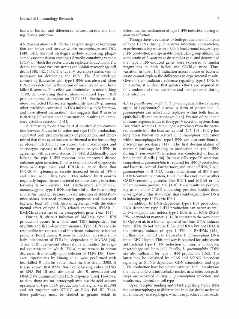
cGas
RNApol III
CDNs
STINGIRF3
IRF5IRF3
/7
Type I IFNs
MDPL. monocytogenes
M. tuberculosis
NOD2
RIP2
RNA
ssRNA/dsRNA
Cytosolic RNAS.L.
entericapneumophila
C. burnetii
ssRNA/dsRNA
RIG-I / MDA-SIRF3
IRF3/7
TRIF
LPSS.C.
entericaburnetii
Endosomal dsRNAS. enterica
TLR4
TLR3
Cytosolic DNA
?
TLR7
TLR9Endosomal ssRNAor DNA
burnetii
MyD88
IRF7
L. pneumophila
M. tuberculosis
L. monocytogenes
C.
C.
trachomatis
burnetii
F. tularensis (STING)
B.
C.
abortus
Cytosolic DNAB. abortus ?
MyD88
?
?
?
?
?
?
Figure 1: Induction of type I IFNs by intracellular bacteria. Type I IFNs can be induced via cytosolic nucleic acid sensors RIG-I, MDA5, RNAPol III, cGAS, and STING or nucleic acid-sensing TLR3, 7 or 9. Bacterial products like LPS andMDP can signal the production of type I IFNsvia TLR4 or NOD2/RIP2, respectively.
8 Journal of Immunology Research
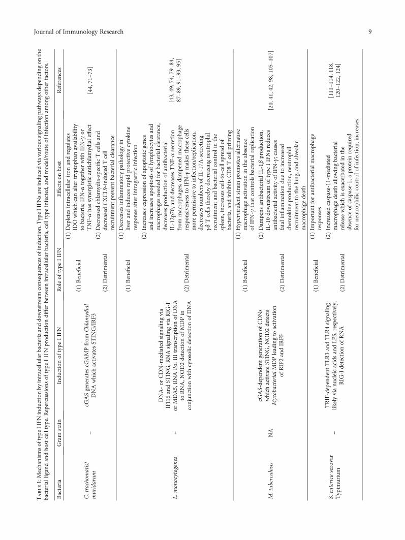
Table1:Mechanism
softype
IIFN
indu
ctionby
intracellularbacteriaanddo
wnstream
consequencesofindu
ction.TypeIIFN
sareindu
cedviavariou
ssignalingpathwaysdepend
ingon
the
bacterialligandandho
stcelltype.R
epercussions
oftype
IIFN
prod
uction
differ
betweenintracellularbacteria,celltypeinfected,and
mod
el/rou
teof
infectionam
ongotherfactors.
Bacteria
Gram
stain
Indu
ctionof
type
IIFN
Roleof
type
IIFN
Effectson
host
References
C.trachom
atis/
muridarum
−cG
ASgeneratescG
AMPfrom
Chlam
ydial
DNAwhich
activatesST
ING/IRF3
(1)Beneficial
(1)Depletesintracellulariron
andregulates
IDO
which
canaltertryptoph
anavailability
tobacteria;IFN
-αtogether
withIFN-γ
orTNF-αhassynergisticantichlamydialeffect
[44,71–73]
(2)Detrimental
(2)Decreased
chlamydia-specificTcells
and
decreasedCXCL9
-ind
uced
Tcell
recruitm
entpreventbacterialclearance
L.monocytogenes
+
DNA-or
CDN-m
ediatedsignalingvia
IFI16andST
ING,R
NAsignalingviaRIG
-Ior
MDA5,RNAPol
IIItranscriptionof
DNA
toRNA,N
OD2detectionof
MDPin
conjun
ctionwithcytosolic
detectionof
DNA
(1)Beneficial
(1)Decreases
inflam
matorypathologyin
liver
andindu
cesrapidprotective
cytokine
respon
seafterintragastricinfection
[43,49,74,79–84,
87–89,91–93,95]
(2)Detrimental
(2)Increasesexpression
ofapop
toticgenes
andincreasesapop
tosisof
lymph
ocytes
and
macroph
ages
needed
forbacterialclearance,
decreasesprod
uction
ofantibacterial
IL-12p70,and
decreasesTNF-αsecretion
from
macroph
ages;d
ampenedmacroph
age
respon
siveness
toIFN-γ
makes
thesecells
moreperm
issive
toinfection/replication,
decreasesnu
mbersof
IL-17A
-secreting
γδTcells
therebydecreasing
neutroph
ilrecruitm
entandbacterialcon
trol
inthe
spleen,increases
cell-to-cellspreadof
bacteria,and
inhibitsCD8Tcellprim
ing
M.tub
erculosis
NA
cGAS-depend
entgeneration
ofCDNs
which
activateST
ING,N
OD2detects
MycobacterialMDPleadingto
activation
ofRIP2andIRF5
(1)Beneficial
(1)Hypervirulent
strain
prom
otes
alternative
macroph
ageactivation
intheabsence
ofIFN-γ
that
controlsbacterialreplication
[20,41,42,98,105–107]
(2)Detrimental
(2)Dam
pens
antibacterialIL-1β
prod
uction
,IL-10do
wnstream
oftype
IIFNsredu
ces
antibacterialactivityof
IFN-γ;causes
fatalinfl
ammationdu
eto
increased
chem
okineprod
uction
,neutrop
hil
recruitm
entto
thelung,and
alveolar
macroph
agedeath
S.enterica
serovar
Typim
urium
−TRIF-dependent
TLR
3andTLR
4signaling
likelyvianu
cleicacidsandLP
S,respectively,
RIG
-Idetectionof
RNA
(1)Beneficial
(1)Im
portantforantibacterialm
acroph
age
respon
ses
[111–114,118,
120–122,124]
(2)Detrimental
(2)Increasedcaspase-11-m
ediated
macroph
agedeathallowingbacterial
releasewhich
isexacerbatedin
the
absenceof
caspase-1,aproteinrequ
ired
forneutroph
iliccontrolo
finfection,
increases
9Journal of Immunology Research

Table1:Con
tinu
ed.
Bacteria
Gram
stain
Indu
ctionof
type
IIFN
Roleof
type
IIFN
Effectson
host
References
RIP1/RIP3-mediatedmacroph
agedeath;
supp
resses
IL-1
cytokine
andneutroph
ilchem
oattractanttranscriptsin
macroph
ages
which
decreasesbacterialcon
trol;
influenza-indu
cedtype
IIFNsnegatively
impactgutmicrobiotaanddecrease
innate
respon
sesin
thegut,increasing
susceptibilityto
S.enterica
F.tularensis
−cG
AS,IFI204,STIN
G,and
IRF3-dependent
(1)Beneficial
(1)TypeIIFN-ind
uced
GBPsactivateAIM
2inflam
masom
eleadingto
macroph
age
pyroptosisandremovalof
replicativeniche
[91,126,135–139]
(2)Detrimental
(2)Decreased
numberof
IL-17A
-secreting
γδTcells
which
areim
portantfor
neutroph
ilrecruitm
entto
thespleen
andbacterialcon
trol
B.abortus
−
RNAPol
IIIand/or
STIN
Gare
requ
ired
fortype
IIFN
indu
ctionin
aMyD
88-dependent
butTRIF-
andTLR
-ind
ependent
manner
(1)Detrimental
(1)Alters
DCmaturationwhich
dampens
DCcytokine
prod
uction
;increases
bacterial
load
dueto
decreasesin
IFN-γ
andNO
prod
uction
;increasesspleno
cyte
apop
tosisandTrailexpression
inmacroph
ages
[36,141,144]
L.pn
eumophila
−
RIG
-Idetectionof
RNAandsubsequent
signalingthroughIPS-1andIRF3,R
NA
Pol
III-depend
enttranscriptionof
DNAto
RNAthen
signalingviaRIG
-IandIPS-1,
CDN
detectionviaST
ING
(1)Beneficial
(1)Con
trolsbacterialreplicationwithinho
stcellvacuoles;p
romotes
inflam
matory
macroph
agepo
larization
and
bacterialclearance
[47,82,150–154]
C.burnetii
−NOD1/2,RIG
-I,and
/orTLR
7/9-mediated
prod
uction
ofIFN-αin
pDCs,involves
IRF7
(1)Beneficial
(1)Adm
inistrationto
lung
was
protective—mechanism
unkn
ownat
thistime
[167,169]
(2)Detrimental
(2)Lack
ofIFNARbenefitedho
st;i.p.
deliveryof
IFN-α
was
harm
fultoho
stperhapsdu
eto
supp
ressionof
necessary
inflam
matorycytokines
10 Journal of Immunology Research
IVB secretion system of L. pneumophila; so, it is plausiblethat C. burnetii is sensed and induces type I IFNs in a mannersimilar to that of L. pneumophila [168]. Additionally, C.burnetii-stimulated pDCs make IFN-α and have upregulatedexpression of genes upstream of type I IFN production likeTLR7/9, IRF7,MyD88, RIG-I,NOD1, andNOD2 [169]. Thus,C. burnetii may induce production of type I IFNs in manyways, though pathways likely differ between cell types. Ourresults provide an excellent example of the need to assessthe impact of type I IFNs on different tissue sites duringbacterial infection.
5. Concluding Remarks
Type I IFNs are potent immunomodulatory signaling mole-cules and have the capacity to diversely affect immuneresponses. Our knowledge of these signaling molecules hasexpanded greatly since their discovery, and now, we knowthey are induced during intracellular bacterial infectionsand have a multitude of effects (Figure 1, Table 1). Theevolutionary importance of type I IFN production in thiscontext is clear, as many intracellular bacteria have devisedmethods of co-opting type I IFN production for their benefit.Enhancing means of cell death and dampening inflammatoryresponses serve as excellent examples of this, but these effectscould also benefit the host. The downstream effects of type IIFNs depend on the route of infection, which PRR signalingpathways are engaged, and the presence of virulence factorsthat allow for intracellular detection of bacterial components.In each of the intracellular bacteria reviewed here, there is noclear-cut evidence as to the effects of type I IFNs being onlybeneficial or only detrimental to the host. This is not surpris-ing because minute differences in the magnitude of type IIFN protein expression and expression of IFNAR on cellsurfaces can tip the balance and define the impact of type IIFNs. Thus, the web of type I IFN signaling is more complexthan previously thought. When type I IFNs are part of theimmune response in a given setting, everything from hostgenetics and health history to current disease state must beevaluated. It has been shown in mice that influenza A-induced type I IFNs increase mycobacterial growth anddisease in coinfected animals [170]. Thus, secondary infec-tions duringM. tuberculosis with pathogens that may inducetype I IFNs could have a drastic impact on antimycobacterialhost responses. Furthermore, treatment for C. burnetii infec-tion is an aggressive long term antibiotic regimen, andconsidering intratracheal delivery of type I IFNs is beneficialin a mouse model of disease, perhaps aerosolized type I IFNsmay be an effective alternative to antibiotics [167, 171].Situations like this highlight the need for more detailedstudies to better understand how, when, and why type I IFNsmay benefit the host or pathogen. This may open newavenues of treatment options beyond antibiotics to combatintracellular bacterial infections.
Abbreviations
AIM2: Absent in melanoma 2BMDM: Bone marrow-derived macrophages
CDN(s): Cyclic dinucleotidecGAMP: Cyclic GMP-AMPcGAS: Cyclic GMP-AMP synthaseCLR(s): C-type lectin receptorGBP(s): Guanylate binding proteinIFN: InterferonIFNAR: Type I IFN receptorMDP: Muramyl dipeptideNLR(s): NOD-like receptorPAMPs: Pathogen-associated molecular patternsPRRs: Pattern recognition receptorsRIP: Receptor interacting protein/receptor interacting
protein kinaseRLR(s): RIG-I-like receptorSesT: Salmonella enterica serovar TyphimuriumSTING: Stimulator of interferon genesTLR(s): Toll-like receptor.
Conflicts of Interest
The authors declare no conflict of interest.
Authors’ Contributions
Deann T. Snyder, Jodi F. Hedges, and Mark A. Jutila wrotethis article.
Acknowledgments
This project was primarily funded through NIH COBRE(P20 RR020185), with partial funding through NIH grant 1R21 AI117441-01A1, Montana University System ResearchInitiative: 51040-MUSRI2015-03, M.J. Murdock CharitableTrust, and TheMontana State University Agricultural Exper-imental Station (USDA/NIFA).
References
[1] V. van Pesch, H. Lanaya, J. C. Renauld, and T. Michiels,“Characterization of the murine alpha interferon gene fam-ily,” Journal of Virology, vol. 78, no. 15, pp. 8219–8228, 2004.
[2] C. Dunn, M. Brunetto, G. Reynolds et al., “Cytokines inducedduring chronic hepatitis B virus infection promote a pathwayfor NK cell-mediated liver damage,” Journal ExperimentalMedicine, vol. 204, no. 3, pp. 667–680, 2007.
[3] M. Sarasin-Filipowicz, E. J. Oakeley, F. H. T. Duong et al.,“Interferon signaling and treatment outcome in chronichepatitis C,” Proceedings of the National Academy of Sciencesof the United States of America, vol. 105, no. 19, pp. 7034–7039, 2008.
[4] E. B. Wilson, D. H. Yamada, H. Elsaesser et al., “Blockadeof chronic type I interferon signaling to control persistentLCMV infection,” Science, vol. 340, no. 6129, pp. 202–207, 2013.
[5] K. A. Hofmeyer, H. Jeon, and X. Zang, “The PD-1/PD-L1(B7-H1) pathway in chronic infection-induced cytotoxic Tlymphocyte exhaustion,” Journal of Biomedicine & Biotech-nology, vol. 2011, Article ID 451694, p. 9, 2011.
[6] I. Teige, A. Treschow, A. Teige et al., “IFN-beta gene deletionleads to augmented and chronic demyelinating experimental
11Journal of Immunology Research
autoimmune encephalomyelitis,” Journal of Immunology,vol. 170, no. 9, pp. 4776–4784, 2003.
[7] C. Guiducci, M. Gong, Z. Xu et al., “TLR recognition of selfnucleic acids hampers glucocorticoid activity in lupus,”Nature, vol. 465, no. 7300, pp. 937–941, 2010.
[8] T. Goldmann, T. Blank, and M. Prinz, “Fine-tuning of typeI IFN-signaling in microglia—implications for homeostasis,CNS autoimmunity and interferonopathies,” Current Opin-ion in Neurobiology, vol. 36, pp. 38–42, 2016.
[9] A. N. Theofilopoulos, R. Baccala, B. Beutler, and D. H.Kono, “Type I interferons (alpha-beta) in immunity andautoimmunity,” Annual Review of Immunology, vol. 23,pp. 307–335, 2004.
[10] R. Hernández-Pando, H. Orozcoe, A. Sampieri et al.,“Correlation between the kinetics of Th1, Th2 cells andpathology in a murine model of experimental pulmonarytuberculosis,” Immunology, vol. 89, no. 1, pp. 26–33, 1996.
[11] E. C. Borden, G. C. Sen, G. Uze et al., “Interferons at age 50:past, current and future impact on biomedicine,” NatureReviews. Drug Discovery, vol. 6, no. 12, pp. 975–990, 2007.
[12] H. M. Lazear, T. J. Nice, and M. S. Diamond, “Interferon-λ:immune functions at barrier surfaces and beyond,” Immu-nity, vol. 43, no. 1, pp. 15–28, 2015.
[13] N. A. de Weerd, J. P. Vivian, T. K. Nguyen et al., “Structuralbasis of a unique interferon-[beta] signaling axis mediated viathe receptor IFNAR1,” Nature Immunology, vol. 14, no. 9,pp. 901–907, 2013.
[14] L. B. Ivashkiv and L. T. Donlin, “Regulation of type Iinterferon responses,” Nature Reviews. Immunology, vol. 14,no. 1, pp. 36–49, 2014.
[15] P. Fitzgerald-Bocarsly, M. Feldman, M. Mendelsohn, S. Curl,and C. Lopez, “Human mononuclear cells which produceinterferon-alpha during NK(HSV-FS) assays are HLA-DRpositive cells distinct from cytolytic natural killer effectors,”Journal of Leukocyte Biology, vol. 43, no. 4, pp. 323–334, 1988.
[16] D. Parker and A. Prince, “Type I interferon response toextracellular bacteria in the airway epithelium,” Trends inImmunology, vol. 32, no. 12, pp. 582–588, 2011.
[17] M. Charrel-Dennis, E. Latz, K. A. Halmen et al., “TLR-independent type I interferon induction in response to anextracellular bacterial pathogen via intracellular recognitionof its DNA,” Cell Host & Microbe, vol. 4, no. 6, pp. 543–554, 2008.
[18] T. B. Clarke and J. N. Weiser, “Intracellular sensors ofextracellular bacteria,” Immunological Reviews, vol. 243,no. 1, pp. 9–25, 2011.
[19] G. M. Boxx and G. Cheng, “The roles of type I interferon inbacterial infection,” Cell Host & Microbe, vol. 19, no. 6,pp. 760–769, 2016.
[20] F. McNab, K. Mayer-Barber, A. Sher, A. Wack, and A.O’Garra, “Type I interferons in infectious disease,” NatureReviews. Immunology, vol. 15, no. 2, pp. 87–103, 2015.
[21] L. Zitvogel, L. Galluzzi, O. Kepp, M. J. Smyth, and G.Kroemer, “Type I interferons in anticancer immunity,”NatureReviews. Immunology, vol. 15, no. 7, pp. 405–414, 2015.
[22] O. Takeuchi and S. Akira, “Pattern recognition receptors andinflammation,” Cell, vol. 140, no. 6, pp. 805–820, 2010.
[23] S. Pandey, T. Kawai, and S. Akira, “Microbial sensing byToll-like receptors and intracellular nucleic acid sensors,”Cold Spring Harbor Perspectives in Biology, vol. 7, no. 1,article a016246, 2015.
[24] A. M. Lundberg, S. K. Drexler, C. Monaco et al., “Key differ-ences in TLR3/poly I:C signaling and cytokine induction byhuman primary cells: a phenomenon absent frommurine cellsystems,” Blood, vol. 110, no. 9, p. 3245, 2007.
[25] R. Barbalat, L. Lau, R. M. Locksley, and G. M. Barton,“Toll-like receptor 2 on inflammatory monocytes inducestype I interferon in response to viral but not bacterial ligands,”Nature Immunology, vol. 10, no. 11, pp. 1200–1207, 2009.
[26] S. Uematsu and S. Akira, “Toll-like receptors and type Iinterferons,” The Journal of Biological Chemistry, vol. 282,no. 21, pp. 15319–15323, 2007.
[27] T. W. Kim, K. Staschke, K. Bulek et al., “A critical role forIRAK4 kinase activity in Toll-like receptor-mediated innateimmunity,” The Journal of Experimental Medicine, vol. 204,no. 5, pp. 1025–1036, 2007.
[28] H. Hemmi, O. Takeuchi, T. Kawai et al., “A Toll-like receptorrecognizes bacterial DNA,” Nature, vol. 408, no. 6813,pp. 740–745, 2000.
[29] A. Eaton-Bassiri, S. B. Dillon, M. Cunningham et al.,“Toll-like receptor 9 can be expressed at the cell surfaceof distinct populations of tonsils and human peripheralblood mononuclear cells,” Infection and Immunity, vol. 72,no. 12, pp. 7202–7211, 2004.
[30] M. Adib-Conquy, D. Scott-Algara, J. M. Cavaillon, andF. Souza-Fonseca-Guimaraes, “TLR-mediated activationof NK cells and their role in bacterial/viral immune responsesin mammals,” Immunology and Cell Biology, vol. 92, no. 3,pp. 256–262, 2014.
[31] T. Kawai and S. Akira, “Pathogen recognition with Toll-likereceptors,” Current Opinion in Immunology, vol. 17, no. 4,pp. 338–344, 2005.
[32] R. Zhan, Q. Han, C. Zhang, Z. Tian, and J. Zhang, “Toll-likereceptor 2 (TLR2) and TLR9 play opposing roles in hostinnate immunity against Salmonella enterica serovar Typhi-murium infection,” Infection and Immunity, vol. 83, no. 4,pp. 1641–1649, 2015.
[33] U. Bhan, G. Trujillo, K. Lyn-Kew et al., “Toll-like receptor9 regulates the lung macrophage phenotype and hostimmunity in murine pneumonia caused by Legionellapneumophila,” Infection and Immunity, vol. 76, no. 7,pp. 2895–2904, 2008.
[34] N. Arpaia, J. Godec, L. Lau et al., “TLR signaling is requiredfor Salmonella typhimurium virulence,” Cell, vol. 144, no. 5,pp. 675–688, 2011.
[35] L. Y. Huang, K. J. Ishii, S. Akira, J. Aliberti, and B. Golding,“Th1-like cytokine induction by heat-killed Brucella abortusis dependent on triggering of TLR9,” Journal of Immunology,vol. 175, no. 6, p. 3964, 2005.
[36] N. Lapaque, A. Muller, L. Alexopoulou, J. C. Howard, andJ. P. Gorvel, “Brucella abortus induces Irgm3 and Irga6expression via type-I IFN by a MyD88-dependent pathway,without the requirement of TLR2, TLR4, TLR5 and TLR9,”Microbial Pathogenesis, vol. 47, no. 6, pp. 299–304, 2009.
[37] S. R. Paludan and A. G. Bowie, “Immune sensing of DNA,”Immunity, vol. 38, no. 5, pp. 870–880, 2013.
[38] E. E. Gray, D.Winship, J. M. Snyder, S. J. Child, A. P. Geballe,and D. B. Stetson, “The AIM2-like receptors are dispensablefor the interferon response to intracellular DNA,” Immunity,vol. 45, no. 2, pp. 255–266, 2016.
[39] L. Sun, J. Wu, F. Du, X. Chen, and Z. J. Chen, “Cyclic GMP-AMP synthase is a cytosolic DNA sensor that activates the
12 Journal of Immunology Research

type-I interferon pathway,” Science, vol. 339, no. 6121, p. 10,2013.
[40] A. C. Collins, H. Cai, T. Li et al., “Cyclic GMP-AMP synthase(cGAS) is an innate immune DNA sensor for Mycobacteriumtuberculosis,” Cell Host & Microbe, vol. 17, no. 6, pp. 820–828, 2015.
[41] R. Wassermann, M. Gulen, C. Sala et al., “Mycobac-terium tuberculosis differentially activates cGAS- andinflammasome-dependent intracellular immune responsesthrough ESX-1,” Cell Host & Microbe, vol. 17, no. 6,pp. 799–810, 2015.
[42] R. O. Watson, S. L. Bell, D. A. MacDuff et al., “Thecytosolic sensor cGAS detects Mycobacterium tuberculo-sis DNA to induce type I interferons and activateautophagy,” Cell Host & Microbe, vol. 17, no. 6,pp. 811–819, 2015.
[43] K. Hansen, T. Prabakaran, A. Laustsen et al., “Listeriamonocytogenes induces IFNβ expression through an IFI16-,cGAS- and STING-dependent pathway,” The EMBO Journal,vol. 33, no. 15, pp. 1654–1666, 2014.
[44] Y. Zhang, L. Yeruva, A. Marinov et al., “The DNA sensor,cyclic GMP-AMP synthase, is essential for induction ofIFN-β during chlamydia trachomatis infection,” Journal ofImmunology, vol. 193, no. 5, pp. 2394–2404, 2014.
[45] S. R. Paludan, “Activation and regulation of DNA-drivenimmune responses,” Microbiology and Molecular BiologyReviews, vol. 79, no. 2, pp. 225–241, 2015.
[46] A. Ablasser, F. Bauernfeind, G. Hartmann, E. Latz, K. A.Fitzgerald, and V. Hornung, “RIG-I dependent sensing ofpoly(dA-dT) via the induction of an RNA polymeraseIII transcribed RNA intermediate,” Nature Immunology,vol. 10, no. 10, pp. 1065–1072, 2009.
[47] Y. H. Chiu, J. B. MacMillan, and Z. J. Chen, “RNA polymer-ase III detects cytosolic DNA and induces type-I interferonsthrough the RIG-I pathway,” Cell, vol. 138, no. 3, pp. 576–591, 2009.
[48] C. X. Koo, K. Kobiyama, Y. J. Shen et al., “RNA polymeraseIII regulates cytosolic RNA:DNA hybrids and intracellularmicroRNA expression,” The Journal of Biological Chemistry,vol. 290, no. 12, pp. 7463–7473, 2015.
[49] Z. Abdullah, M. Schlee, S. Roth et al., “RIG-I detectsinfection with live Listeria by sensing secreted bacterialnucleic acids,” The EMBO Journal, vol. 31, no. 21, pp. 4153–4164, 2012.
[50] E. Dixit and J. C. Kagan, “Intracellular pathogen detection byRIG-I-like receptors,” Advances in Immunology, vol. 117,pp. 99–125, 2013.
[51] H. Kato, O. Takeuchi, E. Mikamo-Satoh et al., “Length-dependent recognition of double-stranded ribonucleicacids by retinoic acid-inducible gene-I and melanomadifferentiation-associated gene 5,” Journal of ExperimentalMedicine, vol. 205, no. 7, pp. 1601–1610, 2008.
[52] S. Jensen and A. R. Thomsen, “Sensing of RNA viruses: areview of innate immune receptors involved in recognizingRNA virus invasion,” Journal of Virology, vol. 86, no. 6,pp. 2900–2910, 2012.
[53] S. Reikine, J. B. Nguyen, and Y. Modis, “Pattern recognitionand signaling mechanisms of RIG-I and MDA5,” Frontiersin Immunology, vol. 5, p. 342, 2014.
[54] K. R. Rodriguez, A. M. Bruns, and C. M. Horvath, “MDA5and LGP2: accomplices and antagonists of antiviral signal
transduction,” Journal of Virology, vol. 88, no. 15, pp. 8194–8200, 2014.
[55] V. Motta, F. Soares, T. Sun, and D. J. Philpott, “NOD-likereceptors: versatile cytosolic sentinels,” Physiological Reviews,vol. 95, no. 1, p. 149, 2014.
[56] G. Chen, M. H. Shaw, Y. G. Kim, and G. Nunez, “NOD-likereceptors: role in innate immunity and inflammatory dis-ease,” Annual Review of Pathology: Mechanisms of Disease,vol. 4, pp. 365–398, 2009.
[57] S. E. Girardin, I. G. Boneca, J. Viala et al., “Nod2 is a generalsensor of peptidoglycan through Muramyl dipeptide (MDP)detection,” The Journal of Biological Chemistry, vol. 278,no. 11, pp. 8869–8872, 2003.
[58] S. E. Girardin, I. G. Boneca, L. A. M. Carneiro et al.,“Nod1 detects a unique muropeptide from gram-negativebacterial peptidoglycan,” Science, vol. 300, no. 5625,p. 1584, 2003.
[59] Y. Zhong, A. Kinio, and M. Saleh, “Functions of NOD-likereceptors in human diseases,” Frontiers in Immunology,vol. 4, p. 333, 2013.
[60] A. Sabbah, T. H. Chang, R. Harnack et al., “Activation ofinnate immune antiviral responses by Nod2,” Nature Immu-nology, vol. 10, no. 10, pp. 1073–1080, 2009.
[61] T. Watanabe, N. Asano, A. Kitani, I. J. Fuss, T. Chiba, andW. Strober, “NOD1-mediated mucosal host defense againsthelicobacter pylori,” International Journal of Inflammation,vol. 2010, Article ID 476482, p. 6, 2010.
[62] I. M. Dambuza and G. D. Brown, “C-type lectins in immu-nity: recent developments,” Current Opinion in Immunology,vol. 32, pp. 21–27, 2015.
[63] J. C. Hoving, G. J. Wilson, and G. D. Brown, “SignallingC-type lectin receptors, microbial recognition and immu-nity,” Cellular Microbiology, vol. 16, no. 2, pp. 185–194, 2014.
[64] G. Trinchieri, “Type I interferon: friend or foe?” TheJournal of Experimental Medicine, vol. 207, no. 10,pp. 2053–2063, 2010.
[65] S. Kaur and L. C. Platanias, “IFN-[beta]-specific signaling viaa unique IFNAR1 interaction,” Nature Immunology, vol. 14,no. 9, pp. 884–885, 2013.
[66] L. C. Platanias, “Mechanisms of type-I- and type-II-interferon-mediated signalling,” Nature Reviews. Immu-nology, vol. 5, no. 5, pp. 375–386, 2005.
[67] A. K. Perry, G. Chen, D. Zheng, H. Tang, and G. Cheng,“The host type I interferon response to viral and bacterialinfections,” Cell Research, vol. 15, no. 6, pp. 407–422, 2005.
[68] H. Cho and B. L. Kelsall, “The role of type I interferons inintestinal infection, homeostasis, and inflammation,” Immu-nological Reviews, vol. 260, no. 1, pp. 145–167, 2014.
[69] J. Kazar, J. D. Gillmore, and F. B. Gordon, “Effect ofinterferon and interferon inducers on infections with anonviral intracellular microorganism, chlamydia tracho-matis,” Infection and Immunity, vol. 3, no. 6, pp. 825–832, 1971.
[70] L. M. de la Maza, E. M. Peterson, J. M. Goebel, C. W. Fennie,and C. W. Czarniecki, “Interferon-induced inhibition ofchlamydia trachomatis: dissociation from antiviral andantiproliferative effects,” Infection and Immunity, vol. 47,no. 3, pp. 719–722, 1985.
[71] T. Ishihara, M. Aga, K. Hino et al., “Inhibition of Chlamydiatrachomatis growth by human interferon-alpha: mechanismsand synergistic effect with interferon-gamma; and tumor
13Journal of Immunology Research

necrosis factor-alpha,” Biomedical Research, vol. 26, no. 4,pp. 179–185, 2005.
[72] P. Puccetti, “On watching the watchers: IDO and type I/IIIFN,” European Journal of Immunology, vol. 37, no. 4,pp. 876–879, 2007.
[73] U. M. Nagarajan, D. Prantner, J. D. Sikes et al., “Type Iinterferon signaling exacerbates Chlamydia muridarumgenital infection in a murine model,” Infection and Immu-nity, vol. 76, no. 10, pp. 4642–4648, 2008.
[74] M. Rayamajhi, J. Humann, K. Penheiter, K. Andreasen,and L. L. Lenz, “Induction of IFN-alpha-beta enables Listeriamonocytogenes to suppress macrophage activation by IFN-gamma,” The Journal of Experimental Medicine, vol. 207,no. 2, pp. 327–337, 2010.
[75] B. Lukas and J. Hruskova, “A virus inhibitor circulating inthe blood of chickens, induced by Francisella tularensisand Listeria monocytogenes,” Folia Microbiologia (Praha),vol. 12, no. 2, pp. 157–161, 1967.
[76] E. A. Havell, “Augmented induction of interferons duringListeria monocytogenes infection,” The Journal of InfectiousDiseases, vol. 153, no. 5, pp. 960–969, 1986.
[77] E. A. Havell, “Listeria monocytogenes-induced interferon-gamma primes the host for production of tumor necrosisfactor and interferon-alpha/beta,” The Journal of InfectiousDiseases, vol. 167, no. 6, pp. 1364–1371, 1993.
[78] C. E. Witte, K. A. Archer, C. S. Rae, J. D. Sauer, J. J.Woodward, and D. A. Portnoy, “Chapter 8 - innateimmune pathways triggered by Listeria monocytogenesand their role in the induction of cell-mediated immunity,”in Advances in Immunology Immunity to Listeria monocyto-genes, R. U. A. J. Emil, Ed., vol. 113, pp. 135–156, AcademicPress, Cambridge, MA, USA, 2012.
[79] K. A. Archer, J. Durack, and D. A. Portnoy, “STING-dependent type I IFN production inhibits cell-mediatedimmunity to Listeria monocytogenes,” PLoS Pathogens,vol. 10, no. 1, article e1003861, 2014.
[80] C. A. Hagmann, A. M. Herzner, Z. Abdullah et al., “RIG-Idetects triphosphorylated RNA of Listeria monocytogenesduring infection in non-immune cells,” PLoS One, vol. 8,no. 4, article e62872, 2013.
[81] J. H. Leber, G. T. Crimmins, S. Raghavan, N. P. Meyer-Morse, J. S. Cox, and D. A. Portnoy, “Distinct TLR- andNLR-mediated transcriptional responses to an intracellularpathogen,” PLoS Pathogens, vol. 4, no. 1, article e6, 2008.
[82] D. B. Stetson and R. Medzhitov, “Recognition of cytosolicDNA activates an IRF3-dependent innate immune response,”Immunity, vol. 24, no. 1, pp. 93–103, 2006.
[83] J. J. Woodward, A. T. Iavarone, and D. A. Portnoy,“C-di-AMP secreted by intracellular Listeria monocytogenesactivates a host type I interferon response,” Science,vol. 328, no. 5986, pp. 1703–1705, 2010.
[84] D. L. Burdette, K. M. Monroe, K. Sotelo-Troha et al., “STINGis a direct innate immune sensor of cyclic-di-GMP,” Nature,vol. 478, no. 7370, pp. 515–518, 2011.
[85] K. T. Schwartz, J. D. Carleton, S. J. Quillin, S. D. Rollins,D. A. Portnoy, and J. H. Leber, “Hyperinduction of hostbeta interferon by a Listeria monocytogenes strain naturallyoverexpressing the multidrug efflux pump MdrT,” Infectionand Immunity, vol. 80, no. 4, pp. 1537–1545, 2012.
[86] K. J. Ishii, T. Kawagoe, S. Koyama et al., “TANK-bindingkinase-1 delineates innate and adaptive immune responses
to DNA vaccines,” Nature, vol. 451, no. 7179, pp. 725–729, 2008.
[87] R. M. O’Connell, S. K. Saha, S. A. Vaidya et al., “Type Iinterferon production enhances susceptibility to Listeriamonocytogenes infection,” Journal Experimental Medicine,vol. 200, no. 4, pp. 437–445, 2004.
[88] J. A. Carrero, B. Calderon, and E. R. Unanue, “Type Iinterferon sensitizes lymphocytes to apoptosis and reducesresistance to Listeria infection,” Journal Experimental Medi-cine, vol. 200, no. 4, p. 535, 2004.
[89] V. Auerbuch, D. G. Brockstedt, N. Meyer-Morse, M.O’Riordan, and D. A. Portnoy, “Mice lacking the type Iinterferon receptor are resistant to Listeria monocytogenes,”The Journal of Experimental Medicine, vol. 200, no. 4,pp. 527–533, 2004.
[90] T. Reimer, M. Schweizer, and T. W. Jungi, “Type I IFNinduction in response to Listeria monocytogenes in humanmacrophages: evidence for a differential activation of IFNregulatory factor 3 (IRF3),” Journal of Immunology,vol. 179, no. 2, p. 1166, 2007.
[91] T. Henry, G. S. Kirimanjeswara, T. Ruby et al., “Type Iinterferon signaling constrains IL-17A/F secretion by γδ Tcells during bacterial infections,” Journal of Immunology,vol. 184, no. 7, pp. 3755–3767, 2010.
[92] S. E. Osborne, B. Sit, A. Shaker et al., “Type I interferonpromotes cell-to-cell spread of Listeria monocytogenes,”Cellular Microbiology, vol. 19, no. 3, 2017.
[93] S. J. Kearney, C. Delgado, E. M. Eshleman, K. K. Hill, B. P.O’Connor, and L. L. Lenz, “Type I interferons down regulatemyeloid cell IFNGR by inducing recruitment of an Egr3/Nab1 complex that silences ifngr1 transcription,” Journal ofImmunology, vol. 191, no. 6, pp. 3384–3392, 2013.
[94] M. G. Pitts, T. Myers-Morales, and S. E. F. D’Orazio, “Type IIFN does not promote susceptibility to foodborne Listeriamonocytogene,” Journal of Immunology, vol. 196, no. 7,p. 3109, 2016.
[95] E. Kernbauer, V. Maier, I. Rauch, M. Müller, and T. Decker,“Route of infection determines the impact of type Iinterferons on innate immunity to Listeria monocytogenes,”PLoS One, vol. 8, no. 6, article e65007, 2013.
[96] B. Lechartier, J. Rybniker, A. Zumla, and S. T. Cole,“Tuberculosis drug discovery in the post-post-genomic era,”EMBO Molecular Medicine, vol. 6, no. 2, pp. 158–168, 2014.
[97] C. Manca, L. Tsenova, S. Freeman et al., “Hypervirulent M.Tuberculosis W/Beijing strains upregulate type I IFNs andincrease expression of negative regulators of the Jak-Statpathway,” Journal of Interferon & Cytokine Research,vol. 25, no. 11, pp. 694–701, 2005.
[98] A. Dorhoi, V. Yeremeev, G. Nouailles et al., “Type I IFN sig-naling triggers immunopathology in tuberculosis-susceptiblemice by modulating lung phagocyte dynamics,” EuropeanJournal of Immunology, vol. 44, no. 8, pp. 2380–2393, 2014.
[99] S. A. Stanley, J. E. Johndrow, P. Manzanillo, and J. S. Cox,“The type I IFN response to infection with Mycobacteriumtuberculosis requires ESX-1-mediated secretion and contrib-utes to pathogenesis,” Journal of Immunology, vol. 178, no. 5,p. 3143, 2007.
[100] M. P. R. Berry, C. M. Graham, F. W. McNab et al.,“An interferon-inducible neutrophil-driven blood transcrip-tional signature in human tuberculosis,” Nature, vol. 466,no. 7309, pp. 973–977, 2010.
14 Journal of Immunology Research

[101] A. K. Pandey, Y. Yang, Z. Jiang et al., “NOD2, RIP2 andIRF5 play a critical role in the type I interferon responseto Mycobacterium tuberculosis,” PLoS Pathogens, vol. 5,no. 7, article e1000500, 2009.
[102] E. J. M. Stoop, W. Bitter, and A. M. van der Sar, “Tuberclebacilli rely on a type VII army for pathogenicity,” Trends inMicrobiology, vol. 20, no. 10, pp. 477–484, 2012.
[103] B. B. Mishra, P. Moura-Alves, A. Sonawane et al., “Myco-bacterium tuberculosis protein ESAT-6 is a potent activatorof the NLRP3/ASC inflammasome,” Cellular Microbiology,vol. 12, no. 8, pp. 1046–1063, 2010.
[104] H. Saiga, S. Kitada, Y. Shimada et al., “Critical role of AIM2 inMycobacterium tuberculosis infection,” International Immu-nology, vol. 24, no. 10, pp. 637–644, 2012.
[105] C. Manca, L. Tsenova, A. Bergtold et al., “Virulence of aMycobacterium tuberculosis clinical isolate in mice isdetermined by failure to induce Th1 type immunity and isassociated with induction of IFN-alpha-beta,” PNAS,vol. 98, no. 10, pp. 5752–5757, 2001.
[106] F. Bouchonnet, N. Boechat, M. Bonay, and A. J. Hance,“Alpha/Beta interferon impairs the ability of human macro-phages to control growth of Mycobacterium bovis BCG,”Infection and Immunity, vol. 70, no. 6, pp. 3020–3025, 2002.
[107] L. Moreira-Teixeira, J. Sousa, F. W. McNab et al., “TypeI IFN inhibits alternative macrophage activation duringMycobacterium tuberculosis infection and leads to enhancedprotection in the absence of IFN-γ signaling,” Journal ofImmunology, vol. 197, no. 12, pp. 4714–4726, 2016.
[108] M. Lorkowski, A. Felipe-López, C. A. Danzer, N. Hansmeier,and M. Hensel, “Salmonella enterica invasion of polarizedepithelial cells is a highly cooperative effort,” Infection andImmunity, vol. 82, no. 6, pp. 2657–2667, 2014.
[109] C. G. Forest, E. Ferraro, S. C. Sabbagh, and F. Daigle,“Intracellular survival of Salmonella enterica serovar Typhiin human macrophages is independent of Salmonellapathogenicity island (SPI)-2,”Microbiology, vol. 156, Part 12,pp. 3689–3698, 2010.
[110] O. Steele-Mortimer, J. H. Brumell, L. A. Knodler, S. Méresse,A. Lopez, and B. B. Finlay, “The invasion-associated type IIIsecretion system of Salmonella enterica serovar Typhimur-ium is necessary for intracellular proliferation and vacuolebiogenesis in epithelial cells,” Cellular Microbiology, vol. 4,no. 1, pp. 43–54, 2002.
[111] P. Broz, K. Newton, M. Lamkanfi, S. Mariathasan, V. M.Dixit, and D. M. Monack, “Redundant roles for inflam-masome receptors NLRP3 and NLRC4 in host defenseagainst Salmonella,” The Journal of Experimental Medicine,vol. 207, no. 8, pp. 1745–1755, 2010.
[112] P. Broz, T. Ruby, K. Belhocine et al., “Caspase-11 increasessusceptibility to Salmonella infection in the absence ofcaspase-1,” Nature, vol. 490, no. 7419, pp. 288–291, 2012.
[113] K. A. Owen, C. J. Anderson, and J. E. Casanova, “Salmo-nella suppresses the TRIF-dependent type I interferonresponse in macrophages,” MBio, vol. 7, no. 1, articlee02051–15, 2016.
[114] M. Schmolke, J. R. Patel, E. de Castro et al., “RIG-I detectsmRNA of intracellular Salmonella enterica serovar Typhi-murium during bacterial infection,” MBio, vol. 5, no. 2,article e01006–14, 2014.
[115] M. Lara-Tejero, F. S. Sutterwala, Y. Ogura et al., “Role ofthe caspase-1 inflammasome in Salmonella typhimurium
pathogenesis,” The Journal of Experimental Medicine,vol. 203, no. 6, pp. 1407–1412, 2006.
[116] B. Raupach, S. K. Peuschel, D. M. Monack, and A.Zychlinsky, “Caspase-1-mediated activation of interleukin-1b (IL-1β) and IL-18 contributes to innate immunedefenses against Salmonella enterica serovar Typhimuriuminfection,” Infection and Immunity, vol. 74, no. 8,pp. 4922–4926, 2006.
[117] N. Kayagaki, S.Warming, M. Lamkanfi et al., “Non-canonicalinflammasome activation targets caspase-11,” Nature,vol. 479, no. 7371, pp. 117–121, 2011.
[118] E. A. Miao, D. P. Mao, N. Yudkovsky et al., “Innate immunedetection of the type III secretion apparatus through theNLRC4 inflammasome,”Proceedings of theNational Academyof Sciences of the United States of America, vol. 107, no. 7,pp. 3076–3080, 2010.
[119] G. Bukholm, B. P. Berdal, C. Haug, and M. Degré, “Mousefibroblast interferon modifies Salmonella typhimuriuminfection in infant mice,” Infection and Immunity, vol. 45,no. 1, pp. 62–66, 1984.
[120] N. Robinson, S. McComb, R. Mulligan, R. Dudani, L.Krishnan, and S. Sad, “Type I interferon induces necroptosisin macrophages during infection with Salmonella entericaserovar Typhimurium,” Nature Immunology, vol. 13, no. 10,pp. 954–962, 2012.
[121] P. Vandenabeele, W. Declercq, F. Van Herreweghe, andT. Vanden Berghe, “The role of the kinases RIP1 andRIP3 in TNF-induced necrosis,” Science Signaling, vol. 3,no. 115, p. re4, 2010.
[122] D. J. Perkins, R. Rajaiah, S. M. Tennant et al., “Salmonellatyphimurium co-opts the host type I IFN system to restrictmacrophage innate immune transcriptional responses selec-tively,” Journal of Immunology, vol. 195, no. 5, pp. 2461–2471, 2015.
[123] C. Dilantika, E. R. Sedyaningsih, M. R. Kasper et al.,“Influenza virus infection among pediatric patients reportingdiarrhea and influenza-like illness,” BMC Infectious Diseases,vol. 10, no. 1, p. 3, 2010.
[124] E. Deriu, G. M. Boxx, X. He et al., “Influenza virus affectsintestinal microbiota and secondary Salmonella infection inthe gut through type I interferons,” PLoS Pathogens, vol. 12,no. 5, article e1005572, 2016.
[125] M. A. Freudenberg, T. Merlin, C. Kalis, Y. Chvatchko, H.Stübig, and C. Galanos, “Cutting edge: a murine, IL-12-independent pathway of IFN-gamma induction by gram-negative bacteria based on STAT4 activation by type I IFNand IL-18 signaling,” Journal of Immunology, vol. 169,no. 4, pp. 1665–1668, 2002.
[126] T. Henry, A. Brotcke, D. S. Weiss, L. J. Thompson, and D. M.Monack, “Type I interferon signaling is required for activa-tion of the inflammasome during Francisella infection,” TheJournal of Experimental Medicine, vol. 204, no. 5, pp. 987–994, 2007.
[127] C. Checroun, T. D. Wehrly, E. R. Fischer, S. F. Hayes, andJ. Celli, “Autophagy-mediated reentry of Francisella tular-ensis into the endocytic compartment after cytoplasmicreplication,” Proceedings of the National Academy of Sciencesof the United States of America, vol. 103, no. 39, pp. 14578–14583, 2006.
[128] L. A. Allen, “Interview with Dr. Lee-Ann Allen regardingpivotal advance: Francisella tularensis LVS evades killing byhuman neutrophils via inhibition of the respiratory burst
15Journal of Immunology Research

and phagosome escape,” Journal of Leukocyte Biology, vol. 80,no. 6, pp. 1222–1223, 2006.
[129] J. D. Hall, M. D. Woolard, B. M. Gunn et al., “Infected-host-cell repertoire and cellular response in the lungfollowing inhalation of Francisella tularensis Schu S4,LVS, or U112,” Infection and Immunity, vol. 76, no. 12,pp. 5843–5852, 2008.
[130] G. S. Baron and F. E. Nano, “MglA and MglB are requiredfor the intramacrophage growth of Francisella novicida,”Molecular Microbiology, vol. 29, no. 1, pp. 247–259, 1998.
[131] M. Santic, M. Molmeret, K. E. Klose, S. Jones, and Y. A.Kwaik, “The Francisella tularensis pathogenicity islandprotein IglC and its regulator MglA are essential for modulat-ing phagosome biogenesis and subsequent bacterial escapeinto the cytoplasm,” Cellular Microbiology, vol. 7, no. 7,pp. 969–979, 2005.
[132] S. Mariathasan, D. S. Weiss, V. M. Dixit, and D. M. Monack,“Innate immunity against Francisella tularensis is dependenton the ASC/caspase-1 axis,” The Journal of ExperimentalMedicine, vol. 202, no. 8, pp. 1043–1049, 2005.
[133] M. A. Gavrilin, I. J. Bouakl, N. L. Knatz et al., “Internalizationand phagosome escape required for Francisella to inducehuman monocyte IL-1β processing and release,” Proceedingsof the National Academy of Sciences of the United States ofAmerica, vol. 103, no. 1, pp. 141–146, 2006.
[134] A. M. Hajjar, M. D. Harvey, S. A. Shaffer et al., “Lack ofin vitro and in vivo recognition of Francisella tularensissubspecies lipopolysaccharide by Toll-like receptors,” Infec-tion and Immunity, vol. 74, no. 12, pp. 6730–6738, 2006.
[135] K. M. Storek, N. A. Gertsvolf, M. B. Ohlson, and D. M.Monack, “cGAS and Ifi204 cooperate to produce type I IFNsin response to Francisella infection,” Journal of Immunology,vol. 194, no. 7, pp. 3236–3245, 2015.
[136] K. A. Fitzgerald and V. A. K. Rathinam, “GBPs take AIMat Francisella,” Nature Immunology, vol. 16, no. 5,pp. 443–444, 2015.
[137] S. M. Man, R. Karki, R. K. S. Malireddi et al., “The tran-scription factor IRF1 and guanylate-binding proteins targetactivation of the AIM2 inflammasome by Francisella infec-tion,” Nature Immunology, vol. 16, no. 5, pp. 467–475, 2015.
[138] E. Meunier, P. Wallet, R. F. Dreier et al., “Guanylate-bindingproteins promote activation of the AIM2 inflammasome dur-ing infection with Francisella novicida,” Nature Immunology,vol. 16, no. 5, pp. 476–484, 2015.
[139] B. H. Kim, A. R. Shenoy, P. Kumar, R. Das, S. Tiwari, andJ. D. MacMicking, “A family of IFN-gamma-inducible 65-kDGTPases protects against bacterial infection,” Science,vol. 332, no. 6030, pp. 717–721, 2011.
[140] J. Celli, C. de Chastellier, D. M. Franchini, J. Pizarro-Cerda,E. Moreno, and J. P. Gorvel, “Brucella evades macrophagekilling via VirB-dependent sustained interactions with theendoplasmic reticulum,” Journal Experimental. Medicine,vol. 198, no. 4, pp. 545–556, 2003.
[141] S. P. Salcedo, M. I. Marchesini, H. Lelouard et al., “Brucellacontrol of dendritic cell maturation is dependent on theTIR-containing protein Btp1,” PLoS Pathogens, vol. 4, no. 2,article e21, 2008.
[142] X. Li and Y. He, “Caspase-2-dependent dendritic celldeath, maturation, and priming of T cells in response toBrucella abortus infection,” PLoS One, vol. 7, no. 8, articlee43512, 2012.
[143] M. T. Gomes, P. C. Campos, L. A. de Almeida et al., “The roleof innate immune signals in immunity to Brucella abortus,”Frontiers in Cellular and Infection Microbiology, vol. 2,p. 130, 2012.
[144] L. A. de Almeida, N. B. Carvalho, F. S. Oliveira et al., “MyD88and STING signaling pathways are required for IRF3-mediated IFN−+¦ induction in response to Brucella abortusinfection,” PLoS One, vol. 6, no. 8, article e23135, 2011.
[145] C. M. Roux, H. G. Rolán, R. L. Santos et al., “Brucella requiresa functional Type IV secretion system to elicit innate immuneresponses in mice,” Cellular Microbiology, vol. 9, no. 7,pp. 1851–1869, 2007.
[146] A. L. Neild and C. R. Roy, “Immunity to vacuolar pathogens:what can we learn from Legionella?” Cellular Microbiology,vol. 6, no. 11, pp. 1011–1018, 2004.
[147] G. Segal, M. Feldman, and T. Zusman, “The Icm/Dot type-IVsecretion systems of Legionella pneumophila and Coxiellaburnetii,” FEMS Microbiology Reviews, vol. 29, no. 1,pp. 65–81, 2005.
[148] A. M. Richards, J. E. Von Dwingelo, C. T. Price, and Y. AbuKwaik, “Cellular microbiology and molecular ecology ofLegionella-amoeba interaction,” Virulence, vol. 4, no. 4,pp. 307–314, 2013.
[149] G. Schiavoni, C. Mauri, D. Carlei, F. Belardelli, M. CastellaniPastoris, and E. Proietti, “Type I IFN protects permissivemacrophages from Legionella pneumophila infectionthrough an IFN-γ-independent pathway,” Journal of Immu-nology, vol. 173, no. 2, pp. 1266–1275, 2004.
[150] B. Opitz, M. Vinzing, V. van Laak et al., “Legionella pneumo-phila induces IFNβ in lung epithelial cells via IPS-1 and IRF3,which also control bacterial replication,” The Journal of Bio-logical Chemistry, vol. 281, no. 47, pp. 36173–36179, 2006.
[151] K. M. Monroe, S. M. McWhirter, and R. E. Vance, “Iden-tification of host cytosolic sensors and bacterial factorsregulating the type I interferon response to Legionellapneumophila,” PLoS Pathogens, vol. 5, no. 11, articlee1000665, 2009.
[152] A. A. Abdul-Sater, I. Tattoli, L. Jin et al., “Cyclic-di-GMP andcyclic-di-AMP activate the NLRP3 inflammasome,” EMBOReports, vol. 14, no. 10, pp. 900–906, 2013.
[153] J. Lippmann, H. C. Müller, J. Naujoks et al., “Dissection of atype I interferon pathway in controlling bacterial intracellularinfection in mice,” Cellular Microbiology, vol. 13, no. 11,pp. 1668–1682, 2011.
[154] C. R. Plumlee, C. Lee, A. A. Beg, T. Decker, H. A.Shuman, and C. Schindler, “Interferons direct an effectiveinnate response to Legionella pneumophila infection,” TheJournal of Biological Chemistry, vol. 284, no. 44, pp. 30058–30066, 2009.
[155] A. A. Abdul-Sater, A. Majoros, C. Plumlee et al., “DifferentSTAT transcription complexes drive early and delayedresponses to type I IFNs,” Journal of Immunology, vol. 195,no. 1, pp. 210–216, 2015.
[156] D. K. Y. Ang, C. V. L. Oates, R. Schuelein et al., “Cuttingedge: pulmonary Legionella pneumophila is controlled byPlasmacytoid dendritic cells but not type I IFN,” Journalof Immunology, vol. 184, no. 10, pp. 5429–5433, 2010.
[157] M. Calverley, S. Erickson, A. J. Read, and A. G. Harmsen,“Resident alveolar macrophages are susceptible to andpermissive of Coxiella burnetii infection,” PLoS One, vol. 7,no. 12, article e51941, 2012.
16 Journal of Immunology Research
[158] R. A. Heinzen, M. A. Scidmore, D. D. Rockey, and T.Hackstadt, “Differential interaction with endocytic and exo-cytic pathways distinguish parasitophorous vacuoles of Cox-iella burnetii and chlamydia trachomatis,” Infection andImmunity, vol. 64, no. 3, pp. 796–809, 1996.
[159] S. Meconi, C. Capo, M. Remacle-Bonnet, G. Pommier, D.Raoult, and J. L. Mege, “Activation of protein tyrosine kinasesby Coxiella burnetii: role in actin cytoskeleton reorganizationand bacterial phagocytosis,” Infection and Immunity, vol. 69,no. 4, pp. 2520–2526, 2001.
[160] P. S. Romano, M. G. Gutierrez, W. Berón, M. Rabinovitch,and M. I. Colombo, “The autophagic pathway is activelymodulated by phase II Coxiella burnetii to efficiently replicatein the host cell,” Cellular Microbiology, vol. 9, no. 4,pp. 891–909, 2007.
[161] A. O. Barry, N. Boucherit, G. Mottola et al., “Impairedstimulation of p38α-MAPK/Vps41-HOPS by LPS frompathogenic Coxiella burnetii prevents trafficking to microbi-cidal phagolysosomes,” Cell Host & Microbe, vol. 12, no. 6,pp. 751–763, 2012.
[162] J. Dellacasagrande, C. Capo, D. Raoult, and J. L. Mege, “IFN-γmediated control of Coxiella burnetii survival in monocytes:the role of cell apoptosis and TNF,” Journal of Immunology,vol. 162, no. 4, pp. 2259–2265, 1999.
[163] A. Honstettre, G. Imbert, E. Ghigo et al., “Dysregulation ofcytokines in acute Q fever: role of interleukin-10 and tumornecrosis factor in chronic evolution of Q fever,” The Journalof Infectious Diseases, vol. 187, no. 6, pp. 956–962, 2003.
[164] E. Ghigo, A. Honstettre, C. Capo, J. P. Gorvel, D. Raoult,and J. L. Mege, “Link between impaired maturation ofphagosomes and defective Coxiella burnetii killing inpatients with chronic Q fever,” The Journal of InfectiousDiseases, vol. 190, no. 10, pp. 1767–1772, 2004.
[165] S. Meghari, Y. Bechah, C. Capo et al., “Persistent Coxiellaburnetii infection in mice overexpressing IL-10: an efficientmodel for chronic Q fever pathogenesis,” PLoS Pathogens,vol. 4, no. 2, article e23, 2008.
[166] K. P.Williams, J. J. Gillespie, B.W. S. Sobral et al., “Phylogenyof gammaproteobacteria,” Journal of Bacteriology, vol. 192,no. 9, pp. 2305–2314, 2010.
[167] J. F. Hedges, A. Robison, E. Kimmel et al., “Type I IFNcounters or promotes Coxiella burnetii replication dependenton tissue,” Infection and Immunity, vol. 84, no. 6, pp. 1815–1825, 2016.
[168] E. J. van Schaik, C. Chen, K. Mertens, M. M. Weber, and J. E.Samuel, “Molecular pathogenesis of the obligate intracellularbacterium Coxiella burnetii,” Nature Reviews. Microbiology,vol. 11, no. 8, pp. 561–573, 2013.
[169] M. B. Ka, S. Mezouar, A. Ben Amara et al., “Coxiella burnetiiinduces inflammatory interferon-like signature in plasmacy-toid dendritic cells: a new feature of immune response in Qfever,” Frontiers in Cellular and Infection Microbiology,vol. 6, p. 70, 2016.
[170] P. S. Redford, K. D. Mayer-Barber, F. W. McNab et al.,“Influenza a virus impairs control of Mycobacterium tuber-culosis coinfection through a type I interferon receptor-dependent pathway,” The Journal of Infectious Diseases,vol. 209, no. 2, pp. 270–274, 2014.
[171] G. J. Kersh, “Antimicrobial therapies for Q fever,” ExpertReview of Anti-Infective Therapy, vol. 11, no. 11, pp. 1207–1214, 2013.
17Journal of Immunology Research

Submit your manuscripts athttps://www.hindawi.com
Stem CellsInternational
Hindawi Publishing Corporationhttp://www.hindawi.com Volume 2014
Hindawi Publishing Corporationhttp://www.hindawi.com Volume 2014
MEDIATORSINFLAMMATION
of
Hindawi Publishing Corporationhttp://www.hindawi.com Volume 2014
Behavioural Neurology
EndocrinologyInternational Journal of
Hindawi Publishing Corporationhttp://www.hindawi.com Volume 2014
Hindawi Publishing Corporationhttp://www.hindawi.com Volume 2014
Disease Markers
Hindawi Publishing Corporationhttp://www.hindawi.com Volume 2014
BioMed Research International
OncologyJournal of
Hindawi Publishing Corporationhttp://www.hindawi.com Volume 2014
Hindawi Publishing Corporationhttp://www.hindawi.com Volume 2014
Oxidative Medicine and Cellular Longevity
Hindawi Publishing Corporationhttp://www.hindawi.com Volume 2014
PPAR Research
The Scientific World JournalHindawi Publishing Corporation http://www.hindawi.com Volume 2014
Immunology ResearchHindawi Publishing Corporationhttp://www.hindawi.com Volume 2014
Journal of
ObesityJournal of
Hindawi Publishing Corporationhttp://www.hindawi.com Volume 2014
Hindawi Publishing Corporationhttp://www.hindawi.com Volume 2014
Computational and Mathematical Methods in Medicine
OphthalmologyJournal of
Hindawi Publishing Corporationhttp://www.hindawi.com Volume 2014
Diabetes ResearchJournal of
Hindawi Publishing Corporationhttp://www.hindawi.com Volume 2014
Hindawi Publishing Corporationhttp://www.hindawi.com Volume 2014
Research and TreatmentAIDS
Hindawi Publishing Corporationhttp://www.hindawi.com Volume 2014
Gastroenterology Research and Practice
Hindawi Publishing Corporationhttp://www.hindawi.com Volume 2014
Parkinson’s Disease
Evidence-Based Complementary and Alternative Medicine
Volume 2014Hindawi Publishing Corporationhttp://www.hindawi.com
Related Documents