Behavioral/Systems/Cognitive Geometric Cues Influence Head Direction Cells Only Weakly in Nondisoriented Rats Rebecca Knight, Robin Hayman, Lin Lin Ginzberg, and Kathryn Jeffery Institute of Behavioural Neuroscience, Department of Cognitive, Perceptual and Brain Sciences, Division of Psychology and Language Sciences, University College London, London WC1H 0AP, United Kingdom The influential hypothesis that environmental geometry is critical for spatial orientation has been extensively tested behaviorally, and yet findings have been conflicting. Head direction (HD) cells, the neural correlate of the sense of direction, offer a window into the processes underlying directional orientation and may help clarify the issue. In the present study, HD cells were recorded as rats foraged in enclosures of varying geometry, with or without simultaneous manipulation of landmarks and self-motion cues (path integration). All geometric enclosures had single-order rotational symmetry and thus completely polarized the environment. They also had unique features, such as corners, which could, in principle, act as landmarks. Despite these strongly polarizing geometric cues, HD cells in nondisoriented rats never rotated with these shapes. In contrast, when a cue card (white or gray) was added to one wall, HD cells readily rotated with the enclosure. When path integration was disrupted by disorienting the rat, HD cells rotated with the enclosure even without the landmark. Collectively, these findings indicate that geometry exerts little or no influence on heading computations in nondisoriented rats, but it can do so in disoriented rats. We suggest that geometric processing is only a weak influence, providing a backup system for heading calculations and recruited only under conditions of disorientation. Introduction One of the most influential hypotheses concerning spatial orien- tation in animals is the geometric module hypothesis of Cheng (1986), which proposes that environmental geometry overrides features in orienting animals. This hypothesis has inspired much behavioral testing in many different species, but results have been mixed (for review, see Cheng and Newcombe, 2005), and the contribution of geometric processing to orientation remains in- completely understood (Cheng, 2008). A possible complicating factor in behavioral studies is that determining an animal’s heading calculation relies on mea- surement of a behavioral response such as goal localization, which is the end product of numerous processes over and above orientation per se, such as memory for the goal, use of environmental features as beacons rather than orienting cues, and navigational decision making. One way around this diffi- culty is to supplement behavioral observation with observa- tion of head direction (HD) cells (Taube, 1995), which are neurons that encode the animals’ directional heading in the horizontal plane and probably underlie the animal’s sense of direction (Taube et al., 1996). Because the HD signal stands as proxy for the animal’s directional sense, it provides a useful means of assessing the animal’s calculated heading in the ab- sence of behavioral confounds. We therefore used this signal to explore the use of geometric cues in heading determination. We used three distinctly polarized geometric environments—a trapezoid, an isosceles triangle and a teardrop (Table 1)— which varied in their geometric complexity and ambiguity and which were each situated within the center of a circular cur- tained arena. We show here that, surprisingly, despite the strong polarization of the environments, the HD cells in non- disoriented animals did not use the environments for orienta- tion but instead remained aligned with the outside room. To rule out the possibility that room cues were unusually strong in our environment, we created fourth and fifth conditions by attaching either a gray or white cue card to the shortest wall of the triangle. As is typical (Taube et al., 1990b), HD cells ro- tated readily with the white, highly salient cue card and par- tially with the gray, less salient cue card. Finally, because behavioral experiments have typically used disoriented ani- mals (Cheng, 1986; Wall et al., 2004; Pecchia and Vallortigara, 2010), and because orientation has also been shown to mod- ulate the learning of geometric cues (Batty et al., 2009), we added a sixth condition where rats were disoriented between exposures to the triangle. Now HD cells oriented their firing using environment geometry. Our results thus suggest that the influence of geometry on HD cells, which has been reported before (Clark et al., 2010), is evident only in highly disoriented rats. Under normal (non- disoriented) circumstances, it seems that environment geom- Received May 5, 2011; revised Aug. 17, 2011; accepted Aug. 22, 2011. Author contributions: R.K. and K.J. designed research; R.K., R.H., and L.L.G. performed research; R.K. and R.H. analyzed data; R.K. and K.J. wrote the paper. This work was supported by EU FP7 (“Spacebrain”) and Wellcome Trust grants to K.J., and a Biotechnology and Biological Sciences Research Council studentship to R.K. and L.L.G. We thank Dina Lew for comments on the manuscript. The authors declare no financial conflicts of interest. This article is freely available online through the J Neurosci Open Choice option. Correspondence should be addressed to Kathryn Jeffery, Institute of Behavioural Neuroscience, Department of Cognitive, Perceptual and Brain Sciences, Division of Psychology and Language Sciences, University College London, London WC1H 0AP, UK. E-mail: [email protected]. DOI:10.1523/JNEUROSCI.2257-11.2011 Copyright © 2011 the authors 0270-6474/11/3115681-12$15.00/0 The Journal of Neuroscience, November 2, 2011 • 31(44):15681–15692 • 15681

Welcome message from author
This document is posted to help you gain knowledge. Please leave a comment to let me know what you think about it! Share it to your friends and learn new things together.
Transcript
Behavioral/Systems/Cognitive
Geometric Cues Influence Head Direction Cells Only Weaklyin Nondisoriented Rats
Rebecca Knight, Robin Hayman, Lin Lin Ginzberg, and Kathryn JefferyInstitute of Behavioural Neuroscience, Department of Cognitive, Perceptual and Brain Sciences, Division of Psychology and Language Sciences, UniversityCollege London, London WC1H 0AP, United Kingdom
The influential hypothesis that environmental geometry is critical for spatial orientation has been extensively tested behaviorally, and yetfindings have been conflicting. Head direction (HD) cells, the neural correlate of the sense of direction, offer a window into the processesunderlying directional orientation and may help clarify the issue. In the present study, HD cells were recorded as rats foraged inenclosures of varying geometry, with or without simultaneous manipulation of landmarks and self-motion cues (path integration). Allgeometric enclosures had single-order rotational symmetry and thus completely polarized the environment. They also had uniquefeatures, such as corners, which could, in principle, act as landmarks. Despite these strongly polarizing geometric cues, HD cells innondisoriented rats never rotated with these shapes. In contrast, when a cue card (white or gray) was added to one wall, HD cells readilyrotated with the enclosure. When path integration was disrupted by disorienting the rat, HD cells rotated with the enclosure even withoutthe landmark. Collectively, these findings indicate that geometry exerts little or no influence on heading computations in nondisorientedrats, but it can do so in disoriented rats. We suggest that geometric processing is only a weak influence, providing a backup system forheading calculations and recruited only under conditions of disorientation.
IntroductionOne of the most influential hypotheses concerning spatial orien-tation in animals is the geometric module hypothesis of Cheng(1986), which proposes that environmental geometry overridesfeatures in orienting animals. This hypothesis has inspired muchbehavioral testing in many different species, but results have beenmixed (for review, see Cheng and Newcombe, 2005), and thecontribution of geometric processing to orientation remains in-completely understood (Cheng, 2008).
A possible complicating factor in behavioral studies is thatdetermining an animal’s heading calculation relies on mea-surement of a behavioral response such as goal localization,which is the end product of numerous processes over andabove orientation per se, such as memory for the goal, use ofenvironmental features as beacons rather than orienting cues,and navigational decision making. One way around this diffi-culty is to supplement behavioral observation with observa-tion of head direction (HD) cells (Taube, 1995), which areneurons that encode the animals’ directional heading in the
horizontal plane and probably underlie the animal’s sense ofdirection (Taube et al., 1996). Because the HD signal stands asproxy for the animal’s directional sense, it provides a usefulmeans of assessing the animal’s calculated heading in the ab-sence of behavioral confounds. We therefore used this signalto explore the use of geometric cues in heading determination.
We used three distinctly polarized geometric environments—atrapezoid, an isosceles triangle and a teardrop (Table 1)—which varied in their geometric complexity and ambiguity andwhich were each situated within the center of a circular cur-tained arena. We show here that, surprisingly, despite thestrong polarization of the environments, the HD cells in non-disoriented animals did not use the environments for orienta-tion but instead remained aligned with the outside room. Torule out the possibility that room cues were unusually strongin our environment, we created fourth and fifth conditions byattaching either a gray or white cue card to the shortest wall ofthe triangle. As is typical (Taube et al., 1990b), HD cells ro-tated readily with the white, highly salient cue card and par-tially with the gray, less salient cue card. Finally, becausebehavioral experiments have typically used disoriented ani-mals (Cheng, 1986; Wall et al., 2004; Pecchia and Vallortigara,2010), and because orientation has also been shown to mod-ulate the learning of geometric cues (Batty et al., 2009), weadded a sixth condition where rats were disoriented betweenexposures to the triangle. Now HD cells oriented their firingusing environment geometry.
Our results thus suggest that the influence of geometry onHD cells, which has been reported before (Clark et al., 2010),is evident only in highly disoriented rats. Under normal (non-disoriented) circumstances, it seems that environment geom-
Received May 5, 2011; revised Aug. 17, 2011; accepted Aug. 22, 2011.Author contributions: R.K. and K.J. designed research; R.K., R.H., and L.L.G. performed research; R.K. and R.H.
analyzed data; R.K. and K.J. wrote the paper.This work was supported by EU FP7 (“Spacebrain”) and Wellcome Trust grants to K.J., and a Biotechnology and
Biological Sciences Research Council studentship to R.K. and L.L.G. We thank Dina Lew for comments on themanuscript.
The authors declare no financial conflicts of interest.This article is freely available online through the J Neurosci Open Choice option.Correspondence should be addressed to Kathryn Jeffery, Institute of Behavioural Neuroscience, Department of
Cognitive, Perceptual and Brain Sciences, Division of Psychology and Language Sciences, University College London,London WC1H 0AP, UK. E-mail: [email protected].
DOI:10.1523/JNEUROSCI.2257-11.2011Copyright © 2011 the authors 0270-6474/11/3115681-12$15.00/0
The Journal of Neuroscience, November 2, 2011 • 31(44):15681–15692 • 15681
etry has little or no influence on heading computations innavigation.
Materials and MethodsSubjectsSixteen adult male Lister Hooded rats (weighing between 273 and 465 gat the time of surgery) were housed individually [11:11 h light:dark, with1 h (�2) simulated dawn/dusk] on a food-restricted diet sufficient tomaintain 90% of free-feeding weight, with ad libitum access to water. Allprocedures were licensed by the UK Home Office subject to the restric-tions and provisions contained in the Animals (Scientific Procedures)Act 1986.
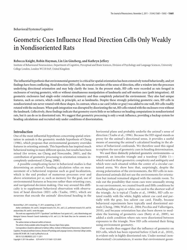
ApparatusThe experiment was performed in a circular arena 250 cm diameter,surrounded by thick black curtains. The arena was lit from directly above(�250 lux of light—approximately the same light level as an indooroffice). Adjacent to the light was a video camera to record the locationand orientation of the rat, and a small radio generating white noise toovershadow external uncontrolled auditory cues. Recordings in thisarena took place inside one of three geometric enclosures, which wereconstructed from hardboard, painted black, and had no floor or ceiling(Fig. 1 A). The enclosures comprised a trapezoid, an isosceles triangle,and a teardrop, chosen for the reasons outlined below.
Trapezoid. The trapezoid was regular (mirror-symmetric). The paral-lel walls were 60 (short wall) and 120 (long wall) cm long; the angled wallswere both 70 cm long. All walls were 60 cm high. This enclosure had twopairs of congruent angles of 65° and 115°, respectively, so geometricinformation emanating from the corners would be ambiguous. Thus, anyability of the directional system to use geometry to orient unambiguouslywould require either an ability to discriminate wall length (since therewas a unique shortest wall and a unique longest wall) or an ability tointegrate information from more than one corner, or from a corner anda wall, simultaneously.
Isosceles triangle. The triangle enclosure was designed as a response tothe finding that HD cells did not rotate with the trapezoid. The trianglehad two equal long walls of 122 cm and a single short wall of 60 cm,making a single unique angle of 30° and two congruent angles of 75°
flanking the base of the triangle. The height of the walls was 60 cm. Weconsidered that the increased aspect ratio of the triangle, together withthe presence of one very obvious, narrow corner, would enhance thelikelihood that HD cells would use this feature of the geometry to orient,if orientation by geometric cues occurs at all.
Teardrop. The teardrop was introduced following the surprising find-ing that HD cells did not rotate with the isosceles triangle either. Wehypothesized that perhaps the cells were unable to use wall length as acue, and that the three angles of the triangle might not have been suffi-ciently discriminable to aid orientation. Thus, the teardrop possessedonly a single corner with an angle similar to that in the triangle (30°). IfHD cells can use corners as a geometric cue, then the teardrop shouldprovide unambiguous polarizing information to them.
Triangle plus gray or white cue card. The failure of HD cells to rotatetheir firing with the above enclosures might have been due to their smallsize—perhaps HD cells cannot use information of such close proximity.Alternatively, perhaps enclosure size is irrelevant but the cells prefervisual information, which was impoverished in the geometric enclosuresdue to the low contrast provided by corners etc. In these manipulations,therefore, a cue card of high contrast (white) or low contrast (gray) wasintroduced to the isosceles triangle. As we will show, cells rotated readilywith the white cue card and somewhat with the gray card.
For each session, one of these enclosures was placed in the center of thearena, directly onto two black plastic sheets that could be wiped androtated between trials to scramble olfactory cues. The orientation of thearena on the first trial of each day was pseudorandomized so thatthe orientation was never the same for consecutive days. To maximizethe chance of the rats relying on the geometric properties of the shape fororientation, no polarizing extramaze cues were placed anywhere in thearena. For cue card trials, either a white or a gray A3 card was taped to theinside of the isosceles triangle’s shortest wall (Fig. 1 A).
Surgery and electrodesAll rats were implanted at the start of the experiment with moveablemicroelectrodes. Four tetrodes were constructed from four interwound,25-�m-diameter platinum–iridium wire (California Fine Wire). The te-trodes were held in a microdrive assembly (Axona) that allowed them tobe lowered or raised, with one full turn of the screw equal to an incrementof 200 �m dorsoventrally. During surgery, the animals were anesthetizedwith isoflurane and oxygen (3 l/min). Animals were placed on a stereo-taxic frame, with lambda and bregma in the horizontal plane. Micro-drives were fixed to the skull with six 1.6 mm jewelers’ screws (PrecisionTechnology Supplies) and dental cement. One of the screws was solderedto a ground wire to enable the animal to be electronically grounded. Theelectrodes were lowered into the brain in areas known to contain HDcells: the postsubiculum (PoS), the anterodorsal thalamic nucleus(ADN), the retrosplenial cortex (RSP), or the lateral mammillary nuclei(LMN). Four HD cells were also recorded from two animals that wereimplanted for a separate experiment using coordinates for the CA1 re-gion of the hippocampus, which has been shown to contain HD cells(Leutgeb et al., 2000). Once the electrodes were implanted, a metallicsleeve was pulled down over the remaining exposed wires. Postsurgery,the animals were monitored periodically until they awoke. All animalswere given at least 1 week to recover following the surgery.
Screening proceduresRecording commenced 1 week after surgery. Recording was done usingmultichannel recording equipment (DacqUSB, Axona). The rats wereconnected to the recording device via lightweight wires and a socketattached to the microdrive plug. The potentials recorded on each of the16 electrodes of the four tetrodes were passed through AC-coupled unitygain operational amplifiers mounted on the rat’s head and fed to therecording system. The signal was amplified (�20,000�), bandpass fil-tered (300 Hz–7 kHz), collected, and stored on a computer. Each of thefour wires of one tetrode was recorded differentially with respect to thewires of other tetrodes. Screening for HD cells took place in a roomseparate from the actual experimental room to minimize the learning ofextraneous cues in the recording environment by the rats. During screen-ing, rats were encouraged to completely sample the floor surface by for-
Table 1. A summary of the six conditions, along with the type of geometric cuesavailable in each condition (not drawn to scale), the manipulation to pathintegration, and whether or not visual landmark information was available
Condition Geometry Path integration Visual landmark
Trapezoid Orientated Absent
Triangle Orientated Absent
Teardrop Orientated Absent
White cue card Orientated Present
Gray cue card Orientated Present
Disorientation Disorientated Absent
15682 • J. Neurosci., November 2, 2011 • 31(44):15681–15692 Knight et al. • HD Cells Are Only Weakly Influenced by Geometry
aging for Coco Pops (Kellogg’s). A directional head-stage with two LEDarrays, one large and one small, aligned opposite each other at a separa-tion of 10 cm, was used along with a video camera attached to the ceilingto track the rat’s location and head direction. If no HD cell activity waspresent, the electrodes were lowered by �50 �m.
Recording proceduresWhen an HD cell was found, the rat was taken into the experimentalroom in an opaque holding box and placed on a raised platform boxoutside of the curtained enclosure to acclimatize to the room for 5 min.The rat was connected to the recording device while outside of the cur-tained enclosure, then returned to the holding box, which was carriedinto the enclosure through one of three joins in the curtains and placed ina pseudorandom location on the floor within the curtained area. After 1min, the rat was taken out of the holding box, carried �1 m, and placedin the center of the geometric enclosure, using a different point ofentry for each trial. The lightweight wires were attached by clips to
two pieces of elastic string suspended fromthe ceiling and the recording session, whichcomprised five individual trials, com-menced. Food was thrown into the environ-ment to encourage rats to sample the wholespace. During this time, the empty holdingbox was moved to a new location in the cur-tained enclosure to prevent it from becom-ing a stable directional cue. Each trial lastedfor 240 s, after which the wires were un-clipped from the string and the rat was re-moved from the environment. The rat wasplaced back in the holding box, which now oc-cupied a new location in the curtained enclo-sure. The rat remained in the holding box whilethe environments were rotated.
While the rat was in the holding box, the twofloor cards were flipped, shuffled, and cleanedwith water. The geometric enclosure was alsoflipped upside down and rotated by either90° (clockwise or counterclockwise) or 180°.Rotations were pseudorandom, so that eachrecording session contained one 90° clock-wise rotation, one 90° counterclockwise rota-tion, and two 180° rotations. Thus, the shapewas aligned with each of the compass axes once(north, south, east, west), except for the lasttrial, when the shape’s orientation was thesame as that in the first trial (Fig. 1 B).
Before the rat was placed back in the newlyrotated environment, it either did or did notundergo a disorientation treatment, which dis-rupts path integration. For five of the six con-ditions, the rat was not disoriented, becauseprevious findings have shown that this can pre-vent a rat from forming a stable relationshipbetween its internal sense of direction and theenvironment (Knierim et al., 1995). How-ever, in the remaining condition, the holdingbox was manually spun by the experimenter(approximately one rotation per second)while they walked around the curtained en-closure for �1 min.
Then, for all six condition types, the rat wastaken out of the holding box and placed back inthe center of the shape for a further 240 s trial.The protocol was repeated until the rat hadbeen recorded in the five trials, at which pointthe session was complete and the rat was takenout of the curtained enclosure in the holdingbox, disconnected from the recording cable,and returned to the home cage. The presenta-tion order for each condition was pseudoran-
domized, ensuring that each of the six conditions was the firstcondition for at least one rat and the last condition for at least one rat(Table 2).
Data analysisCluster-cutting software (Tint; Axona) was used to analyze the data off-line. The peak-to-peak amplitude for one electrode was plotted againsteach of the other electrodes in the tetrode, creating clusters of spikes thatallowed cells to be isolated by hand. All cells with a peak firing rate of�1.0 Hz across the session were excluded from further analysis. For theremaining data, polar plots (firing rate as a funciton of head direction)were created for each cell’s directional activity using a smoothing kernelof 5°. To calculate each cell’s mean firing direction, peak firing rate, anddirectional firing range for each trial, the animal’s head direction wasdivided into 60 6° bins and smoothed in Matlab (Mathworks) using asmoothing kernel of 5°. To calculate the directional firing range, a trian-
Figure 1. A, The dimensions of the environments used for each condition, along with photos of the A3 cue cards attached to theshortest wall of the isosceles triangle. All environments were constructed using hardboard and painted matte black. The height ofall the environments measured 60 cm. The teardrop shape environment was constructed from a flexible piece of hardboardinserted inside the isosceles triangle. B, An example of a protocol used in one session in the triangle condition. The outline of atriangular environment indicates how the environments were rotated between trials inside the curtained environment. Eachsession consisted of two 180° and two 90° [clockwise (CW) and counterclockwise (CCW)] rotations. To achieve this, 90° and 180°rotations had to be interleaved, which meant that four sequences of rotations were possible. These four sequences werepseudorandomized.
Knight et al. • HD Cells Are Only Weakly Influenced by Geometry J. Neurosci., November 2, 2011 • 31(44):15681–15692 • 15683
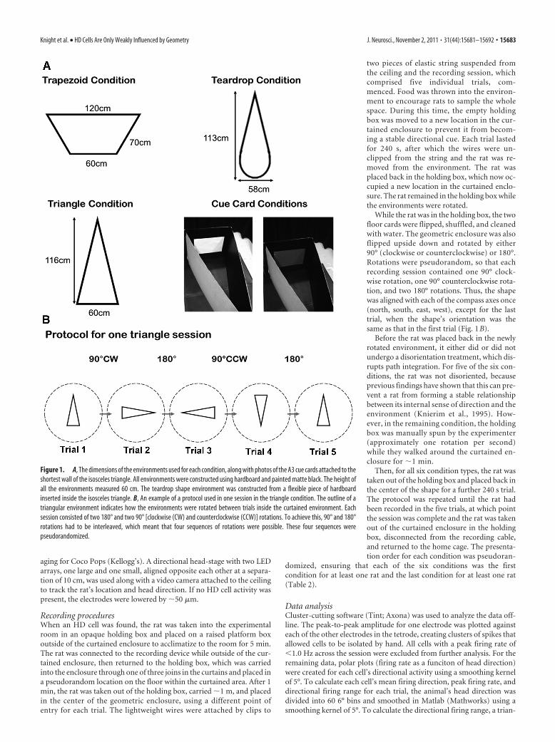
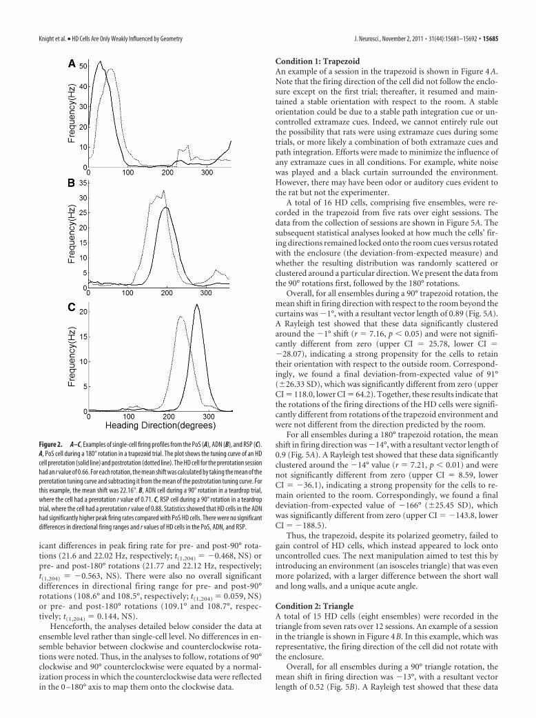
gle was fitted to a firing rate versus HD plot. Using Taube et al.’s (1990a)criteria, left and right maximum and left and right minimum data pointswere selected to form a triangular shape. A linear set of values was thenextrapolated for the right and left legs. The base of the triangle (where thetwo legs intersected the x-axis) was then taken as the directional firingrange. To assess the degree to which the data deviated from the circularmean, mean vector lengths were calculated on unsmoothed data. Meanvector lengths (r) measured the circular spread of data with values rang-ing from 0 to 1. Mean vector length r values that are closer to 1 indicateless variance in the dataset. HD cells that had an r value of �0.3 within agiven session were excluded (van der Meer et al., 2010). Figure 2 A showsan example of one HD cell from the PoS that had an r value of 0.66pre-180° rotation. Figure 2, B and C, shows an example of one HD cellfrom the ADN with an r value of 0.71 and from an RSP cell with an r valueof 0.88, respectively.
Because HD cells from a given animal always behave coherently(Taube et al., 1990b) and cannot be treated as independent data, fromthis point onwards in the analysis any subsequent calculations were per-formed using the circular mean values of cells that had been recordedsimultaneously, i.e., as an ensemble (ranging from 1 to 7 cells). All anal-ysis was done using the CircStat Matlab toolbox (Berens, 2009). To ana-lyze the rotation of HD cells from one trial to the next, the circular meandirection of cells in the first trial was subtracted from the circular meandirection of cells in the second trial (Fig. 2) to provide a measure of howfar the ensemble had rotated. This was repeated for each subsequent trialof the session so that each session produced four rotation estimates.These mean rotations were then subtracted from the predicted angle ofshift, based on how much the environment had rotated, to produceabsolute deviations from expected rotation. The mean vector length ofabsolute deviations for each condition was calculated. The Rayleigh test(Berens, 2009) was then used to determine whether these absolute meandeviations clustered around a particular direction. Circular one- andtwo-sample t tests (Berens, 2009) were used to compare absolute meandeviation values against zero (the predicted deviation given perfectenclosure-following) and to compare absolute mean deviation valuesbetween conditions, respectively. A mean absolute deviation, which wasnot significantly different from zero, indicates that the HD cells followedthe rotation of the shaped enclosure.
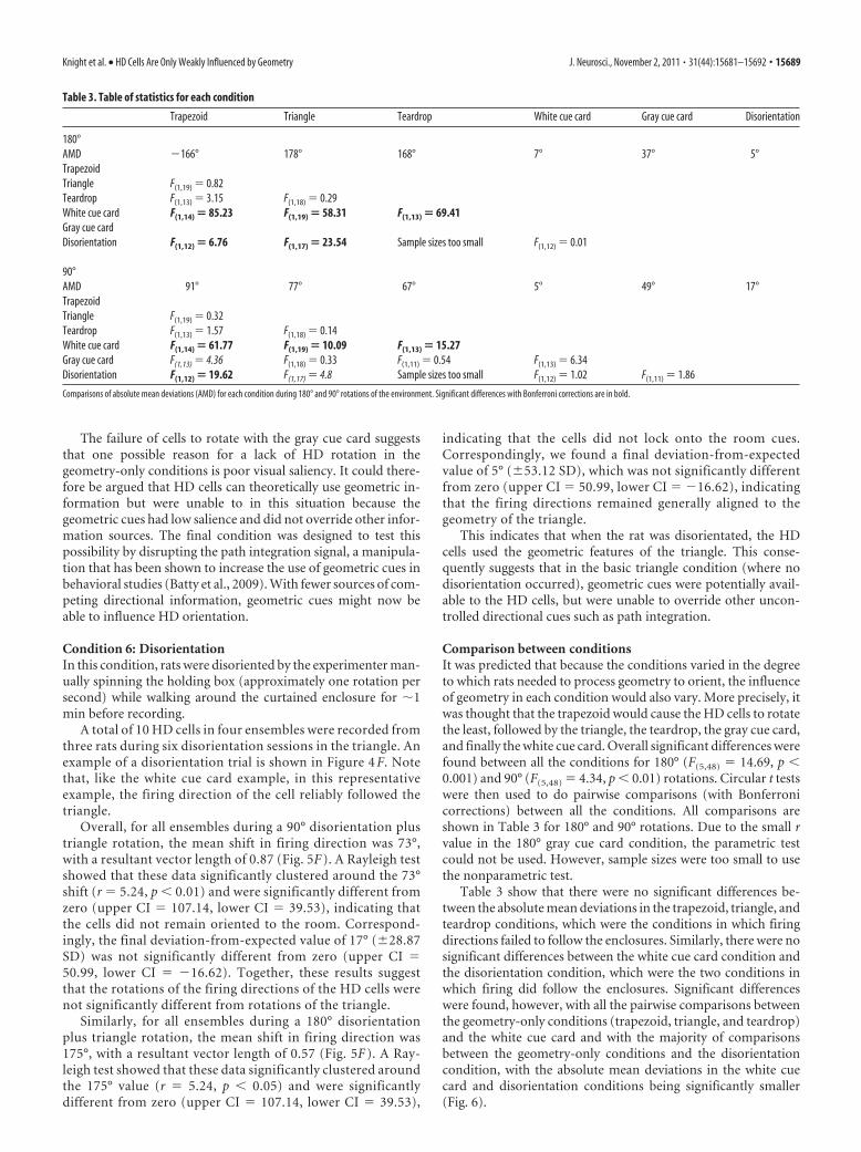
Histological analysisAt the end of testing, the rats were deeply anesthetized with isofluranebefore they were injected with sodium pentobarbital. They were thentranscardially perfused using saline followed by paraformaldehyde (4%).The brains were removed and stored in paraformaldehyde (4%) for atleast 1 week before sectioning. The brains were sliced at 40 �m on afreezing microtome. The sections were then mounted and stained withcresyl violet (Fig. 3).
ResultsThe environments used in the recordings are shown in Figure 1A,and a schematic protocol of one recording session (a sequence offive trials) is shown in Figure 1B. A total of 56 individual HD cellswere recorded from 16 rats during 47 sessions. Simultaneouslyrecorded cells were grouped together as single ensembles, rangingfrom one to seven cells, of which there were 22 in total. Below, wefirst present descriptive data from the individual neurons, andthen present the data from the ensembles, which we take as proxyfor the rat’s internal direction sense.
Fifty-six cells met the inclusion criteria, with a peak firing rateof �1.0 Hz and a mean vector length (r) of �0.3 (van der Meer etal., 2010). Of these 56 cells, histological analysis (Fig. 3) showedthat 24 were recorded in the PoS, 16 in the ADN, 12 in the RSP,and one in the LMN (Table 2). The three remaining cells wererecorded from rats implanted using CA1 coordinates [where HDcells are sometimes found (Leutgeb et al., 2000)], although thiscould not be confirmed with histology. Overall, these 56 HD cellshad an average peak firing rate of 21.6 Hz and an average direc-tional firing range of 109°. Circular statistical analysis determinedthe mean vector length, or r value, which is a measure of thespread of the data ranging from 1 (perfectly concentrated) to 0(randomly distributed around all directions). The mean r valuefor the complete HD cell dataset was 0.5.
Comparisons were made between these brain areas to deter-mine whether there were significant differences in peak firingrate, directional firing range, and r values of the cells. As only onecell was recorded from the LMN and the three cells implantedwith CA1 coordinates could not be confirmed, these areas wereexcluded from comparisons. Of the three parameters, peak firingrate (F(2,49) � 6.04, p � 0.05) showed overall significant differ-ences. Post hoc tests (with Bonferroni corrections) at an alphalevel of 0.05 found that cells in the ADN had significantly higherpeak firing rates compared with the PoS (upper CI � 39.87, lowerCI � 5.76). Figure 2 shows prerotation and postrotation tuningcurves of one cell recorded in the ADN, one cell in the PoS, andone cell in the RSP.
During each session, rats were exposed to one of the environ-ments over five trials. Between trials, the environments were ro-tated by 90° clockwise, 90° counterclockwise, or 180°, duringwhich time we took care not to disorient the rats (except forcondition 6) (Knierim et al., 1995). There were no overall signif-
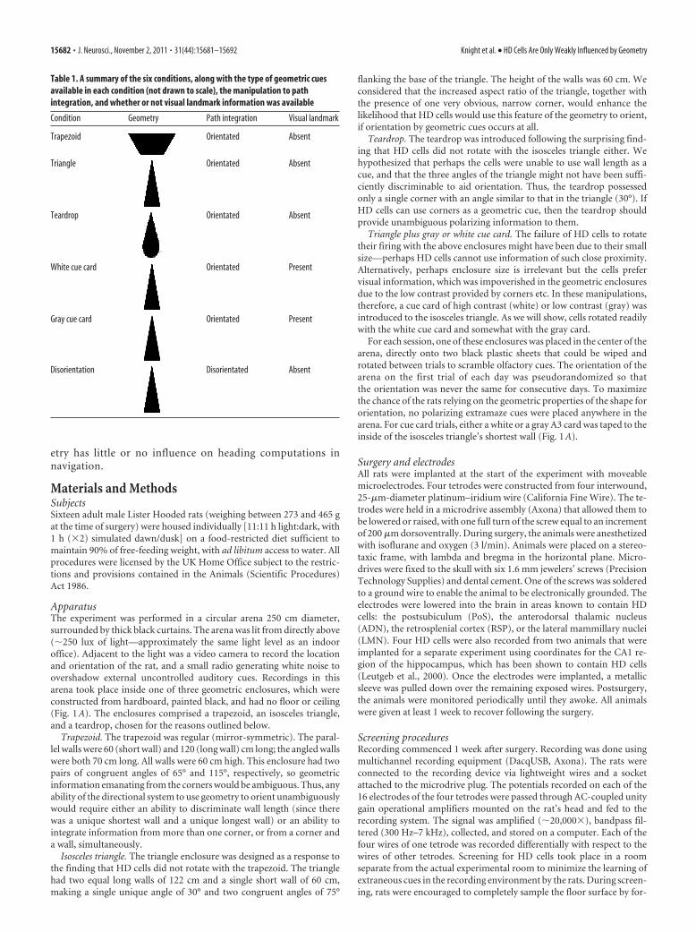
Table 2. The presentation order of conditions for each rat
Rat Location 1st condition 2nd condition 3rd condition 4th condition
R318 CA1 (Total 2 cells) Triangle �2 (2 ensembles)R321 PoS (Total 9 cells) Trapezoid �4 (1 ensemble) Triangle (1 ensemble)R330 CA1 (Total 1 cells) Trapezoid (1 ensemble)R333 ADN (Total 1 cell) Trapezoid (1 ensemble) Triangle (1 ensemble)R344 PoS (Total 8 cells) Triangle �5 (1 ensemble) Disorientation (1 ensemble) Trapezoid (1 ensemble)R371 PoS (Total 5 cells) White card (1 ensemble) Triangle (1 ensemble) Trapezoid (1 ensemble)R388 RSP (Total 1 cell) White card (1 ensemble)R404 RSP (Total 2 cells) Gray card (1 ensemble) Teardrop (1 ensemble)R409 ADN (Total 3 cells) White card (1 ensemble) Teardrop (2 ensembles) Triangle (1 ensemble) Grey card (1 ensemble)R410 ADN (Total 1 cell) Gray card (1 ensemble)R420 ADN (Total 1 cells) Gray card �2 (2 ensembles) White card �2 (1 ensemble) Gray card (1 ensemble) Disorientation �2 (1 ensemble)R1693 LMN (Total 1 cell) Triangle (1 ensemble)R1696 RSP (Total 4 cells) Gray card (1 ensemble) Teardrop �2 (2 ensembles)R1697 RSP (Total 2 cells) White card (1 ensemble) Teardrop (1 ensemble)R1704 RSP (Total 3 cells) Teardrop (1 ensemble) White card (1 ensemble)R433 PoS (Total 2 cells) Disorientation �2 (2 ensembles)
All conditions were presented first for at least one rat and presented last for at least one rat in order to counterbalance any order effects. The table also presents the brain area where each rat was implanted, the total number of HD cellsrecorded in each rat, and the total number of ensembles recorded in each condition for each rat.
15684 • J. Neurosci., November 2, 2011 • 31(44):15681–15692 Knight et al. • HD Cells Are Only Weakly Influenced by Geometry
icant differences in peak firing rate for pre- and post-90° rota-tions (21.6 and 22.02 Hz, respectively; t(1,204) � �0.468, NS) orpre- and post-180° rotations (21.77 and 22.12 Hz, respectively;t(1,204) � �0.563, NS). There were also no overall significantdifferences in directional firing range for pre- and post-90°rotations (108.6° and 108.5°, respectively; t(1,204) � 0.059, NS)or pre- and post-180° rotations (109.1° and 108.7°, respec-tively; t(1,204) � 0.144, NS).
Henceforth, the analyses detailed below consider the data atensemble level rather than single-cell level. No differences in en-semble behavior between clockwise and counterclockwise rota-tions were noted. Thus, in the analyses to follow, rotations of 90°clockwise and 90° counterclockwise were equated by a normal-ization process in which the counterclockwise data were reflectedin the 0 –180° axis to map them onto the clockwise data.
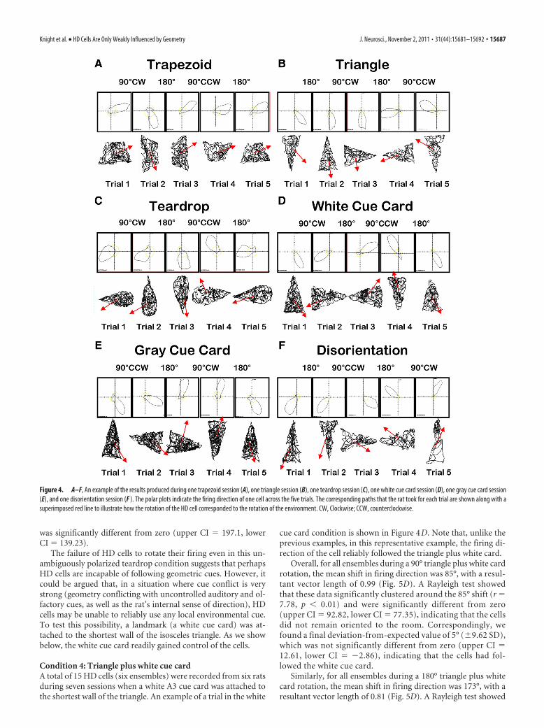
Condition 1: TrapezoidAn example of a session in the trapezoid is shown in Figure 4A.Note that the firing direction of the cell did not follow the enclo-sure except on the first trial; thereafter, it resumed and main-tained a stable orientation with respect to the room. A stableorientation could be due to a stable path integration cue or un-controlled extramaze cues. Indeed, we cannot entirely rule outthe possibility that rats were using extramaze cues during sometrials, or more likely a combination of both extramaze cues andpath integration. Efforts were made to minimize the influence ofany extramaze cues in all conditions. For example, white noisewas played and a black curtain surrounded the environment.However, there may have been odor or auditory cues evident tothe rat but not the experimenter.
A total of 16 HD cells, comprising five ensembles, were re-corded in the trapezoid from five rats over eight sessions. Thedata from the collection of sessions are shown in Figure 5A. Thesubsequent statistical analyses looked at how much the cells’ fir-ing directions remained locked onto the room cues versus rotatedwith the enclosure (the deviation-from-expected measure) andwhether the resulting distribution was randomly scattered orclustered around a particular direction. We present the data fromthe 90° rotations first, followed by the 180° rotations.
Overall, for all ensembles during a 90° trapezoid rotation, themean shift in firing direction with respect to the room beyond thecurtains was �1°, with a resultant vector length of 0.89 (Fig. 5A).A Rayleigh test showed that these data significantly clusteredaround the �1° shift (r � 7.16, p � 0.05) and were not signifi-cantly different from zero (upper CI � 25.78, lower CI ��28.07), indicating a strong propensity for the cells to retaintheir orientation with respect to the outside room. Correspond-ingly, we found a final deviation-from-expected value of 91°(�26.33 SD), which was significantly different from zero (upperCI � 118.0, lower CI � 64.2). Together, these results indicate thatthe rotations of the firing directions of the HD cells were signifi-cantly different from rotations of the trapezoid environment andwere not different from the direction predicted by the room.
For all ensembles during a 180° trapezoid rotation, the meanshift in firing direction was �14°, with a resultant vector length of0.9 (Fig. 5A). A Rayleigh test showed that these data significantlyclustered around the �14° value (r � 7.21, p � 0.01) and werenot significantly different from zero (upper CI � 8.59, lowerCI � �36.1), indicating a strong propensity for the cells to re-main oriented to the room. Correspondingly, we found a finaldeviation-from-expected value of �166° (�25.45 SD), whichwas significantly different from zero (upper CI � �143.8, lowerCI � �188.5).
Thus, the trapezoid, despite its polarized geometry, failed togain control of HD cells, which instead appeared to lock ontouncontrolled cues. The next manipulation aimed to test this byintroducing an environment (an isosceles triangle) that was evenmore polarized, with a larger difference between the short walland long walls, and a unique acute angle.
Condition 2: TriangleA total of 15 HD cells (eight ensembles) were recorded in thetriangle from seven rats over 12 sessions. An example of a sessionin the triangle is shown in Figure 4B. In this example, which wasrepresentative, the firing direction of the cell did not rotate withthe enclosure.
Overall, for all ensembles during a 90° triangle rotation, themean shift in firing direction was �13°, with a resultant vectorlength of 0.52 (Fig. 5B). A Rayleigh test showed that these data
Figure 2. A–C, Examples of single-cell firing profiles from the PoS (A), ADN (B), and RSP (C).A, PoS cell during a 180° rotation in a trapezoid trial. The plot shows the tuning curve of an HDcell prerotation (solid line) and postrotation (dotted line). The HD cell for the prerotation sessionhad an r value of 0.66. For each rotation, the mean shift was calculated by taking the mean of theprerotation tuning curve and subtracting it from the mean of the postrotation tuning curve. Forthis example, the mean shift was 22.16°. B, ADN cell during a 90° rotation in a teardrop trial,where the cell had a prerotation r value of 0.71. C, RSP cell during a 90° rotation in a teardroptrial, where the cell had a prerotation r value of 0.88. Statistics showed that HD cells in the ADNhad significantly higher peak firing rates compared with PoS HD cells. There were no significantdifferences in directional firing ranges and r values of HD cells in the PoS, ADN, and RSP.
Knight et al. • HD Cells Are Only Weakly Influenced by Geometry J. Neurosci., November 2, 2011 • 31(44):15681–15692 • 15685

significantly clustered around the 13° shift (r � 6.78, p � 0.05)and were not significantly different from zero (upper CI � 57.87,lower CI � �32.66), indicating, as with the trapezoid, a strongpropensity for the cells to remain oriented to the room. Cor-respondingly, we found a final deviation-from-expected valueof 77° (�56.0 SD), which was significantly different from zero(upper CI � 122.61, lower CI � 31.51). Together, these resultsindicate that the rotations of the firing directions of the HDcells were significantly different from rotations of the triangleenvironment and were not different from the direction pre-dicted by the room.
Similarly, for all ensembles during a 180° triangle rotation, themean shift in firing direction was 2°, with a resultant vector lengthof 0.75 (Fig. 5B). A Rayleigh test showed that these data signifi-cantly clustered around the 2° value (r � 9.72, p � 0.01) and werenot significantly different from zero (upper CI � 29.22, lowerCI � �25.21), indicating a strong propensity for the cells toremain oriented to the room. Correspondingly, we found a finaldeviation-from-expected value of 178° (�40.69 SD), which wassignificantly different from zero (upper CI � 205.12, lower CI �151.26).
One possible reason for the failure of cells to lock onto thegeometry of the triangle is that from the perspective of the rat, thecorners look similar and the wall lengths are hard to discriminate.This seems surprising, but nevertheless, we attempted to make theenvironment less rotationally ambiguous by reducing the number ofcorners to one (the teardrop enclosure).
Condition 3: TeardropA total of 12 HD cells (seven ensembles) were recorded in theteardrop from five rats over seven sessions. An example of a ses-sion in the teardrop is shown in Figure 4C. Note that, as in thetrapezoid and the triangle, the firing direction of the cell did notrotate with the enclosure.
Overall, for all ensembles during a 90° teardrop rotation, themean shift in firing direction was �23°, with a resultant vectorlength of 0.77 (Fig. 5C). A Rayleigh test showed that these datasignificantly clustered around the 23° shift (r � 5.41, p � 0.01)and were not significantly different from zero (upper CI � 60.73,lower CI � �14.9), indicating a strong propensity for the cells toremain oriented to the room. Correspondingly, we found a finaldeviation-from-expected value of 67° (�38.62 SD), which wassignificantly different from zero (upper CI � 104.85, lower CI �29.79). Together, these results indicate that the rotations of thefiring directions of the HD cells were significantly different fromrotations of the teardrop environment and were not differentfrom the direction predicted by the room.
Similarly, for all ensembles during a 180° teardrop rotation,the mean shift in firing direction was 12°, with a resultant vectorlength of 0.9 (Fig. 5C). A Rayleigh test showed that these datasignificantly clustered around the 12° value (r � 6.29, p � 0.01)and were not significantly different from zero (upper CI � 41.25,lower CI � �17.19), indicating a strong propensity for the cellsto remain oriented to the room. Correspondingly, we found afinal deviation-from-expected value of 168° (�25.75 SD), which
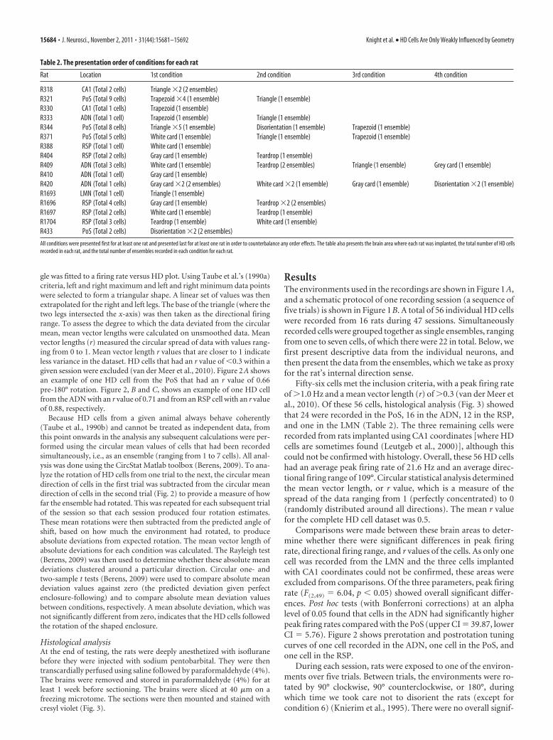
Figure 3. A–D, Histology from postsubiculum (Post; A), ADN (B), retrosplenial cortex (dysgranular, RSD; granular a, RSGa; granular b, RSGb; granular c, RSGc; C), and lateral mammillary nucleus(LM; D). An example slice is shown for each brain region, along with the corresponding red dot on the template (Paxinos and Watson, 2004). Each black dot represents the recording site for theremaining rats implanted in that region. The histology for the two rats implanted in the CA1 region of the hippocampus could not be recovered. They were, however, implanted using the followingcoordinates: anterior/posterior: �3.5; medial/lateral: 2.0; dorsal/ventral: �1.5. Place cells were also found in this region. R371, R333, R1704, and R1693 are the identifiers of the rats whose datais shown here.
15686 • J. Neurosci., November 2, 2011 • 31(44):15681–15692 Knight et al. • HD Cells Are Only Weakly Influenced by Geometry
was significantly different from zero (upper CI � 197.1, lowerCI � 139.23).
The failure of HD cells to rotate their firing even in this un-ambiguously polarized teardrop condition suggests that perhapsHD cells are incapable of following geometric cues. However, itcould be argued that, in a situation where cue conflict is verystrong (geometry conflicting with uncontrolled auditory and ol-factory cues, as well as the rat’s internal sense of direction), HDcells may be unable to reliably use any local environmental cue.To test this possibility, a landmark (a white cue card) was at-tached to the shortest wall of the isosceles triangle. As we showbelow, the white cue card readily gained control of the cells.
Condition 4: Triangle plus white cue cardA total of 15 HD cells (six ensembles) were recorded from six ratsduring seven sessions when a white A3 cue card was attached tothe shortest wall of the triangle. An example of a trial in the white
cue card condition is shown in Figure 4D. Note that, unlike theprevious examples, in this representative example, the firing di-rection of the cell reliably followed the triangle plus white card.
Overall, for all ensembles during a 90° triangle plus white cardrotation, the mean shift in firing direction was 85°, with a resul-tant vector length of 0.99 (Fig. 5D). A Rayleigh test showedthat these data significantly clustered around the 85° shift (r �7.78, p � 0.01) and were significantly different from zero(upper CI � 92.82, lower CI � 77.35), indicating that the cellsdid not remain oriented to the room. Correspondingly, wefound a final deviation-from-expected value of 5° (�9.62 SD),which was not significantly different from zero (upper CI �12.61, lower CI � �2.86), indicating that the cells had fol-lowed the white cue card.
Similarly, for all ensembles during a 180° triangle plus whitecard rotation, the mean shift in firing direction was 173°, with aresultant vector length of 0.81 (Fig. 5D). A Rayleigh test showed
Figure 4. A–F, An example of the results produced during one trapezoid session (A), one triangle session (B), one teardrop session (C), one white cue card session (D), one gray cue card session(E), and one disorientation session (F ). The polar plots indicate the firing direction of one cell across the five trials. The corresponding paths that the rat took for each trial are shown along with asuperimposed red line to illustrate how the rotation of the HD cell corresponded to the rotation of the environment. CW, Clockwise; CCW, counterclockwise.
Knight et al. • HD Cells Are Only Weakly Influenced by Geometry J. Neurosci., November 2, 2011 • 31(44):15681–15692 • 15687
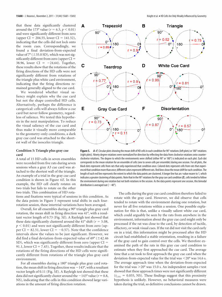
that these data significantly clusteredaround the 173° value (r � 6.5, p � 0.01)and were significantly different from zero(upper CI � 204.55, lower CI � 141.52),indicating that the cells did not lock ontothe room cues. Correspondingly, wefound a final deviation-from-expectedvalue of 7° (�35.0 SD), which was not sig-nificantly different from zero (upper CI �38.96, lower CI � �24.64). Together,these results show that the rotations of thefiring directions of the HD cells were notsignificantly different from rotations ofthe triangle plus white card environment,indicating that the firing directions re-mained generally aligned to the cue card.
We wondered whether visual sa-liency might explain why the cue cardbut not the shape controlled HD cells.Alternatively, perhaps the difference iscategorical: cells will always follow a cuecard but never follow geometry, regard-less of salience. We tested this hypothe-sis in the next manipulation. To reducethe visual saliency of the cue card andthus make it visually more comparableto the geometry-only conditions, a darkgray cue card was attached to the short-est wall of the isosceles triangle.
Condition 5: Triangle plus gray cuecardA total of 15 HD cells in seven ensembleswere recorded from five rats during sevensessions when a gray A3 cue card was at-tached to the shortest wall of the triangle.An example of a trial in the gray cue cardcondition is shown in Figure 4E. In thisexample, the HD cell clearly rotates ontwo trials but fails to rotate on the othertwo trials. This combination of HD rota-tions and fixations were typical of sessions in this condition. Asthe data points in Figure 5 represent total shifts in each four-rotation session, these intertrial variations have been averaged.
Overall, for all ensembles during a 90° triangle plus gray cardrotation, the mean shift in firing direction was 41°, with a resul-tant vector length of 0.73 (Fig. 5E). A Rayleigh test showed thatthese data significantly clustered around the 41° shift (r � 5.08,p � 0.01) and were not significantly different from zero (up-per CI � 82.51, lower CI � �0.57). Note that the confidenceintervals show the values to be just significant. However, wedid find a final deviation-from-expected value of 49° (�42.46SD), which was significantly different from zero (upper CI �91.1, lower CI � 7.45). Together, these results indicate that therotations of the firing directions of the HD cells were signifi-cantly different from rotations of the triangle plus gray cardenvironment.
For all ensembles during a 180° triangle plus gray card rota-tion, the mean shift in firing direction was �143°, with a resultantvector length of 0.11 (Fig. 5E). A Rayleigh test showed that thesedata did not significantly cluster around the �143° value (r � 0.8,NS), indicating that the cells in this condition showed large vari-ations in the amount of firing direction rotation.
The cells during the gray cue card condition therefore failed torotate with the gray card. However, we did observe that cellstended to rotate with the environment during one rotation, butnever for all five rotations within a session. One possible expla-nation for this is that, unlike a visually salient white cue card,which could arguably be seen by the rats from anywhere in theenvironment, information about the gray cue card might only beprocessed if the rat was close to the card, by detection of tactile,olfactory, or weak visual cues. If the rat did not visit the card earlyon in a trial, this information might be processed after the HDcircuit had established a stable orientation, resulting in a failureof the gray card to gain control over the cells. We therefore ex-amined the path of the rats in this gray cue card condition toestimate when they first approached the cue card. The averagetime that a rat took to first approach the gray cue card when thedeviation-from-expected value for the trial was �20° was 14.6 s.The average approach time when the deviation-from-expectedfor the trial was �20° was 15.0 s. An independent samples t testshowed that these approach times were not significantly different(t(33) � 0.055, NS). These findings suggest that this proximityhypothesis is unlikely. However, no behavioral measures weretaken during the trial, so definitive conclusions cannot be drawn.
Figure 5. A–F, Circular plots showing the mean shift of HD cells in each condition for 90° rotations (left plots) or 180° rotations(right plots). Ninety degree rotations were normalized for direction by reflecting the data from clockwise rotations onto counter-clockwise rotations. The degree to which the environments were shifted (either 90° or 180°) is indicated on each plot. Each dotcorresponds to the mean rotation for an ensemble of cells (one to seven cells per ensemble) during one session. For all plots, theblack dots represent cells from rats that only experienced that condition once. Colored dots represent cells from rats that experi-enced that condition more than once; different colors represent different rats. Red lines show the mean shift for each condition. Thelength of each red line represents the extent to which the data points are clustered. A longer line has an r value nearer to 1, whichindicates a greater clustering of data points. Note that in the 90° rotations for the gray cue card condition (E), cells tended to followthe environment during one rotation but not both rotations in the session. As the data points represent one session, this bimodaldistribution is averaged out (�40°).
15688 • J. Neurosci., November 2, 2011 • 31(44):15681–15692 Knight et al. • HD Cells Are Only Weakly Influenced by Geometry
The failure of cells to rotate with the gray cue card suggeststhat one possible reason for a lack of HD rotation in thegeometry-only conditions is poor visual saliency. It could there-fore be argued that HD cells can theoretically use geometric in-formation but were unable to in this situation because thegeometric cues had low salience and did not override other infor-mation sources. The final condition was designed to test thispossibility by disrupting the path integration signal, a manipula-tion that has been shown to increase the use of geometric cues inbehavioral studies (Batty et al., 2009). With fewer sources of com-peting directional information, geometric cues might now beable to influence HD orientation.
Condition 6: DisorientationIn this condition, rats were disoriented by the experimenter man-ually spinning the holding box (approximately one rotation persecond) while walking around the curtained enclosure for �1min before recording.
A total of 10 HD cells in four ensembles were recorded fromthree rats during six disorientation sessions in the triangle. Anexample of a disorientation trial is shown in Figure 4 F. Notethat, like the white cue card example, in this representativeexample, the firing direction of the cell reliably followed thetriangle.
Overall, for all ensembles during a 90° disorientation plustriangle rotation, the mean shift in firing direction was 73°,with a resultant vector length of 0.87 (Fig. 5F ). A Rayleigh testshowed that these data significantly clustered around the 73°shift (r � 5.24, p � 0.01) and were significantly different fromzero (upper CI � 107.14, lower CI � 39.53), indicating thatthe cells did not remain oriented to the room. Correspond-ingly, the final deviation-from-expected value of 17° (�28.87SD) was not significantly different from zero (upper CI �50.99, lower CI � �16.62). Together, these results suggestthat the rotations of the firing directions of the HD cells werenot significantly different from rotations of the triangle.
Similarly, for all ensembles during a 180° disorientationplus triangle rotation, the mean shift in firing direction was175°, with a resultant vector length of 0.57 (Fig. 5F ). A Ray-leigh test showed that these data significantly clustered aroundthe 175° value (r � 5.24, p � 0.05) and were significantlydifferent from zero (upper CI � 107.14, lower CI � 39.53),
indicating that the cells did not lock onto the room cues.Correspondingly, we found a final deviation-from-expectedvalue of 5° (�53.12 SD), which was not significantly differentfrom zero (upper CI � 50.99, lower CI � �16.62), indicatingthat the firing directions remained generally aligned to thegeometry of the triangle.
This indicates that when the rat was disorientated, the HDcells used the geometric features of the triangle. This conse-quently suggests that in the basic triangle condition (where nodisorientation occurred), geometric cues were potentially avail-able to the HD cells, but were unable to override other uncon-trolled directional cues such as path integration.
Comparison between conditionsIt was predicted that because the conditions varied in the degreeto which rats needed to process geometry to orient, the influenceof geometry in each condition would also vary. More precisely, itwas thought that the trapezoid would cause the HD cells to rotatethe least, followed by the triangle, the teardrop, the gray cue card,and finally the white cue card. Overall significant differences werefound between all the conditions for 180° (F(5,48) � 14.69, p �0.001) and 90° (F(5,48) � 4.34, p � 0.01) rotations. Circular t testswere then used to do pairwise comparisons (with Bonferronicorrections) between all the conditions. All comparisons areshown in Table 3 for 180° and 90° rotations. Due to the small rvalue in the 180° gray cue card condition, the parametric testcould not be used. However, sample sizes were too small to usethe nonparametric test.
Table 3 show that there were no significant differences be-tween the absolute mean deviations in the trapezoid, triangle, andteardrop conditions, which were the conditions in which firingdirections failed to follow the enclosures. Similarly, there were nosignificant differences between the white cue card condition andthe disorientation condition, which were the two conditions inwhich firing did follow the enclosures. Significant differenceswere found, however, with all the pairwise comparisons betweenthe geometry-only conditions (trapezoid, triangle, and teardrop)and the white cue card and with the majority of comparisonsbetween the geometry-only conditions and the disorientationcondition, with the absolute mean deviations in the white cuecard and disorientation conditions being significantly smaller(Fig. 6).
Table 3. Table of statistics for each condition
Trapezoid Triangle Teardrop White cue card Gray cue card Disorientation
180°AMD �166° 178° 168° 7° 37° 5°TrapezoidTriangle F(1,19) � 0.82Teardrop F(1,13) � 3.15 F(1,18) � 0.29White cue card F(1,14) � 85.23 F(1,19) � 58.31 F(1,13) � 69.41Gray cue cardDisorientation F(1,12) � 6.76 F(1,17) � 23.54 Sample sizes too small F(1,12) � 0.01
90°AMD 91° 77° 67° 5° 49° 17°TrapezoidTriangle F(1,19) � 0.32Teardrop F(1,13) � 1.57 F(1,18) � 0.14White cue card F(1,14) � 61.77 F(1,19) � 10.09 F(1,13) � 15.27Gray cue card F(1,13) � 4.36 F(1,18) � 0.33 F(1,11) � 0.54 F(1,13) � 6.34Disorientation F(1,12) � 19.62 F(1,17) � 4.8 Sample sizes too small F(1,12) � 1.02 F(1,11) � 1.86
Comparisons of absolute mean deviations (AMD) for each condition during 180° and 90° rotations of the environment. Significant differences with Bonferroni corrections are in bold.
Knight et al. • HD Cells Are Only Weakly Influenced by Geometry J. Neurosci., November 2, 2011 • 31(44):15681–15692 • 15689
DiscussionThis study explored the use of geometric cues by HD cells todetermine whether geometry is an important source of orientinginformation (Cheng, 1986). We found that when rats were notdisoriented, HD cells did not use even a highly polarized environ-mental geometry to orient their firing directions. Instead, firingdirections generally remained aligned to the room. When ratswere strongly disoriented, HD cells rotated with the bare enclo-sures. This demonstrates that HD cells can process geometrywhen other cues, such as landmarks and path integration, areunavailable, but this effect was relatively weak and failed tooverride path integration. This finding is important in light ofprevious suggestions that geometric cues are preferentially in-volved in reorientation—the so-called “geometric module hy-pothesis” (Cheng, 1986, 2008). Our results suggest this may bethe case only when path integration is severely disrupted fol-lowing disorientation.
There are two aspects of note in our findings: first, that HDcells did not appear sensitive to geometry in oriented rats; andsecond, that they were sensitive in disoriented rats. These twosituations are discussed in turn below.
Influence of geometric cues in oriented animalsRelatively few behavioral studies have examined the influenceof geometry in nondisoriented animals. Lourenco and Hut-tenlocher (2006) found that children used geometric cues tofind a goal when disoriented by self-rotation, but did not usethe same cues when it was the environment that had beenrotated instead. Batty et al. (2009) found that disorientation ofrats during training caused a relative preference for the use ofgeometric cues (as opposed to internal, path integration cues)during testing, although disorientation before the test itselfdid not increase the use of geometry any further. It appearsfrom these studies that when orientation (path integration)cues were present, they influenced choices: findings that areconsistent with our observation that when orientation or vi-sual cues were present, HD cells apparently preferred themover geometry.
Studies of neuronal responses to changes in geometry have, bycontrast, produced results that seem to contradict ours. Kubieand Ranck (1983) and Muller and Kubie (1987) found that hip-pocampal place cells remapped when nondisoriented animalswere placed in environments of differing geometry. However, theenvironments also differed in nongeometric aspects (e.g., the ma-terial of the walls), information to which place cells are known to
be sensitive (Anderson and Jeffery, 2003). It is not knownwhether HD cells changed orientation following these manipula-tions. These studies therefore do not speak directly to the issue ofHD cell geometric processing.
Direct observation of HD cells has also, however, suggesteda possible influence of geometry. Golob et al. (2001) foundthat HD cells changed orientation in response to the shift ofthe animals (without disorientation) from a square enclosureto a rectangular one. In these experiments, rats were trained tolocate a corner of the square arena, which was polarized with acue card, for water reward. When the arena was changed to arectangle, cells rotated their firing directions on 12 of 13 oc-casions. These results suggest the processing of geometric cuesby HD cells; however, most of the shifts in firing directionwere multiples of 90°, suggesting that what changed was thecells’ choice of corner to orient by. Given that the rats had beentrained to specifically attend to corners to locate the reward, itis possible the HD cells were using corners as landmarks,rather than processing geometry as a holistic property of theenvironment. Thus, it is not clear whether this alteration rep-resents true geometric processing by the HD cells.
An apparent effect of environment geometry on HD cellswas also found by Dudchenko and Zinyuk (2005), who re-corded HD cells in two T-mazes that were located in adjacentrooms and oriented differently. They found that HD cells ro-tated between the two rooms so as to maintain the same ori-entation with respect to the maze. However, while the cellswere clearly orienting using the apparatus, it may not havebeen geometry per se that the cells were using. The rats werealways started from the same place in the maze, and studies ofplace cells have shown that start position can be an orientingcue for place cells (Sharp et al., 1990), presumably via the headdirection cells. Furthermore, the maze would provide a visu-ally unique panorama from most locations, which means theHD cells would have potential visual orienting cues even ifthey did not process geometry directly.
Thus, although prior studies of HD cells have shown respon-siveness to manipulations of environmental geometry, it has notbeen shown that the relevant factor was geometry itself and notsome feature or other cue also present. The fact that our environ-ments were relatively low in unique features, despite their dis-tinctly polarized geometry, may explain why HD cells in ouroriented rats preferred to use path integration or the cue cardwhen these were available.
Influence of geometric cues in disoriented animalsOur finding that geometric cues by themselves have only aweak influence on HD cell orientation seems surprising giventhe substantial behavioral literature suggesting a primacy forgeometric cues over landmarks (Cheng, 1986; for review, seeCheng and Newcombe, 2005). However, most behavioralstudies have been undertaken in disoriented rats (Cheng,1986; Maurer and Derivaz, 2000; Wall et al., 2004; Cheng andGallistel, 2005; Skov-Rackette and Shettleworth, 2005; Gibsonet al., 2007; Maes et al., 2009), in which path integration hasbeen disrupted. The present results suggest that geometriccues are able to influence HD cells if other sources of orientinginformation are absent.
When other orienting cues are absent, our findings accordwith previous research on HD cells. Taube et al. (1990b) foundthat when a square enclosure was changed to a rectangle, HDcells in disoriented rats rotated their firing directions, whileGolob and Taube (1997) found that HD cells in disoriented
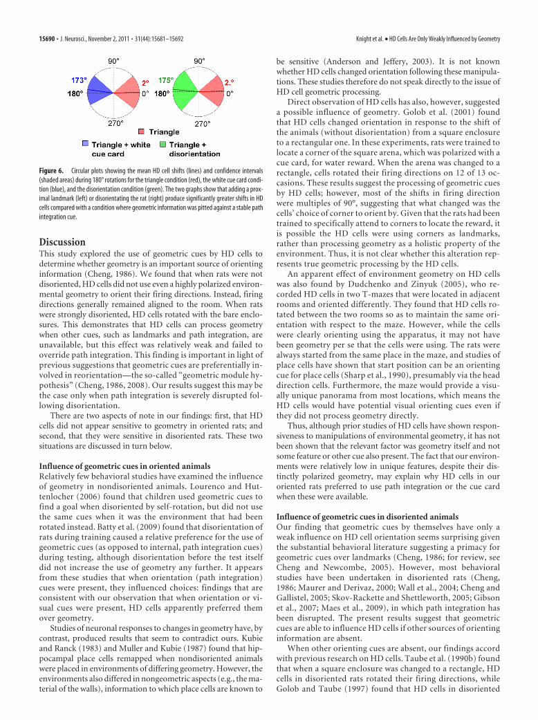
Figure 6. Circular plots showing the mean HD cell shifts (lines) and confidence intervals(shaded areas) during 180° rotations for the triangle condition (red), the white cue card condi-tion (blue), and the disorientation condition (green). The two graphs show that adding a prox-imal landmark (left) or disorientating the rat (right) produce significantly greater shifts in HDcells compared with a condition where geometric information was pitted against a stable pathintegration cue.
15690 • J. Neurosci., November 2, 2011 • 31(44):15681–15692 Knight et al. • HD Cells Are Only Weakly Influenced by Geometry
rats with hippocampal lesions altered their firing when anenclosure was changed from a cylinder to a square, triangle, orpentagon. More recently, Clark et al. (2010) found that geo-metric cues could override the effect of a single landmark.These rotations may have been due to geometric processing,but may, as noted above, also result from processing of specificfeatures arising from the geometry. Further experiments willbe needed to test whether true geometric processing occursduring reorientation.
Interestingly, Clark et al. (2010) found that geometric cuestended not to override the effects of multiple landmarks, suggest-ing that the hierarchy of cue preference is salience-dependentrather than absolute. We found the use of a landmark to besalience-dependent when we manipulated its visual contrast.Thus, the switch between the use of different cues may resultfrom salience-based competition rather than any absolute hier-archy of cue use. Previous studies have also found a switch indirectional cue use by HD cells under some circumstances. Forexample, Knierim et al. (1995) found that a cue card was morelikely to gain control over HD cells in rats that had learned aboutthe cue while they were nondisoriented than in rats that had beendisoriented during training. All animals were tested under thesame disorientation conditions, suggesting that the cue card hadlost salience because of the disorientation-training procedure.This finding is reminiscent of the Batty et al. (2009) behavioralstudy, in which disorientation during training caused a reductionin the use of geometric cues during testing. These experimentsshow that the influence of directional cues on HD cells can bereweighted with experience and is not absolute.
Our results additionally indicate that the orientation state ofthe animal at the time of testing influences cue use too. Thefinding that disorientation instantaneously modulates the rela-tive influence of geometric cues is interesting because disorienta-tion does not degrade the HD signal. This suggests that whateverdecision process is responsible for assigning precedence to aninput, it knows about the state of the path integrator. Thus, itseems able to distinguish between mild disorientation, whenthere is merely a slight disagreement between the path integratorand the environmental cues (as when the shape was rotated), andcomplete disorientation. HD cells seemed to use path integrationin the former case and geometry in the latter. Since HD cells inthoroughly disoriented rats still fired strongly and with tight (al-beit misdirected) tuning curves, the implication is that the signalabout orientation state (mild vs severe), and thus the decision touse or ignore geometry, occurs outside the HD cells themselves.Potentially, there is an input that tells the HD cells that severedisorientation has occurred and therefore to weight externalcues, including geometry, more heavily than normal. Such ascheme could provide an interesting model system for the studyof Bayesian integration processes (Cheng et al., 2007).
Why, adaptively speaking, should geometry be such a weak cuefor HD cell orientation? We can only speculate, but it is worth notingthat the HD cell system is probably old, in evolutionary terms,whereas the ecological lifestyles of rats living in bounded spaces aremore recent. Many animals that share a common ancestor with ro-dents, such as fish and birds, live in open spaces that lack a cleargeometry. Furthermore, it is not clear that even in bounded spaces, itis geometry per se that is the relevant feature, rather than the visualpanorama that derives from geometric configurations (Cheung etal., 2008; Sturzl et al., 2008). Further work is needed to determinewhat aspect of geometry influences HD cells in disoriented rats.
In summary, we have explored the use of geometric cues byHD cells and found that, in nondisoriented rats, their influence
does not appear as strong as the influence of other simultaneouslypresent cues such as a landmark or path integration. Conversely,in disoriented rats, in which path integration was disabled, geom-etry did have an effect, suggesting that it is processed, even if itseffect is only weak. Since disorientation is presumably an unusualcondition in the normal life of a rat, the implication is that localgeometry is not a primary guide to the HD system in rodents, butmay be used as a weak backup system for reorientation whenother cues are unavailable.
ReferencesAnderson MI, Jeffery KJ (2003) Heterogeneous modulation of place cell
firing by changes in context. J Neurosci 23:8827– 8835.Batty ER, Hoban L, Spetch ML, Dickson CT (2009) Rats’ use of geometric,
featural and orientation cues to locate a hidden goal. Behav Processes82:327–334.
Berens P (2009) CircStat: a MATLAB toolbox for circular statistics. J StatSoftw 31:1–21.
Cheng K (1986) A purely geometric module in the rat’s spatial representa-tion. Cognition 23:149 –178.
Cheng K (2008) Whither geometry? Troubles of the geometric module.Trends Cogn Sci 12:355–361.
Cheng K, Gallistel CR (2005) Shape parameters explain data from spatialtransformations: comment on Pearce et al (2004) and Tommasi and Polli(2004). J Exp Psychol Anim Behav Process 31:254 –259.
Cheng K, Newcombe NS (2005) Is there a geometric module for spatialorientation? Squaring theory and evidence. Psychon Bull Rev 12:1–23.
Cheng K, Shettleworth SJ, Huttenlocher J, Rieser JJ (2007) Bayesian integra-tion of spatial information. Psychol Bull 133:625– 637.
Cheung A, Stuzl W, Zeil J, Cheng K (2008) The information content ofpanoramic images. II. View-based navigation in nonrectangular experi-mental arenas. J Exp Psychol Anim Behav Process 34:15–30.
Clark BJ, Harris MJ, Taube JS (2010) Control of anterodorsal thalamic headdirection cells by environmental boundaries: comparison with conflictingdistal landmarks. Hippocampus. Advance online publication. RetrievedNov 25, 2010. doi:10.1002/hipo.20880.
Dudchenko PA, Goodridge JP, Taube JS (1997) The effects of disorientationon visual landmark control of head direction cell orientation. Exp BrainRes 115:375–380.
Gibson BM, Wilks TJ, Kelly DM (2007) Rats (Rattus norvegicus) encode theshape of an array of discrete objects. J Comp Psychol 121:130 –144.
Golob EJ, Taube JS (1997) Head direction cells and episodic spatial infor-mation in rats without a hippocampus. Proc Natl Acad Sci U S A 94:7645–7650.
Golob EJ, Stackman RW, Wong AC, Taube JS (2001) On the behavioralsignificance of head direction cells: neural and behavioral dynamics dur-ing spatial memory tasks. Behav Neurosci 115:285–304.
Knierim JJ, Kudrimoti HS, McNaughton BL (1995) Place cells, head direc-tion cells, and the learning of landmark stability. J Neurosci15:1648 –1659.
Kubie, J and Ranck J-B Jr (1983) Sensory-behavioral correlates in individualhippocampus neurons in three situations: space and context. In: Thehippocampus (Seifert W, ed), pp 433– 447. New York: Academic.
Leutgeb S, Ragozzino KE, Mizumori SJ (2000) Convergence of head direc-tion and place information in the CA1 region of hippocampus. Neurosci-ence 100:11–19.
Lourenco SF, Huttenlocher J (2006) How do young children determine lo-cation? Evidence from disorientation tasks. Cognition 100:511–529.
Maes JH, Fontanari L, Regolin L (2009) Spatial reorientation in rats (Rattusnorvegicus): use of geometric and featural information as a function ofarena size and feature location. Behav Brain Res 201:285–291.
Maurer R, Derivaz V (2000) Rats in a transparent Morris water maze useelemental and configural geometry of landmarks as well as distance to thepool wall. Spatial Cogn Comput 2:135–156.
Muller RU, Kubie JL (1987) The effects of changes in the environment onthe spatial firing of hippocampal complex-spike cells. J Neurosci7:1951–1968.
Paxinos G, Watson C (2004) The rat brain in stereotaxic coordinates. NewYork: Academic.
Knight et al. • HD Cells Are Only Weakly Influenced by Geometry J. Neurosci., November 2, 2011 • 31(44):15681–15692 • 15691

Pecchia T, Vallortigara G (2010) Reorienting strategies in a rectangular ar-ray of landmarks by domestic chicks (Gallus gallus). J Comp Psychol124:147–158.
Sharp PE, Kubie JL, Muller RU (1990) Firing properties of hippocampalneurons in a visually symmetrical environment: contributions of multiplesensory cues and mnemonic processes. J Neurosci 10:3093–3105.
Skov-Rackette SI, Shettleworth SJ (2005) What do rats learn about the ge-ometry of object arrays? Tests with exploratory behavior. J Exp PsycholAnim Behav Process 31:142–154.
Sturzl W, Cheung A, Cheng K, Zeil J (2008) The information content ofpanoramic images. I. The rotational errors and the similarity of views inrectangular experimental arenas. J Exp Psychol Anim Behav Process34:1–14.
Taube JS (1995) Place cells recorded in the parasubiculum of freely movingrats. Hippocampus 5:569 –583.
Taube JS, Muller RU, Ranck JB Jr (1990a) Head-direction cells recordedfrom the postsubiculum in freely moving rats. I. Description and quanti-tative analysis. J Neurosci 10:420 – 435.
Taube JS, Muller RU, Ranck JB Jr (1990b) Head-direction cells recordedfrom the postsubiculum in freely moving rats. II. Effects of environmentalmanipulations. J Neurosci 10:436 – 447.
Taube JS, Goodridge JP, Golob EJ, Dudchenko PA, Stackman RW (1996)Processing the head direction cell signal: a review and commentary. BrainRes Bull 40:477– 484.
van der Meer MA, Richmond Z, Braga RM, Wood ER, Dudchenko PA(2010) Evidence for the use of an internal sense of direction in homing.Behav Neurosci 124:164 –169.
Wall PL, Botly LC, Black CK, Shettleworth SJ (2004) The geometric modulein the rat: independence of shape and feature learning in a food findingtask. Learn Behav 32:289 –298.
15692 • J. Neurosci., November 2, 2011 • 31(44):15681–15692 Knight et al. • HD Cells Are Only Weakly Influenced by Geometry
Related Documents