Please cite this article in press as: Nymark, P., et al., Genotoxicity of polyvinylpyrrolidone-coated silver nanoparticles in BEAS 2B cells. Toxicology (2012), http://dx.doi.org/10.1016/j.tox.2012.09.014 ARTICLE IN PRESS G Model TOX-51086; No. of Pages 11 Toxicology xxx (2012) xxx–xxx Contents lists available at SciVerse ScienceDirect Toxicology jou rn al hom epage: www.elsevier.com/locate/toxicol Genotoxicity of polyvinylpyrrolidone-coated silver nanoparticles in BEAS 2B cells Penny Nymark a , Julia Catalán a,d,∗ , Satu Suhonen a , Hilkka Järventaus a , Renie Birkedal b , Per Axel Clausen b , Keld Alstrup Jensen b , Minnamari Vippola a,c , Kai Savolainen a , Hannu Norppa a a Nanosafety Research Center and Safe New Technologies, Work Environment Development, Finnish Institute of Occupational Health, Helsinki, Finland b Danish Centre for Nanosafety, National Research Centre for the Working Environment, Copenhagen, Denmark c Department of Materials Science, Tampere University of Technology, Tampere, Finland d Department of Anatomy, Embryology and Genetics, University of Zaragoza, Zaragoza, Spain a r t i c l e i n f o Article history: Received 6 June 2012 Accepted 28 September 2012 Available online xxx Keywords: Human bronchial epithelial cells Chromosomal damage DNA damage Micronuclei Silver nanoparticles a b s t r a c t Silver nanoparticles (AgNPs) are widely utilized in various consumer products and medical devices, espe- cially due to their antimicrobial properties. However, several studies have associated these particles with toxic effects, such as inflammation and oxidative stress in vivo and cytotoxic and genotoxic effects in vitro. Here, we assessed the genotoxic effects of AgNPs coated with polyvinylpyrrolidone (PVP) (aver- age diameter 42.5 ± 14.5 nm) on human bronchial epithelial BEAS 2B cells in vitro. AgNPs were dispersed in bronchial epithelial growth medium (BEGM) with 0.6 mg/ml bovine serum albumin (BSA). The AgNP were partially well-dispersed in the medium and only limited amounts (ca. 0.02 g Ag + ion/l) could be dissolved after 24 h. The zeta-potential of the AgNPs was found to be highly negative in pure water but was at least partially neutralized in BEGM with 0.6 mg BSA/ml. Cytotoxicity was measured by cell number count utilizing Trypan Blue exclusion and by an ATP-based luminescence cell viability assay. Genotoxicity was assessed by the alkaline single cell gel electrophoresis (comet) assay, the cytokinesis-block micro- nucleus (MN) assay, and the chromosomal aberration (CA) assay. The cells were exposed to various doses (0.5–48 g/cm 2 corresponding to 2.5–240 g/ml) of AgNPs for 4 and 24 h in the comet assay, for 48 h in the MN assay, and for 24 and 48 h in the CA assay. DNA damage measured by the percent of DNA in comet tail was induced in a dose-dependent manner after both the 4-h and the 24-h exposures to AgNPs, with a statistically significant increase starting at 16 g/cm 2 (corresponding to 60.8 g/ml) and doubling of the percentage of DNA in tail at 48 g/cm 2 . However, no induction of MN or CAs was observed at any of the doses or time points. The lack of induction of chromosome damage by the PVP-coated AgNPs is possibly due to the coating which may protect the cells from direct interaction with the AgNPs, either by reducing ion leaching from the particles or by causing extensive agglomeration of the nanoparticles, with a possible reduction of the cellular uptake. © 2012 Elsevier Ireland Ltd. All rights reserved. 1. Introduction Silver nanoparticles (AgNPs) are presently the most commer- cialized of all nanomaterials. According to the Woodrow-Wilson database, about 24% of all consumer products claiming to contain engineered nanomaterials have nanosilver (Ahamed et al., 2010; Johnston et al., 2010; www.nanotechproject.org). AgNPs are eas- ily synthesized and a common way to control the growth, shape and size of the particles is to use polyvinylpyrrolidone (PVP) as a structure directing polymer (Al-Saidi et al., 2011; Mdluli et al., 2009, 2011). AgNPs have a wide range of applications in electronics, paints, clothing, food, cosmetics, and medical devices due to their ∗ Corresponding author at: Finnish Institute of Occupational Health, Topeliuksenkatu 41 a A, FI-00250 Helsinki, Finland. Tel.: +358 30 4742210; fax: +358 30 4742805. E-mail address: Julia.Catalan@ttl.fi (J. Catalán). catalytic, optic, magnetic and antibacterial properties (Ng et al., 2010). The latest potential applications of AgNPs include medical treatment – for example the HIV-1 virus has been reported to be inhibited by AgNPs (Ahamed et al., 2010). Nevertheless, biological and toxicological studies on AgNPs are still rather few, and the toxic properties of various forms of AgNPs have not been clearly defined yet (for extensive reviews see Ahamed et al., 2010; Johnston et al., 2010). The most common route of exposure is thought to be inhalation among workers who han- dle or produce nanomaterials (Lee et al., 2010). In vivo experiments in mice and rats have suggested that inhaled AgNPs are able to reach several organs, including the lungs and the liver, and to cross the blood–brain barrier (Genter et al., 2012; reviewed in Ahamed et al., 2010). Short-term in vivo exposure of mice and rats to sev- eral different sizes and doses of silver nanoparticles (NPs) through inhalation and intraperitoneal injection was reported to induce oxidative stress and inflammation (Park et al., 2011a; reviewed in Ahamed et al., 2010). DNA damage detected by the single cell gel 0300-483X/$ – see front matter © 2012 Elsevier Ireland Ltd. All rights reserved. http://dx.doi.org/10.1016/j.tox.2012.09.014

Welcome message from author
This document is posted to help you gain knowledge. Please leave a comment to let me know what you think about it! Share it to your friends and learn new things together.
Transcript
G
T
G
PPa
b
c
d
a
ARAA
KHCDMS
1
cdeJiaa2p
Tf
0h
ARTICLE IN PRESS Model
OX-51086; No. of Pages 11
Toxicology xxx (2012) xxx– xxx
Contents lists available at SciVerse ScienceDirect
Toxicology
jou rn al hom epage: www.elsev ier .com/ locate / tox ico l
enotoxicity of polyvinylpyrrolidone-coated silver nanoparticles in BEAS 2B cells
enny Nymarka, Julia Catalána,d,∗, Satu Suhonena, Hilkka Järventausa, Renie Birkedalb,er Axel Clausenb, Keld Alstrup Jensenb, Minnamari Vippolaa,c, Kai Savolainena, Hannu Norppaa
Nanosafety Research Center and Safe New Technologies, Work Environment Development, Finnish Institute of Occupational Health, Helsinki, FinlandDanish Centre for Nanosafety, National Research Centre for the Working Environment, Copenhagen, DenmarkDepartment of Materials Science, Tampere University of Technology, Tampere, FinlandDepartment of Anatomy, Embryology and Genetics, University of Zaragoza, Zaragoza, Spain
r t i c l e i n f o
rticle history:eceived 6 June 2012ccepted 28 September 2012vailable online xxx
eywords:uman bronchial epithelial cellshromosomal damageNA damageicronuclei
ilver nanoparticles
a b s t r a c t
Silver nanoparticles (AgNPs) are widely utilized in various consumer products and medical devices, espe-cially due to their antimicrobial properties. However, several studies have associated these particleswith toxic effects, such as inflammation and oxidative stress in vivo and cytotoxic and genotoxic effectsin vitro. Here, we assessed the genotoxic effects of AgNPs coated with polyvinylpyrrolidone (PVP) (aver-age diameter 42.5 ± 14.5 nm) on human bronchial epithelial BEAS 2B cells in vitro. AgNPs were dispersedin bronchial epithelial growth medium (BEGM) with 0.6 mg/ml bovine serum albumin (BSA). The AgNPwere partially well-dispersed in the medium and only limited amounts (ca. 0.02 �g Ag+ ion/l) could bedissolved after 24 h. The zeta-potential of the AgNPs was found to be highly negative in pure water butwas at least partially neutralized in BEGM with 0.6 mg BSA/ml. Cytotoxicity was measured by cell numbercount utilizing Trypan Blue exclusion and by an ATP-based luminescence cell viability assay. Genotoxicitywas assessed by the alkaline single cell gel electrophoresis (comet) assay, the cytokinesis-block micro-nucleus (MN) assay, and the chromosomal aberration (CA) assay. The cells were exposed to various doses(0.5–48 �g/cm2 corresponding to 2.5–240 �g/ml) of AgNPs for 4 and 24 h in the comet assay, for 48 h inthe MN assay, and for 24 and 48 h in the CA assay. DNA damage measured by the percent of DNA in comettail was induced in a dose-dependent manner after both the 4-h and the 24-h exposures to AgNPs, with
2
a statistically significant increase starting at 16 �g/cm (corresponding to 60.8 �g/ml) and doubling ofthe percentage of DNA in tail at 48 �g/cm2. However, no induction of MN or CAs was observed at anyof the doses or time points. The lack of induction of chromosome damage by the PVP-coated AgNPs ispossibly due to the coating which may protect the cells from direct interaction with the AgNPs, eitherby reducing ion leaching from the particles or by causing extensive agglomeration of the nanoparticles,of th
with a possible reduction. Introduction
Silver nanoparticles (AgNPs) are presently the most commer-ialized of all nanomaterials. According to the Woodrow-Wilsonatabase, about 24% of all consumer products claiming to containngineered nanomaterials have nanosilver (Ahamed et al., 2010;ohnston et al., 2010; www.nanotechproject.org). AgNPs are eas-ly synthesized and a common way to control the growth, shapend size of the particles is to use polyvinylpyrrolidone (PVP) as
Please cite this article in press as: Nymark, P., et al., Genotoxicity of polyviny(2012), http://dx.doi.org/10.1016/j.tox.2012.09.014
structure directing polymer (Al-Saidi et al., 2011; Mdluli et al.,009, 2011). AgNPs have a wide range of applications in electronics,aints, clothing, food, cosmetics, and medical devices due to their
∗ Corresponding author at: Finnish Institute of Occupational Health,opeliuksenkatu 41 a A, FI-00250 Helsinki, Finland. Tel.: +358 30 4742210;ax: +358 30 4742805.
E-mail address: [email protected] (J. Catalán).
300-483X/$ – see front matter © 2012 Elsevier Ireland Ltd. All rights reserved.ttp://dx.doi.org/10.1016/j.tox.2012.09.014
e cellular uptake.© 2012 Elsevier Ireland Ltd. All rights reserved.
catalytic, optic, magnetic and antibacterial properties (Ng et al.,2010). The latest potential applications of AgNPs include medicaltreatment – for example the HIV-1 virus has been reported to beinhibited by AgNPs (Ahamed et al., 2010).
Nevertheless, biological and toxicological studies on AgNPsare still rather few, and the toxic properties of various forms ofAgNPs have not been clearly defined yet (for extensive reviews seeAhamed et al., 2010; Johnston et al., 2010). The most common routeof exposure is thought to be inhalation among workers who han-dle or produce nanomaterials (Lee et al., 2010). In vivo experimentsin mice and rats have suggested that inhaled AgNPs are able toreach several organs, including the lungs and the liver, and to crossthe blood–brain barrier (Genter et al., 2012; reviewed in Ahamedet al., 2010). Short-term in vivo exposure of mice and rats to sev-
lpyrrolidone-coated silver nanoparticles in BEAS 2B cells. Toxicology
eral different sizes and doses of silver nanoparticles (NPs) throughinhalation and intraperitoneal injection was reported to induceoxidative stress and inflammation (Park et al., 2011a; reviewed inAhamed et al., 2010). DNA damage detected by the single cell gel
ING Model
T
2 icolog
edooe0ta2sObioameeXaeAcsed
spprttptmHo2giiNdsactsst
tbta
2
2
awctri
ARTICLEOX-51086; No. of Pages 11
P. Nymark et al. / Tox
lectrophoresis (comet) assay was observed in rats at intravenousoses higher than 20 mg/kg (Tiwari et al., 2011), but no inductionf micronucleated erythrocytes was detected in another report onrally exposed rats, using doses between 30 and 1000 mg/kg (Kimt al., 2008). Neither intraperitoneal injection in mice (9 ± 6 nm,.05, 0.5, 0.7, 1.0, 1.6, 2.5, 3.3 and 5 × 10⁻³ g Ag+ ion/l) nor inhala-ion (18 nm, 0.7, 1.4 and 2.9 × 10⁶ particles/cm3) in rats causedny genotoxic effects (Kim et al., 2011a; Ordzhonikidze et al.,009). However, since there are only four in vivo genotoxicitytudies available on AgNPs in mammals (Kim et al., 2008, 2011a;rdzhonikidze et al., 2009; Tiwari et al., 2011), this issue needs toe addressed further. In contrast to the in vivo studies, in vitro stud-
es with AgNPs have usually shown genotoxic effects – inductionf DNA strand breaks, micronuclei, and chromosomal aberrations –t low non-cytotoxic doses in different types of human and mam-alian cells (Ahamed et al., 2008, 2010; Asare et al., 2012; AshaRani
t al., 2009; Flower et al., 2012; Hackenberg et al., 2011; Kawatat al., 2009; Kim et al., 2010, 2011b; Li et al., 2011; Park et al., 2011b;u et al., 2012). Also, a dose-dependent increase in bulky DNAdducts was reported in human lung adenocarcinoma cells afterxposure to 0.5–15 �g/ml AgNPs for 24 h (Foldbjerg et al., 2011).
low non-cytotoxic dose (0.5 �g/ml) of 7–10 nm AgNPs increasedell viability in human hepatoma cells, possibly due to hormesis, i.e.timulatory effects of low levels of potentially toxic agents (Kawatat al., 2009). Such effects may lead to proliferation of cells with DNAamage in crucial genes, e.g. cancer associated genes.
Several mechanisms of toxicity have been proposed for AgNPs,uch as soluble fraction or leaching of ionic Ag (Ag+) from thearticles, association of AgNPs with the cell membrane causinghysical damage and cell membrane malfunction, induction ofeactive oxygen species (ROS) due to particle penetration throughhe cell membrane, and protein–particle interaction inhibiting pro-ein function (Johnston et al., 2010; El Badawy et al., 2011). It seemsrobable that a combination of different mechanisms contributeso the toxicity of AgNPs. For example, the solubility of Ag particles
ainly appears to be a direct function of the available surface area.ence, the size and coating of the particles will affect the efficiencyf ion release and the overall toxicity of the particles (Ahamed et al.,010; Johnston et al., 2010; Kawata et al., 2009). One study sug-ested that surface charge, which is affected by different coatings,s a more important factor determining cytotoxicity than size, sincet offers a mechanism of interaction between oppositely chargedPs and cells (El Badawy et al., 2011). Finally, exposures using pre-ispersion in a medium may cause initial dissolution of the AgNPs,o that soluble Ag+ is already present at the point of dosing, whichgain will be affected (precipitation or further dissolution) by theonditions in the cell medium and at the site of deposition and byranslocation through the biological system used. An example ofuch complex reactions was shown in a study on the distribution ofilver in the gastrointestinal system of rats following oral exposureo AgNPs (14 ± 4 nm) (Loeschner et al., 2011).
Here, we studied the genotoxicity of AgNPs (average diame-er of primary particles 42.5 ± 14.5 nm) coated with PVP in humanronchial epithelial BEAS 2B cells. According to the manufacturer,his type of AgNPs is mainly used as antimicrobial and conductivedditives.
. Materials and methods
.1. Characterization of silver nanoparticles (AgNPs)
Commercially available powdered AgNPs (NGAP NP Ag-2103), with an aver-ge diameter of 42.5 ± 14.5 nm, were donated by NANOGAP (Milladoiro, Spain;
Please cite this article in press as: Nymark, P., et al., Genotoxicity of polyvin(2012), http://dx.doi.org/10.1016/j.tox.2012.09.014
ww.nanogap.es) for use in the NANODEVICE project. The original materialonsisted of AgNPs dispersed in alcohols or glycols. This product was transferredo a sub-contractor who prepared a powder, which contained 85% (w/w) Ag. Theesidual 15 wt.% of the powder was not reported. The powder sample was dividednto subsamples (named NRCWE-009) which was distributed to the project partners.
PRESSy xxx (2012) xxx– xxx
The powdered AgNPs were characterized with a transmission electron micro-scope (Jeol JEM 2010 TEM, Tokyo, Japan). Samples were prepared in two ways: first,the powdered AgNP aggregates were placed onto an amorphous carbon foil coppergrid as such and second, the AgNP aggregates were dispersed into ethanol and adrop of this dispersion was placed onto the amorphous carbon foil copper grid. Ele-mental analysis of the AgNPs was carried out by an energy dispersive spectroscope(ThermoNoran Vantage EDS, Breda, the Netherlands) attached to the Jeol JEM 2010TEM.
X-ray diffraction analysis was completed using a Bruker D8 Advanced diffrac-tometer (Bruker AXS GmbH, Karslruhe, Germany). The powder was analyzed on aquartz plate and attached using vacuum grease. The measurements were performedat room temperature (25 ◦C) in transmission mode with Bragg–Brentano geometry.The instrument had a sealed Cu X-ray tube run at 40 kV and 40 mA, wavelength1.5406 A (CuK�1) from a primary beam Ge monochromator, linear position sensitivedetector (Lynx-eye) with an opening angle of 3.3◦ , and a fixed divergence slit of 0.2◦ .Step size was 0.015◦ 2�, and the samples were measured from 4◦ to 92◦ in 2�.
Specific surface area was analyzed by BET (Brünauer–Emmett–Teller) tech-nique as a commercial service from Quantachrome GmbH (Odelzhausen, Germany).Approximately 1.8 g was degassed for 1 h at 160 ◦C and then analyzed in a QUAN-TACHROME AUTOSORB-3, with Krypton, at 77 K. Determination by nitrogen was notpossible due to a very low surface area of the degassed sample.
Thermogravimetric analysis (TGA) was done by a Mettler Toledo TGA/SDTA 851e(Mettler Toledo A/S, Denmark). The alumina crucible had a volume of 70 �l. Thesample was measured from 25 ◦C to 800 ◦C in an oxygen flow of 10 ml/min and aheating rate of 10 K/min.
The PVP coating of the AgNPs was identified and quantified by a combinationof several analytical techniques. Extraction of the thermal degradation product ofPVP formed in 200 ◦C methanol, 2-pyrrolidone, was performed using pressurizedliquid extraction (PLE; Dionex ASE 200 accelerated solvent extractor) as previ-ously described (Clausen et al., 2012). The extracts were centrifuged at 20,000 × gfor 60 min and 1–40 �l analyzed with thermal desorption and gas chromatogra-phy mass spectrometry (TD–GC–MS), as described (Clausen et al., 2012). Extractionof the thermal degradation products of PVP formed in a hot steam of He, 2-pyrrolidone and N-vinyl-2-pyrrolidone, was performed using thermal desorptionin tubes containing Tenax TA at temperatures from 200 to 400 ◦C and analyzedat the conditions described above. The identities were confirmed by comparisonwith authentic standards of 2-pyrrolidone (>99%, Product No. 240338, Aldrich) andN-vinyl-2-pyrrolidone (>99%, Product No. V3409, Aldrich). The formation of N-vinyl-2-pyrrolidone by thermal degradation of PVP in a hot (300 ◦C) stream of He waschosen for the quantification of PVP on the AgNP. PVP with average molecular weight(MW) of 40,000 (Product No. PVP40T, Sigma–Aldrich) was dissolved in methanol andinjected into Tenax TA tubes which were treated and analyzed as described above.The calibration curve was GC–MS area counts of N-vinyl-2-pyrrolidone vs. amountof PVP. The AgNP was extracted by sonication for 1 h in methanol. The extractswere centrifuged at 20,000 × g for 60 min and injected into Tenax TA tubes whichwere treated and analyzed as the tubes with PVP. From the calibration curve theTD–GC–MS area counts of N-vinyl-2-pyrrolidone formed by the AgNP could directlybe related to the amount of PVP. The repeat-unit of the polymer coating on theAgNP was found by matrix assisted laser desorption ionization time of flight massspectroscopy (MALDI-TOF, Autoflex II, Bruker Daltonics) and the mass spectrumcompared to that of pure PVP (MW 40,000). The MALDI-TOF sample preparationwas made according to Malvagna et al. (2002). Dextran with MW ∼6000 (ProductNo. 31388, Sigma–Aldrich) was used for mass calibration of the MALDI-TOF.
2.2. Preparation of AgNP exposure dispersions
A stock solution of AgNPs (4 mg/ml) was prepared in bronchial epithelial growthmedium (BEGM; Clonetics, Walkerwille, MD, USA) containing 0.6 mg/ml bovineserum albumin (BSA; Sigma–Aldrich, Steinheim, Germany), and the solution wassonicated at 37 ◦C for 20 min using a 37 kHz Elmasonic Ultrasound Cleaner (Elma-sonic, Singen, Germany). Immediately thereafter, dispersions for the exposures wereprepared by efficient vortexing between pipettings. The exposure dispersions weresonicated for another 20 min and immediately applied to the cells.
2.3. Characterization of the exposure medium
The hydrodynamic size (dH) of the silver particles in the stock suspension andtheir zeta-potential was determined at 37 ◦C in Nanopure-filtered water, MilliQ-filtered water added 0.6% BSA (w/v) and in the in vitro exposure medium (BEGM with0.6% (w/v) BSA). The measurements were performed by dynamic laser scattering(DLS) using a Zetasizer Nano equipped with a 4 mW 633 nm He–Ne laser and oper-ated by the Malvern DTS vs. 6.20 Software (Malvern Ltd, UK). Size-measurementswere completed in 700 �l polystyrene DLS cuvettes, and zeta-potentials were deter-mined in standard Malvern disposable folded capillary cells. Measurement positions
ylpyrrolidone-coated silver nanoparticles in BEAS 2B cells. Toxicology
and conditions were selected in the automatic mode. The zeta-potentials weredetermined using Smoluchowski’s equation. The viscosity and optical parametersof water were used for both DLS sizing and determination of zeta-potential. The vis-cosity (0.789 ± 0.038 mPa s) was determined in 10-ml samples using a SV-10 VibroViscometer (A&D Company Ltd., Japan).
ING Model
T
icology
2Dtfs2ecua4
ccpepTutwfiacspu
2
plitcw3
2
mp(Ud
ccvcacrv
2
bfld6cH
pfGipit
2
v
ARTICLEOX-51086; No. of Pages 11
P. Nymark et al. / Tox
The solubility limit (So) of Ag+ in BEGM with 0.6 mg/ml BSA was determined at5 ◦C using a stirred (100 rpm) thermostatic flow-cell wrapped in aluminium foil.ata were obtained by stepwise addition of 25 �l water with 56.5 mg AgNO3/ml
o the exposure medium added 2% (w/v) NaNO3 to achieve enough ionic strengthor ISE measurements. The Ag+ concentration was determined using a Ag+ ISE (ionelective electrode) (ELIT 8211, Nico 2000 Ltd, London, UK) connected to a PHM-40 multimeter (Hach Lange, Denmark). Equilibration was allowed for 15 min afterach addition. Thereafter measurements were done for 3 min at 30 s intervals toonform measurement stability. The last four measurements were averaged andsed for calculation. Data in the linear dose–response region were used to establish
regression curve for calculation of So. So was determined as the average of at least doses above the saturation limit (similar voltage readings on the ISE).
Finally dissolution tests of the specific NGAP AgNPs were performed in aell-incubator at 5% CO2 and 37 ◦C to resemble in vitro test conditions. Duplicate con-entrations of 0, 0.03, 0.1 and 0.3 mg/ml were tested in a 24-well SensorDish® ReaderH and O2-reader plates (PreSens GmbH, Germany). Samples were dosed into thexposure mediums from a 2 mg/ml batch BEGM plus 0.6 mg/ml BSA medium pre-ared by ultrasound treatment in an Elmasonic bath sonicator as described above.he final test dispersions were prepared in the SDR reader plates immediately afterltrasound treatment was finished and placed on a shaker (100 rpm) in the incuba-or for 24 h. After completion of the dissolution experiment, the 2 ml added to eachell were sampled through a syringe, filtered through a 0.2 �m CAMECA syringelter. Then 1.5 ml was collected in a 2 ml Eppendorph centrifuge tube for each dosend centrifuged for 20 min at 20,000 × g and 25 ◦C in an Ole Dich centrifuge. Afterentrifugation, 1.25 ml sample was retrieved from each duplicate and pooled in aingle 5–10 ml sample tube and added 1 ml 2% HNO3 in nanopure water to preventrecipitation during storage. Samples were stored in darkness at room temperaturentil measurement of dissolved Ag+ using the ISE electrode.
.4. Cell culture
Transformed human bronchial epithelial BEAS 2B cells, exhibiting an epithelialhenotype (Reddel et al., 1988), were obtained from the American Type Culture Col-
ection through LGC Promochem AB (Borås, Sweden). The BEAS 2B cells were grownn serum-free BEGM medium at 37 ◦C in a humidified atmosphere of 5% CO2. Twenty-housand log-phase BEAS 2B cells were plated on 24-well plates (for cytotoxicity andomet assays; Nunc, Roskilde, Denmark) 2 days prior to exposure, and 250,000 cellsere grown in T25 culture flasks (for MN and CA assays; Nunc, Roskilde, Denmark)
days prior to exposure, i.e. until semiconfluency.
.5. Cytotoxicity
Semiconfluent cells on 24-well plates (culture area 1.9 cm2/well; cultureedium volume 1 ml/well) were exposed to 500 �l per well of ultrasonicated dis-
ersions of AgNPs for 4, 24 and 48 h at doses 0.5, 1, 2, 4, 6, 8, 12, 24 and 48 �g/cm2
corresponding to 1.9, 3.8, 7.6, 15.2, 22.8, 30.4, 45.6, 91.2, and 182.4 �g/ml).ntreated controls were included at all time points. All the doses were done inuplicate, and the experiments were repeated twice.
Cytotoxicity was measured using the Trypan Blue dye exclusion technique (afterollecting cells by trypsination), i.e. by counting the number of living (unstained)ells using a phase-contrast microscopy, and by the CellTiter-Glo® luminescent celliability assay (Promega, Madison, USA), which determines the number of viableells based on the quantification of ATP. ATP reflects the presence of metabolicallyctive cells. Cell number was expressed as the percentage of viable cells in the treatedultures in comparison with the control cultures. These assays reflect all treatment-elated effects (necrosis, cell cycle delay, and apoptosis) that reduce the number ofiable cells.
.6. Comet assay
The comet assay (single cell gel electrophoresis) was used to study DNA strandreaks and alkaline labile sites in BEAS 2B cells following AgNP exposure. Semicon-uent cultures on 24-well plates were exposed (500 �l/well) for 4 and 24 h to sevenoses of AgNPs: 2, 4, 8, 16, 24, 36 and 48 �g/cm2 (corresponding to 7.6, 15.2, 30.4,0.8, 91.2, 136.8, 182.4 �g/ml). The doses were chosen according to the Trypan Blueytotoxicity assay. Untreated and positive (20 mM hydrogen peroxide, Riedel-deaen, Seelze, Germany) controls were included in all series.
The comet assay was performed in alkaline conditions (pH >13) as describedreviously (Lindberg et al., 2009). The slides were coded, and one scorer per-ormed the comet analysis using a fluorescence microscope (Axioplan 2, Zeiss, Jena,ermany) and an interactive automated comet counter (Komet 5.5, Kinetic Imag-
ng Ltd., Liverpool, UK). The percentage of DNA in the comet tail from 100 cellser replicate was used as a measure of the amount of DNA damage. In each exper-
ment, two replicates per dose were included, and the experiment was repeatedwice.
Please cite this article in press as: Nymark, P., et al., Genotoxicity of polyviny(2012), http://dx.doi.org/10.1016/j.tox.2012.09.014
.7. Micronucleus assay
Semiconfluent cells in T25 flasks (culture area 25 cm2/flask, culture mediumolume 5 ml/flask) were exposed for 48 h to seven doses of AgNP: 2, 4, 8, 16, 24, 36
PRESS xxx (2012) xxx– xxx 3
and 48 �g/cm2 (corresponding to 10, 20, 40, 80, 120, 180 and 240 �g/ml). The doseswere chosen based on the Trypan Blue cytotoxicity assay. Cytochalasin B (Cyt-B,9 �g/ml; Sigma–Aldrich, Steinheim, Germany) was added to the cell cultures after6 h of exposure, to induce binucleation of dividing cells. Untreated controls and pos-itive controls (150 ng/ml mitomycin C [MMC], Sigma–Aldrich, Steinheim, Germany)were also included.
After the exposure, the cells were trypsinized for 20 min, phosphate-bufferedsaline (PBS) containing 10% foetal bovine serum (FBS; Gibco, Paisley, UK) was added,and the cells were centrifuged at 1100 rpm for 5 min. The supernatant was removed,and PBS was added to the cell suspension. After centrifugation and removal of thesupernatant, the cells were incubated in 5 ml of hypotonic solution (50% RPMI indistillate water) for less than 2 min. The cells were again centrifuged and first fixedin 3:1 methanol–acetic acid and then in 97% methanol–3% acetic acid. The cellswere then spread on microscopy slides and left to dry overnight. The slides werestained with acridine orange (32 �g/ml in Sørensen’s buffer, pH 6.8) for 1 min andrinsed in Sørensen’s buffer thrice for 3 min. Finally, the slides were stained with4′ ,6-diamidino-2-phenylindole (DAPI, 5 �g/ml) for 5 min, then rinsed in tap waterand allowed to dry. The stained and fixed slides were kept protected from light at4 ◦C until analysis.
The slides were coded, and the frequency of micronucleated cells in 2000binucleate cells per dose (1000 cells/replicate) was analyzed by one scorerusing an Axioplan 2E Universal microscope (Zeiss, Jena, Germany). Binucleatecells were identified with a 40× lens using a green/red (FITC/TRITC) dou-ble filter. MN in the cells was verified with a DAPI filter, to ensure DNAcontent.
Cytokinesis block proliferation index (CBPI) was calculated from 200 cells perculture (Surrallés et al., 1995) as follows: CBPI = [(no. mononucleate cells) + 2(no.binucleate cells) + 3(no. multinucleate cells)]/(total no. cells).
2.8. Chromosomal aberration assay
Semiconfluent cultures in T25 flasks were exposed for 24 h to six doses ofAgNPs: 2, 4, 8, 16, 24 and 48 �g/cm2 (corresponding to 10, 20, 40, 80, 120, 180and 240 �g/ml), and for 48 h to 0.5, 1, 2, 4, 6, and 8 �g/cm2 (corresponding to 2.5,5, 10, 20, 30 and 40 �g/ml). The doses were chosen according to the Trypan Bluecytotoxicity assays for the 24 h treatment. In the 48-h treatment, the low rate ofmetaphases prevented analysis at doses higher than 8 �g/cm2. Untreated controlsand positive controls (50 ng/ml mitomycin C, Sigma–Aldrich, Steinheim, Germany)were included in all series.
Colcemid (500 ng/flask; Gibco, Paisley, UK) was added to the cultures forthe last 4 h of culture, and the cells were collected by trypsination andharvested by a hypotonic treatment (0.56% KCl, 5 min), followed by threefixations with methanol–glacial acetic acid (3:1). Microscopic slides were pre-pared by air-drying and stained with 4% Giemsa for 5 min in Sørensen’s buffer(pH 7.0).
Chromosomal aberrations (CAs) were analyzed from coded slides by an experi-enced microscopist utilizing Metafer metaphase finder (MetaSystems; Altlussheim,Germany) equipped with a Zeiss Axioplan2 microscope. One hundred metaphaseswere scored per replicate, and two replicates were done per dose (200 metaphasesper dose). The scoring criteria used for CAs were essentially as described by Savage(1976), except that chromatid gaps were defined as suggested by Brøgger et al.(1984). In addition, the mitotic index, i.e., the proportion of total cells that under-went mitosis at the time of harvesting, was calculated from 1000 cells per replicate(2000 cells per dose).
2.9. Statistics
Two-way and one-way analyses of variance (ANOVA) were applied to exam-ine, respectively, whether the percentage of DNA in tail (Comet), or the frequencyof micronucleated cells or the CBPI values were statistically significantly affectedby the in vitro exposure to the AgNPs in comparison with the untreated controlcultures. Since differences among experiments (for the Comet assay) have previ-ously been reported, experiment was included as a second factor (in addition to thenanoparticle dose) at the corresponding ANOVAs, to reduce the residual variabilityof the model. Tukey’s test was applied for an a posteriori comparison among themeans.
For the statistical analysis, chromosomal aberrations (CAs) were groupedinto chromatid-type (gaps included and excluded) and chromosome-type aber-rations. In addition, effects on the frequency of total CAs (gaps includedor excluded) were assessed. Kruskal–Wallis test (non-parametric one-wayANOVA) was applied to examine whether the percentage of cells with CAsor the mitotic index were statistically significantly affected by the in vitroexposure to the nanomaterials in comparison with the untreated controlcultures.
For all the assays, linear regression analysis was applied to examine whether a
lpyrrolidone-coated silver nanoparticles in BEAS 2B cells. Toxicology
linear dose–response could be observed. The difference between the positive con-trol and the untreated control was assessed by a two-sample t-test. Differenceswere interpreted to be significant if the P-value was <0.05. All statistical anal-yses were performed by the Statistix for Windows 2.0 programme (Tallahassee,USA).
ARTICLE IN PRESSG Model
TOX-51086; No. of Pages 11
4 P. Nymark et al. / Toxicology xxx (2012) xxx– xxx
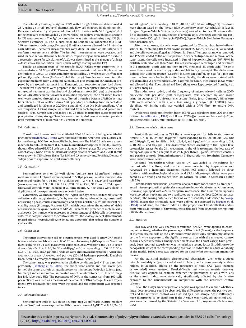
Fig. 1. Characterization of the AgNPs by transmission electron microscopy. TEM micrographs showing the NGAP silver nanomaterial morphology in dry powdered form( logy ina anol, s
3
3
micoyptt(
wwmowb
lstt
ta1
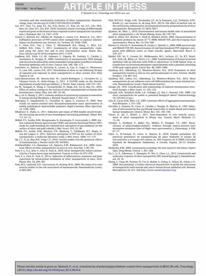
Bastarrachea et al., 2011). To test this property, the AgNPs wereheated to 400 ◦C in a stream of helium which was analyzed withTD–GC–MS. This experiment showed release of both 2-pyrrolidone
a–c) and after dispersing the NGAP powder into ethanol (d–f). The NGAP morphond notable amount of binder phase. When the NGAP powder is dispersed into eth
. Results
.1. Characterization of AgNPs
In transmission electron microscopy (TEM), the AgNP powderainly appeared as micron-scale aggregates with AgNPs embedded
n an organic matrix (Fig. 1a–c). According to in situ EDS analysis, Agonstituted more than 80 wt.% of the aggregates. Also some carbon,xygen, nitrogen and silicon were detected, but a quantitative anal-sis on light elements could not be carried out reliably. When theowdered AgNPs were dispersed in ethanol, the NPs were releasedo form a dispersion of primary particle sizes corresponding wello the average diameter of 42.5 ± 14.5 nm given by the producerFig. 1d–f).
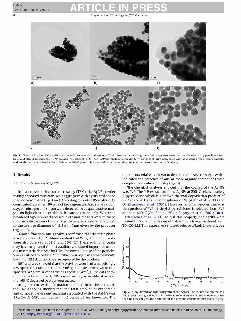
X-ray diffraction (XRD) analysis confirmed that the main phaseas pure silver (Fig. 2). Minor unidentified X-ray diffraction peaksere also observed at 32.3◦ and 36.0◦ 2�. These additional peaksay have originated from crystalline-associated impurities or the
rganic matrix observed by TEM. The crystallite size of the particlesas calculated to be 41 ± 2 nm, which was again in agreement with
oth the TEM data and the size reported by the producer.BET analyses showed that the AgNP powder had a surprisingly
ow specific surface area of 0.03 m2/g. The theoretical value of apherical 42.5 nm silver particle is about 13.4 m2/g. The data showhat the surfaces of the AgNPs are not readily accessible, at least inhe 160 ◦C degassed powder aggregates.
Please cite this article in press as: Nymark, P., et al., Genotoxicity of polyvin(2012), http://dx.doi.org/10.1016/j.tox.2012.09.014
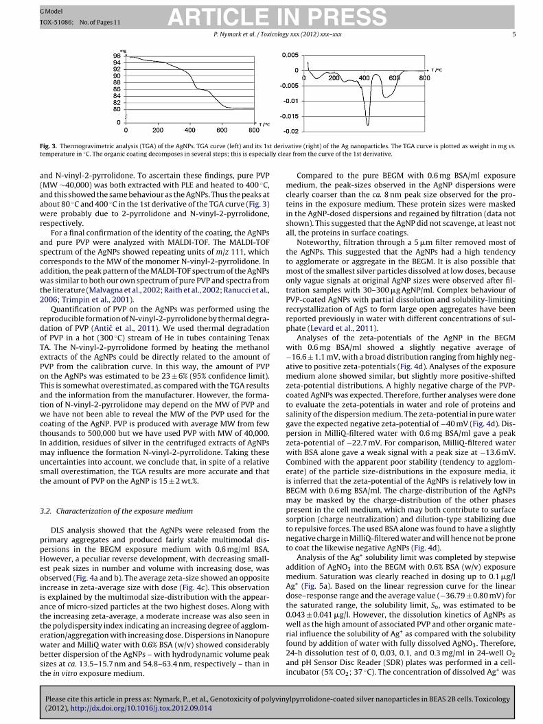
In agreement with information obtained from the producer,he TGA-analyses showed that the total amount of evaporablend combustible organic material associated with the AgNPs was5 ± 2 wt.% (95% confidence limit) corrected for buoyancy. The
the dry form consists of large aggregates with nanosized silver primary particlesilver nanoparticles are spread out effectively.
organic material was shown to decompose in several steps, whichindicated the presence of one or more organic compounds withcomplex molecular chemistry (Fig. 3).
The chemical analyses showed that the coating of the AgNPswas PVP. The PLE extraction of the AgNPs at 200 ◦C released solely2-pyrrolidone which is a known thermal degradation product ofPVP at about 100 ◦C in atmospheres of N2 (Antic et al., 2011) andO2 (Bogatyrev et al., 2001). However, another known degrada-tion product of PVP, N-vinyl-2-pyrrolidone, is released from PVPat about 400 ◦C (Antic et al., 2011; Bogatyrev et al., 2001; Loría-
ylpyrrolidone-coated silver nanoparticles in BEAS 2B cells. Toxicology
Fig. 2. X-ray diffraction (XRD) diagram of the AgNPs. The counts are plotted as afunction of the angle given in 2�. The broad reflections seen in the sample indicatesthe small crystal size. The positions for the silver reflections are marked with grey.
ARTICLE IN PRESSG Model
TOX-51086; No. of Pages 11
P. Nymark et al. / Toxicology xxx (2012) xxx– xxx 5
F t derivt ly clea
a(aawr
ascawt2
rdoTePoTatwctImust
3
ppHeoiiattewbst
ig. 3. Thermogravimetric analysis (TGA) of the AgNPs. TGA curve (left) and its 1semperature in ◦C. The organic coating decomposes in several steps; this is especial
nd N-vinyl-2-pyrrolidone. To ascertain these findings, pure PVPMW ∼40,000) was both extracted with PLE and heated to 400 ◦C,nd this showed the same behaviour as the AgNPs. Thus the peaks atbout 80 ◦C and 400 ◦C in the 1st derivative of the TGA curve (Fig. 3)ere probably due to 2-pyrrolidone and N-vinyl-2-pyrrolidone,
espectively.For a final confirmation of the identity of the coating, the AgNPs
nd pure PVP were analyzed with MALDI-TOF. The MALDI-TOFpectrum of the AgNPs showed repeating units of m/z 111, whichorresponds to the MW of the monomer N-vinyl-2-pyrrolidone. Inddition, the peak pattern of the MALDI-TOF spectrum of the AgNPsas similar to both our own spectrum of pure PVP and spectra from
he literature (Malvagna et al., 2002; Raith et al., 2002; Ranucci et al.,006; Trimpin et al., 2001).
Quantification of PVP on the AgNPs was performed using theeproducible formation of N-vinyl-2-pyrrolidone by thermal degra-ation of PVP (Antic et al., 2011). We used thermal degradationf PVP in a hot (300 ◦C) stream of He in tubes containing TenaxA. The N-vinyl-2-pyrrolidone formed by heating the methanolxtracts of the AgNPs could be directly related to the amount ofVP from the calibration curve. In this way, the amount of PVPn the AgNPs was estimated to be 23 ± 6% (95% confidence limit).his is somewhat overestimated, as compared with the TGA resultsnd the information from the manufacturer. However, the forma-ion of N-vinyl-2-pyrrolidone may depend on the MW of PVP ande have not been able to reveal the MW of the PVP used for the
oating of the AgNP. PVP is produced with average MW from fewhousands to 500,000 but we have used PVP with MW of 40,000.n addition, residues of silver in the centrifuged extracts of AgNPs
ay influence the formation N-vinyl-2-pyrrolidone. Taking thesencertainties into account, we conclude that, in spite of a relativemall overestimation, the TGA results are more accurate and thathe amount of PVP on the AgNP is 15 ± 2 wt.%.
.2. Characterization of the exposure medium
DLS analysis showed that the AgNPs were released from therimary aggregates and produced fairly stable multimodal dis-ersions in the BEGM exposure medium with 0.6 mg/ml BSA.owever, a peculiar reverse development, with decreasing small-st peak sizes in number and volume with increasing dose, wasbserved (Fig. 4a and b). The average zeta-size showed an oppositencrease in zeta-average size with dose (Fig. 4c). This observations explained by the multimodal size-distribution with the appear-nce of micro-sized particles at the two highest doses. Along withhe increasing zeta-average, a moderate increase was also seen inhe polydispersity index indicating an increasing degree of agglom-ration/aggregation with increasing dose. Dispersions in Nanopure
Please cite this article in press as: Nymark, P., et al., Genotoxicity of polyviny(2012), http://dx.doi.org/10.1016/j.tox.2012.09.014
ater and MilliQ water with 0.6% BSA (w/v) showed considerablyetter dispersion of the AgNPs – with hydrodynamic volume peakizes at ca. 13.5–15.7 nm and 54.8–63.4 nm, respectively – than inhe in vitro exposure medium.
ative (right) of the Ag nanoparticles. The TGA curve is plotted as weight in mg vs.r from the curve of the 1st derivative.
Compared to the pure BEGM with 0.6 mg BSA/ml exposuremedium, the peak-sizes observed in the AgNP dispersions wereclearly coarser than the ca. 8 nm peak size observed for the pro-teins in the exposure medium. These protein sizes were maskedin the AgNP-dosed dispersions and regained by filtration (data notshown). This suggested that the AgNP did not scavenge, at least notall, the proteins in surface coatings.
Noteworthy, filtration through a 5 �m filter removed most ofthe AgNPs. This suggested that the AgNPs had a high tendencyto agglomerate or aggregate in the BEGM. It is also possible thatmost of the smallest silver particles dissolved at low doses, becauseonly vague signals at original AgNP sizes were observed after fil-tration samples with 30–300 �g AgNP/ml. Complex behaviour ofPVP-coated AgNPs with partial dissolution and solubility-limitingrecrystallization of AgS to form large open aggregates have beenreported previously in water with different concentrations of sul-phate (Levard et al., 2011).
Analyses of the zeta-potentials of the AgNP in the BEGMwith 0.6 mg BSA/ml showed a slightly negative average of−16.6 ± 1.1 mV, with a broad distribution ranging from highly neg-ative to positive zeta-potentials (Fig. 4d). Analyses of the exposuremedium alone showed similar, but slightly more positive-shiftedzeta-potential distributions. A highly negative charge of the PVP-coated AgNPs was expected. Therefore, further analyses were doneto evaluate the zeta-potentials in water and role of proteins andsalinity of the dispersion medium. The zeta-potential in pure watergave the expected negative zeta-potential of −40 mV (Fig. 4d). Dis-persion in MilliQ-filtered water with 0.6 mg BSA/ml gave a peakzeta-potential of −22.7 mV. For comparison, MilliQ-filtered waterwith BSA alone gave a weak signal with a peak size at −13.6 mV.Combined with the apparent poor stability (tendency to agglom-erate) of the particle size-distributions in the exposure media, itis inferred that the zeta-potential of the AgNPs is relatively low inBEGM with 0.6 mg BSA/ml. The charge-distribution of the AgNPsmay be masked by the charge-distribution of the other phasespresent in the cell medium, which may both contribute to surfacesorption (charge neutralization) and dilution-type stabilizing dueto repulsive forces. The used BSA alone was found to have a slightlynegative charge in MilliQ-filtered water and will hence not be proneto coat the likewise negative AgNPs (Fig. 4d).
Analysis of the Ag+ solubility limit was completed by stepwiseaddition of AgNO3 into the BEGM with 0.6% BSA (w/v) exposuremedium. Saturation was clearly reached in dosing up to 0.1 �g/lAg+ (Fig. 5a). Based on the linear regression curve for the lineardose–response range and the average value (−36.79 ± 0.80 mV) forthe saturated range, the solubility limit, So, was estimated to be0.043 ± 0.041 �g/l. However, the dissolution kinetics of AgNPs aswell as the high amount of associated PVP and other organic mate-rial influence the solubility of Ag+ as compared with the solubility
lpyrrolidone-coated silver nanoparticles in BEAS 2B cells. Toxicology
found by addition of water with fully dissolved AgNO3. Therefore,24-h dissolution test of 0, 0.03, 0.1, and 0.3 mg/ml in 24-well O2and pH Sensor Disc Reader (SDR) plates was performed in a cell-incubator (5% CO2; 37 ◦C). The concentration of dissolved Ag+ was
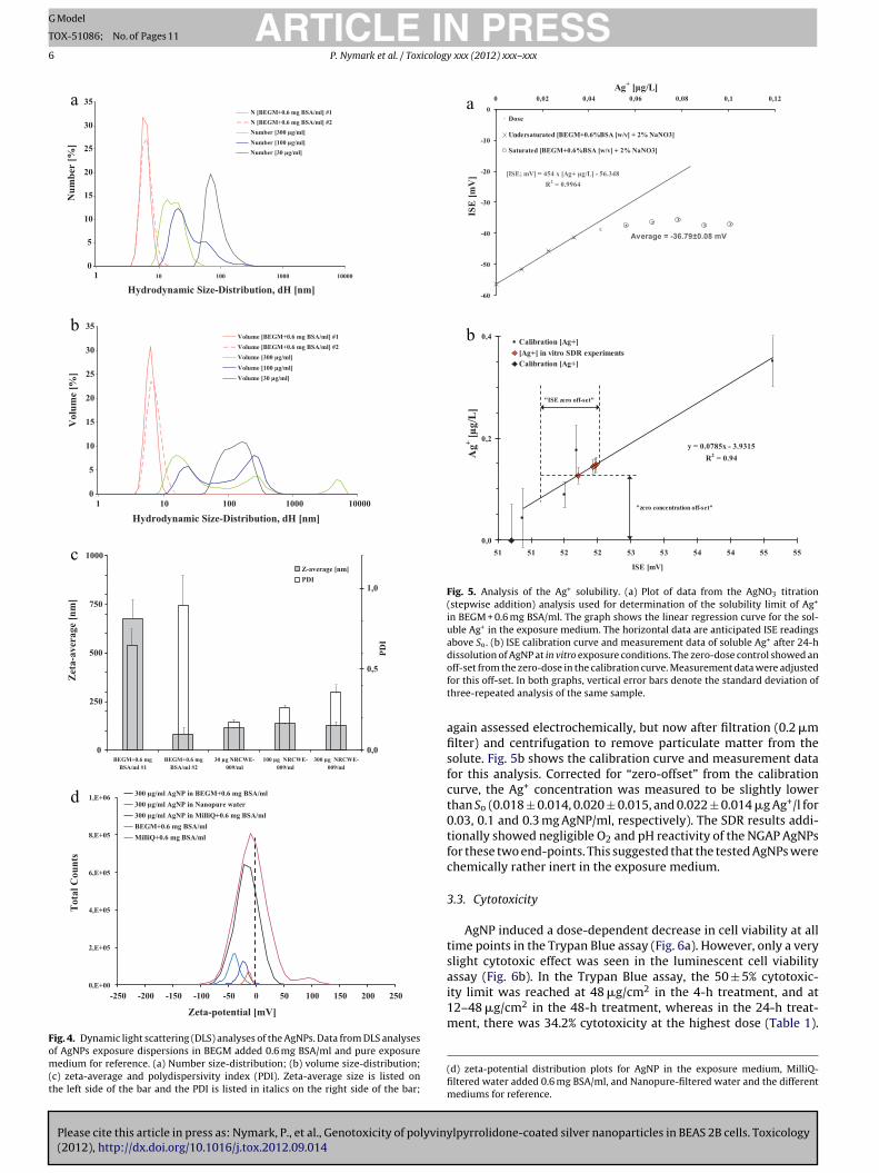
Please cite this article in press as: Nymark, P., et al., Genotoxicity of polyvin(2012), http://dx.doi.org/10.1016/j.tox.2012.09.014
ARTICLE IN PRESSG Model
TOX-51086; No. of Pages 11
6 P. Nymark et al. / Toxicology xxx (2012) xxx– xxx
0
5
10
15
20
25
30
35
1 10 100 1000 10000
Hydrodynamic Size-Distribution, dH [nm]
Nu
mb
er [
%]
N [BEGM+0.6 mg BSA/ml] #1
N [BEGM+0.6 mg BSA/ml] #2
Number [300 µg/ml]
Number [100 µg/ml]
Number [30 µg/ml]
0
5
10
15
20
25
30
35
1 10 100 1000 10000
Hydrodynamic Size-Distribution, dH [nm]
Volu
me
[%]
Volume [BEGM+0.6 mg BSA/ml] #1
Volume [BEGM+0.6 mg BSA/ml] #2
Volume [300 µg/ml]
Volume [100 µg/ml]
Volume [30 µg/ml]
0
250
500
750
1000
BEGM+0.6 mg
BSA/ml #1
BEGM+0.6 mg
BSA/ml #2
30 µg NRCWE-
009/ml
100 µg NRCWE-
009/ml
300 µg NRCWE-
009/ml
Zet
a-a
ver
age
[nm
]
0,0
0,5
1,0
PD
I
Z-average [nm]
PDI
0,E+00
2,E+05
4,E+05
6,E+05
8,E+05
1,E+06
-250 -200 -150 -100 -50 0 50 100 150 200 250
Zeta-potential [mV]
Tota
l C
ou
nts
300 µg/ml AgNP in BEGM+0.6 mg BSA/ml
300 µg/ml AgNP in Nanopure water
300 µg/ml AgNP in MilliQ+0.6 mg BSA/ml
BEGM+0.6 mg BSA/ml
MilliQ+0.6 mg BSA/ml
a
b
c
d
Fig. 4. Dynamic light scattering (DLS) analyses of the AgNPs. Data from DLS analysesof AgNPs exposure dispersions in BEGM added 0.6 mg BSA/ml and pure exposuremedium for reference. (a) Number size-distribution; (b) volume size-distribution;(c) zeta-average and polydispersivity index (PDI). Zeta-average size is listed onthe left side of the bar and the PDI is listed in italics on the right side of the bar;
-60
-50
-40
-30
-20
-10
0
0 0,02 0,04 0,06 0,08 0,1 0,12
Ag+ [µg/L]
ISE
[m
V]
0,0
0,2
0,4
51 51 52 52 53 53 54 54 55 55
ISE [mV]
Ag
+ [
µg
/L]
a
b
Fig. 5. Analysis of the Ag+ solubility. (a) Plot of data from the AgNO3 titration(stepwise addition) analysis used for determination of the solubility limit of Ag+
in BEGM + 0.6 mg BSA/ml. The graph shows the linear regression curve for the sol-uble Ag+ in the exposure medium. The horizontal data are anticipated ISE readingsabove So. (b) ISE calibration curve and measurement data of soluble Ag+ after 24-hdissolution of AgNP at in vitro exposure conditions. The zero-dose control showed an
off-set from the zero-dose in the calibration curve. Measurement data were adjustedfor this off-set. In both graphs, vertical error bars denote the standard deviation ofthree-repeated analysis of the same sample.again assessed electrochemically, but now after filtration (0.2 �mfilter) and centrifugation to remove particulate matter from thesolute. Fig. 5b shows the calibration curve and measurement datafor this analysis. Corrected for “zero-offset” from the calibrationcurve, the Ag+ concentration was measured to be slightly lowerthan So (0.018 ± 0.014, 0.020 ± 0.015, and 0.022 ± 0.014 �g Ag+/l for0.03, 0.1 and 0.3 mg AgNP/ml, respectively). The SDR results addi-tionally showed negligible O2 and pH reactivity of the NGAP AgNPsfor these two end-points. This suggested that the tested AgNPs werechemically rather inert in the exposure medium.
3.3. Cytotoxicity
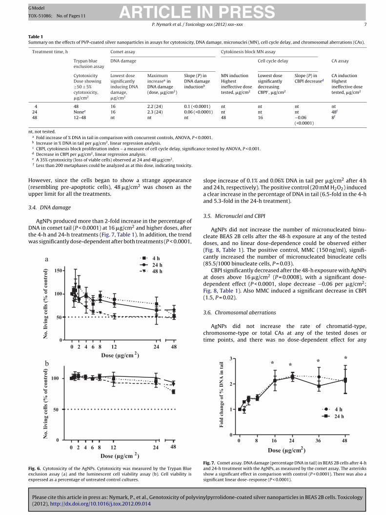
AgNP induced a dose-dependent decrease in cell viability at alltime points in the Trypan Blue assay (Fig. 6a). However, only a veryslight cytotoxic effect was seen in the luminescent cell viability
ylpyrrolidone-coated silver nanoparticles in BEAS 2B cells. Toxicology
assay (Fig. 6b). In the Trypan Blue assay, the 50 ± 5% cytotoxic-ity limit was reached at 48 �g/cm2 in the 4-h treatment, and at12–48 �g/cm2 in the 48-h treatment, whereas in the 24-h treat-ment, there was 34.2% cytotoxicity at the highest dose (Table 1).
(d) zeta-potential distribution plots for AgNP in the exposure medium, MilliQ-filtered water added 0.6 mg BSA/ml, and Nanopure-filtered water and the differentmediums for reference.
ARTICLE IN PRESSG Model
TOX-51086; No. of Pages 11
P. Nymark et al. / Toxicology xxx (2012) xxx– xxx 7
Table 1Summary on the effects of PVP-coated silver nanoparticles in assays for cytotoxicity, DNA damage, micronuclei (MN), cell cycle delay, and chromosomal aberrations (CAs).
Treatment time, h Comet assay Cytokinesis block MN assay
Trypan blueexclusion assay
DNA damage Cell cycle delay CA assay
CytotoxicityDose showing≥50 ± 5%cytotoxicity,�g/cm2
Lowest dosesignificantlyinducing DNAdamage,�g/cm2
Maximumincreasea inDNA damage(dose, �g/cm2)
Slope (P) inDNA damageinductionb
MN inductionHighestineffective dosetested, �g/cm2
Lowest dosesignificantlydecreasingCBPIc, �g/cm2
Slope (P) inCBPI decreased
CA inductionHighestineffective dosetested, �g/cm2
4 48 16 2.2 (24) 0.1 (<0.0001) nt nt nt nt24 Nonee 16 2.3 (24) 0.06 (<0.0001) nt nt nt 48f
48 12–48 nt nt nt 48 16 −0.06(<0.0001)
8f
nt, not tested.a Fold increase of % DNA in tail in comparison with concurrent controls, ANOVA, P < 0.0001.b Increase in % DNA in tail per �g/cm2, linear regression analysis.c CBPI, cytokinesis block proliferation index – a measure of cell cycle delay, significance tested by ANOVA, P < 0.001.
H(u
3
Dtw
Fee
d Decrease in CBPI per �g/cm2, linear regression analysis.e A 35% cytotoxicity (loss of viable cells) observed at 24 and 48 �g/cm2.f Less than 200 metaphases could be analyzed as at this dose, indicating toxicity.
owever, since the cells began to show a strange appearanceresembling pre-apoptotic cells), 48 �g/cm2 was chosen as thepper limit for all the treatments.
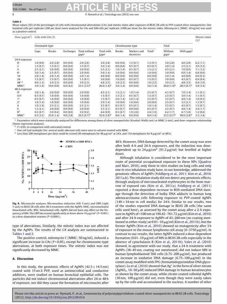
.4. DNA damage
Please cite this article in press as: Nymark, P., et al., Genotoxicity of polyviny(2012), http://dx.doi.org/10.1016/j.tox.2012.09.014
AgNPs produced more than 2-fold increase in the percentage ofNA in comet tail (P < 0.0001) at 16 �g/cm2 and higher doses, after
he 4-h and 24-h treatments (Fig. 7, Table 1). In addition, the trendas significantly dose-dependent after both treatments (P < 0.0001,
0
100
150
2
50
48 4 6 8 12 24
4 h
24 h
48 h
0
Dose (µg/cm2)
No
. li
vin
g c
ells
(%
of
con
tro
l)
0
100
2 4 6 8 12
50
48240
Dose (µg/cm2)
No
. li
vin
g c
ells
(%
of
con
tro
l)
a
b
ig. 6. Cytotoxicity of the AgNPs. Cytotoxicity was measured by the Trypan Bluexclusion assay (a) and the luminescent cell viability assay (b). Cell viability isxpressed as a percentage of untreated control cultures.
slope increase of 0.1% and 0.06% DNA in tail per �g/cm2 after 4 hand 24 h, respectively). The positive control (20 mM H2O2) induceda clear increase in the percentage of DNA in tail (6.5-fold in the 4-hand 5.3-fold in the 24-h treatment).
3.5. Micronuclei and CBPI
AgNPs did not increase the number of micronucleated binu-cleate BEAS 2B cells after the 48-h exposure at any of the testeddoses, and no linear dose-dependence could be observed either(Fig. 8, Table 1). The positive control, MMC (150 ng/ml), signifi-cantly increased the number of micronucleated binucleate cells(85.5/1000 binucleate cells, P = 0.03).
CBPI significantly decreased after the 48-h exposure with AgNPsat doses above 16 �g/cm2 (P = 0.0008), with a significant dose-dependent effect (P < 0.0001, slope decrease −0.06 per �g/cm2;Fig. 8, Table 1). Also MMC induced a significant decrease in CBPI(1.5, P = 0.02).
3.6. Chromosomal aberrations
lpyrrolidone-coated silver nanoparticles in BEAS 2B cells. Toxicology
AgNPs did not increase the rate of chromatid-type,chromosome-type or total CAs at any of the tested doses ortime points, and there was no dose-dependent effect for any
0
1
2
3
4 h
24 h
8 16 24 36 48
***
*
0
Dose (µg/cm2)
Fold
ch
an
ge
of
% D
NA
in
tail
Fig. 7. Comet assay. DNA damage (percentage DNA in tail) in BEAS 2B cells after 4-hand 24-h treatment with the AgNPs, as measured by the comet assay. The asterisksshow a significant effect in comparison with control (P < 0.0001). There was also asignificant linear dose–response (P < 0.0001).
ARTICLE IN PRESSG Model
TOX-51086; No. of Pages 11
8 P. Nymark et al. / Toxicology xxx (2012) xxx– xxx
Table 2Mean values (SD) of the percentages of cells with chromosomal aberrations (CAs) and mitotic index after exposure of BEAS 2B cells to PVP-coated silver nanoparticles. Onehundred cells per replicate (200 per dose) were analyzed for CAs and 500 cells per replicate (1000 per dose) for the mitotic index. Mitomycin C (MMC; 50 ng/ml) was usedas a positive control.
Dose (�g/cm2) Cells with CAs (%) Mitotic index(%)a
Chromatid-type Chromosome-type Total
Gaps Breaks Exchanges Total withoutgapsa
Total withgapsa
Breaks Markers anddicentrics
Totala Withoutgapsa
With gapsa
24-h exposure0c 1.0 (0.0) 2.0 (2.8) 0.0 (0.0) 2.0 (2.8) 3.0 (2.8) 0.0 (0.0) 1.5 (0.7) 1.5 (0.7) 3.0 (2.8) 4.0 (2.8) 6.2 (1.7)2 1.5 (0.7) 1.5 (0.7) 0.0 (0.0) 1.5 (0.7) 3.0 (1.4) 0.0 (0.0) 0.5 (0.7) 0.5 (0.7) 2.0 (1.4) 3.5 (2.1) 3.9 (3.3)4 2.5 (2.1) 3.0 (0.0) 0.0 (0.0) 3.0 (0.0) 5.5 (2.1) 1.0 (1.4) 0.5 (0.7) 1.5 (2.1) 4.5 (2.1) 7.0 (0.0) 7.1 (1.3)8c 3.0 (1.4) 2.5 (0.7) 0.0 (0.0) 2.0 (0.0) 5.0 (1.4) 1.0 (0.0) 0.0 (0.0) 1.0 (0.0) 3.0 (0.0) 6.0 (1.4) 6.8 (0.0)16 2.0 (1.4) 2.0 (1.4) 0.0 (0.0) 2.0 (1.4) 4.0 (0.0) 0.0 (0.0) 0.0 (0.0) 0.0 (0.0) 2.0 (1.4) 4.0 (0.0) 6.0 (0.3)24 1.5 (0.7) 1.5 (0.7) 0.0 (0.0) 1.5 (0.7) 3.0 (0.0) 1.0 (0.0) 0.5 (0.7) 1.5 (0.7) 3.0 (0.0) 4.5 (0.7) 6.9 (0.4)48c,d 0.9 (1.3) 3.9 (2.3) 0.0 (0.0) 3.9 (2.3) 4.8 (3.5) 1.8 (2.5) 0.0 (0.0) 1.8 (2.5) 3.9 (2.3) 4.8 (3.5) 6.9 (1.0)MMCc 3.0 (1.4) 19.0 (0.0) 6.0 (4.2) 23.5 (3.5)b 26.0 (1.4)b 3.0 (1.4) 0.0 (0.0) 3.0 (1.4) 26.0 (1.4)b 28.5 (0.7)b 3.8 (3.4)
48-h exposure0c 3.0 (1.4) 2.0 (0.0) 0.0 (0.0) 2.0 (0.0) 4.5 (2.1) 1.5 (2.1) 1.0 (1.4) 2.5 (0.7) 4.5 (0.7) 7.0 (1.4) 1.1 (0.1)0.5c 0.5 (0.7) 1.0 (0.0) 0.0 (0.0) 1.0 (0.0) 1.5 (0.7) 1.5 (2.1) 0.5 (0.7) 1.5 (0.7) 2.5 (0.7) 3.0 (1.4) 1.1 (0.7)1c 0.5 (0.7) 1.0 (0.0) 0.0 (0.0) 1.0 (0.0) 1.5 (0.7) 2.0 (1.4) 2.0 (1.4) 2.5 (0.7) 3.5 (0.7) 4.0 (1.4) 1.0 (0.3)2c 1.0 (1.4) 1.0 (0.0) 0.0 (0.0) 1.0 (0.0) 2.0 (1.4) 1.0 (0.0) 1.0 (0.0) 2.0 (0.0) 2.5 (0.7) 3.5 (2.1) 1.3 (0.7)4 1.0 (1.4) 2.5 (2.1) 0.0 (0.0) 2.5 (2.1) 3.5 (0.7) 0.5 (0.7) 0.5 (0.7) 1.0 (1.4) 3.5 (0.7) 4.5 (0.7) 1.5 (0.7)6 1.5 (2.1) 2.0 (0.0) 0.0 (0.0) 2.0 (0.0) 3.5 (2.1) 0.5 (0.7) 0.0 (0.0) 0.5 (0.7) 2.5 (0.7) 4.0 (1.4) 2.4 (1.7)8d 0.5 (0.7) 3.9 (0.2) 0.0 (0.0) 3.9 (0.2) 4.4 (0.9) 0.5 (0.7) 1.9 (2.6) 2.4 (1.9) 6.2 (1.7) 6.7 (1.0) 1.2 (0.6)MMCc 6.5 (3.5) 25.0 (1.4) 9.0 (2.8) 29.5 (0.7)b 35.0 (2.8)b 8.0 (1.4) 0.0 (0.0) 8.0 (1.4) 33.5 (0.7)b 39.0 (2.8)b 2.5 (1.8)
a Parameters which were statistically analyzed for differences among doses of silver nanoparticles (Kruskal–Wallis test) or MMC (t-test), and dose–response relationship(linear regression analysis).
b P < 0.05, in comparison with untreated control.c One cell had multiple CAs; several multi-aberrant cells were seen in cultures treated
d Less than 200 metaphases per dose could be scored (99 metaphases for 48 �g/cm2 at
0
5
10
15
20
25
30
1.0
1.2
1.4
1.6
1.8
2.0
MNBNCs/1000 BNCs
CBPI
0 4 8 16 24 36 48
Dose (µg/cm2)
No
. M
NB
NC
s/1
00
0 B
NC
s
CB
PI
Fig. 8. Micronuclei analyses. Micronucleus induction (left Y-axis) and CBPI (rightY-axis) in BEAS 2B cells after 48-h treatment with the AgNPs. MNC, micronucleatedbqi
tbT
sas
4
capo
inucleated cells; BNC, binucleated cells. AgNPs did not have any effect on the fre-uency of MN. The CBPI decreased significantly at doses above 16 �g/cm2 (P < 0.001)
n a dose-dependent manner (P < 0.0001).
ype of aberrations. Similarly, the mitotic index was not affectedy the AgNPs. The results of the CA analysis are summarized inables 1 and 2.
The positive control, mitomycin C (MMC; 50 ng/ml), induced aignificant increase in CAs (P < 0.05), except for chromosome-typeberrations, at both exposure times. The mitotic index was notignificantly decreased by MMC.
. Discussion
In this study, the genotoxic effects of AgNPs (42.5 ± 14.5 nm)
Please cite this article in press as: Nymark, P., et al., Genotoxicity of polyvin(2012), http://dx.doi.org/10.1016/j.tox.2012.09.014
oated with 15 wt.% PVP, used as antimicrobial and conductivedditives, were studied on human bronchial epithelial cells. Thearticles did not induce chromosomal aberrations after 24 or 48 hf exposure, nor did they cause the formation of micronuclei after
with MMC. 24 h, and 154 metaphases for 8 �g/cm2 at 48 h).
48 h. However, DNA damage detected by the comet assay was seenafter both 4-h and 24-h exposures, and the induction was dose-dependent up to 24 �g/cm2 (91.2 �g/ml) but levelled at higherdoses.
Although inhalation is considered to be the most importantroute of potential occupational exposure to these NPs (Quadrosand Marr, 2010), only three in vitro studies on lung cells and onlyone in vivo inhalation study have, to our knowledge, addressed thegenotoxic effects of AgNPs (Foldbjerg et al., 2011; Kim et al., 2010,2011a,b). The inhalation study did not detect any genotoxic effects,through analysis of micronucleated erythrocytes in the bone mar-row of exposed rats (Kim et al., 2011a). Foldbjerg et al. (2011)reported a dose-dependent increase in ROS-mediated DNA dam-age through the detection of bulky DNA adducts in human lungadenocarcinoma cells following exposure to PVP-coated AgNPs(149 ± 34 nm in cell media) for 24 h. Similar to our results, twoof the studies reported DNA damage in BEAS 2B cells (the samecells used here), as assessed by the comet assay after a 2-h expo-sure to AgNPs of <100 nm at 190.43–761.72 �g/ml (Kim et al., 2010)and after 24-h exposure to AgNPs of 43–260 nm (no coating men-tioned in either study) at 0.01–10 �g/ml (Kim et al., 2011b), but theformer AgNPs (Kim et al., 2010) showed no mutagenicity after 3 hof exposure in the mouse lymphoma cell assay (0–3750 �g/ml). Incontrast to our results, the latter AgNPs induced a dose-dependentformation (0.01–10 �g/ml) of MN in BEAS 2B cells especially in theabsence of cytochalasin B (Kim et al., 2011b). Vales et al. (2010)showed, in agreement with our study, that a 24-h treatment withAgNPs (38–45 nm, coating not mentioned) did not induce MN inhuman lymphoblastoid TK6 cells (6.75–200 �g/ml), but producedan increase in oxidative DNA damage (6.75–100 �g/ml) in thecomet assay modified with FPG (formamidopyrimidine DNA glyco-sylase). Lu et al. (2010) showed that Ag+ in the form of silver nitrate
ylpyrrolidone-coated silver nanoparticles in BEAS 2B cells. Toxicology
(AgNO3, 10–50 �M) induced DNA damage in human keratinocytesas shown by the comet assay, while citrate-coated colloidal AgNPs(30 nm, 100 �g/ml) did not, even though they were easily takenup by the cells and accumulated in the nucleus. A number of other
ING Model
T
icology
iatweK
23rr(AaosaedKtod(
t(aAfpace
4sehcowTwpsoto1(t
eaeiitDtN2tps
ARTICLEOX-51086; No. of Pages 11
P. Nymark et al. / Tox
n vitro studies have shown that AgNPs (in the size range 1–50 nm)re able to induce DNA and chromosomal damage in different cellypes and model organisms, such as human and mouse fibrobasts asell as Caenorhabditis elegans (Ahamed et al., 2008, 2010; AshaRani
t al., 2009; Ellegaard-Jensen et al., 2012; Hackenberg et al., 2011;awata et al., 2009; Li et al., 2011; Xu et al., 2012).
Two groups showed that neither inhalation (0.7, 1.4 and.9 × 106 particles/cm3, 6 h/day for 90 days) nor oral exposure (30,00 and 1000 mg/kg/day for 28 days) to AgNPs (18 nm and 60 nm,espectively) induces formation of micronucleated erythrocytes inats, although the Ag was shown to accumulate in several tissuesKim et al., 2008, 2011a). Arora et al. (2009) reported that althoughgNPs (7–20 nm) were able to enter primary mouse fibroblastsnd liver cells, antioxidant mechanisms seemed to protect fromxidative damage at concentrations below ½ IC50 – in contrast toecondary established cell lines where the oxidative stress associ-ted with AgNPs exposure seemed to result in DNA damage (Arorat al., 2008, 2009). Thus, in vitro and in vivo studies seem to contra-ict each other, regarding the effects of AgNPs, as also reviewed byim et al. (2012). These contrasting results indicate that the geno-
oxic effects of AgNPs are either false positives as a consequencef in vitro-related effects (Walmsley, 2005) or that they are highlyependent on cell type – or AgNP coating – as stated by Park et al.2009).
Finally, research on the cytotoxic effects of AgNPs are also con-roversial and both very high cytotoxicity at as low as 2–5 �g/mlBar-Ilan et al., 2009; Hussain et al., 2005; Soto et al., 2007) as wells almost no cytotoxicity (up to 100 �g/ml) have been reported forgNPs (Samberg et al., 2012). Conflicting results, such as these, call
or more efficient characterization of the NPs, especially regardingarticle size, coatings and surface charge, since these probablyffect both cyto- and genotoxicity. However, it may be even morerucial to have a broad understanding of the fate of the NPs in thexposure system.
The average crystallite size of the AgNP studied here was1 ± 2 nm and they were found to be associated with 15 wt.% PVPome of which was thought to be present as surface coating in thexposure medium. The surface charge of the dispersed AgNP wasighly negative (−40 mV) in water, but appeared partially if notompletely neutralized in the exposure medium. The zeta-potentialf AgNPs in the exposure medium showed a weak negative averageith a wide distribution ranging from positive to highly negative.
he low zeta-potential in the exposure medium is in accordanceith the partial agglomeration of the AgNPs observed in the sam-les. In water with added BSA, the surface charge was −22.7 mV,upporting the hypothesis that partial or complete neutralizationccurs in the exposure medium. The Ag+ solubility (AgNO3) inhe BEGM medium with 0.6 mg/ml BSA was found to be in therder of 0.04 �g/ml. Twenty-four-hour dissolution tests using 30,00 and 300 �g/ml AgNPs indicated that only a very low amount<0.022 �g/l) of Ag+ was dissolved in the exposure medium duringhe experiment.
It has been suggested that the toxicity of AgNPs may bexplained by a combination of effects of size, coating, Ag-solubilitynd uptake rates due to agglomeration into micro-sized agglom-rates in the exposure medium (Ellegaard-Jensen et al., 2012). Thenduction of DNA damage could be due to the release of Ag+ ionsn the dispersion medium. Our TEM and BET analyses showed thathe powdered AgNP were embedded in a matrix consisting of PVP.ispersion in ethanol revealed good dispersion by dissolution of
he matrix, but a coating may still have been present around thePs since PVP can chemisorb to the AgNP surface (Al-Saidi et al.,
Please cite this article in press as: Nymark, P., et al., Genotoxicity of polyviny(2012), http://dx.doi.org/10.1016/j.tox.2012.09.014
011; Mdluli et al., 2011). It is likely that this coating reducedhe ion leaching of the particles. This hypothesis was further sup-orted by the finding that the So of Ag+ in the BEGM mediumupplemented with BSA was less than 1 �g/l, and the amount of
PRESS xxx (2012) xxx– xxx 9
dissolved Ag+ in the exposure medium was slightly lower, rangingfrom 0.018 ± 0.014 to 0.022 ± 0.014 �g/l at 30–300 �g/ml AgNP.Thus, the dissolution kinetics and potentially also the associatedorganics and surface coatings affected the solubility of AgNPs.
The solubility of the AgNPs used in this study was quite lowand remained similar at the three doses tested, even though thehigher dose was 10 times higher than the lowest one. Neverthe-less, there was a linear increase in DNA damage induction at AgNPdoses of 30.4–91.2 �g/ml (corresponding to 8–24 �g/cm2). Hence,the data strongly suggests that the observed effects of the AgNPsdo not only arise from dissolved Ag+ in the dispersion medium. Itis possible that the release of ions from the NPs is more efficientlyperformed inside the cells than in the exposure medium, eitherdue to the coating being degraded by the cell (Schrand et al., 2008),or due to the AgNPs being ionized in the cells by a Trojan-horsetype mechanism (Park et al., 2010). Thus, the results of this studypoint towards a combinatorial mechanism where AgNPs are takenup by human bronchial epithelial cells and Ag+ is released fromthe particles in acidic conditions prevailing in lysosomes, result-ing in DNA damage. Indeed, both dissolved Ag+ and the specificproperties of AgNPs have been proposed to contribute to the toxiceffects of AgNPs (Johnston et al., 2010). AgNPs (7–10 nm) stabilizedwith polyethylenimine as well as the soluble Ag2CO3 were shownto induce the expression of DNA repair genes, while polystyreneNPs did not (Kawata et al., 2009). In contrast, the same investi-gators found that only AgNPs (7–10 nm), but neither Ag2CO3 norpolystyrene NPs induced MN in human hepatoma cells at doses upto 3 �g/ml (Kawata et al., 2009). In addition, Kawata et al. (2009)showed that Ag+ contributes mainly to the cytotoxic and stress-associated effects of AgNPs, but not to the DNA damaging effects,as demonstrated by an MN assay using the strong Ag+ ligand, cys-tein.
In the present study, DNA damage increased linearly up to24 �g/cm2, but levelled at higher doses. Such a dose–responsemight reflect the particle uptake capacity of the cells. Kim et al.(2011b) have reported that uptake of AgNPs into BEAS 2B cellsmay involve an endocytic pathway, which (they referred to Jianget al., 2008) depends on the size of the AgNPs, with the great-est cellular response occurring at 40–50 nm, an optimal particlesize for uptake through endocytosis, whereas bigger agglomerateswould be taken up less efficiently. Since our AgNPs had a high ten-dency to agglomerate in the BSA-supplemented BEGM mediumwith increasing doses, the uptake kinetics might also be levelledwhen the agglomerates start to be too large. Kim et al. (2011b)claimed that, despite aggregation of the AgNPs they used (≤100 nm,0.01–10 g/ml; no coating mentioned) in the BEGM medium,40–59 nm NPs still accounted for approximately 50% of all AgNPmass, and should have induced cellular responses. The authorsdid not explain how they assessed these calculations from theirDLS data.
In addition to size, also coating and surface charge play a cru-cial role in the uptake of NPs (Ahamed et al., 2010; Johnston et al.,2010; El Badawy et al., 2011). The presence of organic coatings has,indeed, been reported to the one hand result in more extensivecytotoxicity (Ahamed et al., 2008) and, on the other hand, to ren-der the particles biocompatible (Schrand et al., 2008). However, thebiocompatibility of, e.g. polysaccharide-coated AgNPs was shownto change over time, possibly due to the coating being degraded bythe cell (Schrand et al., 2008). Another report showed that driedcitrate-coated AgNPs (i.e. in powder form) were toxic to kerati-nocytes at 100 �g/ml, while dried PVP-coated AgNPs and silvernanoprisms showed no cytotoxicity at the same dose (Lu et al.,
lpyrrolidone-coated silver nanoparticles in BEAS 2B cells. Toxicology
2010). Furthermore, Samberg et al. (2010) reported that AgNPs arenot toxic by themselves, but residual soluble contaminants increasecell mortality. They found that washed uncoated AgNPs as well asAgNPs coated with polyaromatic graphitic carbon, were less toxic
ING Model
T
1 icolog
tk
bc((cstretou
ordtactstatHtIstacsBr2en
sdctGwaDcJdec
C
A
wlC
ARTICLEOX-51086; No. of Pages 11
0 P. Nymark et al. / Tox
han unwashed/uncoated particles to porcine skin and in humaneratinocytes (Samberg et al., 2010).
Surface charge (i.e. zeta-potential) is another feature that haseen proposed to have a large impact on toxicity, since positivelyharged NPs may interact with negatively charged componentse.g. DNA) in cells more readily than negatively charged particlesZhang et al., 2008). In a study on bacteria, the most positivelyharged NPs were the most toxic (El Badawy et al., 2011). However,ince positively charged AgNPs seem to be significantly more toxichan Ag+ ions, it is probable that physical interaction plays a largeole in AgNP toxicity, in addition to the effect of Ag+ (El Badawyt al., 2011). Our AgNPs were probably completely neutralized byhe constituents in the exposure medium, since the zeta-potentialf the AgNP was only slightly negative. This may also affect theirptake by the BEAS 2B cells.
Kim et al. (2011b) concluded that the genotoxic effects theybserved in BEAS 2B cells might be related to the generation ofadical oxygen species (ROS) by the AgNPs, producing oxidativeamage. Their AgNPs seemed to have a higher genotoxic capacityhan ours since, using the same cell line and treatment time (24-h)s we did, they found a 4-fold increase in DNA damage (using theomet assay) at their highest dose (10 �g/ml), which was similaro our lowest dose (7.6 �g/ml). The Comet assay is able to detectingle- and double-strand DNA breaks (Collins, 2004), but whilehe former can efficiently be repaired by an excision repair mech-nism, repair of the latter is more error-prone and could give riseo permanent chromosome damage (Mladenov and Iliakis, 2011).owever, extensive induction of single-strand breaks could exceed
he repair capacity of the cell, also resulting in chromosomal breaks.n the current study, we could not observe induction of chromo-omal aberrations or micronuclei, suggesting an efficient repair ofhe AgNP-induced DNA breaks. In fact, the induction of DNA dam-ge was already observed after 4-h exposure, with a dose–responseurve which was highly similar to the one observed after 24-h expo-ure. These results suggest, firstly, that the uptake of AgNPs by theEAS 2B cells seems to be quite fast. In fact, Kim et al. (2011b)eported the presence of aggregates within the nucleus after the4-h treatment. Secondly, that the cells seem to cope with the gen-rated DNA damage, repairing it in an efficient way, since there iso increase in damage with increasing time exposure.
Nevertheless, although PVP-coated AgNPs, such as the onestudied here, were not able to induce permanent chromosomalamage in human bronchial cells, there is much evidence of AgNPlastogenicity in other types of cells (Ahamed et al., 2010). Fur-hermore, AgNPs (20 nm) were found to induce up-regulation ofADD45B in human dermal fibroblasts at a 200 �M dose, whichas not seen with nanogold (Ma et al., 2011). GADD45B (growth
rrest and DNA damage-inducible, beta) is strongly associated withNA damage and cancer. In addition, AgNPs have been shown toause DNA damage as well as oxidative and carcinogenic stress inapanese medaka, while Ag+ only induced inflammation, metallicetoxification responses, and a low overall stress response (Chaet al., 2009). Thus, the particles should be produced and used withaution until further studies indicate safety.
onflict of interest statement
The authors declare that there is no conflict of interest.
cknowledgements
Please cite this article in press as: Nymark, P., et al., Genotoxicity of polyvin(2012), http://dx.doi.org/10.1016/j.tox.2012.09.014
Technical assistance from Yahia Kembouche (NRCWE) in DLS asell as Ag+ solubility analysis is greatly appreciated. The research
eading to these results has received funding from the Europeanommission under grant agreement FP7-211464-2 (NANODEVICE).
PRESSy xxx (2012) xxx– xxx
References
Ahamed, M., Karns, M., Goodson, M., Rowe, J., Hussain, S.M., Schlager, J.J., Hong, Y.,2008. DNA damage response to different surface chemistry of silver nanoparti-cles in mammalian cells. Toxicol. Appl. Pharmacol. 233, 404–410.
Ahamed, M., AlSalhi, M.S., Siddiqui, M.K.J., 2010. Silver nanoparticle applications andhuman health. Clin. Chim. Acta 411, 1841–1848.
Al-Saidi, W.A., Feng, H., Fichthorn, K.A., 2011. Adsorption of polyvinylpyrrolidone onAg surfaces: insight into a structure-directing agent. Nano Lett. 12, 997–1001.
Antic, V.V., Antic, M.P., Kronimus, A., Oing, K., Schwarzbauer, J., 2011. Quantitativedetermination of poly(vinylpyrrolidone) by continuous-flow off-line pyrolysis-GC/MS. J. Anal. Appl. Pyrolysis 90, 93–99.
Arora, S., Jain, J., Rajwade, J.M., Paknikar, K.M., 2008. Cellular responses induced bysilver nanoparticles: in vitro studies. Toxicol. Lett. 179, 93–100.
Arora, S., Jain, J., Rajwade, J.M., Paknikar, K.M., 2009. Interactions of silver nanopar-ticles with primary mouse fibroblasts and liver cells. Toxicol. Appl. Pharmacol.236, 310–318.
Asare, N., Instanes, C., Sandberg, W.J., Refsnes, M., Schwarze, P., Kruszewski, M.,Brunborg, G., 2012. Cytotoxic and genotoxic effects of silver nanoparticles intesticular cells. Toxicology 291, 65–72.
AshaRani, P.V., Low Kah Mun, G., Hande, M.P., Valiyaveettil, S., 2009. Cytotoxicityand genotoxicity of silver nanoparticles in human cells. ACS Nano 3, 279–290.
Bar-Ilan, O., Albrecht, R.M., Fako, V.E., Furgeson, D.Y., 2009. Toxicity assessmentof multisized gold and silver nanoparticles in Zebrafish embryos. Small 16,1897–1910.
Bogatyrev, V.M., Borisenko, N.V., Pokrovskii, V.A., 2001. Thermal degradation ofpolyvinylpyrrolidone on the surface of pyrogenic silica. Russ. J. Appl. Chem. 74,839–844.
Brøgger, A., Norum, R., Hansteen, I.L., Clausen, K.O., Skårdal, K., Mitelman, F., Kolnig,A.M., Strömbeck, B., Nordenson, I., Andersson, G., Jakobsson, K., Mäki-Paakkanen,J., Norppa, H., Järventaus, H., Sorsa, M., 1984. Comparison between five Nordiclaboratories on scoring of human lymphocyte chromosome aberrations. Hered-itas 100, 209–218.
Collins, A.R., 2004. The comet assay for DNA damage and repair: principles, applica-tions, and limitations. Mol. Biotechnol. 26, 249–261.
Chae, Y.J., Pham, C.H., Lee, J., Bae, E., Yi, J., Gu, M.B., 2009. Evaluation of the toxicimpact of silver nanoparticles on Japanese medaka (Oryzias latipes). Aquat. Tox-icol. 94, 320–327.
Clausen, P.A., Liu, Z., Kofoed-Sørensen, V., Little, J.C., Wolkoff, P., 2012. The influenceof temperature on the emission of di(2-ethylhexyl)phthalate (DEHP) from PVCflooring in the emission cell FLEC. Environ. Sci. Technol. 46, 909–915.
El Badawy, A.M., Silva, R.G., Morris, B., Scheckel, K.G., Suidan, M.T., Tolaymat, T.M.,2011. Surface charge-dependent toxicity of silver nanoparticles. Environ. Sci.Technol. 45, 283–287.
Ellegaard-Jensen, L., Jensen, K.A., Johansen, A., 2012. Nano-silver inducesdose–response effects on the nematode Caenorhabditis elegans. Ecotoxicol. Envi-ron. Saf., http://dx.doi.org/10.1016/j.ecoenv.2012.03.003.
Flower, N.A.L., Brabu, B., Revathy, M., Gopalakrishnan, C., Raja, S.V.K., Murugan, S.S.,Kumaravel, T.S., 2012. Characterization of synthesized silver nanoparticles andassessment of its genotoxicity potentials using the alkaline comet assay. Mutat.Res. 742, 61–65.
Foldbjerg, R., Dang, D., Autrup, H., 2011. Cytotoxicity and genotoxicity of silvernanoparticles in the human lung cancer cell line, A549. Arch. Toxicol. 85,743–750.
Genter, M.B., Newman, N.C., Shertzer, H.G., Ali, S.F., Bolon, B., 2012. Distributionand systemic effects of intranasally administrated 25 nm silver nanoparticles inadult mice. Toxicol. Pathol. 00, 1–10.
Hackenberg, S., Scherzed, A., Kessler, M., Hummel, S., Technau, A., Froelich, K.,Ginzkey, C., Koehler, C., Hagen, R., Kleinsasser, N., 2011. Silver nanoparticles:evaluation of DNA damage, toxicity and functional impairment in human mes-enchymal stem cells. Toxicol. Lett. 201, 27–33.
Hussain, S.M., Hess, K.L., Gearhart, J.M., Geiss, K.T., Schlager, J.J., 2005. In vitro toxicityof nanoparticles in BRL 3A rat liver cells. Toxicol. in Vitro 19, 975–983.
Jiang, W., Kim, B.Y., Rutka, J.T., Chan, W.C., 2008. Nanoparticle-mediated cellularresponse is size-dependent. Nat. Nanotechnol. 3, 145–150.
Johnston, H., Hutchison, G., Christensen, F., Peters, S., Hankin, S., Stone, V., 2010. Areview of the in vivo and in vitro toxicity of silver and gold particulates: particleattributes and biological mechanisms responsible for the observed toxicity. Crit.Rev. Toxicol. 40, 328–346.
Kawata, K., Osawa, M., Okabe, S., 2009. In vitro toxicity of silver nanoparticles atnoncytotoxic doses to HepG2 human hepatoma cells. Environ. Sci. Technol. 43,6046–6051.
Kim, Y.S., Kim, J.S., Cho, H.S., Rha, D.S., Kim, J.M., Park, J.D., Choi, B.S., Lim, R., Chang,H.K., Chung, Y.H., Kwon, I.H., Jeong, J., Han, B.S., Yu, I.J., 2008. Twenty-eight-day oral toxicity, genotoxicity, and gender-related tissue distribution of silvernanoparticles in Sprague-Dawley rats. Inhal. Toxicol. 20, 575–583.
Kim, Y.J., Yang, S., Ryu, J.C., 2010. Cytotoxicity and genotoxicity of nano-silver inmammalian cell lines. Mol. Cell Toxicol. 6, 119–125.
Kim, J.S., Sung, J.H., Ji, J.H., Song, K.S., Lee, J.H., Kang, C.S., Yu, I.J., 2011a. In vivogenotoxicity of silver nanoparticles after 90-day silver nanoparticle inhalationexposure. Saf. Health Work 2, 34–38.
ylpyrrolidone-coated silver nanoparticles in BEAS 2B cells. Toxicology
Kim, H.R., Kim, M.J., Lee, S.Y., Oh, S.M., Chung, K.H., 2011b. Genotoxic effects of sil-ver nanoparticles stimulated by oxidative stress in human normal bronchialepithelial (BEAS 2B) cells. Mutat. Res. 726, 129–135.
Kim, J.S., Song, K.S., Sung, J.H., Ryu, H.R., Choi, B.G., Cho, H.S., Lee, J.K., Yu, I.J., 2012.Genotoxicity, acute oral and dermal toxicity, eye and dermal irritation and
ING Model
T
icology
L
L
L
L
L
L
L
M
M
M
M
M
N
O
P
P
P
ARTICLEOX-51086; No. of Pages 11
P. Nymark et al. / Tox
corrosion and skin sensitisation evaluation of silver nanoparticles. Nanotoxi-cology, http://dx.doi.org/10.3109/17435390.2012.676099.
ee, H.Y., Choi, Y.J., Jung, E.J., Yin, H.Q., Kwon, J.T., Kim, J.E., Im, H.T., Cho, M.H.,Kim, J.H., Kim, H.Y., Lee, B.H., 2010. Genomics-based screening of differentiallyexpressed genes in the brains of mice exposed to silver nanoparticles via inhala-tion. J. Nanopart. Res. 12, 1567–1578.
evard, C., Reinsch, B.C., Michel, M., Oumahi, C., Lowry, G.V., Brown Jr., G.E., 2011.Sulfidation processes of PVP-coated silver nanoparticles in aqueous solution:impact on dissolution rate. Environ. Sci. Technol. 45, 5260–5266.
i, Y., Chen, D.H., Yan, J., Chen, Y., Mittelstaedt, R.A., Zhang, Y., Biris, A.S.,Heflich, R.H., Chen, T., 2011. Genotoxicity of silver nanoparticles evalu-ated using the Ames test and in vitro micronucleus assay. Mutat. Res.,http://dx.doi.org/10.1016/j.mrgentox.2011.11.010.
indberg, H.K., Falck, G.C.-M., Suhonen, S., Vippola, M., Vanhala, E., Catalán, J.,Savolainen, K., Norppa, H., 2009. Genotoxicity of nanomaterials: DNA damageand micronuclei induced by carbon nanotubes and graphite nanofibres in humanbronchial epithelial cells in vitro. Toxicol. Lett. 186, 166–173.
oeschner, K., Hadrup, N., Qvortrup, K., Larsen, A., Gao, X., Vogel, U., Mortensen,A., Lam, H.R., Larsen, E.H., 2011. Distribution of silver in rats following 28 daysof repeated oral exposure to silver nanoparticles or silver acetate. Part. FibreToxicol. 8, 18.
oría-Bastarrachea, M., Herrera-Kao, W., Cauich-Rodríguez, J., Cervantes-Uc, J.,Vázquez-Torres, H., Ávila-Ortega, A., 2011. A TG/FTIR study on the thermaldegradation of poly(vinyl pyrrolidone). J. Therm. Anal. Calorim. 104, 737–742.
u, W., Senapati, D., Wang, S., Tovmachenko, O., Singh, A.K., Yu, H., Ray, P.C., 2010.Effect of surface coating on the toxicity of silver nanomaterials on human skinkeratinocytes. Chem. Phys. Lett. 487, 92–96.
a, J., Lü, X., Huang, Y., 2011. Genomic analysis of cytotoxicity response to nanosilverin human dermal fibroblasts. J. Biomed. Nanotechnol. 7, 263–275.
alvagna, P., Impallomeni, G., Cozzolino, R., Spina, E., Garozzo, D., 2002. Newresults on matrix-assisted laser desorption/ionization mass spectrometry ofwidely polydisperse hydrosoluble polymers. Rapid Commun. Mass Spectrom.16, 1599–1603.
ladenov, E., Iliakis, G., 2011. Induction and repair of DNA double strand breaks:the increasing spectrum of non-homologous end joining pathways. Mutat. Res.711, 61–72.
dluli, P.S., Sosibo, N.M., Revaprasadu, N., Karamanis, P., Leszczynski, J., 2009. Sur-face enhanced Raman spectroscopy (SERS) and density functional theory (DFT)study for understanding the regioselective adsorption of pyrrolidinone on thesurface of silver and gold colloids. J. Mol. Struct. 935, 32–38.
dluli, P.S., Sosibo, N.M., Mashazi, P.N., Nyokong, T., Tshikhudo, R.T., Skepu, A.,van der Lingen, E., 2011. Selective adsorption of PVP on the surface of silvernanoparticles: a molecular dynamics study. J. Mol. Struct. 1004, 131–137.
g, C.T., Li, J.J., Bay, B.H., Yung, L.Y., 2010. Current studies into the genotoxic effectsof nanomaterials. J. Nucleic Acids (PII: 947859).
rdzhonikidze, C.G., Ramaiyya, L.K., Egorova, E.M., Rubanovich, A.V., 2009. Geno-toxic effects of silver nanoparticles on mice in vivo. Acta Nat. 3, 99–101.
ark, E.-J., Yi, J., Kim, Y., Choi, K., Park, K., 2010. Silver nanoparticles induce cytotox-icity by a Trojan-horse type mechanism. Toxicol. in Vitro 24, 872–878.
ark, E.-J., Choi, K., Park, K., 2011a. Induction of inflammatory responses and gene
Please cite this article in press as: Nymark, P., et al., Genotoxicity of polyviny(2012), http://dx.doi.org/10.1016/j.tox.2012.09.014
expression by intratracheal instillation of silver nanoparticles in mice. Arch.Pharm. Res. 34, 299–307.
ark, M.V., Lankveld, D.P., van Loveren, H., de Jong, W.H., 2009. The status of in vitrotoxicity studies in the risk assessment of nanomaterials. Nanomedicine (Lond.)4, 669–685.
PRESS xxx (2012) xxx– xxx 11
Park, M.V.D.Z., Neigh, A.M., Vermeulen, J.P., de la Fonteyne, L.J.J., Verharen, H.W.,Briedé, J.J., van Loveren, H., de Jong, W.H., 2011b. The effect of particle size onthe cytotoxicity, inflammation, developmental toxicity and genotoxicity of silvernanoparticles. Biomaterials 32, 9810–9817.
Quadros, M., Marr, L., 2010. Environmental and human health risks of aerosolizedsilver nanoparticles. J. Air Waste Manag. Assoc. 60, 770–781.
Raith, K., Kühn, A.V., Rosche, F., Wolf, R., Neubert, R.H.H., 2002. Characterization ofpovidone products by means of 13C-NMR, MALDI-TOF, and electrospray massspectrometry. Pharm. Res. 19, 556–560.
Ranucci, E., Ferruti, P., Annunziata, R., Gerges, I., Spinelli, G., 2006. NMR spectroscopyand MALDI-TOF MS characterisation of end-functionalised PVP oligomers pre-pared with different esters as chain transfer agents. Macromol. Biosci. 6,216–227.
Reddel, R.R., Ke, Y., Gerwin, B.I., McMenamin, M.G., Lechner, J.F., Su, R.T., Brash,D.E., Park, J.B., Rhim, J.S., Harris, C.C., 1988. Transformation of human bronchialepithelial cells by infection with SV40 or adenovirus-12 SV40 hybrid virus, ortransfection via strontium phosphate coprecipitation with a plasmid containingSV40 early region genes. Cancer Res. 48, 1904–1909.
Samberg, M.E., Oldenburg, S.J., Monteiro-Riviere, N.A., 2010. Evaluation of silvernanoparticle toxicity in skin in vivo and keratinocytes in vitro. Environ. HealthPerspect. 118, 407–413.
Samberg, M.E., Loboa, E.G., Oldenburg, S.J., Monteiro-Riviere, N.A., 2012. Silvernanoparticles do not influence stem cell differentiation but cause minimal tox-icity. Nanomedicine (Lond.) (Epub ahead of print).
Savage, J.R., 1976. Classification and relationships of induced chromosomal struc-tural changes. J. Med. Genet. 13, 103–122.
Schrand, A.M., Braydich-Stolle, L.K., Schlager, J.J., Dai, L., Hussain, S.M., 2008. Cansilver nanoparticles be useful as potential biological labels? Nanotechnology19, 235104 (13 pp.).
Soto, K., Garza, K.M., Murr, L.E., 2007. Cytotoxic effects of aggregated nanomaterials.Acta Biomater. 3, 351–358.
Surrallés, J., Xamena, N., Creus, A., Catalán, J., Norppa, H., Marcos, R., 1995. Induc-tion of micronuclei by five pyrethroid insecticides in whole-blood and isolatedhuman lymphocyte cultures. Mutat. Res. 341, 169–184.
Tiwari, D., Jin, T., Behari, J., 2011. Dose-dependent in vivo toxicity assess-ment of silver nanoparticle in Wistar rats. Toxicol. Mech. Methods 21,13–24.
Trimpin, S., Eichhorn, P., Räder, H.J., Müllen, K., Knepper, T.P., 2001. Recal-citrance of poly(vinylpyrrolidone): evidence through matrix-assisted laserdesorption-ionization time-of-flight mass spectrometry. J. Chromatogr. A 938,67–77.
Vales, G., El-Yamani, N., Creus, A., Marcos, R., 2010. Estudio preliminar delpotencial genotóxico de nanopartículas de plata mediante el ensayo demicronúcleos y el ensayo del cometa. In: XIX Congreso de la SEMA (SociedadEspanola de Mutagénesis Ambiental), A Coruna, Espana, 20–22 Octubre2010.
Walmsley, R.M., 2005. Genotoxicity screening: the slow march to the future. ExpertOpin. Drug Metab. Toxicol. 1, 261–268.
Xu, L., Li, X., Takemura, T., Hanagata, N., Wu, G., Chou, L.L., 2012. Genotoxicity andmolecular response of silver nanoparticle (NP)-based hydrogel. J. Nanobiotech-
lpyrrolidone-coated silver nanoparticles in BEAS 2B cells. Toxicology
nol. 10, 16.Zhang, Y., Yang, M., Portney, N., Cui, D., Budak, G., Ozbay, E., Ozkan, M., Ozkan, C.S.,
2008. Zeta potential: a surface electrical characteristic to probe the interactionof nanoparticles with normal and cancer human breast epithelial cells. Biomed.Microdevices 10, 321–328 http://www.nanotechproject.org/
Related Documents