MOLECULAR AND CELLULAR BIOLOGY, 0270-7306/00/$04.0010 Dec. 2000, p. 9103–9112 Vol. 20, No. 24 Copyright © 2000, American Society for Microbiology. All Rights Reserved. Genomic Targeting of Methylated DNA: Influence of Methylation on Transcription, Replication, Chromatin Structure, and Histone Acetylation DIRK SCHU ¨ BELER, 1 MATTHEW C. LORINCZ, 1 DANIEL M. CIMBORA, 1 ² AGNES TELLING, 1 YONG-QUING FENG, 2 ERIC E. BOUHASSIRA, 2 AND MARK GROUDINE 1,3 * Division of Basic Sciences, Fred Hutchinson Cancer Research Center, Seattle, Washington 98109 1 ; Department of Radiation Oncology, University of Washington School of Medicine, Seattle, Washington 98195 3 ; and Division of Hematology, Department of Medicine, Albert Einstein College of Medicine, Bronx, New York 10461 2 Received 20 July 2000/Returned for modification 10 August 2000/Accepted 26 September 2000 We have developed a strategy to introduce in vitro-methylated DNA into defined chromosomal locations. Using this system, we examined the effects of methylation on transcription, chromatin structure, histone acetylation, and replication timing by targeting methylated and unmethylated constructs to marked genomic sites. At two sites, which support stable expression from an unmethylated enhancer-reporter construct, introduction of an in vitro-methylated but otherwise identical construct results in specific changes in transgene conformation and activity, including loss of the promoter DNase I-hypersensitive site, localized hypoacetyla- tion of histones H3 and H4 within the reporter gene, and a block to transcriptional initiation. Insertion of methylated constructs does not alter the early replication timing of the loci and does not result in de novo methylation of flanking genomic sequences. Methylation at the promoter and gene is stable over time, as is the repression of transcription. Surprisingly, sequences within the enhancer are demethylated, the hypersensitive site forms, and the enhancer is hyperacetylated. Nevertheless, the enhancer is unable to activate the methylated and hypoacetylated reporter. Our findings suggest that CpG methylation represses transcription by interfering with RNA polymerase initiation via a mechanism that involves localized histone deacetylation. This repression is dominant over a remodeled enhancer but neither results in nor requires region-wide changes in DNA replication or chromatin structure. In vertebrates, methylation of DNA occurs predominantly at the cytosine of CpG dinucleotides. This reversible modification is required for mouse development (33), plays an active role in X-chromosome inactivation and imprinting (25), and may be involved in tissue-specific gene repression (4) and in the silenc- ing of parasitic sequences (52). Dynamic changes in methyl- ation have been implicated in malignant transformation (26), and thus far two genetic disorders have been correlated to defects in genes involved in maintenance of methylation and methylation-induced repression (18). The predominant consequence of methylation is transcrip- tional repression, which can be mediated either directly, by blocking the binding of transcription factors to CpG containing binding sites (23), or indirectly by proteins that specifically bind to methylated DNA via a methyl-CpG-binding domain (MDB) (37). Recently, several MBD-containing proteins have been described (19), of which four have been implicated in transcriptional repression. These proteins are thought to mod- ify chromatin structure by recruiting histone deacetylase (HDAC) activity to methylated DNA, resulting in a repressive nucleosomal structure (reviewed in references 1 and 43). The repressive effect of methylation on a given gene depends on the nature of its control elements (such as enhancer and promoter) (2), the density of methylated CpGs (21), the pro- tein environment of a given cell type, and the chromosomal context of the gene, which can support or repress transcription. Thus, to determine the consequences of methylation on gene activity, it is important to compare unmethylated and methyl- ated DNAs in the same cellular system and at the same posi- tion in the genome. The availability of methylases from bacte- ria permits the methylation of plasmid DNA in vitro prior to transfer into vertebrate cells. Thus far, standard techniques of gene transfer involving injection or transfection have been used to introduce such in vitro-methylated DNA into cells to determine the effects of DNA methylation on expression and/or chromatin structure. Studies using this experimental approach have contributed much information to our current understanding of methylation-induced repression. However, this approach is limited by the non-chromosomal-chromatin structure and the absence of replication in the case of the nonintegrated DNA and by the influences of copy number and different integration site(s) on transcription of the transgene(s) in the case of the stable transfections. Here we show that in vitro-methylated DNA can be effi- ciently targeted into defined genomic sites using Cre recombi- nase. In order to analyze the mechanism of methylation-in- duced repression, as well as the dynamics of the methylation pattern, we used this approach to compare methylated and unmethylated DNA after insertion into the same chromosomal position. We targeted two genomic loci, both of which support expression from an unmethylated transgene (9), with either a fully methylated construct or an unmethylated, but otherwise identical control. Our results suggest that DNA methylation at a genomic site permissive for transcription is stably propagated and is suffi- cient to repress transcription. This repression occurs in the absence of de novo methylation of adjacent DNA and without a change in the early timing of replication, suggesting that methylation does not result in a widespread change in the * Corresponding author. Mailing address: Fred Hutchinson Cancer Research Center, 1100 Fairview Ave. N, A3-025, Seattle, WA 98109. Phone: (206) 667-4497. Fax: (206) 667-5894. E-mail: [email protected]. ² Present address: Myriad Genetics, Salt Lake City, UT 84108. 9103 at FRIEDRICH MIESCHER INSTITUTE on February 15, 2008 mcb.asm.org Downloaded from

Welcome message from author
This document is posted to help you gain knowledge. Please leave a comment to let me know what you think about it! Share it to your friends and learn new things together.
Transcript

MOLECULAR AND CELLULAR BIOLOGY,0270-7306/00/$04.0010
Dec. 2000, p. 9103–9112 Vol. 20, No. 24
Copyright © 2000, American Society for Microbiology. All Rights Reserved.
Genomic Targeting of Methylated DNA: Influence of Methylationon Transcription, Replication, Chromatin Structure, and
Histone AcetylationDIRK SCHUBELER,1 MATTHEW C. LORINCZ,1 DANIEL M. CIMBORA,1† AGNES TELLING,1
YONG-QUING FENG,2 ERIC E. BOUHASSIRA,2 AND MARK GROUDINE1,3*
Division of Basic Sciences, Fred Hutchinson Cancer Research Center, Seattle, Washington 981091; Department ofRadiation Oncology, University of Washington School of Medicine, Seattle, Washington 981953; and Division of
Hematology, Department of Medicine, Albert Einstein College of Medicine, Bronx, New York 104612
Received 20 July 2000/Returned for modification 10 August 2000/Accepted 26 September 2000
We have developed a strategy to introduce in vitro-methylated DNA into defined chromosomal locations.Using this system, we examined the effects of methylation on transcription, chromatin structure, histoneacetylation, and replication timing by targeting methylated and unmethylated constructs to marked genomicsites. At two sites, which support stable expression from an unmethylated enhancer-reporter construct,introduction of an in vitro-methylated but otherwise identical construct results in specific changes in transgeneconformation and activity, including loss of the promoter DNase I-hypersensitive site, localized hypoacetyla-tion of histones H3 and H4 within the reporter gene, and a block to transcriptional initiation. Insertion ofmethylated constructs does not alter the early replication timing of the loci and does not result in de novomethylation of flanking genomic sequences. Methylation at the promoter and gene is stable over time, as is therepression of transcription. Surprisingly, sequences within the enhancer are demethylated, the hypersensitivesite forms, and the enhancer is hyperacetylated. Nevertheless, the enhancer is unable to activate the methylatedand hypoacetylated reporter. Our findings suggest that CpG methylation represses transcription by interferingwith RNA polymerase initiation via a mechanism that involves localized histone deacetylation. This repressionis dominant over a remodeled enhancer but neither results in nor requires region-wide changes in DNAreplication or chromatin structure.
In vertebrates, methylation of DNA occurs predominantly atthe cytosine of CpG dinucleotides. This reversible modificationis required for mouse development (33), plays an active role inX-chromosome inactivation and imprinting (25), and may beinvolved in tissue-specific gene repression (4) and in the silenc-ing of parasitic sequences (52). Dynamic changes in methyl-ation have been implicated in malignant transformation (26),and thus far two genetic disorders have been correlated todefects in genes involved in maintenance of methylation andmethylation-induced repression (18).
The predominant consequence of methylation is transcrip-tional repression, which can be mediated either directly, byblocking the binding of transcription factors to CpG containingbinding sites (23), or indirectly by proteins that specificallybind to methylated DNA via a methyl-CpG-binding domain(MDB) (37). Recently, several MBD-containing proteins havebeen described (19), of which four have been implicated intranscriptional repression. These proteins are thought to mod-ify chromatin structure by recruiting histone deacetylase(HDAC) activity to methylated DNA, resulting in a repressivenucleosomal structure (reviewed in references 1 and 43).
The repressive effect of methylation on a given gene dependson the nature of its control elements (such as enhancer andpromoter) (2), the density of methylated CpGs (21), the pro-tein environment of a given cell type, and the chromosomalcontext of the gene, which can support or repress transcription.Thus, to determine the consequences of methylation on gene
activity, it is important to compare unmethylated and methyl-ated DNAs in the same cellular system and at the same posi-tion in the genome. The availability of methylases from bacte-ria permits the methylation of plasmid DNA in vitro prior totransfer into vertebrate cells. Thus far, standard techniques ofgene transfer involving injection or transfection have beenused to introduce such in vitro-methylated DNA into cells todetermine the effects of DNA methylation on expressionand/or chromatin structure. Studies using this experimentalapproach have contributed much information to our currentunderstanding of methylation-induced repression. However,this approach is limited by the non-chromosomal-chromatinstructure and the absence of replication in the case of thenonintegrated DNA and by the influences of copy number anddifferent integration site(s) on transcription of the transgene(s)in the case of the stable transfections.
Here we show that in vitro-methylated DNA can be effi-ciently targeted into defined genomic sites using Cre recombi-nase. In order to analyze the mechanism of methylation-in-duced repression, as well as the dynamics of the methylationpattern, we used this approach to compare methylated andunmethylated DNA after insertion into the same chromosomalposition. We targeted two genomic loci, both of which supportexpression from an unmethylated transgene (9), with either afully methylated construct or an unmethylated, but otherwiseidentical control.
Our results suggest that DNA methylation at a genomic sitepermissive for transcription is stably propagated and is suffi-cient to repress transcription. This repression occurs in theabsence of de novo methylation of adjacent DNA and withouta change in the early timing of replication, suggesting thatmethylation does not result in a widespread change in the
* Corresponding author. Mailing address: Fred Hutchinson CancerResearch Center, 1100 Fairview Ave. N, A3-025, Seattle, WA 98109.Phone: (206) 667-4497. Fax: (206) 667-5894. E-mail: [email protected].
† Present address: Myriad Genetics, Salt Lake City, UT 84108.
9103
at FR
IED
RIC
H M
IES
CH
ER
INS
TIT
UT
E on F
ebruary 15, 2008 m
cb.asm.org
Dow
nloaded from

structure of the locus. Furthermore, we show that the enhancerbecomes demethylated and remodeled but is not sufficient toovercome the repression, which occurs at the level of transcrip-tional initiation. Consistent with the model of HDAC recruit-ment by methylated DNA (1), we observe hypoacetylation ofhistones H3 and H4 at the methylated regions of the transgene,implicating a localized histone deacetylation as the cause ofrepression.
MATERIALS AND METHODS
Vectors and in vitro methylation. The targeting plasmid pL1HS2EGFP1L wasconstructed by standard methods; the complete sequence is available on request.It contains L1 and 1L loxP sites as defined previously (10) flanking a HS2fragment of the human b-globin LCR (GenBank HUMHBB file 7764 to 9218)linked to the human b-globin promoter (fragment 2374 to 144 relative to thecap site) driving an enhanced green fluorescent protein (EGFP) reporter gene.The EGFP reporter consist of the simian virus 40 (SV40) 16S-19S splicing sitesfused to the EGFP coding sequences (fragment NcoI-NotI, positions 677 to 1401of Clontech [Palo Alto, Calif.] plasmid pEGFP-N1) and to SV40 polyadenylationsites. The SV40 16S-19S splicing sites and the poly(A) signal were derived fromClontech plasmid pCMVBeta. In vitro methylation was performed with SssImethylase (NEB) according to the protocol supplied by the manufacturer, fol-lowed by organic extraction and ethanol precipitation. Completeness of reactionwas verified by full resistance to digestion with the methylation-sensitive enzymesHpaII and HhaI.
Cell lines and gene targeting. MEL cell clones RL5 and RL6 contain a HYTKfusion gene flanked by inverted loxP sites (9). These cells were cultivated inDulbecco’s modified Eagle’s medium supplemented with 10% calf serum andsplit every 4 days. Prior to Cre-mediated targeting, cells were cultured in mediumsupplemented with 750 mg of hygromycin (Roche) per ml to select cells express-ing the HYTK fusion gene. After selection, 4 3 106 cells were cotransfected with25 mg of pL1HS2EGFP1L, 20 mg of Cre expression plasmid (CMV-Cre) (17),and 200 mg of sonicated salmon sperm DNA as a carrier in a BTX electroporatorset to 250 V and 1,100 mF. Cells were plated in nonselective media and split after3 days into media containing 10 mM ganciclovir to select against HYTK-express-ing cells. After 1 week in selection, dilutions were plated to obtain single clones,which were then expanded and analyzed by genomic DNA Southern blot.
FACS analysis. For GFP expression analysis, a single-cell suspension washarvested and washed with staining media (phosphate-buffered saline supple-mented with 3% calf serum). Cells were resuspended in staining media supple-mented with 1 mg of propidium iodide (PI) per ml for live-dead discrimination.Fluorescence-activated cell sorter (FACS) analysis was carried out on a FACS-Calibur cytometer (Becton Dickinson) equipped with the standard fluoresceinfilter set. Data on a minimum of 10,000 live cells were collected and analyzedwith the software CellQuest (Becton Dickinson).
Nuclease sensitivity analysis. DNase I digestion of nuclei and subsequentSouthern blot analyses were performed as described previously (11). The com-plete GFP coding region was used as a probe.
Replication timing analysis. Replication timing was analyzed essentially asdescribed elsewhere (7). Exponentially growing cells were pulse-labeled withbromodeoxyuridine (BrdU) and fixed. After being stained with PI, cells weresorted into different phases of the cell cycle according to DNA content, andBrdU-containing nascent DNA was purified by immunoprecipitation with anantibody against BrdU-DNA (Becton Dickinson). PCR (23 cycles) was per-formed using 2 ml (500 cell equivalents) of each nascent strand sample as atemplate. Southern blots were prepared and probed with radiolabeled probessynthesized by random priming the equivalent PCR product, amplified sepa-rately from a clone containing the transgene. In each experiment, genomic DNAfrom the same clone was included as a control for the strength and specificity ofthe PCR. All primers were specific and yielded a single primary product.
Analysis of histone acetylation. Chromatin fixation and purification were per-formed as described earlier (46). Exponentially growing cells (2 3 108) were fixedin 150 ml of Dulbecco’s modified Eagle’s medium with 1% formaldehyde for 3min at room temperature. After sonication, protein-DNA complexes were puri-fied by isopycnic centrifugation (40). DNA content of cross-linked chromatin wasquantified using a Hoefer Instruments fluorometer. Polyclonal antibodies againstall acetylated isoforms of histone H4 (aH4-Ac) and against histone H3 acety-lated at lysines 9 and 14 (aH3-Ac) were purchased from Upstate Biotechnology.Immunoprecipitation conditions for both antisera were as described elsewhere(46). Quantitative PCR of input and antibody-bound chromatin was performedwith 1 to 2 ng of DNA as a template in a total volume of 25 ml with theappropriate primer pairs. Primers for transgene sequences were designed andtested to be specific and to give a product size ranging from 340 to 380 bp. Theprimer pair for the mouse amylase gene (amy416) gives a product of 400 bp,allowing us to perform duplex PCR with any of the transgene primer sets. A totalof 0.1 ml of [a32P]dCTP (NEN) was added to each reaction. For each sequence,PCR reactions were performed in parallel under conditions of linear amplifica-tion (see Fig. 2 in reference 46; also data not shown) in a Perkin-Elmer 9600thermocycler, for 27 cycles, using identical temperature profiles for all primer
pairs. One-third of the reaction was subjected to electrophoresis on a 5% non-denaturing polyacrylamide gel, and products were quantified with a Phospho-rImager and the ImageQuant software (Molecular Dynamics).
Nuclear run-on analysis. Nuclear run-on assays were performed as describedearlier (31) using [a-32P]CTP as the label. A 369-bp fragment, starting 47 bpdownstream of the cap site and ending 170 bp into the GFP reading frame wasused as a promoter-proximal probe, generated by PCR using the primer pairroGFP112. The distal probe was generated with the primer pair GFP112 andcorresponds to the 39 half of the GFP gene (bp 232 to 611 of the reading frame).
Methylation analysis. Southern blot analysis to detect the methylation state ofHpaII sites was carried out using standard procedures. Bisulfite conversion wasconducted as described previously (34). To obtain the methylation status of theenhancer, nested PCR of converted genomic DNA was carried out with primerpairs 1bisHS2-1 and 2bisHS2-1 in the first round (30 cycles; annealing temper-ature, 50°C) and 1bisHS2-2 and 2bisHS2-2 in the second round of PCR (29cycles; annealing temperature, 50°C). For the b promoter, primer pairs 1bisbpr1and 2bisbpr1 (30 cycles; annealing temperature, 50°C) and 1bisbpr2 and2bisbpr1 (29 cycles; annealing temperature, 50°C) were used in the first andsecond rounds, respectively. PCR products were cloned using the TA Cloning Kit(Invitrogen, Carlsbad, Calif.), and individual clones were sequenced with an ABIPRISM 377 DNA sequencer (Perkin-Elmer) as described earlier (34).
Primer sequences. Listed are the names, product sizes, and sequences (inparentheses) of primers used in this study. The sequences in the transgene wereas follows: GFP112, 377 bp, GFP-1 (ACATGAAGCAGCACGACTTC) andGFP-2 (TGCTCAGGTAGTGGTTGTC); roGFP112, 369 bp, roGFP-1 (ACCGGTGGTCGAGGAACTGA) and roGFP-2 (AGGGCACGGGCAGCTTGC);hubPr115, 342 bp, hubPr-1 (TGCTTACCAAGCTGTGATTCC) and hubPr-5(GTGTCTGTTTGAGGTTGCTAG); huHS2114, 343 bp, huHS2-1 (TTCCAGCATCCTCATCTCTGA) and huHS2-4 (TTTAGTCAGGTGGTCAGCTTCTC); mouse amylase 2.1y gene amy416, 401 bp, Amy4 (TCAGTTGTAATTCTCCTTGTACGG) and Amy6 (CATTCCTTGGCAATATCAACC); amyl112, 370bp, mAmyl1 (AGCACTGAGGATTCAGTCTATG) and mAmyl2, (CCCGTACAAGGAGAATTACAAC); and mouse b-globin 59Ey (located 1.1 kb 59 of theEy start codon), 376 bp, 5Ey-3 (GCACATGGATGCAGTTAAACAC) and5Ey-4 (GAGTGACAGTGTAGAGAAGATG). The primers for bisulfite con-verted DNA of the transgene were as follows: 1bisHS2-1 (GTTATATTTTTGTGTGTTTTTATTAGTGAT), 1bisHS2-2 (TATAGTTTAAGTATGAGTAGTTTTGGTTAG), 1bisHS2-2 (TATAGTTTAAGTATGAGTAGTTTTGGTTAG), 2bisHS2-2 (TACACATATATTAATAAAACCTAATTCTAC), 1bisbpr-1(ATATGAAATAAGGATATGGAAGAGGAAGGT), 1bisbpr-2 (TTTTAAGGTATTTTTGGATAGTTAGGTGGT), and 2bisbpr-1 (CAAACCTAAAAATAAAAACAACATCCACTA).
RESULTS
Experimental strategy. Our goal was to define the effects ofDNA methylation in a defined chromosomal position. To ac-complish this, we targeted control and in vitro-methylatedDNA to the same sites in the genome. The likely repressiveeffect of methylation on transcription precluded the use ofhomologous or site-specific recombination targeting strategies,which depend upon the expression of a marker gene on theDNA molecule to be inserted. Instead, we made use of recom-binase-mediated cassette exchange (RMCE), which allows thetargeted insertion of a DNA cassette by selection against theHYTK fusion gene introduced during the original derivationof the targeting sites (10, 47).
We chose two genomic sites (RL5 and RL6) in mouse eryth-roleukemia (MEL) cells which can be targeted with Cre-re-combinase using RMCE (Fig. 1A [9, 10]). A DNA constructcontaining the HS2 enhancer element from the human b-glo-bin LCR, the human b-globin gene promoter, and the GFPreporter gene was either left unmodified or in vitro-methylatedwith SssI methylase (which methylates every CpG) and subse-quently inserted into these sites.
Clones were derived and analyzed by Southern blotting forlegitimate exchange of the cassette as shown in Fig. 1B. RL5-HS2 and RL6-HS2 refer to the unmethylated construct in-serted into RL5 and RL6, respectively, and RL5-HS2meth andRL6-HS2meth refer to the in vitro-methylated constructs atthese sites. The targeting efficiency, measured as the percent-age of ganciclovir-resistant clones that have been correctlytargeted, varied between 40 and 80% (data not shown). In vitromethylation of the plasmid did not decrease the targeting ef-
9104 SCHUBELER ET AL. MOL. CELL. BIOL.
at FR
IED
RIC
H M
IES
CH
ER
INS
TIT
UT
E on F
ebruary 15, 2008 m
cb.asm.org
Dow
nloaded from

ficiency or the total number of clones, suggesting that CpGmethylation does not interfere with recombinase activity. Thus,Cre-RMCE is suitable to target in vitro-methylated DNA intopreviously marked genomic sites.
Methylation-induced repression in permissive genomicsites. To measure the effect of methylation on reporter geneexpression, GFP fluorescence was analyzed by flow cytometry.As shown in Fig. 1C, both RL5 and RL6 insertion sites supportpancellular GFP expression from the unmethylated transgeneat high levels. In contrast, at both genomic sites, methylation ofthe reporter construct represses GFP expression in all cellsanalyzed. At RL5, GFP expression from the methylated con-struct is reduced to just above the background fluorescencelevel. Analysis of steady-state RNA using Northern blot anal-ysis revealed that the residual GFP fluorescence reflects low-level transcription (data not shown). A similar expression pat-tern for the unmethylated and in vitro-methylated constructwas observed at RL5 and RL6 when the transgene was inte-grated in the opposite orientation, indicating that in bothgenomic sites these expression characteristics are not orienta-tion dependent (data not shown). The repressed and activeexpression states of the methylated and unmethylated trans-genes, respectively, are stable over at least 12 weeks in culture,corresponding to ca. 100 cell divisions (Fig. 1C). The expres-sion status at the RL5 integration site did not change evenafter 10 months in culture, whereas at RL6 noticeable silencingof the unmethylated transgene was observed in one orientationby the fourth month of culture, as described elsewhere (9).
Maintenance of methylation. The stable expression states ofthe unmethylated and premethylated transgenes at RL5, evenafter extended periods in culture, suggest that the originalmethylation state is propagated in vivo. To determine if themethylation state of the introduced cassette is faithfully main-tained, genomic DNA was isolated immediately after clonederivation (day 14) and after an additional 10 weeks in culture(day 90). First, the extent of methylation of the transgene andthe adjacent genomic sequence was characterized by Southernblotting using the methylation-sensitive restriction enzymeHpaII. The position of each restriction site and the DNAfragment used as a probe are shown in Fig. 2A. The HpaII sitesin the GFP gene of the unmethylated construct at RL5 (RL5-HS2) are susceptible to digestion at the early and late timepoints, suggesting that no de novo methylation has occurred. Incontrast, digestion of the RL5-HS2meth clone yields a largerfragment, indicating that these sites are methylated. The XbaI/HpaII digest reveals that all nine HpaII sites in the promoterand the GFP gene are blocked, suggesting that this part of thetransgene remains completely methylated. However, the re-sulting fragment is smaller than a fragment obtained with theXbaI digest alone, indicating that digestion occurs at an en-dogenous, unmethylated HpaII site outside of the transgene.Thus methylation is stably maintained in this part of the trans-gene, and we find no evidence for de novo methylation at oneCpG in the flanking genomic DNA.
The BglII/HpaII restriction digest reveals the methylationstatus of the GFP gene, promoter, enhancer, and 59-flankinggenomic DNA (Fig. 2A). This digest yields a 2.4-kb fragmentin case of RL5-HS2meth, which is indicative of methylation of
FIG. 1. (A) Principle of Cre-RMCE with inverted loxP sites. First, a stablecell line is generated with a construct encoding the positive-negative selectablemarker gene HYTK (a fusion of hygromycin B phosphotransferase and herpessimplex virus thymidine kinase) flanked by inverted loxP sites. For the replace-ment reaction, a construct containing a similar set of loxP sites flanking thecassette to be recombined is transfected together with a Cre recombinase ex-pression plasmid. Recombination between the loxP sites in the two constructsresults in exchange of the cassettes and loss of the TK-negative selectablemarker. The inverted loxP sites on the same DNA molecule can also recombine,resulting in the inversion of the intervening DNA (10). Ganciclovir is used toselect against cells that still express the HYTK gene, allowing isolation of cellsthat have undergone the targeting reaction. (B) Representative Southern blotanalysis of clones derived from RL6 using a restriction enzyme and probecombination that allows determination of the correct integration and orientation.Clones containing the transgene exclusively in one orientation (lanes 1, 2, 4, 5,and 6) were further analyzed, a mixture of both orientations (lane 3), or addi-tional random insertions of the targeting construct (lane 7) were discarded. (C)Expression of the reporter gene, as measured by flow cytometry, is independentof the time in culture and is repressed by in vitro methylation. After targetedinsertion into RL5 and RL6, clones containing the unmethylated transgene(RL5-HS2 and RL6-HS2 [black]) or the in vitro-methylated transgene (RL5-HS2meth and RL6-HS2meth [grey]) were analyzed by FACS either immediately
after derivation (upper profile) or after an additional 10 weeks in culture (lowerprofile). The original RL5 and RL6 clones (containing only the HYTK marker)served as a negative control (white). The fluorescence values shown reflect thedifference between the median of the transgene containing clone and that of theGFP negative parental clone.
VOL. 20, 2000 TARGETED DNA METHYLATION AND TRANSCRIPTIONAL REPRESSION 9105
at FR
IED
RIC
H M
IES
CH
ER
INS
TIT
UT
E on F
ebruary 15, 2008 m
cb.asm.org
Dow
nloaded from

all eight HpaII sites in the coding region and promoter butdemethylation of a HpaII site in the enhancer. This site ispartially demethylated at the early time point and fully de-methylated after 10 weeks in culture.
To further characterize the extent of demethylation in thisregion, we mapped the methylation state of all CpGs in theenhancer and promoter region using bisulfite conversion andsequencing (34; see also Materials and Methods). This tech-nique allows the analysis of the methylation state of any cyto-sine, independent of its sequence context. Primers to PCRamplify the bisulfite-converted genomic DNA were chosen tobe specific for the core of HS2 or the promoter; the resultingmethylation data are shown in Fig. 2B. Consistent with theSouthern blot analysis, the promoter is methylated in the RL5-HS2meth clone, indicating that methylation is maintained inthis region. However, the CpGs present in the HS2 enhancerare unmethylated in both RL5-HS2 and RL5-HS2meth, thelatter suggesting that demethylation of the enhancer has oc-curred in vivo (Fig. 2B).
In summary, the introduced methylation is stably maintainedat the promoter and coding region and no spreading of meth-ylation into adjacent genomic DNA occurs. However, the en-hancer is demethylated in the in vitro-methylated clone. De-spite this demethylation, the enhancer is unable to overcomethe methylation-induced repression.
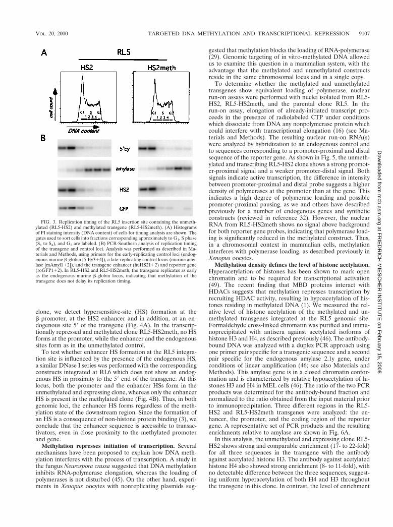
Early timing of replication in the methylated state. Earlytiming of DNA replication has been associated in many sys-tems with active transcription, open chromatin structure, andhypomethylation of the DNA, whereas late replication hasbeen correlated with transcriptional inactivity, closed chroma-tin structure, and hypermethylation (48). Thus, we sought todetermine whether the targeted introduction of methylationaffects the replication timing at both integration sites (RL5 andRL6) by determining the relative abundance of specificgenomic sequences in nascent DNA synthesized during differ-ent windows of the cell cycle (Fig. 3 and reference 7). Expo-nentially growing cells were pulse-labeled with BrdU andsorted by FACS into different fractions of the cell cycle basedon their DNA content. BrdU-labeled DNA was enriched byimmunoprecipitation and analyzed by PCR, using primers spe-cific for the transgene or endogenous loci with a known timingof replication. As a control for early replication, we used theendogenous mouse b-globin locus and as a control for latereplication we used the mouse amylase 2.1y gene, which wehave shown previously to be late replicating in erythroid cells(7). The unmethylated transgene in the clone RL5-HS2 repli-cates early during S phase in comparison to the late controland as early as the mouse b-globin locus (Fig. 3). The replica-tion timing of the RL5 locus is also early in the methylated andtranscriptionally repressed clone RL5-HS2meth (Fig. 3), whichis indistinguishable from the unmethylated clone. At the RL6locus we find the same result: early timing of replication withboth the unmethylated and the methylated constructs (data notshown). Thus, at both genomic sites, methylation of the trans-gene does not interfere with its early replication. We concludethat the establishment of methylation-induced repression nei-ther requires nor results in late replication of these genomicregions.
Remodeling of the enhancer is not influenced by the localmethylation state. Despite the localized demethylation of HS2observed in the RL5-HS2meth transgene, the enhancer is un-able to activate transcription. Thus, we asked whether meth-ylation of the promoter and gene interferes with remodeling ofthe enhancer. Nuclei were isolated and incubated with increas-ing amount of DNase I, and the resulting genomic DNA wasanalyzed on a Southern blot. In the unmethylated RL5-HS2
FIG. 2. Maintenance of the methylation status. (A) Map of the L1-HS2GFP-1L transgene, including locations of the HpaII (black diamonds) XbaI(X), BglII (B), and loxP sites (black triangles). For Southern blot analyses,genomic DNA from the early and late time points was digested with either XbaIor BglII, in combination with the methylation-sensitive enzyme HpaII, and hy-bridized with a GFP probe (black bar). The unmethylated clone RL5-HS2 yieldsa 600-bp fragment with both HpaII-containing digests at both time points, indi-cating that no de novo methylation of the GFP gene has occurred. The invitro-methylated clone RL5-HS2meth shows methylation of all HpaII sites in thetransgene, with the exception of the three HpaII sites at the 59 end of thetransgene, as indicated by the 2.4-kb fragment obtained with a BglII/HpaII digest(see the text). (B) Detailed mapping of the methylation status of the enhancerand promoter region. Genomic DNA from the late time point was bisulfiteconverted, and the sequences of interest were PCR amplified, subcloned, andsequenced (see Materials and Methods). The positions of primers are indicatedby open triangles. Open or filled circles correspond to unmethylated or methyl-ated CpGs, respectively.
9106 SCHUBELER ET AL. MOL. CELL. BIOL.
at FR
IED
RIC
H M
IES
CH
ER
INS
TIT
UT
E on F
ebruary 15, 2008 m
cb.asm.org
Dow
nloaded from
clone, we detect hypersensitive-site (HS) formation at theb-promoter, at the HS2 enhancer and in addition, at an en-dogenous site 59 of the transgene (Fig. 4A). In the transcrip-tionally repressed and methylated clone RL5-HS2meth, no HSforms at the promoter, while the enhancer and the endogenoussites form as in the unmethylated control.
To test whether enhancer HS formation at the RL5 integra-tion site is influenced by the presence of the endogenous HS,a similar DNase I series was performed with the correspondingconstructs integrated at RL6 which does not show an endog-enous HS in proximity to the 59 end of the transgene. At thislocus, both the promoter and the enhancer HSs form in theunmethylated and expressing clone, whereas only the enhancerHS is present in the methylated clone (Fig. 4B). Thus, in bothgenomic loci, the enhancer HS forms regardless of the meth-ylation state of the downstream region. Since the formation ofan HS is a consequence of non-histone protein binding (3), weconclude that the enhancer sequence is accessible to transac-tivators, even in close proximity to the methylated promoterand gene.
Methylation represses initiation of transcription. Severalmechanisms have been proposed to explain how DNA meth-ylation interferes with the process of transcription. A study inthe fungus Neurospora crassa suggested that DNA methylationinhibits RNA-polymerase elongation, whereas the loading ofpolymerases is not disturbed (45). On the other hand, experi-ments in Xenopus oocytes with nonreplicating plasmids sug-
gested that methylation blocks the loading of RNA-polymerase(29). Genomic targeting of in vitro-methylated DNA allowedus to examine this question in a mammalian system, with theadvantage that the methylated and unmethylated constructsreside in the same chromosomal locus and in a single copy.
To determine whether the methylated and unmethylatedtransgenes show equivalent loading of polymerase, nuclearrun-on assays were performed with nuclei isolated from RL5-HS2, RL5-HS2meth, and the parental clone RL5. In therun-on assay, elongation of already-initiated transcript pro-ceeds in the presence of radiolabeled CTP under conditionswhich dissociate from DNA any nonpolymerase protein whichcould interfere with transcriptional elongation (16) (see Ma-terials and Methods). The resulting nuclear run-on RNA(s)were analyzed by hybridization to an endogenous control andto sequences corresponding to a promoter-proximal and distalsequence of the reporter gene. As shown in Fig. 5, the unmeth-ylated and transcribing RL5-HS2 clone shows a strong promot-er-proximal signal and a weaker promoter-distal signal. Bothsignals indicate active transcription, the difference in intensitybetween promoter-proximal and distal probe suggests a higherdensity of polymerases at the promoter than at the gene. Thisindicates a high degree of polymerase loading and possiblepromoter-proximal pausing, as we and others have describedpreviously for a number of endogenous genes and syntheticconstructs (reviewed in reference 32). However, the nuclearRNA from RL5-HS2meth shows no signal above backgroundfor both reporter gene probes, indicating that polymerase load-ing is significantly reduced in the methylated construct. Thus,in a chromosomal context in mammalian cells, methylationinterferes with polymerase loading, as described previously inXenopus oocytes.
Methylation density defines the level of histone acetylation.Hyperacetylation of histones has been shown to mark openchromatin and to be required for transcriptional activation(49). The recent finding that MBD proteins interact withHDACs suggests that methylation represses transcription byrecruiting HDAC activity, resulting in hypoacetylation of his-tones residing in methylated DNA (1). We measured the rel-ative level of histone acetylation of the methylated and un-methylated transgenes integrated at the RL5 genomic site.Formaldehyde cross-linked chromatin was purified and immu-noprecipitated with antisera against acetylated isoforms ofhistone H3 and H4, as described previously (46). The antibody-bound DNA was analyzed with a duplex PCR approach usingone primer pair specific for a transgenic sequence and a secondpair specific for the endogenous amylase 2.1y gene, underconditions of linear amplification (46; see also Materials andMethods). This amylase gene is in a closed chromatin confor-mation and is characterized by relative hypoacetylation of hi-stones H3 and H4 in MEL cells (46). The ratio of the two PCRproducts was determined for the antibody-bound fraction andnormalized to the ratio obtained from the input material priorto immunoprecipitation. Three different regions in the RL5-HS2 and RL5-HS2meth transgenes were analyzed: the en-hancer, the promoter, and the coding region of the reportergene. A representative set of PCR products and the resultingenrichments relative to amylase are shown in Fig. 6A.
In this analysis, the unmethylated and expressing clone RL5-HS2 shows strong and comparable enrichment (17- to 22-fold)for all three sequences in the transgene with the antibodyagainst acetylated histone H3. The antibody against acetylatedhistone H4 also showed strong enrichment (8- to 11-fold), withno detectable difference between the three sequences, suggest-ing uniform hyperacetylation of both H4 and H3 throughoutthe transgene in this clone. In contrast, the level of enrichment
FIG. 3. Replication timing of the RL5 insertion site containing the unmeth-ylated (RL5-HS2) and methylated transgene (RL5-HS2meth). (A) Histogramsof PI staining intensity (DNA content) of cells for timing analysis are shown. Thegates used to sort cells into fractions corresponding approximately to G1, S phase(S1 to S4), and G2 are labeled. (B) PCR-Southern analysis of replication timingof the transgene and control loci. Analysis was performed as described in Ma-terials and Methods, using primers for the early-replicating control loci (endog-enous murine b-globin [59Ey314]), a late-replicating control locus (murine amy-lase [mAmyl112]), and the transgene enhancer (huHS2112) and reporter gene(roGFP112). In RL5-HS2 and RL5-HS2meth, the transgene replicates as earlyas the endogenous murine b-globin locus, indicating that methylation of thetransgene does not delay its replication timing.
VOL. 20, 2000 TARGETED DNA METHYLATION AND TRANSCRIPTIONAL REPRESSION 9107
at FR
IED
RIC
H M
IES
CH
ER
INS
TIT
UT
E on F
ebruary 15, 2008 m
cb.asm.org
Dow
nloaded from

for H4 and H3 in the methylated clone RL5-HS2meth variesamong the three sequences, with the highest level of enrich-ment at HS2, an intermediate level at the promoter, and thelowest level at the reporter gene (Fig. 6A). A direct compari-son of the H3 acetylation between methylated and unmethyl-ated constructs shows that the methylated clone is almost two-fold less acetylated at HS2, threefold less acetylated at thepromoter, and over sixfold less acetylated at the gene. Theextent of this localized deacetylation directly correlates withthe CpG density, which is highest in the GFP gene (see Fig.2A), indicating that methylation density defines the degree oflocal hypoacetylation. These results are consistent with therecruitment of HDAC-containing complexes by MBDs andsuggest that this recruitment results in a very localized deacety-lation.
DISCUSSION
Genomic targeting of methylated DNA results in stabletranscriptional repression. We have targeted in vitro-methyl-ated DNA into the genome to analyze methylation-inducedrepression at defined genomic insertion sites. DNA methyl-ation studies have typically utilized either nonchromosomaltemplates, such as transiently transfected plasmids (2, 29),drug-selectable episomal constructs (21), or stably integratedtransgenes (12, 30). While these experiments have been infor-mative, nonchromosomal templates do not necessarily resem-ble the chromatin structure of chromosomal DNA and thusmay not accurately reflect the effect of CpG methylation ontranscription and chromatin structure. While stable transfec-tion results in chromosomal integration, current protocols do
FIG. 4. Analysis of enhancer and promoter remodeling. Nuclei were isolated and digested with increasing concentrations of DNase I. Subsequently, genomic DNAwas isolated, digested with Bgl II, and hybridized with a probe corresponding to the GFP gene. (A) Analysis of integration site RL5 with the unmethylated (RL5-HS2)and methylated (RL5-HS2meth) transgene. (B) Analysis of integration site RL6. Each hypersensitive site detected is marked with an arrow.
9108 SCHUBELER ET AL. MOL. CELL. BIOL.
at FR
IED
RIC
H M
IES
CH
ER
INS
TIT
UT
E on F
ebruary 15, 2008 m
cb.asm.org
Dow
nloaded from

not allow for control of the copy number or integration site,and thus different constructs are analyzed in different chromo-somal contexts. Variable effects of different integration sites ontranscription and chromatin structure are well documented(13, 20), complicating the analysis of cell lines harboring stablytransfected reporter constructs. The Cre-RMCE targetingstrategy described here circumvents these limitations, permit-ting the stable introduction of unmethylated and in vitro-meth-ylated DNA at the same integration site.
The construct we analyzed contains the GFP reporter genedriven by the human b-globin promoter and the HS2 enhancerelement from the human b-globin locus control region. Thisplasmid was either unmethylated or methylated in vitro at allCpGs using the bacterial methyltransferase SssI and intro-duced with similar high efficiency into two defined genomicintegration sites that support stable expression from an un-methylated transgene. While the unmethylated construct isstably expressed even after long-term passage in culture, invitro methylation of the reporter results in strongly reducedexpression at either integration site (Fig. 1C), suggesting thatthe methylation is maintained and is responsible for this re-pression.
Methylation of the transgene does not alter the methylationstate, chromatin structure, or replication timing of flankingDNA. Consistent with the active expression state of the un-methylated transgene over time, we find no de novo methyl-ation of this construct. On the other hand repression of the invitro-methylated transgene is stable, a result consistent withthe maintenance of its methylation at the promoter and thereporter gene. It has been proposed that spreading of methyl-ation in cis into nonmethylated DNA is one mechanism bywhich de novo methylation occurs (50); however, we do notobserve de novo methylation in the genomic DNA adjacent tothe methylated construct. In addition, a DNase I-hypersensi-tive site flanking one of the insertion sites is present indepen-
dent of the methylation state of the transgene. These observa-tions suggest that the introduced patch of methylated DNA atthe promoter and gene is propagated through cell division butis not sufficient to cause de novo methylation or to alter thechromatin structure of flanking DNA.
At many loci, replication timing is correlated with transcrip-tional activity; expressed loci are early replicating and silentloci are late replicating (15). Since transcriptional activatorsmay be limited in late S phase, late replication itself may playa role in gene repression (reviewed in reference 48). As wouldbe predicted, the active unmethylated constructs are early rep-licating. Surprisingly, at both genomic sites, the silent, in vitro-methylated constructs are also early replicating, indicating thata change in replication timing to late S phase is neither arequirement for, nor a consequence of, methylation-inducedrepression at these genomic loci. The maintenance methylaseDNMT1, which preferentially binds to hemimethylated DNA,was recently reported to be associated with HDACs (14, 44) atreplication foci. This interaction may ensure that, independentof the timing of replication, even hemimethylated DNA is in arepressive chromatin state.
Taken together, the lack of methylation spreading, the pres-ervation of early replication timing, and the presence of aflanking HS after integration of the methylated construct sug-gest that transcriptional repression is not due to widespreadchanges in the activity or structure of the locus per se butrather to the local effects of methylation on the transgeneitself.
Methylation-induced repression of transcriptional initia-tion is dominant over a demethylated and remodeled en-hancer. Analysis of the methylation status of the transgenes bybisulfite sequencing reveals that methylation at the promoterand gene is maintained over time, whereas the enhancer isdemethylated after integration of the in vitro-methylated con-struct. Previous reports suggest that the binding of transacti-vators to DNA can interfere with the maintenance of methyl-ation, probably by masking the CpG dinucleotide after DNAreplication (22, 35). Thus, the observed demethylation at theenhancer could be a consequence of transcription factor bind-ing to the enhancer.
Given the demethylated state of the enhancer, it is perhapsnot surprising that the enhancer HS still forms (Fig. 3). SinceHS formation requires non-histone protein binding (3), theenhancer of the methylated construct is occupied despite itsclose proximity to a high density of methylated CpGs. Never-theless, this is not sufficient to overcome methylation-inducedrepression, and we conclude that methylation-induced repres-sion does not result from inhibition of transcription factorbinding at the enhancer. In contrast, the methylation is main-tained over the promoter and gene and the promoter HS doesnot form. Consequently, the promoter and/or gene are the sitesat which the methylation-induced repression mechanism oper-ates.
To directly address whether polymerase loading or elonga-tion are effected by DNA methylation, we used the nuclearrun-on assay and measured the density of polymerases on theunmethylated and methylated transgenes. Previous studies us-ing in vitro-methylated DNA containing mammalian promot-ers injected into Xenopus oocytes (29) suggest that methylationresults in a block to transcription initiation. In contrast, exper-iments in N. crassa suggest that a block to transcriptionalelongation is the major mechanism for methylation-inducedrepression in this organism (45). Our results indicate that, ona chromosomal template in mammalian cells, methylation in-terferes with transcriptional initiation. However, we cannotrule out that methylation has an additional effect on transcrip-
FIG. 5. Nuclear run-on analysis to determine the density of polymerases inthe unmethylated and methylated transgenes at the RL5 integration site. Nucleifrom the RL5-HS2 clone, containing the unmethylated transgene, the RL5-HS2meth clone, containing the methylated transgene and the control parentalclone RL5 without the transgene were isolated. Nuclear run-on assays wereperformed in the presence of radioactively labeled CTP, and nascent RNA washybridized to three different DNA probes: a-actin as an endogenous control;59GFP, a promoter-proximal fragment containing the 39 end of the b-globinpromoter and the 59 half of the GFP gene (a PCR product generated with theprimer pair roGFP112); and 39GFP, a promoter-distal fragment containingmost of the 39 half of the GFP coding region (generated with the primer pairGFP112). The actively expressing clone RL5-HS2 yields a higher signal for theproximal than for the distal probe, indicating that promoter-proximal pausing ofpolymerases occurs (see the text). In contrast, RL5-HS2meth gives no signalabove background for either probes, suggesting a strong reduction of polymeraseloading in the methylated state.
VOL. 20, 2000 TARGETED DNA METHYLATION AND TRANSCRIPTIONAL REPRESSION 9109
at FR
IED
RIC
H M
IES
CH
ER
INS
TIT
UT
E on F
ebruary 15, 2008 m
cb.asm.org
Dow
nloaded from

tional elongation, since such an effect would be masked by therepressed initiation.
Is localized deacetylation of histones sufficient for repres-sion? Two mechanisms of methylation-induced transcriptionalrepression have been proposed. The binding of a subset oftranscription factors is sensitive to methylation of their cognatebinding sites (23), suggesting that CpG methylation of a pro-moter could directly block transactivator binding. However, adirect block of binding is unlikely to be responsible for therepression of the b-globin promoter used in this study, as the120-bp element upstream of the initiation site does not containany CpGs (Fig. 2B), yet is sufficient for promoter activity (ref-erence 36 and references therein). An alternative mechanismof transcriptional repression involving the MBD family of pro-teins has been proposed (reviewed in reference 1). These pro-teins interact with, or are integral components of, complexeswhich include HDACs (27, 38, 39, 51, 53), suggesting thatrecruitment of HDAC activity, resulting in a modified nucleo-somal structure, is a common motif in methylation-inducedrepression. Accordingly, it has been shown that methylatedtransgenes are hypoacetylated (6, 8, 12, 42). Here, we observea reduction of histone acetylation at the promoter and gene ofthe in vitro-methylated transgene. The degree of deacetylationis more prominent for histone H3 than for H4 and correlates
with the density of methylated CpGs: the GFP gene, which hasa high density of CpGs, is the most hypoacetylated, while thepromoter and enhancer, with lower densities, are acetylated toa lesser extent. This localized deacetylation suggests that therecruited HDACs act only on nearby nucleosomes, a resultconsistent with that reported for the HDAC activity of theyeast Sin3A complex (28).
In several studies treatment with the HDAC inhibitor tri-chostatin A (TSA) partially relieved the transcriptional repres-sion of in vitro-methylated constructs (6, 8), whereas in othersno reactivation was observed (34, 41). Here, TSA treatment ofcells containing the inactive, methylated transgene did notresult in reactivation of reporter gene transcription (data notshown), a result consistent with our previous study in MELcells showing that a densely methylated provirus containing thesame reporter gene could not be reactivated by TSA alone(34). Since the HDACs currently known to be involved inmethylation-induced repression are at least partially sensitiveto TSA treatment, we speculated that a lack of reactivationindicates an additional HDAC independent mode of repres-sion (34). However, the recent finding that the HDAC activityof yeast SIR2 (24) and yeast HOS3 (5) is not inhibited by TSAindicates that TSA does not inhibit all HDAC activity, and it
FIG. 6. Chromatin immunoprecipitation analysis of histone H3 and H4 acetylation in different regions of the methylated and unmethylated transgene. Antibodiesrecognizing all acetylated isoforms of H4 (H4) or histone H3 acetylated at lysines 9 and 14 (H3) were used for immunoprecipitation. PCRs were performed on the inputand antibody-bound chromatin fractions in the presence of a radiolabeled nucleotide under conditions of linear amplifications, as we have shown previously (seereference 46 and Materials and Methods). One primer pair amplifies a sequence from the transgene and the other amplifies a sequence from the endogenous mouseamylase 2.1y gene. The PCR products from the input (I) and antibody-bound DNA (H3 and H4) were electrophoresed on a nondenaturing acrylamide gel; arepresentative gel is shown. (B) Quantification of duplex PCR products from three independent immunoprecipitation experiments. The transgene/amylase ratio fromeach bound fraction was standardized by dividing by the transgene/amylase ratio from the input material to determine the relative enrichment of transgenic sequencesduring the immunoprecipitation. The mean value and standard error of the mean for the enrichment are plotted (see the text). The x axis is drawn at 1, which reflectsno enrichment.
9110 SCHUBELER ET AL. MOL. CELL. BIOL.
at FR
IED
RIC
H M
IES
CH
ER
INS
TIT
UT
E on F
ebruary 15, 2008 m
cb.asm.org
Dow
nloaded from

remains to be determined if methylation-induced repressioninvolves a TSA-resistant HDAC activity.
Together, our experiments show that DNA methylation re-sults in a localized histone deacetylation without affecting thechromatin structure and replication timing of the insertion site.Previously, we have shown that the transcriptionally activeb-globin promoter in its native location is hyperacetylated (46),and we speculated that this hyperacetylation is required foractivation. Thus, a localized hypoacetylation mediated by CpGmethylation may be sufficient to account for the observed re-pression.
ACKNOWLEDGMENTS
This work was supported by a fellowship from the Deutsche For-schungsgemeinschaft to D.S.; NIH fellowship GM 19767/01 to M.C.L.;a fellowship from the American Cancer Society to D.M.C.; and NIHgrants HL38655, DK56845, and HL554350 to E.E.B. and grantsDK44746, HL57620, and CA54337 to M.G.
We thank Jurgen Bode, Steven Fiering, Ross Hardison, AntonKrumm, and the members of the Groudine lab for helpful suggestions;Claire Francastel for helpful comments on the manuscript; and JoanHamilton, David Scalzo, Jennifer Stout, and Urszula Maliszewski fortechnical assistance.
REFERENCES1. Bird, A. P., and A. P. Wolffe. 1999. Methylation-induced repression—belts,
braces, and chromatin. Cell 99:451–454.2. Boyes, J., and A. Bird. 1992. Repression of genes by DNA methylation
depends on CpG density and promoter strength: evidence for involvement ofa methyl-CpG binding protein. EMBO J. 11:327–333.
3. Boyes, J., and G. Felsenfeld. 1996. Tissue-specific factors additively increasethe probability of the all-or-none formation of a hypersensitive site. EMBOJ. 15:2496–2507.
4. Brandeis, M., M. Ariel, and H. Cedar. 1993. Dynamics of DNA methylationduring development. Bioessays 15:709–713.
5. Carmen, A. A., P. R. Griffin, J. R. Calaycay, S. E. Rundlett, Y. Suka, and M.Grunstein. 1999. Yeast HOS3 forms a novel trichostatin A-insensitive ho-modimer with intrinsic histone deacetylase activity. Proc. Natl. Acad. Sci.USA 96:12356–12361.
6. Chen, W. Y., and T. M. Townes. 2000. Molecular mechanism for silencingvirally transduced genes involves histone deacetylation and chromatin con-densation. Proc. Natl. Acad. Sci. USA 97:377–382.
7. Cimbora, D. M., D. Schubeler, A. Reik, J. Hamilton, C. Francastel, E. M.Epner, and M. Groudine. 2000. Long-distance control of origin choice andreplication timing in the human beta-globin locus are independent of thelocus control region. Mol. Cell. Biol. 20:5581–5591.
8. Eden, S., T. Hashimshony, I. Keshet, H. Cedar, and A. W. Thorne. 1998.DNA methylation models histone acetylation. Nature 394:842.
9. Feng, Y. Q., M. C. Lorincz, S. Fiering, J. M. Greally, and E. Bouhassira.Position effects are influenced by the orientation of a transgene with respectto flanking chromatin. Mol. Cell. Biol., in press.
10. Feng, Y. Q., J. Seibler, R. Alami, A. Eisen, K. A. Westerman, P. Leboulch, S.Fiering, and E. E. Bouhassira. 1999. Site-specific chromosomal integrationin mammalian cells: highly efficient CRE recombinase-mediated cassetteexchange. J. Mol. Biol. 292:779–785.
11. Forrester, W. C., E. Epner, M. C. Driscoll, T. Enver, M. Brice, T. Papayan-nopoulou, and M. Groudine. 1990. A deletion of the human beta-globinlocus activation region causes a major alteration in chromatin structure andreplication across the entire beta-globin locus. Genes Dev. 4:1637–1649.
12. Forrester, W. C., L. A. Fernandez, and R. Grosschedl. 1999. Nuclear matrixattachment regions antagonize methylation-dependent repression of long-range enhancer-promoter interactions. Genes Dev. 13:3003–3014.
13. Francastel, C., M. C. Walters, M. Groudine, and D. I. Martin. 1999. Afunctional enhancer suppresses silencing of a transgene and prevents itslocalization close to centrometric heterochromatin. Cell 99:259–269.
14. Fuks, F., W. A. Burgers, A. Brehm, L. Hughes-Davies, and T. Kouzarides.2000. DNA methyltransferase Dnmt1 associates with histone deacetylaseactivity. Nat. Genet. 24:88–91.
15. Goldman, M. A., G. P. Holmquist, M. C. Gray, L. A. Caston, and A. Nag.1984. Replication timing of genes and middle repetitive sequences. Science224:686–692.
16. Groudine, M., M. Peretz, and H. Weintraub. 1981. Transcriptional regula-tion of hemoglobin switching in chicken embryos. Mol. Cell. Biol. 1:281–288.
17. Gu, H., Y. R. Zou, and K. Rajewsky. 1993. Independent control of immu-noglobulin switch recombination at individual switch regions evidencedthrough Cre-loxP-mediated gene targeting. Cell 73:1155–1164.
18. Hendrich, B. 2000. Methylation moves into medicine. Curr. Biol. 10:R60–R63.
19. Hendrich, B., and A. Bird. 1998. Identification and characterization of afamily of mammalian methyl-CpG binding proteins. Mol. Cell. Biol. 18:6538–6547.
20. Henikoff, S. 1992. Position effect and related phenomena. Curr. Opin. Genet.Dev. 2:907–912.
21. Hsieh, C. L. 1994. Dependence of transcriptional repression on CpG meth-ylation density. Mol. Cell. Biol. 14:5487–5494.
22. Hsieh, C. L. 1999. Evidence that protein binding specifies sites of DNAdemethylation. Mol. Cell. Biol. 19:46–56.
23. Iguchi-Ariga, S. M., and W. Schaffner. 1989. CpG methylation of the cAMP-responsive enhancer/promoter sequence TGACGTCA abolishes specificfactor binding as well as transcriptional activation. Genes Dev. 3:612–619.
24. Imai, S., C. M. Armstrong, M. Kaeberlein, and L. Guarente. 2000. Tran-scriptional silencing and longevity protein Sir2 is an NAD-dependent histonedeacetylase. Nature 403:795–800.
25. Jaenisch, R. 1997. DNA methylation and imprinting: why bother? TrendsGenet. 13:323–329.
26. Jones, P. A., and P. W. Laird. 1999. Cancer epigenetics comes of age. Nat.Genet. 21:163–167.
27. Jones, P. L., G. J. Veenstra, P. A. Wade, D. Vermaak, S. U. Kass, N.Landsberger, J. Strouboulis, and A. P. Wolffe. 1998. Methylated DNA andMeCP2 recruit histone deacetylase to repress transcription. Nat. Genet.19:187–191.
28. Kadosh, D., and K. Struhl. 1998. Targeted recruitment of the Sin3-Rpd3histone deacetylase complex generates a highly localized domain of re-pressed chromatin in vivo. Mol. Cell. Biol. 18:5121–5127.
29. Kass, S. U., N. Landsberger, and A. P. Wolffe. 1997. DNA methylationdirects a time-dependent repression of transcription initiation. Curr. Biol.7:157–165.
30. Keshet, I., J. Lieman-Hurwitz, and H. Cedar. 1986. DNA methylation affectsthe formation of active chromatin. Cell 44:535–543.
31. Krumm, A., L. B. Hickey, and M. Groudine. 1995. Promoter-proximal paus-ing of RNA polymerase II defines a general rate-limiting step after tran-scription initiation. Genes Dev. 9:559–572.
32. Krumm, A., T. Meulia, and M. Groudine. 1993. Common mechanisms forthe control of eukaryotic transcriptional elongation. Bioessays 15:659–665.
33. Li, E., T. H. Bestor, and R. Jaenisch. 1992. Targeted mutation of the DNAmethyltransferase gene results in embryonic lethality. Cell 69:915–926.
34. Lorincz, M. C., D. Schubeler, S. C. Goeke, M. Walters, M. Groudine, andD. I. Martin. 2000. Dynamic analysis of proviral induction and de novomethylation: implications for a histone deacetylase-independent, methyl-ation density-dependent mechanism of transcriptional repression. Mol. Cell.Biol. 20:842–850.
35. Matsuo, K., J. Silke, O. Georgiev, P. Marti, N. Giovannini, and D. Rungger.1998. An embryonic demethylation mechanism involving binding of tran-scription factors to replicating DNA. EMBO J. 17:1446–1453.
36. Myers, R. M., K. Tilly, and T. Maniatis. 1986. Fine structure genetic analysisof a beta-globin promoter. Science 232:613–618.
37. Nan, X., R. R. Meehan, and A. Bird. 1993. Dissection of the methyl-CpGbinding domain from the chromosomal protein MeCP2. Nucleic Acids Res.21:4886–4892.
38. Nan, X., H. H. Ng, C. A. Johnson, C. D. Laherty, B. M. Turner, R. N.Eisenman, and A. Bird. 1998. Transcriptional repression by the methyl-CpG-binding protein MeCP2 involves a histone deacetylase complex. Nature393:386–389.
39. Ng, H. H., Y. Zhang, B. Hendrich, C. A. Johnson, B. M. Turner, H. Erdju-ment-Bromage, P. Tempst, D. Reinberg, and A. Bird. 1999. MBD2 is atranscriptional repressor belonging to the MeCP1 histone deacetylase com-plex. Nat. Genet. 23:58–61.
40. Orlando, V., H. Strutt, and R. Paro. 1997. Analysis of chromatin structure byin vivo formaldehyde cross-linking. Methods 11:205–214.
41. Osborne, C. S., P. Pasceri, R. Singal, T. Sukonnik, G. D. Ginder, and J. Ellis.1999. Amelioration of retroviral vector silencing in locus control regionbeta-globin-transgenic mice and transduced F9 embryonic cells. J. Virol.73:5490–5496.
42. Pikaart, M. J., F. Recillas-Targa, and G. Felsenfeld. 1998. Loss of transcrip-tional activity of a transgene is accompanied by DNA methylation andhistone deacetylation and is prevented by insulators. Genes Dev. 12:2852–2862.
43. Razin, A. 1998. CpG methylation, chromatin structure, and gene silenc-ing—a three-way connection. EMBO J. 17:4905–4908.
44. Rountree, M. R., K. E. Bachman, and S. B. Baylin. 2000. DNMT1 bindsHDAC2 and a new co-repressor, DMAP1, to form a complex at replicationfoci. Nat. Genet. 25:269–277.
45. Rountree, M. R., and E. U. Selker. 1997. DNA methylation inhibits elonga-tion but not initiation of transcription in Neurospora crassa. Genes Dev.11:2383–2395.
46. Schubeler, D., C. Francastel, D. M. Cimbora, A. Reik, D. I. Martin, and M.Groudine. 2000. Nuclear localization and histone acetylation: a pathway forchromatin opening and transcriptional activation of the human beta-globinlocus. Genes Dev. 14:940–950.
47. Seibler, J., D. Schubeler, S. Fiering, M. Groudine, and J. Bode. 1998. DNA
VOL. 20, 2000 TARGETED DNA METHYLATION AND TRANSCRIPTIONAL REPRESSION 9111
at FR
IED
RIC
H M
IES
CH
ER
INS
TIT
UT
E on F
ebruary 15, 2008 m
cb.asm.org
Dow
nloaded from

cassette exchange in ES cells mediated by Flp recombinase: an efficientstrategy for repeated modification of tagged loci by marker-free constructs.Biochemistry 37:6229–6234.
48. Simon, I., and H. Cedar. 1996. Temporal order of DNA replication, p.387–408. In M. DePamphilis (ed.), DNA replication in eukaryotic cells. ColdSpring Harbor Laboratory Press, Cold Spring Harbor, N.Y.
49. Struhl, K. 1998. Histone acetylation and transcriptional regulatory mecha-nisms. Genes Dev. 12:599–606.
50. Tollefsbol, T. O., and C. A. Hutchison III. 1997. Control of methylationspreading in synthetic DNA sequences by the murine DNA methyltrans-
ferase. J. Mol. Biol. 269:494–504.51. Wade, P. A., A. Gegonne, P. L. Jones, E. Ballestar, F. Aubry, and A. P. Wolffe.
1999. Mi-2 complex couples DNA methylation to chromatin remodelling andhistone deacetylation. Nat. Genet. 23:62–66.
52. Yoder, J. A., C. P. Walsh, and T. H. Bestor. 1997. Cytosine methylation andthe ecology of intragenomic parasites. Trends Genet. 13:335–340.
53. Zhang, Y., H. H. Ng, H. Erdjument-Bromage, P. Tempst, A. Bird, and D.Reinberg. 1999. Analysis of the NuRD subunits reveals a histone deacetylasecore complex and a connection with DNA methylation. Genes Dev. 13:1924–1935.
9112 SCHUBELER ET AL. MOL. CELL. BIOL.
at FR
IED
RIC
H M
IES
CH
ER
INS
TIT
UT
E on F
ebruary 15, 2008 m
cb.asm.org
Dow
nloaded from
Related Documents


![Histone Deacetylase Inhibitors in Clinical Studies as ......reverse activities of HATs and HDA Cs regulate gene expression thr ough chromatin modifications [4,5]. Histone acetylation](https://static.cupdf.com/doc/110x72/60ceab9bacd7766c844c979d/histone-deacetylase-inhibitors-in-clinical-studies-as-reverse-activities.jpg)
