DOI: 10.1126/science.1143837 , 1921 (2007); 317 Science et al. Hilary G. Morrison, Giardia lamblia Intestinal Parasite Genomic Minimalism in the Early Diverging www.sciencemag.org (this information is current as of September 27, 2007 ): The following resources related to this article are available online at http://www.sciencemag.org/cgi/content/full/317/5846/1921 version of this article at: including high-resolution figures, can be found in the online Updated information and services, http://www.sciencemag.org/cgi/content/full/317/5846/1921/DC1 can be found at: Supporting Online Material http://www.sciencemag.org/cgi/content/full/317/5846/1921#otherarticles , 24 of which can be accessed for free: cites 42 articles This article http://www.sciencemag.org/cgi/collection/genetics Genetics : subject collections This article appears in the following http://www.sciencemag.org/about/permissions.dtl in whole or in part can be found at: this article permission to reproduce of this article or about obtaining reprints Information about obtaining registered trademark of AAAS. is a Science 2007 by the American Association for the Advancement of Science; all rights reserved. The title Copyright American Association for the Advancement of Science, 1200 New York Avenue NW, Washington, DC 20005. (print ISSN 0036-8075; online ISSN 1095-9203) is published weekly, except the last week in December, by the Science on September 27, 2007 www.sciencemag.org Downloaded from

Welcome message from author
This document is posted to help you gain knowledge. Please leave a comment to let me know what you think about it! Share it to your friends and learn new things together.
Transcript
DOI: 10.1126/science.1143837 , 1921 (2007); 317Science
et al.Hilary G. Morrison,Giardia lambliaIntestinal Parasite
Genomic Minimalism in the Early Diverging
www.sciencemag.org (this information is current as of September 27, 2007 ):The following resources related to this article are available online at
http://www.sciencemag.org/cgi/content/full/317/5846/1921version of this article at:
including high-resolution figures, can be found in the onlineUpdated information and services,
http://www.sciencemag.org/cgi/content/full/317/5846/1921/DC1 can be found at: Supporting Online Material
http://www.sciencemag.org/cgi/content/full/317/5846/1921#otherarticles, 24 of which can be accessed for free: cites 42 articlesThis article
http://www.sciencemag.org/cgi/collection/geneticsGenetics
: subject collectionsThis article appears in the following
http://www.sciencemag.org/about/permissions.dtl in whole or in part can be found at: this article
permission to reproduce of this article or about obtaining reprintsInformation about obtaining
registered trademark of AAAS. is aScience2007 by the American Association for the Advancement of Science; all rights reserved. The title
CopyrightAmerican Association for the Advancement of Science, 1200 New York Avenue NW, Washington, DC 20005. (print ISSN 0036-8075; online ISSN 1095-9203) is published weekly, except the last week in December, by theScience
on
Sep
tem
ber
27, 2
007
ww
w.s
cien
cem
ag.o
rgD
ownl
oade
d fr
om
baseline spontaneous activity (~1 spike/s) is typically toolow to detect signal decreases.
16. A. von Stein, J. Sarnthein, Int. J. Psychophysiol. 38, 301(2000).
17. One might expect to observe visually elicited oxygenresponses during the evoked intervals of the TMS trial(fig. S2B). However, stimulus-evoked responses areconsiderably smaller and more variable than TMS-induced oxygen responses, and are therefore negligiblein the current paradigm (fig. S6).
18. A. Shmuel, M. Augath, A. Oeltermann, N. K. Logothetis,Nat. Neurosci. 9, 569 (2006).
19. B. N. Pasley, B. A. Inglis, R. D. Freeman, Neuroimage 36,269 (2007).
20. J. Niessing et al., Science 309, 948 (2005).21. On the basis of previous work in awake animals (22) and
the similarity of neurovascular organization (23, 24)across the cortex, we expect that preserved coupling afterTMS will generalize to a broad range of cortical regionsand physiological states.
22. J. Berwick et al., J. Cereb. Blood Flow Metab. 22, 670 (2002).23. C. Iadecola, Nat. Rev. Neurosci. 5, 347 (2004).
24. H. M. Duvernoy, S. Delon, J. L. Vannson, Brain Res. Bull.7, 519 (1981).
25. A. Pascual-Leone, J. Valls-Sole, E. M. Wassermann,M. Hallett, Brain 117, 847 (1994).
26. B. Takano et al., Neuroimage 23, 849 (2004).27. Y. Z. Huang, M. J. Edwards, E. Rounis, K. P. Bhatia,
J. C. Rothwell, Neuron 45, 201 (2005).28. F. Maeda, J. P. Keenan, J. M. Tormos, H. Topka,
A. Pascual-Leone, Clin. Neurophysiol. 111, 800 (2000).29. H. Wang, X. Wang, H. Scheich, Neuroreport 7, 521
(1996).30. S. F. Cooke, T. V. Bliss, Brain 129, 1659 (2006).31. C. Holscher, R. Anwyl, M. J. Rowan, J. Neurosci. 17, 6470
(1997).32. V. Wespatat, F. Tennigkeit, W. Singer, J. Neurosci. 24,
9067 (2004).33. J. Jacobs, M. J. Kahana, A. D. Ekstrom, I. Fried,
J. Neurosci. 27, 3839 (2007).34. T. Paus et al., J. Neurophysiol. 79, 1102 (1998).35. H. R. Siebner et al., Neuroimage 14, 883 (2001).36. A. Pascual-Leone et al., J. Clin. Neurophysiol. 15, 333
(1998).
37. M. L. Kringelbach, N. Jenkinson, S. L. Owen, T. Z. Aziz,Nat. Rev. Neurosci. 8, 623 (2007).
38. A. M. Speer et al., Biol. Psychiatry 54, 826 (2003).39. A. T. Sack et al., Cereb. Cortex (2007).40. C. C. Ruff et al., Curr. Biol. 16, 1479 (2006).41. We thank our colleagues at the University of California,
Berkeley, and anonymous reviewers for their helpfulcomments, and R. Bartholomew, N. Lines, and L. Gibson forassistance in developing the electrophysiological apparatus.Supported by research and CORE grants from the NationalEye Institute (EY01175 and EY03176, respectively) and byNSF graduate research fellowship 2003014861.
Supporting Online Materialwww.sciencemag.org/cgi/content/full/317/5846/1918/DC1Materials and MethodsFigs. S1 to S9References
13 June 2007; accepted 21 August 200710.1126/science.1146426
Genomic Minimalism in theEarly Diverging Intestinal ParasiteGiardia lambliaHilary G. Morrison,1* Andrew G. McArthur,1 Frances D. Gillin,2 Stephen B. Aley,3Rodney D. Adam,4 Gary J. Olsen,5 Aaron A. Best,6 W. Zacheus Cande,7 Feng Chen,8Michael J. Cipriano,1 Barbara J. Davids,2 Scott C. Dawson,9 Heidi G. Elmendorf,10Adrian B. Hehl,11 Michael E. Holder,1 Susan M. Huse,1 Ulandt U. Kim,1 Erica Lasek-Nesselquist,1Gerard Manning,12 Anuranjini Nigam,4 Julie E. J. Nixon,1 Daniel Palm,13Nora E. Passamaneck,1 Anjali Prabhu,4 Claudia I. Reich,5 David S. Reiner,2 John Samuelson,14Staffan G. Svard,15 Mitchell L. Sogin1
The genome of the eukaryotic protist Giardia lamblia, an important human intestinal parasite, iscompact in structure and content, contains few introns or mitochondrial relics, and has simplifiedmachinery for DNA replication, transcription, RNA processing, and most metabolic pathways. Proteinkinases comprise the single largest protein class and reflect Giardia’s requirement for a complexsignal transduction network for coordinating differentiation. Lateral gene transfer from bacterial andarchaeal donors has shaped Giardia’s genome, and previously unknown gene families, for example,cysteine-rich structural proteins, have been discovered. Unexpectedly, the genome shows littleevidence of heterozygosity, supporting recent speculations that this organism is sexual. This genomesequence will not only be valuable for investigating the evolution of eukaryotes, but will also beapplied to the search for new therapeutics for this parasite.
G iardia lamblia (syn. G. intestinalis, G.duodenalis) is the most prevalent para-sitic protist in the United States, where
its incidence may be as high as 0.7% (1). World-wide, giardiasis is common among people withpoor fecal-oral hygiene, and major modes oftransmission include contaminated water sup-plies or sexual activity. Flagellated giardialtrophozoites attach to epithelial cells of the smallintestine, where they can cause disease withouttriggering a pronounced inflammatory response.There are no known virulence factors or toxins,and variable expression of surface proteins mayallow evasion of host immune responses andadaptation to different host environments. Troph-ozoites can differentiate into infectious cysts thatare transmitted through feces.
Unusual features of this enigmatic protist in-clude the presence of two similar, transcription-
ally active diploid nuclei and the absence ofmitochondria and peroxisomes. Giardia is amember of the Diplomonadida, which includesboth free-living (e.g., Trepomonas) and parasiticspecies. The phylogenetic position of diplomonadsand related excavate taxa is perplexing. RibosomalRNA (rRNA), vacuolar ATPase (adenosine triphos-phatase), and elongation factor phylogenies iden-tify Giardia as a basal eukaryote (2–4). Othergene trees position diplomonads as one of manyeukaryotic lineages that diverged nearly simulta-neously with the opisthokonts and plants. Dis-coveries of a mitochondrial-like cpn60 gene anda mitosome imply that the absence of respiringmitochondria in Giardia may reflect adaptationto a microaerophilic life-style rather than diver-gence before the endosymbiosis of the mitochon-drial ancestor (5, 6). Because of its impact onhuman disease and its relevance to understanding
the evolution of eukaryotes, we embarked upon agenome analysis of G. lamblia.
The genome of G. lamblia WB clone C6(ATCC50803) is ~11.7MB in size, distributed onfive chromosomes. The edited draft genome se-quence contains 306 contigs on 92 scaffolds(Supporting Online Material). The genome iscompact. We identified 6470 open readingframes (ORFs) with a mean intergenic distanceof 372 base pairs (bp) (Table 1). Approximately77% of the assembled sequence defines ORFs, ofwhich 1800 overlap and 1500more arewithin 100nucleotides (nt) of an adjacent ORF. Serial anal-ysis of gene expression (SAGE) and cDNA se-quences provided transcriptional evidence for4787 of theseORFs (Supporting OnlineMaterial).
Although the total number of ORFs is similarto that of yeast, many specific giardial pathwaysappear simple in comparison with those of othereukaryotic organisms. Giardia’s genome encodesa simplified form of many cellular processes:fewer and more basic subunits, incorporation ofsingle-domain bacterial- and archaeal-like en-
1Marine Biological Laboratory, Woods Hole, MA 02543–1015, USA. 2Department of Pathology, Division of InfectiousDiseases, University of California, San Diego, CA 92103–8416, USA. 3Department of Biological Sciences, Universityof Texas at El Paso, El Paso, TX 79968–0519, USA. 4De-partments of Medicine and Immunobiology, University ofArizona College of Medicine, Tucson, AZ 85724–5049, USA.5Department of Microbiology, University of Illinois atUrbana-Champaign, Urbana, IL 61801, USA. 6Departmentof Biology, Hope College, Holland, MI 49423, USA. 7Universityof California, Berkeley, CA 94720–3200, USA. 8University ofPennsylvania, Philadelphia, PA 19194, USA. 9University ofCalifornia, Davis, CA 95616, USA. 10Biology Department,Georgetown University, Washington, DC 20057, USA. 11In-stitute of Parasitology, University of Zürich, CH-8057 Zürich,Switzerland. 12Razavi Newman Center for Bioinformatics, TheSalk Institute for Biological Studies, La Jolla, CA 92037–1099,USA. 13Centre for Microbiological Preparedness, SwedishInstitute for Infectious Disease Control, 171 82 Solna, Sweden.14Department of Molecular and Cell Biology, Boston UniversityGoldman School of Dental Medicine, Boston, MA 02118–2932,USA. 15Department of Cell and Molecular Biology, UppsalaUniversity, SE-751 24 Uppsala, Sweden.
*To whom correspondence should be addressed. E-mail:[email protected]
www.sciencemag.org SCIENCE VOL 317 28 SEPTEMBER 2007 1921
REPORTS
on
Sep
tem
ber
27, 2
007
ww
w.s
cien
cem
ag.o
rgD
ownl
oade
d fr
om
zymes, and a limited metabolic repertoire com-monly observed in parasites. We did not detectthese missing components in searches of assem-bled and unassembled reads; however, they maybe highly divergent and difficult to recognize.Others may be nonessential or functionally re-dundant with other proteins in the same or anotherpathway. The host may provide essential meta-bolic products for an incomplete pathway, but thisis a highly improbable explanation for missingstructural proteins or subunits of core machinery.
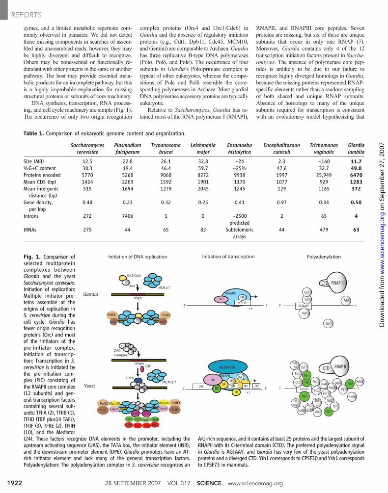
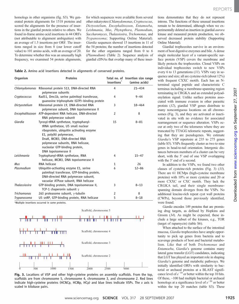
DNA synthesis, transcription, RNA process-ing, and cell cycle machinery are simple (Fig. 1).The occurrence of only two origin recognition
complex proteins (Orc4 and Orc1/Cdc6) inGiardia and the absence of regulatory initiationproteins (e.g., Cdt1, Dpb11, Cdc45, MCM10,and Gemini) are comparable to Archaea.Giardiahas three replicative B-type DNA polymerases(Pola, Pold, and Pole). The occurrence of foursubunits in Giardia’s Pola/primase complex istypical of other eukaryotes, whereas the compo-sitions of Pole and Pold resemble the corre-sponding polymerases in Archaea. Most giardialDNApolymerase accessory proteins are typicallyeukaryotic.
Relative to Saccharomyces, Giardia has re-tained most of the RNA polymerase I (RNAPI),
RNAPII, and RNAPIII core peptides. Sevenproteins are missing, but six of these are uniquesubunits that occur in only one RNAP (7).Moreover, Giardia contains only 4 of the 12transcription initiation factors present in Saccha-romyces. The absence of polymerase core pep-tides is unlikely to be due to our failure torecognize highly diverged homologs in Giardia,because the missing proteins represented RNAP-specific elements rather than a random samplingof both shared and unique RNAP subunits.Absence of homologs to many of the uniquesubunits required for transcription is consistentwith an evolutionary model hypothesizing that
Fig. 1. Comparison ofselected multiproteincomplexes betweenGiardia and the yeastSaccharomyces cerevisiae.Initiation of replication:Multiple initiator pro-teins assemble at theorigins of replication inS. cerevisiae during thecell cycle. Giardia hasfewer origin recognitionproteins (Orc) and mostof the initiators of thepre-initiator complex.Initiation of transcrip-tion: Transcription in S.cerevisiae is initiated bythe pre-initiation com-plex (PIC) consisting ofthe RNAPII core complex(12 subunits) and gen-eral transcription factorscontaining several sub-units: TFIIA (2), TFIIB (1),TFIID (TBP plus14 TAFs),TFIIF (3), TFIIE (2), TFIIH(10), and the Mediator(24). These factors recognize DNA elements in the promoter, including theupstream activating sequence (UAS), the TATA box, the initiator element (INR),and the downstream promoter element (DPE). Giardia promoters have an AT-rich initiator element and lack many of the general transcription factors.Polyadenylation: The polyadenylation complex in S. cerevisiae recognizes an
A/U-rich sequence, and it contains at least 25 proteins and the largest subunit ofRNAPII with its C-terminal domain (CTD). The preferred polyadenylation signalin Giardia is AGTAAY, and Giardia has very few of the yeast polyadenylationproteins and a diverged CTD. Yth1 corresponds to CPSF30 and Ysh1 correspondsto CPSF73 in mammals.
Origin
Orc1/Cdc6
MCM 2-7
4Orc4
Initiation of DNA replication
Giardia
YeastMCM 2-7
ORCComplex
14
2 36
5
Cdt1
Cdc6
Origin
Polyadenylation
5’ AGTAAAY 3’
Yth1
Ysh1
Pap1
Glc7
Pab1
CTD RNAP II
5’ A/U-rich 3’
YOR179C Cft1
Pta1
Yth1 Fip1
Ysh1
Cft2
Pap1
Psf2
Rna14 Hrp1
YKL059C
Swd2p
YKL018W
Pcf11
Rna15
SSU 72
Mpe1
Ref2
Glc7
Pti1
Clp1
Pab1Nab2p
Nab4p
CTD RNAP II
RPA RPA
Sid3 Sid3Sid2Dpb11
Psf1 Pfs2 Pfs3Sld5
Mcm10
Cdc45
Mcm10
Cdc45
Polα PolαPolε Polε
Polα PolαPolε Polε
Initiation of transcription
3’A/T-rich
5’+1
5’ 3’+1
TATA
TBP
TBP
UASIID
IIB
IIH
IIFIIH
INR
MEDIATOR
RNAPII
IIE
IIA
RNAPII
DPE
Table 1. Comparison of eukaryotic genome content and organization.
Saccharomycescerevisiae
Plasmodiumfalciparum
Trypanosomabrucei
Leishmaniamajor
Entamoebahistolytica
Encephalitozooncuniculi
Trichomonasvaginalis
Giardialamblia
Size (MB) 12.5 22.8 26.1 32.8 ~24 2.3 ~160 11.7%G+C content 38.3 19.4 46.4 59.7 ~25% 47.6 32.7 49.0Proteins encoded 5770 5268 9068 8272 9938 1997 25,949 6470Mean CDS (bp) 1424 2283 1592 1901 1170 1077 929 1283Mean intergenic
distance (bp)515 1694 1279 2045 1245 129 1165 372
Gene density,per kbp
0.48 0.23 0.32 0.25 0.41 0.97 0.34 0.58
Introns 272 7406 1 0 ~2500predicted
2 65 4
tRNAs 275 44 65 83 Subtelomericarrays
44 479 63
28 SEPTEMBER 2007 VOL 317 SCIENCE www.sciencemag.org1922
REPORTS
on
Sep
tem
ber
27, 2
007
ww
w.s
cien
cem
ag.o
rgD
ownl
oade
d fr
om
class-specific polymerase subunits arose after thedivergence of diplomonads.
A single intron with a noncanonical 5′ splicesite was identified in a 2Fe-2S ferredoxin gene,along with components of the spliceosome (8). Wegenerated trophozoite cDNAs and examined align-ments of conserved proteins to identify otherpossible introns. We found three candidates, ingenes for ribosomal protein L7A, a dynein light-chain protein, and an unknown protein. Two wereconfirmed by reverse transcription–polymerasechain reaction, and the RPL7A intron wasindependently reported (9). These new candidatesshow canonical GT/AG splice sites and contain anAC-repeat motif, [AC]CT[GA]AC[AC]CACAG(fig. S1). The AC-repeat motif is very like thatcommon to Trichomonas introns [ACTAACA-CACAG (10)], suggesting a shared splicingmechanism. An intron has also been reported inthe excavate Carpediomonas (11).
Giardia’s machinery for RNA processingis less complex than that of other eukaryotes,but the presumed polyadenylation signal(AGUAAA) (12) resembles that of othereukaryotes (AAUAAA). Searches for Giardiasequences that are similar to the many poly-adenylation factors in yeast and other eukary-otes identified relatively few homologs (Fig. 1).Giardia has a relative paucity of enzymes forposttranslational modification. Like Plasmodium,it lacks the vast majority of genes encodingglycosyltransferases and so makes the shortestN-glycan precursor yet identified, dolichol-PP-GlcNAc2 (13). Giardia, like Trypanosoma andArchaea, has a single-subunit oligosaccharyl-transferase for transferring N-glycans from thelipid precursor to the peptide (14), compared witheight in yeast and humans. Unlike most eukary-otes, Giardia has an N-glycan–independentquality-control system for protein folding (e.g.,chaperones, protein disulfide isomerases, andpeptidyl-prolyl cis-trans isomerases) and proteindegradation. Giardia has fewer nucleotide sugartransporters than any other eukaryotic genome,including just one for uridine 5´-diphosphate(UDP)–GlcNAc (15). Giardia is missing the setof glycosyltransferases that typically modify N-and O-linked glycans in the Golgi lumen.Instead, Giardia has a cytosolic glycosyltrans-ferase, rare among protists, which adds O-linkedGlcNAc to Ser and Thr of cytosolic proteins(15).
Giardia has a conventional endoplasmicreticulum (ER) with conserved chaperones(BiP, Hsp90, DnaJ), but is unusual in havingfive protein disulfide isomerases, each with onlya single active site (16), and in lacking the Ero1protein that drives disulfide formation in the ERlumen. Membrane transport in Giardia is unlikethat of other parasitic protozoa (17, 18). Despitethe highly polarized cell structure, there is noconclusive evidence for a stacked Golgi ap-paratus or cisternae for posttranslational mat-uration of secretory cargo except in encystingtrophozoites. Only a few Rabs, SNAREs (soluble
N-ethylmaleimide–sensitive factor attachment pro-tein receptors), and a small number of adaptorprotein (AP) complexes participate in vesicledocking and membrane fusion. Unlike all othereukaryotes that have at least three AP complexes,Giardia encodes only two. The presence of onlytwo APs with no indication of pseudogenes ororphan subunits argues for a simple membranetransport system in Giardia.
Two rounds of cytokinesis, accompanied by asingle round of nuclear division, occur duringexcystation. Giardia’s transcriptionally equiva-lent nuclei must synchronously divide in troph-ozoites and form quadrinucleate, 16N cysts (19).The presence of homologs to yeast Cin8, polokinase, aurora kinase, and antiparallel micro-tubule bundling proteins suggests that the nec-essary spindle apparatus machinery is present.We identified giardial homologs of several mi-totic exit network (MEN) proteins, indicating thatregulation of cytokinesis in Giardia may besimilar to that of yeast in whichMENcoordinatesnuclear division with cytokinesis. Homologs ofactin, cyclin-dependent kinases, and the mitoticcyclins A and B are present in Giardia. How-ever, the lack of myosin indicates that the actin-myosin cleavage furrow previously found in alleukaryotes is not present in Giardia. Possibly anonmyosin, adhesion-dependent cytokinesismechanism exists in Giardia, as in some mutantsof Dictyostelium (20).
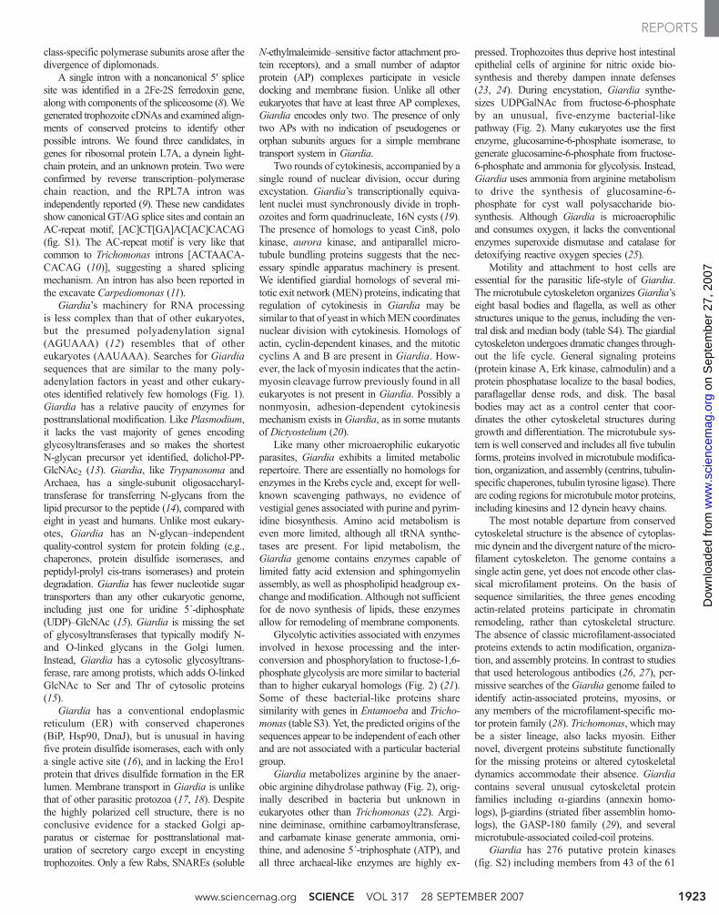
Like many other microaerophilic eukaryoticparasites, Giardia exhibits a limited metabolicrepertoire. There are essentially no homologs forenzymes in the Krebs cycle and, except for well-known scavenging pathways, no evidence ofvestigial genes associated with purine and pyrim-idine biosynthesis. Amino acid metabolism iseven more limited, although all tRNA synthe-tases are present. For lipid metabolism, theGiardia genome contains enzymes capable oflimited fatty acid extension and sphingomyelinassembly, as well as phospholipid headgroup ex-change and modification. Although not sufficientfor de novo synthesis of lipids, these enzymesallow for remodeling of membrane components.
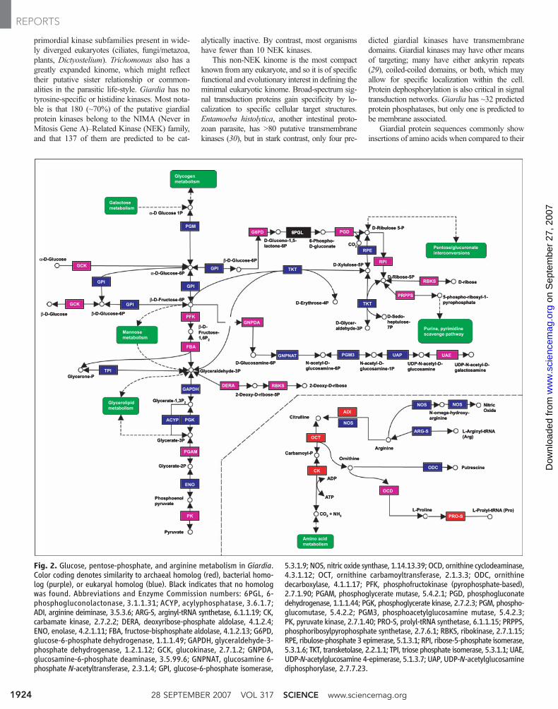
Glycolytic activities associated with enzymesinvolved in hexose processing and the inter-conversion and phosphorylation to fructose-1,6-phosphate glycolysis are more similar to bacterialthan to higher eukaryal homologs (Fig. 2) (21).Some of these bacterial-like proteins sharesimilarity with genes in Entamoeba and Tricho-monas (table S3). Yet, the predicted origins of thesequences appear to be independent of each otherand are not associated with a particular bacterialgroup.
Giardia metabolizes arginine by the anaer-obic arginine dihydrolase pathway (Fig. 2), orig-inally described in bacteria but unknown ineukaryotes other than Trichomonas (22). Argi-nine deiminase, ornithine carbamoyltransferase,and carbamate kinase generate ammonia, orni-thine, and adenosine 5´-triphosphate (ATP), andall three archaeal-like enzymes are highly ex-
pressed. Trophozoites thus deprive host intestinalepithelial cells of arginine for nitric oxide bio-synthesis and thereby dampen innate defenses(23, 24). During encystation, Giardia synthe-sizes UDPGalNAc from fructose-6-phosphateby an unusual, five-enzyme bacterial-likepathway (Fig. 2). Many eukaryotes use the firstenzyme, glucosamine-6-phosphate isomerase, togenerate glucosamine-6-phosphate from fructose-6-phosphate and ammonia for glycolysis. Instead,Giardia uses ammonia from arginine metabolismto drive the synthesis of glucosamine-6-phosphate for cyst wall polysaccharide bio-synthesis. Although Giardia is microaerophilicand consumes oxygen, it lacks the conventionalenzymes superoxide dismutase and catalase fordetoxifying reactive oxygen species (25).
Motility and attachment to host cells areessential for the parasitic life-style of Giardia.The microtubule cytoskeleton organizesGiardia’seight basal bodies and flagella, as well as otherstructures unique to the genus, including the ven-tral disk and median body (table S4). The giardialcytoskeleton undergoes dramatic changes through-out the life cycle. General signaling proteins(protein kinase A, Erk kinase, calmodulin) and aprotein phosphatase localize to the basal bodies,paraflagellar dense rods, and disk. The basalbodies may act as a control center that coor-dinates the other cytoskeletal structures duringgrowth and differentiation. The microtubule sys-tem is well conserved and includes all five tubulinforms, proteins involved in microtubule modifica-tion, organization, and assembly (centrins, tubulin-specific chaperones, tubulin tyrosine ligase). Thereare coding regions for microtubulemotor proteins,including kinesins and 12 dynein heavy chains.
The most notable departure from conservedcytoskeletal structure is the absence of cytoplas-mic dynein and the divergent nature of the micro-filament cytoskeleton. The genome contains asingle actin gene, yet does not encode other clas-sical microfilament proteins. On the basis ofsequence similarities, the three genes encodingactin-related proteins participate in chromatinremodeling, rather than cytoskeletal structure.The absence of classic microfilament-associatedproteins extends to actin modification, organiza-tion, and assembly proteins. In contrast to studiesthat used heterologous antibodies (26, 27), per-missive searches of the Giardia genome failed toidentify actin-associated proteins, myosins, orany members of the microfilament-specific mo-tor protein family (28). Trichomonas, which maybe a sister lineage, also lacks myosin. Eithernovel, divergent proteins substitute functionallyfor the missing proteins or altered cytoskeletaldynamics accommodate their absence. Giardiacontains several unusual cytoskeletal proteinfamilies including a-giardins (annexin homo-logs), b-giardins (striated fiber assemblin homo-logs), the GASP-180 family (29), and severalmicrotubule-associated coiled-coil proteins.
Giardia has 276 putative protein kinases(fig. S2) including members from 43 of the 61
www.sciencemag.org SCIENCE VOL 317 28 SEPTEMBER 2007 1923
REPORTS
on
Sep
tem
ber
27, 2
007
ww
w.s
cien
cem
ag.o
rgD
ownl
oade
d fr
om
primordial kinase subfamilies present in wide-ly diverged eukaryotes (ciliates, fungi/metazoa,plants, Dictyostelium). Trichomonas also has agreatly expanded kinome, which might reflecttheir putative sister relationship or common-alities in the parasitic life-style. Giardia has notyrosine-specific or histidine kinases. Most nota-ble is that 180 (~70%) of the putative giardialprotein kinases belong to the NIMA (Never inMitosis Gene A)–Related Kinase (NEK) family,and that 137 of them are predicted to be cat-
alytically inactive. By contrast, most organismshave fewer than 10 NEK kinases.
This non-NEK kinome is the most compactknown from any eukaryote, and so it is of specificfunctional and evolutionary interest in defining theminimal eukaryotic kinome. Broad-spectrum sig-nal transduction proteins gain specificity by lo-calization to specific cellular target structures.Entamoeba histolytica, another intestinal proto-zoan parasite, has >80 putative transmembranekinases (30), but in stark contrast, only four pre-
dicted giardial kinases have transmembranedomains. Giardial kinases may have other meansof targeting; many have either ankyrin repeats(29), coiled-coiled domains, or both, which mayallow for specific localization within the cell.Protein dephosphorylation is also critical in signaltransduction networks. Giardia has ~32 predictedprotein phosphatases, but only one is predicted tobe membrane associated.
Giardial protein sequences commonly showinsertions of amino acids when compared to their
Fig. 2. Glucose, pentose-phosphate, and arginine metabolism in Giardia.Color coding denotes similarity to archaeal homolog (red), bacterial homo-log (purple), or eukaryal homolog (blue). Black indicates that no homologwas found. Abbreviations and Enzyme Commission numbers: 6PGL, 6-phosphogluconolactonase, 3.1.1.31; ACYP, acylyphosphatase, 3.6.1.7;ADI, arginine deiminase, 3.5.3.6; ARG-S, arginyl-tRNA synthetase, 6.1.1.19; CK,carbamate kinase, 2.7.2.2; DERA, deoxyribose-phosphate aldolase, 4.1.2.4;ENO, enolase, 4.2.1.11; FBA, fructose-bisphosphate aldolase, 4.1.2.13; G6PD,glucose-6-phosphate dehydrogenase, 1.1.1.49; GAPDH, glyceraldehyde-3-phosphate dehydrogenase, 1.2.1.12; GCK, glucokinase, 2.7.1.2; GNPDA,glucosamine-6-phosphate deaminase, 3.5.99.6; GNPNAT, glucosamine 6-phosphate N-acetyltransferase, 2.3.1.4; GPI, glucose-6-phosphate isomerase,
5.3.1.9; NOS, nitric oxide synthase, 1.14.13.39; OCD, ornithine cyclodeaminase,4.3.1.12; OCT, ornithine carbamoyltransferase, 2.1.3.3; ODC, ornithinedecarboxylase, 4.1.1.17; PFK, phosphofructokinase (pyrophosphate-based),2.7.1.90; PGAM, phosphoglycerate mutase, 5.4.2.1; PGD, phosphogluconatedehydrogenase, 1.1.1.44; PGK, phosphoglycerate kinase, 2.7.2.3; PGM, phospho-glucomutase, 5.4.2.2; PGM3, phosphoacetylglucosamine mutase, 5.4.2.3;PK, pyruvate kinase, 2.7.1.40; PRO-S, prolyl-tRNA synthetase, 6.1.1.15; PRPPS,phosphoribosylpyrophosphate synthetase, 2.7.6.1; RBKS, ribokinase, 2.7.1.15;RPE, ribulose-phosphate 3 epimerase, 5.1.3.1; RPI, ribose-5-phosphate isomerase,5.3.1.6; TKT, transketolase, 2.2.1.1; TPI, triose phosphate isomerase, 5.3.1.1; UAE,UDP-N-acetylglucosamine 4-epimerase, 5.1.3.7; UAP, UDP-N-acetylglucosaminediphosphorylase, 2.7.7.23.
28 SEPTEMBER 2007 VOL 317 SCIENCE www.sciencemag.org1924
REPORTS
on
Sep
tem
ber
27, 2
007
ww
w.s
cien
cem
ag.o
rgD
ownl
oade
d fr
om
homologs in other organisms (fig. S3). We gen-erated protein alignments for 1518 proteins andscored the alignments for the presence of inser-tions in the giardial protein relative to others. Wefound in-frame amino acid insertions in 44 ORFs(not attributable to alignment ambiguities) withan average of 1.5 insertions per ORF. The inser-tions ranged in size from 8 (our lower cutoffvalue) to 101 amino acids, with an average of 20.To determine whether this was an unusually highfrequency, we examined 54 protein alignments,
for which sequences were available from severalother eukaryotes (Chlamydomonas,Cryptococcus,Dictyostelium, Encephalitozoon, Entamoeba,Leishmania, Mus, Phytopthora, Plasmodium,Saccharomyces, Thalassiosira, Trichomonas, andTrypanosoma; Supporting Online Material).Giardia sequences showed 15 insertions in 11 ofthe 54 proteins; the number of insertions detectedfor the other organisms ranged from 0 to 6(Plasmodium) (Table 2). Sequence analysis ofgiardial cDNAs that overlap many of these inser-
tions demonstrates that they do not representintrons. The functions of these unusual insertionsremain to be determined, although when we ex-perimentally deleted an insertion in giardial aurorakinase and measured protein production, we ob-served decreased protein stability (SupportingOnline Material).
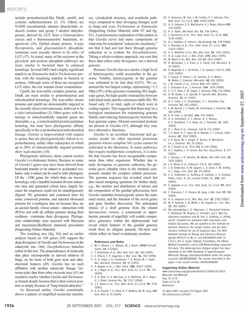
Giardial trophozoites survive in an environ-ment of host digestive enzymes and bile. A densesingle molecular layer of a variant-specific sur-face protein (VSP) covers the membrane andlikely protects the trophozoites. Clonal VSPs onindividual trophozoites switch to new VSPsevery 6 to 13 generations (31). VSPs vary in se-quence and size; all are cysteine-rich (about 12%)with frequent CXXC motifs. Each has an N-terminal signal peptide and characteristic Cterminus including a membrane-spanning regionterminating in CRGKA and an extended polyad-enylation signal. Unlike surface proteins asso-ciated with immune evasion in other parasiticprotists (32), giardial VSP genes distribute tomany noncontiguous locations on all chromo-somes (Fig. 3), and they are activated or inacti-vated in situ with no evidence for associatedrearrangement or sequence alteration. VSPs oc-cur at only two of the telomeres where they aretruncated by TTAGG telomeric repeats, suggest-ing that they are pseudogenes. We estimateGiardia’s VSP repertoire at 235 to 275 genes(table S5). VSPs frequently cluster as two to ninegenes in head-to-tail orientation. Intergenic dis-tances between members of a cluster can be veryshort, with the 5′ end of one VSP overlappingwith the 3′ end of a second.
In addition to the VSPs, we found two otherclasses of cysteine-rich proteins (Fig. 3) (33).There are 61 HCMps (high-cysteine membraneproteins) with 10% or more cysteine and 20 ormore CXXC or CXC motifs. They lack theCRGKA tail, and their single membrane-spanning domain diverges from the VSPs. Noadditional leucine-rich repeat cyst wall proteins(CWPs), beyond those previously identified,were found.
Giardia encodes 149 proteins that are promis-ing drug targets, as defined by Hopkins andGroom (34). As might be expected, these in-clude a large subset of the kinases, e.g., TOR(target of rapamycin) (table S6).
When attached to the surface of the intestinalmucosa,Giardia trophozoites have ample oppor-tunity to pick up genes from bacteria and toscavenge products of host and bacterial metabo-lism. Like that of both Trichomonas andEntamoeba, Giardia’s genome contains manylateral gene transfer (LGT) candidates, indicatingthat LGT has played an important role in shapingGiardia’s genome and metabolic pathways. Weinitially identified ORFs with similarity to bac-terial or archaeal proteins at a BLAST signifi-cance level of e−10 or better within the top 10 hits.Of these, ~100 had multiple bacterial or archaealhomologs at a significance level of e−30 or betterwithin the top 20 matches (table S3). These
Table 2. Amino acid insertions detected in alignments of conserved proteins.
Organism Proteins Total no. ofinsertions*
Insertion size range(amino acids)
Chlamydomonas Ribosomal protein S13, DNA-directed RNApolymerase subunits
4 21–434
Cryptococcus Rad51, Dmc1b, serine palmitoyl transferase,guanosine triphosphate (GTP)–binding protein
4 9–44
Dictyostelium Ribosomal protein L9, DNA-directed RNApolymerase subunit, DNA topoisomerase II
3 18–464
Encephalitozoon ATP-dependent RNA helicase, DNA-directedRNA polymerase subunit
2 8
Giardia Tyrosyl-tRNA synthetase, tryptophanyl-tRNA synthetase, U5 small nuclearriboprotein, ubiquitin activating enzymeE1, poly(A) polymerase,DnaK, MCM3, DNA-directed RNApolymerase subunits, RNA helicase,nucleolar GTP-binding protein,DNA topoisomerase II
15 8–81
Leishmania Tryptophanyl-tRNA synthetase, RNAhelicase, MCM3, DNA topoisomerase II
4 15–47
Mus RNA helicase 1 26Plasmodium Ubiquitin-activating enzyme E1, serine
palmitoyl transferase, GTP-binding protein,DNA-directed RNA polymerase subunit,vacuolar ATPase subunit, RNA helicase
6 12–86
Thalassiosira GTP-binding protein, DNA topoisomerase II,TCP-1 chaperonin subunit g
3 8–13
Trichomonas 26S proteasome subunit, g-tubulin 2 8Trypanosoma U5 snRP, GTP-binding protein, RNA helicase 5 8–18*Multiple insertions occurred in some proteins.
Fig. 3. Locations of VSP and other high-cysteine proteins on assembly scaffolds. From the top,scaffolds are from chromosome 5, chromosome 4, chromosome 3, and chromosome 2. Red linesindicate high-cysteine proteins (HCNCp, HCMp, HCp) and blue lines indicate VSPs. The x axis isscaled in kilobase pairs.
www.sciencemag.org SCIENCE VOL 317 28 SEPTEMBER 2007 1925
REPORTS
on
Sep
tem
ber
27, 2
007
ww
w.s
cien
cem
ag.o
rgD
ownl
oade
d fr
om
include proteobacterial-like DnaK, cpn60, andcysteine sulfurtransferase (6, 35). Others areNADH (nicotinamide adenine dinucleotide, re-duced) oxidase and group 3 alcohol dehydro-genase, derived by LGT from a Gram-positivecoccus and a thermoanaerobic bacterium, re-spectively (36). Hybrid cluster protein, A-typeflavoprotein, and glucosamine-6 phosphateisomerase were recently shown to be relics ofLGT (37). As noted, many of the enzymes in theglycolytic and pentose phosphate pathways aremore similar to bacterial than to eukaryalhomologs. Several ORFs had a highly significantmatch to an Entamoeba and/or Trichomonas pro-tein, with the remaining matches to bacteria orarchaea. Although some of these are recognizedLGT relics, the rest warrant closer examination.
Cpn60, the iron-sulfur complex proteins, andDnaK are most similar to proteobacterial andmitochondrial homologs. The iron-sulfur clusterproteins and cpn60 are demonstrably targeted tothe recently discovered mitosome, believed to bea relict mitochondrion (5). Other genes with ho-mology to mitochondrially targeted genes aredetectable, e.g., amitochondrial protein peptidasehomolog, but none have phylogenetic affinityspecifically to the a-proteobacteria/mitochondriallineage. Giardia is impoverished with respectto genes that are phylogenetically linked to a-proteobacteria, unlike other eukaryotes in whichup to 20% of mitochondrially targeted proteinsshow such ancestry (38).
Phylogenetic inference alone cannot resolveGiardia’s evolutionary history. Because so manyof Giardia’s genes may have been derived fromhorizontal transfer or be subject to accelerated evo-lution, only a subset can be used to infer phylogeny.Of the ~1500 genes for which there are knownhomologs, only a handful included diverse eukary-otic taxa and generated robust trees, largely be-cause the sequences could not be unambiguouslyaligned. We generated and examined trees formany conserved proteins, and selected ribosomalproteins for a multigene data set because they arean ancient family, whose nature—interaction withrRNAs and with all cellular proteins during theirsynthesis—constrains their divergence. Phyloge-netic relationships were assessed with Bayesianand maximum-likelihood statistical procedures(Supporting Online Material).
The resulting tree (fig. S4) and an earlieranalysis based on 100 genes (39) support thedeep divergence ofGiardia andTrichomonas in theeukaryotic tree. Only Encephalitozoon branchesearlier in this tree. The preponderance of moleculardata place microsporidia as derived relatives offungi, on the basis of both gene trees and ultra-structural features (40). Giardia has no suchaffiliation with another eukaryotic lineage. Ge-nome-scale data from other excavate taxa (41) areneeded to resolve whetherGiardia and Trichomon-as branch deeply because that is their correct posi-tion or simply because of “long branch attraction.”
As discussed earlier, Giardia consistentlyshows a pattern of simplified molecular machin-
ery, cytoskeletal structure, and metabolic path-ways compared to later diverging lineages suchas fungi and even Trichomonas or Entamoeba(Supporting Online Material; table S7 and fig.S5). A parsimonious explanation of this pattern isthat Giardia never had many components ofwhat may be considered “eukaryotic machinery,”not that it had and lost them through genomereduction as is evident for Encephalitozoon.Taking a whole-evidence approach, one sees thatthese data reflect early divergence, not a derivedgenome.
Because Giardia has two nuclei, a high levelof heterozygosity could accumulate in the ge-nome. Notably, heterozygosity in the genomewas estimated to be less than 0.01%. We ex-amined the two largest contigs, representing >1.2Mbp (10% of the genome) containing 482 single-copy genes, for high-qualitymismatches betweenindividual reads and the consensus (table S8).Wefound only 25 in total, eight of which were incoding regions. This suggests that there may be abiological mechanism for maintaining genomefidelity and reducing heterozygosity between thefour genome copies. Meiosis-associated proteinsare present in Giardia (42), although they mayhave alternative functions.
Giardia is an excellent functional and ge-nomic model for other intestinal protozoanparasites whose complete life cycles cannot bereplicated in the laboratory. In many pathwaysthat require multiprotein complexes, it is nota-ble that Giardia has fewer recognizable compo-nents than other organisms. Whether due toearly divergence or genomic reduction, the ge-nome gives valuable clues to the minimal com-ponents needed for complex cellular processes.The genome sequence has revealed much butalso raised intriguing questions for further study,e.g., the number and distribution of introns andthe composition of the giardial spliceosome, howGiardia maintains homozygosity across the sepa-rated nuclei, and the function of the novel genesand gene families discovered. The anticipatedrelease of a draft genome from the relatedSpironucleus vortens, a commensal or oppor-tunistic parasite of angelfish, will enable compar-ative genomics within the diplomonads andreveal which features of the giardial genomeresult from its obligate parasitic life-style andwhich reflect its basal evolutionary position.
References and Notes1. M. C. Hlavsa, J. C. Watson, M. J. Beach, MMWR Surveill.
Summ. 54, 9 (2005).2. T. Hashimoto et al., Mol. Biol. Evol. 12, 782 (1995).3. E. Hilario, J. P. Gogarten, J. Mol. Evol. 46, 703 (1998).4. D. D. Leipe, J. H. Gunderson, T. A. Nerad, M. L. Sogin,
Mol. Biochem. Parasitol. 59, 41 (1993).5. A. Regoes et al., J. Biol. Chem. 280, 30557 (2005).6. A. J. Roger et al., Proc. Natl. Acad. Sci. U.S.A. 95, 229
(1998).7. A. A. Best, H. G. Morrison, A. G. McArthur, M. L. Sogin,
G. J. Olsen, Genome Res. 14, 1537 (2004).8. J. E. Nixon et al., Proc. Natl. Acad. Sci. U.S.A. 99, 3701
(2002).9. A. G. Russell, T. E. Shutt, R. F. Watkins, M. W. Gray, BMC
Evol. Biol. 5, 45 (2005).
10. S. Vanacova, W. Yan, J. M. Carlton, P. J. Johnson, Proc.Natl. Acad. Sci. U.S.A. 102, 4430 (2005).
11. A. G. Simpson, E. K. MacQuarrie, A. J. Roger, Nature 419,270 (2002).
12. R. D. Adam, Microbiol. Rev. 55, 706 (1991).13. J. Samuelson et al., Proc. Natl. Acad. Sci. U.S.A. 102,
1548 (2005).14. D. J. Kelleher, R. Gilmore, Glycobiology 16, 47R (2006).15. S. Banerjee et al., Proc. Natl. Acad. Sci. U.S.A. 104,
11676 (2007).16. A. G. McArthur et al., Mol. Biol. Evol. 18, 1455 (2001).17. H. D. Luján et al., J. Biol. Chem. 270, 4612 (1995).18. M. Marti et al., Mol. Biol. Cell 14, 1433 (2003).19. R. Bernander, J. E. Palm, S. G. Svard, Cell. Microbiol. 3,
55 (2001).20. A. Nagasaki, E. L. de Hostos, T. Q. Uyeda, J. Cell Sci. 115,
2241 (2002).21. S. Suguri, K. Henze, L. B. Sanchez, D. V. Moore,
M. Muller, J. Eukaryot. Microbiol. 48, 493 (2001).22. J. M. Carlton et al., Science 315, 207 (2007).23. L. Eckmann et al., J. Immunol. 164, 1478 (2000).24. E. Li, P. Zhou, S. M. Singer, J. Immunol. 176, 516 (2006).25. D. M. Brown, J. A. Upcroft, P. Upcroft, Mol. Biochem.
Parasitol. 72, 47 (1995).26. D. E. Feely, J. V. Schollmeyer, S. L. Erlandsen, Exp.
Parasitol. 53, 145 (1982).27. E. M. Narcisi, J. J. Paulin, M. Fechheimer, J. Parasitol. 80,
468 (1994).28. R. D. Vale, J. Cell Biol. 163, 445 (2003).29. H. G. Elmendorf, S. C. Rohrer, R. S. Khoury,
R. E. Bouttenot, T. E. Nash, Int. J. Parasitol. 35, 1001(2005).
30. D. L. Beck et al., Eukaryot. Cell 4, 722 (2005).31. T. E. Nash, H. T. Lujan, M. R. Mowatt, J. T. Conrad, Infect.
Immun. 69, 1922 (2001).32. J. D. Barry, M. L. Ginger, P. Burton, R. McCulloch,
Int. J. Parasitol. 33, 29 (2003).33. B. J. Davids et al., PLoS ONE 1, e44 (2006).34. A. L. Hopkins, C. R. Groom, Nat. Rev. Drug Discov. 1, 727
(2002).35. J. Tachezy, L. B. Sanchez, M. Muller, Mol. Biol. Evol. 18,
1919 (2001).36. J. E. Nixon et al., Eukaryot. Cell 1, 181 (2002).37. J. O. Andersson, A. M. Sjogren, L. A. Davis, T. M. Embley,
A. J. Roger, Curr. Biol. 13, 94 (2003).38. S. G. Andersson, O. Karlberg, B. Canback, C. G. Kurland,
Philos. Trans. R. Soc. London B Biol. Sci. 358, 165(2003).
39. E. Bapteste et al., Proc. Natl. Acad. Sci. U.S.A. 99, 1414(2002).
40. F. Thomarat, C. P. Vivares, M. Gouy, J. Mol. Evol. 59, 780(2004).
41. A. G. Simpson et al., Mol. Biol. Evol. 19, 1782 (2002).42. M. A. Ramesh, S. B. Malik, J. M. Logsdon Jr., Curr. Biol.
15, 185 (2005).43. We acknowledge J. D. Silberman, S. (Pacocha) Preheim,
S. Birkeland, M. Shapiro, V. Seshadri, and J. Woo forlaboratory assistance and M. Pop, S. Salzburg, G. Hinkle,and R. Campbell for assistance with informatics andhelpful discussions. We are grateful to The Institute forGenomic Research, the Sanger Centre, and the JointGenome Institute for use of sequence data. The NIH/National Institute of Allergy and Infectious Diseases(grants AI43273 to M.L.S. and AI42488/AI51687 toF.D.G.), the G. Unger Vetlesen Foundation, the EllisonMedical Foundation, and LI-COR Biotechnology supportedthis work. This whole-genome shotgun project has beendeposited at the DNA Databank of Japan/EuropeanMolecular Biology Laboratory/GenBank under the projectaccession AACB00000000. The version described in thispaper is the second version, AACB02000000.
Supporting Online Materialwww.sciencemag.org/cgi/content/full/317/5846/1921/DC1Materials and MethodsFigs. S1 to S5Tables S1 to S8References
16 April 2007; accepted 20 August 200710.1126/science.1143837
28 SEPTEMBER 2007 VOL 317 SCIENCE www.sciencemag.org1926
REPORTS
on
Sep
tem
ber
27, 2
007
ww
w.s
cien
cem
ag.o
rgD
ownl
oade
d fr
om
Related Documents
![The [unseen] Modernist Eye: Minimalism, Defamiliarization ... · Minimalism, Defamiliarization and the Advertising Film. ... [unseen] Modernist Eye: Minimalism, Defamiliarization](https://static.cupdf.com/doc/110x72/5ac0ff807f8b9a433f8c5be6/the-unseen-modernist-eye-minimalism-defamiliarization-defamiliarization.jpg)