Genomic Islands as a Marker to Differentiate between Clinical and Environmental Burkholderia pseudomallei Thanatchaporn Bartpho 1,2 , Thidathip Wongsurawat 3 , Surasakdi Wongratanacheewin 1,2 , Adel M. Talaat 4 , Nitsara Karoonuthaisiri 3 , Rasana W. Sermswan 2,5 * 1 Department of Microbiology, Faculty of Medicine, Khon Kaen University, Khon Kaen, Thailand, 2 Melioidosis Research Center, Khon Kaen University, Khon Kaen, Thailand, 3 National Center for Genetic Engineering and Biotechnology, Pathumthani, Thailand, 4 Department of Animal Health and Biomedical Sciences, University of Wisconsin- Madison, Wisconsin, United States of America, 5 Department of Biochemistry, Faculty of Medicine, Khon Kaen University, Khon Kaen, Thailand Abstract Burkholderia pseudomallei, as a saprophytic bacterium that can cause a severe sepsis disease named melioidosis, has preserved several extra genes in its genome for survival. The sequenced genome of the organism showed high diversity contributed mainly from genomic islands (GIs). Comparative genome hybridization (CGH) of 3 clinical and 2 environmental isolates, using whole genome microarrays based on B. pseudomallei K96243 genes, revealed a difference in the presence of genomic islands between clinical and environmental isolates. The largest GI, GI8, of B. pseudomallei was observed as a 2 sub- GI named GIs8.1 and 8.2 with distinguishable %GC content and unequal presence in the genome. GIs8.1, 8.2 and 15 were found to be more common in clinical isolates. A new GI, GI16c, was detected on chromosome 2. Presences of GIs8.1, 8.2, 15 and 16c were evaluated in 70 environmental and 64 clinical isolates using PCR assays. A combination of GIs8.1 and 16c (positivity of either GI) was detected in 70% of clinical isolates and 11.4% of environmental isolates (P,0.001). Using BALB/c mice model, no significant difference of time to mortality was observed between K96243 isolate and three isolates without GIs under evaluation (P.0.05). Some virulence genes located in the absent GIs and the difference of GIs seems to contribute less to bacterial virulence. The PCR detection of 2 GIs could be used as a cost effective and rapid tool to detect potentially virulent isolates that were contaminated in soil. Citation: Bartpho T, Wongsurawat T, Wongratanacheewin S, Talaat AM, Karoonuthaisiri N, et al. (2012) Genomic Islands as a Marker to Differentiate between Clinical and Environmental Burkholderia pseudomallei. PLoS ONE 7(6): e37762. doi:10.1371/journal.pone.0037762 Editor: Olivier Neyrolles, Institut de Pharmacologie et de Biologie Structurale, France Received November 17, 2011; Accepted April 24, 2012; Published June 1, 2012 Copyright: ß 2012 Bartpho et al. This is an open-access article distributed under the terms of the Creative Commons Attribution License, which permits unrestricted use, distribution, and reproduction in any medium, provided the original author and source are credited. Funding: Funding was provided by Thailand Research Fund RMU4980044, Higher Education Research Promotion and National Research University Project of Thailand, Office of the Higher Education Commission, through the Center of Excellence in Specific Health Problems in Greater Mekong Sub-region cluster (SHeP- GMS), Khon Kaen University, and the Thailand Research Fund through the Royal Golden Jubilee Ph.D. Program (Grant No. PHD/0116/2548). The funders had no role in study design, data collection and analysis, decision to publish, or preparation of the manuscript. Competing Interests: The authors have declared that no competing interests exist. * E-mail: [email protected] Introduction B. pseudomallei is a Gram-negative bacillus that causes melioidosis. It can be found as an environmental saprophyte in soil or stagnant water in the endemic areas of Southeast Asia and northern Australia [1]. Soil therefore is an important reservoir of the organism. The depth of soils between 15 and 30 cm, with at least 10% of moisture and pH 5–6 were reported to be related to the presence of the organism in soil in northeast Thailand [2]. B. pseudomallei is recognized as a Category B agent of bioterrorism by the Centers for Disease Control and Prevention, USA (CDC) [3]. It accounts for 20% of community-acquired septicemias in the northeast of Thailand [4]. N.J. White reported that severe melioidosis cases in Thailand resulted in approximately 50% mortality [5]. Melioidosis presents as a variety of clinical manifestations ranging from acute, sub-acute, chronic, or sub- clinical with the commonest presentation as pneumonia [4]. Three main routes of B. pseudomallei infection are ingestion, inhalation and inoculation. Although inhalation is reportedly a common infection route, the actual contribution of each route is unclear [6]. Currently, there is no effective vaccine for melioidosis, and relapse is common and is at an unacceptable rate [6]. Molecular typing methods revealed a large diversity among B. pseudomallei isolates, from both environmental and clinical specimens with significant differences in the classification indices between these two sources [7,8] but isolates from each source can be classified into the same molecular type [6]. Moreover, the clinical and environmental isolates showed no difference in virulence as observed in a mouse model [9]. The genotypes of isolates from each source, however, were not investigated in the study. Nevertheless, the identification of the potentially virulent organism in soil is still important to be used in the control and tracking during an outbreak. Accessory genes from genome analysis organize B. pseudomallei into three broad clusters of clinical, environmental and animal groups but overlap is still observed [10,11]. At present, there is no specific typing method to distinguish the organism in soil as to whether it is saprophytic or a potentially virulent organism. B. pseudomallei K96243 was the first strain whose genome was sequenced and analyzed [12]. As of now, there are several genome sequences of B. pseudomallei from various countries submitted to GenBank. Comparative genomic data indicated a high complexity of the genome obtained through horizontal gene acquisition that is an important feature of recent genetic evolution and that has resulted in a genetically diverse pathogenic species [10,13]. The composition of bacterial PLoS ONE | www.plosone.org 1 June 2012 | Volume 7 | Issue 6 | e37762

Welcome message from author
This document is posted to help you gain knowledge. Please leave a comment to let me know what you think about it! Share it to your friends and learn new things together.
Transcript
Genomic Islands as a Marker to Differentiate betweenClinical and Environmental Burkholderia pseudomalleiThanatchaporn Bartpho1,2, Thidathip Wongsurawat3, Surasakdi Wongratanacheewin1,2, Adel M. Talaat4,
Nitsara Karoonuthaisiri3, Rasana W. Sermswan2,5*
1Department of Microbiology, Faculty of Medicine, Khon Kaen University, Khon Kaen, Thailand, 2Melioidosis Research Center, Khon Kaen University, Khon Kaen, Thailand,
3National Center for Genetic Engineering and Biotechnology, Pathumthani, Thailand, 4Department of Animal Health and Biomedical Sciences, University of Wisconsin-
Madison, Wisconsin, United States of America, 5Department of Biochemistry, Faculty of Medicine, Khon Kaen University, Khon Kaen, Thailand
Abstract
Burkholderia pseudomallei, as a saprophytic bacterium that can cause a severe sepsis disease named melioidosis, haspreserved several extra genes in its genome for survival. The sequenced genome of the organism showed high diversitycontributed mainly from genomic islands (GIs). Comparative genome hybridization (CGH) of 3 clinical and 2 environmentalisolates, using whole genome microarrays based on B. pseudomallei K96243 genes, revealed a difference in the presence ofgenomic islands between clinical and environmental isolates. The largest GI, GI8, of B. pseudomallei was observed as a 2 sub-GI named GIs8.1 and 8.2 with distinguishable %GC content and unequal presence in the genome. GIs8.1, 8.2 and 15 werefound to be more common in clinical isolates. A new GI, GI16c, was detected on chromosome 2. Presences of GIs8.1, 8.2, 15and 16c were evaluated in 70 environmental and 64 clinical isolates using PCR assays. A combination of GIs8.1 and 16c(positivity of either GI) was detected in 70% of clinical isolates and 11.4% of environmental isolates (P,0.001). Using BALB/cmice model, no significant difference of time to mortality was observed between K96243 isolate and three isolates withoutGIs under evaluation (P.0.05). Some virulence genes located in the absent GIs and the difference of GIs seems to contributeless to bacterial virulence. The PCR detection of 2 GIs could be used as a cost effective and rapid tool to detect potentiallyvirulent isolates that were contaminated in soil.
Citation: Bartpho T, Wongsurawat T, Wongratanacheewin S, Talaat AM, Karoonuthaisiri N, et al. (2012) Genomic Islands as a Marker to Differentiate betweenClinical and Environmental Burkholderia pseudomallei. PLoS ONE 7(6): e37762. doi:10.1371/journal.pone.0037762
Editor: Olivier Neyrolles, Institut de Pharmacologie et de Biologie Structurale, France
Received November 17, 2011; Accepted April 24, 2012; Published June 1, 2012
Copyright: � 2012 Bartpho et al. This is an open-access article distributed under the terms of the Creative Commons Attribution License, which permitsunrestricted use, distribution, and reproduction in any medium, provided the original author and source are credited.
Funding: Funding was provided by Thailand Research Fund RMU4980044, Higher Education Research Promotion and National Research University Project ofThailand, Office of the Higher Education Commission, through the Center of Excellence in Specific Health Problems in Greater Mekong Sub-region cluster (SHeP-GMS), Khon Kaen University, and the Thailand Research Fund through the Royal Golden Jubilee Ph.D. Program (Grant No. PHD/0116/2548). The funders had norole in study design, data collection and analysis, decision to publish, or preparation of the manuscript.
Competing Interests: The authors have declared that no competing interests exist.
* E-mail: [email protected]
Introduction
B. pseudomallei is a Gram-negative bacillus that causes
melioidosis. It can be found as an environmental saprophyte in
soil or stagnant water in the endemic areas of Southeast Asia and
northern Australia [1]. Soil therefore is an important reservoir of
the organism. The depth of soils between 15 and 30 cm, with at
least 10% of moisture and pH 5–6 were reported to be related to
the presence of the organism in soil in northeast Thailand [2]. B.
pseudomallei is recognized as a Category B agent of bioterrorism
by the Centers for Disease Control and Prevention, USA (CDC)
[3]. It accounts for 20% of community-acquired septicemias in
the northeast of Thailand [4]. N.J. White reported that severe
melioidosis cases in Thailand resulted in approximately 50%
mortality [5]. Melioidosis presents as a variety of clinical
manifestations ranging from acute, sub-acute, chronic, or sub-
clinical with the commonest presentation as pneumonia [4].
Three main routes of B. pseudomallei infection are ingestion,
inhalation and inoculation. Although inhalation is reportedly
a common infection route, the actual contribution of each route
is unclear [6]. Currently, there is no effective vaccine for
melioidosis, and relapse is common and is at an unacceptable
rate [6]. Molecular typing methods revealed a large diversity
among B. pseudomallei isolates, from both environmental and
clinical specimens with significant differences in the classification
indices between these two sources [7,8] but isolates from each
source can be classified into the same molecular type [6].
Moreover, the clinical and environmental isolates showed no
difference in virulence as observed in a mouse model [9]. The
genotypes of isolates from each source, however, were not
investigated in the study. Nevertheless, the identification of the
potentially virulent organism in soil is still important to be used
in the control and tracking during an outbreak. Accessory genes
from genome analysis organize B. pseudomallei into three broad
clusters of clinical, environmental and animal groups but overlap
is still observed [10,11]. At present, there is no specific typing
method to distinguish the organism in soil as to whether it is
saprophytic or a potentially virulent organism.
B. pseudomallei K96243 was the first strain whose genome was
sequenced and analyzed [12]. As of now, there are several
genome sequences of B. pseudomallei from various countries
submitted to GenBank. Comparative genomic data indicated
a high complexity of the genome obtained through horizontal
gene acquisition that is an important feature of recent genetic
evolution and that has resulted in a genetically diverse
pathogenic species [10,13]. The composition of bacterial
PLoS ONE | www.plosone.org 1 June 2012 | Volume 7 | Issue 6 | e37762
genomes can be altered rapidly and dramatically through
a variety of processes including horizontal gene transfer (HGT)
which incorporates genetic elements from another organism
directly into the genome resulting in genomic islands [14]. These
sequences can permanently alter bacterial genotypes and result in
adaptation to their environment by genome optimization [11].
The term genomic island was introduced to describe regions that
contained a diverse range of functions, such as (i) the ability to
utilize novel carbon and nitrogen sources (metabolic islands); (ii)
the ability to break down novel compounds (degradation islands);
(iii) resistance to antibiotic and heavy metals (resistance islands);
and (iv) the ability to cause disease (pathogenic islands) [14,15].
Some pathogens may therefore cause disease by using virulence
factors obtained through HGT [16]. Thus, the genomic islands
may play an important role in the development of new species,
subspecies, and also development of pathotypes [17]. Sixteen GIs
were first identified in the B. pseudomallei K96243 genome by
Holden, et al. [12] and several distinct GIs were depicted from
comparative genomes of 5 B. pseudomallei strains [13]. GIs 7 and
14 were found to be a part of the bacterial core genome and
others may variably contribute at least, in part, to pathogenesis
and adaptation to external environments [10]. Analysis of 5 GIs,
GI 2, 6, 9, 11 and 16, as representative of various functions in
each GI showed high diversity among clinical and environmental
isolates and showed no difference between these two sources
[18]. Several GIs in B. pseudomallei contain metabolic or
virulence-related genes that contribute to fitness of the organism
and may be selectively present in clinical isolates. These GIs in
particular could then be used as a marker to identify potentially
virulent organisms in soil used for agriculture or in other
environments in the endemic areas.
The whole genome DNA microarray has been used as a tool
for genomic comparisons to determine presence or absence of
genes in a single hybridization experiment [11]. These arrays
provide a powerful method to investigate the plasticity of the
genome that may reflect the future bacterial capabilities within
a short time. For example, DNA microarrays have been used to
investigate genetic contents of closely related species such as
Brucella spp. [19], Streptomyces coelicolor A3 (2) [20], and absence or
divergence of Streptomyces coelicolor M145 genes in S. lividans TK21
[21] or genome plasticity in Mycobacterium avium subspecies [22].
The DNA microarray is employed to study the contribution of L-
arabinose metabolism to the virulence of B. pseudomallei [23].
Comparative genomic hybridization analysis also helps describing
the genome evolution through genes lost among B. pseudomallei, B.
mallei and B. thailandensis [24,25].
In this study, the whole genome DNA microarray (Burkholderia
mallei/pseudomallei microarray) version 2 from the Pathogen
Functional Genomics Resource Center was used. The 9,826
oligos, representing all 5,854 ORFs of B. pseudomallei K96243, are
designed based on ORF sequences across the genomes of
Burkholderia mallei ATCC 2344, B. pseudomallei K96243 and B.
pseudomallei 1710b to cover as many ORFs of all strains as possible,
without unnecessary duplication. The genetic DNA of 2 environ-
mental and 3 clinical isolates were compared with that of B.
pseudomallei K96243 on the DNA microarray slides to identify
regions of genomes that could be used to distinguish clinical from
environmental isolates. PCR was subsequently used to confirm the
presence of selected regions that distinguish the source of the
organism in 70 soil and 64 clinical isolates.
Results
Microarray comparison of genomic DNA from five B.pseudomallei clinical and environmental isolates with B.pseudomallei K96243 strainThis microarray comparison only allows the detection of regions
that are missing in other B. pseudomallei isolates relative to the
K96243 genome, but could not detect the presence of unique
regions of the other isolates. Hybridization results revealed the
presence of the majority of K96243 ORFs across the five genomes,
as indicated by similar ORFs with signal intensity levels from the
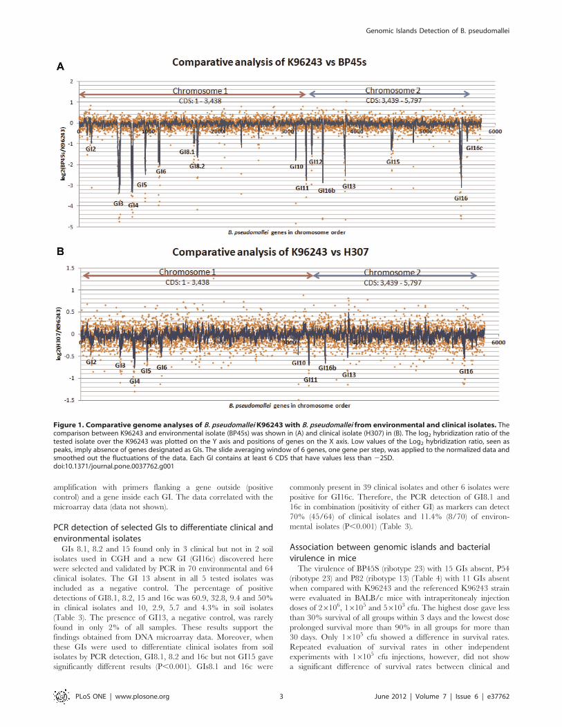
microarray comparison (i.e. log2 ratios are close to zero; Figure 1).
Genes with log2 hybridization ratios of at least minus two standard
deviations less than the overall mean were considered potentially
absent or divergent in the tested isolates. These absent genes when
clustered and consisting of at least six consecutive genes were
designated as a genomic island (GI). The comparative results
between environmental (Figure 1A, BP45s) or clinical isolates
(Figure 1B, H307) with K96243 are shown as the log2 ratio for
each gene position. There were 15 GIs (peaks) detected as absent
in five B. pseudomallei isolates when compared with the K96243
(Table 1), of which most corresponded to the K96243 GIs (GI2, 3,
4, 5, 6, 8, 10, 11, 12, 13, 15 and 16) reported by Holden et al. [12]
and the GI16b as reported by Tuanyok et al. [13]. The largest GI,
GI8 (92.3 kb), in the K96243 strain was seen as two separated
absent regions of 15.7 kb (CDS coordinates BPSL1638-
BPSL1656) and 21.8kb (CDS coordinates BPSL1693-1708A) with
distinguishable %GC contents (Table 1). They were assigned here
as GIs8.1 and 8.2 because they were located at the same reference
genomic location with GI8 in K96243 but contained different
gene contents. Since the methods used here compared gene
compositions with the K96243, it can only report regions present
in or absent from the K96243, but cannot identify the unique gene
composition in other isolates. Therefore, the GI nomenclature as
proposed by Tuanyok et al. [13] may not be properly applied.
There were four conserved GIs (GIs1, 7, 9 and 14) present in all 5
isolates, of which GIs7 and 9 were reported to be a part of the core
genome of B. pseudomallei strain [12].
Comparative genome hybridization in this study also detected
a new GI, named GI16c located downstream from GI16 on
chromosome 2, with the size of 8.3 kb determined by the
microarray. It consists of eight contiguous probes (CDS coordinates
BPSS2148-BPSS2154) containing common characteristics of a GI
such as a transposase gene and insertion element. It appeared to be
absent in all soil isolates and present in 1/3 of tested clinical isolates.
Moreover, from the log2 hybridization ratios of genes from
each isolate to those of a clinical isolate of the reference K96243,
higher similarity (shorter peaks, closer to 0) was observed in each
GI in the clinical isolates than in the soil isolates (Figure 1). The
overall profiles from the comparison of environmental isolates (2
total) and clinical isolates (3 total) to the K96243 were also
different (Figure S1). When the presence or absence of each GI
in these 5 isolates was compared with K96243, clinical isolates
(H307, P54, P82) contained lower numbers of absent genomic
islands (10–11 absent GIs) than environmental isolates (BP45s
and BP28L). GIs 6, 12 and 16c in clinical isolates showed some
variability in the GI patterns. A total of eight regions (GIs2, 3, 4,
5, 11, 13, 16 and 16b) were absent in both tested clinical and soil
isolates. GIs 8.1, 8.2 and 15 were absent in the soil isolates but
present in all of the clinical isolates. Their functions are related
to metabolism, cellular process, biosynthesis and transport
proteins (Table 2).
The absence of the GIs3, 4, 6, 10, 13, 15 and 16c identified by
DNA microarray was selected for further confirmation by PCR
Genomic Islands Detection of B. pseudomallei
PLoS ONE | www.plosone.org 2 June 2012 | Volume 7 | Issue 6 | e37762
amplification with primers flanking a gene outside (positive
control) and a gene inside each GI. The data correlated with the
microarray data (data not shown).
PCR detection of selected GIs to differentiate clinical andenvironmental isolatesGIs 8.1, 8.2 and 15 found only in 3 clinical but not in 2 soil
isolates used in CGH and a new GI (GI16c) discovered here
were selected and validated by PCR in 70 environmental and 64
clinical isolates. The GI 13 absent in all 5 tested isolates was
included as a negative control. The percentage of positive
detections of GI8.1, 8.2, 15 and 16c was 60.9, 32.8, 9.4 and 50%
in clinical isolates and 10, 2.9, 5.7 and 4.3% in soil isolates
(Table 3). The presence of GI13, a negative control, was rarely
found in only 2% of all samples. These results support the
findings obtained from DNA microarray data. Moreover, when
these GIs were used to differentiate clinical isolates from soil
isolates by PCR detection, GI8.1, 8.2 and 16c but not GI15 gave
significantly different results (P,0.001). GIs8.1 and 16c were
commonly present in 39 clinical isolates and other 6 isolates were
positive for GI16c. Therefore, the PCR detection of GI8.1 and
16c in combination (positivity of either GI) as markers can detect
70% (45/64) of clinical isolates and 11.4% (8/70) of environ-
mental isolates (P,0.001) (Table 3).
Association between genomic islands and bacterialvirulence in miceThe virulence of BP45S (ribotype 23) with 15 GIs absent, P54
(ribotype 23) and P82 (ribotype 13) (Table 4) with 11 GIs absent
when compared with K96243 and the referenced K96243 strain
were evaluated in BALB/c mice with intraperitonealy injection
doses of 26106, 16105 and 56103 cfu. The highest dose gave less
than 30% survival of all groups within 3 days and the lowest dose
prolonged survival more than 90% in all groups for more than
30 days. Only 16105 cfu showed a difference in survival rates.
Repeated evaluation of survival rates in other independent
experiments with 16105 cfu injections, however, did not show
a significant difference of survival rates between clinical and
Figure 1. Comparative genome analyses of B. pseudomallei K96243 with B. pseudomallei from environmental and clinical isolates. Thecomparison between K96243 and environmental isolate (BP45s) was shown in (A) and clinical isolate (H307) in (B). The log2 hybridization ratio of thetested isolate over the K96243 was plotted on the Y axis and positions of genes on the X axis. Low values of the Log2 hybridization ratio, seen aspeaks, imply absence of genes designated as GIs. The slide averaging window of 6 genes, one gene per step, was applied to the normalized data andsmoothed out the fluctuations of the data. Each GI contains at least 6 CDS that have values less than 22SD.doi:10.1371/journal.pone.0037762.g001
Genomic Islands Detection of B. pseudomallei
PLoS ONE | www.plosone.org 3 June 2012 | Volume 7 | Issue 6 | e37762
environmental isolates (P = 0.6) or between each isolate and
K96243 (data not shown).
Discussion
Comparative genome hybridization of five tested isolates to
K96243 indicated their average core genome close to 86% as
reported by Sim et al. [10]. The microarray-based CGH
technology has been shown as a remarkable tool for the
identification and fine discrimination between close species, and
additionally provided insight into the adaptation to its ecological
niche as reported in L. taiwanensis BL263 [26]. The variability of
GIs among clinical isolates was higher than that of environmen-
tal isolates. The different overall comparison profiles between
clinical and environmental isolates, as shown in figure 1 and
figure S1 of the other 3 isolates can clearly distinguish the source
of isolation. Even though the transfer of GIs is assumed to be
spontaneous, stress responses may provide selective pressure
responsible for variation of GIs and gene insertion in each GI of
the clinical isolates may play an important role for host
adaptation. The absence or divergence of B. pseudomallei GIs
found by comparative genome hybridization using the K96243
sequenced strain as a reference was consistent with the recent
studies using bioinformatics tools or in silico analysis [12,13,18].
Moreover, two out of four GIs (GsI7 and 9) found to be common
among the 5 tested isolates are also reported as parts of the core
genome by Sim et al.[10]. Interestingly, the method was able to
identify a new GI of the genomic islands (GI 16c) on
chromosome 2, which was not previously reported [12,13]. A
small difference in %GC (69.5%) of GI16c when compared to
the rest of the B. pseudomallei genome (68%) and a lack of flanking
tRNA and 39 end repeat may make it difficult to be detected by
other genome sequence comparison methods. GI16c, however,
contains transpose and insertion element genes which are
involved in genome mobility and are common characteristics of
GIs. This GI may be in the stage of evolutionary regression that
eliminated some GI functions [27].
The GI 8, which is the largest GI of K96243, was identified in
this study to contain 2 absent regions with a distinguishable %GC.
The percentage of these 2 regions present in 64 clinical isolates was
also clearly different. This GI region was reported to contain
several types of gene contents that varied in size (15.8–92.3 kb)
[13]. The hyper-variation in this location supported the cause of
genome plasticity created by various GIs. The comparative
genome analysis of the B. pseudomallei genomes available in the
public domain [28] also indicated a common finding of variability
in the number of genes and gene shuffling within each GI obtained
through horizontal transfer from other organisms.
The PCRdetection for the presence of five GIs, GIs2, 6, 9, 11 and
16, with various functions, among 186 environmental and clinical
isolates was reported to be no different [18]. Two environmental
isolates in this study were carefully picked from an undisturbed soil
location in Nampong district, Khon Kaen province as soil
representative isolates.When they were used together with 3 clinical
isolates in comparative genome hybridization, 3 GIs (GIs8.1, 8.2
and 15) were clinically specific GIs as they were only identified in
clinical but not soil isolates. Their functions are related to
metabolism and transport proteins that may support the survival
of the organism inside their host. The PCR detection of these GIs
indicated their low presentation in soil isolates (3–10%) but varied
among clinical isolates. Interestingly, the new GI16c was rarely
detected in soil samples but was present in up to 50% in clinical
isolates. GIs8.1, 8.2 and 16c can significantly distinguish clinical
from soil isolates. The PCR detection using a combination of
primers specific to GIs8.1 and 16c was positive up to 70% of clinical
isolates. The access of B. pseudomallei genomes through the VISTA
component of Integrated Microbial Genomes (IMG) [29] was done
to investigate the presence of these 2 GIs (GIs8.1 and 16c) in 22
publically available genome sequences, of which 4 are complete and
18 are drafts [30]. Twenty-one of them are clinical isolates from
several countries including Thailand and 1 is an environmental
isolate (S13) fromSingapore.GIs8.1 and 16cwere present in 57 (12/
21) and 62 (13/21)% of clinical isolates and 71.4% by 2 GIs
detection. None of them were present in the S13 soil isolate [28]. A
combination of the 3 primer sets for GIs8.1, 8.2 and 16c aids the
percentage of clinical isolate detections in this study to 76.6%
(Table 3). Therefore, the development of multiplex PCR for GI8.1
and 16c detection or all these 3 GIs can be applied to detect
potentially virulent isolates in soil in endemic areas of northeast
Thailand in a single process. As GIs in each B. pseudomallei isolate are
diverse and may be changed through adaptation and evolution,
further evaluation of this PCR detection with isolates from other
areas is therefore advised before it can be applied to other regions of
the world.
In this study, it is clear that differences of time to mortality were
not observed in BALB/c mice model after inoculation with B.
pseudomallei clinical and environmental isolates with the same
ribotype (ribotype 23) and another clinical isolate of ribotype 13
[8] with a different absence of GIs. Clinically specific GIs encode
several genes with metabolic and transport functions that might be
important in generation and acquisition of nutrients. Examples of
these genes are the ABC transport system, hydrolase, oxygenase and
the Gnt-R family of regulatory proteins. The islands also include
genes encoded for the outer membrane porin protein and a surface
exposed protein such as BPSL1705, a Yad A- like protein (adhesion
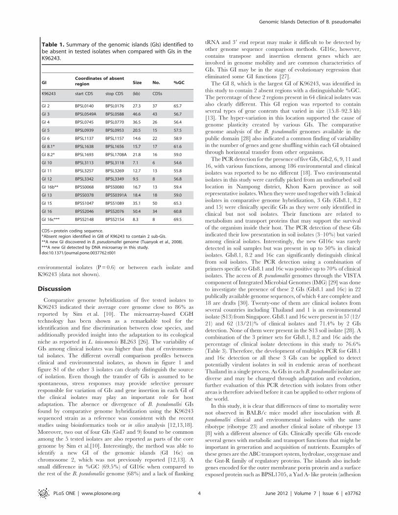
Table 1. Summary of the genomic islands (GIs) identified tobe absent in tested isolates when compared with GIs in theK96243.
GICoordinates of absentregion Size No. %GC
K96243 start CDS stop CDS (kb) CDSs
GI 2 BPSL0140 BPSL0176 27.3 37 65.7
GI 3 BPSL0549A BPSL0588 46.6 43 56.7
GI 4 BPSL0745 BPSL0770 36.5 26 56.4
GI 5 BPSL0939 BPSL0953 20.5 15 57.5
GI 6 BPSL1137 BPSL1157 14.6 22 58.9
GI 8.1* BPSL1638 BPSL1656 15.7 17 61.6
GI 8.2* BPSL1693 BPSL1708A 21.8 16 59.0
GI 10 BPSL3113 BPSL3118 7.1 6 54.6
GI 11 BPSL3257 BPSL3269 12.7 13 55.8
GI 12 BPSL3342 BPSL3349 9.5 8 56.8
GI 16b** BPSS0068 BPSS0080 16.7 13 59.4
GI 13 BPSS0378 BPSS0391A 18.4 18 59.0
GI 15 BPSS1047 BPSS1089 35.1 50 65.3
GI 16 BPSS2046 BPSS2076 50.4 34 60.8
GI 16c*** BPSS2148 BPSS2154 8.3 8 69.5
CDS =protein coding sequence.*Absent region identified in GI8 of K96243 to contain 2 sub-GIs.**A new GI discovered in B. pseudomallei genome (Tuanyok et al., 2008).***A new GI detected by DNA microarray in this study.doi:10.1371/journal.pone.0037762.t001
Genomic Islands Detection of B. pseudomallei
PLoS ONE | www.plosone.org 4 June 2012 | Volume 7 | Issue 6 | e37762
gene of Yersinia sp.). These genes were suggested to promote the
virulence of B. pseudomallei through mediating host-cell interactions
and therefore were previously suggested to be important in bacterial
survival or pathogenesis [12,31]. The BPSL1705 gene, however,
was confirmed by PCR amplification to be absent in all tested
isolates but was still present in the B. pseudomallei K96243 (data not
shown). Therefore, these genes might not play a direct role in the
virulence of this bacteriumas previously suggested, or there are some
other alternatives genes with similar functions to this gene in other
isolates. A similar story was also found for the cell surface protein or
hemagglutinin-related protein gene (BPSS2053) (in GI 16) that was
absent in all tested isolates but present in theK96243. This gene was
proposed to play a role in virulence by reducing the adherence to
human buccal epithelial cells [10]. Therefore, the difference in
severity of the disease should depend at least on a combination of
genes with more influence from host’s status as seen by the high risk
of this disease in diabetic people.Nevertheless, the possibility that the
absent GI may be associated with acquisition of the bacteria that
were omitted by direct inoculation or the low sample size or even
simply that mice are different from humans has to be considered
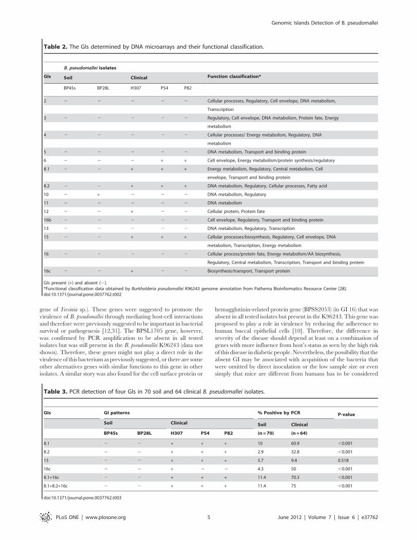
Table 2. The GIs determined by DNA microarrays and their functional classification.
B. pseudomallei isolates
GIs Soil Clinical Function classification*
BP45s BP28L H307 P54 P82
2 2 2 2 2 2 Cellular processes, Regulatory, Cell envelope, DNA metabolism,
Transcription
3 2 2 2 2 2 Regulatory, Cell envelope, DNA metabolism, Protein fate, Energy
metabolism
4 2 2 2 2 2 Cellular processes/ Energy metabolism, Regulatory, DNA
metabolism
5 2 2 2 2 2 DNA metabolism, Transport and binding protein
6 2 2 2 + + Cell envelope, Energy metabolism/protein synthesis/regulatory
8.1 2 2 + + + Energy metabolism, Regulatory, Central metabolism, Cell
envelope, Transport and binding protein
8.2 2 2 + + + DNA metabolism, Regulatory, Cellular processes, Fatty acid
10 2 + 2 2 2 DNA metabolism, Regulatory
11 2 2 2 2 2 DNA metabolism
12 2 2 + 2 2 Cellular protein, Protein fate
16b 2 2 2 2 2 Cell envelope, Regulatory, Transport and binding protein
13 2 2 2 2 2 DNA metabolism, Regulatory, Transcription
15 2 2 + + + Cellular processes/biosynthesis, Regulatory, Cell envelope, DNA
metabolism, Transcription, Energy metabolism
16 2 2 2 2 2 Cellular process/protein fate, Energy metabolism/AA biosynthesis,
Regulatory, Central metabolism, Transcription, Transport and binding protein
16c 2 2 + 2 2 Biosynthesis/transport, Transport protein
GIs present (+) and absent (2).*Functional classification data obtained by Burkholderia pseudomallei K96243 genome annotation from Pathema Bioinformatics Resource Center [28].doi:10.1371/journal.pone.0037762.t002
Table 3. PCR detection of four GIs in 70 soil and 64 clinical B. pseudomallei isolates.
GIs GI patterns % Positive by PCR P-value
Soil Clinical Soil Clinical
BP45s BP28L H307 P54 P82 (n =70) (n=64)
8.1 2 2 + + + 10 60.9 ,0.001
8.2 2 2 + + + 2.9 32.8 ,0.001
15 2 2 + + + 5.7 9.4 0.518
16c 2 2 + 2 2 4.3 50 ,0.001
8.1+16c 2 2 + + + 11.4 70.3 ,0.001
8.1+8.2+16c 2 2 + + + 11.4 75 ,0.001
doi:10.1371/journal.pone.0037762.t003
Genomic Islands Detection of B. pseudomallei
PLoS ONE | www.plosone.org 5 June 2012 | Volume 7 | Issue 6 | e37762
before it can be concluded that the virulence of each isolate is not
different.
Besides examining the virulence genes located in some absent
GIs, PCR was used to investigate the presence of genes outside the
GIs that encode for other known virulence determinants such as
type III protein secretion systems (bsaQ), that are potential
virulence determinants such as surface polysaccharides
(BPSL2800), fimbriae and pili (BPSS0120) [12]. They were all
present in all 5 tested isolates. This may suggest that these other
virulence factors present in the B. pseudomallei genome are critical
to maintain its virulence. Similarly, regulatory genes known to
promote survival such as a sigma factor (rpoS) [32] were present in
all isolates. These factors in B. pseudomallei may contribute to the
fitness of the organism as well or even more than those obtained
recently from horizontal gene transfer.
In addition, the microarray data analysis of all isolates revealed
the presence of other virulence genes such as types I, II, III and
VI protein secretion systems [33], exoproteins and genes known
to promote survival in diverse and challenging environments
including secondary metabolite biosynthesis genes (siderophore,
malleobactin biosynthesis cluster, superoxide detoxification, Ser
proteases, phospholipase C, catalase, etc.) [12,34,35,36,37],
sigma factor [32,38], quorum sensing [39], reactive nitrogen
intermediates [40], O-antigen [41], chaperonin GroEL [42],
multidrug effect pump, secondary metabolism functions or drug
resistance functions, intracellular stress and motility and chemo-
taxis [12]. Therefore, these data confirm the previous report
from Ulett et al, 2001 that there is no difference in virulence
between clinical and environmental isolates. As their work does
not analyze the genotype of the compared isolates, this study
could then provide additional details to summarize that the
outcome of the infection mainly depends on the immune
response or susceptibility of the host rather than the genetic
background of the bacteria.
In conclusion, through whole genome comparative analysis by
DNA microarray hybridization, a new GI (GI16c) present on
chromosome 2 of B. pseudomallei was identified. The technique was
also able to show different hybridization profiles between clinical
and environmental isolates and identify 3 GIs that were found
more commonly in clinical isolates. The use of PCR to detect
GI8.1 and GI16c is a simple and cost effective method to screen
for potentially virulent isolates in the environment in endemic
areas, and it can further be applied for pathophysiological studies
of the bacteria.
Materials and Methods
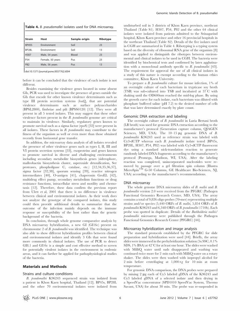
Strains and culture conditionsB. pseudomallei K96243 sequenced strain was isolated from
a patient in Khon Kaen hospital, Thailand [12]. BP45s, BP28L
and the other 70 environmental isolates were isolated from
undisturbed soil in 3 districts of Khon Kaen province, northeast
Thailand (Table S1). H307, P54, P82 and the other 64 clinical
isolates were isolated from patients admitted to the Srinagarind
hospital, Khon Kaen province and other 16 provincial hospitals in
the northeast Thailand (Table S2). Details of the five isolates used
in CGH are summarized in Table 4. Ribotyping is a typing system
based on the diversity of ribosomal RNA gene of the organism [8]
and was applied to distinguish the ribotypes between environ-
mental and clinical isolates to be used in CGH. The bacteria were
identified by biochemical tests and confirmed by latex agglutina-
tion with a monoclonal antibody specific to B. pseudomallei [43].
The requirement for approval the use of all clinical isolates in
a study of this nature is exempt according to the human ethics
committee, Khon Kaen University.
To prepare a B. pseudomallei culture for mouse infection, 1% of
an overnight culture of each bacterium in trypticase soy broth
(TSB) was sub-cultured into TSB and incubated at 37uC with
shaking until the OD600nm reached the mid-log phase. By using
the growth curve for each isolate, each bacterium was diluted with
phosphate buffered saline (pH 7.2) to the desired number of cells
that was later determined exactly by plate count.
Genomic DNA extraction and labelingThe overnight culture of B. pseudomallei in Luria Bertani broth
(LB broth) was used for genomic DNA extraction according to the
manufacturer’s protocol (Generation capture column, QIAGEN
Sciences, MD, USA). The 10–15 mg genomic DNA of B.
pseudomallei K96243 used as reference strain was labeled with
Cy3-dCTP whereas each B. pseudomallei isolate tested (BP45s,
BP28L, H307, P54, P82) was labeled with Cy5-dCTP fluorescent
dye using a standard nick-translation reaction to generate
randomly labeled DNA fragments according to the manufacturer’s
protocol (Promega, Madison, WI, USA). After the labeling
reaction was completed, unincorporated nucleotides were re-
moved by passage over a gel filtration spin column (Illustra
MicroSpinTM G-50 Columns, GE Healthcare Bio-Sciences, NJ,
USA) according to the manufacturer’s recommendations.
DNA microarrayThe whole genome DNA microarray slides of B. mallei and B.
pseudomallei version 2.0 were received from the PFGRC (Pathogen
Functional Genomics Research Center, MD, USA). The array
contains a total of 9,826 oligo probes (70-mer) representing multiple
strains and/or species (5,440 ORFs of B. mallei, 5,854 ORFs of B.
pseudomalleiK96243 and 6,348 ORFs of B. pseudomallei 1710b). Each
probe was spotted in duplicate. Details of the Burkholderia mallei/
pseudomallei microarray were published through the Pathogen
Functional Genomics Research Center (PFGRC) [44].
Microarray hybridization and image analysisThe standard protocols established by the PFGRC for slide
preparation and hybridization were used [44]. Briefly, the array
slideswere immersed in the prehybridization solution (5x SSC, 0.1%
SDS, 1%BSA) at 42uC for at least one hour. The slides were washed
with MilliQ water until suds disappeared and washing was
continued twice more for 2 min each with MilliQ water on a rotary
shaker. The slides were then washed with isopropyl alcohol for
2 min before centrifuging at 1,0006g for 10 min at room
temperature.
For genomic DNA comparison, the DNA probes were prepared
by mixing 2 mg each of Cy5 labeled gDNA of the K96243 and
Cy3 labeled gDNA of a selected isolate and then drying in
a SpeedVac concentrator (SPD1010 SpeedVac System, Thermo
Savant, USA) for about 30 min. The probe was re-suspended in
Table 4. B. pseudomallei isolates used for DNA microarray.
Strain Host Sample origin Ribotype
BP45S Environment Soil 23
BP28L Environment Soil 13
H307 Male, 54 years Blood 23
P54 Female, 50 years Pus 23
P82 Male, 56 years Pus 13
doi:10.1371/journal.pone.0037762.t004
Genomic Islands Detection of B. pseudomallei
PLoS ONE | www.plosone.org 6 June 2012 | Volume 7 | Issue 6 | e37762
50 ml hybridization solution (50% formamide, 5x SSC, 0.1% SDS)
and hybridized onto the post-processed slides while the LifterSlip
Microarray Coverslips (Erie Scientific, USA) were placed over the
array slides and incubated at 42uC in a water bath for 16–20 h.
Each slide was then washed according to the standard protocols
recommended by PFGRC.
Microarray data analysisThe hybridized slides were scanned with a GenePix 4000B laser
scanner (Molecular Devices, Sunnyvale, CA, USA) and initially
analyzed with GenePix Pro 6.1 software. Briefly, signals below the
threshold level (the total pixel intensity of the spot is 65% lower
than the median background intensity at both wavelengths) or
‘‘empty’’ (ORF without oligo) and ‘‘bad’’ (flagged by visual
inspection of the spot images) spots were removed before
normalization. The data were normalized within each array by
the scaled print-tip (Lowess) method [45,46] and across arrays
using the Aroma package [47,48] available from Centre for
Mathematical Sciences, Lund, Sweden [49], using R project
environment [50]. The analysis for the presence or absence of GIs
was observed based on K96243 ORFs on the microarray. The low
values of Log2 hybridization ratios imply absence of genes
visualized by Treeview software was provided as figure S2. The
CDS coordinates of genes in 15 GIs were listed as table S3. The
microarray data have been deposited in NCBIs Gene Expression
Omnibus [51] and all data is MIAME compliant which are
accessible through GEO Series accession number GSE19558 [52]
for comparative genomics hybridizations.
GC content analysis of GIsTo calculate %GC content of each GI, a Python computer
script was written following a method of Madigan et al. [53] by
using complete nucleotide sequences of B. pseudomallei K96243
chromosomes 1 and 2 (downloaded from NCBI, GenBank:
NC_006350.1 and NC_006351.1) as the input data.
PCR verification of microarray dataThe presence or absence of GIs3, 4, 6, 10, 13 and 16c in 5
tested isolates from the microarray hybridization was confirmed
using primers flanking a gene outside (positive control) and a gene
inside each GI by using TaqDNA polymerase (Roche Applied
Science, Indianapolis, IN). Further confirmation of the presence of
some selected virulence genes; FliC, bsaQ, rpoS, BPSL2800,
BPSS0120, BPSL1705 and BPSS2053 was also performed.
Primers for amplification of each GI and important genes were
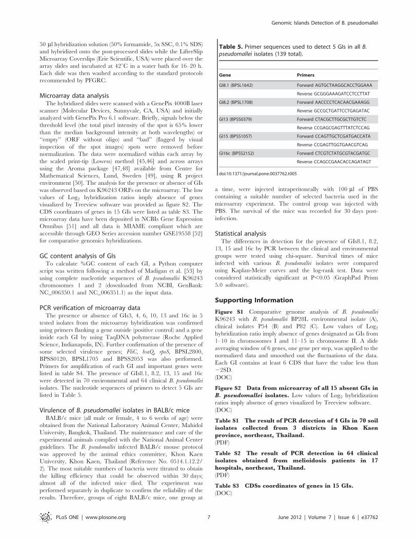
listed in table S4. The presence of GIs8.1, 8.2, 13, 15 and 16c
were detected in 70 environmental and 64 clinical B. pseudomallei
isolates. The nucleotide sequences of primers to detect 5 GIs are
listed in Table 5.
Virulence of B. pseudomallei isolates in BALB/c miceBALB/c mice (all male or female, 4 to 6 weeks of age) were
obtained from the National Laboratory Animal Center, Mahidol
University, Bangkok, Thailand. The maintenance and care of the
experimental animals complied with the National Animal Center
guidelines. The B. pseudomallei infected BALB/c mouse protocol
was approved by the animal ethics committee, Khon Kaen
University, Khon Kaen, Thailand (Reference No. 0514.1.12.2/
2). The most suitable numbers of bacteria were titrated to obtain
the killing efficiency that could be observed within 30 days;
almost all of the infected mice died. The experiment was
performed separately in duplicate to confirm the reliability of the
results. Therefore, groups of eight BALB/c mice, one group at
a time, were injected intraperitoneally with 100 ml of PBS
containing a suitable number of selected bacteria used in the
microarray experiment. The control group was injected with
PBS. The survival of the mice was recorded for 30 days post-
infection.
Statistical analysisThe differences in detection for the presence of GIs8.1, 8.2,
13, 15 and 16c by PCR between the clinical and environmental
groups were tested using chi-square. Survival times of mice
infected with various B. pseudomallei isolates were compared
using Kaplan-Meier curves and the log-rank test. Data were
considered statistically significant at P,0.05 (GraphPad Prism
5.0 software).
Supporting Information
Figure S1 Comparative genome analysis of B. pseudomallei
K96243 with B. pseudomallei BP28L environmental isolate (A),
clinical isolates P54 (B) and P82 (C). Low values of Log2hybridization ratio imply absence of genes designated as GIs from
1–10 in chromosomes I and 11–15 in chromosome II. A slide
averaging window of 6 genes, one gene per step, was applied to the
normalized data and smoothed out the fluctuations of the data.
Each GI contains at least 6 CDS that have the value less than
22SD.
(DOC)
Figure S2 Data from microarray of all 15 absent GIs inB. pseudomallei isolates. Low values of Log2 hybridization
ratios imply absence of genes visualized by Treeview software.
(DOC)
Table S1 The result of PCR detection of 4 GIs in 70 soilisolates collected from 3 districts in Khon Kaenprovince, northeast, Thailand.
(PDF)
Table S2 The result of PCR detection in 64 clinicalisolates obtained from melioidosis patients in 17hospitals, northeast, Thailand.
(PDF)
Table S3 CDSs coordinates of genes in 15 GIs.
(DOC)
Table 5. Primer sequences used to detect 5 GIs in all B.pseudomallei isolates (139 total).
Gene Primers
GI8.1 (BPSL1642) Forward AGTGCTAAGGCACCTGGAAA
Reverse GCGGGAAAGATCCTCCTTAT
GI8.2 (BPSL1708) Forward AACCCCTCACAACGAAAGG
Reverse GCCGCTGATTCCTGAGATAC
GI13 (BPSS0379) Forward CTACGCTTGCGCTTGTCTC
Reverse CCGAGCGAGTTTATCTCCAG
GI15 (BPSS1057) Forward CCAGTTGCTCGATGACCATA
Reverse CCGAGTTGGTGAACGTCAG
GI16c (BPSS2152) Forward CTCGTCTATGCGTACGATGC
Reverse CCAGCCGAACACCAGATAGT
doi:10.1371/journal.pone.0037762.t005
Genomic Islands Detection of B. pseudomallei
PLoS ONE | www.plosone.org 7 June 2012 | Volume 7 | Issue 6 | e37762
Table S4 Primer sequences used for GIs confirmationand specific genes amplification.
(DOC)
Acknowledgments
We thank Pathogen Functional Genomics Research Center (PFGRC),
NIAID, USA for donating the microarray slides. We are very grateful to
Emeritus Professor James A Will, University of Wisconsin-Madison, USA
for editing the manuscript.
Author Contributions
Conceived and designed the experiments: RWS AT NK SW. Performed
the experiments: TB TW. Analyzed the data: TB TW RWS NK.
Contributed reagents/materials/analysis tools: RWS NK AT. Wrote the
paper: RWS TB NK.
References
1. Currie BJ (2003) Melioidosis: an important cause of pneumonia in residents ofand travellers returned from endemic regions. Eur Respir J 22: 542–550.
2. Palasatien S, Lertsirivorakul R, Royros P, Wongratanacheewin S,
Sermswan RW (2008) Soil physicochemical properties related to the presenceof Burkholderia pseudomallei. Trans R Soc Trop Med Hyg 102 Suppl 1: S5–9.
3. Woods DE (2002) The use of animal infection models to study the pathogenesis
of melioidosis and glanders. Trends Microbiol 10: 483–484; discussion 484–485.
4. Chaowagul W, White NJ, Dance DA, Wattanagoon Y, Naigowit P, et al. (1989)
Melioidosis: a major cause of community-acquired septicemia in northeastern
Thailand. J Infect Dis 159: 890–899.
5. White NJ (2003) Melioidosis. Lancet 361: 1715–1722.
6. Cheng AC, Currie BJ (2005) Melioidosis: epidemiology, pathophysiology, and
management. Clin Microbiol Rev 18: 383–416.
7. Vesaratchavest M, Tumapa S, Day NP, Wuthiekanun V, Chierakul W, et al.
(2006) Nonrandom distribution of Burkholderia pseudomallei clones in relation togeographical location and virulence. J Clin Microbiol 44: 2553–2557.
8. Sermswan RW, Wongratanacheewin S, Trakulsomboon S, Thamlikitkul V
(2001) Ribotyping of Burkholderia pseudomallei from clinical and soil isolates inThailand. Acta Trop 80: 237–244.
9. Ulett GC, Currie BJ, Clair TW, Mayo M, Ketheesan N, et al. (2001) Burkholderia
pseudomallei virulence: definition, stability and association with clonality.Microbes Infect 3: 621–631.
10. Sim SH, Yu Y, Lin CH, Karuturi RK, Wuthiekanun V, et al. (2008) The core
and accessory genomes of Burkholderia pseudomallei: implications for humanmelioidosis. PLoS Pathog 4: e1000178.
11. Dobrindt U, Hacker J (2001) Whole genome plasticity in pathogenic bacteria.Curr Opin Microbiol 4: 550–557.
12. Holden MT, Titball RW, Peacock SJ, Cerdeno-Tarraga AM, Atkins T, et al.
(2004) Genomic plasticity of the causative agent of melioidosis, Burkholderia
pseudomallei. Proc Natl Acad Sci U S A 101: 14240–14245.
13. Tuanyok A, Leadem BR, Auerbach RK, Beckstrom-Sternberg SM, Beckstrom-
Sternberg JS, et al. (2008) Genomic islands from five strains of Burkholderia
pseudomallei. BMC Genomics 9: 566.
14. Hacker J, Carniel E (2001) Ecological fitness, genomic islands and bacterialpathogenicity. A Darwinian view of the evolution of microbes. EMBO Rep 2:
376–381.
15. Dobrindt U, Hochhut B, Hentschel U, Hacker J (2004) Genomic islands inpathogenic and environmental microorganisms. Nat Rev Microbiol 2: 414–424.
16. Ahmed N, Dobrindt U, Hacker J, Hasnain SE (2008) Genomic fluidity and
pathogenic bacteria: applications in diagnostics, epidemiology and intervention.Nat Rev Microbiol 6: 387–394.
17. Dobrindt U, Agerer F, Michaelis K, Janka A, Buchrieser C, et al. (2003) Analysis
of genome plasticity in pathogenic and commensal Escherichia coli isolates by useof DNA arrays. J Bacteriol 185: 1831–1840.
18. Tumapa S, Holden MT, Vesaratchavest M, Wuthiekanun V,Limmathurotsakul D, et al. (2008) Burkholderia pseudomallei genome plasticity
associated with genomic island variation. BMC Genomics 9: 190.
19. Rajashekara G, Glasner JD, Glover DA, Splitter GA (2004) Comparative whole-genome hybridization reveals genomic islands in Brucella species. J Bacteriol
186: 5040–5051.
20. Weaver D, Karoonuthaisiri N, Tsai HH, Huang CH, Ho ML, et al. (2004)Genome plasticity in Streptomyces: identification of 1 Mb TIRs in the S. coelicolor
A3(2) chromosome. Mol Microbiol 51: 1535–1550.
21. Jayapal KP, Lian W, Glod F, Sherman DH, Hu WS (2007) Comparativegenomic hybridizations reveal absence of large Streptomyces coelicolor genomic
islands in Streptomyces lividans. BMC Genomics 8: 229.
22. Wu CW, Glasner J, Collins M, Naser S, Talaat AM (2006) Whole-genome
plasticity among Mycobacterium avium subspecies: insights from comparative
genomic hybridizations. J Bacteriol 188: 711–723.
23. Moore RA, Reckseidler-Zenteno S, Kim H, Nierman W, Yu Y, et al. (2004)
Contribution of gene loss to the pathogenic evolution of Burkholderia pseudomalleiand Burkholderia mallei. Infect Immun 72: 4172–4187.
24. Ong C, Ooi CH, Wang D, Chong H, Ng KC, et al. (2004) Patterns of large-
scale genomic variation in virulent and avirulent Burkholderia species. GenomeRes 14: 2295–2307.
25. Kim HS, Schell MA, Yu Y, Ulrich RL, Sarria SH, et al. (2005) Bacterial genome
adaptation to niches: divergence of the potential virulence genes in threeBurkholderia species of different survival strategies. BMC Genomics 6: 174.
26. Sarmiento-Rubiano LA, Berger B, Moine D, Zuniga M, Perez-Martinez G, et
al. (2010) Characterization of a novel Lactobacillus species closely related to
Lactobacillus johnsonii using a combination of molecular and comparative
genomics methods. BMC Genomics 11: 504.
27. Juhas M, van der Meer JR, Gaillard M, Harding RM, Hood DW, et al. (2009)Genomic islands: tools of bacterial horizontal gene transfer and evolution.
FEMS Microbiol Rev 33: 376–393.
28. Pathema Bioinformatics Resource Center. Available: http://pathema.jcvi.org/
Pathema. Accessed 10 Jan 2011.
29. Vista Tools for comparative genomic. Available: http://genome.lbl.gov/vista/index.shtml. Accessed 14 Nov 2011.
30. IMG Integrated Microbial Gemones. Available: http://img.jgi.doe.gov/cgi-
bin/w/main.cgi. Accessed 16 Dec 2010.
31. Tiyawisutsri R, Holden MT, Tumapa S, Rengpipat S, Clarke SR, et al. (2007)
Burkholderia Hep_Hag autotransporter (BuHA) proteins elicit a strong antibodyresponse during experimental glanders but not human melioidosis. BMC
Microbiol 7: 19.
32. Subsin B, Thomas MS, Katzenmeier G, Shaw JG, Tungpradabkul S, et al.(2003) Role of the stationary growth phase sigma factor RpoS of Burkholderia
pseudomallei in response to physiological stress conditions. J Bacteriol 185:7008–7014.
33. Shalom G, Shaw JG, Thomas MS (2007) In vivo expression technologyidentifies a type VI secretion system locus in Burkholderia pseudomallei that is
induced upon invasion of macrophages. Microbiology 153: 2689–2699.
34. Loprasert S, Sallabhan R, Whangsuk W, Mongkolsuk S (2002) The Burkholderiapseudomallei oxyR gene: expression analysis and mutant characterization. Gene
296: 161–169.
35. Singh R, Wiseman B, Deemagarn T, Donald LJ, Duckworth HW, et al. (2004)
Catalase-peroxidases (KatG) exhibit NADH oxidase activity. J Biol Chem 279:43098–43106.
36. Korbsrisate S, Tomaras AP, Damnin S, Ckumdee J, Srinon V, et al. (2007)
Characterization of two distinct phospholipase C enzymes from Burkholderia
pseudomallei. Microbiology 153: 1907–1915.
37. Vellasamy KM, Vasu C, Puthucheary SD, Vadivelu J (2009) Comparativeanalysis of extracellular enzymes and virulence exhibited by Burkholderia
pseudomallei from different sources. Microb Pathog 47: 111–117.
38. Vanaporn M, Vattanaviboon P, Thongboonkerd V, Korbsrisate S (2008) TherpoE operon regulates heat stress response in Burkholderia pseudomallei. FEMS
Microbiol Lett 284: 191–196.
39. Song Y, Xie C, Ong YM, Gan YH, Chua KL (2005) The BpsIR quorum-
sensing system of Burkholderia pseudomallei. J Bacteriol 187: 785–790.
40. Loprasert S, Sallabhan R, Whangsuk W, Mongkolsuk S (2003) Compensatoryincrease in ahpC gene expression and its role in protecting Burkholderia
pseudomallei against reactive nitrogen intermediates. Arch Microbiol 180:498–502.
41. Sarkar-Tyson M, Thwaite JE, Harding SV, Smither SJ, Oyston PC, et al. (2007)
Polysaccharides and virulence of Burkholderia pseudomallei. J Med Microbiol 56:
1005–1010.
42. Woo PC, Woo GK, Lau SK, Wong SS, Yuen K (2002) Single gene targetbacterial identification. groEL gene sequencing for discriminating clinical
isolates of Burkholderia pseudomallei and Burkholderia thailandensis. Diagn MicrobiolInfect Dis 44: 143–149.
43. Samosornsuk N, Lulitanond A, Saenla N, Anuntagool N, Wongratanacheewin S,
et al. (1999) Short report: evaluation of a monoclonal antibody-based latex
agglutination test for rapid diagnosis of septicemic melioidosis. Am J Trop MedHyg 61: 735–737.
44. Burkholderia mallei/pseudomallei microarray version 2 Description. Available:
http://pfgrc.jcvi.org/index.php/microarray/array_description/burkholderia_mallei/version2.html. Accessed 20 Jul 2010.
45. Quackenbush J (2002) Microarray data normalization and transformation. NatGenet 32 Suppl. pp 496–501.
46. Yang YH, Dudoit S, Luu P, Lin DM, Peng V, et al. (2002) Normalization for
cDNA microarray data: a robust composite method addressing single andmultiple slide systematic variation. Nucleic Acids Res 30: e15.
47. Bengtsson H, Jonsson G, Vallon-Christersson J (2004) Calibration and
assessment of channel-specific biases in microarray data with extended
dynamical range. BMC Bioinformatics 5: 177.
48. Bengtsson H, Hossjer O (2006) Methodological study of affine transformationsof gene expression data with proposed robust non-parametric multi-dimensional
normalization method. BMC Bioinformatics 7: 100.
49. Centre for Mathematical Sciences, Lund, Sweden. Available: http://www.maths.lth.se/help/R/aroma. Accessed 15 Aug 2010.
Genomic Islands Detection of B. pseudomallei
PLoS ONE | www.plosone.org 8 June 2012 | Volume 7 | Issue 6 | e37762

50. R project environment. Available: http://cran.r-project.org. Accessed 15 Aug
2010.51. Edgar R, Domrachev M, Lash AE (2002) Gene Expression Omnibus: NCBI
gene expression and hybridization array data repository. Nucleic Acids Res 30:
207–210.
52. GEO Series accession number GSE1 Available: http://www.ncbi.nlm.nih.gov/
geo/query/acc.cgi?acc =GSE19558. Accessed 14 May 2011.
53. Madigan MT, Martinko JM, Parker J (2003) Brock biology of microorganisms.
Upper Saddle River: Prentice Hall. 1019 p.
Genomic Islands Detection of B. pseudomallei
PLoS ONE | www.plosone.org 9 June 2012 | Volume 7 | Issue 6 | e37762
Related Documents