Genomic Characterisation of Invasive Non-Typhoidal Salmonella enterica Subspecies enterica Serovar Bovismorbificans Isolates from Malawi Christina Bronowski 1. , Maria C. Fookes 2. , Ruth Gilderthorp 2 , Kevin E. Ashelford 3 , Simon R. Harris 2 , Amos Phiri 4 , Neil Hall 3 , Melita A. Gordon 1,4,5 , John Wain 6 , Charles A. Hart 1 , Paul Wigley 1 , Nicholas R. Thomson 2" *, Craig Winstanley 1" 1 Institute of Infection and Global Health, University of Liverpool, Liverpool, United Kingdom, 2 Pathogen Genomics, The Wellcome Trust Sanger Institute, Hinxton, Cambridge, United Kingdom, 3 Institute of Integrative Biology, University of Liverpool, Liverpool, United Kingdom, 4 Malawi-Liverpool-Wellcome Trust Clinical Research Program, Queen Elizabeth Hospital, Blantyre, Malawi, 5 Department of Medicine, College of Medicine, University of Malawi, Malawi, 6 Department of Medical Microbiology, University of East Anglia, Norwich Research Park, Norwich, United Kingdom Abstract Background: Invasive Non-typhoidal Salmonella (iNTS) are an important cause of bacteraemia in children and HIV-infected adults in sub-Saharan Africa. Previous research has shown that iNTS strains exhibit a pattern of gene loss that resembles that of host adapted serovars such as Salmonella Typhi and Paratyphi A. Salmonella enterica serovar Bovismorbificans was a common serovar in Malawi between 1997 and 2004. Methodology: We sequenced the genomes of 14 Malawian bacteraemia and four veterinary isolates from the UK, to identify genomic variations and signs of host adaptation in the Malawian strains. Principal Findings: Whole genome phylogeny of invasive and veterinary S. Bovismorbificans isolates showed that the isolates are highly related, belonging to the most common international S. Bovismorbificans Sequence Type, ST142, in contrast to the findings for S. Typhimurium, where a distinct Sequence Type, ST313, is associated with invasive disease in sub-Saharan Africa. Although genome degradation through pseudogene formation was observed in ST142 isolates, there were no clear overlaps with the patterns of gene loss seen in iNTS ST313 isolates previously described from Malawi, and no clear distinction between S. Bovismorbificans isolates from Malawi and the UK. The only defining differences between S. Bovismorbificans bacteraemia and veterinary isolates were prophage-related regions and the carriage of a S. Bovismorbificans virulence plasmid (pVIRBov). Conclusions: iNTS S. Bovismorbificans isolates, unlike iNTS S. Typhiumrium isolates, are only distinguished from those circulating elsewhere by differences in the mobile genome. It is likely that these strains have entered a susceptible population and are able to take advantage of this niche. There are tentative signs of convergent evolution to a more human adapted iNTS variant. Considering its importance in causing disease in this region, S. Bovismorbificans may be at the beginning of this process, providing a reference against which to compare changes that may become fixed in future lineages in sub-Saharan Africa. Citation: Bronowski C, Fookes MC, Gilderthorp R, Ashelford KE, Harris SR, et al. (2013) Genomic Characterisation of Invasive Non-Typhoidal Salmonella enterica Subspecies enterica Serovar Bovismorbificans Isolates from Malawi. PLoS Negl Trop Dis 7(11): e2557. doi:10.1371/journal.pntd.0002557 Editor: Ruifu Yang, Beijing Institute of Microbiology and Epidemiology, China Received July 5, 2013; Accepted October 11, 2013; Published November 14, 2013 Copyright: ß 2013 Bronowski et al. This is an open-access article distributed under the terms of the Creative Commons Attribution License, which permits unrestricted use, distribution, and reproduction in any medium, provided the original author and source are credited. Funding: The MRC funded 454 sequencing (Grant code G0600805, awarded to JW) and CB through a MRC capacity building studenship (Grant code G0500534, awarded to CAH and CW), while Illumina sequencing was funded though the Wellcome Trust under grant WT098051 (NRT) (http://www.mrc.ac.uk/index.htm; http://www.wellcome.ac.uk). The funders had no role in study design, data collection and analysis, decision to publish, or preparation of the manuscript. Competing Interests: The authors have declared that no competing interests exist. * E-mail: [email protected] . These authors contributed equally to this work. " NRT and CW also contributed equally to this work. Introduction Invasive Non-typhoidal Salmonella (iNTS) are a major cause of morbidity and mortality in sub-Saharan Africa. Especially in young children, iNTS are either the first or second most common cause of bacteraemia [1,2], meningitis and septic arthritis [3,4] with high morbidity. HIV infection is the primary risk factor for iNTS bacteraemia in adults, and it has been suggested that iNTS emerged together with the HIV pandemic in sub-Saharan Africa [5]. The most important clinical risk factors for iNTS disease in children are malnutrition, malaria and anaemia, with one in five cases of NTS bacteraemia in children also associated with HIV infection [2,6,7]. There is considerable interest in identifying any underlying bacterial genetic basis for the apparent increase in invasiveness and transmission of African NTS strains. PLOS Neglected Tropical Diseases | www.plosntds.org 1 November 2013 | Volume 7 | Issue 11 | e2557

Welcome message from author
This document is posted to help you gain knowledge. Please leave a comment to let me know what you think about it! Share it to your friends and learn new things together.
Transcript

Genomic Characterisation of Invasive Non-TyphoidalSalmonella enterica Subspecies enterica SerovarBovismorbificans Isolates from MalawiChristina Bronowski1., Maria C. Fookes2., Ruth Gilderthorp2, Kevin E. Ashelford3, Simon R. Harris2,
Amos Phiri4, Neil Hall3, Melita A. Gordon1,4,5, John Wain6, Charles A. Hart1, Paul Wigley1,
Nicholas R. Thomson2"*, Craig Winstanley1"
1 Institute of Infection and Global Health, University of Liverpool, Liverpool, United Kingdom, 2 Pathogen Genomics, The Wellcome Trust Sanger Institute, Hinxton,
Cambridge, United Kingdom, 3 Institute of Integrative Biology, University of Liverpool, Liverpool, United Kingdom, 4 Malawi-Liverpool-Wellcome Trust Clinical Research
Program, Queen Elizabeth Hospital, Blantyre, Malawi, 5 Department of Medicine, College of Medicine, University of Malawi, Malawi, 6 Department of Medical
Microbiology, University of East Anglia, Norwich Research Park, Norwich, United Kingdom
Abstract
Background: Invasive Non-typhoidal Salmonella (iNTS) are an important cause of bacteraemia in children and HIV-infectedadults in sub-Saharan Africa. Previous research has shown that iNTS strains exhibit a pattern of gene loss that resembles thatof host adapted serovars such as Salmonella Typhi and Paratyphi A. Salmonella enterica serovar Bovismorbificans was acommon serovar in Malawi between 1997 and 2004.
Methodology: We sequenced the genomes of 14 Malawian bacteraemia and four veterinary isolates from the UK, to identifygenomic variations and signs of host adaptation in the Malawian strains.
Principal Findings: Whole genome phylogeny of invasive and veterinary S. Bovismorbificans isolates showed that theisolates are highly related, belonging to the most common international S. Bovismorbificans Sequence Type, ST142, incontrast to the findings for S. Typhimurium, where a distinct Sequence Type, ST313, is associated with invasive disease insub-Saharan Africa. Although genome degradation through pseudogene formation was observed in ST142 isolates, therewere no clear overlaps with the patterns of gene loss seen in iNTS ST313 isolates previously described from Malawi, and noclear distinction between S. Bovismorbificans isolates from Malawi and the UK. The only defining differences between S.Bovismorbificans bacteraemia and veterinary isolates were prophage-related regions and the carriage of a S.Bovismorbificans virulence plasmid (pVIRBov).
Conclusions: iNTS S. Bovismorbificans isolates, unlike iNTS S. Typhiumrium isolates, are only distinguished from thosecirculating elsewhere by differences in the mobile genome. It is likely that these strains have entered a susceptiblepopulation and are able to take advantage of this niche. There are tentative signs of convergent evolution to a more humanadapted iNTS variant. Considering its importance in causing disease in this region, S. Bovismorbificans may be at thebeginning of this process, providing a reference against which to compare changes that may become fixed in futurelineages in sub-Saharan Africa.
Citation: Bronowski C, Fookes MC, Gilderthorp R, Ashelford KE, Harris SR, et al. (2013) Genomic Characterisation of Invasive Non-Typhoidal Salmonella entericaSubspecies enterica Serovar Bovismorbificans Isolates from Malawi. PLoS Negl Trop Dis 7(11): e2557. doi:10.1371/journal.pntd.0002557
Editor: Ruifu Yang, Beijing Institute of Microbiology and Epidemiology, China
Received July 5, 2013; Accepted October 11, 2013; Published November 14, 2013
Copyright: � 2013 Bronowski et al. This is an open-access article distributed under the terms of the Creative Commons Attribution License, which permitsunrestricted use, distribution, and reproduction in any medium, provided the original author and source are credited.
Funding: The MRC funded 454 sequencing (Grant code G0600805, awarded to JW) and CB through a MRC capacity building studenship (Grant code G0500534,awarded to CAH and CW), while Illumina sequencing was funded though the Wellcome Trust under grant WT098051 (NRT) (http://www.mrc.ac.uk/index.htm;http://www.wellcome.ac.uk). The funders had no role in study design, data collection and analysis, decision to publish, or preparation of the manuscript.
Competing Interests: The authors have declared that no competing interests exist.
* E-mail: [email protected]
. These authors contributed equally to this work.
" NRT and CW also contributed equally to this work.
Introduction
Invasive Non-typhoidal Salmonella (iNTS) are a major cause of
morbidity and mortality in sub-Saharan Africa. Especially in
young children, iNTS are either the first or second most common
cause of bacteraemia [1,2], meningitis and septic arthritis [3,4]
with high morbidity. HIV infection is the primary risk factor for
iNTS bacteraemia in adults, and it has been suggested that iNTS
emerged together with the HIV pandemic in sub-Saharan Africa
[5]. The most important clinical risk factors for iNTS disease in
children are malnutrition, malaria and anaemia, with one in five
cases of NTS bacteraemia in children also associated with HIV
infection [2,6,7]. There is considerable interest in identifying any
underlying bacterial genetic basis for the apparent increase in
invasiveness and transmission of African NTS strains.
PLOS Neglected Tropical Diseases | www.plosntds.org 1 November 2013 | Volume 7 | Issue 11 | e2557

Strain collections of iNTS isolates from Africa are dominated by
the serovars Typhimurium and Enteritidis [8–12]. However a
seven year study of iNTS isolates associated with bacteraemia in
Malawi showed that S. Bovismorbificans was the third most
common serovar, with 46 cases, which accounts for 1% of the total
number of NTS isolates [11]. In contrast to this, a study of
Salmonella bacteraemia in developed countries (Finland, Denmark,
Canada and Australia) showed that among 490 bacteraemia NTS
isolates, isolated between 2000 and 2007, only one was S.
Bovismorbificans (0.2%) [13]. However, S. Bovismorbificans has
been found responsible for gastroenteritis outbreaks: in Malaysia
between 1973 and 1996 S. Bovismorbificans accounted for 2–11%
of salmonellosis cases [14,15] as well as isolated outbreaks all over
Europe and around the world [16,17][18], S. Bovismorbificans
phage type 32 (PT32) [19,20].
It has been shown that bacteraemia isolates of S. Typhimurium
from Kenya and Malawi belong to a distinct Multi Locus
Sequence Typing (MLST) group, ST313, that harbours a specific
repertoire of prophages and shows evidence of specific patterns of
genome degradation with many parallels to human-specific
Salmonella serovars such as S. Typhi and Paratyphi A, which cause
acute invasive disease [21]. ST313 is significantly distant from the
common gastroenteritis-associated S. Typhimurium ST19 [22].
These studies have raised the possibility that bacteremia-associated
iNTS serovars that were previously able to infect a broad host
range are becoming human-host adapted [23,24].
Multidrug resistance is also a significant factor in the emergence
of iNTS strains in Africa, resulting in a reliance on fluoroquino-
lones [11,25]. While S. Typhimurium and Enteritidis isolates from
Malawi exhibited resistance to commonly-used antimicrobials
(including ampicillin, co-trimoxazole and chloramphenicol), iNTS
S. Bovismorbificans isolates have remained comparatively suscep-
tible to these commonly-used antimicrobials.
Here, we report the genome sequence of S. Bovismorbificans
3114 (ST142), a paediatric bacteraemia isolate from Malawi, and
describe a detailed analysis of iNTS strains of S. Bovismorbificans
causing disease in humans in Malawi and compare them to other
isolates from the same region and from veterinary isolates from the
UK. We investigated whether there are markers of adaptation to
the human host, similar to those described in other iNTS serovars
from this region.
Materials and Methods
Bacterial strains and antimicrobial resistance profilingMalawian S. Bovismorbificans isolates used in this study were
taken from a previous study by Gordon et al [11] and date from
1997 to 2004. S. Bovismorbificans serovar designations for
Malawian strains were confirmed by serotyping at the National
Salmonella Reference Laboratory, Galway, Republic of Ireland.
Veterinary strains of S. Bovismorbificans were obtained from
Professor Paul Barrow (University of Nottingham), and were taken
from a collection dating from the 1970s and 1980s. All Salmonella
strains were stored in 10% (v/v) glycerol broth at 280uC. The
antimicrobial susceptibility profiles of the four veterinary strains
were determined by the disc diffusion method, in accordance with
BSAC guidelines (http://bsac.org.uk/wp-content/uploads/2012/
02/Version-11.1-2012-Final-.pdf), using a total of 11 antimicro-
bials: (AML10 (amoxicillin 10 mg), AMC30 (amoxicillin/clavula-
nic acid 30 mg), CTX30 (cefotaxime 30 mg), CN 10 (gentamicin
10 mg), CIP 1 (ciprofloxacin 1 mg), W 2.5 (trimethoprim 2.5 mg),
NA 30 (nalidixic acid 30 mg), RL25 (sulphamethoxazole 25 mg),
C10 (chloramphenicol 10 mg), TET30 (tetracycline 30 mg), CXM
5 (cefuroxime sodium 5 mg), RD 2 (rifampicin 2 mg), CAZ30
(ceftazidime 30 mg), S 25 (streptomycin 25 mg). The susceptibility
profiles of the human S. Bovismorbificans isolates from Malawi
have been determined previously, in accordance with BSAC
guidelines [11].
Genomic DNA extraction and genome sequencingSalmonella strains were cultured in Luria broth overnight at 37uC
shaking at 200 rpm. Genomic DNA extractions were performed
using the Wizard Genomic DNA Purification Kit (A1120,
Promega, Madison, USA) as described in the manufacturer’s
instructions.
The genome of S. Bovismorbificans strain 3114 was sequenced
using the Roche 454 Genome Sequencer FLX (GS-FLX)
following the manufacturer’s instructions (Roche 454 Life Science,
Branford, CT, USA). In brief, each sample was made into both a
paired-end and fragment library using the standard FLX
chemistry for 454. Fragment libraries were prepared by fragmen-
tation, attachment of adapter sequences, refinement of the ends
and selection of adapted molecules. Paired-end libraries were
produced by hydroshear shearing, circularisation, addition of
adapters and selection, as for the fragment library. Both libraries
were amplified by emPCR and fragment-containing beads were
recovered and enriched. Sequencing primers were added and each
library was deposited onto a quarter of a PicoTitrePlate plate and
sequenced.
Multiplexed Illumina standard libraries were prepared for S.
Bovismorbificans 3114 and 17 additional strains following
standard protocols with 200 bp inserts and sequenced on the
Illumina Genome Analyzer II. Paired end sequence runs were
performed with 54 bp read length. Raw sequence data is
submitted to the public data repository, ENA, under accession
ERP000181.
S. Bovismorbificans strain 3114 genome sequenceassembly
For S. Bovismorbificans 3114 454 data, reads from the fragment
and paired-end libraries were de-novo assembled into contigs
Author Summary
Bacteraemia and meningitis caused by non-typhoidalSalmonella (including serovars Typhimurium, Enteritidisand Bovismorbificans) are a serious health issue in sub-Saharan Africa, particularly in young children and HIV-infected adults. Previous work has indicated that a distinctS. Typhimurium sequence type, ST313, has evolved andspread in these countries, and may be more human-adapted than isolates found in the developed world. Wetherefore investigated the genomes of Salmonella entericaserovar Bovismorbificans bacteraemia isolates from Malawiand compared them to genomes of veterinary S.Bovismorbificans isolates from the UK using Next Genera-tion Sequencing Technology and subsequent genomiccomparisons to establish if there is a genetic basis for thisincrease in invasive disease observed among African NTS.Contrary to the previous findings for S. Typhimurium,where a distinct ST is found only in sub-Saharan Africa, wediscovered that the S. Bovismorbificans isolates fromMalawi belong to the most common ST of the serovarand the genome is highly conserved across all sequencedisolates. The major differences between UK veterinary andAfrican human isolates were due to prophage regionsinserted into the genomes of African isolates, coupled witha higher prevalence of a virulence plasmid compared tothe UK isolates.
Genomic Analysis of Salmonella Bovismorbificans
PLOS Neglected Tropical Diseases | www.plosntds.org 2 November 2013 | Volume 7 | Issue 11 | e2557

using the Roche 454 Newbler assembler (version 2.0.01.12) with
default settings.
Illumina data was then used to extend and order the 454-
assembled contigs using the PAGIT package [26] as follows: 454
contigs were extended using ICORN [26], and the resulting
contigs were ordered and orientated with respect to the genome of
S. Typhimurium LT2 using ABACAS [27]. Finally, gap closure
was attempted where possible using IMAGE [26].
For each scaffold-contig in turn, putative coding sequences
(CDSs) were predicted using Glimmer version 3.02 (http://www.
cbcb.umd.edu/software/glimmer/). A further in-house Perl script
was then used to identify and correct those CDSs likely to have
been split due to sequencing errors when handling homopolymer
repeats. This involved BLASTP alignment of CDS protein
translations against a database of translations generated from
previously annotated Salmonella genomes, the identifications of
likely indels within homopolymer regions, the modification of
coding sequence feature positions to correct errors, and the
merging of relevant CDSs. Where such modification occurred, this
was recorded as metadata (in the form of the eventual GenBank
feature note field). Such CDSs were also marked with the
exception flag set to ‘low-quality sequence region’ for the final
GenBank submission to signify poor quality sequencing.
A putative function was then assigned to each gene by BLASTn
(NCBI Blast 2.2.17) comparison with a database of sequences
generated from the previously annotated genome of S. Typhimur-
ium LT2. Putative tRNA genes were detected using tRNAscan-SE
1.23 (ftp://selab.janelia.org/pub/software/tRNAscan-SE/). A
pseudochromosome, consisting of a concatenation of contigs
arranged into scaffolds, with 100 Ns separating adjoining scaffolds,
was prepared for comparison purposes; the final version of the
3114 genome was manually curated using Artemis [28,29].
The S. Bovismorbificans 3114 chromosome has been submitted
to EMBL under accession number HF969015, and its 93.8 kb
virulence plasmid under accession number HF969016.
S. Bovismorbificans Illumina genome assemblyIllumina sequence data for the 17 additional genomes was
assembled as follows: for each strain, Velvet [30] was used to
create multiple assemblies by varying the kmer size between 66%
and 90% of the read length. From these assemblies, the one with
the best N50 was chosen and contigs which were shorter than the
insert size length were removed. An assembly improvement step
was then run on the chosen assembly. The contigs of the assembly
were scaffolded by iteratively running SSPACE [31]. Then gaps
identified as 1 or more N’s, were targeted for closure by running
120 iterations of GapFiller [32]. Finally, the reads were aligned
back to the improved assembly using SMALT (http://www.
sanger.ac.uk/resources/software/smalt/) and a set of statistics was
produced for assessing the QC of the assembly.
All of the software developed is freely available for download
from GitHub (https://github.com/sanger-pathogens) under an
open source license, GNU GPL 3. The improvement step of the
pipeline is also available as a standalone Perl module from CPAN
(http://search.cpan.org/,ajpage/) (see Table S1 for assembly
data and statistics).
Pseudogene analysisPseudogenes were identified, using ACT comparisons [33], by
comparing the genome of strain 3114, first to S. Typhiumurium
LT2 and then to the genomes of S. Typhimurium D23580,
SL1344 and DT104. Pseudogenes were identified according to
whether CDS showed frameshift mutations, missing N- or C-
terminals or carried nonsense mutations. Once pseudogenes were
identified in the 3114 genome, their orthologous sequences were
checked in the assemblies of a further six S. Bovismorbificans
isolates, from both Malawi (3180, D1253, D993, A1668,) and the
UK (653308, 276608). These isolates were chosen as representa-
tives of distinct subclades in the tree. In some cases it was not
possible to identify mutations due to gaps in the sequence; these
are highlighted in Table S5.
Multi Locus Sequence Typing (MLST)MLST sequences were obtained from Illumina reads and
sequence types were assigned through the MLST website (http://
www.mlst.net/). Ambiguous results for some individual loci were
subsequently confirmed by PCR amplification of the locus and
sequencing of the PCR product (Beckman Coulter). In order to
build the MLST-based phylogenetic tree of Figure S1, the
concatenated sequences of the seven MLST loci of S. Bovismorbi-
ficans 3114 and the most common S. enterica STs from published
databases were loaded into SeaView v3.2 [34]. The phylogeny was
reconstructed using PhyML [35] within the Seaview package, and
FigTree v1.3.1 [36] was used to edit and label the final figure.
Construction of a pan genome pseudomoleculeFor the purpose of mapping and visualization of the genomic
content of all S. Bovismorbificans samples, a pseudomolecule was
constructed comprising the reference 3114 genome (chromosome
and virulence plasmid) and all non-redundant accessory regions
found by tblastx in each sample assembly with respect to the others
in an iterative manner. Briefly we performed pairwise compar-
isons, firstly of one isolate against the reference sequence (genome
and its plasmid). Regions in the comparator that were not present
in the reference were identified and added to the end of the
reference sequence to form a pan genome pseudomolecule. This
was repeated in an iterative process involving manual curation of
the sequences to be included in the growing pseudo molecule.
Read mapping, single nucleotide polymorphism (SNP)calling and construction of the phylogenetic tree of theS. Bovismorbificans sample set
Mapping of illumina reads per sample was carried out against
this resulting pseudomolecule using SMALT (http://www.sanger.
ac.uk/resources/software/smalt/) without mapping to repeats.
SNP calling was performed as previously described [37]. In order
to construct a robust phylogenetic tree a Bayesian approach [38],
which identifies high density SNPs and recombinant regions and
ignores them when constructing the phylogeny, was used. A
detailed list of sites removed from the chromosome, 48,423 bases
in total, is summarised in Table S2 and the alignment of variant
sites used to construct the final phylogenetic tree is presented in
Table S3. In order to construct the phylogeentic tree shown in
Figure 1 an initial tree using S. Heidelberg str SL476 (acc
no. CP001120) (see Figure S1), as an outgroup was constructed.
This identified the root position in the ingroup and final tree.
Using this root position the final tree shown in Figure 1 was
constructed using all 954 variant sites (Table S3) within the
chomosome: A maximum likelyhood approach (RAxML) was used
to construct the initial bipartitions tree followed by a reconstruc-
tion of the SNPs onto the tree branches using delayed
transformation (DELTRAN) parsimony [39].
Mapped illumina read data was saved in Bam format [40] and
converted to coverage files (number of reads mapped to each base
coordinate of the reference) with an in-house script. To aid
visualisation the read depth per base position of each isolate
against the pan-pseudomolecule reference sequence was
Genomic Analysis of Salmonella Bovismorbificans
PLOS Neglected Tropical Diseases | www.plosntds.org 3 November 2013 | Volume 7 | Issue 11 | e2557
constructed (Figure 1). Base positions with 0 or 1–14 reads
mapped were coloured white or grey, respectively. Base positions
with 156coverage or greater are coloured black. The cutoff of 15
or more times coverage per base position was selected because it
was just below the minimum median coverage obtained across all
of the isolates we sequenced (16 to 316coverage [data not shown];
see Figure 1). Using the observed coverage regions .4000 bps
showing a significant deviation from the median coverage were
identified by manual curation and checked against the genome
assembly.
Accession numbersThe raw sequence data is available under the accession number
ERP000181 at the European Nucleotide Archive, ENA. The
sequence and annotation data for S. Bovismorbificans strain 3114
chromosome and virulence plasmid, pVIRBov are available from
ENA under accession numbers HF969015-HF969016.
Results and Discussion
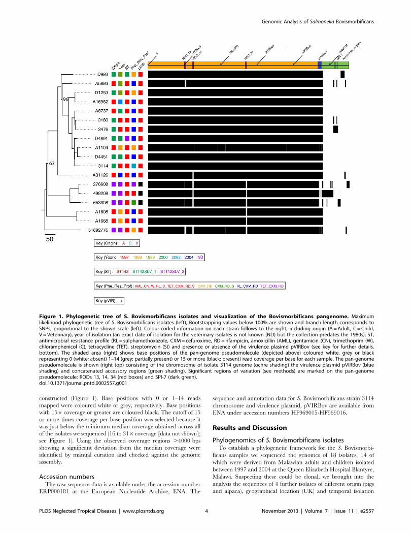
Phylogenomics of S. Bovismorbificans isolatesTo establish a phylogenetic framework for the S. Bovismorbi-
ficans samples we sequenced the genomes of 18 isolates, 14 of
which were derived from Malawian adults and children isolated
between 1997 and 2004 at the Queen Elizabeth Hospital Blantyre,
Malawi. Suspecting these could be clonal, we brought into the
analysis the sequences of 4 further isolates of different origin (pigs
and alpaca), geographical location (UK) and temporal isolation
Figure 1. Phylogenetic tree of S. Bovismorbificans isolates and visualization of the Bovismorbificans pangenome. Maximumlikelihood phylogenetic tree of S. Bovismorbificans isolates (left), Bootstrapping values below 100% are shown and branch length corresponds toSNPs, proportional to the shown scale (left). Colour-coded information on each strain follows to the right, including origin (A = Adult, C = Child,V = Veterinary), year of isolation (an exact date of isolation for the veterinary isolates is not known (ND) but the collection predates the 1980s), ST,antimicrobial resistance profile (RL = sulphamethoxazole, CXM = cefuroxime, RD = rifampicin, amoxicillin (AML), gentamicin (CN), trimethoprim (W),chloramphenicol (C), tetracycline (TET), streptomycin (S)) and presence or absence of the virulence plasimd pVIRBov (see key for further details,bottom). The shaded area (right) shows base positions of the pan-genome pseudomolecule (depicted above) coloured white, grey or blackrepresenting 0 (white; absent) 1–14 (grey; partially present) or 15 or more (black; present) read coverage per base for each sample. The pan-genomepseudomolecule is shown (right top) consisting of the chromosome of isolate 3114 genome (ochre shading) the virulence plasmid pVIRBov (blueshading) and concatenated accessory regions (green shading). Significant regions of variation (see methods) are marked on the pan-genomepseudomolecule: RODs 13, 14, 34 (red boxes) and SPI-7 (dark green).doi:10.1371/journal.pntd.0002557.g001
Genomic Analysis of Salmonella Bovismorbificans
PLOS Neglected Tropical Diseases | www.plosntds.org 4 November 2013 | Volume 7 | Issue 11 | e2557

(1970s/80s) to provide context to investigate wider serovar
variations. Genomic DNA from the S. Bovismorbificans isolates
were assayed using either 454 or multiplex Illumina sequencing
(see methods). The genome data of these sequences are
summarised in Table 1. Only chromosomal SNPs were used to
construct the maximum likelihood phylogenetic tree (see methods)
shown in Figure 1 (left), for which the root branch (leading to
sample 51892776) was previously identified by including an
outgroup (see methods). Recombinant regions and regions that
were unlikely to reflect the core phylogeny, such as prophage, were
removed from this analysis (48,423 total sites; Table S2).
Amongst our samples, we found a total of 954 variable sites
randomly distributed around the S. Bovismorbificans chromo-
some, which is approximately one SNP per 4,742 bp or just under
0.001% nucleotide divergence, within the core regions. It is
evident that the human and animal isolates are intermixed. To
give an idea of the level of nucleotide divergence within the core
genome the animal isolate 51892776 closest to the root, was
separated by 328 or 341 SNPs from the reference human isolate
3114 or most the divergent isolate shown in the tree, respectively.
Consistent with this, we extracted the sequences of the MLST loci
from the whole genome sequence data. All strains belonged to the
major S. Bovismorbificans sequence type ST142 except two
Malawian strains D993 and D4891 which were single locus
variants (SLV) of ST142 (see Table 1) but were not found to be
located on long branches on the tree.
It is evident from Figure 1 (left) that the human S.
Bovismorbificans isolates taken in Malawi are phylogenetically
extremely closely related, when compared to each other. This level
of sequence divergence is comparable to the evolutionary distance
between the S. Typhimurium lineages causing invasive disease in
Africa [41] which form two distinct lineages (differentiated from
each other by 455 SNPs), that are separated from the nearest
gastroenteritis lineages by .700 SNPs [41]. However, when
including the animal isolates from the UK it is apparent that this
limited variation is a feature of S. Bovismorbificans as a serovar
despite temporal, geographic and host differences. Also, in contrast
to the S. Typhimurium lineages causing invasive disease, there was
no clustering within the human samples with age or year of
isolation.
Together these data suggested that the S. Bovismorbificans
isolates causing invasive disease in Malawi, unlike S. Typhimur-
ium, were not a specialised clade, at least according to the core
phylogeny, therefore we looked at the accessory genome for clues
to the observed differences in disease outcome for these isolates
causing invasive disease in Malawi.
Accessory genomic regions within S. BovismorbificansST142 isolates
In order to visualise the variation across S. Bovismorbificans
isolates we constructed a pan-genome. To do this we concatenated
the whole genome sequence of strain 3114 (chromosome and
Table 1. S. Bovismorbificans isolates used in this study.
StrainIsolationyear Host Age# Outcome pVIRBov
AntimicrobialResistance profile MLST ST* ROD13 ROD14 ROD34
AccessoryGenomeSize (bp)
human
3114 1997 child ND ND + RL_CXM_RD 142 + + + 0
3180 1997 child ND ND + RL_CXM_RD 142 + + + 38,420
3476 1997 child ND ND + RL_CXM_RD 142 + + + 131,557
D993 1999 child ND ND + CXM_RD 142SLV_1 + + + 110,972
D1253 1999 child 4M ND + CXM_RD 142 + + + 5,577
D4451 2000 child 1Y11M ND + RL_CXM_RD 142 + + + 4,791
D4891 2000 child 11M ND + RL_CXM_RD 142SLV_2 + + + 4,791
A1104 1998 adult ND 3 + CXM_RD 142 + + + 4,791
A1608 1998 adult ND ND + RL_CXM_RD 142 + + 2 7,893
A1668 1998 adult ND 1 + RL_CXM_RD 142 + + 2 7,893
A16982 2002 adult ND 2 + RL_CXM_RD 142 + + + 4,735
A31126 2004 adult 23Y 1 + RL_CXM_RD 142 + + + 34,498
A5893 1999 adult 35Y ND + RL_CXM_RD 142 + + + 40,582
A8737 2000 adult 30Y ND + RL_CXM_RD 142 + + + 5,695
veterinary
499208 ,1980 alpaca N/A N/A 2 AML_CN_W_RL_C_TET_CXM_RD_S 142 / 2 2 289,627
653308 ,1980 pig N/A N/A 2 CXM_RD_S 142 / 2 2 88,266
276608 ,1980 pig N/A N/A 2 TET_CXM_RD 142 / 2 2 121,149
51892776 ,1980 pig N/A N/A + CXM_RD 142 2 2 2 36,589
The table summarizes properties of the isolates used in this study, including the presence or absence of ROD13, -14 and -34, the presence of the virulence plasmidpVIRBov and the size of the accessory genome in each of the addtional 17 Illumina-sequenced S. Bovismorbificans genomes obtained in comparison to strain 3114.#M = months, Y = years ; outcome 1 = death, 2 = survived, 3 = unknown, ND = no data; N/A = not applicable; resistance profile = sulphamethoxazole (RL), cefuroxime(CXM), rifampicin (RD), amocixillin (AML), gentamicin (CN), trimethoprim (W), chloramphenicol (C), tetracycline (TET), streptomycin (S); ST refers to the MLST sequencetype as determined by Illumina sequencing and sequencing of PCR amplicons,*SLV = Single Locus Variant; ROD13/ = partial or different ROD present.doi:10.1371/journal.pntd.0002557.t001
Genomic Analysis of Salmonella Bovismorbificans
PLOS Neglected Tropical Diseases | www.plosntds.org 5 November 2013 | Volume 7 | Issue 11 | e2557

virulence plasmid) as well as the regions found to be variable
present in one or more isolate, in a non-redundant manner (see
methods). Figure 1 (right) shows the read coverage of all isolates
included in this study mapped against the pan-genome pseudo-
molecule (see methods).
There are three main regions of difference (RODs) in
chromosome of strain 3114, denoted ROD_13, ROD_14 and
ROD_34. These are the only significant regions of difference in
the core genome being either absent or partially present in the four
veterinary strains and variably distributed in the human isolates
(Table 1 and Figure 1). They are predicted to encode prophage;
ROD_34 and ROD_13 are highly similar to the S. Typhimurium
prophage elements Gifsy-1 and Gifsy-2, respectively, and have
therefore been termed Gifsy-like (See Table S4). ROD14
represents a novel 46.4 kb prophage inserted into a spermidine/
putrescine operon on the S. Bovismorbificans 3114 genome (See
below; Table S4).
A distinctive region of variation between human (carried in all)
and animal samples (variably present) was a ,93 kb virulence
plasmid, here named pVIRBov (Figure 2(B)). pVIRBov is highly
simlar to the S. Typhimurium LT2 virulence plasmid, pSLT (See
Figure 2(B) and S2) and, like pSLT, carries the defining spv
virulence gene cassette and the pef (plasmid-encoded fimbriae)
operon mediating adhesion to murine intestinal epithelial cells
[42]. Located downstream of the pef operon is the rcK (resistance to
complement killing) gene. RcK is required by S. Typhimurium for
survival in macrophages and for virulence in mice [43]. pVIRBov
also shows a 7.463 kb deletion, and a 6.705 kb insertion,
compared to pSLT. The deleted region contains a putative gene
for single strand binding protein B (ssbB), as well as a number of
putative genes encoding membrane-associated proteins. The
insertion relative to pSLT includes a pilA-like gene (SBOV4711),
repC (SBOV47701), a gene encoding a putative outer membrane
protein (SBOV47871), traJ (SBOV48411), the primary activator of
tra (polycistronic transfer) operon expression [44] and a number of
hypothetical proteins. pVIRBov does not carry rsk (resistance to
serum killing), thought to be associated with the control of serum
resistance [45]. In contrast to a previous PCR based screen of S.
Bovismorbificans isolates, these data show that 100% of the
human bacteraeamia isolates and only one in four of the UK
veterinary isolates carry pVIRBov [46].
In pairwise comparisons with respect to the S. Bovismorbificans
str 3114 genome, eight human isolates (D1253, D4891, D4451,
A1104, A1608, A1668, A16892 and A8737) were almost identical
in genomic content to the reference, with accessory regions of only
4–8 kb (summarised in Table 1). The veterinary isolate 499208
and the paediatric bacteraemia isolate 3476 both carry the largest
accessory genomic regions, of ,290 kb and 132 kb, respectively
(Table 1). The accessory genome of 3476 contains a ROD inserted
at a t-RNA(Phe) -downstream of the CDS homologue to
SBOV31771 in the 3114 strain-, showing high sequence homology
to the SPI-7 of the human restricted pathogen S. Typhi CT18
(Figure 1, Figure S3). SPI-7 is a large mosaic pathogenicity island
carrying a collection of virulence-related genes; versions of SPI-7
have been identified in S. enterica serovars Typhi, Paratyphi C, and
Dublin [47–49]. This particular version of sample 3476 also
encodes a putative Vi capsule although lacks the sopE phage of S.
Typhi CT18. It also contains an operon related to carbohydrate
metabolism and extracellular structure modifications (Figure S3).
We did not find any genomic scar or any other evidence to show
that this island might have been contained in any of the other
human isolates or most recent ancestors.
Despite the existence of substantial accessory regions in half of
the samples sequenced, they were mostly found to be unique to
single isolates without correlation to the phylogenetic inference or
the disease outcome.
Comparative analysis of Pseudogene carriage amongst S.Bovismorbificans and S. Typhimurium isolates
In addition to gene gain, functional gene loss plays an important
role in the adaptation of the Salmonella to different lifestyles, with
host restricted Salmonella carrying over 200 pseudogenes
[21,22,47,50–52] compared to their broad host range counter-
parts. It is also evident that the patterns of gene loss are
nonrandom with nonsense mutations and frame-shifts being
over-represented in genes that are associated with aspects of
virulence or host interaction. The parallels in the patterns of
pseudogene accumulation is also a feature of the iNTS S.
Typhimurium isolate D23580, causing invasive disease in Malawi.
By comparing the genome of strain 3114 to those of S.
Typhimurium D23580, SL1344 and DT104, a total of 43
pseudogenes were identified in S. Bovismorbificans 3114. Six
further S. Bovismorbificans isolates, which were chosen as
representatives of distinct groups on the phylogenetic tree were
also analysed for pseudogene content (summarised results in Table
S5)
Of the 43 pseudogenes identified, 15 (34.9%) were conserved in
all of the six S. Bovismorbificans isolates tested, but intact in the
three S. Typhimurium strains. There was no clear distinction in
pseudogene carriage between human bacteraemia (Malawi) and
veterinary (UK) S. Bovismorbificans isolates, with only a single
pseudogene specific to Malawian isolates. Five (11.5%) pseudo-
genes have been identified as pseudogenes in all seven S.
Bovismorbificans and all three of the S. Typhimurium isolates
(Table S5).
Genes that are pseudogenes in all seven S. Bovismorbificans
isolates tested, but intact in all three S. Typhimurium isolates
(D23580, SL1344, DT104), include genes involved in sugar
metabolism, a putative autotransporter of the haemagglutinin
family (SBOV37821) and putative surface-exposed virulence
protein BigA (SBOV35501).
We observed no clear link between pseudogene carriage and
source (human and veterinary isolates). Although the extent of
pseudogene formation in S. Bovismorbificans does not compare to
the host adapted Salmonella serovars or to the iNTS Typhimurium
D23580, there are tantalising glimpses that suggest S. Bovismor-
bificans as a species may carry pseudogenes, such as BigA that are
consistent with host specialisation. What is clear, though, is that
this has not been driven by the emergence of a new dominant
lineage adapted to humans in Africa as we have seen before with S.
Typhimurium ST313.
Detailed comparison of S. Bovismorbificans with other S.enterica genomes
Since this is the first time a S. Bovismorbificans genome has
been described and to identify functions that may be involved in
the apparent plastiticity in pathogenicity we performed whole-
genome comparison between S. Bovismorbificans and S. Typhi-
murium LT2. These comparisons revealed a high level of synteny
and colinearity, with no inversions (Figure 3). A comparison of S.
Bovismorbificans genome statistics with other serovars is summar-
ized in Table 2. Figure 2(A) shows orthologous genes, identified by
reciprocal fasta searches, in 10 S. enterica subspecies 1 strains from
eight Salmonella serovars as well as wider members of the
Enterobacteriaceae. This analysis showed that S. Bovismorbificans
gene content broadly resembles those common to S. enterica
subspecies 1 NTS serovars. Genome comparisons to S.
Genomic Analysis of Salmonella Bovismorbificans
PLOS Neglected Tropical Diseases | www.plosntds.org 6 November 2013 | Volume 7 | Issue 11 | e2557

Genomic Analysis of Salmonella Bovismorbificans
PLOS Neglected Tropical Diseases | www.plosntds.org 7 November 2013 | Volume 7 | Issue 11 | e2557

Typhimurium LT2 show that, while SPI-1,-2,-4,-5,-9 and -11 are
largely synonymous, SPI-3,-6,-10 and -12 show deletions com-
pared to the genome of S. Typhimurium LT2. Of note is SPI-6,
formerly known as SCI (Salmonella enterica centisome 7 island),
which is approximately 10.6 kb in size, compared to the 47 kb
SPI-6 in the genome of S. Typhimurium LT2, and simply retains
part of the fimbrial saf operon while lacking the Type VI secretion
system (T6SS) encoded by this island. The T6SS is thought to play
a role in adaptation to different lifestyles and environments,
particularly animal hosts. SPI-6 T6SS was found to be absent from
serovars Enteritidis, Gallinarum, Agona, Javiana, Virchow and
IIIb 61:1, v:1,5,(7) [53]. Like S. Typhimurium LT2, SPI-13 and -
14, are largely absent from S. Bovismorbificans [54] as are SPI-15
and -17 (Figure 3 see Table S6 for details on SPI repertoires)
S. Bovismorbificans 3114 and S. Typhimurium LT2 share the
same repertoire of 13 fimbrial operons (stf, saf, stb, fim, stc, std, lpf,
stj, sth, bcf, sti, csg and pef) although safA of the saf operon is absent
from the genome of strain 3114. The positions of the fimbrial
operons within the S. Bovismorbificans 3114 genome are
summarized in Figure 3.
Comparison of S. Bovismorbificans with other S. entericagenomes: Regions of difference (RODs)
Regions of difference (RODs) were defined as insertions (or
replacements) in the genome of S. Bovismorbificans 3114 when
compared to published S. Typhimurium genomes (see methods;
summarized in Table S4). A total of 27 RODs were identified
many of which were predicted to encode proteins of unknown
function. The most significant class of RODs were those related to
prophage elements (ROD7,-12, -13, -14, -17, -21, -30, -31, 34;
ROD 13, -14 and -34 are described above). Prophages are
important sources of genomic variation in Salmonella, with most
serovars being polylysogenic [55,56]. Cryptic prophages have been
shown to contribute to bacterial survival in adverse environments.
They have been shown to help bacteria overcome acid, osmotic
and oxidative stresses, influence growth and biofilm formation and
contribute significantly to resistance to ß-lactams and quinolones
[57].
In comparison to the genome of S. Typhimurium LT2, the
genome of S. Bovismorbificans 3114 has a number of deletions or
variations in regions related to common Salmonella prophages.
There are no putative genes matching the common Salmonella
prophage Fels-1 and Fels-2, with the exception of the Fels-1 ybjP
gene. Also absent, compared to S. Typhimurium LT2, are
inducible prophages Gifsy-1 and Gifsy-2, which have been
replaced by prophage-like RODs 34 and 13 respectively (See
above; Table S4). Although, ROD13 presents a partial match to
Gifsy-2, unlike Gifsy-2, it does not carry the same genetic cargo
sodC1 which is associated with intracellular survival [58] or gtgA,
which together with sodCI and gtgB is also absent from Gifsy-1 of S.
Typhi [59]. ROD34 is 45.8 kb in size and carries Gifsy-1 like
regions in both terminal regions, as well as one Fels-1 like region.
ROD14 represents a novel prophage 46.4 kb in size with some
similarities to a predicted E. coli (UMKN88) phage and to
Bacteriophage P27. The cargo of ROD14 largely constitutes
hypothetical proteins with the exception of the SPI-2 effector sifA
gene (SBOV11471) which is essential for Sif formation, a process
Figure 2. (A) Representation of the S. Bovismorbificans chromosome. From the outside in, the outer Circle 1 shows the size in base pairs.Circles 2 and 3 show the position of CDS transcribed in a clockwise and anti-clockwise direction, respectively. Circle 4 shows Regions of Difference(RODs) common to several NTS, including pathogenicity islands (blue), fimbrial operons (orange) and phages (pink), while Circle 5 shows (RODs) in S.Bovismorbificans that are different or absent from S. Typhimurium (magenta). Circles 6 to 20 show orthologous genes of S. Bovismorbificans (asdetermined by reciprocal FASTA analysis) in: S. Typhimurium (LT2), S. Typhimurium (D23580), S. Typhimurium (SL1344), S. Enteritidis (SEN), S.Cholaeraesuis (Schol), S. Paratyphi A (SpA), S. Paratyphi C (ParaC), S. Typhi (CT18), S. Gallinarum (SGAL) and S. Arizonae in red, E. coli (M1655) and E. coli(Sakai) in blue and Yersinia enterocolitica (YE), Yersinia pestis (YPSTB) and Y. pestis (YP91001) in green. Circle 21 shows a plot of G+C content (in a 10-kbwindow). Circle 22 shows a plot of GC skew ([G _ C]/[G+C]; in a 10-kb window). Genes in circles 3 and 4 are color-coded according to the function oftheir gene products: dark green, membrane or surface structures; yellow, central or intermediary metabolism; cyan, degradation of macromolecules;red, information transfer/cell division; cerise, degradation of small molecules; pale blue, regulators; salmon pink, pathogenicity or adaptation; black,energy metabolism; orange, conserved hypothetical; pale green, unknown; and brown, pseudogenes. (B). The virulence plasmid of S.Bovismorbificans 3114 pVIRBov. From the outside: Circle 1 shows the size in basepairs, Circle 2 and 3 show CDSs in a clockwise and anti-clockwise direction, respectively. Circle 4 shows othologous genes of pVIRBov in pSLT of S. Typhimurium LT2 (red) as determined by reciprocal fastaanalysis. Circle 4 shows a plot of G+C content (in a 10-kb window). Circle 5 shows a plot of GC skew ([G _ C]/[G+C]; in a 10-kb window). Genes incircles 2 and 3 are colour-coded according to the function of their gene products: dark green, membrane or surface structures; cyan, degradation ofmacromolecules; red, information transfer/cell division; pale blue, regulators; salmon pink, pathogenicity or adaptation; black, energy metabolism;orange, conserved hypothetical; pale green, unknown.doi:10.1371/journal.pntd.0002557.g002
Figure 3. ACT comparison (http://www.sanger.ac.uk/Software/ACT) between S. Bovismorbificans 3114 and S. Typhimurium LT2.Showing amino acid matches between the complete six-frame translations (computed using TBLASTX) of the whole-genome sequences of S.Bovismorbificans and S. Typhimurium (LT2). Forward and reverse strands of DNA are shown for each genome (light grey horizontal bars). The red barsbetween the DNA lines represent individual TBLASTX matches, with inverted matches colored blue. The position of all the fimbrial operons marked inorange, the positions of Salmonella pathogenicity islands (SPI) are marked in blue, the position of prophages inserted into the genome are marked inpink. Analogous features are coloured the same.doi:10.1371/journal.pntd.0002557.g003
Genomic Analysis of Salmonella Bovismorbificans
PLOS Neglected Tropical Diseases | www.plosntds.org 8 November 2013 | Volume 7 | Issue 11 | e2557

linked to SCV (Salmonella Containing Vacuole) integrity [60,61].
The remaining prophage regions show no similarity to other
Salmonella prophage but are present in all of the strains we have
sequenced, regardless of source (summarised in Table S4)
S. Bovismorbificans ST142 antibiotic resistancephenotypes and genotypes
Multidrug resistance is a serious problem in sub-Saharan Africa.
S. Typhimurium and S. Enteritidis isolates from Malawi exhibit
extensive resistance profiles. The empirical treatment for adults
with sepsis in Malawi was chloramphenicol and benzyl penicillin.
With the emergence of chloramphenicol resistance in S. Typhimur-
ium isolates in 2002, treatment was switched to ciprofloxacin
and parenteral gentamicin was added. While S. Typhimurium
isolates have been resistant to ampicillin and trimethoprim-
sulphamethoxazole for a long time, a dramatic increase of
resistance to ampicillin, trimethoprim-sulphamethoxazole and
chloramphenicol was observed in S. Enteritidis isolates in 1999 [62].
Phenotypic antibiotic resistance profiles (Table 1 and Figure 1)
were obtained for all S. Bovismorbificans samples in this study. Of
the 14 Malawian S. Bovismorbificans isolates, 11 showed
resistance against sulphamethoxazole, cefuroxime and rifampicin,
while three isolates were resistant to cefuroxime and rifampicin
only. Contrary to the findings for S. Typhimurium and S.
Enteritidis, S. Bovismorbificans isolates remain susceptible to
chloramphenicol and ampicillin, and resistance to sulphamethox-
azole and cefuroxime follows no dicernible temporal distribution.
Table 3 and Table S7 summarize putative resistance related
genes identified in both the core and accessory genomic regions of
all S. Bovismorbificans isolates. Consistent with other studies in
enteric bacteria, linking antibiotic resistance phenotype and
genotype was problematic [63]. However, we were able to identify
a number of putative b-lactamase genes in S. Bovismorbificans
that may explain the resistance to cephalosporins, such as
Cefuroxime. Moreover all the S. Bovismorbificans isolates
(including the veterinary ones) carry the mutation in the rpoB
gene associated with resistance to rifampicin [64–66] (Figure S4
shows the rpoB gene alignment of strain 3114 together with those
of S. Typhimurium LT2 and DT104). Despite phenotypic
resistance to sulphamethoxazole no sul genes could be identified
in any of the 14 Malawian S. Bovismorbificans isolates to explain
this.
Prevalence of tuberculosis (TB) is high in HIV-infected patients
[67], and co-infection with invasive NTS and TB is common [68].
Rifampicin is the standard treatment for TB. Ours and previous
data show that all of the NTS isolates, regardless of serovar, were
resistant to rifampicin [11]. It is not possible to comment on
whether co-infection and treatment for TB might be a causal
mechanism for the emergence of resistance among S. Bovimorbi-
ficans or simply allowed an already resistant S. Bovimorbificans to
exploit this niche. There is, however, strong evidence antimicro-
bial resistance, particularly to chloramphenicol in the case of S.
Typhimurium have been key drivers in the spread of Salmonella
pathovars across Africa [41].
A number of additional putative resistance genes were identified
in the S. Bovismorbificans core genome, including those directed
against aminoglycosides (aadA), streptomycin/spectinomycin (aphA)
and trimethoprim (dhfr1) (Table S7).
For completeness the four veterinary UK isolates compared to
the human isolates showed an even more diverse antimicrobial
resistance profiles (summarised in Table 1), some of them showing
an extensive array of resistance related genes, perhaps associated
with the type of antibiotics and dosages used in the UK at the time
they were isolated.
Table 2. Properties of the S. Bovismorbificans 3114 chromosome compared to other S. enterica chromosomes [22,47,51,72].
Bovismorbificans Typhimurium Typhimurium Typhi Enteritidis Gallinarum
strain 3114 D23850 LT2 CT18 P125109 (PT4) 287/91
Size (bp) 4677483 4879400 4857432 4809037 4685846 4658697
percent G&C 52.19 52.19 52.22 52.09 52.17 52.2
No of CDS 4671 4521 4451 4599 4318 4274
coding density 86.60% 86.30% 86.80% 87.60% 85.50% 79.90%
average gene size 877 947 947 958 953 939
pseudogenes 43 77 25 204 113 309
doi:10.1371/journal.pntd.0002557.t002
Table 3. Antimicrobial resistance genes identified in the S. Bovismorbificans genome.
Resistance gene Mechanism of resistance Resistance to* SBOV CDS DT104 CDS
aadA streptomycin/spectinomycin adenyltransferase SPT, STR SBOV12521 SDT1236
aphA aminoglycoside phosphotransferase KAN SBOV43311 SDT4236
dhfr1 dihydrofolate reductase TMP SBOV00351 SDT0092
putative b-lactamase b-lactamases PENs SBOV25431 SDT2527
b-lactamase domain SBOV29851 SDT2944
putative b-lactamase SBOV38301 SDT3717
This table summarizes antimicrobial resistance genes identified in the S. Bovismorbificans core genome in comparison to the S. Typhiumurium DT104 genome.*SPT = spectinomycin, STR = streptomycin,TMP = trimethoprim, KAN = kanamycin, PENs = penicillins.doi:10.1371/journal.pntd.0002557.t003
Genomic Analysis of Salmonella Bovismorbificans
PLOS Neglected Tropical Diseases | www.plosntds.org 9 November 2013 | Volume 7 | Issue 11 | e2557

ConclusionIn conclusion all S. Bovismorbificans isolates included in this
study showed extremely close phylogenetic relationships regardless
of source, place of isolation, host or disease outcome, even though
morbidity and mortality caused by NTS is much more severe in
sub-Saharan Africa and the developing world [22,68]. Genome
comparisons between the Malawian bacteraemia and UK
veterinary isolates showed few clear differences. In our study, all
of the bacteraemia isolates from Malawi were of the most
prevalent S. Bovismorbificans sequence type, ST142.
Unlike iNTS S. Typhimurium isolates causing invasive disease
in Malawi there is no evidence that functional gene loss was a
significant feature of the evolution and adaptation to a more
invasive lifestyle for African S. Bovismorbificans isolates. The only
differences from those strains circulating elsewhere were in the
mobile genome, largely prophage, and the presence of the
virulence plasmid (only in one of four of the UK veterinary
samples). However comparing the accessory genomic variations of
the African S. Bovismorbificans isolates, such as the apparently
random presence or absence of SPI-7, it strongly suggested that
those causing disease originate from a mixed population of
bacteria circulating within the region and that invasive disease by
this serovar was caused by multiple sporadic independent
bacteraemia infections.
All isolates, regardless of source, appear to display multiple
phenotypic and genotypic drug resistance markers. In Malawi
this is likely to have been essential to colonise a susceptible
population, which tend to take regular antibiotic therapy.
Although there is no obvious sign of convergent evolution to a
more human adapted iNTS variant of S. Bovismorbificans, these
strains -considering their importance in causing disease in this
region-, may be at the very beginning of this process and so this
study provides the reference point against which to compare
changes that may become fixed in future lineages in sub-Saharan
Africa. This study also highlights the likely importance of the
patterns of evolutionary change we have previously highlighted in
S. Typhimurium and show how, given the opportunity, multiple
Salmonella serovars are able to cause more acute disease in
susceptible populations.
Supporting Information
Figure S1 Concatenated MLST sequences of S. entericasubsp. I published on the MLST database (www.mlst.net). Tip labels show major lineages of the Salmonella enterica subsp.
I serovar identified (ST11-S. Enteritidis, ST15-S. Heidelberg,
ST19-S. Typhimurium and ST142-S. Bovismorbificans. S. Typhi
ST1 has been included as an outlier.
(TIF)
Figure S2 ACT comparison (http://www.sanger.ac.uk/Software/ACT) between S. Bovismorbificans virulenceplasmid pVIRBov (top) and S. Typhimurium LT2virulence plasmid pSLT (AJ011572, bottom). Showing
amino acid matches between the complete six-frame translations
(computed using TBLASTX) sequences of pVIRBov and pSLT.
Forward and reverse strands of DNA are shown for each genome
(light grey horizontal bars). The blue bars between the DNA lines
represent individual TBLASTX matches, with inverted matches
colored red. All genes present are colour-coded according to the
function of their gene products: dark green, membrane or surface
structures; cyan, degradation of macromolecules; red, information
transfer/cell division; pale blue, regulators; salmon pink, patho-
genicity or adaptation; black, energy metabolism; orange,
conserved hypothetical; pale green, unknown. Analogous features
are coloured the same.
(TIF)
Figure S3 A SPI7 island on the accessory genome of S.Bovismorbificans 3476. An EasyFig representation [69]
showing comparison between the sequence of the reference S
Bovismorbificans str 3114 (A) at the location where the SPI7 island
of sample 3476 (B) is inserted on its own genome and with
respect to S. Typhi CT18 (C). The new 97 kb SPI7 island (B) is
most similar to that of S Typhi CT18 (C), containing an operon
extra (genes in orange, marked B1) involved in carbohydrate
modifications.
(TIF)
Figure S4 ClustalW2 alignment of rpoB from S.Typhimurium LT2, DT104 and S. Bovismorbificans3114. Snapshot of ClustalW2 alignment [70,71] of a section of
the predicted amino acid sequences of rpoB from S. Typhimur-
ium LT2, S. Typhimurium DT104 and S. Bovismorbificans
3114, highlighting the single amino acid change detected in
3114.
(TIFF)
Figure S5 Putative function of CDS in S. Bovismorbifi-cans accessory genomes according to blastx, measuredin kilobases (kb) (http://blast.ncbi.nlm.nih.gov/Blast.cgi).(TIF)
Table S1 Assembly data and statistics for Illuminasequenced genomes of S. Bovismorbificans isolates.(XLSX)
Table S2 Nucleotide sites masked when reconstructingthe phylogeny of S. Bovismorbificans isolates.(XLS)
Table S3 SNP profile for all S. Bovismorbificansisolates sequenced in this study.(XLS)
Table S4 Regions of difference (RODs) determined byACT comparison of S. Bovismorbificans 3114 and S.Typhimurium LT2.(DOCX)
Table S5 Summary of pseudogenes identified in strain3114.(DOCX)
Table S6 Comparison of Salmonella PathogenicityIslands (SPI) repertoire of S. Bovismorbificans 3114and S. Typhimurium LT2.(DOCX)
Table S7 Resistance related genes identified in SBovismorbificans samples.(XLSX)
Acknowledgments
We would like to thank Theresa Feltwell (Wellcome Trust Sanger Institute)
and Dr Margaret Hughes (CGR - UoL) who were involved in Illumina
sequencing and Roche 454 sequencing, respectively. We would also like to
thank Dr Ian Goodhead for help with genome curation and submission to
the ENA database. We would further like to acknowledge Ms Winifred
Dove and Mrs Amanda Hall for their help with antimicrobial susceptibility
testing, serotyping and help with strain information. We would also like to
thank Professor Paul Barrow for providing us with the veterinary S.
Bovismorbificans isolates.
Genomic Analysis of Salmonella Bovismorbificans
PLOS Neglected Tropical Diseases | www.plosntds.org 10 November 2013 | Volume 7 | Issue 11 | e2557

Author Contributions
Conceived and designed the experiments: CB MCF CW NRT CAH PW
JW. Performed the experiments: CB MCF. Analyzed the data: CB MCF
RG KEA NRT CW MAG NH. Contributed reagents/materials/analysis
tools: KEA NH SRH MAG AP JW NRT CW. Wrote the paper: CB CW
MCF PW NRT.
References
1. Bahwere P, Levy J, Hennart P, Donnen P, Lomoyo W, et al. (2001) Community-
acquired bacteremia among hospitalized children in rural central Africa.
Int J Infect Dis 5: 180–188.
2. Berkley JA, Lowe BS, Mwangi I, Williams T, Bauni E, et al. (2005) Bacteremia
among children admitted to a rural hospital in Kenya. N Engl J Med 352: 39–
47.
3. Graham SM (2002) Salmonellosis in children in developing and developed
countries and populations. Curr Opin Infect Dis 15: 507–512.
4. Lavy CB, Peek AC, Manjolo G (2005) The incidence of septic arthritis in
Malawian children. Int Orthop 29: 195–196.
5. Okoro CK, Kingsley RA, Connor TR, Harris SR, Parry CM, et al. (2012)
Intracontinental spread of human invasive Salmonella Typhimurium pathovar-
iants in sub-Saharan Africa. Nat Genet 44: 1215–1221.
6. Green SD, Cheesbrough JS (1993) Salmonella bacteraemia among young
children at a rural hospital in western Zaire. Ann Trop Paediatr 13: 45–53.
7. Graham SM, Molyneux EM, Walsh AL, Cheesbrough JS, Molyneux ME, et al.
(2000) Nontyphoidal Salmonella infections of children in tropical Africa. Pediatr
Infect Dis J 19: 1189–1196.
8. Kassa-Kelembho E, Mbolidi CD, Service YB, Morvan J, Minssart P(2003)
Bacteremia in adults admitted to the Department of Medicine of Bangui
Community Hospital (Central African Republic). Acta Trop 89: 67–72.
9. Kariuki S, Revathi G, Kariuki N, Kiiru J, Mwituria J, et al. (2006)
Characterisation of community acquired non-typhoidal Salmonella from
bacteraemia and diarrhoeal infections in children admitted to hospital in
Nairobi, Kenya. BMC Microbiol 6: 101.
10. Gilks CF, Brindle RJ, Otieno LS, Simani PM, Newnham RS, et al. (1990) Life-
threatening bacteraemia in HIV-1 seropositive adults admitted to hospital in
Nairobi, Kenya. Lancet 336: 545–549.
11. Gordon MA, Graham SM, Walsh AL, Wilson L, Phiri A, et al. (2008) Epidemics
of invasive Salmonella enterica serovar enteritidis and S. enterica Serovar
typhimurium infection associated with multidrug resistance among adults and
children in Malawi. Clin Infect Dis 46: 963–969.
12. Sigauque B, Roca A, Mandomando I, Morais L, Quinto L, et al. (2009)
Community-acquired bacteremia among children admitted to a rural hospital in
Mozambique. Pediatr Infect Dis J 28: 108–113.
13. Laupland KB, Schonheyder HC, Kennedy KJ, Lyytikainen O, Valiquette L, et
al. (2010) Salmonella enterica bacteraemia: a multi-national population-based
cohort study. BMC Infect Dis 10: 95.
14. Lee WS, Puthucheary SD, Boey CC (1998) Non-typhoid Salmonella gastro-
enteritis. J Paediatr Child Health 34: 387–390.
15. Jegathesan M (1984) Salmonella serotypes isolated from man in Malaysia over
the 10-year period 1973–1982. J Hyg (Lond) 92: 395–399.
16. Gilsdorf A, Jansen A, Alpers K, Dieckmann H, van Treeck U, et al. (2005) A
nationwide outbreak of Salmonella Bovismorbificans PT24, Germany, Decem-
ber 2004–March 2005. Euro Surveill 10: E050324 050321.
17. Puohiniemi R, Heiskanen T, Siitonen A (1997) Molecular epidemiology of two
international sprout-borne Salmonella outbreaks. J Clin Microbiol 35: 2487–
2491.
18. Schiellerup P, Neimann J, Hald TM, Ethelberg S (2001) [Outbreak of
Salmonella Bovismorbificans infection]. Ugeskr Laeger 163: 5683.
19. Stafford RJ, McCall BJ, Neill AS, Leon DS, Dorricott GJ, et al. (2002) A
statewide outbreak of Salmonella bovismorbificans phage type 32 infection in
Queensland. Commun Dis Intell 26: 568–573.
20. Nastasi A, Mammina C, Aleo A (1994) Epidemic dissemination of Salmonella
enterica spp. enterica serovar Bovismorbificans in southern Italy in the years
1989–1991. Eur J Epidemiol 10: 81–84.
21. Holt KE, Thomson NR, Wain J, Langridge GC, Hasan R, et al. (2009)
Pseudogene accumulation in the evolutionary histories of Salmonella enterica
serovars Paratyphi A and Typhi. BMC Genomics 10: 36.
22. Kingsley RA, Msefula CL, Thomson NR, Kariuki S, Holt KE, et al. (2009)
Epidemic multiple drug resistant Salmonella Typhimurium causing invasive
disease in sub-Saharan Africa have a distinct genotype. Genome Res 19: 2279–
2287.
23. Moran NA, Plague GR (2004) Genomic changes following host restriction in
bacteria. Curr Opin Genet Dev 14: 627–633.
24. Feasey NA, Dougan G, Kingsley RA, Heyderman RS, Gordon MA (2012)
Invasive non-typhoidal salmonella disease: an emerging and neglected tropical
disease in Africa. Lancet 379: 2489–2499.
25. Kariuki S, Revathi G, Kariuki N, Muyodi J, Mwituria J, et al. (2005) Increasing
prevalence of multidrug-resistant non-typhoidal salmonellae, Kenya, 1994–
2003. Int J Antimicrob Agents 25: 38–43.
26. Swain MT, Tsai IJ, Assefa SA, Newbold C, Berriman M, et al. (2012) A post-
assembly genome-improvement toolkit (PAGIT) to obtain annotated genomes
from contigs. Nat Protoc 7: 1260–1284.
27. Assefa S, Keane TM, Otto TD, Newbold C, Berriman M (2009) ABACAS:
algorithm-based automatic contiguation of assembled sequences. Bioinformatics
25: 1968–1969.
28. Rutherford K, Parkhill J, Crook J, Horsnell T, Rice P, et al. (2000) Artemis:
sequence visualization and annotation. Bioinformatics 16: 944–945.
29. Carver T, Berriman M, Tivey A, Patel C, Bohme U, et al. (2008) Artemis and
ACT: viewing, annotating and comparing sequences stored in a relational
database. Bioinformatics 24: 2672–2676.
30. Zerbino DR, Birney E (2008) Velvet: algorithms for de novo short read assembly
using de Bruijn graphs. Genome Res 18: 821–829.
31. Boetzer M, Henkel CV, Jansen HJ, Butler D, Pirovano W (2011) Scaffolding
pre-assembled contigs using SSPACE. Bioinformatics 27: 578–579.
32. Boetzer M, Pirovano W (2012) Toward almost closed genomes with GapFiller.
Genome Biol 13: R56.
33. Carver TJ, Rutherford KM, Berriman M, Rajandream MA, Barrell BG, et al.
(2005) ACT: the Artemis Comparison Tool. Bioinformatics 21: 3422–3423.
34. Gouy M, Guindon S, Gascuel O (2010) SeaView version 4: A multiplatform
graphical user interface for sequence alignment and phylogenetic tree building.
Mol Biol Evol 27: 221–224.
35. Guindon S, Dufayard JF, Lefort V, Anisimova M, Hordijk W, et al. (2010) New
algorithms and methods to estimate maximum-likelihood phylogenies: assessing
the performance of PhyML 3.0. Syst Biol 59: 307–321.
36. Rambaut A, Drummond A (2009) FigTree v1. 3.1. Computer program and
documentation distributed by the author at http://tree bio ed ac uk/software.
37. Harris SR, Feil EJ, Holden MT, Quail MA, Nickerson EK, et al. (2010)
Evolution of MRSA during hospital transmission and intercontinental spread.
Science 327: 469–474.
38. Croucher NJ, Harris SR, Fraser C, Quail MA, Burton J, et al. (2011) Rapid
pneumococcal evolution in response to clinical interventions. Science 331: 430–
434.
39. Farris JS (1970) Methods for Computing Wagner Trees. Systematic Zoology 19:83–92.
40. Li H, Handsaker B, Wysoker A, Fennell T, Ruan J, et al. (2009) The Sequence
Alignment/Map format and SAMtools. Bioinformatics 25: 2078–2079.
41. Okoro CK, Kingsley RA, Connor TR, Harris SR, Parry CM, et al. (2012)
Intracontinental spread of human invasive Salmonella Typhimurium pathovar-iants in sub-Saharan Africa. Nat Genet 44: 1215–1221.
42. Baumler AJ, Tsolis RM, Bowe FA, Kusters JG, Hoffmann S, et al. (1996) The
pef fimbrial operon of Salmonella typhimurium mediates adhesion to murine
small intestine and is necessary for fluid accumulation in the infant mouse. Infect
Immun 64: 61–68.
43. Heffernan EJ, Harwood J, Fierer J, Guiney D (1992) The Salmonella
typhimurium virulence plasmid complement resistance gene rck is homologous
to a family of virulence-related outer membrane protein genes, including pagC
and ail. J Bacteriol 174: 84–91.
44. Frost LS, Ippen-Ihler K, Skurray RA (1994) Analysis of the sequence and gene
products of the transfer region of the F sex factor. Microbiol Rev 58: 162–210.
45. Vandenbosch JL, Kurlandsky DR, Urdangaray R, Jones GW (1989) Evidence of
coordinate regulation of virulence in Salmonella typhimurium involving the rsk
element of the 95-kilobase plasmid. Infect Immun 57: 2566–2568.
46. Ezquerra E, Burnens AP, Frith K, Costas M, Stanley J (1993) Molecular
genotype analysis of Salmonella bovismorbificans. Mol Cell Probes 7: 45–54.
47. Parkhill J, Dougan G, James KD, Thomson NR, Pickard D, et al. (2001)
Complete genome sequence of a multiple drug resistant Salmonella enterica
serovar Typhi CT18. Nature 413: 848–852.
48. Liu WQ, Liu GR, Li JQ, Xu GM, Qi D, et al. (2007) Diverse genome structures
of Salmonella paratyphi C. BMC Genomics 8: 290.
49. Morris C, Tam CK, Wallis TS, Jones PW, Hackett J (2003) Salmonella enterica
serovar Dublin strains which are Vi antigen-positive use type IVB pili for
bacterial self-association and human intestinal cell entry. Microbial pathogenesis
35: 279–284.
50. McClelland M, Florea L, Sanderson K, Clifton SW, Parkhill J, et al. (2000)Comparison of the Escherichia coli K-12 genome with sampled genomes of a
Klebsiella pneumoniae and three salmonella enterica serovars, Typhimurium,
Typhi and Paratyphi. Nucleic Acids Res 28: 4974–4986.
51. Thomson NR, Clayton DJ, Windhorst D, Vernikos G, Davidson S, et al. (2008)
Comparative genome analysis of Salmonella Enteritidis PT4 and Salmonella
Gallinarum 287/91 provides insights into evolutionary and host adaptation
pathways. Genome Res 18: 1624–1637.
52. Chiu CH, Tang P, Chu C, Hu S, Bao Q, et al. (2005) The genome sequence of
Salmonella enterica serovar Choleraesuis, a highly invasive and resistant
zoonotic pathogen. Nucleic Acids Res 33: 1690–1698.
53. Blondel CJ, Jimenez JC, Contreras I, Santiviago CA (2009) Comparative
genomic analysis uncovers 3 novel loci encoding type six secretion systemsdifferentially distributed in Salmonella serotypes. BMC Genomics 10: 354.
Genomic Analysis of Salmonella Bovismorbificans
PLOS Neglected Tropical Diseases | www.plosntds.org 11 November 2013 | Volume 7 | Issue 11 | e2557
54. Shah DH, Lee MJ, Park JH, Lee JH, Eo SK, et al. (2005) Identification
of Salmonella gallinarum virulence genes in a chicken infection modelusing? PCR-based signature-tagged mutagenesis. Microbiology 151: 3957–
3968.
55. Brussow H, Canchaya C, Hardt WD (2004) Phages and the evolution ofbacterial pathogens: from genomic rearrangements to lysogenic conversion.
Microbiol Mol Biol Rev 68: 560–602, table of contents.56. Thomson N, Baker S, Pickard D, Fookes M, Anjum M, et al. (2004) The role of
prophage-like elements in the diversity of Salmonella enterica serovars. J Mol
Biol 339: 279–300.57. Wang X, Kim Y, Ma Q, Hong SH, Pokusaeva K, et al. (2010) Cryptic
prophages help bacteria cope with adverse environments. Nature communica-tions 1: 147.
58. Ammendola S, Ajello M, Pasquali P, Kroll JS, Langford PR, et al. (2005)Differential contribution of sodC1 and sodC2 to intracellular survival and
pathogenicity of Salmonella enterica serovar Choleraesuis. Microbes Infect 7:
698–707.59. Figueroa-Bossi N, Uzzau S, Maloriol D, Bossi L (2001) Variable assortment of
prophages provides a transferable repertoire of pathogenic determinants inSalmonella. Mol Microbiol 39: 260–271.
60. Beuzon CR, Salcedo SP, Holden DW (2002) Growth and killing of a Salmonella
enterica serovar Typhimurium sifA mutant strain in the cytosol of different hostcell lines. Microbiology 148: 2705–2715.
61. Ibarra JA, Steele-Mortimer O (2009) Salmonella–the ultimate insider.Salmonella virulence factors that modulate intracellular survival. Cell Microbiol
11: 1579–1586.62. Gordon MA, Graham SM, Walsh AL, Wilson L, Phiri A, et al. (2008) Epidemics
of invasive Salmonella enterica serovar enteritidis and S. enterica Serovar
typhimurium infection associated with multidrug resistance among adults and
children in Malawi. Clinical infectious diseases : an official publication of the
Infectious Diseases Society of America 46: 963–969.63. Mather AE, Reid SW, Maskell DJ, Parkhill J, Fookes MC, et al. (2013)
Distinguishable Epidemics of Multidrug-Resistant Salmonella Typhimurium
DT104 in Different Hosts. Science 341: 1514–1517.64. Wehrli W (1983) Rifampin: mechanisms of action and resistance. Reviews of
infectious diseases 5 Suppl 3: S407–411.65. Floss HG, Yu TW (2005) Rifamycin-mode of action, resistance, and
biosynthesis. Chemical reviews 105: 621–632.
66. Campbell EA, Korzheva N, Mustaev A, Murakami K, Nair S, et al. (2001)Structural mechanism for rifampicin inhibition of bacterial rna polymerase. Cell
104: 901–912.67. Bedell RA, Anderson ST, van Lettow M, Akesson A, Corbett EL, et al. (2012)
High prevalence of tuberculosis and serious bloodstream infections inambulatory individuals presenting for antiretroviral therapy in Malawi. PLoS
One 7: e39347.
68. Gordon MA, Banda HT, Gondwe M, Gordon SB, Boeree MJ, et al. (2002) Non-typhoidal salmonella bacteraemia among HIV-infected Malawian adults: high
mortality and frequent recrudescence. AIDS 16: 1633–1641.69. Sullivan MJ, Petty NK, Beatson SA (2011) Easyfig: a genome comparison
visualizer. Bioinformatics 27: 1009–1010.
70. Larkin MA, Blackshields G, Brown NP, Chenna R, McGettigan PA, et al. (2007)Clustal W and Clustal X version 2.0. Bioinformatics 23: 2947–2948.
71. Goujon M, McWilliam H, Li W, Valentin F, Squizzato S, et al. (2010) A newbioinformatics analysis tools framework at EMBL-EBI. Nucleic Acids Res 38:
W695–699.72. McClelland M, Sanderson KE, Spieth J, Clifton SW, Latreille P, et al. (2001)
Complete genome sequence of Salmonella enterica serovar Typhimurium LT2.
Nature 413: 852–856.
Genomic Analysis of Salmonella Bovismorbificans
PLOS Neglected Tropical Diseases | www.plosntds.org 12 November 2013 | Volume 7 | Issue 11 | e2557
Related Documents


![Pork Contaminated with Salmonella enterica Serovar …aem.asm.org/content/76/14/4601.full.pdfstudy indicates that in Germany S. enterica serovar 4,[5],12:i: strains isolated from pig,](https://static.cupdf.com/doc/110x72/5b30ee7e7f8b9a81728b54ae/pork-contaminated-with-salmonella-enterica-serovar-aemasmorgcontent76144601fullpdfstudy.jpg)

