Genomic Approach to Study Floral Development Genes in Rosa sp. Annick Dubois 1. , Arnaud Remay 4. , Olivier Raymond 1. , Sandrine Balzergue 2. , Aure ´ lie Chauvet 1 , Marion Maene 1 , Yann Pe ´ crix 3 , Shu-Hua Yang 1 , Julien Jeauffre 4 , Tatiana Thouroude 4 , Ve ´ ronique Boltz 1 , Marie- Laure Martin-Magniette 2 , Ste ´ phane Janczarski 1 , Fabrice Legeai 5 , Jean-Pierre Renou 2,4 , Philippe Vergne 1 , Manuel Le Bris 3 , Fabrice Foucher 4 , Mohammed Bendahmane 1 * 1 Laboratoire Reproduction et De ´ veloppement des Plantes, Institut Nationale de la Recherche Agronomique, Centre National de la Recherche Scientifique, Ecole Normale Supe ´ rieure, Lyon, France, 2 Unite ´ de Recherche en Ge ´ nomique Ve ´ge ´ tale, Institut Nationale de la Recherche Agronomique, Centre National de la Recherche Scientifique, Evry, France, 3 Institut Me ´ diterrane ´ en d’Ecologie et de Pale ´oe ´ cologie, Centre National de la Recherche Scientifique, Universite ´ Paul Ce ´ zanne-Aix-Marseille III, Marseille, France, 4 UMR Ge ´ne ´ tique et Horticulture, Institut Nationale de la Recherche Agronomique, Agrocampus Ouest, Universite ´ d’Angers, Beaucouze ´ , France, 5 UMR Bio3P IRISA Equipe Symbiose Campus de Beaulieu, Institut Nationale de la Recherche Agronomique, Rennes, France Abstract Cultivated for centuries, the varieties of rose have been selected based on a number of flower traits. Understanding the genetic and molecular basis that contributes to these traits will impact on future improvements for this economically important ornamental plant. In this study, we used scanning electron microscopy and sections of meristems and flowers to establish a precise morphological calendar from early rose flower development stages to senescing flowers. Global gene expression was investigated from floral meristem initiation up to flower senescence in three rose genotypes exhibiting contrasted floral traits including continuous versus once flowering and simple versus double flower architecture, using a newly developed Affymetrix microarray (Rosa1_Affyarray) tool containing sequences representing 4765 unigenes expressed during flower development. Data analyses permitted the identification of genes associated with floral transition, floral organs initiation up to flower senescence. Quantitative real time PCR analyses validated the mRNA accumulation changes observed in microarray hybridizations for a selection of 24 genes expressed at either high or low levels. Our data describe the early flower development stages in Rosa sp, the production of a rose microarray and demonstrate its usefulness and reliability to study gene expression during extensive development phases, from the vegetative meristem to the senescent flower. Citation: Dubois A, Remay A, Raymond O, Balzergue S, Chauvet A, et al. (2011) Genomic Approach to Study Floral Development Genes in Rosa sp.. PLoS ONE 6(12): e28455. doi:10.1371/journal.pone.0028455 Editor: Miguel A. Blazquez, Instituto de Biologı ´a Molecular y Celular de Plantas, Spain Received November 2, 2011; Accepted November 8, 2011; Published December 14, 2011 Copyright: ß 2011 Dubois et al. This is an open-access article distributed under the terms of the Creative Commons Attribution License, which permits unrestricted use, distribution, and reproduction in any medium, provided the original author and source are credited. Funding: This work was funded by the ‘‘Biologie Ve ´ge ´ tale’’ and the ‘‘Ge ´ne ´ tique et Ame ´ lioration des Plantes’’ Departments of the French Institut National de la Recherche Agronomique, and by the Re ´ gion Rho ˆ nes-Alpes. Dr. Maene, Dr. Pecrix and Dr. Remay were supported by funds from the Re ´gion Rho ˆ ne Alpes (Dr. Maene), The Region PACA (Dr. Pecrix) and by a joint grant from Re ´ gion Pays de la Loire and the French ‘Institut National de la Recherche Agronomique’’ (Dr. Remay). The funders had no role in study design, data collection and analysis, decision to publish, or preparation of the manuscript. Competing Interests: The authors have declared that no competing interests exist. * E-mail: [email protected] . These authors contributed equally to this work. Introduction Roses are widely used as garden ornamental plants and cut flowers. A few flowering traits of roses are essential for the plants commercial value. Examples of these traits are plant architecture, continuous flowering, flower development, function and senes- cence, scent biosynthesis, reproduction and resistance to biotic and abiotic stresses. However, little is known about the molecular mechanisms that control these traits. This dearth of information limits the scope of rational selection to improve the ornamental plants. During the past decade, using model species such as Arabidopsis thaliana, tobacco, Brachypodium distachyon, rice or maize, researchers significantly enhanced our understanding of the various aspects of plant development and resistance to biotic and abiotic stresses, and of the molecular and genetic pathways associated with these aspects. However, these model species are not suitable for the studies of other flowering traits such as recurrent blooming, scent production and double flower character. Rose represents an interesting ornamental model species to address some of these aspects. Cultivated roses have a very ancient history. The two major areas of rose domestication were China and the peri-mediterra- nean area encompassing part of Europe and Middle East, where Rosa chinensis Jacq. and R. gallica L. (respectively) were bred and contributed predominantly to the subsequent selection process. Artificial crossing between Asian and European roses gave birth to ‘‘modern rose cultivars’’. Although testimonies and historical records have documented major crosses that led to modern roses, the genetic basis on which the modern rose cultivars are established is still poorly understood [1]. It has been reported that about 8 to 20 species out of about 200 wild species have contributed to the origin of present cultivars [2,3,4]. In Rosa sp., EST sequencing has identified novel genes whose expression is associated with several rose traits [5,6] such as the PLoS ONE | www.plosone.org 1 December 2011 | Volume 6 | Issue 12 | e28455

Welcome message from author
This document is posted to help you gain knowledge. Please leave a comment to let me know what you think about it! Share it to your friends and learn new things together.
Transcript

Genomic Approach to Study Floral Development Genesin Rosa sp.Annick Dubois1., Arnaud Remay4., Olivier Raymond1., Sandrine Balzergue2., Aurelie Chauvet1, Marion
Maene1, Yann Pecrix3, Shu-Hua Yang1, Julien Jeauffre4, Tatiana Thouroude4, Veronique Boltz1, Marie-
Laure Martin-Magniette2, Stephane Janczarski1, Fabrice Legeai5, Jean-Pierre Renou2,4, Philippe Vergne1,
Manuel Le Bris3, Fabrice Foucher4, Mohammed Bendahmane1*
1 Laboratoire Reproduction et Developpement des Plantes, Institut Nationale de la Recherche Agronomique, Centre National de la Recherche Scientifique, Ecole Normale
Superieure, Lyon, France, 2 Unite de Recherche en Genomique Vegetale, Institut Nationale de la Recherche Agronomique, Centre National de la Recherche Scientifique,
Evry, France, 3 Institut Mediterraneen d’Ecologie et de Paleoecologie, Centre National de la Recherche Scientifique, Universite Paul Cezanne-Aix-Marseille III, Marseille,
France, 4 UMR Genetique et Horticulture, Institut Nationale de la Recherche Agronomique, Agrocampus Ouest, Universite d’Angers, Beaucouze, France, 5 UMR Bio3P IRISA
Equipe Symbiose Campus de Beaulieu, Institut Nationale de la Recherche Agronomique, Rennes, France
Abstract
Cultivated for centuries, the varieties of rose have been selected based on a number of flower traits. Understanding thegenetic and molecular basis that contributes to these traits will impact on future improvements for this economicallyimportant ornamental plant. In this study, we used scanning electron microscopy and sections of meristems and flowers toestablish a precise morphological calendar from early rose flower development stages to senescing flowers. Global geneexpression was investigated from floral meristem initiation up to flower senescence in three rose genotypes exhibitingcontrasted floral traits including continuous versus once flowering and simple versus double flower architecture, using anewly developed Affymetrix microarray (Rosa1_Affyarray) tool containing sequences representing 4765 unigenes expressedduring flower development. Data analyses permitted the identification of genes associated with floral transition, floralorgans initiation up to flower senescence. Quantitative real time PCR analyses validated the mRNA accumulation changesobserved in microarray hybridizations for a selection of 24 genes expressed at either high or low levels. Our data describethe early flower development stages in Rosa sp, the production of a rose microarray and demonstrate its usefulness andreliability to study gene expression during extensive development phases, from the vegetative meristem to the senescentflower.
Citation: Dubois A, Remay A, Raymond O, Balzergue S, Chauvet A, et al. (2011) Genomic Approach to Study Floral Development Genes in Rosa sp.. PLoSONE 6(12): e28455. doi:10.1371/journal.pone.0028455
Editor: Miguel A. Blazquez, Instituto de Biologıa Molecular y Celular de Plantas, Spain
Received November 2, 2011; Accepted November 8, 2011; Published December 14, 2011
Copyright: � 2011 Dubois et al. This is an open-access article distributed under the terms of the Creative Commons Attribution License, which permitsunrestricted use, distribution, and reproduction in any medium, provided the original author and source are credited.
Funding: This work was funded by the ‘‘Biologie Vegetale’’ and the ‘‘Genetique et Amelioration des Plantes’’ Departments of the French Institut National de laRecherche Agronomique, and by the Region Rhones-Alpes. Dr. Maene, Dr. Pecrix and Dr. Remay were supported by funds from the Region Rhone Alpes (Dr.Maene), The Region PACA (Dr. Pecrix) and by a joint grant from Region Pays de la Loire and the French ‘Institut National de la Recherche Agronomique’’ (Dr.Remay). The funders had no role in study design, data collection and analysis, decision to publish, or preparation of the manuscript.
Competing Interests: The authors have declared that no competing interests exist.
* E-mail: [email protected]
. These authors contributed equally to this work.
Introduction
Roses are widely used as garden ornamental plants and cut
flowers. A few flowering traits of roses are essential for the plants
commercial value. Examples of these traits are plant architecture,
continuous flowering, flower development, function and senes-
cence, scent biosynthesis, reproduction and resistance to biotic and
abiotic stresses. However, little is known about the molecular
mechanisms that control these traits. This dearth of information
limits the scope of rational selection to improve the ornamental
plants. During the past decade, using model species such as
Arabidopsis thaliana, tobacco, Brachypodium distachyon, rice or maize,
researchers significantly enhanced our understanding of the
various aspects of plant development and resistance to biotic and
abiotic stresses, and of the molecular and genetic pathways
associated with these aspects. However, these model species are
not suitable for the studies of other flowering traits such as
recurrent blooming, scent production and double flower character.
Rose represents an interesting ornamental model species to
address some of these aspects.
Cultivated roses have a very ancient history. The two major
areas of rose domestication were China and the peri-mediterra-
nean area encompassing part of Europe and Middle East, where
Rosa chinensis Jacq. and R. gallica L. (respectively) were bred and
contributed predominantly to the subsequent selection process.
Artificial crossing between Asian and European roses gave birth to
‘‘modern rose cultivars’’. Although testimonies and historical
records have documented major crosses that led to modern roses,
the genetic basis on which the modern rose cultivars are
established is still poorly understood [1]. It has been reported
that about 8 to 20 species out of about 200 wild species have
contributed to the origin of present cultivars [2,3,4].
In Rosa sp., EST sequencing has identified novel genes whose
expression is associated with several rose traits [5,6] such as the
PLoS ONE | www.plosone.org 1 December 2011 | Volume 6 | Issue 12 | e28455

scent associated genes O-methyltransferases and alcohol acetyl-
transferase and floral associated genes [6,7,8,9,10,11,12,13]. EST
sequences were also used to generate a rose DNA microarray
comprising 350 selected ESTs [14]. Using this microarray,
researchers discovered several novel floral initiation genes and
flower scent–related candidate genes (i.e. germacrene D-synthase
encoding genes) [15]. However, this array contains only a limited
number of sequences that represent genes expressed at late petal
development stages.
With publicly available rose gene sequences, we generated a
microarray and studied the gene expression throughout floral
development, from the initial floral transition to floral senescence.
We created an annotated flower EST database corresponding to
4834 genes and used the sequences to develop an Affymetrix
microarray. With this microarray, we compared the transcriptome
at different floral development stages. We found a good correlation
between the microarray data and real time quantitative RT-PCR
(qPCR) data for selected genes whose expression coincides with
early, mid and late flower development stages. This dataset can
help identify new rose genes associated with floral initiation, flower
development and senescence.
Results and Discussion
Staging the floral transition and flower development inRosa sp
Understanding the genetic basis of flower formation in
ornamental plants such as roses is particularly important for
future cultivar improvement. We first analyzed the visible
morphological modifications during the floral process, from the
vegetative meristem to the senescent flower using three rose
cultivars, Rosa wichurana, R. chinensis cv. Old Blush and R. x hybrida
cv. Felicite et Perpetue. Rosa wichurana and R. chinensis cv. Old
Blush, two diploid roses, are among the few roses genotypes that
were used in the numerous crossings and hybridizations to create
the modern roses [2,16]. For example R. chinensis cv. Old Blush
contributed major traits, like recurrent flowering and components
of the characteristic ‘tea scent’ of modern roses [5,9,17], and R.
wichurana is a non recurrent flowering rose that contributed the
climbing trait for some garden roses [17]. The third rose, R. x
hybrida cv. Felicite et Perpetue (FP) is a cultivated hybrid. These
three cultivars were chosen because they have very different
flowering habits. For example R. chinensis cv. Old Blush was chosen
to study floral organogenesis, maturation and senescence, as it
flowers all year long in our greenhouse at ENS, Lyon. However,
continuing flowering limits our ability to sample enough vegetative
meristems for transcriptome analyses. Therefore, to collect
sufficient number of meristems, we also chose non recurrent
flowering roses, R. wichurana and R. x hybrida cv. Felicite et Perpetue
in greenhouse and field conditions at INRA, Angers.
Rose flowers are composed of four organ types arranged in
whorls, from the outer to the inner sepals, petals, stamens and
carpels. Flower development stages have been determined for
model plants such as A. thaliana [18]. However, these development
stages cannot be directly applied to the rose flower development.
In contrast to A. thaliana flowers that are composed of four
concentric whorls, rose flowers are composed of one whorl of 5
sepals and multiple whorls of petals, of stamens and of carpels.
Furthermore, the floral architecture of modern roses differs from
that of wild-type roses. For instance, modern rose varieties exhibit
double flower character of high number of petals and modified
numbers of stamens and carpels, whereas wild-type roses have 5
petals. Scanning electron microscopy (SEM) was used to image
floral initiation in Rosa sp (Figure 1). Based on these imaging data,
we divided the floral initiation process into three stages. After bud
outgrowth, the vegetative meristem is dome-shaped and narrow
with leaf primordia on its flanks (Stage VM1 for vegetative
meristem; Figure 1A, a, d). This structure is typical of a vegetative
meristem as previously described [19]. Rapidly, when the new
stems have acquired three fully expanded leaves, the meristem
enlarges, emerges and leaf primordia are now invisible (Stage
VM2, Figure 1A, b, e). We defined this VM2 stage as ‘‘pre-floral
stage’’. Then, the meristem becomes floral characterized by a flat,
large and doming structure (Stage FM for floral meristem;
Figure 1A, c, f). These morphological changes were similar in
the non-recurrent flowering roses, R. wichurana and R. x hybrida cv.
Felicite et Perpetue. Similar enlargement and doming of the
meristem were observed during the floral initiation in other related
Rosaceae [20].
Sections of floral meristem and young flower buds (Figure 1A,
g–k) were used to define the floral organogenesis steps in R.
chinensis cv. Old Blush. Five morphologically distinct developmen-
tal stages were easily distinguished under a dissecting microscope.
At flower development stage 1, the floral bud is surrounded by
bracts, the floral meristem is flat and five sepal primordia are
visible. Floral organs subsequently form following a radial gradient
so that the most external organs are the more differentiated. At
stage 2, petal primordia are apparent on the flank of the
hypanthium. At development stage 3 stamens primordia appear
on the flank of the hypanthium while petal primordia continue
developing. At stage 4, carpel primordia are the last organs that
appear in the center of the hypanthium, while the other organs
continue developing. At stage 5, all floral organs are apparent, and
the hypanthium starts to sink below the perianth and stamens.
During the onward development stages the hypanthium continues
to form and the flower becomes clearly visible (Figure 1 B). The
four types of floral organs continue developing and flowers start
opening (VP stage for visible petals) (Figure 1 B). Then the flower
fully opens (OF stage for open flower), and finally senesces (SF
stage for senescing flowers).
Rose EST database creation and Rosa1_Affymetrixcustom array design
We collected the available rose genes sequences (ESTs and
mRNA) and built a comprehensive database. Using sequence
clustering, we generated a dataset comprising 4765 unique
sequences (clusters and singletons) and deposited them in http://
urgi.versailles.inra.fr/GnpSeq.
For most of the clusters, one representative EST was chosen
based the following criteria. Its sequence is larger than 600
nucleotides and preferably corresponding to the 59 end gene
sequence. Because the rose is highly heterozygous, such strategy
should prevent using chimerical sequences that might have been
obtained during the clustering process. However, 343 clusters did
not meet the criteria above. For these 343 clusters, two or more
ESTs representing the unique sequence were used. In total, 5175
unique rose EST sequences representing 4765 unique sequences
were used for the Rosa1_Affymetrix array design and a total of
6,289 probe sets including Affymetrix control probesets were
designed. The arrays were manufactured by Affymetrix (http://
www.affymetrix.com).
Array sequences annotationWe used the Blastx algorithm against the nr database to identify
the best protein hits for the 5175 unique rose sequences, and
analyzed these results using Blast2go software [21]. 3959
sequences (76.5%) produced a significant match with one or more
entry in the database. Among the 3959 sequences, 222 (5.6%)
Rosa Gene Expression during Flower Development
PLoS ONE | www.plosone.org 2 December 2011 | Volume 6 | Issue 12 | e28455

could not be mapped with GO terms and 3737 had at least 1 GO
term. For 1439 sequences, full automatic annotations were
obtained. Analysis of GO biological process mapping showed that
out of these 1439 sequences, 700 (48.6% of mapped sequences)
were annotated as involved in primary metabolism processes and
only 43 were annotated as putative secondary metabolism genes.
120 sequences (8.33% of mapped sequences) were mapped with
the GO:0010468 annotation corresponding to regulation of gene
expression. GO molecular function analysis showed that 38
sequences (2.6% of mapped sequences) had putative transcription
factor activity (GO:00037000). The complete list of these
sequences represented in the array, giving the first Blastx hit, the
Blast2go computed annotation and gene ontology, is shown in
Table S1. About 23.5% of the rose sequences produced no
significant Blast hit in the gene databases. It is likely that the
sequences of these genes have diverged far enough to render the
annotation difficult. These highly divergent genes may have
evolved functions that are be specific to the Rosa genus or
Rosaceae family and are therefore of particular interest.
Gene expression associated with rose floral initiationWe analyzed the transcriptomes of R. wichurana (Rw) and R. x
hybrida cv. Felicite et Perpetue (FP) during floral initiation.
Specifically, we compared vegetative (VM1) to pre-floral (VM2)
stages and pre-floral to floral (FM) stages (Figure 2A). Such
comparisons can uncover on genes potentially involved in the
control of floral initiation. The rationale is that the genes up-
regulated between vegetative and pre-floral buds are expected to
be putative floral activators. Conversely, genes repressed between
vegetative and pre-floral stages are expected to be putative floral
inhibitors.
824 genes in R. wichurana and 652 genes in R. x hybrida cv.
Felicite et Perpetue had a dynamic expression pattern between
vegetative meristem (VM1) and pre-floral meristem (VM2) (Tables
S2 and S3). Between VM1, VM2 and floral meristem (FM) stages,
302 (Rw) and 104 (FP) of these genes continued to be differentially
expressed. During the VM1 to VM2 transition, 336 (Rw) and 301
(FP) genes were up-regulated between vegetative and floral stages,
hence they represent candidates associated with floral initiation.
488 (Rw) and 351 (FP) genes were down-regulated and they are
thus potential floral initiation repressors (Tables S2 and S3). To
increase the confidence in the discovery of genes associated with
floral induction, the overlapping genes from both datasets (Rw and
FP) were selected. 258 differentially expressed genes during the
VM1 to VM2 transition were common between FP and Rw
samples. Among these genes, 222 out of 258 (86%) presented the
following expression pattern. 131 genes are down-regulated
between VM1 and VM2 stages and are thus putative floral
repressors (top list in Table 1 and complete list in Table S4A). 91
gene are up-regulated between VM1 and VM2 stages and are thus
putative floral activators (top list in Table 1 and complete list in
Table S4B). Altogether, these genes are interesting candidates for
studying floral initiation in Rosa sp.
Among the putative rose floral activators, the expression of the
putative rose homologues of SOC1 (RhSOC1) and APETALA1
(RhAP1) were induced during the floral initiation both in R.
wichurana and in R. x hybrida cv. Felicite et Perpetue (Tables S2 and
S3; Figure 3), in agreement with previously reported data [13].
Therefore, like in Arabidopsis [22,23], in Rosa sp the expression of
RhSOC1 and RhAP1 suggests that these genes may have similar
function as floral integrator and floral meristem identity regulator,
respectively. Among the genes that were differentially expressed in
Figure 1. Rose flower development stages. A. (a) to (f): Morphology of the floral transition in one-time flowering roses (R. wichurana) Schematicrepresentation of the different stages observed during the floral transition in spring is shown in the upper panel from a vegetative meristem (VM) to afloral meristem (FM). a to c: Light microscopy of cross section of meristems. d to f: Environmental scanning electron microscopy images. Black bar:10 mm. (g) to (k): Rose flower organogenesis stages. Cross sections of floral meristem and young flower buds. Images representing initiation of sepals(stages 1, g), petals (stage 2, i), stamens (stages 3, h) and carpels (stage 4, j). k: hypanthium starts introverting below the floral organs (stages 5). Blackbar: 50 mm (g,h,i); 200 mm (j,k). B. Visible rose flower stages. Pictures of rose flowers at flower bud with visible petals (stage VP), open flower stage(OF) and senescing Flower stage (SF).doi:10.1371/journal.pone.0028455.g001
Rosa Gene Expression during Flower Development
PLoS ONE | www.plosone.org 3 December 2011 | Volume 6 | Issue 12 | e28455

both roses during floral initiation, six (BI978989, BI978732,
BI978794, EC589388, BQ104046, EC586448) showed similarities
to genes involved in auxin transport or auxin signalling. Two
auxin-repressed homologues (BI978989 and BI978794) were
down-regulated and two auxin-induced homologues (BI978732
and BI978794) were up-regulated during the floral initiation
process in Rw and FP, suggesting dynamic auxin signalling in the
rose apex during the floral initiation and the organogenesis of the
inflorescence meristems. Auxin and ethylene often interact
synergistically [24–25]. We found genes involved in ethylene
signalling were down-regulated during floral initiation in Rw and
FP. These genes (EC586386 and AY919867) showed similarities
with EIN and EIL genes [26]. EIN and EIL transcription factors
are positive regulators of the ethylene signalling [27]. In Arabidopsis,
ethylene delayed flowering as acs mutant flowered later [28]. In
addition, during the floral initiation in Rw, two genes showing
similarity with ethylene synthesis gene, ACC oxydase (AF441282)
and ACC synthase (BQ105189) are down-regulated. Therefore,
during the floral initiation, decrease in ethylene production may
lead to diminution of EIN/EIL transcription factor and reduction
of the ethylene signalling. These expression data suggest that
ethylene and auxin may be involved in floral initiation process in
rose although further experiments will be necessary to validate
these hypotheses.
Gene expression associated with rose floral developmentWe harvested six pools of samples corresponding to different
flower development stages in R. chinensis cv. Old Blush (Figure 1)
and compared the transcriptome in successive stages (Figure 2B).
We found three distinct groups with common genes (T-test). These
groups corresponded to early, mid and late floral development
(Figure 2B). A total of 135, 401 and 456 sequences appeared
significantly and differentially regulated at least once during early,
mid and late flower development stages, respectively.
To validate and evaluate the accuracy of the microarray data,
we performed quantitative real-time PCR (qPCR). Twenty four
genes were selected from the microarray transcriptomics compar-
isons based on previous bibliographic reports and/or deregulation
levels, then, using qPCR, we further characterized the expression
profiles (Figure 3; Figure S1). The correlation between the
microarray results and those obtained by qPCR was assessed by
calculating the Pearson’s product moment correlation coefficient
[47,48] (Table S5). Pearson’s correlation coefficient was calculated
between each pair of fold change as estimated by microarray and
qPCR experiments. The statistical significance of each Pearson’s
correlation coefficient was assessed using the cor.test routine in R.
A global correlation coefficient of 0.858 calculated by the average
of every gene was observed. These results indicate that our
microarrays are able to detect consistently both low and high fold-
changes with high accuracy in different experimental conditions
(Table S5).
Transcriptome analyses during early flower development135 genes were differentially expressed at during early floral
organogenesis. Among these genes, 46 were found differentially
expressed between stages 1+2 and 3+4 and 105 genes were
differentially expressed between stages 3+4 and 5 (Table 2 and
Table S6). An ACC synthase (AY803737) putative homologue was
among the highly up-regulated genes between stages 1+2 and 3+4.
In Arabidopsis, there are nine ACC synthases, many of which are
expressed in the flower [29,30]. The floral organ identity MADS-
box encoding genes [31,32,33], such as an APETALA3 homologue
(RhTM6/MASAKOB3 AB055966, Figure 3), the AGAMOUS
ortholog (RhAG, AB025645, Figure 3), or the rose PISTILLATA
Figure 2. Description of the comparisons performed using micrarrays. A. To identify genes associated with floral initiation in Rosa using R.wichurana (Rw), R. x hybrida cv. Felicite et Perpetue (FP); Comparisons were done in the 2 genotypes; VM1: vegetative meristem stage; VM2: pre-floralmeristems; MF: floral meristem. B. Schematic representation showing the rose flower development stages from flower organogenesis (stage 1) toonset of senescing flowers (stage SF). Arrows indicates the different transcriptome comparisons. VP: flower bud with visible petals; OF: open flower;SF: Senescing flower.doi:10.1371/journal.pone.0028455.g002
Rosa Gene Expression during Flower Development
PLoS ONE | www.plosone.org 4 December 2011 | Volume 6 | Issue 12 | e28455
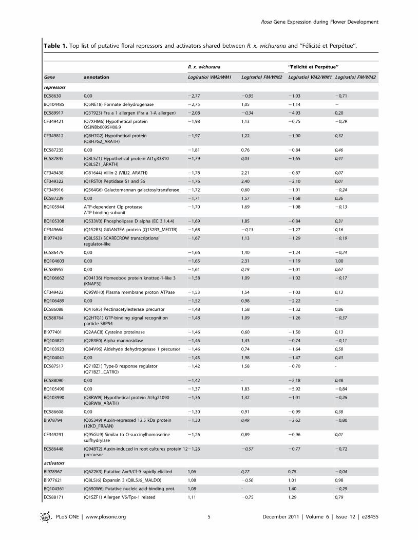
Table 1. Top list of putative floral repressors and activators shared between R. x. wichurana and ‘‘Felicite et Perpetue’’.
R. x. wichurana ‘‘Felicite et Perpetue’’
Gene annotation Log(ratio) VM2/WM1 Log(ratio) FM/WM2 Log(ratio) VM2/WM1 Log(ratio) FM/WM2
repressors
EC58630 0,00 22,77 20,95 21,03 20,71
BQ104485 (Q5NE18) Formate dehydrogenase 22,75 1,05 21,14 2
EC589917 (Q3T923) Fra a 1 allergen (Fra a 1-A allergen) 22,08 20,34 24,93 0,20
CF349421 (Q7XHM6) Hypothetical proteinOSJNBb0095H08.9
21,98 1,13 20,75 20,29
CF349812 (Q8H7G2) Hypothetical protein(Q8H7G2_ARATH)
21,97 1,22 21,00 0,32
EC587235 0,00 21,81 0,76 20,84 0,46
EC587845 (Q8L5Z1) Hypothetical protein At1g33810(Q8L5Z1_ARATH)
21,79 0,03 21,65 0,41
CF349438 (O81644) Villin-2 (VILI2_ARATH) 21,78 2,21 20,87 0,07
CF349322 (Q1RST0) Peptidase S1 and S6 21,76 2,40 22,10 0,01
CF349916 (Q564G6) Galactomannan galactosyltransferase 21,72 0,60 21,01 20,24
EC587239 0,00 21,71 1,57 21,68 0,36
BQ105944 ATP-dependent Clp proteaseATP-binding subunit
21,70 1,69 21,08 20,13
BQ105308 (Q533V0) Phospholipase D alpha (EC 3.1.4.4) 21,69 1,85 20,84 0,31
CF349664 (Q1S2R3) GIGANTEA protein (Q1S2R3_MEDTR) 21,68 20,13 21,27 0,16
BI977439 (Q8L553) SCARECROW transcriptionalregulator-like
21,67 1,13 21,29 20,19
EC586479 0,00 21,66 1,40 21,24 20,24
BQ104603 0,00 21,65 2,31 21,19 1,00
EC588955 0,00 21,61 0,19 21,01 0,67
BQ106662 (O04136) Homeobox protein knotted-1-like 3(KNAP3))
21,58 1,09 21,02 20,17
CF349422 (Q9SWH0) Plasma membrane proton ATPase 21,53 1,54 21,03 0,13
BQ106489 0,00 21,52 0,98 22,22 2
EC586088 (Q41695) Pectinacetylesterase precursor 21,48 1,58 21,32 0,86
EC588764 (Q2HTG1) GTP-binding signal recognitionparticle SRP54
21,48 1,09 21,26 20,37
BI977401 (Q2AAC8) Cysteine proteinase 21,46 0,60 21,50 0,13
BQ104821 (Q2R3E0) Alpha-mannosidase 21,46 1,43 20,74 20,11
BQ103923 (Q84V96) Aldehyde dehydrogenase 1 precursor 21,46 0,74 21,64 0,58
BQ104041 0,00 21,45 1,98 21,47 0,43
EC587517 (Q71BZ1) Type-B response regulator(Q71BZ1_CATRO)
21,42 1,58 20,70 -
EC588090 0,00 21,42 - 22,18 0,48
BQ105490 0,00 21,37 1,83 25,92 20,84
BQ103990 (Q8RWI9) Hypothetical protein At3g21090(Q8RWI9_ARATH)
21,36 1,32 21,01 20,26
EC586608 0,00 21,30 0,91 20,99 0,38
BI978794 (Q05349) Auxin-repressed 12.5 kDa protein(12KD_FRAAN)
21,30 0,49 22,62 20,80
CF349291 (Q9SGU9) Similar to O-succinylhomoserinesulfhydrylase
21,26 0,89 20,96 0,01
EC586448 (Q94BT2) Auxin-induced in root cultures protein 12precursor
21,26 20,57 20,77 20,72
activators
BI978967 (Q6Z2K3) Putative Avr9/Cf-9 rapidly elicited 1,06 0,27 0,75 20,04
BI977621 (Q8L5J6) Expansin 3 (Q8L5J6_MALDO) 1,08 20,50 1,01 0,98
BQ104361 (Q650W6) Putative nucleic acid-binding prot. 1,08 - 1,40 20,29
EC588171 (Q1SZF1) Allergen V5/Tpx-1 related 1,11 20,75 1,29 0,79
Rosa Gene Expression during Flower Development
PLoS ONE | www.plosone.org 5 December 2011 | Volume 6 | Issue 12 | e28455

ortholog (RhPI/MASAKO BP, AB038462), were among the genes
whose expression was up-regulated between stages 1+2 and 3+4 or
between stages 3+4 and 5. Interestingly, genes that are predicted
to have functions in cell wall remodelling, such as putative
extracellular lipases (BQ106293, EC586717, EC588243,
BI978064, BI977386, BQ105800), xyloglucan endotransglucosy-
lase/hydrolase 2 (XTH2, DQ320658) [34], expansins (BI977621,
EC589557), putative pectin esterase (BQ105504) and pectate lyase
(BQ103887, BQ105987) were up-regulated between stages 3+4
and 5. This result supports the idea that very active cell wall
remodeling coincides with the beginning of organ elongation that
occurs mainly at stage 5. A putative gibberellin 2-oxidase
(BQ105545) was up-regulated early during flower development.
In Arabidopsis, a similar up-regulation of genes implicated in
gibberellins metabolism and signaling have been described at early
floral development [35,36]. In agreement with previously
published data, our microarray analysis suggests that gibberellins
are important during early floral development of rose plants
[13,37]. Among the genes that showed strong down-regulation
between stages 1+2 and 3+4, we found the putative orthologues of
PERIANTHIA (PAN), AP1 and SOC1 (AGL20). In Arabidopsis, PAN,
AP1 and SOC1 are expressed in the floral meristem, but their
expression is down-regulated in the subsequent steps during floral
organs differentiation [36,38,39,40], hence in agreement with the
observed down-regulation of the rose homologues between flower
development stages 1+2 and 3+4.
Early to late floral development transitionSequences corresponding to 401 genes were detected as
differentially regulated between stages 5 and VP. Among these
genes, 233 were down-regulated and 168 were up-regulated (see
Table 3 for a selection of genes and Table S7 for full list). Genes
that exhibit strong similarities to genes involved in carotene,
flavonoid and anthocyanin biosynthesis are up-regulated between
stages 5 and VP. Among these genes, putative phytoene synthase
(BI979026), zeta carotene desaturase (CF349648), lycopene beta-
cyclase (BQ105122) are likely to be involved in carotenoid
biosynthesis. The expression of UDP-glucose anthocyanidin-o-
glucosyltransferase (AB201048/RhGT1), previously involved in
anthocyanin synthesis [41], was strongly up-regulated. A similar
strong up-regulation was observed for genes encoding putative
phenylalanine ammonia-lyase (BQ105227), chalcone synthase
(EC587811), flavonol synthase (AB038247) and anthocyanidin
synthase (BI977949) (Figure 3). Altogether, these genes are likely
good candidates involved in anthocyanins biosynthesis in rose
petals.
Interestingly, genes predicted to encode five putative cyclins
(EC586028, EC586517, EC587578, EC588351, and EC588489)
and a putative cyclin dependent kinase (EC589228) are strongly
down-regulated during floral organ morphogenesis. This down-
regulation may reflect the transition from mitotic growth to post-
mitotic growth where floral organs grow through cell expansion.
Recently, Vanneste et al. showed that the transcriptional down-
regulation of A2 type cyclins is a direct link between develop-
mental programming and cell-cycle exit in Arabidopsis thaliana [42].
Fifteen genes encoding putative transcription factors were up-
regulated, while nine were down-regulated. Among the up-
regulated transcription factors, we found the putative orthologue
of SHP (AB025643) [32] and a putative NAC domain protein
(BI978992, Figure 3). BI978992 is homologous to Arabidopsis
NAC2, a gene expressed in ovule integuments. The differential
expression of NAC2 between stages 5 and visible petals (VP)
suggests its putatively conserved function with the Arabidopsis
NAC2. Three putative MYB transcription factors were also up-
R. x. wichurana ‘‘Felicite et Perpetue’’
Gene annotation Log(ratio) VM2/WM1 Log(ratio) FM/WM2 Log(ratio) VM2/WM1 Log(ratio) FM/WM2
EC586116 0,00 1,12 21,17 1,14 0,34
EC589388 (Q1SHH7) Auxin responsive SAUR protein 1,14 20,02 1,52 0,47
BI978946 (Q93Z01) AT5g58730 1,20 21,03 1,55 20,17
RoAGL20 (Q7Y137) MADS-box protein PTM5 1,22 - 0,97 0,14
BQ105514 0,00 1,23 20,74 0,72 20,02
BI977348 (Q94AQ7) Hypothetical protein At5g11280 1,23 0,50 0,73 20,47
BQ103904 (Q41696) Cysteine protease precursor 1,25 22,35 2,55 0,20
EC589855 0,00 1,27 20,03 0,71 20,44
BI978115 (Q84W81) Hypothetical protein At5g49800 1,27 0,34 1,85 20,57
EC586690 Q2QXK7) F-box domain, putative 1,31 20,91 1,36 20,02
EC588294 (Q1S0D0) Glyoxalase/bleomycin resistance protein 1,32 20,46 0,77 1,19
EC588783 (Q9LUC1) Putative protein At3g14740 1,34 0,27 1,19 20,31
RoAP1a (Q283Q1) APETALA1 protein 1,38 20,40 2,06 0,88
EC587486 0,00 1,42 20,62 1,38 20,35
BI978732 (P32293) Auxin-induced protein 22A 1,47 20,36 1,27 1,28
BQ104100 0,00 1,55 20,94 1,49 1,03
BQ105108 (O65744) GDP dissociation inhibitor 1,63 23,09 2,13 0,17
Log(ratio) of intensities are represented, italicized numbers represent ratios for which the p-value of the Bonferroni test was higher than 0.05. -: no value could becalculated.doi:10.1371/journal.pone.0028455.t001
Table 1. Cont.
Rosa Gene Expression during Flower Development
PLoS ONE | www.plosone.org 6 December 2011 | Volume 6 | Issue 12 | e28455

regulated (CF349636, BQ104100 and BI978095, Figure 3). These
rose MYBs may be involved in organ elongation, as they share
about 67% protein sequence similarity with AtMYB21, known to
be involved in gibberellins/jasmonate-mediated control of stamen
filament elongation [43].
Late floral development456 genes were differentially regulated at least once during the
late phases of floral development, i.e. from visible petal (VP) stage
to senescent flower (SF) stage. Most of these genes showed similar
expression pattern when we compared stages VP to OF (open
flower) or stages VP to SF (See Table 4 for top list, and Table S8
for full data). This result indicates that the transcriptome becomes
less dynamic at senescence stages and thus not so many differences
are detected when comparing samples OF and SF to the VP
sample. Gene ontology analysis showed that among the up-
regulated genes, the three GO terms chlorophyll catabolic process,
heterocycle catabolic process and cellular nitrogen compound
catabolic process were significantly overrepresented as compared
to the whole annotated set; the four GO terms nucleus,
macromolecule biosynthetic process, intracellular non-mem-
brane-bounded organelle and ribonucleoprotein complex were
Figure 3. Real time quantitative RT-PCR (qPCR) analysis of six selected differentially expressed genes during rose floralorganogenesis, floral opening and senescence in R. chinensis cv. Old Blush. qPCR data (black histograms) are compared to the microarrayhybridization data (white histograms). Microarray data is presented regardless of Bonferroni test success. Each pair of histograms represent successivecomparisons between floral development stages 1+2, 3+4, 5, visible petals (VP), open flower (OF) and senescing flowers (SF).doi:10.1371/journal.pone.0028455.g003
Rosa Gene Expression during Flower Development
PLoS ONE | www.plosone.org 7 December 2011 | Volume 6 | Issue 12 | e28455

underrepresented. We could identify two genes encoding stay-
green protein homologues (BI978267 and BQ106457) that are
strongly up-regulated upon petal elongation and remain highly
expressed throughout the final petal senescing process. Stay-green
proteins have a major role in chlorophyll and photosynthetic
pigments degradation and have been repeatedly described to be
associated with the processes of fruit ripening and organ
senescence [44]. Surprisingly, no gene related to ethylene
biosynthesis or signaling was detected as differentially expressed
during late floral development. However the RbXTH1 and
RbEXPA1 genes, both induced during ethylene-triggered and field
abscission [34,45], were strongly up-regulated between VP and
OF stages and remained as such in senescing flowers. Among the
down-regulated genes, the two GO terms protein metabolic
process and plasma membrane were underrepresented as
compared to the whole set (whole microarray GO terms) and
the eight GO terms acyltransferase activity, acyl-carrier-protein
biosynthetic process, acyl carrier activity, cellular carbohydrate
metabolic process, polysaccharide metabolic process, fatty acid
biosynthetic process, lipase activity and defense response to fungus
were overrepresented (Table 5). The enrichment in the latter set
may represent a slowdown of general metabolic pathways at the
onset of flower senescence. Similar results were reported in A.
thaliana during organs senescence where a down-regulation of the
photosynthetic machinery accompanied by a reduction in
expression of many cell wall biosynthetic genes reflecting a
cessation of growth during senescence [46].
ConclusionsWe established a calendar of the floral initiation and
development for the rose and developed a rose microarray that
harbors sequence from genes expressed during the floral transition
and whole floral development process in Rosa sp, from initiation up
to senescing flowers. This microarray and the floral development
calendar were successfully used to identify genes whose expression
correlated with different flower development stages.These multiple
datasets represent an extensive study of rose floral development.
This resource can be helpful to select candidate genes potentially
involved in different horticultural traits, such as flowering, floral
architecture, scent production and emission, senescence and
abscission. We used the microarray developed herein to identify
genes whose expression is associated with some of these rose
important traits, such as flower initiation, development and
senescence. Rosa1_Affyarray harbors sequences from ESTs found
in petals of different rose genotypes [5,14] (http://urgi.versailles.
inra.fr/GnpSeq) and thus may be helpful to identify genes
associated with other rose traits such as scent biosynthesis and/
or emission genes. The rose is among the species that exhibit the
highest scent complexity [47–48] [12] and some scent biosynthesis
pathways are unique to the rose or not yet identified in other
model species including other members of the Rosaceae genus
[11,49]. QTLs have been identified to be associated to several
important traits of the rose [50]. However, the heterozygous
genome of the rose complicates the breeding programs to select for
several traits simultaneously. The identification of genes whose
expression correlates with important ornamental traits can
facilitate and accelerate candidate gene identification for rose
breeding by marker assisted selection or genomic selection. For
example, this dataset can provide researchers with a useful
resource on the expression of candidate genes within a given
mapping interval. Furthermore, the rapidly progressing high
throughput sequencing technologies should allow the generation of
precise genetic maps for the rose that could be combined to
refined transcriptomics approaches to identify the genes respon-
sible for important horticultural traits in the rose, and allow
subsequent marker-assisted selection.
Table 2. List of selected floral organogenesis associated genes in R. chinensis cv Old Blush.
R. chinensis cv Old Blush
Gene annotation Stages 3+4 vs 1+2 Stages 5 vs 3+4
AY803737 Rosa hybrid cultivar 1-aminocyclopropane-1-carboxylase synthase 2 (ACS2) 2,99 21,21
AB055966 Rosa rugosa MASAKO B3 mRNA for MADS-box protein, 2,67 1,04
AB025645 Rosa rugosa MASAKO C2 mRNA for MADS-box protein, 2,94 1,37
CF349463 (Q1S9M3) Lipase, active site (Q1S9M3_MEDTR) 2,68 -
BI978064 (Q9M8Y5) Putative GDSL-motif lipase/acylhydrolase (Q9M8Y5_ARATH) 2,10 1,17
BI977386 (Q9M8Y5) Putative GDSL-motif lipase/acylhydrolase (Q9M8Y5_ARATH) 1,99 1,08
EC586717 (Q1S3U7) Lipolytic enzyme, GDS-L (Q1S3U7_MEDTR) 1,69 1,16
BQ105800 (Q1SAY6) Lipolytic enzyme, GDSL (Q1SAY6_MEDTR) 2,76 0,91
DQ320658 Rosa6borboniana xyloglucan endotransglucosylase/hydrolase 2 (Xth2) 2,55 0,74
BI977621 (Q8L5J6) Expansin 3 (Q8L5J6_MALDO) 20,89 1,26
EC589557 (Q9SBT1) Expansin (Q9SBT1_FRAAN) 0,48 1,20
BQ105987 (Q94FT6) Pectate lyase B (Fragment) (Q94FT6_FRAAN) 0,75 1,27
BQ103887 (Q52PJ2) Ripening-related pectate lyase (Q52PJ2_MANIN) 1,21 1,11
BQ105504 (Q7X9B1) Pectinesterase (EC 3.1.1.11) (Q7X9B1_FRAAN) 1,44 1,35
BQ105545 (Q4W8C3) Gibberellin 2-oxidase (Q4W8C3_PHAAN) 0,42 21,35
RoPAN (Q9SX27) Putative bZIP transcription factor, PERIANTHIA (Q9SX27_ARATH) 21,94 22,08
RoAGL20 (Q7Y137) POPTM (Q7Y137) MADS-box protein PTM5 22,77 20,03
RoAP1b (Q2XUP6) MADS-box protein 20,98 23,15
Log(ratio) of intensities are represented, italicized numbers represent ratios for which the p-value of the Bonferroni test was higher than 0.05.doi:10.1371/journal.pone.0028455.t002
Rosa Gene Expression during Flower Development
PLoS ONE | www.plosone.org 8 December 2011 | Volume 6 | Issue 12 | e28455

Materials and Methods
Plant materialR. wichurana was obtained from ‘Jardin de Bagatelle’ (Paris,
France) and R. x hybrida cv. Felicite et Perpetue from the Loubert
Nursery (Rosier sur Loire, France). Plants were grown outdoors
on their own roots as previously described [13]. In spring, at
different time points (see results), terminal parts of the growing
shoot were harvested and partly dissected (removal of young
leaves). R. chinensis cv. Old Blush was propagated by cuttings
from the Lyon Botanical Garden. Plants were grown in the
greenhouse with 16 h/8 h day/night and 25uC/14uC day/night
temperature. No specific permits were required for the
described filed studies, no specific permissions were required
for these locations, the location is not privately owned or
protected, and the field studies did not involve endangered or
protected species.
Light microscopy and SEM imaging of meristems andearly flower development
Samples were dissected under a binocular stereomicroscope and
then fixed in 4% glutaraldehyde (v/v) in 0.1 M phosphate buffer
(pH 7.2) for 2 h at 4uC under vacuum. Samples were dehydrated
in a graded ethanol series and embedded in Technovit 7100 [51].
Sections of 1.5 to 2.0 mm (Leica RM 2165 microtome) were
stained with toluidine blue and examined under an Olympus
BH2-RFC microscope coupled to a 3CCD Sony camera.
For scanning electron microscopy, terminal part of the shoot
was carefully dissected. After a fixation in 4% glutaraldehyde (v/v),
followed by post-fixation with osmium tetroxide, the sample was
dehydrated in a graded alcohol series and in acetone. Dehydration
was completed by critical point drying. Sample were then coated
with gold (MED 020 BALTEC) and observed with a JEOL JSM-
63017 scanning electron microscope.
RNA samples preparationTwo independent biological replicates were produced for each
samples at different stages. For each biological repetition and each
point, RNA samples were obtained by pooling vegetative or floral
tissue from at least five different plants. For R. chinensis cv. Old
Blush samples, meristems or flowers were dissected and collected
individually on plants at developmental growth stages, cultivated
in greenhouse conditions as previously described [55]. For R.
wichurana and R. x hybrida cv. Felicite et Perpetue, RNA was
extracted from non-dissected buds, including either the vegetative
meristem and its surrounding leaves or the pre-floral/floral
meristem and its surrounding leaves and bracts.Total RNA was
extracted using RNeasy Plant Mini Kit (Qiagen) according to the
supplier’s instructions.
AFFYMETRIX Array hybridizationRNA samples were checked for their integrity on The Agilent
2100 bioanalyzer according to the Agilent Technologies (Wald-
broon, Germany).
Table 3. List of selected genes associated with early to late flower development in R. chinensis cv Old Blush.
R. chinensis cv Old Blush
Gene annotation 5 vs PA
BI978095 (P93474) Myb26 8,00
BI978992 (Q50J79) NAM-like protein 5,28
AB038247 Rosa hybrid cultivar ‘Kardinal’ FLS mRNA for flavonol synthase 4,67
BQ105122 (Q9SEA0) Lycopene beta-cyclase 4,30
EC587811 (Q84UT9) Chalcone synthase 3,27
BQ104100 MYB domain class transcription factor 3,01
AB025643 Rosa rugosa MASAKO D1 mRNA for MADS-box protein. 3,00
CF349648 (Q5W5X6) Zeta-carotene desaturase ZDS2 2,97
BI979026 (Q2VEY1) Putative phytoene synthase 2,89
BI977949 (Q5UL09) Anthocyanidin synthase 2,18
AB201048 RhGT1 UDP-glucose: anthocyanidin 5,3-O-glucosyltransferase, 2,18
CF349636 (Q9ATD1) GHMYB9 2,12
BQ105227 (Q9M567) Phenylalanine ammonia-lyase 2 2,11
EC586028 (Q9SNV1) Cyclin D3a (Fragment) 22,12
AB201051 RhGT4 mRNA UDP-glucose: flavonol 3-O-glucosyltransferase 22,18
EC587392 (Q8S342) Putative anthocyanidine rhamnosyl-transferase 22,45
EC587578 (Q6T2Z6) Cyclin d3 23,83
EC586734 (Q08733) Aquaporin PIP1.3 24,52
RhCyc2 (Q9SBQ4) CYCB1-1 protein 24,65
EC588351 (Q9SBQ4) CYCB1-1 protein 24,72
EC58848 (P93557) Mitotic cyclin 24,77
EC589228 (Q94EX2) At1g76540/cyclin dependent kinase 25,05
EC586517 (Q4JF78) Cyclin-dependent kinase B 25,26
Log(ratio) of intensities are represented, for all ratios the p-value of the Bonferroni test was lower than 0.05.doi:10.1371/journal.pone.0028455.t003
Rosa Gene Expression during Flower Development
PLoS ONE | www.plosone.org 9 December 2011 | Volume 6 | Issue 12 | e28455
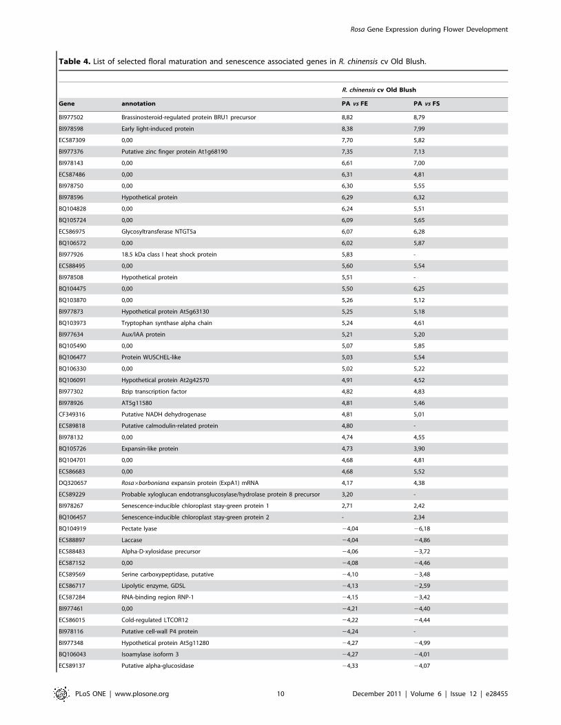
Table 4. List of selected floral maturation and senescence associated genes in R. chinensis cv Old Blush.
R. chinensis cv Old Blush
Gene annotation PA vs FE PA vs FS
BI977502 Brassinosteroid-regulated protein BRU1 precursor 8,82 8,79
BI978598 Early light-induced protein 8,38 7,99
EC587309 0,00 7,70 5,82
BI977376 Putative zinc finger protein At1g68190 7,35 7,13
BI978143 0,00 6,61 7,00
EC587486 0,00 6,31 4,81
BI978750 0,00 6,30 5,55
BI978596 Hypothetical protein 6,29 6,32
BQ104828 0,00 6,24 5,51
BQ105724 0,00 6,09 5,65
EC586975 Glycosyltransferase NTGT5a 6,07 6,28
BQ106572 0,00 6,02 5,87
BI977926 18.5 kDa class I heat shock protein 5,83 -
EC588495 0,00 5,60 5,54
BI978508 Hypothetical protein 5,51 -
BQ104475 0,00 5,50 6,25
BQ103870 0,00 5,26 5,12
BI977873 Hypothetical protein At5g63130 5,25 5,18
BQ103973 Tryptophan synthase alpha chain 5,24 4,61
BI977634 Aux/IAA protein 5,21 5,20
BQ105490 0,00 5,07 5,85
BQ106477 Protein WUSCHEL-like 5,03 5,54
BQ106330 0,00 5,02 5,22
BQ106091 Hypothetical protein At2g42570 4,91 4,52
BI977302 Bzip transcription factor 4,82 4,83
BI978926 AT5g11580 4,81 5,46
CF349316 Putative NADH dehydrogenase 4,81 5,01
EC589818 Putative calmodulin-related protein 4,80 -
BI978132 0,00 4,74 4,55
BQ105726 Expansin-like protein 4,73 3,90
BQ104701 0,00 4,68 4,81
EC586683 0,00 4,68 5,52
DQ320657 Rosa6borboniana expansin protein (ExpA1) mRNA 4,17 4,38
EC589229 Probable xyloglucan endotransglucosylase/hydrolase protein 8 precursor 3,20 -
BI978267 Senescence-inducible chloroplast stay-green protein 1 2,71 2,42
BQ106457 Senescence-inducible chloroplast stay-green protein 2 - 2,34
BQ104919 Pectate lyase 24,04 26,18
EC588897 Laccase 24,04 24,86
EC588483 Alpha-D-xylosidase precursor 24,06 23,72
EC587152 0,00 24,08 24,46
EC589569 Serine carboxypeptidase, putative 24,10 23,48
EC586717 Lipolytic enzyme, GDSL 24,13 22,59
EC587284 RNA-binding region RNP-1 24,15 23,42
BI977461 0,00 24,21 24,40
EC586015 Cold-regulated LTCOR12 24,22 24,44
BI978116 Putative cell-wall P4 protein 24,24 -
BI977348 Hypothetical protein At5g11280 24,27 24,99
BQ106043 Isoamylase isoform 3 24,27 24,01
EC589137 Putative alpha-glucosidase 24,33 24,07
Rosa Gene Expression during Flower Development
PLoS ONE | www.plosone.org 10 December 2011 | Volume 6 | Issue 12 | e28455

Two mg of total RNA were used to synthesize biotin-labeled
cRNAs with the One-cycle cDNA synthesis kit (Affymetrix, Santa
Clara, CA). Superscript II reverse transcriptase and T7-oligo (dT)
primers were used to synthesize the single strand of cDNA at 42uCduring 1 hour followed by the synthesis of the double stranded
cDNA by using DNA ligase, DNA polymerase I and RNaseH
during 2 hours at 16uC. Clean up of the double-stranded cDNA
was performed with Sample Cleanup Module (Affymetrix) followed
by in vitro transcription (IVT) in presence of biotin-labeled UTP
using GeneChipH IVT labelling Kit (Affymetrix). Quantity of the
labelled-cRNA with RiboGreenH RNA Quantification Reagent
(Turner Biosystems, Sunnyvale, CA) was determined after cleanup
by the Sample Cleanup Module (Affymetrix). Fragmentation of
10 mg of labelled-cRNA was carried out for 35 minutes at 94uC,
followed by hybridization during 16 hours at 45uC to Affymetrix
GeneChipH Rosa1 Genome Array representing approximately
4869 genes. After hybridization, the arrays were washed with 2
different buffers (stringent: 66 SSPE, 0.01% Tween-20 and non-
stringent: 100 mM MES, 0.1 M [Na+], 0.01% Tween-20) and
stained with a complex solution including Streptavidin R-
Phycoerythrin conjugate (Invitrogen/molecular probes, Carlsbad,
CA) and anti Streptavidin biotinylated antibody (Vectors laborato-
ries, Burlingame, CA). The washing and staining steps were
performed in a GeneChipH Fluidics Station 450 (Affymetrix). The
Affymetrix GeneChipH Rosa1 Genome Arrays were finally scanned
with the GeneChipH Scanner 3000 7G piloted by the GeneChipHOperating Software (GCOS).
Statistical Analysis of Microarray DataThe data were normalized with the gcrma algorithm [52],
available in the Bioconductor package [53]. To determine differen-
tially expressed genes, we performed a usual two group t-test that
assumes equal variance between groups. The variance of the gene
expression per group is a homoscedastic variance, where genes
displaying extremes of variance (too small or too large) were excluded.
The raw P values were adjusted by the Bonferroni method, which
controls the Family Wise Error Rate (FWER) [54]. A gene is declared
differentially expressed if the Bonferroni P-Value is less than 0.05.
Data DepositionAll this steps were performed on Affymetrix plateform at INRA-
URGV, Evry. The raw. CEL files were imported in R software for
data analysis. All raw and normalized data are available through
the CATdb database (AFFY_PetalDvt_Lyon_Rose, [55]) and
from the Gene Expression Omnibus (GEO) repository at the
National Center for Biotechnology Information (NCBI) [56],
accession number GSE18357.
Validation of genes expression using quantitative real-time PCR
Only genes that were involved in floral development were analyzed
for microarray data validation. One microgram total RNA (treated
with DNAse) was used in a reverse transcription assay with RevertAid
M-MuLV Reverse Transcriptase (Fermentas, Burlington, Ontario).
Target cDNAs were quantified by qPCR using FastStart universal
SYBR green master (Roche, Basel, Switzerland) on a Step-OnePlus
Real-Time PCR System (Applied Biosystems, Foster City, CA USA).
Expression levels were normalized with RhaTubuline, RhGAPDH and
RhEF1a reference genes. These genes were validated as reference genes
using the GeNorm application [57]. Three independent biological
replicates (pools of dissected flowers from at least 5 different plants)
were used for each experiment and two qPCR technical replicates were
R. chinensis cv Old Blush
Gene annotation PA vs FE PA vs FS
EC587785 0,00 24,37 23,91
EC586984 Putative beta-expansin 24,38 24,99
CF349422 Plasma membrane proton ATPase 24,41 23,64
BI977751 0,00 24,49 24,43
EC589098 Phosphoethanolamine N-methyltransferase 1 24,49 25,79
BQ106293 GDSL-motif lipase/hydrolase-like protein 24,51 26,32
CF349724 Glucosyltransferase-like protein 24,74 24,10
BQ106328 Pathogenesis-related transcriptional factor and ERF 25,10 -
EC586884 Proline-rich protein APG-like 25,11 23,72
CF349712 Senescence-inducible gene protein 25,12 25,47
BI978135 Plant lipid transfer protein/Par allergen 25,16 27,04
CF349692 Putative alcohol oxidase 25,17 26,66
EC588080 Hypothetical protein (At2g35760/T20F21.5) 25,32 25,20
BI977262 Putative lipase 25,48 26,04
CF349791 Globulin-like protein (Fragment) 25,67 25,53
AB121046 phloroglucinol O-methyltransferase, complete cds 26,13 25,06
BI978206 0,00 28,51 -
BI978064 Putative GDSL-motif lipase/acylhydrolase 210,47 210,51
BI977386 Putative GDSL-motif lipase/acylhydrolase 211,60 211,42
Log(ratio) of intensities are represented, for all ratios the p-value of the Bonferroni test was lower than 0.05.doi:10.1371/journal.pone.0028455.t004
Table 4. Cont.
Rosa Gene Expression during Flower Development
PLoS ONE | www.plosone.org 11 December 2011 | Volume 6 | Issue 12 | e28455

performed for each biological replicate. Primer sequences are available
in Table S9. The correlation between the microarray results, and those
obtained by qPCR was assessed by calculating the Pearson’s product
moment correlation coefficient [58,59].
Supporting Information
Figure S1 Real time quantitative RT-PCR (qPCR)analysis of 18 selected differentially expressed genesduring rose floral organogenesis and senescence in R.chinensis cv Old Blush.
(TIFF)
Table S1 Full array sequences annotation and ontology.
(XLSX)
Table S2 Genes differentially expressed during floralinitiation in R. wichurana.
(XLSX)
Table S3 Genes differentially expressed during floralinitiation in R. x hybrida cv. Felicite et Perpetue.
(XLSX)
Table S4 List of genes repressed (A) or activated (B)during flower initiation.
(XLSX)
Table S5 Microarray and qRT-PCR results of 25selected genes with their replicate-level Pearson corre-lation.
(DOCX)
Table S6 Genes differentially expressed during earlyfloral organogenesis in R. chinensis cv. Old Blush.
(XLSX)
Table S7 Genes differentially expressed during floralorgan elongation in R. chinensis cv. Old Blush.
(XLSX)
Table S8 Genes differentially expressed during floweropening and senescence in R. chinensis cv. Old Blush.
(XLSX)
Table S9 Primers used in this study.
(DOC)
Acknowledgments
We thank Judit Szecsi and Sylvie Baudino for critical reading of the
manuscript. We thank Alexis Lacroix, Isabelle Desbouchages, Priscilla
Angelot and N. Dousset and J. Chameau taking care of the plants, M.
Thellier and Michel Chevalier for the histological analysis, S/Georgeault
and R. Filmontt for the SEM studies.
Table 5. Gossip analysis of GO terms enrichment in late flower development dataset (genes that are differentially expressed atleast once during floral maturation and senescence).
GO Term Name FDR FWERsingle testp-Value
# in testgroup
# inreferencegroup
# nonannotedtest
# nonannotedreferencegroup Over/Under
Late floral developmentupregulated genes
GO:0005634 nucleus 0.0 0.0 0.012 0 107 56 1290 under
GO:0009059 macromoleculebiosynthetic process
0.0 0.0 0.028 0 88 56 1309 under
GO:0043232 intracellular non-membrane-boundedorganelle
0.0 0.0 0.030 0 86 56 1311 under
GO:0030529 ribonucleoproteincomplex
0.0 0.0 0.030 0 84 56 1313 under
GO:0015996 chlorophyll catabolicprocess
0.008 0.008 5.43E-5 3 0 53 1397 over
GO:0046700 heterocycle catabolicprocess
0.028 0.062 5.13E-4 3 2 53 1395 over
GO:0044270 cellular nitrogencompound catabolicprocess
0.028 0.062 5.13E-4 3 2 53 1395 over
Late floral developmentdownregulated genes
GO:0019538 protein metabolicprocess
0.0 0.0 0.009 5 225 73 1150 under
GO:0005886 plasma membrane 0.012 0.006 0.013 4 192 74 1183 under
GO:0008415 acyltransferase activity 0.016 0.025 1.62E-4 7 17 71 1358 over
GO:0042967 acyl-carrier-proteinbiosynthetic process
0.016 0.028 2.15E-4 7 18 71 1357 over
GO:0000036 acyl carrier activity 0.027 0.079 5.73E-4 3 1 75 1374 over
GO:0044262 cellular carbohydratemetabolic process
0.027 0.084 6.73E-4 15 99 63 1276 over
GO:0005976 polysaccharidemetabolic process
0.027 0.086 7.08E-4 10 48 68 1327 over
The reference group that was used corresponds to the full annotated sequences (sequences with GO terms) of the microarray.doi:10.1371/journal.pone.0028455.t005
Rosa Gene Expression during Flower Development
PLoS ONE | www.plosone.org 12 December 2011 | Volume 6 | Issue 12 | e28455

Author Contributions
Conceived and designed the experiments: MB AD OR. Performed the
experiments: AD AR OR SB AC MM YP SHY JJ TT VB MLMM SJ JPR
PV MLB FF. Analyzed the data: AD OR SB MLMM PV MLB FF MB.
Contributed reagents/materials/analysis tools: FL. Wrote the paper: AD
MB.
References
1. Martin M, Piola F, Chessel D, Jay M, Heizmann P (2001) The domestication
process of the Modern Rose: genetic structure and allelic composition of the rose
complex. Theoretical-and-Applied-Genetics 102: 398–404.
2. De Vries DP, Dubois L (1996) Rose breeding: past, present, prospects. Acta
Horticulturae 424: 241–248.
3. Gudin S (2001) Rose breeding technologies. Acta Horticulturae 547: 23–26.
4. Reynders-Aloisi S, Bollereau P (1996) Characterisation of genetic diversity in
genus Rosa by Randomly Amplified Polymorphic DNA. Acta Horticulturae
424: 253–259.
5. Channeliere S, Riviere S, Scalliet G, Szecsi J, Jullien F, et al. (2002) Analysis of
gene expression in rose petals using expressed sequence tags. FEBS Lett 515:
35–38.
6. Foucher F, Chevalier M, Corre C, Soufflet-Freslon V, Legeai F, et al. (2008)
New resources for studying the rose flowering process. Genome 51: 827–837.
7. Guterman I, Masci T, Chen X, Negre F, Pichersky E, et al. (2006) Generation of
phenylpropanoid pathway-derived volatiles in transgenic plants: rose alcohol
acetyltransferase produces phenylethyl acetate and benzyl acetate in petunia
flowers. Plant Mol Biol 60: 555–563.
8. Lavid N, Wang J, Shalit M, Guterman I, Bar E, et al. (2002) O-
methyltransferases involved in the biosynthesis of volatile phenolic derivatives
in rose petals. Plant Physiol 129: 1899–1907.
9. Scalliet G, Journot N, Jullien F, Baudino S, Magnard JL, et al. (2002)
Biosynthesis of the major scent components 3,5-dimethoxytoluene and 1,3,5-
trimethoxybenzene by novel rose O-methyltransferases. Febs Letters 523: PII
S0014-5793(0002)02956-02953.
10. Scalliet G, Lionnet C, Le Bechec M, Dutron L, Magnard JL, et al. (2006) Role of
petal-specific orcinol O-methyltransferases in the evolution of rose scent. Plant
Physiology 140: 18–29.
11. Scalliet G, Piola F, Douady CJ, Rety S, Raymond O, et al. (2008) Scent
evolution in Chinese roses. Proceedings Of The National Academy Of Sciences
Of The United States Of America 105: 5927–5932.
12. Shalit M, Guterman I, Volpin H, Bar E, Tamari T, et al. (2003) Volatile ester
formation in roses. Identification of an acetyl-coenzyme A. Geraniol/Citronellol
acetyltransferase in developing rose petals. Plant Physiol 131: 1868–1876.
13. Remay A, Lalanne D, Thouroude T, Le Couviour F, Hibrand-Saint Oyant L,
et al. (2009) A survey of flowering genes reveals the role of gibberellins in floral
control in rose. Theor Appl Genet 119: 767–781.
14. Guterman I, Shalit M, Menda N, Piestun D, Dafny-Yelin M, et al. (2002) Rose
scent: Genomics approach to discovering novel floral fragrance-related genes.
Plant Cell 14: 2325–2338.
15. Guterman I, Masci T, Chen XL, Negre F, Pichersky E, et al. (2006) Generation
of phenylpropanoid pathway-derived volatiles in transgenic plants: Rose alcohol
acetyltransferase produces phenylethyl acetate and benzyl acetate in petunia
flowers. Plant Molecular Biology 60: 555–563.
16. Gudin S (2000) Rose: Genetics and breeding. In: Plant Breeding Reviews 17:
159–189.
17. Krussmann G (1981) The complete book of roses. Portland: Timber Press. xii,.
436 p.
18. Smyth DR, Bowman JL, Meyerowitz EM (1990) Early flower development in
Arabidopsis. Plant Cell 2: 755–767.
19. Chimonidou D (2003) Morphology and Anatomy: ‘Flower Development and
Abscission zone’. ENCYCLOPEDIA OF ROSE SCIENCE. Amesterdam:
Elsevier Academic Press. pp 504–512.
20. Foster T, Johnston R, Seleznyova A (2003) A morphological and quantitative
characterization of early floral development in apple (Malus6domestica Borkh.).
Ann Bot 92: 199–206.
21. Gotz S, Garcia-Gomez JM, Terol J, Williams TD, Nagaraj SH, et al. (2008)
High-throughput functional annotation and data mining with the Blast2GO
suite. Nucleic Acids Res 36: 3420–3435.
22. Amasino R (2010) Seasonal and developmental timing of flowering. Plant J 61:
1001–1013.
23. Fornara F, de Montaigu A, Coupland G (2010) SnapShot: Control of flowering
in Arabidopsis. Cell 141: 550, 550 e551–552.
24. Bennett MJ, Swarup R, Perry P, Hagenbeek D, Van Der Straeten D, et al.
(2007) Ethylene upregulates auxin biosynthesis in Arabidopsis seedlings to
enhance inhibition of root cell elongation. Plant Cell 19: 2186–2196.
25. Kang BG, Newcomb W, Burg SP (1971) Mechanism of Auxin-Induced Ethylene
Production. Plant Physiology 47: 504–&.
26. Ma N, Tan H, Liu X, Xue J, Li Y, et al. (2006) Transcriptional regulation of
ethylene receptor and CTR genes involved in ethylene-induced flower opening
in cut rose (Rosa hybrida) cv. Samantha. J Exp Bot 57: 2763–2773.
27. Helariutta Y, Bishopp A, Mahonen AP (2006) Signs of change: hormone
receptors that regulate plant development. Development 133: 1857–1869.
28. Theologis A, Tsuchisaka A, Yu GX, Jin HL, Alonso JM, et al. (2009) A
Combinatorial Interplay Among the 1-Aminocyclopropane-1-Carboxylate
Isoforms Regulates Ethylene Biosynthesis in Arabidopsis thaliana. Genetics
183: 979–1003.
29. Tsuchisaka A, Theologis A (2004) Heterodimeric interactions among the 1-
amino-cyclopropane-1-carboxylate synthase polypeptides encoded by theArabidopsis gene family. Proc Natl Acad Sci U S A 101: 2275–2280.
30. Yamagami T, Tsuchisaka A, Yamada K, Haddon WF, Harden LA, et al. (2003)Biochemical diversity among the 1-amino-cyclopropane-1-carboxylate synthase
isozymes encoded by the Arabidopsis gene family. J Biol Chem 278:
49102–49112.
31. Dubois A, Raymond O, Maene M, Baudino S, Langlade NB, et al. (2010)
Tinkering with the C-function: a molecular frame for the selection of doubleflowers in cultivated roses. PLoS One 5: e9288.
32. Kitahara K, Hibino Y, Aida R, Matsumoto S (2004) Ectopic expression of therose AGAMOUS-like MADS-box genes ‘MASAKO C1 and D1’ causes similar
homeotic transformation of sepal and petal in Arabidopsis and sepal in Torenia.Plant Science 166: 1245–1252.
33. Kitahara K, Hirai S, Fukui H, Matsumoto S (2001) Rose MADS-box genes‘MASAKO BP and B3’ homologous to class B floral identity genes. Plant
Science 161: 549–557.
34. Singh AP, Tripathi SK, Nath P, Sane AP (2011) Petal abscission in rose is
associated with the differential expression of two ethylene-responsive xyloglucan
endotransglucosylase/hydrolase genes, RbXTH1 and RbXTH2. J Exp Bot.
35. Kaufmann K, Wellmer F, Muino JM, Ferrier T, Wuest SE, et al. (2010)
Orchestration of floral initiation by APETALA1. Science 328: 85–89.
36. Wellmer F, Alves-Ferreira M, Dubois A, Riechmann JL, Meyerowitz EM (2006)
Genome-wide analysis of gene expression during early Arabidopsis flowerdevelopment. PLoS Genet 2: e117.
37. Roberts AV, Blake PS, Lewis R, Taylor JM, Dunstan DI (1999) The Effect ofGibberellins on Flowering in Roses. J Plant Growth Regul 18: 113–119.
38. Borner R, Kampmann G, Chandler J, Gleissner R, Wisman E, et al. (2000) AMADS domain gene involved in the transition to flowering in Arabidopsis.
Plant J 24: 591–599.
39. Das P, Ito T, Wellmer F, Vernoux T, Dedieu A, et al. (2009) Floral stem cell
termination involves the direct regulation of AGAMOUS by PERIANTHIA.Development 136: 1605–1611.
40. Maier AT, Stehling-Sun S, Wollmann H, Demar M, Hong RL, et al. (2009)Dual roles of the bZIP transcription factor PERIANTHIA in the control of floral
architecture and homeotic gene expression. Development 136: 1613–1620.
41. Ogata J, Kanno Y, Itoh Y, Tsugawa H, Suzuki M (2005) Plant biochemistry:
anthocyanin biosynthesis in roses. Nature 435: 757–758.
42. Vanneste S, Coppens F, Lee E, Donner TJ, Xie Z, et al. (2011) Developmental
regulation of CYCA2s contributes to tissue-specific proliferation in Arabidopsis.EMBO J 30: 3430–3441.
43. Cheng H, Song S, Xiao L, Soo HM, Cheng Z, et al. (2009) Gibberellin actsthrough jasmonate to control the expression of MYB21, MYB24, and MYB57 to
promote stamen filament growth in Arabidopsis. PLoS Genet 5: e1000440.
44. Hortensteiner S (2009) Stay-green regulates chlorophyll and chlorophyll-binding
protein degradation during senescence. Trends Plant Sci 14: 155–162.
45. Nath P, Sane AP, Tripathi SK (2007) Petal abscission in rose (Rosa bourboniana
var Gruss an Teplitz) is associated with the enhanced expression of an alpha
expansin gene, RbEXPA1. Plant Science 172: 481–487.
46. Stead A, Rogers HJ, Roberts JA, Wagstaff C (2007) Programmed cell death
during floral development and senescence. Comparative Biochemistry andPhysiology a-Molecular & Integrative Physiology 146: S199–S200.
47. Kovats ES (1987) Composition of essential oils: Part 7. Bulgarian oil of rose(Rosa Damascena mill.). Journal of Chromatography A 406: 185–222.
48. Nakamura S (1987) Scent and component analysis of the Hybrid Tea Rose.Perfumer & flavorist 112: 43–45.
49. Kaminaga Y, Schnepp J, Peel G, Kish CM, Ben-Nissan G, et al. (2006) Plantphenylacetaldehyde synthase is a bifunctional homotetrameric enzyme that
catalyzes phenylalanine decarboxylation and oxidation. J Biol Chem 281:23357–23366.
50. Spiller M, Berger RG, Debener T (2010) Genetic dissection of scent metabolicprofiles in diploid rose populations. Theor Appl Genet 120: 1461–1471.
51. Baayen RP, Kroes GMLW, Lange W (1998) Histology of root rot of flaxseedlings (Linum usitatissimum) infected by Fusarium oxysporum f.sp. lini.
European Journal of Plant Pathology 104: 725–736.
52. Irizarry RA, Ooi SL, Wu Z, Boeke JD (2003) Use of mixture models in a
microarray-based screening procedure for detecting differentially represented
yeast mutants. Stat Appl Genet Mol Biol 2: Article1.
53. Gentleman R, Carey V (2002) Bioconductor. RNews 2: 1116.
54. Ge Y, Dudoit S, Speed TP (2003) Resampling-based multiple testing for
microarray data analysis. TEST 12: 1–44.
55. Gagnot S, Tamby JP, Martin-Magniette ML, Bitton F, Taconnat L, et al. (2008)
CATdb: a public access to Arabidopsis transcriptome data from the URGV-
CATMA platform. Nucleic Acids Research 36: D986–D990.
Rosa Gene Expression during Flower Development
PLoS ONE | www.plosone.org 13 December 2011 | Volume 6 | Issue 12 | e28455

56. Barrett T, Troup DB, Wilhite SE, Ledoux P, Rudnev D, et al. (2007) NCBI
GEO: mining tens of millions of expression profiles–database and tools update.Nucleic Acids Res 35: D760–765.
57. Speleman F, Vandesompele J, De Preter K, Pattyn F, Poppe B, et al. (2002)
Accurate normalization of real-time quantitative RT-PCR data by geometricaveraging of multiple internal control genes. Genome Biology 3.
58. Coppack SW (1990) Limitations of the Pearson product-moment correlation.
Clin Sci (Lond) 79: 287.
59. Liu Y, Meng Q, Chen R, Wang J, Jiang S, et al. (2004) A new method to
evaluate the similarity of chromatographic fingerprints: weighted pearson
product-moment correlation coefficient. J Chromatogr Sci 42: 545–550.
Rosa Gene Expression during Flower Development
PLoS ONE | www.plosone.org 14 December 2011 | Volume 6 | Issue 12 | e28455
Related Documents