2850–2859 Nucleic Acids Research, 2001, Vol. 29, No. 13 © 2001 Oxford University Press Genome-wide detection of alternative splicing in expressed sequences of human genes Barmak Modrek, Alissa Resch, Catherine Grasso and Christopher Lee* Department of Chemistry and Biochemistry, University of California, 611 Charles E. Young Drive East, Los Angeles, CA 90095-1570, USA Received January 26, 2001; Revised and Accepted May 5, 2001 ABSTRACT We have identified 6201 alternative splice relation- ships in human genes, through a genome-wide analysis of expressed sequence tags (ESTs). Starting with ∼2.1 million human mRNA and EST sequences, we mapped expressed sequences onto the draft human genome sequence and only accepted splices that obeyed the standard splice site consensus. A large fraction (47%) of these were observed multiple times, indicating that they comprise a substantial fraction of the mRNA species. The vast majority of the detected alternative forms appear to be novel, and produce highly specific, biologically meaningful control of function in both known and novel human genes, e.g. specific removal of the lysosomal targeting signal from HLA-DM β chain, replacement of the C-terminal transmembrane domain and cytoplasmic tail in an FC receptor β chain homolog with a different transmembrane domain and cytoplasmic tail, likely modulating its signal transduction activity. Our data indicate that a large proportion of human genes, probably 42% or more, are alternatively spliced, but that this appears to be observed mainly in certain types of molecules (e.g. cell surface receptors) and systemic functions, particularly the immune system and nervous system. These results provide a comprehensive dataset for understanding the role of alternative splicing in the human genome, accessible at http://www.bioinfor- matics.ucla.edu/HASDB. INTRODUCTION Alternative splicing is an important mechanism for modulating gene function. It can change how a gene acts in different tissues and developmental states by generating distinct mRNA isoforms composed of different selections of exons. Alterna- tive splicing has been implicated in many processes, including sex determination (1), apoptosis (2) and acoustic tuning in the ear (3). Recently, it has been suggested that if alternative splicing is widespread in the human genome, it could represent a relatively efficient expansion of the genome’s ‘vocabulary’ of variant genes, by producing multiple functional forms of many genes. Its functional implications can be simple, gener- ating a single alternative form, or can produce remarkable diversity. In the Drosophila gene Dscam, combinatorial alter- native splicing of ‘cassettes’ of exons reminiscent of the combinatorial generation of immunoglobulin diversity, produces thousands of distinct functional isoforms (4). This gene, homologous to the human gene for Down’s syndrome cell adhesion molecule (DSCAM), appears to be involved in neuronal guidance, where such diversity could be useful as a molecular ‘address’. Alternative splicing has been studied intensively in hundreds of human genes (1,5), and it appears to be widespread, occurring in 5–30% of human genes (6,7) or perhaps as many as 35–40% (8,9). Recently, it has been reported that alternative splicing can be detected in expressed sequence tag (EST) sequencing (9) and has been analyzed in a collection of full-length mRNAs (8). Based upon estimates of the total number of human genes (10,11), it is likely that at least 10 000–20 000 human genes are alternatively spliced. However, currently only 899 alterna- tively spliced human genes are catalogued in the Alternative Splicing Database of Mammals (AsMamDB) (12). We have performed a genome-wide analysis of alternative splicing based on human expressed sequence data, which greatly expands our knowledge of this central function in human molec- ular biology (Table 1). We have identified tens of thousands of splices, and thousands of alternative splices, in several thousand human genes. We have mapped all of these onto the draft human genome sequence, and verified that the putative splice junctions detected in the expressed sequences map onto genomic exon–intron junctions that match the known splice site consensus. Based on this genome-wide analysis of gene structure and alternative splicing, we have constructed a Human Alternative Splicing Database, at http://www.bioinformatics.ucla.edu/HASDB. In this paper we also show how our database can be used to study the impact of alternative splicing on protein function. We present an initial analysis of the patterns and functional role of alternative splicing across the human genome. As we seek to show with examples in this paper, our data- base could be a useful resource to researchers who have found a new cDNA or human gene and wish to find additional infor- mation. It can help answer a wide range of questions, e.g. ‘Are the two bands on a western blot due to alternative splicing?’ or ‘Do the genes in protein family X all use alternative splicing as a mechanism to modulate function?’ The database integrates a variety of data for each gene, ranging from genomic map loca- tion to gene structure, with links to external resources such as *To whom correspondence should be addressed. Tel: +1 310 825 7374; Fax: +1 310 267 0248; Email: [email protected]

Welcome message from author
This document is posted to help you gain knowledge. Please leave a comment to let me know what you think about it! Share it to your friends and learn new things together.
Transcript
2850–2859 Nucleic Acids Research, 2001, Vol. 29, No. 13 © 2001 Oxford University Press
Genome-wide detection of alternative splicing inexpressed sequences of human genesBarmak Modrek, Alissa Resch, Catherine Grasso and Christopher Lee*
Department of Chemistry and Biochemistry, University of California, 611 Charles E. Young Drive East, Los Angeles,CA 90095-1570, USA
Received January 26, 2001; Revised and Accepted May 5, 2001
ABSTRACT
We have identified 6201 alternative splice relation-ships in human genes, through a genome-wideanalysis of expressed sequence tags (ESTs).Starting with ∼2.1 million human mRNA and ESTsequences, we mapped expressed sequences ontothe draft human genome sequence and onlyaccepted splices that obeyed the standard splice siteconsensus. A large fraction (47%) of these wereobserved multiple times, indicating that theycomprise a substantial fraction of the mRNA species.The vast majority of the detected alternative formsappear to be novel, and produce highly specific,biologically meaningful control of function in bothknown and novel human genes, e.g. specific removalof the lysosomal targeting signal from HLA-DM βchain, replacement of the C-terminal transmembranedomain and cytoplasmic tail in an FC receptor βchain homolog with a different transmembranedomain and cytoplasmic tail, likely modulating itssignal transduction activity. Our data indicate that alarge proportion of human genes, probably 42% ormore, are alternatively spliced, but that this appearsto be observed mainly in certain types of molecules(e.g. cell surface receptors) and systemic functions,particularly the immune system and nervous system.These results provide a comprehensive dataset forunderstanding the role of alternative splicing in thehuman genome, accessible at http://www.bioinfor-matics.ucla.edu/HASDB.
INTRODUCTION
Alternative splicing is an important mechanism for modulatinggene function. It can change how a gene acts in differenttissues and developmental states by generating distinct mRNAisoforms composed of different selections of exons. Alterna-tive splicing has been implicated in many processes, includingsex determination (1), apoptosis (2) and acoustic tuning in theear (3). Recently, it has been suggested that if alternativesplicing is widespread in the human genome, it could representa relatively efficient expansion of the genome’s ‘vocabulary’of variant genes, by producing multiple functional forms of
many genes. Its functional implications can be simple, gener-ating a single alternative form, or can produce remarkablediversity. In the Drosophila gene Dscam, combinatorial alter-native splicing of ‘cassettes’ of exons reminiscent of thecombinatorial generation of immunoglobulin diversity,produces thousands of distinct functional isoforms (4). Thisgene, homologous to the human gene for Down’s syndromecell adhesion molecule (DSCAM), appears to be involved inneuronal guidance, where such diversity could be useful as amolecular ‘address’.
Alternative splicing has been studied intensively in hundredsof human genes (1,5), and it appears to be widespread, occurringin 5–30% of human genes (6,7) or perhaps as many as 35–40%(8,9). Recently, it has been reported that alternative splicingcan be detected in expressed sequence tag (EST) sequencing(9) and has been analyzed in a collection of full-length mRNAs(8). Based upon estimates of the total number of human genes(10,11), it is likely that at least 10 000–20 000 human genes arealternatively spliced. However, currently only 899 alterna-tively spliced human genes are catalogued in the AlternativeSplicing Database of Mammals (AsMamDB) (12).
We have performed a genome-wide analysis of alternativesplicing based on human expressed sequence data, which greatlyexpands our knowledge of this central function in human molec-ular biology (Table 1). We have identified tens of thousands ofsplices, and thousands of alternative splices, in several thousandhuman genes. We have mapped all of these onto the draft humangenome sequence, and verified that the putative splice junctionsdetected in the expressed sequences map onto genomic exon–intronjunctions that match the known splice site consensus. Based onthis genome-wide analysis of gene structure and alternativesplicing, we have constructed a Human Alternative SplicingDatabase, at http://www.bioinformatics.ucla.edu/HASDB. In thispaper we also show how our database can be used to study theimpact of alternative splicing on protein function. We presentan initial analysis of the patterns and functional role of alternativesplicing across the human genome.
As we seek to show with examples in this paper, our data-base could be a useful resource to researchers who have founda new cDNA or human gene and wish to find additional infor-mation. It can help answer a wide range of questions, e.g. ‘Arethe two bands on a western blot due to alternative splicing?’ or‘Do the genes in protein family X all use alternative splicing asa mechanism to modulate function?’ The database integrates avariety of data for each gene, ranging from genomic map loca-tion to gene structure, with links to external resources such as
*To whom correspondence should be addressed. Tel: +1 310 825 7374; Fax: +1 310 267 0248; Email: [email protected]
Nucleic Acids Research, 2001, Vol. 29, No. 13 2851
GenBank, OMIM, SWISS-PROT etc. It provides a detailedalignment of the ESTs, mRNAs, genomic DNA and proteinsequence, showing single nucleotide polymorphisms (SNPs)(13), exons and introns, splice site junctions, alternative splicesand, most importantly, the raw experimental evidence for all ofthese features, including chromatogram traces from publicEST sequencing projects.
MATERIALS AND METHODS
Data sources
Our analysis is based on two major types of data: humangenomic sequence assemblies and human EST sequences.Human genomic assembly sequences (accession no.NT_XXXX) and ‘draft’ BAC clone sequences [accession nosACXXXX, ALXXXXX, etc. (14)] were downloaded fromNCBI (ftp://ncbi.nlm.nih.gov/genome/seq and ftp://ncbi.nlm.nih.gov/genbank/gbhtgXXX.seq.gz). For partiallysequenced clones, ‘draft’ fragments of 4 kb or longer contin-uous sequence were included in our analysis. All the workdescribed in this paper is based on the October 2000 release ofthese data. Human EST sequences were downloaded fromUNIGENE (ftp://ncbi.nlm.nih.gov/repository/UniGene). Weused the EST clustering provided by UNIGENE, and did notperform our own re-clustering of the EST sequences. All thework described in this paper is based on the December 2000release of UNIGENE.
Genomic mapping of expressed sequence clusters
Consensus sequences from our previous analysis of humanexpressed sequences (13) were searched against a database ofhuman genomic assembly sequences using BLAST (15). Weused consensus sequences to eliminate non-consensus featuresof each UNIGENE cluster, such as EST sequencing errors,chimeric ESTs or contamination by a minority of similar butparalogous sequences. The consensus sequence excludes thesefeatures, and should prevent them from affecting the genomicsearch. Our assembly and consensus analysis of UNIGENEwas previously described as part of SNP discovery fromhuman expressed sequences (13), and the consensus sequencesare available at http://www.bioinformatics.ucla.edu/snp/.Briefly, after assembly, the maximum likelihood traversal of
the EST–mRNA alignment is generated using dynamicprogramming, producing a consensus sequence that excludesminority features such as sequencing errors, sequence differ-ences due to paralog contamination, unaligned ends and insertsdue to chimeric sequences or unspliced introns.
The search for the genomic location of each UNIGENEcluster was performed in two stages. First, we identify thecandidate gene regions in the genomic sequence for a givenconsensus using a BLAST threshold of E < 10–50 and a nucle-otide mismatch penalty of 11. Secondly, to check the candidategene location, we searched for radiation hybrid mapping datafor sequence tagged sites (STS) linked with this gene. Candi-date regions that did not agree with the STS mapping informa-tion for the cluster were discarded. Thirdly, we identified theputative exons, by using a lower threshold (E < 10–10) that willreport shorter exons. The resulting BLAST hits must span theentire consensus, allowing only up to 100 bp of unmatchedsequence at the consensus ends. Allowing BLAST this shortunmatched region at the ends is necessary, since it may notidentify very small exons reliably. Genomic candidates areassessed in order of ascending expectation value, until a candi-date passes our second BLAST stage. The matching genomicregion, plus 2 kb on either end to allow for short or fragmen-tary exons at the ends that BLAST may have missed, is alignedwith the complete set of ESTs and mRNAs for the UNIGENEcluster using dynamic programming (16,17), truncating thegap extension penalty beyond 16 bp to allow for introns. Thefull EST and mRNA sequence must match the genomicsequence to be kept for the alternative splicing analysis. If anEST has 6 bp or more of insert relative to genomic, it isexcluded. Using this procedure we mapped 47 422 of the86 244 UNIGENE clusters onto the genomic sequence. Basedon our analysis of chromosome 22, and comparison withNCBI’s Acembly gene mapping, we estimate a false negativerate for our mapping procedure of 20%, and an upper bound forits false positive rate of 3–5% (see Results).
Alternative splicing analysis
Splicing is detected by a computational procedure thatanalyzes the genomic–EST–mRNA multiple sequence align-ment. Briefly, the gene structure is marked on the genomicsequence, based on its alignment with ESTs and mRNAs, bydrawing a connection between each pair of genomic letters
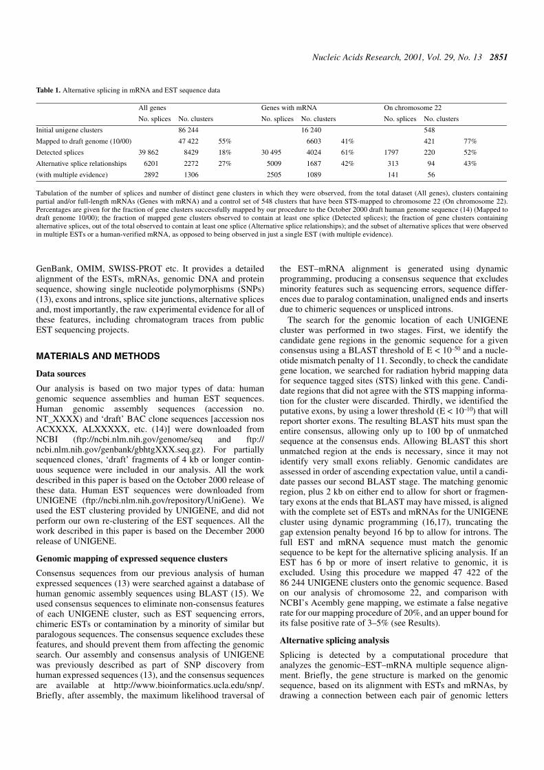
Table 1. Alternative splicing in mRNA and EST sequence data
Tabulation of the number of splices and number of distinct gene clusters in which they were observed, from the total dataset (All genes), clusters containingpartial and/or full-length mRNAs (Genes with mRNA) and a control set of 548 clusters that have been STS-mapped to chromosome 22 (On chromosome 22).Percentages are given for the fraction of gene clusters successfully mapped by our procedure to the October 2000 draft human genome sequence (14) (Mapped todraft genome 10/00); the fraction of mapped gene clusters observed to contain at least one splice (Detected splices); the fraction of gene clusters containingalternative splices, out of the total observed to contain at least one splice (Alternative splice relationships); and the subset of alternative splices that were observedin multiple ESTs or a human-verified mRNA, as opposed to being observed in just a single EST (with multiple evidence).
All genes Genes with mRNA On chromosome 22
No. splices No. clusters No. splices No. clusters No. splices No. clusters
Initial unigene clusters 86 244 16 240 548
Mapped to draft genome (10/00) 47 422 55% 6603 41% 421 77%
Detected splices 39 862 8429 18% 30 495 4024 61% 1797 220 52%
Alternative splice relationships 6201 2272 27% 5009 1687 42% 313 94 43%
(with multiple evidence) 2892 1306 2505 1089 141 56
2852 Nucleic Acids Research, 2001, Vol. 29, No. 13
aligned to a pair of letters in an expressed sequence that areadjacent (i.e. nucleotide i and i+1). Thus, an exon is identifiedby a contiguous segment of connected letters, an intron by acontiguous segment of unconnected letters and a splice by aconnection that jumps from one genomic letter to a distantgenomic letter. Thus, a candidate splice is detected as a gapbetween two exons that match a single contiguous region ofone or more ESTs. We report splices only for connections thatskip >10 bp in the genomic sequence (representing an effectiveminimum intron length) to screen out sequencing error oralignment heterogeneity artefacts. Individual splice observa-tions from different ESTs are joined together when their 5′splice sites match within 6 bp in the genomic sequence, andtheir 3′ splice sites also match within 6 bp. This level of variationis permitted to screen out sequencing error and alignment artefactsthat could give spurious alternative splices. All candidatesplices were checked against the standard consensus splicingsequences, and all candidates with mismatches were discarded.It is possible that some of these mismatches may be viabledeviations from the consensus sequence and represent realsplices. However, we have excluded them from considerationin the results presented in this paper. This procedure wasdesigned to be robust, and even in cases with a mis-assembledgenomic sequence should not report spurious splices. Instead,genomic mis-assembly would likely cause mismatch with theESTs, and complete exclusion of the cluster from our analysis.It should be noted that EST–mRNA versus genomic sequencealignments occasionally contain degenerate alignment positions,in which one or more nucleotides are identical in the genomicsequence on either side of a gap (intron). In this case our soft-ware checks each of the equivalent alignments to identify thecorrect splice junction.
Alternative splices are reported when two detected splicesoverlap in the genomic sequence (and thus are mutually exclu-sive events). One important consequence of this definition isthat alternative splicing always requires positive evidence(i.e. strong match of EST to genomic) on both sides of eachcompared splice. An alternative splice will never be reportedsimply because one EST is longer or shorter than others, oreven if vector sequence was attached at one end. [Vectorsequences are screened out of UNIGENE (18) data. However,it is still important to note that heterogeneity at EST ends willnot give rise to reported alternative splices.] All splices, alter-native splices, individual splice observations in specificsequences, source library information, gene information,genomic mapping information, etc. are stored within a rela-tional database (MySQL), and are accessible for query via theweb (http://www.bioinformatics.ucla.edu/HASDB). To assessthe fraction of alternative splices detected based on mRNA,EST versus mRNA or EST versus EST evidence (Fig. 5D), weused a database query to compute these numbers for all thealternative splices in our database.
Functional analysis of alternative splicing
We have performed extensive visual analysis and verificationof our results, for hundreds of different genes. We used theGeneMine software system (19) to validate all aspects of thegenomic mapping of our clusters, the exons, introns, splicesites, alternative splicing analysis and impact on protein structureand function, by thoroughly examining each of these features inthe genomic–EST–mRNA multiple sequence alignments. The
GeneMine software is freely available to academic researchers(http://www.bioinformatics.ucla.edu/genemine).
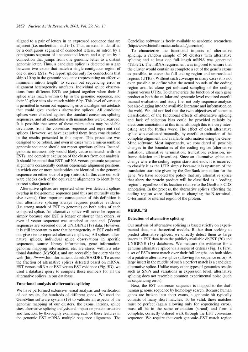
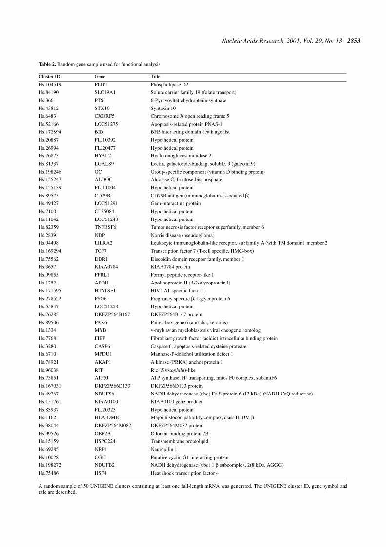
To characterize the functional impacts of alternativesplicing, a random sample of 50 clusters with alternativesplicing and at least one full-length mRNA was generated(Table 2). The mRNA requirement was imposed to ensure thatthe cluster would contain as complete a set of the gene’s exonsas possible, to cover the full coding region and untranslatedregions (UTRs). Without such coverage in many cases it is noteven possible to define what the actual bounds of the codingregion are, let alone get unbiased sampling of the codingregion versus UTRs. To characterize the function of each geneproduct at both the cellular and systemic level required carefulmanual evaluation and study (i.e. not only sequence analysisbut also digging into the available literature and information onthe web). We did not feel that the twin objectives of accurateclassification of the functional effects of alternative splicingand lack of selection bias could be provided reliably byelectronic annotations at this time, although this is a very inter-esting area for further work. The effect of each alternativesplice was evaluated manually, by careful examination of thecomplete alignment and available information using the Gene-Mine software. Most importantly, we considered all possiblechanges in the boundaries of the coding region (alternativeinitiation, alternative termination, truncation, extension, in-frame deletion and insertion). Since an alternative splice canchange where the coding region starts and ends, it is incorrectto classify it as the UTR simply because it is upstream of thetranslation start site given by the GenBank annotation for thegene. We have adopted the policy that any alternative splicethat alters the protein product will be classified as a ‘codingregion’, regardless of its location relative to the GenBank CDSannotation. In the process, the alternative splices affecting thecoding region were identified as changing the N-terminal,C-terminal or internal region of the protein.
RESULTS
Detection of alternative splicing
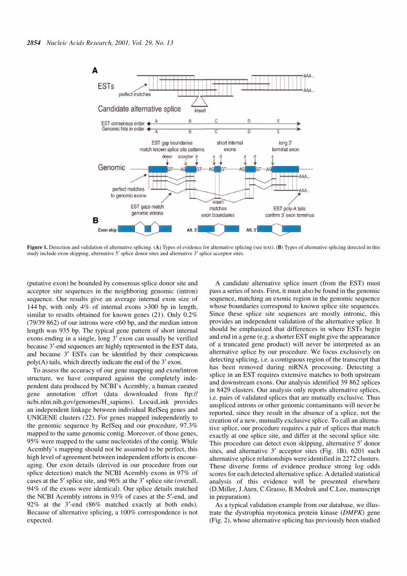
Our analysis of alternative splicing is based strictly on experi-mental data, not theoretical models. Rather than seeking topredict alternative splices, we directly detect them as largeinserts in EST data from the publicly available dbEST (20) andUNIGENE (18) databases. We measure the evidence for agenuine alternative splice via a series of criteria (Fig. 1). First,a set of ESTs must match over their full lengths, on both sidesof a putative alternative splice (allowing for sequence error). Alarge insert in the middle of such a perfect match is a candidatealternative splice. Unlike many other types of genomics resultssuch as SNPs and variations in expression level, alternativesplicing does not resemble common experimental noise (suchas sequencing error).
Next, the EST consensus sequence is mapped to the drafthuman genome sequence by homology search. Because humangenes are broken into short exons, a genomic hit typicallyconsists of many short matches. To be valid, these matchesmust be perfect (again allowing only for sequencing error),must all be in the same orientation (strand) and form acomplete, correctly ordered walk through the EST consensussequence. We require that each genomic–EST match region
Nucleic Acids Research, 2001, Vol. 29, No. 13 2853
Table 2. Random gene sample used for functional analysis
A random sample of 50 UNIGENE clusters containing at least one full-length mRNA was generated. The UNIGENE cluster ID, gene symbol andtitle are described.
Cluster ID Gene Title
Hs.104519 PLD2 Phospholipase D2
Hs.84190 SLC19A1 Solute carrier family 19 (folate transport)
Hs.366 PTS 6-Pyruvoyltetrahydropterin synthase
Hs.43812 STX10 Syntaxin 10
Hs.6483 CXORF5 Chromosome X open reading frame 5
Hs.52166 LOC51275 Apoptosis-related protein PNAS-1
Hs.172894 BID BH3 interacting domain death agonist
Hs.20887 FLJ10392 Hypothetical protein
Hs.26994 FLJ20477 Hypothetical protein
Hs.76873 HYAL2 Hyaluronoglucosaminidase 2
Hs.81337 LGALS9 Lectin, galactoside-binding, soluble, 9 (galectin 9)
Hs.198246 GC Group-specific component (vitamin D binding protein)
Hs.155247 ALDOC Aldolase C, fructose-bisphosphate
Hs.125139 FLJ11004 Hypothetical protein
Hs.89575 CD79B CD79B antigen (immunoglobulin-associated β)
Hs.49427 LOC51291 Gem-interacting protein
Hs.7100 CL25084 Hypothetical protein
Hs.11042 LOC51248 Hypothetical protein
Hs.82359 TNFRSF6 Tumor necrosis factor receptor superfamily, member 6
Hs.2839 NDP Norrie disease (pseudoglioma)
Hs.94498 LILRA2 Leukocyte immunoglobulin-like receptor, subfamily A (with TM domain), member 2
Hs.169294 TCF7 Transcription factor 7 (T-cell specific, HMG-box)
Hs.75562 DDR1 Discoidin domain receptor family, member 1
Hs.3657 KIAA0784 KIAA0784 protein
Hs.99855 FPRL1 Formyl peptide receptor-like 1
Hs.1252 APOH Apolipoprotein H (β-2-glycoprotein I)
Hs.171595 HTATSF1 HIV TAT specific factor I
Hs.278522 PSG6 Pregnancy specific β-1-glycoprotein 6
Hs.55847 LOC51258 Hypothetical protein
Hs.76285 DKFZP564B167 DKFZP564B167 protein
Hs.89506 PAX6 Paired box gene 6 (aniridia, keratitis)
Hs.1334 MYB v-myb avian myeloblastosis viral oncogene homolog
Hs.7768 FIBP Fibroblast growth factor (acidic) intracellular binding protein
Hs.3280 CASP6 Caspase 6, apoptosis-related cysteine protease
Hs.6710 MPDU1 Mannose-P-dolichol utilization defect 1
Hs.78921 AKAP1 A kinase (PRKA) anchor protein 1
Hs.96038 RIT Ric (Drosophila)-like
Hs.73851 ATP5J ATP synthase, H+ transporting, mitos F0 complex, subunitF6
Hs.167031 DKFZP566D133 DKFZP566D133 protein
Hs.49767 NDUFS6 NADH dehydrogenase (ubq) Fe-S protein 6 (13 kDa) (NADH CoQ reductase)
Hs.151761 KIAA0100 KIAA0100 gene product
Hs.83937 FLJ20323 Hypothetical protein
Hs.1162 HLA-DMB Major histocompatibility complex, class II, DM βHs.38044 DKFZP564M082 DKFZP564M082 protein
Hs.99526 OBP2B Odorant-binding protein 2B
Hs.15159 HSPC224 Transmembrane proteolipid
Hs.69285 NRP1 Neuropilin 1
Hs.10028 CG1I Putative cyclin G1 interacting protein
Hs.198272 NDUFB2 NADH dehydrogenase (ubq) 1 β subcomplex, 2(8 kDa, AGGG)
Hs.75486 HSF4 Heat shock transcription factor 4
2854 Nucleic Acids Research, 2001, Vol. 29, No. 13
(putative exon) be bounded by consensus splice donor site andacceptor site sequences in the neighboring genomic (intron)sequence. Our results give an average internal exon size of144 bp, with only 4% of internal exons >300 bp in length,similar to results obtained for known genes (21). Only 0.2%(79/39 862) of our introns were <60 bp, and the median intronlength was 935 bp. The typical gene pattern of short internalexons ending in a single, long 3′ exon can usually be verifiedbecause 3′-end sequences are highly represented in the EST data,and because 3′ ESTs can be identified by their conspicuouspoly(A) tails, which directly indicate the end of the 3′ exon.
To assess the accuracy of our gene mapping and exon/intronstructure, we have compared against the completely inde-pendent data produced by NCBI’s Acembly, a human curatedgene annotation effort (data downloaded from ftp://ncbi.nlm.nih.gov/genomes/H_sapiens). LocusLink providesan independent linkage between individual RefSeq genes andUNIGENE clusters (22). For genes mapped independently tothe genomic sequence by RefSeq and our procedure, 97.3%mapped to the same genomic contig. Moreover, of those genes,95% were mapped to the same nucleotides of the contig. WhileAcembly’s mapping should not be assumed to be perfect, thishigh level of agreement between independent efforts is encour-aging. Our exon details (derived in our procedure from oursplice detection) match the NCBI Acembly exons in 97% ofcases at the 5′ splice site, and 96% at the 3′ splice site (overall,94% of the exons were identical). Our splice details matchedthe NCBI Acembly introns in 93% of cases at the 5′-end, and92% at the 3′-end (86% matched exactly at both ends).Because of alternative splicing, a 100% correspondence is notexpected.
A candidate alternative splice insert (from the EST) mustpass a series of tests. First, it must also be found in the genomicsequence, matching an exonic region in the genomic sequencewhose boundaries correspond to known splice site sequences.Since these splice site sequences are mostly intronic, thisprovides an independent validation of the alternative splice. Itshould be emphasized that differences in where ESTs beginand end in a gene (e.g. a shorter EST might give the appearanceof a truncated gene product) will never be interpreted as analternative splice by our procedure. We focus exclusively ondetecting splicing, i.e. a contiguous region of the transcript thathas been removed during mRNA processing. Detecting asplice in an EST requires extensive matches to both upstreamand downstream exons. Our analysis identified 39 862 splicesin 8429 clusters. Our analysis only reports alternative splices,i.e. pairs of validated splices that are mutually exclusive. Thusunspliced introns or other genomic contaminants will never bereported, since they result in the absence of a splice, not thecreation of a new, mutually exclusive splice. To call an alterna-tive splice, our procedure requires a pair of splices that matchexactly at one splice site, and differ at the second splice site.This procedure can detect exon skipping, alternative 5′ donorsites, and alternative 3′ acceptor sites (Fig. 1B). 6201 suchalternative splice relationships were identified in 2272 clusters.These diverse forms of evidence produce strong log oddsscores for each detected alternative splice. A detailed statisticalanalysis of this evidence will be presented elsewhere(D.Miller, J.Aten, C.Grasso, B.Modrek and C.Lee, manuscriptin preparation).
As a typical validation example from our database, we illus-trate the dystrophia myotonica protein kinase (DMPK) gene(Fig. 2), whose alternative splicing has previously been studied
Figure 1. Detection and validation of alternative splicing. (A) Types of evidence for alternative splicing (see text). (B) Types of alternative splicing detected in thisstudy include exon skipping, alternative 5′ splice donor sites and alternative 3′ splice acceptor sites.

Nucleic Acids Research, 2001, Vol. 29, No. 13 2855
extensively. In DMPK, we identified three alternative splicesin the EST data, all of which are verified by independent exper-imental results in the existing literature (23). Of the three alter-native splices, one deletes the last 15 bp of exon 8, anotherskips exon 12 and exon 13, and the last deletes just 4 bp in exon14. Figure 2 shows one of these alternative splice formsincluding junction and quality of match of the EST evidenceversus the genomic sequence.
Novel alternative splice forms of a known gene
Figure 3 shows several novel alternative splices detected in awell-studied gene, HLA-DM β. Eighty ESTs from UNIGENEcluster Hs.1162 align to form a consensus sequence, which inturn matches an ordered series of segments on one strand ofchromosome 6. The EST sequences match the genomicsequence closely, consistent with sequencing error. The ESTsequences mark out a long 3′ exon (359 bp) plus a series of fiveshort exons, whose sizes (36–288 bp) match the rangeexpected for internal exons. This matches the known genelocation and structure for HLA-DM β (24,25). Eight splices areobserved in these ESTs, where sequence matching one exonicregion skips directly to a downstream exonic region as indi-cated in Figure 3A. The 16 putative exon boundaries impliedby the ESTs map precisely to strong consensus splice acceptorand donor sites in the genomic sequence (Fig. 3C).
Four different alternatively spliced forms of HLA-DM β areobserved: splices 3+4+5 (including exons IV and V in themRNA product); splices 6+5 (skipping exon IV); splices 3+7(excluding exon V); splice 8 (skipping exons IV and V).Exons IV and V are 117 and 36 bp in length, and thus thesealternative splices are all in-frame. The protein coding regionbegins in exon I and ends in exon VI, so these splices producefour different forms of the HLA-DM β chain that differ at theirC-terminus.
Analysis of these forms reveals a remarkably simple andintriguing functional effect. HLA-DM is essential for theloading of class II MHC molecules with exogenous peptideantigens, a key step in antigen presentation and activation ofthe humoral immune response. This is thought to occur in earlylysosomal compartments. HLA-DM is normally targeted tolysosomes, and its β chain contains a transmembrane domainanchoring its C-terminus (26,27). Exon IV is short, and corre-sponds precisely to the transmembrane domain. Exon V is veryshort, and encodes the lysosomal targeting signal YTPL,whose first residue begins at the start of the exon. Thus, thealternative splice regulates HLA-DM’s targeting to endosomalcompartments (by including or excluding the YTPL signal), aswell as its anchoring to the membrane. Given HLA-DM’simportance in antigen processing and presentation by class IIMHC, this regulation is functionally interesting. Removing itstargeting signal would likely redirect HLA-DM first to theplasma membrane, so that it would travel to lysosomes viaendocytic pathways, altering the kinetics and conditions inwhich it first encounters class II MHC. It appears that the genestructure of the HLA-DM β gene has been carefully ‘designed’to enable control of HLA-DM function, by pulling out both thetransmembrane helix and the lysosomal targeting signal intoseparate short exons (IV, V) that can be alternatively splicedin-frame (exon VI supplies the last 4 amino acids of theprotein, identical in all forms). The alternatively spliced formswere detected in uterus (two ESTs), placenta, lymph, stomachand colon. Despite the fact that HLA-DM is the subject ofintense research, we have not been able to find any report ofsuch alternative splicing in the published literature, and it is
Figure 2. Alternative splicing of DMPK. (A) Gene structure for exons XII–XVof the DMPK gene, in contig NT000991 of chromosome 19. Two splice formsare shown, one observed in an mRNA (mRNA1) and one in an EST (EST1).(B) Example sequence evidence for the two splice forms. Sequence EST1skips directly from exon XII to exon XV. We detected three alternative spliceforms in DMPK; all are confirmed by the experimental literature (23).
A
B
Figure 3. Alternative splicing of HLA-DMB. (A) Genomic structure of theHLA-DM β gene, in contig NT001520 of chromosome 6. Exons are shown asfilled boxes, and the observed splices are shown on top of the genomicsequence. (B) The four alternative forms of HLA-DM β mRNA inferred fromthe expressed sequence data, colored to show the exons. The protein readingframe is indicated by an arrow beneath each form, showing the transmembranedomain (TM) and lysosomal targeting signal (LT). (C) The splice donor andacceptor sites for the eight putative splices observed in HLA-DM β. The pri-mary consensus site sequences are highlighted in black and secondary consen-sus sequences (5) are marked in magenta.
2856 Nucleic Acids Research, 2001, Vol. 29, No. 13
thought to be novel by an expert on HLA-DM (E.Mellins,personal communication).
Scope of alternative splicing in human genes
Our genome-wide analysis detected thousands of alternativesplices in the current, publicly available human genome data(Table 1). 6201 alternative splice relationships were detectedin which two splices shared a common donor or acceptor site,but spliced to a different site on their other end (i.e. exon skip-ping, alternative 5′ splice donor site or alternative 3′ spliceacceptor site; Fig. 1B). We found alternative splices in 27% ofgenes for which we had enough expressed sequence to covermore than a single exon. However, this estimate, based onanalysis of all EST clusters, likely underestimates the realoccurrence of alternative splicing, because the available ESTdata typically cover only a small part of the complete gene. Totest this hypothesis, we analyzed the alternative splicing rate ingenes for which mRNA sequence was available (representingall or part of the full gene). We detected one or more alterna-tive splice forms in 42% of these genes, significantly higherthan the rate observed in EST-only clusters. This is in closeagreement with a previous study of mRNA-based expressedsequence clusters (8). Since fragmentation of the genomicsequence can also block complete coverage of a gene, weassessed the rate of alternative splice detection in genesmapped to chromosome 22. Of these, 43% contained alterna-tive splices, including both mRNA and EST-only clusters.
The current EST data appears to be incomplete. Our proce-dure identified splices (i.e. multiple exons) in only 18% of themapped EST clusters. However, for clusters that we mapped tochromosome 22 (full genomic) that also had an mRNAsequence, 88% contained at least one splice. A variety offactors such as the fragmentation of the draft human genomesequence, the large size of introns and the tendency of the
ESTs to cluster at the 3′-end bias the current dataset againstfinding full-length genes, and probably underestimate the truelevel of alternative splicing. Moreover, since the current ESTdata for each gene represent only a subset of the tissues and celltypes in which that gene is expressed, it is likely that the totaloccurrence of alternative splicing is much greater than whatour analyses can detect. A large fraction of the EST alternativesplice forms were observed multiple times (from differentclones and different libraries), indicating that they constitute arelatively high fraction of total mRNA. Of our alternativesplices, 2892 (47%) were observed in two or more ESTsequences. These data represent a ‘high confidence’ subset ofthe detected alternative splices.
Our analysis indicates that the vast majority of our databaserepresents novel findings (Fig. 5D). Only 13% of our alterna-tive splices were detected in mRNA sequences from GenBank,which presumably have been thoroughly studied. Theremaining 87% could be detected only with ESTs. Our proce-dure also detected large numbers of alternative splicing eventsin completely novel genes. Approximately 1200 alternativesplices were detected in clusters containing ESTs only.
Alternative splicing in a novel human gene
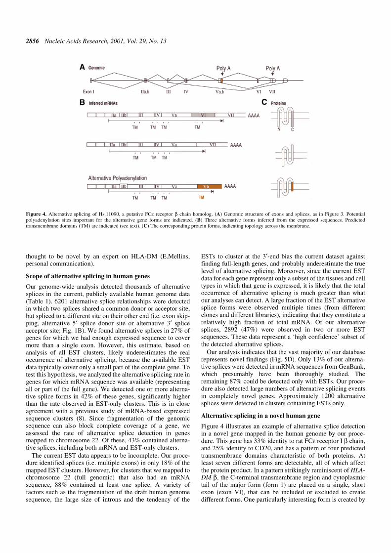
Figure 4 illustrates an example of alternative splice detectionin a novel gene mapped in the human genome by our proce-dure. This gene has 33% identity to rat FCε receptor I β chain,and 25% identity to CD20, and has a pattern of four predictedtransmembrane domains characteristic of both proteins. Atleast seven different forms are detectable, all of which affectthe protein product. In a pattern strikingly reminiscent of HLA-DM β, the C-terminal transmembrane region and cytoplasmictail of the major form (form 1) are placed on a single, shortexon (exon VI), that can be included or excluded to createdifferent forms. One particularly interesting form is created by
Figure 4. Alternative splicing of Hs.11090, a putative FCε receptor β chain homolog. (A) Genomic structure of exons and splices, as in Figure 3. Potentialpolyadenylation sites important for the alternative gene forms are indicated. (B) Three alternative forms inferred from the expressed sequences. Predictedtransmembrane domains (TM) are indicated (see text). (C) The corresponding protein forms, indicating topology across the membrane.
Nucleic Acids Research, 2001, Vol. 29, No. 13 2857
ignoring the normal splice from exon V to exon VI, extendingthe coding region from exon Va for 142 bp (which we havedesignated exon Vb). A polyadenylation site is predicted at theend of this sequence, and the ESTs are observed to terminate inpoly(A) at this point. This alternative termination replaces thecoding region of exons VI and VII with 40 amino acidsencoded by exon Vb [terminated by a STOP codon 23 bpbefore the poly(A) site]. Intriguingly, this replacement C-terminalsequence also contains a predicted transmembrane sequence,and thus neatly substitutes a new C-terminal transmembranedomain and cytoplasmic tail. The cytoplasmic tail in equivalentFC receptor chains plays a key role in activating cytoplasmicsignal transduction molecules (28,29), so this alternative formlikely modulates the signal transduction activity of thisreceptor. This form is detected in placenta and kidney, whilethe majority form was detected in many different libraries.
DISCUSSION
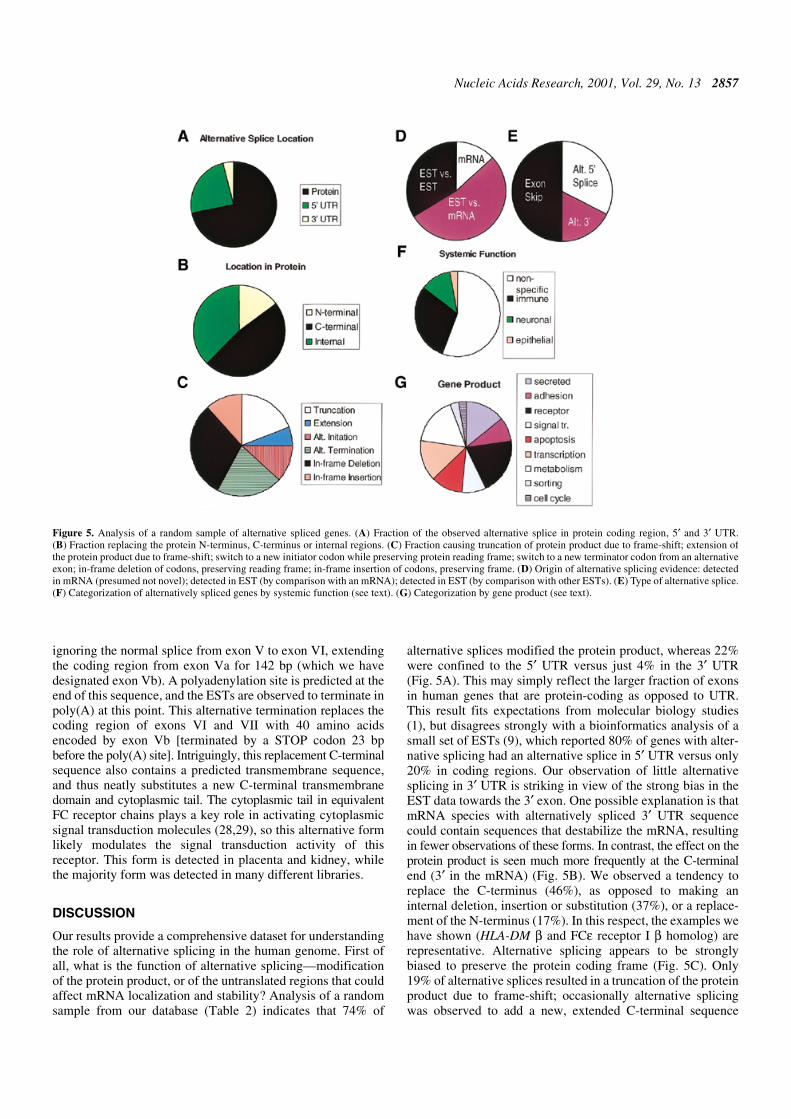
Our results provide a comprehensive dataset for understandingthe role of alternative splicing in the human genome. First ofall, what is the function of alternative splicing—modificationof the protein product, or of the untranslated regions that couldaffect mRNA localization and stability? Analysis of a randomsample from our database (Table 2) indicates that 74% of
alternative splices modified the protein product, whereas 22%were confined to the 5′ UTR versus just 4% in the 3′ UTR(Fig. 5A). This may simply reflect the larger fraction of exonsin human genes that are protein-coding as opposed to UTR.This result fits expectations from molecular biology studies(1), but disagrees strongly with a bioinformatics analysis of asmall set of ESTs (9), which reported 80% of genes with alter-native splicing had an alternative splice in 5′ UTR versus only20% in coding regions. Our observation of little alternativesplicing in 3′ UTR is striking in view of the strong bias in theEST data towards the 3′ exon. One possible explanation is thatmRNA species with alternatively spliced 3′ UTR sequencecould contain sequences that destabilize the mRNA, resultingin fewer observations of these forms. In contrast, the effect on theprotein product is seen much more frequently at the C-terminalend (3′ in the mRNA) (Fig. 5B). We observed a tendency toreplace the C-terminus (46%), as opposed to making aninternal deletion, insertion or substitution (37%), or a replace-ment of the N-terminus (17%). In this respect, the examples wehave shown (HLA-DM β and FCε receptor I β homolog) arerepresentative. Alternative splicing appears to be stronglybiased to preserve the protein coding frame (Fig. 5C). Only19% of alternative splices resulted in a truncation of the proteinproduct due to frame-shift; occasionally alternative splicingwas observed to add a new, extended C-terminal sequence
Figure 5. Analysis of a random sample of alternative spliced genes. (A) Fraction of the observed alternative splice in protein coding region, 5′ and 3′ UTR.(B) Fraction replacing the protein N-terminus, C-terminus or internal regions. (C) Fraction causing truncation of protein product due to frame-shift; extension ofthe protein product due to frame-shift; switch to a new initiator codon while preserving protein reading frame; switch to a new terminator codon from an alternativeexon; in-frame deletion of codons, preserving reading frame; in-frame insertion of codons, preserving frame. (D) Origin of alternative splicing evidence: detectedin mRNA (presumed not novel); detected in EST (by comparison with an mRNA); detected in EST (by comparison with other ESTs). (E) Type of alternative splice.(F) Categorization of alternatively spliced genes by systemic function (see text). (G) Categorization by gene product (see text).
2858 Nucleic Acids Research, 2001, Vol. 29, No. 13
through frame-shift (6%). Alternative splicing resulted in aswitch to a new AUG start site on an alternative exon in 15%of cases. In contrast, replacing the C-terminus by switching toa different exon containing an alternative STOP codonoccurred in 20% of cases. In-frame deletion or insertion of anew sequence in the middle of the protein accounted for 29 and11% of cases, respectively.
In what types of molecules is alternative splicing commonlyobserved? Figure 5G shows a molecular classification of arandom sample of alternatively spliced genes. The most abun-dant category is cell surface functions/receptors (29%), whichincludes membrane-anchored receptors (e.g. CD79B), integralmembrane proteins (e.g. folate transporter SLC19A1) andproteins involved in cell surface adhesion (e.g. lectin,hyaluronoglucosaminidase 2). In two related categories, anadditional 14% of alternatively spliced genes encode secretedproteins (e.g. Norrie disease protein; group-specific compo-nent) and 9% encode signal transduction molecules (e.g. phos-pholipase D2; RIT). The next two major categories aretranscriptional regulation (14%; e.g. MYB, PAX6) and apop-tosis (11%; e.g. BID, PNAS-1). Together, these functions oftransmission, reception and response to cellular signalscomprise >75% of the observed alternatively spliced genes.Proteins involved in metabolism (e.g. aldolase C), andorganelle-specific sorting proteins were also observed. Thissample is by no means comprehensive or exact, but indicates atrend towards cell surface interactions and signaling.
What types of systemic functions are most often affected byalternative splicing? Twenty-nine percent of the alternativelyspliced genes encoded functions specific to the immune system(Fig. 5F; e.g. T-cell specific transcription factor 7, TNFreceptor superfamily member 6). In particular, alternativesplicing of immune system cell surface receptors was veryprevalent. Neuronal functions (e.g. neuropilin, brain-specificaldolase C) comprised 12% of the total. The remaining genespossessed no clearly specific systemic function. These datasuggest alternative splicing may play a large role in immunesystem and nervous system functions which require precisecontrol of cellular differentiation and activation, to processlarge amounts of information. Controlling how each cellresponds to a diverse array of signals can be achieved throughalternative splicing of its receptors and signal transductionmolecules.
How often is alternative splicing clearly associated with aspecific tissue? Based on a sample of 50 genes, ∼14% of alter-natively spliced genes in our dataset showed evidence of tissuespecificity for the minor isoform. This estimate is based on aconservative definition requiring that the minor isoform beobserved multiple times in a specific tissue in which the majorform was not observed. Since in many known cases of tissue-specific alternative splicing both minor and major forms areobserved in the same tissue, this probably misses many casesof real tissue-specificity. Examples include DDR1, discoidindomain receptor, which has a minor form observed in muscle;and CG1I, a putative cyclin G1 interacting protein, which hasisoforms observed specifically in ovary and brain. Within thesmall sample, tissue-specific minor isoforms were observed innovel, uncharacterized genes in brain, colon, testis and pros-tate.
How comprehensive is our dataset, and what are its pros-pects for growth? We have noted two causes of failure by our
procedure to detect alternatively spliced forms that are knownin the literature. First, a given gene may not map yet to the draftgenome, a prerequisite in our procedure for analyzing itssplicing. Secondly, some alternatively spliced mRNA formsare miscategorized as genomic DNA in GenBank, causingthem to be excluded by our procedure. The former seems to bethe most important cause of failure. Despite >90% complete-ness by total nucleotides sequenced, the draft genome used inthis study (October 2000) only enabled mapping of 55% ofUNIGENE expressed sequence clusters, because we require afull-length match versus the expressed gene sequenceconsensus (Table 1). The draft (i.e. incomplete) BAC clonesequences which constituted the majority of this dataset,consisted in large part of short sequence fragments (4–10 kb)separated by unsequenced regions. Such fragments are toosmall to map a typical human gene (10–30 kb) by our conserv-ative procedure. This trend is even stronger for the subset ofgenes that have full-length mRNAs. Of these clusters, only41% could be mapped over their full length to an availablegenomic contig. To check whether this is due to the draftgenome’s fragmentation, we analyzed a subset of gene clustersthat have been mapped by STS to chromosome 22, which hasbeen almost completely sequenced. For these clusters, 77%could be mapped. Thus, given unbroken genomic sequence,our mapping procedure has a false negative rate of ∼20%.These data suggest that completion of the human genomesequence, along with improvements in our algorithms, will atleast double the number of alternative splices detected. Ourdetection of alternative splicing should also grow withincreasing EST data. In our current EST dataset (December2000), splices were detected in only 18% of clusters, reflectingthe fact that the average cluster consists of too few ESTs (oneor two) and is too short (a few hundred base pairs) to covermore than a single exon. This is exaggerated by the strong biasof the ESTs to be from the 3′-end, since 3′ exons tend to bemuch longer than typical internal exons. In contrast, in genesfor which a full-length or partial mRNA sequence was avail-able and which were mapped to a region of full-length genomicsequence (e.g. chromosome 22), 88% contained at least onesplice (and typically many more).
ACKNOWLEDGEMENTS
We wish to thank D. Black, D. Miller, S. Galbraith andD. Eisenberg for their helpful discussions and comments onthis work, and K. Ke for assistance in constructing the HASDBweb site. This work was supported by Department of Energygrant DEFG0387ER60615 and a grant from the SearleScholars Program to C.L. B.M. is a predoctoral traineesupported by NSF IGERT Award #DGE-9987641.
REFERENCES
1. Lopez,A.J. (1998) Alternative splicing of pre-mRNA: developmentalconsequences and mechanisms of regulation. Annu. Rev. Genet., 32,279–305.
2. Boise,L.H., Gonzalez-Garcia,M., Postema,C.E., Ding,L., Lindsten,T.,Turka,L.A., Mao,X., Nunez,G. and Thompson,C.B. (1993) bcl-x, a bcl-2-related gene that functions as a dominant regulator of apoptotic cell death.Cell, 74, 597–608.
3. Fettiplace,R. and Fuchs,P.A. (1999) Mechanisms of hair cell tuning.Annu. Rev. Physiol., 61, 809–834.
Nucleic Acids Research, 2001, Vol. 29, No. 13 2859
4. Schmucker,D., Clemens,J.C., Shu,H., Worby,C.A., Xiao,J., Muda,M.,Dixon,J.E. and Zipursky,S.L. (2000) Drosophila Dscam is an axonguidance receptor exhibiting extraordinary molecular diversity. Cell, 101,671–684.
5. Smith,C.W.J. and Valcarcel,J. (2000) Alternative pre-mRNA splicing: thelogic of combinatorial control. Trends Biochem. Sci., 25, 381–388.
6. Sharp,P.A. (1994) Split genes and RNA splicing. Cell, 77, 805–815.7. Sutcliffe,J.G. and Milner,R.J. (1988) Alternative mRNA splicing: the
Shaker gene. Trends Genet., 4, 297–299.8. Brett,D., Hanke,J., Lehmann,G., Haase,S., Delbruck,S., Krueger,S.,
Reich,J. and Bork,P. (2000) EST comparison indicates 38% of humanmRNAs contain possible alternative splice forms. FEBS Lett., 474, 83–86.
9. Mironov,A.A., Fickett,J.W. and Gelfand,M.S. (1999) Frequent alternativesplicing of human genes. Genome Res., 9, 1288–1293.
10. Liang,F., Holt,I., Pertea,G., Karamycheva,S., Salzberg,S.L. andQuackenbush,J. (2000) Gene Index analysis of the human genomeestimates approximately 120,000 genes. Nat. Genet., 25, 239–240.
11. Ewing,B. and Green,P. (2000) Analysis of expressed sequence tagsindicates 35,000 human genes. Nat. Genet., 25, 232–234.
12. Ji,H., Zhou,Q., Wen,F., Xia,H., Lu,X. and Li,Y. (2001) AsMamDB: analternative splice database of mammals. Nucleic Acids Res., 29, 260–263.
13. Irizarry,K., Kustanovich,V., Li,C., Brown,N., Nelson,S., Wong,W. andLee,C. (2000) Genome-wide analysis of single-nucleotide polymorphismsin human expressed sequences. Nat. Genet., 26, 233–236.
14. Jang,W., Chen,W.C., Sicotte,H. and Schuler,G.D. (1999) Makingeffective use of human genomic sequence data. Trends Genet., 15,284–286.
15. Altschul,S.F., Gish,W., Miller,W., Myers,E.W. and Lipman,D.J. (1990)Basic local alignment search tool. J. Mol. Biol., 215, 403–410.
16. Needleman,S.B. and Wunsch,C.D. (1970) A general method applicable tothe search for similarities in the amino acid sequence of two proteins.J. Mol. Biol., 48, 443–453.
17. Smith,T.F. and Waterman,M.S. (1981) Identification of commonmolecular subsequences. J. Mol. Biol., 147, 195–197.
18. Schuler,G. (1997) Pieces of the puzzle: expressed sequence tags and thecatalog of human genes. J. Mol. Med., 75, 694–698.
19. Lee,C. and Irizarry,K. (2001) The GeneMine system for genome/proteome annotation and collaborative data-mining. IBM Syst. J., 40,in press.
20. Boguski,M.S., Lowe,T.M. and Tolstoshev,C.M. (1993) dbEST—databasefor ‘expressed sequence tags’. Nat. Genet., 4, 332–333.
21. Hawkins,J.D. (1988) A survey on intron and exon lengths. Nucleic AcidsRes., 16, 9893–9905.
22. Pruitt,K.D. and Maglott,D.R. (2001) RefSeq and LocusLink: NCBI gene-centered resources. Nucleic Acids Res., 29, 137–140.
23. Groenen P.J., Wansink,D.G., Coerwinkel,M., van den Broek,W.,Jansen,G. and Wieringa,B. (2000) Constitutive and regulated modes ofsplicing produce six major myotonic dystrophy protein kinase (DMPK)isoforms with distinct properties. Hum. Mol. Genet., 9, 605–616.
24. Kelly,A.P., Monaco,J.J., Cho,S.G. and Trowsdale,J. (1991) A new humanHLA class II-related locus, DM. Nature, 353, 571–573.
25. Shaman,J., von Scheven,E., Morris,P., Chang,M.Y. and Mellins,E. (1995)Analysis of HLA-DMB mutants and -DMB genomic structure.Immunogenetics, 41, 117–124.
26. Sanderson,F., Kleijmeer,M.J., Kelly,A.P., Verwoerd,D., Tulp,A.,Neefjes,J., Geueze,H.J. and Trowsdale,J. (1994) Accumulation of HLA-DM, a regulator of antigen presentation, in MHC class II compartments.Science, 266, 1566–1569.
27. Potter,P.K., Copier,J., Sacks,S.H., Calafat,J., Janssen,H., Neefjes,J. andKelly,A.P. (1999) Accurate intracellular localization of HLA-DMrequires correct spacing of a cytoplasmic YTPL targeting motif relative tothe transmembrane domain. Eur. J. Immunol., 29, 3936–3944.
28. Daeron,M. (1997) Fc receptor biology. Annu. Rev. Immunol., 15, 203–234.29. Kinet,J.P. (1999) The high affinity IgE receptor (FCεRI): from physiology
to pathology. Annu. Rev. Immunol., 17, 931–972.
Related Documents