Genome-Wide Analysis of Menin Binding Provides Insights into MEN1 Tumorigenesis Peter C. Scacheri 1 , Sean Davis 2 , Duncan T. Odom 3 , Gregory E. Crawford 1 , Stacie Perkins 1 , Mohamad J. Halawi 1 , Sunita K. Agarwal 4 , Stephen J. Marx 4 , Allen M. Spiegel 5 , Paul S. Meltzer 2 , Francis S. Collins 1* 1 Genome Technology Branch, National Human Genome Research Institute, National Institutes of Health, Bethesda, Maryland, United States of America, 2 Cancer Genetics Branch, National Human Genome Research Institute, National Institutes of Health, Bethesda, Maryland, United States of America, 3 Whitehead Institute, Cambridge, Massachusetts, United States of America, 4 National Institute of Diabetes and Digestive and Kidney Diseases, National Institutes of Health, Bethesda, Maryland, United States of America, 5 National Institute on Deafness and Other Communication Disorders, National Institutes of Health, Bethesda, Maryland, United States of America Multiple endocrine neoplasia type I (MEN1) is a familial cancer syndrome characterized primarily by tumors of multiple endocrine glands. The gene for MEN1 encodes a ubiquitously expressed tumor suppressor protein called menin. Menin was recently shown to interact with several components of a trithorax family histone methyltransferase complex including ASH2, Rbbp5, WDR5, and the leukemia proto-oncoprotein MLL. To elucidate menin’s role as a tumor suppressor and gain insights into the endocrine-specific tumor phenotype in MEN1, we mapped the genomic binding sites of menin, MLL1, and Rbbp5, to approximately 20,000 promoters in HeLa S3, HepG2, and pancreatic islet cells using the strategy of chromatin-immunoprecipitation coupled with microarray analysis. We found that menin, MLL1, and Rbbp5 localize to the promoters of thousands of human genes but do not always bind together. These data suggest that menin functions as a general regulator of transcription. We also found that factor occupancy generally correlates with high gene expression but that the loss of menin does not result in significant changes in most transcript levels. One exception is the developmentally programmed transcription factor, HLXB9, which is overexpressed in islets in the absence of menin. Our findings expand the realm of menin-targeted genes several hundred-fold beyond that previously described and provide potential insights to the endocrine tumor bias observed in MEN1 patients. Citation: Scacheri PC, Davis S, Odom DT, Crawford GE, Perkins S, et al. (2006) Genome-wide analysis of menin binding provides insights into MEN1 tumorigenesis. PLoS Genet 2(4): e51. DOI: 10.1371/journal.pgen.0020051 Introduction The hallmark of multiple endocrine neoplasia type I (MEN1), is the development of tumors in the parathyroid, anterior pituitary, and enteropancreatic endocrine cells. Other associations occasionally found in MEN1 patients include foregut carcinoids, facial angiofibromas, lipomas, collagenomas, meningiomas, and smooth muscle tumors. MEN1 patients typically inherit loss of function mutations in the MEN1 gene, and tumors arise following loss of the remaining wild-type allele. Thus, the MEN1 gene follows the classic ‘‘two-hit’’ tumor suppressor model first proposed by Knudson for retinoblastoma [1]. Somatic mutations in the MEN1 gene are also frequently found in sporadic parathyroid adenomas, insulinomas, gastrinomas, and lung carcinoids [2– 6], indicating that loss of the MEN1 gene is a major contributor to the development and maintenance of many nonhereditary endocrine tumors. There is currently no prevention of or cure for MEN1 cancers, and cancer treat- ment options are generally limited to surgical interventions. The protein product of MEN1, menin, is a ubiquitously expressed 67-kDa protein found predominantly in the nucleus [7]. Studies in Men1 knockout mice support menin’s role as a tumor suppressor [8,9]. Mice that are homozygous null for Men1 show developmental defects and die at embryonic day 11.5 to 13.5. Heterozygous Men1 mice eventually develop multiple endocrine tumors that arise following somatic loss of the wild-type Men1 allele, and the spectrum of tumors observed in Men1 þ/ mice is remarkably similar to that in human MEN1 kindreds. We and others have also successfully developed conditional knockouts of the Men1 gene, using the Cre-lox system [10–12]. When homo- zygous Men1 ‘‘floxed’’ mice are bred to animals expressing Cre recombinase in the beta cells of the pancreatic islets, the resultant progeny develop multiple pancreatic insulinomas. These insulinomas arise long after homozygous inactivation of the Men1 gene, suggesting that additional somatic events are required for frank tumor formation. Men1 / insulinomas are capable of developing in the absence of chromosomal or microsatellite instability, suggesting that the additional somatic events required for tumor formation are subtle, occurring at either the nucleotide or epigenetic level [13]. Although inherited mutations in the MEN1 gene predispose individuals to several types of tumors, there is a particularly striking predisposition to tumors in endocrine tissues. The development of tumors in this endocrine-specific pattern is quite puzzling as menin appears to be expressed in all tissues. In a previous study, we set out to gain insights to this issue of Editor: Michael Snyder, Yale University, United States of America Received January 10, 2006; Accepted February 23, 2006; Published April 7, 2006 A previous version of this article appeared as an Early Online Release on February 23, 2006 (DOI: 10.1371/journal.pgen.0020051.eor). DOI: 10.1371/journal.pgen.0020051 This is an open-access article distributed under the terms of the Creative Commons Public Domain declaration which stipulates that, once placed in the public domain, this work may be freely reproduced, distributed, transmitted, modified, built upon, or otherwise used by anyone for any lawful purpose. Abbreviations: ChIP-chip, chromatin-immunoprecipitation coupled with micro- array analysis; MEN1, multiple endocrine neoplasia, type I * To whom correspondence should be addressed. E-mail: [email protected] PLoS Genetics | www.plosgenetics.org April 2006 | Volume 2 | Issue 4 | e51 0406

Welcome message from author
This document is posted to help you gain knowledge. Please leave a comment to let me know what you think about it! Share it to your friends and learn new things together.
Transcript
Genome-Wide Analysis of Menin BindingProvides Insights into MEN1 TumorigenesisPeter C. Scacheri
1, Sean Davis
2, Duncan T. Odom
3, Gregory E. Crawford
1, Stacie Perkins
1, Mohamad J. Halawi
1,
Sunita K. Agarwal4
, Stephen J. Marx4
, Allen M. Spiegel5
, Paul S. Meltzer2
, Francis S. Collins1*
1 Genome Technology Branch, National Human Genome Research Institute, National Institutes of Health, Bethesda, Maryland, United States of America, 2 Cancer Genetics
Branch, National Human Genome Research Institute, National Institutes of Health, Bethesda, Maryland, United States of America, 3 Whitehead Institute, Cambridge,
Massachusetts, United States of America, 4 National Institute of Diabetes and Digestive and Kidney Diseases, National Institutes of Health, Bethesda, Maryland, United States
of America, 5 National Institute on Deafness and Other Communication Disorders, National Institutes of Health, Bethesda, Maryland, United States of America
Multiple endocrine neoplasia type I (MEN1) is a familial cancer syndrome characterized primarily by tumors of multipleendocrine glands. The gene for MEN1 encodes a ubiquitously expressed tumor suppressor protein called menin. Meninwas recently shown to interact with several components of a trithorax family histone methyltransferase complexincluding ASH2, Rbbp5, WDR5, and the leukemia proto-oncoprotein MLL. To elucidate menin’s role as a tumorsuppressor and gain insights into the endocrine-specific tumor phenotype in MEN1, we mapped the genomic bindingsites of menin, MLL1, and Rbbp5, to approximately 20,000 promoters in HeLa S3, HepG2, and pancreatic islet cellsusing the strategy of chromatin-immunoprecipitation coupled with microarray analysis. We found that menin, MLL1,and Rbbp5 localize to the promoters of thousands of human genes but do not always bind together. These datasuggest that menin functions as a general regulator of transcription. We also found that factor occupancy generallycorrelates with high gene expression but that the loss of menin does not result in significant changes in most transcriptlevels. One exception is the developmentally programmed transcription factor, HLXB9, which is overexpressed in isletsin the absence of menin. Our findings expand the realm of menin-targeted genes several hundred-fold beyond thatpreviously described and provide potential insights to the endocrine tumor bias observed in MEN1 patients.
Citation: Scacheri PC, Davis S, Odom DT, Crawford GE, Perkins S, et al. (2006) Genome-wide analysis of menin binding provides insights into MEN1 tumorigenesis. PLoS Genet2(4): e51. DOI: 10.1371/journal.pgen.0020051
Introduction
The hallmark of multiple endocrine neoplasia type I(MEN1), is the development of tumors in the parathyroid,anterior pituitary, and enteropancreatic endocrine cells.Other associations occasionally found in MEN1 patientsinclude foregut carcinoids, facial angiofibromas, lipomas,collagenomas, meningiomas, and smooth muscle tumors.MEN1 patients typically inherit loss of function mutations inthe MEN1 gene, and tumors arise following loss of theremaining wild-type allele. Thus, the MEN1 gene follows theclassic ‘‘two-hit’’ tumor suppressor model first proposed byKnudson for retinoblastoma [1]. Somatic mutations in theMEN1 gene are also frequently found in sporadic parathyroidadenomas, insulinomas, gastrinomas, and lung carcinoids [2–6], indicating that loss of the MEN1 gene is a majorcontributor to the development and maintenance of manynonhereditary endocrine tumors. There is currently noprevention of or cure for MEN1 cancers, and cancer treat-ment options are generally limited to surgical interventions.
The protein product of MEN1, menin, is a ubiquitouslyexpressed 67-kDa protein found predominantly in thenucleus [7]. Studies in Men1 knockout mice support menin’srole as a tumor suppressor [8,9]. Mice that are homozygousnull for Men1 show developmental defects and die atembryonic day 11.5 to 13.5. Heterozygous Men1 miceeventually develop multiple endocrine tumors that arisefollowing somatic loss of the wild-type Men1 allele, and thespectrum of tumors observed in Men1þ/� mice is remarkablysimilar to that in human MEN1 kindreds. We and others havealso successfully developed conditional knockouts of the
Men1 gene, using the Cre-lox system [10–12]. When homo-zygous Men1 ‘‘floxed’’ mice are bred to animals expressingCre recombinase in the beta cells of the pancreatic islets, theresultant progeny develop multiple pancreatic insulinomas.These insulinomas arise long after homozygous inactivationof the Men1 gene, suggesting that additional somatic eventsare required for frank tumor formation. Men1�/� insulinomasare capable of developing in the absence of chromosomal ormicrosatellite instability, suggesting that the additionalsomatic events required for tumor formation are subtle,occurring at either the nucleotide or epigenetic level [13].Although inherited mutations in theMEN1 gene predispose
individuals to several types of tumors, there is a particularlystriking predisposition to tumors in endocrine tissues. Thedevelopment of tumors in this endocrine-specific pattern isquite puzzling as menin appears to be expressed in all tissues.In a previous study, we set out to gain insights to this issue of
Editor: Michael Snyder, Yale University, United States of America
Received January 10, 2006; Accepted February 23, 2006; Published April 7, 2006
A previous version of this article appeared as an Early Online Release on February23, 2006 (DOI: 10.1371/journal.pgen.0020051.eor).
DOI: 10.1371/journal.pgen.0020051
This is an open-access article distributed under the terms of the Creative CommonsPublic Domain declaration which stipulates that, once placed in the public domain,this work may be freely reproduced, distributed, transmitted, modified, built upon,or otherwise used by anyone for any lawful purpose.
Abbreviations: ChIP-chip, chromatin-immunoprecipitation coupled with micro-array analysis; MEN1, multiple endocrine neoplasia, type I
* To whom correspondence should be addressed. E-mail: [email protected]
PLoS Genetics | www.plosgenetics.org April 2006 | Volume 2 | Issue 4 | e510406
tissue-specificity in MEN1 by breeding floxed Men1 mice totransgenic mice expressing Cre recombinase from the albuminpromoter, which is predominantly expressed in liver hepato-cytes [14]. This strategy allowed us to assess the loss of meninin liver, a tissue not normally predisposed to developingtumors in humans or mice with heterozygous MEN1 loss offunction mutations. Progeny that were homozygous for afloxed Men1 allele and expressing Cre showed a nearlycomplete loss of Men1 mRNA and corresponding proteinlevels in the liver, yet the overwhelming majority of menin-null livers were histologically normal and remained tumorfree. These findings suggest that in liver, menin’s tumorsuppressor function or functions are dispensable.
Menin has been reported to interact with a multitude ofproteins including JunD, SMAD family members, Pem, NFjB,FANCD2, RPA2, NMMHC II-A, GFAP, vimentin, and Hsp70[15]. The diverse functions of the menin partners suggestroles for menin in transcriptional regulation, DNA process-ing and repair, cytoskeletal organization, and proteindegradation, although to date none of the interactingpartners have been directly proved important in MEN1pathophysiology. No convincing evidence of direct binding ofmenin to DNA has been shown, but menin has been shown toassociate with a multimember protein complex whosecomposition is highly similar to that of the SET1 histonemethyltransferase complex of yeast and humans [16,17].Members of this complex include HCF-2 (host cell factor 2),Rbbp5 (retinoblastoma binding protein 5), WDR5 (WD repeatdomain 5), and trithorax proteins MLL1/MLL2 (mixed-lineage leukemia) and ASH2 (absent, small, or homeotic).The menin-HMT–associated complexes promote methylationof histone H3 tails at lysine 4 (H3 K4), an epigenetic mark thatis generally associated with active gene transcription. At thetime of this study, five target genes positively regulated bymenin and MLL have been reported in various tissues [16–19].These targets include the clustered homeobox genes, HOXA9,c6, and c8, and two cyclin-dependent kinase (CDK) inhibitorsinvolved in cell cycle regulation, p27Kip1 and p18Ink4c.
It has been hypothesized that menin mediates its tumorsuppressor action by regulating histone methylation inpromoters of HOX genes and/or p18, p27, and possibly otherCDK inhibitors [18,19]. Consistent with this hypothesis, H3 K4methylation and expression of p18 and p27 were shown to be
dependent on menin in pancreatic islets [18]. An additionalclue for a role for p18 and p27 in MEN1 pathophysiologycomes from studies in knockout mice [20]. The simultaneousloss of p18 and p27 in mice leads to a tumor spectrum that issimilar to that in human MEN1 and MEN2 patients, includingtumors in the pituitary, parathyroid, thyroid, endocrinepancreas, stomach, and duodenum. These findings raisecritical questions regarding the function of menin and thebasis of endocrine-tumor formation in MEN1. (1) Does meninmediate its tumor suppressor action by governing theexpression of HOX and cell cycle genes alone or throughadditional key targets that have not yet been identified? (2)Does menin only target genes whose transcription is trithoraxdependent, or can menin also target genes independently ofMLL and its associated HMT complex members? (3) Couldthe specific bias for endocrine tumor formation in MEN1result from differences in distinct genes that are targeted bymenin in the endocrine tissues?To gain a more complete understanding of the action of
menin tumor suppression, we identified the genomic occu-pancy of menin and two associated HMT complex members,MLL1 and Rbbp5, using chromatin immunoprecipitationcoupled with DNA microarray analyses (ChIP-chip) [21].Given the evidence that the menin-HMT complex methylateshistone H3 at lysine 4 [16,18], we also mapped the positions ofH3 K4 (trimethylation) by ChIP-chip. Our data in HeLa S3,HepG2, and human pancreatic islet cells indicate that meninoccupies the promoter regions of thousands of human genes,suggesting that menin acts as a global regulator of tran-scription. Menin occupancy frequently coincides with MLL1,Rbbp5, and H3 K4, but menin can also target promotersindependently of the HMT complex. Factor occupancygenerally correlates with high gene expression, and loss ofmenin in pancreatic islets, by and large, does not affecttranscript levels. Last, we present a model for tumorsuppression by MEN1 and offer insights into the endocrinetumor bias observed in MEN1 patients.
Results
Pattern of Menin Binding at HOX Clusters and ElsewhereWe set out to investigate the genomic occupancy of menin
using ChIP-chip. Recent evidence suggesting that meninassociates with an SET1-like HMT complex to regulate HOXgene expression [16,17] prompted us to design a microarraycontaining oligonucleotides tiled at high density across theHOX A, B, C, and D loci. To determine if menin preferentiallybinds to genes that contain homeobox domains, our arraysalso included oligonucleotides corresponding to more than100 homeodomain-containing genes located outside of thefour HOX clusters. We also tiled probes corresponding toapproximately 20 megabases (Mb) of random sequence onChromosome 7, to assess menin occupancy in regions locatedat large distances from genes. As a pilot effort, we hybridizedthese HOX-centered microarrays with menin-chromatinimmunoprecipitated DNA from HeLa S3 cells. A computerprogram incorporating a sliding window and thresholdapproach, ACME (Algorithm for Capturing MicroarrayEnrichment), was used to identify genomic sites enrichedfor menin-binding (Figure 1).Hybridizations to these HOX arrays with menin-chromatin
immunoprecipitated DNA from HeLa S3 cells revealed
PLoS Genetics | www.plosgenetics.org April 2006 | Volume 2 | Issue 4 | e510407
Genomic Occupancy of Menin
Synopsis
In multiple endocrine neoplasia type I, absence of the nuclear factormenin gives rise to endocrine tumors by a mechanism that is poorlyunderstood. Using state-of-the-art genome-wide chromatin-immu-noprecipitation coupled with microarray analysis technology, thispaper significantly enlarges our understanding of the role of meninby greatly extending the number of gene targets where meninbinds. The authors show that while menin frequently colocalizeswith a protein complex that modifies chromatin structure, menincan also bind to many other promoters by an alternativemechanism. They also present data that potentially implicate oneof the menin target genes, HLXB9, in the endocrine specificity oftumorigenesis in multiple endocrine neoplasia, type 1. Furtherexperiments to confirm the role of HLXB9 in tumorigenesis arenecessary and may help explain how the loss of a ubiquitouslyexpressed tumor suppressor gene can give rise to tumors in specifictissues.
striking differences between menin occupancy at the clus-tered HOX genes as compared to elsewhere. For genes locatedoutside the four HOX clusters, menin was frequently localizedto 59 promoter regions, at or very near transcriptional startsites (Figure 2A and 2B). Little to no signal was detected in
intragenic regions, or at 39 untranslated regions. Meninoccupancy at the HOX clusters was more extensive, withbroad footprints that spanned intergenic and intragenicportions of HOX genes (Figure 2C). Specifically, within theHOX A cluster, menin sites clustered at HOXA6, A10, A11, and
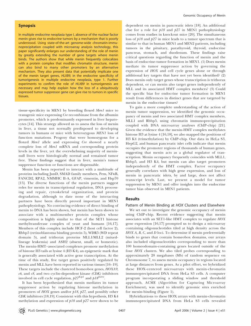
Figure 1. Strategy Used to Analyze Data from Tiled Microarrays
(A and B) Scatterplots of single-array intensities of oligonucleotides obtained from chromatin immunoprecipitation with menin antibodies (B) and acontrol with no antibody (A). Successful experiments are identified as those that show enrichment of multiple probes in the Cy5 channel (chromatinimmoprecipitated DNA) over the Cy3 channel (total genomic DNA).(C) Histogram of mean intensity ratios (log2 scale) from menin chromatin immunoprecipitations from three biological replicates. The distinct tail at theright-hand end corresponds to DNA fragments enriched by menin-ChIP. Genomic sites enriched for factor binding are identified using a computerprogram called ACME. ACME first sorts probes by their genomic location, and then slides a window of user-defined size along tiled regions. ACME thentests whether each window contains a higher than expected number of oligonucletide probes above a user-defined threshold, and then assigns a p-value to each probe on the array. For all arrays used in this study, the window was set to 1,000 bp and the threshold at 90% (indicated by the red bar).(D) Plots showing data before and after processing by ACME. Points in black represent the mean intensity ratio of oligonucleotide probes (right y axis).Points in red indicate corresponding significance values for each data point reported by ACME (left y axis). True-positive signals are represented bymultiple probes that cluster within a given window. Single probes that yield high intensity ratios most likely represent noise and are automaticallyfiltered out by the windowing/threshold analysis.DOI: 10.1371/journal.pgen.0020051.g001
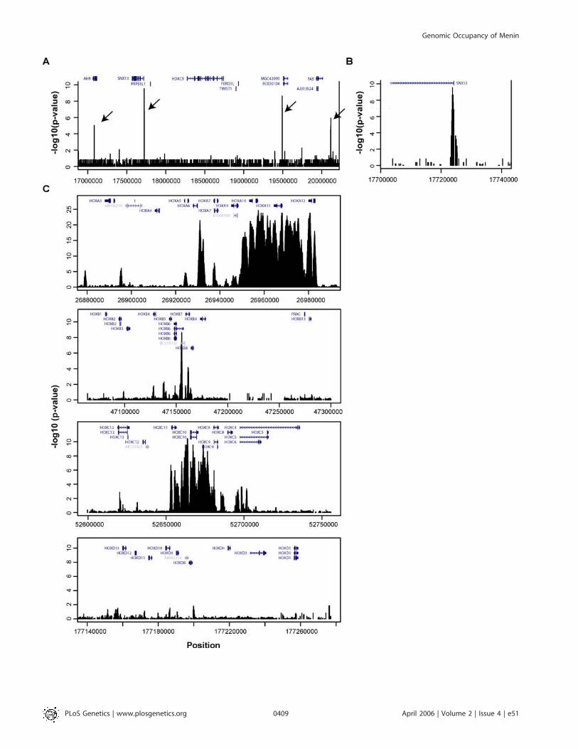
Figure 2. General Pattern of Menin Occupancy in HeLa S3 Cells
(A) Tiled oligonucleotides corresponding to a random 3 MB on Chromosome 7. Four positive signals indicative of menin occupancy are denoted by thearrows and are easily distinguishable from background noise.(B) Expanded view of one positive signal from (A) reveals multiple oligos with significant p-values clustered solely at the transcriptional start site ofSNX13 (Sorting Nexin 13).(C) Pattern of menin occupancy at each of four homeobox clusters.DOI: 10.1371/journal.pgen.0020051.g002
PLoS Genetics | www.plosgenetics.org April 2006 | Volume 2 | Issue 4 | e510408
Genomic Occupancy of Menin
PLoS Genetics | www.plosgenetics.org April 2006 | Volume 2 | Issue 4 | e510409
Genomic Occupancy of Menin
A13. Extensive binding to the HOX C cluster was alsodetected, with broad signals that extended across HOXC11,C10, C9, and portions of C4 and C6. Menin sites were alsodetected near HOXB6 and HOXB7, but the pattern of bindingwas less extensive than that detected at the HOX A and C loci.No significant menin binding was detected at the HOX Dlocus. Menin sites were also detected at several otherhomeobox genes located outside the four HOX clusters, butsimilar to other non-HOX loci, binding was localized to 59
promoter regions (unpublished data). Our finding that meninbinds to HOXA9, C6, and C8 is consistent with prior studies inwhich these three HOX genes were identified by standardChIP-PCR [16,17] and supports the reliability of the ChIP-chip methodology used here for detecting genomic bindingevents by menin.
Genome-Wide Distribution of Menin in Multiple Cell TypesThe results of the pilot study above indicate that, except for
the HOX clusters, menin occupancy is highly specific for 59
regions of genes. Based on these findings, we rationalized thatmenin sites could be identified on a near genome-wide scaleby interrogating only those regions of DNA that correspondto the 59 portions of genes. We therefore designed a secondDNA microarray containing tiling oligonucleotides across the59 regions of approximately 20,000 human genes. Given thebinding pattern of menin at the HOX clusters, this array alsocontained tiled oligos across all four clustered HOX loci, aswell as the complete sequence of 381 genes (�5,000 bp relativeto the transcription start site to þ5,000 bp relative to thetranscription stop site) that showed evidence of meninoccupancy on previous arrays (PCS, unpublished data). Thesepromoter arrays were hybridized with menin-chromatinimmunoprecipitated DNA from human islets, HeLa S3 andHepG2 cells. To assess if the bias for endocrine-tumorformation in MEN1 might be related to differences in distincttarget genes in endocrine cells, we compared sites occupiedin HeLa S3 and HepG2 cells to those identified in humanpancreatic islet cells. As expected, hybridizations with menin-chromatin immunoprecipitated DNA from HeLa S3 cellsrevealed broad signals at the HOX clusters and promoter-specific signals at most other loci (Figure 3A and 3B). InHepG2 and pancreatic islet cells, the vast majority of meninsites were also detected in promoter regions, at or neartranscriptional start sites. Notable differences in the patternof menin binding were detected at the HOX clusters. Whereasmultiple menin sites were detected at the HOX A and Cclusters in HeLa cells, relatively little signal was detected atthese clusters in HepG2 cells and pancreatic islets (Figure 3Aand unpublished data). By using a stringent selectioncriterion of p , 0.0001, we found that menin occupies1,706 sites in HeLa S3 cells, 2,239 sites in HepG2 cells, and1,541 sites in pancreatic islet cells. Sites that were concordantand discordant between each cell type are depicted in a Venndiagram (Figure 3C). The finding that menin binds to a broad
range of human promoters in all three cell types suggests thatmenin has a global role in transcription in both endocrineand non–endocrine-derived cells.
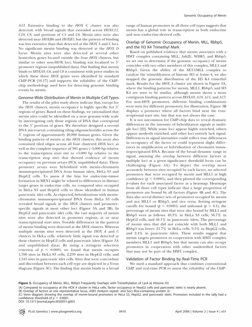
Overlap of Genomic Occupancy of Menin, MLL, Rbbp5,and the H3 K4 Trimethyl MarkBased on published evidence that menin associates with a
HMT complex containing MLL, Ash2L, WDR5, and Rbbp5,we set out to determine if the genomic occupancy of menincoincides with two other members of this complex, MLL1 andRbbp5. Given the ability of the SET1/MLL complex tocatalyze the trimethlyation of histone H3 at lysine 4, we alsomapped the genomic distribution of the H3 K4 trimethylmark. Results for the HOX A cluster are shown in Figure 3A,where the binding patterns for menin, MLL1, Rbbp5, and H3K4 are seen to be similar, although menin shows a morecontiguous binding pattern across HOXA9, A10, A11, and A13.For non-HOX promoters, different binding combinationswere seen for different promoters; for illustration, Figure 3Bdisplays a promoter where all factors bind near the tran-scriptional start site, but that was not always the case.It is not uncommon for ChIP-chip data to reveal dramatic
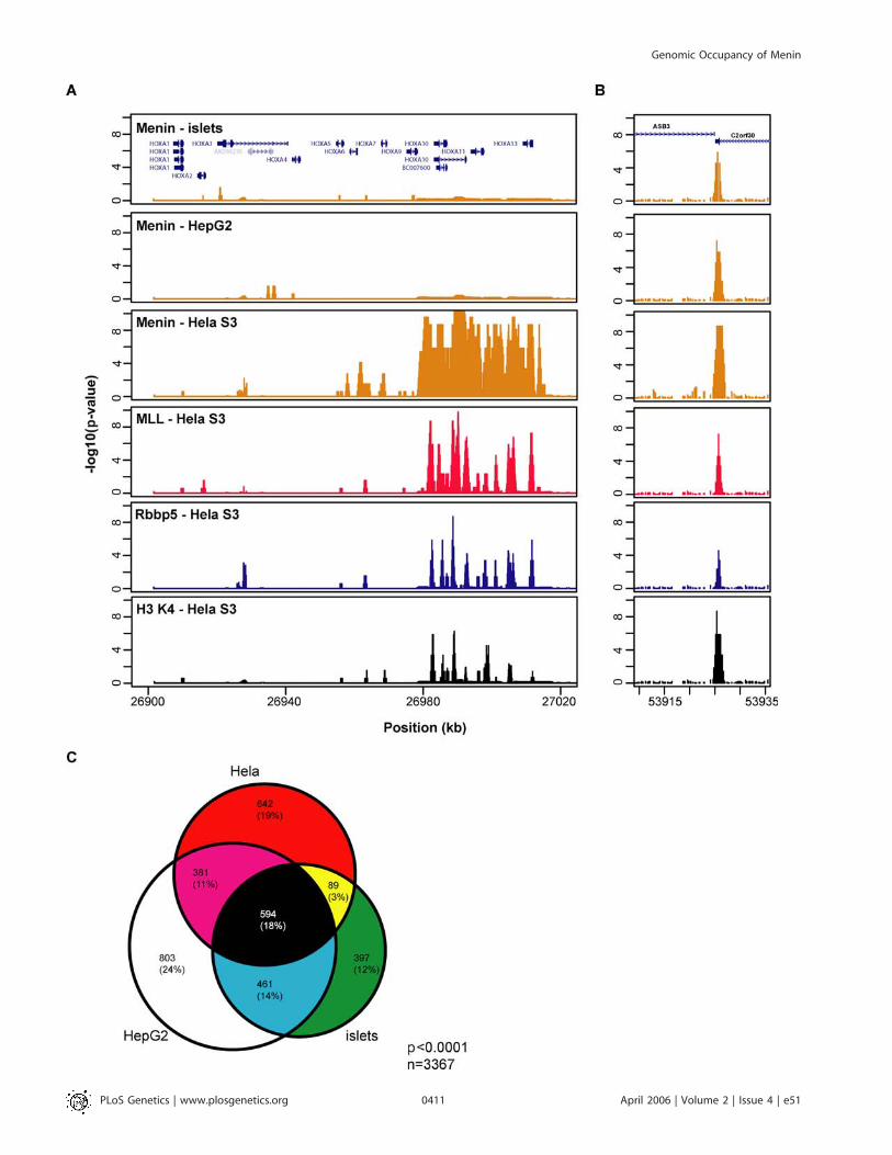
differences in the intensity of hybridization signals at multi-ple loci [22]. While some loci appear highly enriched, othersappear modestly enriched, and other loci entirely lack signal.Differences in signal intensities could reflect true differencesin occupancy of the factor or could represent slight differ-ences in amplification or hybridization of chromatin immu-noprecipitated DNA. Because of the broad dynamic range insignal, assessing the overlap between different factors atmultiple loci at a given significance threshold locus can bechallenging (Figure 4A). To depict the overlap moreaccurately between sites occupied by each factor, we selectedpromoters that were occupied by menin and MLL1 at highconfidence (p , 0.0001), and then plotted the correspondingp-values for each associated factor as a heatmap. Heatmapsfrom all three cell types indicate that a large proportion ofpromoters are bound by all factors (Figure 4B and 4C). Thedata also reveal distinct sets of promoters occupied by meninand not MLL1 or Rbbp5, and vice versa. Setting stringentcutoffs for bound (p , 0.0001) and unbound (p . 0.1), thepercentage of menin sites that were also bound by MLL1 andRbbp5 were as follows: 49.3% in HeLa S3 cells; 56.7% inHepG2 cells, and 46.1% in pancreatic islets. The percentageof menin sites that did not coincide with both MLL1 andRbbp5 was lower: 21.7% in HeLa cells, 9.5% in HepG2 cells,and 2.4% in pancreatic islets. These results suggest thatmenin targets promoters in cooperation with HMT complexmembers MLL1 and Rbbp5, but that menin can also occupypromoters in cooperation with other unidentified factorsthat may not be part of the HMT complex.
Validation of Factor Binding by Real-Time PCRWe used a standard approach that combines conventional
ChIP and real-time PCR to assess the reliability of the ChIP-
Figure 3. Occupancy of Menin, MLL, Rbbp5 Frequently Overlaps with Trimethylation of Lys4 at Histone H3
(A) Compared to occupancy at the HOX A cluster in HeLa cells, factor occupancy in HepG2 cells and pancreatic islets is nearly absent.(B) Overlap of factors at one representative locus, ASB3 (Ankyrin repeat- and Socs Box-containing protein 3).(C) Venn diagram showing the overlap of menin-bound promoters in HeLa S3, HepG2, and pancreatic islets. Promoters included in the tally had aconfidence threshold of p , 0.0001.DOI: 10.1371/journal.pgen.0020051.g003
PLoS Genetics | www.plosgenetics.org April 2006 | Volume 2 | Issue 4 | e510410
Genomic Occupancy of Menin
PLoS Genetics | www.plosgenetics.org April 2006 | Volume 2 | Issue 4 | e510411
Genomic Occupancy of Menin
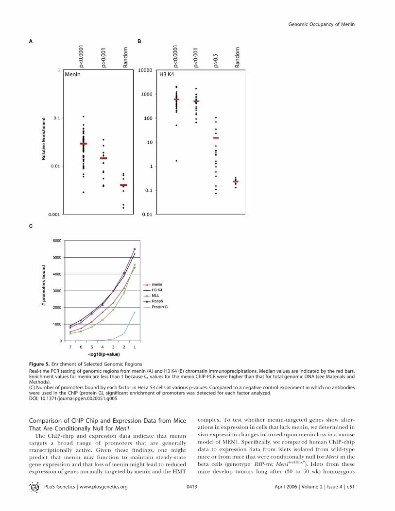
chip method and our approach for analysis of the rawmicroarray data (Figures 4D and 5). We arbitrarily selected 67promoter regions that were identified by our windowing andthreshold analyses to be enriched for menin binding at p ,
0.0001. These sites were compared to 15 random promoterregions (0.001 , p , 1) and seven random regions of thegenome located outside of promoter regions. On average,promoters that had a p-value of ,0.0001 were enriched 7.25-fold over random regions (Figure 5A). Furthermore, of the 67sites that were shown to be enriched by ChIP-chip (p ,
0.0001), 64 (95.5%) were confirmed by real-time PCR to haveenrichment ratios that were greater than those detected forthe randomly selected amplified regions. These data indicatethat sites we determined to be enriched at p , 0.0001 byChIP-chip reliably detect enriched sites of menin binding.Many factor-bound sites probably also exist within the p ¼0.01 to 0.001 confidence interval, but negative control studiessuggest that false-positive rates increase as p-values rise above0.0001 (Figure 5C). Relative enrichment using antibodies toH3 K4 was also tested (Figure 5B). Consistent with the tight
physical association between DNA and histones, enrichmentof H3 K4–bound sites was greater than that for menin sites.
Comparison of ChIP-Chip and Gene ExpressionThe ChIP-chip data in all three cell types indicate that
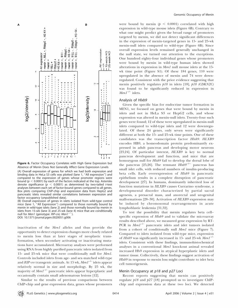
menin occupancy frequently coincides with trimethylation ofhistone H3 at lysine 4, a mark that is generally associatedwith a transcriptionally active state of chromatin. Theseresults prompted us to assess whether factor occupancycorrelates with high levels of gene expression. A systematiccomparison between ChIP-chip and expression data in allthree cell types revealed that genes whose promoters arebound by menin correlate with higher mRNA expressionlevels (Figure 6A and unpublished data). This was true forgene subsets both where menin bound concordantly withMLL and Rbbp5 and where menin bound without thesefactors. These data suggest that menin, in both the presenceand absence of the HMT complex, is generally associatedwith transcriptionally active promoters.
Figure 4. Menin Sites Can Be Bound in the Presence or Absence of HMT Complex Members
(A) Comparison of promoters bound by menin and Rbbp5 showing the broad range in signal intensity for each factor. The�log10 p-value for each siteis plotted on the x and y axes. Vertical and horizontal lines represent confidence thresholds at p , 0.0001. Points in the lower left quadrant representpromoters that are bound by neither menin nor Rbbp5. Points in the upper right quadrant represent promoters occupied by both menin and Rbbp5.Plots comparing menin to MLL and H3 K4 appeared similar (unpublished data).(B and C) Heat maps illustrating the overlap of factor-occupied promoters. Promoters bound by either menin (B) or MLL (C) at the p , 0.0001confidence threshold in HeLa S3 cells were plotted with corresponding p-values for each indicated factor. The heat maps reveal subsets of genes thatare bound by all factors (blue brackets), in addition to subsets that are bound by menin or MLL but not the other factors (green brackets).(D) Real-time PCR validation of randomly selected promoters determined by ChIP-chip to be bound by menin and not Rbbp5 (left), and vice versa(right). Each pair of bars corresponds to a unique promoter region.DOI: 10.1371/journal.pgen.0020051.g004
PLoS Genetics | www.plosgenetics.org April 2006 | Volume 2 | Issue 4 | e510412
Genomic Occupancy of Menin
Comparison of ChIP-Chip and Expression Data from MiceThat Are Conditionally Null for Men1
The ChIP-chip and expression data indicate that menintargets a broad range of promoters that are generallytranscriptionally active. Given these findings, one mightpredict that menin may function to maintain steady-stategene expression and that loss of menin might lead to reducedexpression of genes normally targeted by menin and the HMT
complex. To test whether menin-targeted genes show alter-ations in expression in cells that lack menin, we determined invivo expression changes incurred upon menin loss in a mousemodel of MEN1. Specifically, we compared human ChIP-chipdata to expression data from islets isolated from wild-typemice or frommice that were conditionally null forMen1 in thebeta cells (genotype: RIP-cre; Men1loxP/loxP). Islets from thesemice develop tumors long after (30 to 50 wk) homozygous
Figure 5. Enrichment of Selected Genomic Regions
Real-time PCR testing of genomic regions from menin (A) and H3 K4 (B) chromatin immunoprecipitations. Median values are indicated by the red bars.Enrichment values for menin are less than 1 because Ct values for the menin ChIP-PCR were higher than that for total genomic DNA (see Materials andMethods).(C) Number of promoters bound by each factor in HeLa S3 cells at various p-values. Compared to a negative control experiment in which no antibodieswere used in the ChIP (protein G), significant enrichment of promoters was detected for each factor analyzed.DOI: 10.1371/journal.pgen.0020051.g005
PLoS Genetics | www.plosgenetics.org April 2006 | Volume 2 | Issue 4 | e510413
Genomic Occupancy of Menin
inactivation of the Men1 alleles and thus provide theopportunity to detect expression changes more closely relatedto menin loss than at later stages of neoplastic trans-formation, when secondary activating or inactivating muta-tions have accumulated. Microarray analyses were performedusing RNA from highly purified pancreatic islets isolated from15- and 25-wk mice that were conditionally null for Men1.Controls included islets from age- and sex-matched wild-typeand RIP-cre transgenic animals. At 15 wk,Men1�/� islets appearrelatively normal in size and morphology. By 25 wk, themajority of Men1�/� pancreatic islets appear hyperplastic andoccasionally contain small adenomatous lesions [12].
Similar to the results of previous comparisons betweenChIP-chip and gene expression data, genes whose promoters
were bound by menin (p , 0.0001) correlated with highexpression in wild-type mouse islets (Figure 6B). Contrary towhat one might predict given the broad range of promoterstargeted by menin, we did not detect significant differencesin the expression of menin-targeted genes in 15- and 25-wkmenin-null islets compared to wild-type (Figure 6B). Sinceoverall expression levels remained generally unchanged inthe null state, we turned our attention to the exceptions.One hundred eighty-four individual genes whose promoterswere bound by menin in wild-type human islets showedalterations in expression in Men1 null mouse islets at the 15-wk time point (Figure S1). Of these 184 genes, 110 wereupregulated in the absence of menin and 74 were down-regulated. Consistent with the prior evidence suggesting thatmenin positively regulates p18 in islets [18], p18 (CDKN2C)was found to be significantly reduced in expression inMen1�/� islets.
Analysis of Hlxb9Given the specific bias for endocrine tumor formation in
MEN1, we focused on genes that were bound by menin inislets and not in HeLa S3 or HepG2 cells, and whoseexpression was altered in menin-null islets. Twenty-four suchgenes were found; 12 of these were upregulated in menin-nullislets compared to wild-type islets and 12 were downregu-lated. Of these 24 genes, only seven were significantlydifferent at both the 15- and 25-wk time points. One of thesecandidates was the transcription factor Hlxb9. HLXB9encodes HB9, a homeodomain protein predominantly ex-pressed in adult pancreas and developing motor neurons[23,24]. Of particular interest, HLXB9 is key to normalpancreas development and function, and mice that arehomozygous null for Hlxb9 fail to develop the dorsal lobe ofthe pancreas [25,26]. The remnant Hlxb9�/�- pancreas hassmall islet cells, with reduced numbers of insulin-producingbeta cells. Early overexpression of Hlxb9 in pancreaticepithelium results in a complete disruption of pancreaticdevelopment [27]. In humans, dominantly inherited loss offunction mutations in HLXB9 causes Currarino syndrome, adevelopmental disorder characterized by partial sacralagenesis, a presacral mass, and anorectal and urogenitalmalformations [28–30]. Activation of HLXB9 expression maybe induced by chromosomal rearrangements in acutelymphoblastic leukemia [31,32].To test the possibility that menin regulates beta cell–
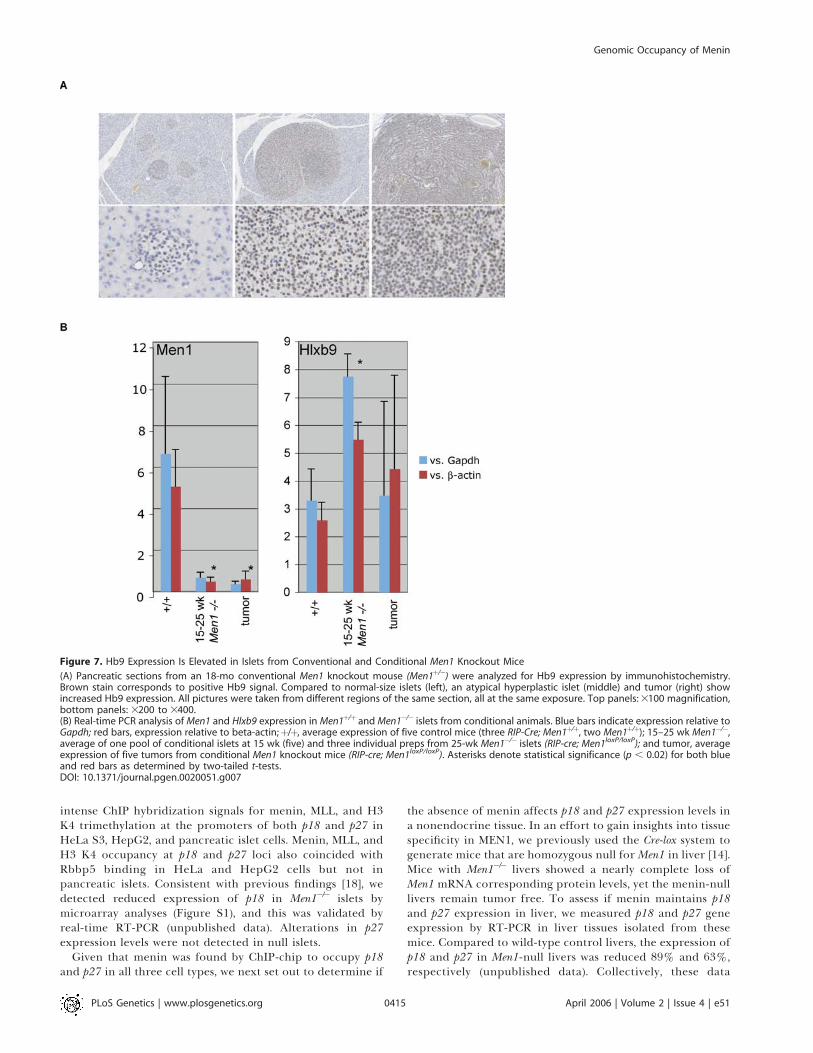
specific expression of Hlxb9 and to validate the microarrayresults described above, we measured gene expression by RT-PCR in Men1�/� pancreatic islets and islet tumors isolatedfrom a cohort of conditionally null Men1 mice (Figure 7).Compared to islets isolated from wild-type mice, expressionof Hlxb9 was significantly increased in 15- and 25-wk Men1�/�
islets. Consistent with these findings, immunohistochemicalanalyses in a conventional Men1 knockout animal revealedincreased Hb9 expression in atypical hyperplastic islets andtumor tissue. Collectively, these findings suggest activation ofHlxb9 in response to menin loss might contribute to islet betacell tumorigenesis.
Menin Occupancy at p18 and p27 LociRecent reports suggesting that menin can positively
regulate p18 and p27 [18] prompted us to investigate ChIP-chip and expression data at these two loci. We detected
Figure 6. Factor Occupancy Correlates with High Gene Expression but
Absence of Menin Does Not Generally Affect Gene Expression Levels
(A) Overall expression of genes for which we had both expression andbinding data in HeLa S3 cells was plotted (lane 1, ‘‘All expression’’) andcompared to the expression of genes whose promoter regions werebound (p , 0.0001) by each of the factors indicated at the top. Asterisksdenote significance (p , 0.0001) as determined by two-tailed t-testanalyses between each set of factor-bound genes compared to all genes.Box plots comparing ChIP-chip and expression data from HepG2 andpancreatic islets revealed similar correlations between expression andfactor occupancy (unpublished data).(B) Overall expression of genes in islets isolated from wild-type controlmice (lane 1, ‘‘All Expression’’) compared to those normally bound bymenin in wild-type islets (lane 2) and those normally bound by menin inislets from 15-wk (lane 3) and 25-wk (lane 4) mice that are conditionallynull for Men1 (genotype: RIP-cre; Men1�/�).DOI: 10.1371/journal.pgen.0020051.g006
PLoS Genetics | www.plosgenetics.org April 2006 | Volume 2 | Issue 4 | e510414
Genomic Occupancy of Menin
intense ChIP hybridization signals for menin, MLL, and H3K4 trimethylation at the promoters of both p18 and p27 inHeLa S3, HepG2, and pancreatic islet cells. Menin, MLL, andH3 K4 occupancy at p18 and p27 loci also coincided withRbbp5 binding in HeLa and HepG2 cells but not inpancreatic islets. Consistent with previous findings [18], wedetected reduced expression of p18 in Men1�/� islets bymicroarray analyses (Figure S1), and this was validated byreal-time RT-PCR (unpublished data). Alterations in p27expression levels were not detected in null islets.
Given that menin was found by ChIP-chip to occupy p18and p27 in all three cell types, we next set out to determine if
the absence of menin affects p18 and p27 expression levels ina nonendocrine tissue. In an effort to gain insights into tissuespecificity in MEN1, we previously used the Cre-lox system togenerate mice that are homozygous null forMen1 in liver [14].Mice with Men1�/� livers showed a nearly complete loss ofMen1 mRNA corresponding protein levels, yet the menin-nulllivers remain tumor free. To assess if menin maintains p18and p27 expression in liver, we measured p18 and p27 geneexpression by RT-PCR in liver tissues isolated from thesemice. Compared to wild-type control livers, the expression ofp18 and p27 in Men1-null livers was reduced 89% and 63%,respectively (unpublished data). Collectively, these data
Figure 7. Hb9 Expression Is Elevated in Islets from Conventional and Conditional Men1 Knockout Mice
(A) Pancreatic sections from an 18-mo conventional Men1 knockout mouse (Men1þ/�) were analyzed for Hb9 expression by immunohistochemistry.Brown stain corresponds to positive Hb9 signal. Compared to normal-size islets (left), an atypical hyperplastic islet (middle) and tumor (right) showincreased Hb9 expression. All pictures were taken from different regions of the same section, all at the same exposure. Top panels: 3100 magnification,bottom panels: 3200 to 3400.(B) Real-time PCR analysis of Men1 and Hlxb9 expression in Men1þ/þ and Men1�/� islets from conditional animals. Blue bars indicate expression relative toGapdh; red bars, expression relative to beta-actin;þ/þ, average expression of five control mice (three RIP-Cre; Men1þ/þ, two Men1þ/þ); 15–25 wk Men1�/�,average of one pool of conditional islets at 15 wk (five) and three individual preps from 25-wk Men1�/� islets (RIP-cre; Men1loxP/loxP); and tumor, averageexpression of five tumors from conditional Men1 knockout mice (RIP-cre; Men1loxP/loxP). Asterisks denote statistical significance (p , 0.02) for both blueand red bars as determined by two-tailed t-tests.DOI: 10.1371/journal.pgen.0020051.g007
PLoS Genetics | www.plosgenetics.org April 2006 | Volume 2 | Issue 4 | e510415
Genomic Occupancy of Menin
support the conclusion that menin enhances expression ofp18 and p27 in both pancreatic islets and liver. Since theabsence of menin gives rise to tumors only in endocrinepancreas, not in liver, mechanisms beyond simple dysregula-tion of p18 and p27 must be involved.
Discussion
We set out to investigate menin’s role as a transcriptionalregulator by identifying genes bound by and dependent onmenin. Prior to this study, the only genes reported to betargeted by menin and MLL were Hoxc6, Hoxc8, HOXA9, p27,and p18. Our findings indicate that menin does not onlytarget specific classes of genes like HOX and CDK inhibitorsbut that menin also targets a very broad range of promotersin multiple tissues. This expands the realm of menin-targetedgenes several hundred-fold. We find that menin occupancyfrequently coincides with members of the HMT complex,MLL1 and Rbbp5, as well as the H3 K4 trimethyl mark, raisingthe possibility that menin functions as a general transcrip-tional regulator that helps maintain stable gene expression.Averaged over thousands of genes, menin correlates withhigher gene expression, but overall expression levels do notdecrease upon loss of menin in null islets. Surprisingly, meninbinds many regions of the genome independently of the HMTcomplex members, suggesting that menin may regulatetranscription by cooperating with other, currently unknownproteins. Possible candidates include JunD, SMADs, NFjB, orother menin-associated transcription factors not part of theSET1/MLL complex.
A fundamental question for many tumor suppressorsyndromes is the tissue specificity of tumor formation. Meninis expressed in nearly all tissues. Why do MEN1-associatedtumors arise primarily in endocrine organs? Could theinteractions between menin, MLL, and HOX gene expressionsuggest an explanation for the endocrine specificity?
MLL is an important regulator of HOX gene expression,and leukemic chromosomal translocations involving MLLappear to act by creating chimeric fusion proteins thatconstitutively activate HOX genes [33–35]. The persistentexpression of HOX genes results in a failure of terminaldifferentiation, and studies in mice have shown that over-expression of certain HOX genes, Hoxa9 and Meis1 inparticular, leads to leukemogenesis [36,37]. Previous studieshave also shown that menin can positively regulate HOXA9 inHeLa cells and Hoxc6 and Hoxc8 in developing mouse embryo[16,17]. Collectively, these findings have led to the enticinghypothesis that tumorigenesis in MEN1, like leukemogenesis,is related to dysregulation of HOX genes normally bound bymenin and MLL. If this hypothesis is valid, however, we wouldhave expected to see menin occupancy at the HOX clusters inpancreatic islets. While we did detect extensive binding ofmenin (and MLL) to the HOX A and C clusters in HeLa S3cells, virtually no signal was detected in either HepG2 cells orpancreatic islets. Moreover, the clustered homeobox genesare expressed at almost undetectable levels in pancreaticislets, and none of them significantly change in expressionfollowing loss of menin. Given these findings and thecontrasts of menin function in myeloid versus endocrinetumors, we believe it is unlikely that dysregulation of HOXgenes in islet cells contributes to MEN1 islet neoplasms.
If HOX genes are not the mediator, then how does menin
loss lead to endocrine tumors? Menin was recently proposedto regulate pancreatic islet growth by promoting histonemethylation and expression of cyclin-dependent kinaseinhibitors p27 and p18 [18,19]. Moreover, the simultaneousloss of p18 and p27 in mice was shown to lead to developmentof tumors in many of the same endocrine tissues as humanswith inherited mutations in the MEN1 and MEN2 genes [20].These studies argue for a major role for p18 and p27 in thetissue-specific tumor phenotype in MEN1. Specifically, loss ofmenin prevents H3 K4 methylation at the promoters of p18and p27, reducing their expression, and ultimately abrogatingtheir inhibitory effect on cyclin-dependent kinases (CDK2and CDK4) and resulting in unrestrained cell growth.Consistent with this hypothesis, we detected menin at thepromoters of p18 and p27 and reduced p18 expression in isletcells that were conditionally null for Men1. However, p18 andp27 concentrations were reduced in Men1�/� liver, which isnot susceptible to developing tumors in heterozygous MEN1patients, or in mice rendered null for menin in liver by aconditional knockout. A separate study showed that menincan regulate p18 and p27 expression in fibroblasts [19], yet thephenotype of MEN1 does not include fibrosarcoma. Theseresults indicate that while dysregulation of p18 and p27 maycontribute to neoplastic transformation in MEN1, these genesalone cannot account for the specific bias for endocrinetumor formation in MEN1.To identify other possible genetic causes for the tissue
specificity of tumor formation, we identified a relatively smallnumber of genes that were both bound by menin and alteredin expression in menin-null islets. One of these genes was thedevelopmentally programmed HLXB9, which codes for tran-scription factor HB9. Hb9 expression was significantlyelevated in pancreatic islets in the absence of menin,suggesting that menin is a transcriptional repressor of
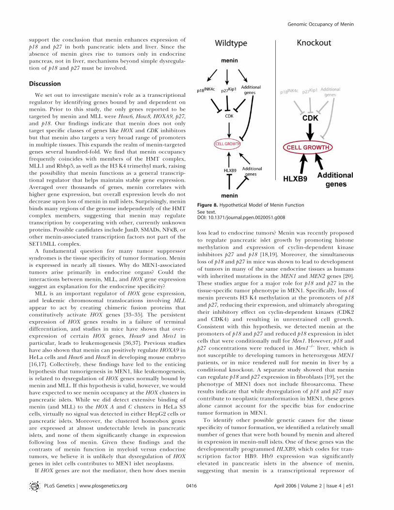
Figure 8. Hypothetical Model of Menin Function
See text.DOI: 10.1371/journal.pgen.0020051.g008
PLoS Genetics | www.plosgenetics.org April 2006 | Volume 2 | Issue 4 | e510416
Genomic Occupancy of Menin

HLXB9. It is also noteworthy that HLXB9 was bound bymenin only in islets, and not in HeLa or HepG2 cells. Thesefindings raise the possibility that the specific bias forendocrine tumor formation in MEN1 could result fromchanges in expression in distinct genes, including HLXB9,that are specifically targeted by menin in endocrine tissues.
Based on our findings and the work of others, we proposethe following model for tumor suppression by menin inpancreatic islets (Figure 8). In both endocrine and non-endocrine tissues, menin mediates transcriptional activationof genes that inhibit cell growth, including, but not limited to,the cyclin-dependent kinase inhibitors p18 and p27. Inaddition, menin can function as a corepressor of tissue-specific genes that promote cell growth, including HLXB9.Menin can mediate its regulatory functions in the presence ofthe HMT complexes or other menin partners. Endocrineneoplasia as a consequence of menin loss results from thecombined effects of decreased p18 and p27 and increasedHLXB9 expression. Additional tissue-specific factors that aredysregulated upon menin loss probably also contribute toneoplasia. Similar studies in additional tissues, such asanterior pituitary or parathyroid, may help reveal thecomplete list of critical targets.
Materials and Methods
Antibodies for chromatin immunoprecipitation. Affinity-purifiedrabbit polyclonal anti-menin (BL342), anti-Rbbp5 (BL766), and anti-MLL1 (BL1289) antibodies were obtained from Bethyl Laboratories(Montgomery, Texas, United States). The menin antibody recognizesan epitope at the C terminus of menin. The Rbbp5 antibody wasraised against a portion of human Rbbp5 encoded within exons 13and 14. The epitope recognized by anti-MLL1 maps to a regionbetween residues 720 and 780 of MLL1. This epitope is found in theN-terminal 300-kDa fragment generated by proteolytic cleavage.Histone H3 K4 antibody (ab8580; Abcam, Cambridge, UnitedKingdom) reacts to trimethylated H3 K4 and shows weak reactivityto dimethylated H3 K4.
Microarrays used for ChIP-chip. Two different DNA tiling micro-array designs were used in this study. Both microarrays were printedby NimbleGen (Madison, Wisconsin, United States). The first arraycontained 190,181 oligonucleotides (70 nucleotides in length)representing HOX clusters A, B, C, and D, and approximately 230loci tiled at an average resolution of one oligo every 150 to 350 bp.Roughly half of these 230 loci harbored genes whose protein productscontain homeodomains. The remaining half were identified aspositive ‘‘hits’’ in a pilot study using a previously described conven-tional microarray that contained portions of promoter regions of13,000 human genes (unpublished data). For each of these 230 genes,oligonucleotides spanning�10 kb relative to the transcriptional startsite to þ10 kb relative to the end of the transcript were tiled. Thecoordinates for DNA corresponding to the HOX clusters based on theJuly 2003 human genome assembly are as follows: HOX A:Chromosome 7: 16,875,358 to 36,981,974; HOX B: Chromosome 17:47,064,003 to 47,300,285; HOX C: Chromosome 12: 52,599,536 to52,755,746; HOX D: Chromosome 2: 177,140,087 to 177,277,119. Thesecond tiling microarray we designed contained oligonucleotidestiled across 19,928 promoter regions. The oligonucleotides weredistributed at a resolution of approximately one 50 mer every 100 bp(1.5 kb approximately each promoter region). This array alsocontained tiling oligonucleotides across 381 genes (�5 kb relative toeach transcriptional start site to þ5 kb relative to the end of eachtranscript) at an average resolution of one oligo every 180 bp. Thearray also harbored tiling oligonucleotides across all four HOXclusters. The coordinates for DNA corresponding to the HOX clusterswere based on the May 2004 human genome assembly and are asfollows: HOX A: Chromosome 7: 26,901,743 to 27,173,542; HOX B:Chromosome 17: 43,958,347 to 44,167,626; HOX C: Chromosome 12:52,616,715 to 52,738,384; HOX D: Chromosome 2: 176,780,780 to176,883,479.
ChIP-chip. The protocol described here was adapted frompreviously published studies [21,38]. Briefly, for each ChIP-chip
experiment, 1 to 23108 cells were crosslinked with 1% formaldehydefor 20 min at room temperature, harvested, and rinsed with 13 PBS.Cell nuclei were isolated, pelleted, and sonicated. DNA fragmentswere enriched by immunoprecipitation with factor-specific anti-bodies. After heat-reversal of the crosslinking, the enriched DNA wasamplified by ligation-mediated PCR (LM-PCR) and then fluorescentlylabeled by using Klenow polymerase and Cy5-labeled dUTP (Amer-sham Biosciences, Piscataway, New Jersey, United States). A sample ofDNA that was not enriched by immunoprecipitation was subjected toLM-PCR and labeled with Cy3-dUTP. ChIP-enriched and unenriched(input) labeled samples were cohybridized to microarrays. Micro-arrays were hybridized 18 to 20 h at 45 8C, washed according to theprotocol described by NimbleGen, and scanned using an AgilentTechnologies (Palo Alto, California, United States) microarrayscanner. As a pilot study, the first custom-designed ‘‘HOX’’ arraywas hybridized with menin chromatin immunoprecipitated DNAfrom HeLa S3 cells from a single experiment. For experiments usingthe second, more promoter-oriented array design, three biologicalreplicates were performed for experiments in HeLa S3 and HepG2cells. In purified pancreatic islet preps, menin ChIP-chip wasperformed in biological quadruplicate, and Rbbp5 and H3 K4experiments in duplicate. Due to limited availability of primary islettissue, ChIP-chip in islet cells using antibodies to MLL1 was doneonce. Islet preparations were treated with formaldehyde in culturemedia between 1 h and 5 d after isolation from pancreata, which werederived from brain-dead organ donors (National Institutes of Health,Institutional Review Board exemption issued by National Institutes ofHealth Office of Human Subjects [IRB Exemption No. 3072]). Foreach ChIP-chip experiment, approximately 30,000 viable isletequivalents were fixed and handled as described above. Islet purityranged from 40% to 90% with 60% to 90% viability.
Analysis of ChIP tiling array data. Raw array data were normalizedusing bi-weight mean using the NimbleScan Version 2.1 software(NimbleGen Systems). Log2 ratios (cy5/cy3) from biological replicateswere averaged. To identify potential sites of enrichment, a windowand threshold strategy was used (Figure 1). Briefly, a 1,000-bp windowwas moved stepwise along the tiled region, centering at every probe.Hybridization signals of probes within each window were tested by v2
analysis to determine if the window contained a higher than expectednumber of probes. These calculations resulted in a single p-valueassociated with each averaged data point. Analyses performed atdifferent window sizes and thresholds yielded similar results(unpublished data). All raw and processed ChIP-chip data is publiclyavailable from Gene Expression Omnibus (GEO) (http://www.ncbi.nlm.nih.gov/geo). The p-value scores for each promoter tested areincluded. The windowing and threshold program we developed,which we named ACME (Algorithm for Capturing MicroarrayEnrichment), is available from the authors upon request.
Real-time PCR validation. Standard ChIP with menin andtrimethyl H3 K4 antibodies was performed on HeLa S3 cells induplicate. PCR primer pairs were designed to amplify 150- to 200-bpfragments from selected genomic regions. For menin, we compared67 promoters determined by ChIP-chip to be enriched for meninbinding at p , 0.0001 to 15 random promoter regions (0.001 , p , 1)and seven random regions located outside of promoter regions. ForH3 K4, we tested 38 promoters that were determined by ChIP-chip tobe highly enriched (p , 0.0001), 20 marginally enriched genes (0.0001, p , 0.001), 17 unenriched promoter regions (p . 0.5), and sevenrandom regions.
Real-time PCRs were carried out in duplicate on each chromatinimmunoprecipitated and input DNA sample using SYBR green PCRmix (Qiagen, Valencia, California, United States) in an AppliedBiosystems 7900HT Fast Real Time-PCR machine (Foster City,California, United States). DNA obtained from menin chromatinimmunoprecipitations was very limited in quantity and could not bequantitated by standard methods. As a result, unequal quantities ofChIP and input DNA were added to PCRs. To account for thedifferences in DNA quantity, for every genomic region studied, a DCtvalue was calculated for each sample by subtracting the Ct value forchromatin immunoprecipitated sample from the Ct value obtainedfor the input. Raising 2 to the DCt power yielded the relative amountof PCR product (relative enrichment). Average values for promotersoccupied by either menin or H3 K4 were compared to thosecalculated for unoccupied promoters and random regions of thegenome. All primer sequences are available upon request.
Comparison of ChIP-chip and expression data. ChIP-chip datafrom islet cells were merged to Affymetrix (Santa Clara, California,United States) islet expression data from the Novartis (version 2) dataset [39]. HeLa S3 and HepG2 ChIP-chip data were merged toexpression data generated in our laboratory using Affymetrix U133A
PLoS Genetics | www.plosgenetics.org April 2006 | Volume 2 | Issue 4 | e510417
Genomic Occupancy of Menin
Plus microarrays (HeLa S3 and HepG2). All ChIP-chip and expressiondatasets were merged by gene symbol. For all three cell types, tworeplicate datasets were averaged. Average raw intensity values wereconverted to log base 2 and normalized using the robust multiarrayaverage method (RMA) [40]. Data for genes represented by multipleprobes were retained in the dataset. This analysis gave 10,148 genes inislet cells and 10,078 genes in HeLa S3 and HepG2 cells for which wehad both binding and expression data.
Microarray analyses were also performed using RNA from highlypure islets prepared from 15-wk-old (n ¼ 5) and 25-wk-old (n ¼ 3)female mice that were conditionally null for Men1 (genotype: RIP-cre,Men1loxP/loxP). Controls included islet RNA prepared from sex-matched wild-type (n ¼ 3) and RIP-cre (n ¼ 2) mice at 15 wk of age.Total RNA isolated from each group of mice was pooled andsubjected to two rounds of amplification three times. Biotin-labeledcRNA was then purified, fragmented, and hybridized to MOE430AAffymetrix GeneChip probe arrays containing more than 22,000probe sets. Two-tailed t-tests using Microsoft Excel were used tocompare expression values from wild-type and RIP-cre controls to15-wk conditionally null mice (RIP-cre, Men1loxP/loxP). The mouseexpression data were merged to the human ChIP-chip data, yielding5,519 unique genes for which we had both expression and bindingdata.
Immunohistochemistry. The pancreas from an 18-mo heterozy-gous Men1 knockout mouse (Men1þ/�) was removed, immediately fixedin 4% paraformaldehyde, and embedded in paraffin. Immunohisto-chemistry of pancreatic sections from an 18-mo conventional Men1knockout mouse (Men1þ/�) was performed following antigen retrievalusing previously described antibodies to Hb9 [25].
Supporting Information
Figure S1. The 184 Genes That Were Found to Separate 15-wk Men1-
Null Islets from Controls Are Depicted in the Red-Green Color-Coded Plot (p , 0.01)
Relative expression levels have been pseudo-colored red and green,with red corresponding to high expression and green correspondingto low expression. The color-coded plot to the right of the expressiondata represents ChIP-chip p-value binding data for each factor. Colorkey: red, p , 0.0001; orange, 0.0001 . p , 0.001; gold, 0.001 . p ,0.01; light yellow, 0.01 . p ,0.1; white, p . 0.1. Twenty-four geneswhose promoters were bound by menin exclusively in islets aredepicted in red and blue. Those in red show statistically significantdifferences in expression at both the 15- and 25-wk time points (p ,0.01).Found at DOI: 10.1371/journal.pgen.0020051.sg001 (142 KB PDF).
Acknowledgments
We give special thanks to Richard Young, Mike Erdos, Darryl Leja,and Yuan Jiang for assistance and helpful discussions. We also thankNimbleGen Systems, Inc. for assistance with tiled microarray design,especially Roland Green and Mike Singer. Primary human pancreaticislet isolates were provided by the Islet Cell Resource Centers.
Author contributions. PCS conceived and designed the experi-ments. PCS, SP, and MJH performed the experiments. PCS, SD, SKA,and FSC analyzed the data. PCS, DTO, GEC, SKA, PSM, and FSCcontributed reagents/materials/analysis tools. SJM and AMS advisedand contributed intellectually. PCS wrote the paper.
Funding. This research was supported by grants from the NationalInstitutes of Health (DTO: NIDDK K25-DK070813; NIDDK R01-DK068655) and the Intramural Research Program of the NationalHuman Genome Research Institute, National Institutes of Health.
Competing interests. The authors have declared that no competinginterests exist. &
References1. Knudson AG Jr. (1971) Mutation and cancer: Statistical study of
retinoblastoma. Proc Natl Acad Sci U S A 68: 820–823.2. Debelenko LV, Brambilla E, Agarwal SK, Swalwell JI, Kester MB, et al. (1997)
Identification of MEN1 gene mutations in sporadic carcinoid tumors of thelung. Hum Mol Genet 6: 2285–2290.
3. Farnebo F, Teh BT, Kytola S, Svensson A, Phelan C, et al. (1998) Alterationsof the MEN1 gene in sporadic parathyroid tumors. J Clin Endocrinol Metab83: 2627–2630.
4. Heppner C, Kester MB, Agarwal SK, Debelenko LV, Emmert-Buck MR, etal. (1997) Somatic mutation of the MEN1 gene in parathyroid tumours. NatGenet 16: 375–378.
5. Zhuang Z, Ezzat SZ, Vortmeyer AO, Weil R, Oldfield EH, et al. (1997)Mutations of the MEN1 tumor suppressor gene in pituitary tumors. CancerRes 57: 5446–5451.
6. Zhuang Z, Vortmeyer AO, Pack S, Huang S, Pham TA, et al. (1997) Somaticmutations of the MEN1 tumor suppressor gene in sporadic gastrinomasand insulinomas. Cancer Res 57: 4682–4686.
7. Guru SC, Goldsmith PK, Burns AL, Marx SJ, Spiegel AM, et al. (1998)Menin, the product of the MEN1 gene, is a nuclear protein. Proc Natl AcadSci U S A 95: 1630–1634.
8. Bertolino P, Tong WM, Galendo D, Wang ZQ, Zhang CX (2003) Hetero-zygous Men1 mutant mice develop a range of endocrine tumors mimickingmultiple endocrine neoplasia type 1. Mol Endocrinol 17: 1880–1892.
9. Crabtree JS, Scacheri PC, Ward JM, Garrett-Beal L, Emmert-Buck MR, et al.(2001) A mouse model of multiple endocrine neoplasia, type 1, developsmultiple endocrine tumors. Proc Natl Acad Sci U S A 98: 1118–1123.
10. Bertolino P, Tong WM, Herrera PL, Casse H, Zhang CX, et al. (2003)Pancreatic beta-cell-specific ablation of the multiple endocrine neoplasiatype 1 (MEN1) gene causes full penetrance of insulinoma development inmice. Cancer Res 63: 4836–4841.
11. Biondi CA, Gartside MG, Waring P, Loffler KA, Stark MS, et al. (2004)Conditional inactivation of the MEN1 gene leads to pancreatic andpituitary tumorigenesis but does not affect normal development of thesetissues. Mol Cell Biol 24: 3125–3131.
12. Crabtree JS, Scacheri PC, Ward JM, McNally SR, Swain GP, et al. (2003) Ofmice and MEN1: Insulinomas in a conditional mouse knockout. Mol CellBiol 23: 6075–6085.
13. Scacheri PC, Kennedy AL, Chin K, Miller MT, Hodgson JG, et al. (2004)Pancreatic insulinomas in multiple endocrine neoplasia, type I knockoutmice can develop in the absence of chromosome instability or micro-satellite instability. Cancer Res 64: 7039–7044.
14. Scacheri PC, Crabtree JS, Kennedy AL, Swain GP, Ward JM, et al. (2004)Homozygous loss of menin is well tolerated in liver, a tissue not affected inMEN1. Mamm Genome 15: 872–877.
15. Agarwal SK, Kennedy PA, Scacheri PC, Novotny EA, Hickman AB, et al.
(2005) Menin molecular interactions: Insights into normal functions andtumorigenesis. Horm Metab Res 37: 369–374.
16. Hughes CM, Rozenblatt-Rosen O, Milne TA, Copeland TD, Levine SS, et al.(2004) Menin associates with a trithorax family histone methyltransferasecomplex and with the hoxc8 locus. Mol Cell 13: 587–597.
17. Yokoyama A, Wang Z, Wysocka J, Sanyal M, Aufiero DJ, et al. (2004)Leukemia proto-oncoprotein MLL forms a SET1-like histone methyltrans-ferase complex with menin to regulate hox gene expression. Mol Cell Biol24: 5639–5649.
18. Karnik SK, Hughes CM, Gu X, Rozenblatt-Rosen O, McLean GW, et al.(2005) Menin regulates pancreatic islet growth by promoting histonemethylation and expression of genes encoding p27Kip1 and p18INK4c.Proc Natl Acad Sci U S A 102: 14659–14664.
19. Milne TA, Hughes CM, Lloyd R, Yang Z, Rozenblatt-Rosen O, et al. (2005)Menin and MLL cooperatively regulate expression of cyclin-dependentkinase inhibitors. Proc Natl Acad Sci U S A 102: 749–754.
20. Franklin DS, Godfrey VL, O’Brien DA, Deng C, Xiong Y (2000) Functionalcollaboration between different cyclin-dependent kinase inhibitors sup-presses tumor growth with distinct tissue specificity. Mol Cell Biol 20: 6147–6158.
21. Odom DT, Zizlsperger N, Gordon DB, Bell GW, Rinaldi NJ, et al. (2004)Control of pancreas and liver gene expression by HNF transcriptionfactors. Science 303: 1378–1381.
22. Bernstein BE, Kamal M, Lindblad-Toh K, Bekiranov S, Bailey DK, et al.(2005) Genomic maps and comparative analysis of histone modifications inhuman and mouse. Cell 120: 169–181.
23. Arber S, Han B, Mendelsohn M, Smith M, Jessell TM, et al. (1999)Requirement for the homeobox gene Hb9 in the consolidation of motorneuron identity. Neuron 23: 659–674.
24. Habener JF, Kemp DM, Thomas MK (2005) Minireview: Transcriptionalregulation in pancreatic development. Endocrinology 146: 1025–1034.
25. Harrison KA, Thaler J, Pfaff SL, Gu H, Kehrl JH (1999) Pancreas dorsal lobeagenesis and abnormal islets of Langerhans in Hlxb9-deficient mice. NatGenet 23: 71–75.
26. Li H, Arber S, Jessell TM, Edlund H (1999) Selective agenesis of the dorsalpancreas in mice lacking homeobox gene Hlxb9. Nat Genet 23: 67–70.
27. Li H, Edlund H (2001) Persistent expression of Hlxb9 in the pancreaticepithelium impairs pancreatic development. Dev Biol 240: 247–253.
28. Belloni E, Martucciello G, Verderio D, Ponti E, Seri M, et al. (2000)Involvement of the HLXB9 homeobox gene in Currarino syndrome. Am JHum Genet 66: 312–319.
29. Hagan DM, Ross AJ, Strachan T, Lynch SA, Ruiz-Perez V, et al. (2000)Mutation analysis and embryonic expression of the HLXB9 Currarinosyndrome gene [erratum appears in Am J Hum Genet (2000) 67: 769]. Am JHum Genet 66: 1504–1515.
30. Lynch SA, Wang Y, Strachan T, Burn J, Lindsay S (2000) Autosomaldominant sacral agenesis: Currarino syndrome. J Med Genet 37: 561–566.
PLoS Genetics | www.plosgenetics.org April 2006 | Volume 2 | Issue 4 | e510418
Genomic Occupancy of Menin
31. Beverloo HB, Panagopoulos I, Isaksson M, van Wering E, van Drunen E, etal. (2001) Fusion of the homeobox gene HLXB9 and the ETV6 gene ininfant acute myeloid leukemias with the t(7;12)(q36;p13). Cancer Res 61:5374–5377.
32. Nagel S, Kaufmann M, Scherr M, Drexler HG, MacLeod RA (2005)Activation of HLXB9 by juxtaposition with MYB via formation oft(6;7)(q23;q36) in an AML-M4 cell line (GDM-1). Genes ChromosomesCancer 42: 170–178.
33. Armstrong SA, Staunton JE, Silverman LB, Pieters R, den Boer ML, et al.(2002) MLL translocations specify a distinct gene expression profile thatdistinguishes a unique leukemia. Nat Genet 30: 41–47.
34. Ayton PM, Cleary ML (2003) Transformation of myeloid progenitors byMLL oncoproteins is dependent on Hoxa7 and Hoxa9. Genes Dev 17:2298–2307.
35. Ferrando AA, Neuberg DS, Staunton J, Loh ML, Huard C, et al. (2002) Gene
expression signatures define novel oncogenic pathways in T cell acutelymphoblastic leukemia. Cancer Cell 1: 75–87.
36. Kumar AR, Hudson WA, Chen W, Nishiuchi R, Yao Q, et al. (2004) Hoxa9influences the phenotype but not the incidence of Mll-AF9 fusion geneleukemia. Blood 103: 1823–1828.
37. Zeisig BB, Milne T, Garcia-Cuellar MP, Schreiner S, Martin ME, et al. (2004)Hoxa9 and Meis1 are key targets for MLL-ENL-mediated cellularimmortalization. Mol Cell Biol 24: 617–628.
38. Ren B, Cam H, Takahashi Y, Volkert T, Terragni J, et al. (2002) E2Fintegrates cell cycle progression with DNA repair, replication, and G(2)/Mcheckpoints. Genes Dev 16: 245–256.
39. Su AI, Wiltshire T, Batalov S, Lapp H, Ching KA, et al. (2004) A gene atlas ofthe mouse and human protein-encoding transcriptomes. Proc Natl AcadSci U S A 101: 6062–6067.
40. Irizarry RA, Bolstad BM, Collin F, Cope LM,Hobbs B, et al. (2003) Summariesof Affymetrix GeneChip probe level data. Nucleic Acids Res 31: e15.
PLoS Genetics | www.plosgenetics.org April 2006 | Volume 2 | Issue 4 | e510419
Genomic Occupancy of Menin
Related Documents