1 Genetics Part II The Genetics of Blood, Sex-linked Traits & Human Genetic Disorders

Welcome message from author
This document is posted to help you gain knowledge. Please leave a comment to let me know what you think about it! Share it to your friends and learn new things together.
Transcript

1
Genetics Part II
The Genetics of Blood, Sex-linked Traits & Human Genetic Disorders

Multiple Alleles
• 3 or more alleles of the same gene that code for a single trait
• In humans, blood type is determined by 3 alleles –A, B, and
• BUT each human can only inherit 2 alleles
Multiple Alleles
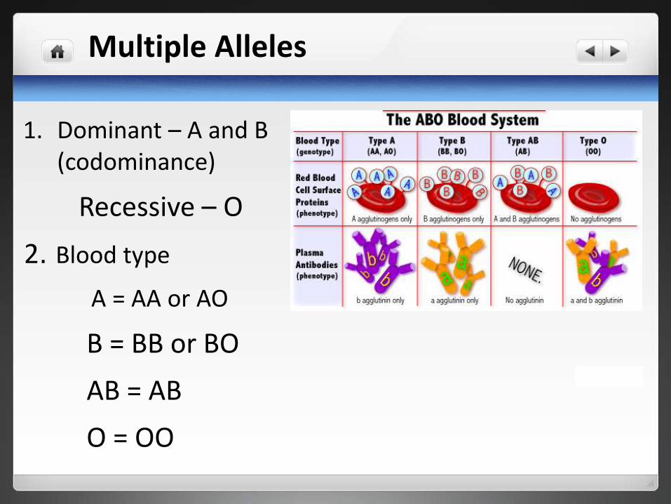
1. Dominant – A and B (codominance)
Recessive – O
2. Blood type
A = AA or AO
B = BB or BO
AB = AB
O = OO

Multiple Alleles
1. Dominant – A and B (codominance)
Recessive – O
2. Blood type
A = AA or AO
B = BB or BO
AB = AB
O = OO

A BWhat would be the possible blood types of children born to a female with type AB blood and a male with type O blood?
AB X OO
AO BO
AO BO
O
O
Children would be type A or B only
Example:

Sex Chromosomes:
Sex Symbol ChromosomePair
HumanSize
Male Y XY smaller
Female X XX larger
6

Sex Chromosomes:7

Other chromosomes:
Other chromosomes are called autosomes.
8

Sex – linked Traits
• Genes for these traits are located only on the Xchromosome (NOT on the Y chromosome)
• X linked alleles always show up in males whetherdominant or recessivebecause males have only oneX chromosome

Example of Humans
Females: 23 homologous pairs
Males: 22 homologous pairs + 1pair different
# Chromosomes Type
46 chromosomes
— 2 sex chromosomes
44 autosomes
Example of Fruit Flies
Females: 4 homologous pairs
Males: 3 homologous pairs + 1 pair different
# Chromosomes Type
8 chromosomes
— 2 sex chromosomes
6 autosomes
10
Sex Linked
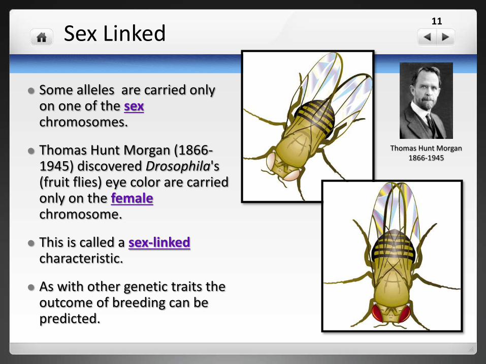
Some alleles are carried only on one of the sexchromosomes.
Thomas Hunt Morgan (1866-1945) discovered Drosophila's (fruit flies) eye color are carried only on the femalechromosome.
This is called a sex-linked characteristic.
As with other genetic traits the outcome of breeding can be predicted.
Thomas Hunt Morgan1866-1945
11

Sex-linked
These traits for fruit fly eye color are written : R stands for redwhile r stands for white.
XR XR Red-eyed
XR Xr Red-eyed
Xr Xr White Eyed
Female
XR Y Red-eyed
Xr Y White Eyed
Male
Possible Genotypes & Phenotypes
12

Sex-linked Parental Cross
Red-eyed female
&
White-eyed male
13

Sex-linked Parental Cross
Red-eyed female&
White-eyed male
XR
XR
Xr Y
14

Sex-linked Parental Cross
Red-eyed female&
White-eyed male
XR Xr XR Y
XR Xr XR Y
XR
XR
Xr Y
15

Sex-linked ƒ1 Cross
Red-eyed female&
Red-eyed male
From Parental Cross
16
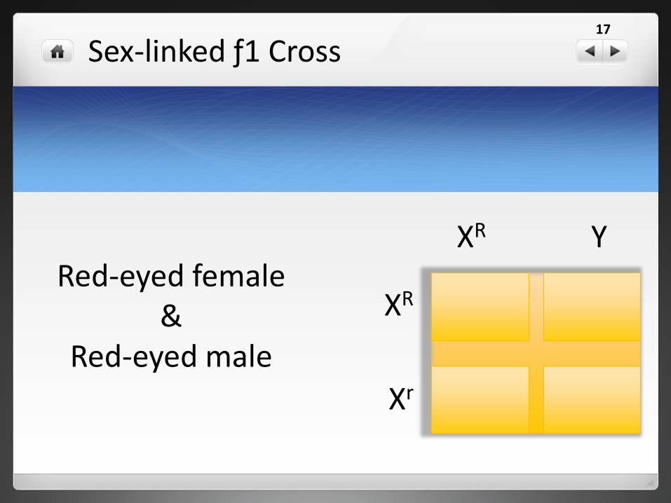
Sex-linked ƒ1 Cross
Red-eyed female&
Red-eyed male
XR Y
XR
Xr
17

Sex-linked ƒ1 Cross
Red-eyed female&
Red-eyed male
XR XR XR Y
XR Xr XR Y
XR
Xr
XR Y
18

Results: ƒ1 cross: ƒ2 generation
XR XR Red-eyed
XR Xr Red-eyed
Female
XR Y Red-eyed
Xr Y White Eyed
Male
Possible Genotypes & Phenotypes
Note: There are not any white-eyed females
19

Human ColorblindnessAn inability to distinguish
between certain colors.
Examples of recessive sex-linked disorders:

Examples of recessive sex-linked disorders:
You should see
58 18
E 17
The most common type is red-green color blindness, where red and green are seen as the same color.
21

HemophiliaBlood doesn’t clot properly.
The ability of the blood to clot is severely reduced, causing the sufferer to bleed severely from even a slight injury. The condition is typically caused by a hereditary lack of a coagulation factor, most often factor VIII.
Examples of recessive sex-linked disorders:

Nondisjunction
Remember: genes are located on chromosomes. When homologous chromosomes fail to segregate during meiosis it is called nondisjunction.
Calvin Bridge's (1889-1938) work with Drosophila ( fruit flies ) determined that to be a female Drosophila there must be two X chromosomes present.
23

Sexing Drosophila
What sex would each of the following be?
XXY
X
Female
Male
24

Sexing Homo sapiens
For humans it has been found that the Y chromosome determines sex.
What sex would each of the following be?
XX
XXX
Female Male•XXY
•XXXYFemale Male
25
Linked Genes:
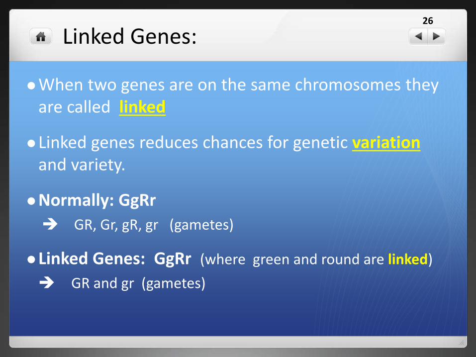
When two genes are on the same chromosomes they are called linked
Linked genes reduces chances for genetic variationand variety.
Normally: GgRr
GR, Gr, gR, gr (gametes)
Linked Genes: GgRr (where green and round are linked)
GR and gr (gametes)
26

Crossing Over
• Crossing over occurs when
homologous chromatids fail to
correctly separate during
prophase I of meiosis.
• This results in offspring that may
have a different genetic make-up
than their parents.
• Gametes that have undergone
this process are called crossing
over.

Maps & Genomes
• Scientist are developing
genetic map (or genome)
which shows the
relationship among different
genes along a chromosome.
• These are used to pin-point
errors in genetic make-up
and help scientists
understand other problems.
Human Genome Karyotype

Karyotype
• A picture of a person's
chromosomes.
• Chromosomes are isolated, stained
and examined under the
microscope.
• Uses the chromosomes in the white
blood cells.
• A picture of the chromosomes is
taken through the microscope.

Comparing Chromosome Numbers30
Looking at DifferentGenomesChromosomal Numbers

Karyotypes of other organisms31
Mus sp. (mouse)
Drosophlia sp. (fruit fly)

Mutations
• Mutation – sudden genetic change (change in base pair sequence of DNA)
• Can be :Harmful mutations – organism less able to survive: genetic disorders, cancer, death
Beneficial mutations – allows organism to better survive: provides genetic variation
Neutral mutations – neither harmful nor helpful to organism
• Mutations can occur in 2 ways: chromosomal mutation or gene/pointmutation

Genetic Disorders:
Lethal genesSickle-Cell AnemiaGalactosemiaPKUTay-Sachs DiseaseDiabetes Mellitus
Sex-linked DiseasesHemophiliaColorblindness
Chromosomal DisordersDown SyndromeTurner SyndromeKlinefelter Syndrome
33

Sickle-Cell Anemia
Mostly affecting those of African ancestry, but also occurs in other ethnic groups, including people who are of Mediterranean and Middle Eastern descent.
More than 70,000 Americans have sickle cell anemia. And about 2 million Americans — 1 in 12 African Americans —have sickle cell trait.
The sickle hemoglobin (HbS) mutation confers a genetic advantage against malaria so carrier frequency is highest in areas where malaria is (or was) endemic (regularly found among particular people or in a certain area )
34

Sickle cell anemia is a blood disorder affecting hemoglobin, the protein found in red blood cells (RBCs), which carries oxygen throughout the body.
Sickle cell anemia occurs when a person inherits two abnormal genes (one from each parent) that cause their RBCs to change shape.
Instead of being flexible and disc-shaped, these cells are more stiff and curved in the shape of the old farm tool known as a sickle — that's where the disease gets its name.
The shape is similar to a crescent moon.
Sickle-Cell Anemia35

Sickle-Cell Anemia
How the trait is passed to offspring
36

Galactosemia
Is a rare genetic metabolic disorder is a disorder that affects how the body processes a simple sugar called galactose.
Galactosemia is not related to and should not be confused with lactose intolerance.
Without treatment, mortality in infants with galactosemia is about 75%.
37

PKU — Phenylketonuria
PKU is short for "phenylketonuria." People with PKU can't process one of the amino acids found in many foods.
The amino acid, called phenylalanine or "Phe" for short, builds up in the body.
Too much Phe is toxic to the brain and can cause many problems.
In infants and children, if PKU is not treated, the resulting high Phe can cause severe mental retardation.
Even if PKU is treated problems like brain changes, lower IQ, and behavior problems may still occur.
In adults and teens, high Phe can cause lower intelligence (IQ), poor focus, mood swings, being irritable, depression, slow reaction time, and other problems.
One of the first tests a baby receives
38
Anamino acid common in milkcannot be broken down and as it builds up it causes mental retardation – newborns are tested for this

Tay-Sachs disease
A metabolic disorder commonly associated with Ashkenazi Jews, has also been found in the French Canadians of Southeastern Quebec, the Cajuns of Southwest Louisiana, and other populations throughout the world.
The severity of expression and the age at onset of Tay-Sachs varies from infantile and juvenile forms that exhibit paralysis, dementia, blindness and early death to a chronic adult form that exhibits neuron dysfunction and psychosis.
Tay-Sachs is an autosomal recessive disease caused by mutations in both alleles of a gene (HEXA) on chromosome 15. HEXA codes for an enzyme found in lysosomes, organelles that break down large molecules for recycling by the cell.
In Tay-Sachs individuals, the enzyme is absent or present only in very reduced amounts, allowing excessive accumulation of the GM2 gangliosidein neurons.
39
Deterioration of the nervous system –early death
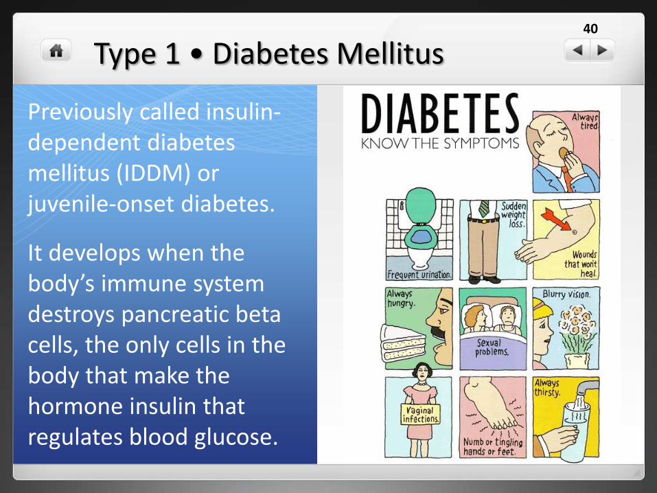
Type 1 • Diabetes Mellitus
Previously called insulin-dependent diabetes mellitus (IDDM) or juvenile-onset diabetes.
It develops when the body’s immune system destroys pancreatic beta cells, the only cells in the body that make the hormone insulin that regulates blood glucose.
40

Type 2 • Diabetes Mellitus
Previously called non-insulin-dependent
diabetes mellitus (NIDDM) or adult-onset
diabetes.
In adults, type 2 diabetes accounts for
about 90% to 95% of all diagnosed cases
of diabetes. It usually begins as insulin
resistance, a disorder in which the cells
do not use insulin properly.
As the need for insulin rises, the pancreas
gradually loses its ability to produce it.
It is associated with older age, obesity,
family history of diabetes, history of
gestational diabetes, impaired glucose
metabolism, physical inactivity, and
race/ethnicity.
African Americans, Hispanic/Latino
Americans, American Indians, and some
Asian Americans and Native Hawaiians or
other Pacific Islanders are at particularly high
risk for type 2 diabetes and its complications.
41

Colorblindness
Color blindness or color vision deficiency is the inability to perceive differences between some of the colors that others can distinguish.
It is most often of genetic nature, but may also occur because of eye, nerve, or brain damage, or exposure to certain chemicals.
42

Colorblindness Tests
25 6 56 45
43

Down Syndrome • Trisomy 21
But a baby with Down syndrome has an extra chromosome #21 (47 instead of 46) or one chromosome has an extra part.
About half of babies with Down syndrome are born with heart defects. Usually, these problems can be corrected by surgery. Some babies may have intestinal problems that also require surgery to fix.
Kids with Down syndrome are more likely to get infections affecting their lungs and breathing. These infections often last longer. They may have eye or ear problems or digestion problems like constipation. Some may develop leukemia, a type of cancer.
Kids with Down syndrome tend to grow and develop more slowly than other children do. They may start walking or talking later than other babies.
About 1 out of every 800 babies born has Down syndrome, no matter what race or nationality the parents are.
44

Down Syndrome • Trisomy 2145

Turner Syndrome • OX
A female does not have the usual pair of two X chromosomes.
Other Names: Bonnevie-Ullrichsyndrome; Gonadal dysgenesis; Monosomy X
Occurring in 1 of 2,500 girls.
Characteristic physical abnormalities, short stature, swelling, broad chest, low hairline, low-set ears, and webbed necks.
46

Turner Syndrome • OX 47

Klinefelter Syndrome • XXY47, XXY, or XXY syndrome is a condition in which males
have an extra X sex chromosome.
While females have an XX chromosomal makeup, and
males an XY, affected individuals have at least two X
chromosomes and at least one Y chromosome.
1 out of every 1,000 males.
One in every 500 males have an extra X chromosome
but do not have the syndrome.
The syndrome can affect different stages of physical,
language and social development. The most common
symptom is infertility. Because they often don't make as
much of the male hormone testosterone as other boys,
teenagers with Klinefelter's syndrome may have less
facial and body hair and may be less muscular than
other boys.
They may have trouble using language to express
themselves. They may be shy and have trouble fitting
in.
48

Klinefelter Syndrome • XXY49

Detecting Human Genetic Disease
Amniocentesis
Ultrasonography
Fetoscopy
DNA tests
50

Detecting Human Genetic Disease
Is a medical procedure used in prenatal
diagnosis of chromosomal abnormalities
and fetal infections
A small amount of amniotic fluid, which
contains fetal tissues, is extracted from the
amnion or amniotic sac surrounding a
developing fetus
The fetal DNA is examined for genetic
abnormalities.
Amniocentesis (amniotic fluid test or AFT)
51

Detecting Human Genetic Disease
Sonography is used to visualize the
embryo or foetus in its mother's uterus
(womb).
The procedure is often a standard part
of prenatal care, as it yields a variety of
information regarding the health of the
mother and of the fetus, as well as
regarding the progress of the
pregnancy.
Ultrasonography
52

Detecting Human Genetic Disease
Is an endoscopic procedure during pregnancy to allow access
to the fetus, the amniotic cavity, the umbilical cord, and the
fetal side of the placenta.
A small (3-4 mm) incision is made in the abdomen, and an
endoscope is inserted through the abdominal wall and uterus
into the amniotic cavity.
Fetoscopy allows medical interventions such as a biopsy or a
laser occlusion of abnormal blood vessels.
Evaluate the fetus for birth defects, such as spina bifida as
well as other defects.
Collect samples of embryo, These samples can then be tested
further for diseases such as hemophilia or sickle cell anemia.
Fetoscopy
53

Detecting Human Genetic Disease
Examines the nucleotides at specific locations on a person's DNA for genetic purposes.
Carrier testing is used to identify people who carry one copy of a gene mutation that, when present in two copies, can cause a genetic disorder.
This type of testing is for individuals who have a family history of a genetic disorder and to people in ethnic groups with an increased risk of specific genetic conditions.
Having both parents tested can provide information about a couple's risk of having a child with a genetic condition.
Immune System
Lupus
Graves' disease
Celiac disease
Multiple sclerosis
Psoriasis
Cardiovascular Conditions
Aneurysm
Atrial fibrillation
Heart disease
Peripheral arterial disease
Venous thromboembolism
Aging
Macular degeneration
Alzheimer's disease
Osteoarthritis
Rheumatoid arthritis
General Health
Obesity
Migraine
Type 1 diabetes
Type 2 diabetes
Cancers
Bladder cancer
Breast cancer
Colorectal cancer
Gastric cancer
Lung cancer
Prostate cancer
Skin cancer
DNA testing
54

Pedigrees
• Graphic representation of how a trait is passed from parents to offspring
• Tips for making a pedigree
1. Circles are for females
2. Squares are for males
3. Horizontal lines connecting a male and a female represent a marriage
4. Vertical line and brackets connect parent to offspring
5. A shaded circle or square indicates a person has the trait
6. A circle or square NOT shaded represents an individual who does NOT have the trait
7. Partial shade indicates a carrier – someone who is heterozygous for the trait

Example: Make a pedigree chart for the following couple.
• Dana is color blind; her husband Jeff is not. They have two boysand two girls.
• HINT: Colorblindness is a recessive sex-linked trait.
XNY
Has trait Can pass trait to offspring
XnXn
Pedigrees
Related Documents