JOURNAL OF CLINICAL MICROBIOLOGY, Dec. 1993, p. 3190-3199 Vol. 31, No. 12 0095-1137/93/123190-10$02.00/0 Copyright X) 1993, American Society for Microbiology Genetic Similarity and Phenotypic Diversity of Commensal and Pathogenic Strains of Candida albicans Isolated from the Oral Cavity JOHN HELLSTEIN,lt HOLLY VAWTER-HUGART,2 PETER FOTOS,l JAN SCHMID,2 AND DAVID R. SOLL2* College of Dentistry' and Department of Biological Sciences,2 University of Iowa, Iowa City, Iowa 52242 Received 21 June 1993/Returned for modification 28 July 1993/Accepted 3 September 1993 Colony phenotype and genetic similarity were assessed within and between groups of commensal and pathogenic strains of Candida albicans collected from the oral cavities of individuals in a single geographical locale. Thirty-eight percent of pathogenic isolates contained predominant or minor variant colony morpholo- gies (other than smooth) when samples from the sites of infection were cultured on plates, while 16% of commensal isolates contained minor variant colony morphologies when samples from the sites of carriage were cultured. The genetic similarities of isolates within and between groups were assessed by DNA fingerprinting by using Southern blot hybridization with the fingerprinting probe Ca3 and analysis with the computer- assisted, automated Dendron system. Both the commensal and the pathogenic groups contained a major cluster of genetically similar C. albicans isolates representing 31 and 33% of the strains in the respective groups. When a combined dendrogram of both commensal and pathogenic isolates was generated, the major clusters of genetically similar isolates in each group mixed into one large cluster. Minor clusters in the individual dendrograms also mixed. These results suggest common clonal origins for commensal and pathogenic strains in the same geographical locale. Candida albicans resides in the oral cavities of a majority of healthy individuals as a commensal organism, causing no apparent morbidity (8, 23). However, under a number of predisposing conditions, the level of C. albicans increases dramatically, resulting in tissue penetration and inflamma- tion of the oral mucosa. Despite its prevalence as a com- mensal organism and the frequency of oral candidiasis, we still have not answered some of the most fundamental questions related to commensal carriage, infection, or the transition from the commensal to the pathogenic state (21, 22). For instance, it is generally assumed that because the organism so commonly inhabits the oral cavities of healthy individuals, the commensal strain is the source of subse- quent infection. This assumption, in turn, suggests that all or most commensal C. albicans organisms are capable of causing an infection, and all strains, by definition, are opportunistic. However, it is also possible that in the tran- sition from a healthy to an infected mouth, there is strain substitution (18). In the past, it was difficult to answer this type of basic epidemiological question because strain comparisons were based on biotyping techniques (7, 9, 12), which compared phenotypes rather than genotypes and, therefore, ran the risk of grouping genetically unrelated strains with similar phenotypes and separating related strains with different phenotypes. In recent years, a number of DNA fingerprint- ing techniques which provide genetic assessments of strain relatedness have been developed (6, 11, 15, 16, 24). One of the most effective of these has been the use of Southern blot * Corresponding author. t Present address: William Beaumont Army Medical Center, Department of Pathology-Dentistry, Fort Bliss, TX 79920. * Present address: Department of Microbiology and Genetics, Massey University, Palmerston North, New Zealand. hybridization with DNA probes containing moderately re- petitive sequences (3, 10, 16, 19, 22, 24). The hybridization patterns generated by this technique are amenable to com- puter-assisted analysis with the Dendron software program (19, 22). The patterns differ between unrelated strains, are constant over hundreds of generations in a single strain (16, 19), and are complex enough to provide similarity coeffi- cients which reflect genetic relatedness (19, 22). In the present study, we used this DNA fingerprinting technique with a newly developed automated Dendron program to test whether a group of commensal strains is genetically distinct from a group of pathogenic strains of C. albicans isolated from the oral cavities of individuals in the same geographical locale. We found no genetic distinctions between commensal and pathogenic strains. Rather, we identified in dendrograms both major and minor clusters of genetically related strains which included both commensal and pathogenic strains suggesting common clonal origins for commensal and patho- genic strains in the Iowa City, Iowa, locale. MATERIALS AND METHODS Group selection. Healthy patients from whom samples were positive for C. albicans on culture and who conformed to the following restrictions were included in the study: (i) no history of symptoms or diagnosis of oral candidiasis, (ii) no immunologic or other compromising condition, (iii) no re- cent use of antibiotics or steroids, and (iv) no history of recurrent C. albicans infections in other body locations. This group consisted of 12 females and 7 males. The average age was 41.5 ± 17.0 years. Patients with oral candidiasis were included in the study if they displayed symptoms of erythe- matous or pseudomembranous candidiasis and samples from the patients were positive for C. albicans on culture. This group consisted of 16 females and 7 males. The average age 3190

Welcome message from author
This document is posted to help you gain knowledge. Please leave a comment to let me know what you think about it! Share it to your friends and learn new things together.
Transcript

JOURNAL OF CLINICAL MICROBIOLOGY, Dec. 1993, p. 3190-3199 Vol. 31, No. 120095-1137/93/123190-10$02.00/0Copyright X) 1993, American Society for Microbiology
Genetic Similarity and Phenotypic Diversity of Commensaland Pathogenic Strains of Candida albicans
Isolated from the Oral CavityJOHN HELLSTEIN,lt HOLLY VAWTER-HUGART,2 PETER FOTOS,l
JAN SCHMID,2 AND DAVID R. SOLL2*
College of Dentistry' and Department of Biological Sciences,2 University of Iowa,Iowa City, Iowa 52242
Received 21 June 1993/Returned for modification 28 July 1993/Accepted 3 September 1993
Colony phenotype and genetic similarity were assessed within and between groups of commensal andpathogenic strains of Candida albicans collected from the oral cavities of individuals in a single geographicallocale. Thirty-eight percent of pathogenic isolates contained predominant or minor variant colony morpholo-gies (other than smooth) when samples from the sites of infection were cultured on plates, while 16% ofcommensal isolates contained minor variant colony morphologies when samples from the sites of carriage werecultured. The genetic similarities of isolates within and between groups were assessed by DNA fingerprintingby using Southern blot hybridization with the fingerprinting probe Ca3 and analysis with the computer-assisted, automated Dendron system. Both the commensal and the pathogenic groups contained a major clusterof genetically similar C. albicans isolates representing 31 and 33% of the strains in the respective groups. Whena combined dendrogram of both commensal and pathogenic isolates was generated, the major clusters ofgenetically similar isolates in each group mixed into one large cluster. Minor clusters in the individualdendrograms also mixed. These results suggest common clonal origins for commensal and pathogenic strainsin the same geographical locale.
Candida albicans resides in the oral cavities of a majorityof healthy individuals as a commensal organism, causing noapparent morbidity (8, 23). However, under a number ofpredisposing conditions, the level of C. albicans increasesdramatically, resulting in tissue penetration and inflamma-tion of the oral mucosa. Despite its prevalence as a com-mensal organism and the frequency of oral candidiasis, westill have not answered some of the most fundamentalquestions related to commensal carriage, infection, or thetransition from the commensal to the pathogenic state (21,22). For instance, it is generally assumed that because theorganism so commonly inhabits the oral cavities of healthyindividuals, the commensal strain is the source of subse-quent infection. This assumption, in turn, suggests that all ormost commensal C. albicans organisms are capable ofcausing an infection, and all strains, by definition, areopportunistic. However, it is also possible that in the tran-sition from a healthy to an infected mouth, there is strainsubstitution (18).
In the past, it was difficult to answer this type of basicepidemiological question because strain comparisons werebased on biotyping techniques (7, 9, 12), which comparedphenotypes rather than genotypes and, therefore, ran therisk of grouping genetically unrelated strains with similarphenotypes and separating related strains with differentphenotypes. In recent years, a number of DNA fingerprint-ing techniques which provide genetic assessments of strainrelatedness have been developed (6, 11, 15, 16, 24). One ofthe most effective of these has been the use of Southern blot
* Corresponding author.t Present address: William Beaumont Army Medical Center,
Department of Pathology-Dentistry, Fort Bliss, TX 79920.* Present address: Department of Microbiology and Genetics,
Massey University, Palmerston North, New Zealand.
hybridization with DNA probes containing moderately re-petitive sequences (3, 10, 16, 19, 22, 24). The hybridizationpatterns generated by this technique are amenable to com-puter-assisted analysis with the Dendron software program(19, 22). The patterns differ between unrelated strains, areconstant over hundreds of generations in a single strain (16,19), and are complex enough to provide similarity coeffi-cients which reflect genetic relatedness (19, 22). In thepresent study, we used this DNA fingerprinting techniquewith a newly developed automated Dendron program to testwhether a group of commensal strains is genetically distinctfrom a group of pathogenic strains of C. albicans isolatedfrom the oral cavities of individuals in the same geographicallocale. We found no genetic distinctions between commensaland pathogenic strains. Rather, we identified in dendrogramsboth major and minor clusters of genetically related strainswhich included both commensal and pathogenic strainssuggesting common clonal origins for commensal and patho-genic strains in the Iowa City, Iowa, locale.
MATERIALS AND METHODS
Group selection. Healthy patients from whom sampleswere positive for C. albicans on culture and who conformedto the following restrictions were included in the study: (i) nohistory of symptoms or diagnosis of oral candidiasis, (ii) noimmunologic or other compromising condition, (iii) no re-cent use of antibiotics or steroids, and (iv) no history ofrecurrent C. albicans infections in other body locations. Thisgroup consisted of 12 females and 7 males. The average agewas 41.5 ± 17.0 years. Patients with oral candidiasis wereincluded in the study if they displayed symptoms of erythe-matous or pseudomembranous candidiasis and samples fromthe patients were positive for C. albicans on culture. Thisgroup consisted of 16 females and 7 males. The average age
3190

CHARACTERIZATION OF ORAL C. ALBICANS 3191
was 55.8 ± 20.5 years. One male in the latter group wassubsequently discovered to be immunocompromised as aresult of AIDS. Complete medical histories were evaluatedfor both groups for inclusion in the study.
Culturing of commensal and pathogenic strains. Five in-traoral sites (buccal mucosa, salivary pool, floor of mouth,dorsum of tongue, palate) of healthy individuals and patientswith candidiasis were individually swabbed with a wet sterileCulturette (C8852-1; American Scientific Products, McGawPark, IL.) by previously described methods (23). Each swabwas vigorously agitated in 5 ml of sterile water, and 0.1 mlwas spread onto each of three agar plates containing thedefined amino acid-rich composition of the medium devel-oped by Lee et al. (5) supplemented with 0.1 ,uM zinc and 70,ug of arginine per ml (2). Colony number and morphologywere assessed after 7 to 9 days of incubation at 25°C. Theisolates from the 19 healthy individuals and 23 patients withcandidiasis were considered in the "basic" groups of com-mensal and pathogenic strains, respectively, in the presentstudy. These strains were collected over a 1-year periodbetween January 1991 and January 1992. Additional com-mensal and pathogenic strains isolated from the oral cavitiesof patients in prior studies in the Iowa City, Iowa, localewere also used when noted in comparisons of strain similar-ities. These strains were referred to as "additional" groups.The additional commensal and pathogenic strains were col-lected between 1988 and 1990. The average ages and geo-graphical locales were similar for the patients from which thebasic and additional pathogenic strains were collected. How-ever, the patients from whom additional commensal strainswere collected were, on average, 15 years younger and hada greater proportion of females than the patients from whombasic commensal strains were collected.
Southern blot hybridization and DNA fingerprint analysis.Strains were fingerprinted with the DNA probe Ca3 bypreviously described methods (19, 24). In brief, DNA wasisolated from cells of each strain by the method of Schererand Stevens (15). The concentration of DNA in each prep-aration was assessed by comparing an ethidium bromide-stained sample with a set of ethidium bromide-stained stan-dards of calf thymus DNA. DNA samples were thenelectrophoresed in a 0.7% agarose gel. Each gel contained 15lanes, with EcoRI-digested DNA of reference strain 3153Ain lanes 2 and 15, 12 test strains in lanes 3 through 14, and a1-kb ladder of molecular mass markers (Bethesda ResearchLaboratory, Gaithersburg, Md.) in lane 1. Gels were run firstat 70 V for 20 min and then at 30 to 40 V until thebromophenol blue marker had traveled 16 cm from the well.Gels were stained with ethidium bromide and were photo-graphed with a UV light source. Gels were then washed,blotted onto a nitrocellulose membrane, hybridized withnick-translated Ca3 probe, and exposed to XAR-S film(Eastman Kodak Co., Rochester, N.Y.) with a CronexLightning Plus intensifying screen (Du Pont Co., Wilming-ton, Del.).
Hybridization patterns were then automatically analyzedwith the newly developed automated Dendron program(Solltech Inc., Iowa City, Iowa) (22). Autoradiogram pat-terns were digitized into the Dendron data base with a Sharpscanner, the digitized images were unwarped (straightened),and lanes were image processed, when necessary, andscanned (22). Bands were automatically identified and inten-sities were classified between 0 (no band) and 3 (highestintensity). The Dendron program then computed the similar-ity coefficient (SA) for every pair of strains on the bases ofthe positions and intensities of the bands in the hybridization
pattern, generating a matrix of similarity coefficients. Den-drograms based on SABs were then generated for selectedstrains by using the unweighted group pair method (19, 20).In an analysis of 45 C. albicans strains, each strain wasfingerprinted twice with the Ca3 probe on separate gels atdifferent times, the mean ± standard deviation SAB for pairswas 0.96 ± 0.03, which is considered a measure of identi-calness for strains analyzed in different gels. For 10 finger-prints of the same strain on the same gel, the mean SAB was1.00 ± 0.00. In an analysis of 30 independently isolated,unrelated C. albicans strains randomly selected from theDendron data base and not part of the present study, theaverage SAB was 0.69 ± 0.11, which is considered a measureof unrelatedness. Lanes on different gels were "neighbored"(the digitized lane images from different gels were placednext to each other), and band positions were normalizedaccording to the band positions of the 3153A referencepatterns bordering each gel by a Dendron subprogram de-veloped for this purpose (17, 23). The digitized lane imagesfrom different gels that were placed next to each other werethen photographed from the monitor screen.
Biotyping by sugr assimilation. The API 20C kit (bio-Merieux, Hazelwood, Mo.) was used to type the species.Isolates from stored slants were streaked onto nutrient agarand were incubated at 25°C for 24 h. Samples were thenanalyzed according to the manufacturer's prescribed methods.
RESULTS
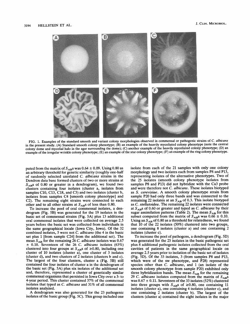
Colony phenotypes and strain identification of the basic setsofcommensal and pathogenic strains. The level of commensalstrain carriage ranged between 6 and 342 colonies per 15plates, with a mean of 52.4 ± 77.0 (Table 1). In contrast, thelevel of colonization with pathogenic strains ranged between9 and 7,500 colonies per 15 plates, with a mean of 569 ±1,594 (Table 2). While only 1 of 19 samples of commensalstrains (5%) contained more than 120 colonies per 15 plates,9 of 23 samples of pathogenic strains (39%) contained morethan 120 colonies per 15 plates (Tables 1 and 2). Thedominant colony morphology of the 19 samples of commen-sal strains was smooth white (Fig. 1A; Table 1). In 3 (16%) ofthe samples of commensal strains (C4, Cll, C14), the minorcolony morphology was heavily myceliated (Fig. 1B or C;Table 1). The dominant colony morphology in 21 of the 23samples of pathogenic strains was also smooth white (Table2). However, the dominant colony morphology in 2 of the 23samples of pathogenic strains was heavily myceliated (P3,P5). In addition, 6 of the 23 samples of pathogenic strains(26%) contained minor colony phenotypes (Table 2). Sam-ples P7, P13, and P19 contained isolates with a minorwrinkled colony morphology (Fig. 1D), sample P8 containedisolates with a star colony morphology (Fig. 1E), sample P17contained isolates with minor wrinkled, ring (Fig. 1F), andstar colony morphologies, and sample P20 contained isolateswith a minor heavily myceliated colony morphology. There-fore, 15% of the commensal isolates had variant colonymorphologies, while 34% of pathogenic isolates had variantcolony morphologies. This difference was possibly signifi-cant, with P < 0.25.
Species identification was assessed in two ways. First,EcoRI-digested DNAs of individual isolates were electro-phoresed and Southern blots were hybridized with the Ca3probe, which is species specific for C. albicans (13). Strainsof C. albicans exhibited a hybridization pattern containing15 to 25 bands, with both highly conserved and variant bands(1). Strains of Candida stellatoidea exhibit patterns close to
VOL. 31, 1993
3192 HELLSTEIN ET AL.
TABLE 1. Colony number, colony phenotypes, and species identification of commensal strains by DNAfingerprinting and sugar assimilation
Sample No. of Major colony Minor colony DNA fingerprint Sugar assimilationno. coloniesa morphologyb morphology' patternd patteme
C1 66 Sm + C.a.C2 36 Sm + C.a.C3 342 Sm + C.a.C4 9 Sm HM +(Sm), +(HM) C.a.(Sm), C.a.(HM)C5 6 Sm NIC6 12 Sm + C.a.C7 108 Sm C.p.C8 75 Sm + C.a.C9 6 Sm C.p.C1o 69 Sm + C.a.C1l 60 Sm HM +(Sm), +(HM) C.a.(Sm),C.a.(HM)C12 21 Sm Sac.C13 18 Sm + C.a.C14 9 Sm HM +(Sm), +(HM) C.a.(Sm),C.a.(HM)C15 18 Sm + C.a.C16 24 Sm + C.a.C17 6 Sm + C.a.C18 21 Sm + C.a.C19 90 Sm + C.a.
a The value is the sum of the number of colonies on 15 plates.b Sm, smooth white phenotype.C In all three samples, (C4, Cll, C14), colonies of isolates of the heavily myceliated phenotype (HM) made up less than 20% of all colonies.d Since the Ca3 probe is relatively specific for C. albicans, a plus sign represents a standard C. albicans-type pattern, and a minus sign represents either no
hybridization or two or fewer bands, as is the case for C. tropicalis (17).e Sugar assimilation tests were performed twice for each sample. The best identification is presented. C.a., C. albicans; C.p., C. parapsilosis; Sac., S.
cerevisiae; NI, not identified by the test; Sm, smooth colony phenotype; HM, heavily myceliated colony phenotype.
those of C. albicans strains (4), while other species exhibitpatterns ranging from two low-intensity, high-molecular-mass bands, in the case of Candida tropicalis, to no bandsfor less related species (13, 25). Second, isolates were
biotyped by their sugar assimilation patterns (9). Isolatesfrom 15 of the 19 samples of commensal strains (79%) typedas C. albicans both by hybridization with the Ca3 probe andby sugar assimilation (Table 1). In the three samples ofcommensal strains in which there were both major and minorcolony phenotypes (C4, Cll, C14), isolates of both pheno-types typed as C. albicans by Ca3 hybridization and sugarassimilation (Table 1). Isolates from 4 of the 19 samples ofcommensal strains (CS, C7, C9, C12) did not hybridizesignificantly with Ca3 and were therefore not C. albicans(Table 1). By sugar assimilation, two (C7, C9) typed asCandida parapsilosis, one (C12) typed as Saccharomycescerevisiae, and one (C5) was unidentifiable.
Isolates from 22 of the 23 samples of pathogenic strains(96%) typed as C. albicans by hybridization with Ca3 andsugar assimilation (Table 2). These included isolates fromsamples P3 and PS, which exhibited a dominant heavilymyceliated phenotype. In the cases of samples P1 and P19,both the major and minor phenotypes of the isolates in thesamples typed as C. albicans by both Ca3 hybridization andsugar assimilation. In the cases of isolates from samples P8and P13, the major smooth phenotype did not type as C.albicans by Ca3 hybridization, while the minor phenotypesdid. In both cases, the major phenotype typed as S. cerevi-siae and the minor phenotype typed as C. albicans by sugarassimilation. In the case of isolates from sample P20, boththe major and minor phenotypes exhibited reduced hybrid-ization patterns and typed as C. stellatoidea by sugar assim-ilation.
General features of hybridization patterns. The Southernblot hybridization patterns of the C. albicans strains probed
with nick-translated Ca3 contained between 10 and 15 majorbands in the molecular mass range of 2.25 to 19 kb (Fig. 2)and between 3 and 8 minor bands in the molecular massrange of less than 2.25 kb (data not shown in Fig. 2). Thepatterns of most C. albicans strains tested so far show thatthe strains contain highly conserved bands at 2.68 and 3.30kb (1). The majority of other bands are moderately variablebetween strains. One or more bands above 7.9 kb have beenfound to be highly variable (1, 24). Digestion of whole-cellDNA with EcoRI resulted in a very clear and highly repro-ducible banding pattern. This is evident in the identicalpatterns of reference strain 3153A, labeled R in Fig. 2. Insome cases, the Ca3 probe did not hybridize to the EcoRI-digested DNA of an isolate (e.g., Fig. 2, lanes 3 and 12). Allof these isolates typed as species other than C. albicans bytheir sugar assimilation patterns (Tables 1 and 2). In somecases, the probe generated a highly reduced pattern (e.g.,Fig. 2, lanes 10 and 18). Again, these strains typed as aspecies other than C. albicans by sugar assimilation (Table2). In rare cases, endonuclease digestion was interfered within repeat experiments (e.g., Fig. 2, lane 8), making it difficultor impossible to analyze. It has been demonstrated that Ca3distinguishes between the great majority of independentisolates by one or more bands (19), and this can be verifiedby a comparison of the independent isolates whose finger-print patterns are shown in Fig. 2.
Genetic similarity of multiple phenotypes from the sameoral cavities. Three sets of samples of commensal strains(C4, Cll, C14) exhibited a predominant smooth phenotype(Fig. 1A) and a minor heavily myceliated phenotype (Fig. 1Band C). To test whether the major and minor phenotypes ineach sample represented the same or different strains, clonesof the smooth and variant phenotypes were fingerprintedwith Ca3. In all three cases, clones of both phenotypes typedas C. albicans by sugar assimilation (Table 1), and the
J. CLIN. MICROBIOL.
CHARACTERIZATION OF ORAL C. ALBICANS 3193
TABLE 2. Colony number, colony phenotypes, and species identification of pathogenic strains by DNA fingerprintingand sugar assimilation
Sample No. of Major colony Minor colony DNA fingeryrint Sugar assimilationno. coloniese morphology" morphology' pattern pattern'P1 225 Sm + C.a.P2 9 Sm + C.a.P3 78 HM + C.a.P4 21 Sm + C.a.P5 24 HM + C.a.P6 12 Sm + C.a.P7 21 Sm Wr + (Sm), + (Wr) C.a. (Sm), C.a. (Wr)P8 42 Sm St - (Sm), + (St) Sac. (Sm), C.a. (St)P9 459 Sm + C.a.PlO 1,200 Sm + C.a.P1l 51 Sm + C.a.P12 150 Sm + C.a.P13 126 Sm Wr - (Sm), + (Wr) Sac. (Sm), C.a. (Wr)P14 36 Sm + C.a.P15 81 Sm + C.a.P16 15 Sm + C.a.P17 2,277 Sm Wr, R, St + (Sm), + (Wr), + (R), C.a. (Sm), C.a. (Wr), C.a. (R), C.a. (St)
+ (St)P18 276 Sm + C.a.P19 72 Sm Wr + (Sm), + (Wr) C.a. (Sm), C.a. (Wr)P20 354 Sm HM +, un (Sm), + unc C. stel. (Sm), C. stel. (HM)
(HM)P21 36 Sm + C.a.P22 18 Sm + C.a.P23 >7,500 Sm + C.a.a The value is the sum of the number of colonies on 15 plates.b Sm, smooth white phenotype; HM, heavily myceliated phenotype.C In all cases, colonies of isolates of minor phenotypes made up less than 25% of all colonies. Wr, wrinkled phenotype; St, star phenotype; R, ring phenotype.d A plus sign represents standard C. albicans-like pattern; a minus sign represents either no hybridization or minor hybridization to 2 or less bands; +un
represents significant hybridization, but uncharacteristic of standard C. albicans strains.I Best identification by sugar assimilation. C.a., C albicans; Sac., S. cerevisiae; C. stel., C. stellatoidea; Sm, smooth colony phenotype; Wr, wrinkled colony
phenotype; St, star colony phenotype; R, ring colony phenotype; HM, heavily myceliated colony phenotype.
fingerprints were either identical (samples C4 and C14) orhighly similar (sample Cll). In Fig. 3A, the Southern blothybridization patterns of clones from samples C4 (smooth)and C4 (heavily myceliated), which were on different gels,were neighbored by the Dendron software program (seeMaterials and Methods) (17, 23). The banding patterns wereidentical (the very-high-molecular-mass broad band at thetop of lane Sm in Fig. 3A proved to be undegraded DNA andnot an analyzable hybridization band. In Fig. 3B, the pat-terns of clones from samples Cll (smooth) and Cll (heavilymyceliated) are presented. The patterns are highly similar,differing by the position of only one high-molecular-massband (noted by an arrow), which has been demonstrated tochange size in the same strain at a higher frequency thanother bands in the hybridization pattern obtained by Ca3fingerprinting (1, 23).
Six samples of pathogenic strains contained strains withmultiple phenotypes. In each case, the multiple phenotypeswere fingerprinted. In four of the six cases (samples P7, P17,P19, and P20), the fingerprint patterns were identical. In Fig.3C, the patterns are presented for clones of P17 ring,smooth, and wrinkled phenotypes. In Fig. 3D, the reducedpatterns of clones from sample P20 (smooth) and P20 (heavi-ly myceliated), which typed as C stellatoidea, were identi-cal. In two of the six cases (samples P8 and P13), strains ofthe smooth dominant phenotype biotyped as S. cerevisiae,and strains of the minor phenotype biotyped as C albicansby sugar assimilation (Table 2). In both cases, the majorstrain, exhibiting the smooth phenotype, did not hybridize
with Ca3, while the strain of the minor phenotype generateda standard C. albicans hybridization pattern (Fig. 4A and B).
Genetic relatedness within each group of commensal andpathogenic isolates. A dendrogram based on SsBS was gen-erated for single isolates from the basic group of 19 samplesof commensal strains (Fig. SA). In the case of samples ofcommensal strains with multiple phenotypes (samples C4,Cll, and C14), only the major colony phenotype was used,since strains of the major and minor phenotypes weredemonstrated to be genetically identical or highly similar(Table 1; Fig. 3A and B). Four of the 19 isolates (samples C5,C7, C9, and C12) did not hybridize with the Ca3 probe (e.g.,C9 in lane 3 of Fig. 2), and in the dendrogram in Fig. 5A, the4 isolates are connected to all other isolates in the basicgroup of commensal strains at an SA of 0.0. By sugarassimilation, the strains in samples C7 and C9 typed as C.parapsilosis, the strain in sample C12 typed as S. cerevisiae,and the strain in sample C5 was not identifiable (Table 1).The isolate in sample C16 had only three identifiable low-molecular-mass bands because of interference in digestion inrepeat analyses. Because the three interpretable bands linedup with common bands in the Ca3 pattern of mainstreamstrains of C. albicans, we believe that this strain is C.albicans, and this was reinforced by its sugar assimilationpattern (Table 1). However, in the dendrogram, it connectedto all other strains with an SAB of 0.20 because of anincomplete pattern. The remaining 14 isolates were con-nected in the dendrograms at SABs of 0.58 or greater. Themean + standard deviation SAB for these 14 isolates com-
VOL. 31, 1993
J. CLIN. MICROBIOL.3194 HELLSTEIN ET AL.
r.T,-r-
FIG. 1. Examples of the standard smooth and variant colony morphologies observed in commensal or pathogenic strains of C. albicansin the present study. (A) Standard smooth colony phenotype; (B) an example of the heavily myceliated colony phenotype (note the centralcolony dome and mycelial halo in the agar surrounding the dome); (C) another example of the heavily myceliated colony phenotype; (D) anexample of the irregular wrinkle colony phenotype; (E) an example of the star colony phenotype; (F) an example of the ring colony phenotype.
puted from the matrix of SABs was 0.64 + 0.09. Using 0.80 as
an arbitrary threshold for genetic similarity (roughly one-halfof randomly selected unrelated C. albicans strains in theDendron data base formed clusters of two or more strains atSABs of 0.80 or greater in a dendrogram), we found twoclusters containing four isolates (cluster a, isolates fromsamples C10, C13, C18, and C3) and two isolates (cluster b,isolates from samples C4 [smooth colony phenotype] andC2). The remaining eight strains were connected to eachother and to all other strains at SABs of less than 0.80.To increase the pool of oral commensal isolates, a den-
drogram (Fig. SB) was generated for the 19 isolates in thebasic set of commensal strains (Fig. 5A) plus 13 additionaloral commensal isolates that were collected on average 2.5years before the basic set was collected from individuals inthe same geographical locale (Iowa City, Iowa). Of the 32combined isolates, S were not C. albicans (the 4 in the basicset plus 1 [from sample C24] from the additional set). Themean SAB for the remaining 26 C. albicans isolates was 0.67
0.10. Seventeen of the 26 C. albicans isolates (63%)clustered into four groups at SABs of .0.80, including one
cluster of 10 isolates (cluster a), one cluster of 3 isolates(cluster d), and two clusters of 2 isolates (clusters b and c).The largest of the four clusters, cluster a (Fig. SB) stillcontained the four isolates of cluster a in the dendrogram ofthe basic set (Fig. 5A) plus six isolates of the additional setand, therefore, represented a cluster of genetically similarcommensal organisms that persisted in Iowa City over a 3- to4-year period. This cluster contained 37% of the commensalisolates that typed as C. albicans and 31% of all commensalisolates analyzed.A dendrogram was also generated for the 23 pathogenic
isolates of the basic group (Fig. SC). This group included one
isolate from each of the 21 samples with only one colonymorphology and two isolates each from samples P8 and P13,representing isolates of the alternative phenotypes. Two ofthe 25 isolates (smooth colony phenotype isolates fromsamples P8 and P13) did not hybridize with the Ca3 probeand were therefore not C. albicans. These isolates biotypedas S. cerevisiae. A smooth colony phenotype strain fromsample P20 had only three bands and was connected to theremaining 22 isolates at an SAB of 0.3. This isolate biotypedas C. stellatoidea. The remaining 22 isolates were connectedat SsBS of 0.60 or greater and typed as C. albicans by theirsugar assimilation patterns (Table 2). The mean SA for thissubset computed from the matrix of SABs was 0.66 ± 0.10.Using an SAB Of 0.80 as a threshold for relatedness, we foundthat 10 of the 22 isolates (50%) clustered into two groups,one containing 8 isolates (cluster a) and one containing 2isolates (cluster c).To increase the pool of pathogens, a dendrogram (Fig. SD)
was generated for the 25 isolates in the basic pathogenic setplus 8 additional pathogenic isolates collected from the oralcavities of patients in the same geographical locale onaverage 2.5 years prior to isolation of the basic set of isolates(Fig. SD). Of the 33 isolates, 3 (from samples P8 and P13,which were of the sw phenotype, and P28) representedspecies other than C. albicans, and 1 (an isolate of thesmooth colony phenotype from sample P20) exhibited onlythree hybridization bands. The mean SAB for the remaining29 C. albicans isolates computed from the matrix of SABswas 0.67 ± 0.11. Seventeen of the 33 isolates (52%) clusteredinto three groups with SABs of .0.80, one containing 11isolates (cluster a), one containing 4 isolates (cluster c), andone containing 2 isolates (cluster b). The largest of theclusters (cluster a) contained the eight isolates in the major

CHARACTERIZATION OF ORAL C. ALBICANS 3195
R 1 2 3 4 5 6 7 8 9 10 1112 R
5.4 -
38 - _00
2.7 ___kb
.40-4
1#A N._ a_*_4 r -p
p-"i' bli
3.8
3.3-
2.7-kb
R 1314 15 16 17 18 19 2021 22 23 24 Rw T.
am" -w- Z
-a~~~~1_0
*;M,'.
FIG. 2. Two representative southern blots of EcoRI-digestedDNA of isolates probed with Ca3. In each blot, test strains arebordered by reference strain 3153A, labeled R. The following lanescontain test strains from the indicated samples: 1, C14-HM (HM isheavily myceliated); 2, P3; 3, C9; 4, P9; 5, P17-St (St is star); 6, C10;7, C17; 8, C16; 9, Cl; 10, P20; 11, P23; 12, C24; 13, P12; 14, P19-Sm(Sm is smooth); 15, C3; 16, P4; 17, P10; 18, P20-Sm (Sm is smooth);19, P1; 20, P11; 21, P14; 22, C8; 23, P19-Wr (Wr is wrinkled); 24,C15.
cluster in the dendrogram of the basic set (Fig. SC) plus threeisolates from the additional set. Cluster a contained 37% ofpathogenic isolates that typed as C. albicans and 33% of allpathogenic isolates analyzed. The second largest cluster(cluster c) in the combined dendrogram (Fig. SD) containedthe two isolates in the minor cluster of the basic pathogenicset (Fig. SC).
Genetic relatedness between commensal and pathogenicstrains. To assess the genetic similarity of the basic set of 19commensal isolates and the basic set of 25 pathogenicisolates, a common dendrogram was generated (Fig. 6A). Ofthe 44 total isolates, 6 were not C. albicans, 1 (an isolate ofthe smooth colony phenotype from sample P20) gave ahighly diminished band pattern, and 1 (from sample C16) wasC. albicans but was uninterpretable. The mean SAB for theremaining 36 C. albicans isolates computed from the matrixof SABs was 0.65 + 0.10. Of these, 21 (58%) clustered intofour groups with SABs of >0.80, with one cluster containing12 isolates (cluster a), two clusters containing 3 isolates each(clusters b and c), and one cluster containing 2 isolates(cluster d). Cluster a contained all isolates in the majorcluster in the dendrogram of the basic set of commensal
FIG. 3. Identical or highly similar Southern blot hybridizationpatterns of Ca3-probed EcoRI-digested DNAs of clones of multiplecolony phenotypes from the same individuals. Since the multiplecolony phenotypes of strains from samples C4 and P20 were
hybridized on different gels, the lanes were "neighbored" (see text)with the Dendron software neighboring program. Note that themultiple phenotypes from samples C4, P17, and P20 exhibitedidentical hybridization patterns, while the multiple phenotypes fromsample Cll exhibited similar but nonidentical patterns, differing bya single high-molecular-mass band, noted by an arrow. Sm, smooth;HM, heavily myceliated; R, ring; Wr, wrinkled. Examples of thevariant phenotypes are presented in Fig. 1.
isolates (Fig. 5A) and the major cluster in the dendrogram ofthe basic set of pathogenic isolates (Fig. SC). Finally, acombined dendrogram was generated for all basic and addi-tional commensal and pathogenic isolates (Fig. 6B). The
A.P8Sm St
B. P13
Sm Wr
440
FIG. 4. Dissimilar Southern blot hybridization patterns of Ca3-probed EcoRI-digested DNAs of multiple colony phenotypes fromthe same individuals. For both P8 and P13, the smooth (Sm) samplesdid not hybridize to Ca3. In both cases, they biotyped as S.cerevisiae by their sugar assimilation patterns. In both cases, strainsof the variant phenotypes hybridized to Ca3 and biotyped as C.albicans by their sugar assimilation patterns. Sm, smooth colonymorphology; St, star colony morphology; Wr, wrinkled colonymorphology. See Fig. 1 for examples of the different colony mor-phologies.
D.P20Sm HM
A.C4Sm HM
3?
iiIs _
4 l--mm
_WA
B.Cll
Sm HM
EU'__ii
* -
.-_3!_
C.P17
R SmWr
Ii8do*40 am lA
as . am
VOL. 31, 1993
54-

3196 HELLSTEIN ET AL.
0 1 .2 .3 .4 .5 .6 .7 .8 .9 1
..................i .........I ...I..... ............I.........I..................I.........I.........
0 .2 .3 .4 .5 .6 .7 .8 .9 1 0 .1 .2 .3 .4 .5 .6 .7 .8 .9SAB SAB
FIG. 5. Individual dendrograms of commensal and pathogenic isolates. (A) Basic group of commensal isolates Cl through C19; (B) basicgroup of commensal isolates Cl through C19 plus 13 additional oral commensal isolates C20 to C32; (C) pathogenic isolates P1 through P23(including the minor phenotypes of P8 and P13); (D) basic group of pathogenic isolates P1 through P23 plus 8 additional oral pathogenicisolates (P24 to P31). An SAB of 0.80 was considered the threshold for a significant degree of similarity. Sm, st, and wr, smooth, star, andwrinkled colony morphologies, respectively.
mean SAB for the 55 C. albicans isolates computed from thematrix of SABs was 0.67 ± 0.11. Again, the 10 members ofcluster a in the individual dendrogram of commensal isolates(Fig. SB) and the 11 members of cluster a in the individualdendrogram of pathogenic isolates (Fig. 5D) combined toform the single large cluster a in the combined dendrogram(Fig. 6B). This cluster contained 38% of the isolates whichtyped as C. albicans. When the stringency for relatednesswas raised from an SAB of 0.80 to an SAB of 0.85, a valuewhich has been used in previous studies of strain relatednesswith the Ca3 probe (17, 18, 23), cluster a subdivided intothree major clusters of three or more isolates (Fig. 6B). Eachof these clusters contained random mixtures of commensaland pathogenic strains.Minor clusters in the individual dendrograms also mixed in
the combined dendrograms (Fig. 6B). Cluster b in the
combined dendrogram (Fig. 6B) contained the two commen-sal isolates in cluster b of the individual dendrogram in Fig.5B and the two pathogenic isolates in cluster b of theindividual dendrogram in Fig. 5D. Cluster c in the combineddendrogram (Fig. 6B) contained the two commensal isolatesin cluster c of the individual dendrogram in Fig. 5B and thefour pathogenic isolates of cluster c in the individual den-drogram in Fig. 5D. Finally, cluster d in the combineddendrogram (Fig. 6B) contained the three commensal iso-lates of cluster d in the individual dendrogram (Fig. 5B) andone new pathogenic isolate.
DISCUSSION
We first examined the phenotypic variability and geneticsimilarity of a basic set of 19 commensal isolates and a basic
J. CLIN. MICROBIOL.

CHARACTERIZATION OF ORAL C. ALBICANS 3197
C
- pg- P19sm- P8st- P 13wr- P6-P15 b- C4sm- C2- C19-P5 a- C3-Plo- P4- P23- P16- P 17sw- C13- CIO- P12
- P22- P3- C 1Ism- C6-PI8 Id- Co- C15- C 14sm- P21P7
0 .1 .2 .3 .4 .5 .6 .7 .8 .9 I
SAB0 . 1 .2 .3 .4 .5 .6 .7 8 .9 I
SABFIG. 6. Combined dendrograms of commensal and pathogenic strains of C. albicans. (A) Combined basic sets of commensal and
pathogenic strains; (B) combined basic and additional sets of commensal and pathogenic strains. See legend to Fig. 5 for details. Sm, st, wr,smooth, star, and wrinkled colony morphologies, respectively.
set of 23 pathogenic isolates from the oral cavities ofindividuals visiting the same clinic in Iowa City over thesame 1-year time period. We then expanded the analysis ofgenetic similarity with additional commensal and pathogenicisolates collected from the oral cavities of individuals in theIowa City locale on average 2.5 years earlier. The studypurposefully included individuals from a single geographicallocale since recent results demonstrated that the majorclusters in a dendrogram based on the Ca3 hybridizationpatterns of strains from Iowa City, Iowa, did not mix withthe major clusters in a dendrogram of strains from AnnArbor, Mich., in combined dendrograms, suggesting thatgenetically distinct strains predominated in the differentgeographical locales (18). Although the basic set of patho-gens in the present study were defined as isolates from theoral cavities of individuals who displayed erythematous orpseudomembranous candidiasis, we assumed that the clonedstrains were involved in inflammation or tissue destruction in
each diagnosed case. It should be kept in mind that thediagnosed symptoms may involve other etiological agents,and that in some cases, C. albicans may represent a second-ary infection or may not be involved in the infection.Therefore, a repeat of the present study with C. albicansisolates from infections such as yeast vaginitis, in which thediagnosis of candidiasis is firmer, is warranted. Such a studyis in progress.Of the 19 samples containing basic commensal strains, all
strains in the samples were found to exhibit a predominantuniform smooth colony morphology when samples fromsites of carriage were immediately plated. Three containedminor variant phenotypes, but in all cases, minor and majorvariant phenotypes proved to represent the same strain byDNA fingerprinting. The results were somewhat different forthe basic set of 23 pathogenic strains. Twenty-one samplescontained strains that exhibited a predominant uniformsmooth colony phenotype, while two samples contained
VOL. 31, 1993
3198 HELLSTEIN ET AL.
strains that exhibited a predominant variant phenotype whensamples from sites of infection were immediately plated. Inaddition, 6 of the 23 samples contained strains with a minorphenotype, and in 1 of these samples, strains of threedifferent variant phenotypes were identified. In four of thesesix samples, strains with the major and minor variant phe-notypes proved to be the same strain by DNA fingerprinting,but in two samples, the predominant phenotype of the strainwas S. cerevisiae and the minor phenotype of the strain wasC. albicans. Therefore, only 16% of samples of commensalstrains contained the variant phenotypes (i.e., other thano-smooth), while 35% of pathogenic samples of pathogenicstrains contained one or more variant phenotypes. Theseresults suggest that pathogenic strains, on average, exhibitmore phenotypic variability at sites of infection than com-mensal strains at sites of carriage. We assume that theincrease in the frequency of variant colony morphologies inpathogenic strains is a result of an increase in the frequencyof phenotypic switching (21).Although the basic group of pathogenic isolates exhibited,
on average, more phenotypic variability than commensalisolates, the levels of genetic diversity of each group were
surprisingly similar, and the genetic similarity between thetwo groups was quite high. Of the 19 basic commensalisolates, 4 (21%) were species other than C. albicans, and ofthe 21 basic pathogenic isolates, 3 (13%) were other species.The SAB of the 14 conventional C. albicans strains in thebasic commensal group was 0.64 + 0.09, and that of the 22conventional C. albicans isolates in the basic pathogenicgroup was 0.66 + 0.10. The SAB Of the expanded group of 26commensal isolates of C. albicans was 0.67 + 0.10, and thatof the expanded group of 29 pathogenic isolates of C.albicans was 0.67 ± 0.11. Both are extremely close to eachother and to the SAB Of 0.69 + 0.03 for 30 unrelated isolatesof C. albicans randomly selected from the Dendron database. These results demonstrate that there is no reduction ingenetic diversity in pathogenic isolates from the oral cavitiesof a group of individuals who were predominantly notimmunocompromised (22 of 23). This is in contrast to a set ofpathogenic isolates from a group of patients with AIDS inLeicester, England (17). This set of isolates exhibited asignificant decrease in genetic diversity when compared withthe genetic diversity of isolates from healthy individualsfrom the same geographical locale, suggesting strain replace-ment in the former group (17).The similarity between commensal and pathogenic iso-
lates from the oral cavities of individuals in Iowa City waseven more evident in combined dendrograms. Isolates in themajor cluster of the individual dendrogram of basic commen-sal strains and isolates in the major cluster of the individualdendrogram of basic pathogenic strains mixed in a combineddendrogram. The same was true when a combined dendro-gram was generated for the expanded groups of commensaland pathogenic strains. The major cluster in the latterdendrogram contained 10 commensal and 11 pathogenicisolates, representing 38% of all C. albicans isolates. Theseresults suggest a common clonal origin in the major cluster ofcommensal and pathogenic strains within the geographicallocale of Iowa City. In addition, two of the three minorclusters (clusters b and c in Fig. 6B) in individual dendro-grams mixed in the combined dendrogram, again supportingthe conclusion that highly related groups of commensal andpathogenic strains share common clonal origins.
Since C. albicans is a diploid organism with no knownsexual cycle (14, 26) and, when tested, has been found tocarry balanced lethal alleles (27), it is highly unlikely that
meiosis is a source of genetic diversity in a C. albicanspopulation. In addition, there has been no indication of anatural transformation system. Therefore, the progeny of acell represents a completely independent lineage in time andis immediately in direct competition with the progeny ofevery other cell for commensal and pathogenic niches. Newalleles which provide a competitive edge are not sharedhorizontally in the population through mating or transforma-tion, and more competitive or more adapted clones willcontinually become enriched in a geographical locale andless competitive clones will disappear. The capacity toassess genetic relatedness by fingerprinting strains with theCa3 probe provides us with a method for monitoring, overtime, the enrichment of competitive strains in a particulargeographical locale. The commensal and pathogenic strainsin cluster a in the combined dendrogram in Fig. 6B appear tohave evolved from a single progenitor which provided itsprogeny with a distinct advantage over other strains for bothcommensalism and pathogenesis. The rate at which a par-ticular strain becomes enriched in a geographical locale, likeIowa City, has not yet been assessed, but the capacity tocompare every new strain with every previous strain ana-lyzed and stored in the Dendron data base will allow us toobtain a rough estimate. In the present study, the basic setsof commensal and pathogen strains were collected on aver-age 2.5 years after the additional sets were collected. Of the38 combined basic commensal and pathogenic strains of C.albicans analyzed, 32% were in cluster a. Of the 19 addi-tional commensal and pathogenic strains collected earlier,47% were in cluster a. If a subsequent analysis of commensaland pathogenic strains of the oral cavity demonstrates fur-ther diminution of isolates in cluster a, it will indicate thatnew strains are being enriched in the Iowa City locale; thesestrains are more competitive than the commensal and patho-genic strains in cluster a. This third analysis is in progress.
ACKNOWLEDGMENTS
We are indebted to S. Woodward for photography.This study was supported by Public Health Service grant Al 23922
(to D.R.S.) and by the U.S. Department of the Army (to J.H.).
REFERENCES1. Anderson, J., T. Srikantha, B. Morrow, S. H. Miyasaki, T. C.
White, N. Agabian, J. Schmid, and D. R. Soil. 1993. Character-ization and partial sequence of the fingerprinting probe Ca3 ofCandida albicans. J. Clin. Microbiol. 31:1472-1480.
2. Bedell, G. W., and D. R. Soll. 1979. Effects of low concentra-tions of zinc on the growth and dimorphism of Candida albi-cans: evidence for zinc-resistant and zinc-sensitive pathwaysfor mycelium formations. Infect. Immun. 26:348-354.
3. Hunter, P. R. 1991. A critical review of typing methods forCandida albicans and their applications. Crit. Rev. Microbiol.17:417-434.
4. Kwon-Chung, K. J., J. B. Hicks, and P. N. Lipke. 1990.Evidence that Candida stellaloidea type II is a mutant ofCandida albicans that does not express sucrose-inhibitablea-glucosidase. Infect. Immun. 58:2804-2808.
5. Lee, K. L., H. R. Buckley, and C. C. Campbell. 1975. An aminoacid liquid synthetic medium for development of mycelial andyeast forms of Candida albicans. Sabouraudia 13:148-153.
6. Magee, B. B., and P. T. Magee. 1987. Electrophoretic karyo-types and chromosome numbers in Candida species. J. Gen.Microbiol. 133:425-430.
7. McCreight, M. C., and D. W. Warnock. 1982. Enhanced differ-entiAtion of isolates of Candida albicans using a modifiedresistogram method. Mykosen 25:589-598.
8. Odds, F. C. 1988. Candida and candidosis. Bailliere Tindall,London.
J. CLIN. MICROBIOL.

CHARACTERIZATION OF ORAL C. ALBICANS 3199
9. Odds, F. C., and A. B. Abbott. 1980. A simple system for thepresumptive identification of Candida albicans and differentia-tion of strains within the species. Sabouraudia 18:301-318.
10. Odds, F. C., D. C. Brawner, J. Straudinger, P. T. Magee, andD. R. Soil. Typing of Candida strains. J. Med. Anim. Mycol., inpress.
11. Olive, P. D., E. J. McManus, W. S. Riggsby, and J. M. Jones.1987. Mitochondrial DNA polymorphism in Candida albicans.J. Infect. Dis. 156:214-215.
12. Polonelli, L., C. Archibusacci, M. Sestito, and G. Morace. 1983.Killer system: a simple method for differentiating Candidaalbicans strains. J. Clin. Microbiol. 17:774-780.
13. Sadhu, C., M. J. McEachern, E. P. Rustchenko-Bulgac, J.Schmid, D. R. Soil, and J. B. Hicks. 1991. Telomeric anddispersed repeat sequences in Candida yeast and their use instrain identification. J. Bacteriol. 173:842-850.
14. Scherer, S., and P. T. Magee. 1990. Genetics of Candidaalbicans. Microbiol. Rev. 54:226-241.
15. Scherer, S., and D. A. Stevens. 1987. Applications of DNAtyping methods to epidemiology and taxonomy of Candidaspecies. J. Clin. Microbiol. 25:675-679.
16. Scherer, S., and D. A. Stevens. 1988. A Candida albicansdispersed, repeated gene family and its epidemiologic applica-tions. Proc. Natl. Acad. Sci. USA 85:1452-1456.
17. Schmid, J., F. C. Odds, M. J. Wiselka, K G. Nicholson, andD. R. Soil. 1992. Genetic similarity and maintenance of Candidaalbicans strains from a group of AIDS patients, demonstratedby DNA fingerprinting. J. Clin. Microbiol. 30:935-941.
18. Schmid, J., M. Rotman, B. Reed, C. L. Pierson, and D. R. Soil.1993. Genetic similarity of Candida albicans strains from vagi-
nitis patients and their partners. J. Clin. Microbiol. 31:39-46.19. Schmid, J., E. Voss, and D. R. Soll. 1990. Computer-assisted
methods for assessing strain relatedness in Candida albicans byfingerprinting with the moderately repetitive sequence Ca3. J.Clin. Microbiol. 28:1236-1243.
20. Sneath, P. H. A., and R. R. Sokal. 1973. Numerical taxonomy,p. 230-234. In The principles and practice of numerical classi-fication. W. H. Freeman & Co., San Francisco.
21. Soll, D. R. 1992. Switching and its possible role in Candidapathogenesis, p. 156-172. In New fungal strategies. ChurchillLivingstone, Edinburgh.
22. Soll, D. R 1993. DNA fingerprinting of Candida albicans. J.Mycol. Med., 3:37-44.
23. Soll, D. R., R. Galask, J. Schmid, C. Hanna, K. Mac, and B.Morrow. 1991. Genetic dissimilarity of commensal strains ofCandida spp. carried in different anatomical locations of thesame healthy women. J. Clin. Microbiol. 29:1702-1710.
24. Soll, D. R., C. J. Langtimm, J. McDowell, J. Hicks, and R.Galask. 1987. High-frequency switching in Candida strainsisolated from vaginitis patients. J. Clin. Microbiol. 25:1611-1622.
25. Soll, D. R., M. Staebell, C. Langtimm, M. Pialler, J. Hicks, andT. V. G. Rao. 1988. Multiple Candida strains in the course of asingle systemic infection. J. Clin. Microbiol. 26:1448-1459.
26. Whelan, W. L. 1987. The genetic of medically important fungi.Crit. Rev. Microbiol. 21:99-170.
27. Whelan, W. L., and D. R. Soil. 1982. Mitotic recombination inCandida albicans: recessive lethal alleles linked to a generequired for methionine biosynthesis. Mol. Gen. Genet. 187:477-485.
VOL. 31, 1993
Related Documents


