KIRSI MÄÄTTÄ Genetic Predisposition to Breast and Ovarian Cancer BRCA1/2-negative families Acta Universitatis Tamperensis 2140

Welcome message from author
This document is posted to help you gain knowledge. Please leave a comment to let me know what you think about it! Share it to your friends and learn new things together.
Transcript
KIRSI MÄÄTTÄ
Genetic Predisposition to Breast and Ovarian Cancer
BRCA1/2-negative families
Acta Universitatis Tamperensis 2140
KIR
SI M
ÄÄ
TTÄ G
enetic Predisposition to B
reast and Ovarian C
ancer A
UT 2140

KIRSI MÄÄTTÄ
Genetic Predisposition to Breast and Ovarian Cancer
BRCA1/2-negative families
ACADEMIC DISSERTATIONTo be presented, with the permission of
the Board of the BioMediTech of the University of Tampere,for public discussion in the auditorium of Finn-Medi 5,
Biokatu 12, Tampere, on 19 February 2016, at 12 o’clock.
UNIVERSITY OF TAMPERE
KIRSI MÄÄTTÄ
Genetic Predisposition to Breast and Ovarian Cancer
BRCA1/2-negative families
Acta Universi tati s Tamperensi s 2140Tampere Universi ty Pres s
Tampere 2016

ACADEMIC DISSERTATIONUniversity of Tampere, BioMediTechLaboratory of Cancer GeneticsFinland
Reviewed by Docent Jukka MoilanenUniversity of OuluFinlandDocent Outi MonniUniversity of HelsinkiFinland
Supervised by Professor Johanna SchleutkerUniversity of TurkuFinlandDocent Satu-Leena LaasanenUniversity of TampereFinland
Copyright ©2016 Tampere University Press and the author
Cover design byMikko Reinikka
Acta Universitatis Tamperensis 2140 Acta Electronica Universitatis Tamperensis 1638ISBN 978-952-03-0040-1 (print) ISBN 978-952-03-0041-8 (pdf )ISSN-L 1455-1616 ISSN 1456-954XISSN 1455-1616 http://tampub.uta.fi
Suomen Yliopistopaino Oy – Juvenes PrintTampere 2016 441 729
Painotuote
Distributor:[email protected]://verkkokauppa.juvenes.fi
The originality of this thesis has been checked using the Turnitin OriginalityCheck service in accordance with the quality management system of the University of Tampere.
CONTENTS
LIST OF ORIGINAL COMMUNICATIONS ................................................................... 7
ABBREVIATIONS ................................................................................................................... 8
ABSTRACT .............................................................................................................................. 13
YHTEENVETO ...................................................................................................................... 15
INTRODUCTION ................................................................................................................. 17
REVIEW OF THE LITERATURE .................................................................................... 19
1 Breast cancer ................................................................................................................. 19
1.1 Breast overview ................................................................................................. 19
1.2 Epidemiology..................................................................................................... 20
1.3 Risk factors ........................................................................................................ 20
1.4 Clinical features and pathology ....................................................................... 23
2 Ovarian cancer .............................................................................................................. 26
2.1 Ovaries overview .............................................................................................. 26
2.2 Epidemiology..................................................................................................... 26
2.3 Risk factors ........................................................................................................ 27
2.4 Clinical features and pathology ....................................................................... 29
3 DNA Damage Response (DDR) pathway ............................................................... 32
3.1 Different DDR repair mechanisms ............................................................... 33
3.2 Key proteins in DDR ....................................................................................... 33
3.3 DDR and cancer ............................................................................................... 35
4 Genetics of cancer ........................................................................................................ 37
4.1 Hallmarks of cancer and mutation signature ................................................ 37
4.2 Tumor suppressor genes ................................................................................. 38

4.3 Oncogenes ......................................................................................................... 38
5 The genetic predisposition to breast and ovarian cancer: susceptibility genes ............................................................................................................................... 40
5.1 High-risk genes: BRCA1 and BRCA2 ........................................................... 41 5.1.1 BRCA1 .............................................................................................. 41 5.1.2 BRCA2 .............................................................................................. 43 5.1.3 Contribution of germline BRCA1/2 mutations to
hereditary breast and ovarian cancer ............................................ 44 5.1.4 Contribution of germline BRCA1/2 mutations to other
cancers .............................................................................................. 45 5.1.5 The germline BRCA1/2 mutation spectrum in Finnish
hereditary breast and/or ovarian cancer families ....................... 45
5.2 High-to-moderate-risk genes involved in cancer syndromes .................... 46 5.2.1 TP53 and Li-Fraumeni syndrome ................................................. 46 5.2.2 ATM and Ataxia-telangiectasia ..................................................... 47 5.2.3 PTEN and Cowden syndrome ..................................................... 49 5.2.4 STK11 and Peutz-Jeghers syndrome ............................................ 49 5.2.5 CDH1 and hereditary diffuse gastric cancer syndrome ............ 50
5.3 Moderate-risk genes ......................................................................................... 51 5.3.1 CHEK2 ............................................................................................. 51 5.3.2 PALB2 .............................................................................................. 53 5.3.3 BRIP1 ................................................................................................ 55 5.3.4 RAD50 .............................................................................................. 55 5.3.5 RAD51C ........................................................................................... 57 5.3.6 FAM175A ........................................................................................ 58 5.3.7 FANCM ........................................................................................... 59
5.4 Low-risk genes .................................................................................................. 60
6 Approaches for novel breast and ovarian cancer susceptibility gene identification .................................................................................................................. 61
6.1 Linkage analysis ................................................................................................. 61
6.2 Mutational screening of candidate genes ...................................................... 61
6.3 Genome-wide association studies .................................................................. 62
6.4 Next-generation sequencing............................................................................ 62
AIMS OF THE STUDY ........................................................................................................ 64
MATERIALS AND METHODS ......................................................................................... 65
1 Study subjects ................................................................................................................ 65
1.1 High-risk HBOC individuals from the Tampere region (I-III)................. 65

1.2 High-risk HBOC individuals from the Turku region (II-III) .................... 66
1.3 Breast or breast and ovarian cancer patients (III) ....................................... 66
1.4 Male breast cancer patients (III) ..................................................................... 67
1.5 Population controls (I-III) .............................................................................. 67
1.6 Ethical aspects (I-III) ....................................................................................... 68
2 Methods ......................................................................................................................... 69
2.1 DNA extraction (I-III) ..................................................................................... 69
2.2 Sanger sequencing (I, III) ................................................................................ 69
2.3 Multiplex Ligation dependent Probe Amplification (MLPA) (I, II) ......................................................................................................................... 74
2.4 High-Resolution Melt (HRM) analysis (I) ..................................................... 74
2.5 SNP genotyping (I, III) .................................................................................... 76
2.6 Copy number variation (CNV) analysis (II) ................................................. 76
2.7 Exome sequencing (III) ................................................................................... 78
2.8 Statistical analyses (I-III) .................................................................................. 79
2.9 Bioinformatics tools (I, III) ............................................................................. 79
2.10 MicroRNA database search (I) ....................................................................... 80
RESULTS .................................................................................................................................. 81
1 Germline sequence variants in BRCA1, BRCA2, CHEK2, PALB2, BRIP1, RAD50, and CDH1, and their contribution to HBOC susceptibility in high-risk families (I) ......................................................................... 81
2 Germline copy number variations and their contribution to HBOC susceptibility (II) ........................................................................................................... 84
3 Identification of HBOC susceptibility genes and gene variants by exome sequencing (III) ............................................................................................................. 87
DISCUSSION .......................................................................................................................... 92
1 Contribution of variants in well-known breast cancer susceptibility genes to high-risk Finnish HBOC families (I, II, III) ............................................. 92
2 Contribution of germline copy number variations to HBOC susceptibility and the identification of candidate genes (II) .................................. 96

3 Identification of novel candidate genes and gene variants in high-risk HBOC families (III) ..................................................................................................... 99
3.1 DNA damage response pathway .................................................................... 99
3.2 Other pathways ............................................................................................... 100
4 Limitations of the study ............................................................................................. 102
5 Future prospects ......................................................................................................... 104
CONCLUSIONS ................................................................................................................... 106
ACKNOWLEDGEMENTS ............................................................................................... 107
REFERENCES ...................................................................................................................... 109

7
LIST OF ORIGINAL COMMUNICATIONS
This thesis is based on the following communications, which are referred by the
corresponding Roman numerals.
I Kuusisto KM, Bebel A, Vihinen M, Schleutker J, Sallinen SL. Screening for BRCA1, BRCA2, CHEK2, PALB2, BRIP1, RAD50, and CDH1 mutations in high-risk Finnish BRCA1/2-founder mutation-negative breast and/or ovarian cancer individuals (2011). Breast Cancer Res. 2011 Feb 28;13(1):R20.
II Kuusisto KM, Akinrinade O, Vihinen M, Kankuri-Tammilehto M, Laasanen SL, Schleutker J. Copy number variation analysis in familial BRCA1/2-negative Finnish breast and ovarian cancer. PLoS One. 2013 Aug 13;8(8):e71802.
III Määttä KM*, Rantapero T*, Lindström A, Nykter M, Kankuri-Tammilehto M, Laasanen SL, Schleutker J. Whole exome sequencing of Finnish hereditary breast cancer families. Submitted.
* equal contribution
8
ABBREVIATIONS
AKT2 V-Akt Murine Thymoma Viral Oncogene Homolog 2
ALDH1 Aldehyde Dehydrogenase 1 Family, Member A1
A-T ataxia-telangiectasia
ATM Ataxia Telangiectasia Mutated
ATP adenosine triphosphate
ATR Ataxia Telangiectasia And Rad3 Related
ATRIP ATR Interacting Protein
BABAM1 BRISC And BRCA1 Complex Member 1
BAP1 BRCA1-Associated Protein 1
BARD1 BRCA1 Associated RING Domain 1
BC breast cancer
BCL2 B-Cell CLL/Lymphoma 2
BNIPL BCL2/Adenovirus E1B 19kD Interacting Protein Like
BRAF B-Raf Proto-Oncogene, Serine/Threonine Kinase
BRCA1 Breast Cancer 1, Early Onset
BRCA2 Breast Cancer 2, Early Onset
BRCC36 BRCA1/BRCA2-Containing Complex, Subunit 3
BRE Brain And Reproductive Organ-Expressed (TNFRSF1A Modulator)
BRIP1 BRCA1 Interacting Protein C-Terminal Helicase 1
CASP8 Caspase 8, Apoptosis-Related Cysteine Peptidase
CCC clear cell carcinoma
Cdc25A Cell Division Cycle 25A
Cdc25C Cell Division Cycle 25C
CDH1 Cadherin 1, Type 1, E-Cadherin (Epithelial)
CDKN2A Cyclin-Dependent Kinase Inhibitor 2A
CDK2 Cyclin-Dependent Kinase 2
CHEK2/CHK2 Checkpoint Kinase 2
CHK1 Checkpoint Kinase 1
CI confidence interval

9
CINP Cyclin-Dependent Kinase 2 Interacting Protein
CNV copy number variation
CSMD1 CUB And Sushi Multiple Domains 1
CtIP Retinoblastoma Binding Protein 8
CTNNB1 Catenin (Cadherin-Associated Protein), Beta 1, 88kDa
DDR DNA damage response
DENND2D DENN/MADD Domain Containing 2D
DSB double-strand break
DSS1 Deleted In Split-Hand/Foot 1
EC endometrioid carcinoma
ECM extracellular matrix
EDN3 Endothelin 3
EFCAB13 EF-Hand Calcium Binding Domain 13
EPHA3 EPH Receptor A3
EPSTI1 Epithelial Stromal Interaction 1 (Breast)
ER estrogen receptor
ERBB2 Erb-B2 Receptor Tyrosine Kinase 2
ERBB4 Erb-B2 Receptor Tyrosine Kinase 4
ERVV-2 Endogenous Retrovirus Group V, Member 2
EXO1 Exonuclease 1
FA Fanconi anemia
FAAP24 Fanconi Anemia-Associated Protein Of 24 KDa
FAM175A Family With Sequence Similarity 175, Member A
FANCD1 Fanconi Anemia, Complementation Group D1
FANCD2 Fanconi Anemia, Complementation Group D2
FANCJ Fanconi Anemia, Complementation Group J
FANCM Fanconi Anemia, Complementation Group M
FANCN Fanconi Anemia, Complementation Group N
FANCO Fanconi Anemia, Complementation Group O
FFPE formalin-fixed paraffin-embedded
FGFR2 Fibroblast Growth Factor Receptor 2
FOCAD Focadhesin
GRB7 Growth Factor Receptor-Bound Protein 7
GWAS genome-wide association study
HBOC hereditary breast and/or ovarian cancer
HDGC hereditary diffuse gastric cancer

10
HER2 Human Epidermal Growth Factor Receptor 2
HGSC high-grade serous carcinoma
HNPCC hereditary nonpolyposis colorectal cancer
HR homologous recombination
HRM high-resolution melt
HUS1 HUS1 Checkpoint Homolog (S. Pompe)
H2AX H2A Histone Family, Member X
KEAP1 Kelch-Like ECH-Associated Protein 1
KRAS Kirsten Rat Sarcoma Viral Oncogene Homolog
LAMA5 Laminin, Alpha 5
LFL Li-Fraumeni Like-Syndrome
LFS Li-Fraumeni Syndrome
LGSC low-grade serous carcinoma
LKB1 Liver Kinase B1
LOF loss-of-function
LSP1 Lymphocyte-Specific Protein 1
MAGEF1 Melanoma Antigen Family F1
MAP3K1 Mitogen-Activated Protein Kinase Kinase Kinase 1, E3 Ubiquitin Protein Ligase
MC mucinous carcinoma
MDC1 Mediator Of DNA-Damage Checkpoint 1
MLH1 MutL Homolog 1
MLPA multiplex ligation-dependent probe amplification
MRE11 Meiotic Recombination 11 Homolog A (S. Cerevisiae)
MRG15 Mortality Factor 4 Like 1
MSH2 MutS Homolog 2
MSH6 MutS Homolog 6
MYC V-Myc Avian Myelocytomatosis Viral Oncogene Homolog
NBR1 Neighbor Of BRCA1 Gene 1
NBR2 Neighbor Of BRCA1 Gene 2
NBS1 Nijmegen Breakage Syndrome 1
NCOA3 Nuclear Receptor Coactivator 3
NGS next-generation sequencing
NHEJ nonhomologous end-joining
NLS nuclear localization signal
OC ovarian cancer

11
OR odds ratio
PALB2 Partner And Localizer Of BRCA2
PARP Poly(ADP-ribose) polymerase
PDZK1 PDZ Domain Containing 1
PIK3CA Phosphatidylinositol-4,5-Bisphosphate 3-Kinase, Catalytic Subunit Alpha
PI3K Phosphoinositide 3-Kinase
PJS Peutz-Jeghers Syndrome
PLAU Plasminogen Activator, Urokinase
PLD1 Phospholipase D1, Phosphatidylcholine-Specific
PMS2 Postmeiotic Segregation Increased (S. Cerevisiae) 2
PON-P Pathogenic-or-Not-Pipeline
PR progesterone receptor
PTEN Phosphatase And Tensin Homolog
PTT protein truncation test
RAD1 RAD1 Checkpoint DNA Exonuclease
RAD9 RAD9 Homolog A (S. Pompe)
RAD18 RAD18 E3 Ubiquitin Protein Ligase
RAD50 RAD50 Homologue (S. Cerevisiae)
RAD51 RAD51 Recombinase
RAD51B RAD51 Paralog B
RAD51C RAD51 Paralog C
RAD51D RAD51 Paralog D
RAD52 RAD52 Homologue (S. Cerevisiae)
RAP80 Receptor Associated Protein 80
RBL2 Retinoblastoma-Like 2
RGMB Repulsive Guidance Molecule Family Member B
RPA2 Replication Protein A2
RRM2B Ribonucleotide Reductase M2B (TP53 Inducible)
SETBP1 SET Binding Protein 1
SNP single nucleotide polymorphism
SNV single nucleotide variant
STK11 Serine/Threonine Kinase 11
S1PR5 Sphingosine-1-Phosphate Receptor 5
TICRR TOPBP1-Interacting Checkpoint And Replication Regulator
TNBC triple-negative breast cancer

12
TNRC9 Trinucleotide Repeat Containing 9
TopBP1 Topoisomerase (DNA) II Binding Protein 1
TOX3 TOX High Mobility Group Box Family Member 3
TP53 Tumor Protein P53
WNT3 Wingless-Type MMTV Integration Site Family, Member 3
WNT10A Wingless-Type MMTV Integration Site Family, Member 10A
XRCC2 X-Ray Repair Complementing Defective Repair In Chinese Hamster Cells 2
XRCC3 X-Ray Repair Complementing Defective Repair In Chinese Hamster Cells 3
53BP1 Tumor Protein P53 Binding Protein 1

13
ABSTRACT
Breast cancer is the most frequent cancer and the most common cause of cancer
deaths among females worldwide. Ovarian cancer is a highly lethal gynecologic
malignancy that is the seventh most common cancer and the eight cause of death
from cancer in women worldwide. In Finland, 4694 new breast cancer cases and 471
new ovarian cancer cases were diagnosed in 2012. Both breast and ovarian cancers
are heterogeneous groups of diseases that can be divided into several subtypes; each
subtype has distinct biological and clinical characteristics and responses to therapies.
The major risk factors of breast and ovarian cancers include age and family history,
and genetic predisposition accounts for as much as 10% of breast and 15% of
ovarian cancers. The two major susceptibility genes for both diseases are BRCA1
and BRCA2, and several other susceptibility genes have been identified. However,
in the majority of high-risk breast and/or ovarian cancer (HBOC) families, the
genetic predisposition factors remain unidentified, making the genetic counseling of
these families challenging. The aim of this study was to obtain new information
about the genetic factors that predispose individuals to breast and ovarian cancer in
the high-risk Finnish BRCA1/2-negative HBOC families. The obtained information
can be utilized in designing more efficient diagnostic, screening, prevention, and
therapeutic strategies for breast and ovarian cancer, as well as new tools for genetic
counseling.
Three different methodological approaches were utilized to identify genetic
predisposition factors in high-risk Finnish BRCA1/2 founder mutation-negative
HBOC families: 1) mutational screening of candidate genes by Sanger sequencing,
TaqMan genotyping assays, the HRM-method, and MLPA; 2) genome-wide copy
number variation analysis using a SNP genotyping array; and 3) exome sequencing
by target enrichment of the protein coding region of the genome and next-generation
sequencing.
A candidate gene approach revealed that previously known pathogenic mutations
in BRCA1 and CHEK2 contribute to 13.4% of cancer cases in HBOC families. The
proportion of CHEK2 mutations was remarkable and clinically relevant.
Additionally, a novel and possibly pathogenic variant was detected in BRCA2. Copy
number variation analysis identified several potential copy number variations that

14
likely increase the risk of HBOC susceptibility and explain the fraction of breast and
ovarian cancer cases. Chromosomal aberrations at 3p11.1, 5q15, 8p23.2, and
19q13.41 were of special interest. Of these, deletions at 3p11.1 and 8p23.2 affected
intronic regions of EPHA3 and CSMD1, respectively, whereas duplication at
19q13.41 disrupted the coding region of the ERVV-2 gene. Moreover, a deletion at
5q15 was located in a non-genic region but was determined to affect regulatory
elements. Exome sequencing analysis focused on DNA damage repair (DDR)
pathway genes. Five variants in DDR genes (ATM, MYC, PLAU, RAD1, and
RRM2B) were enriched in a cohort of HBOC cases compared to controls, suggesting
that these variants may be low-to-moderate risk alleles. A rare variant that may have
clinical relevance was detected in BRCA1. Additionally, a rare variant in RAD50
gene was suggested to predispose to male breast cancer. Moreover, defects in novel
candidate genes targeting other pathways, such as DNA repair and replication,
signaling, apoptosis, and the cell cycle, were identified in early-onset breast cancer
patients. The interesting candidate genes included, for instance, DENND2D,
TICRR, BNIPL, EDN3, and FOCAD.
In conclusion, potential germline sequence alterations and copy number
variations were detected in known susceptibility genes, as well as in novel candidate
genes, and the roles of the variations in HBOC predisposition were indicated. These
findings warrant further confirmation and provide an excellent premise for further
studies.

15
YHTEENVETO
Rintasyöpä on yleisin syöpä ja syöpäkuolemien syy naisilla maailmanlaajuisesti.
Munasarjasyöpä on erittäin huonoennusteinen gynokologinen sairaus joka on
vastaavasti naisten seitsemäksi yleisin syöpä ja kahdeksaksi yleisin syöpäkuolemien
syy. Vuonna 2012 Suomessa diagnosoitiin 4694 uutta rintasyöpätapausta kun taas
uusien munasarjasyöpätapausten määrä oli 471. Rinta- ja munasarjasyöpä ovat
molemmat heterogeeninen joukko sairauksia, jotka voidaan jakaa useisiin alaluokkiin.
Kullakin alaluokalla on tyypilliset biologiset ja kliiniset piirteet sekä terapiavasteet.
Pääriskitekijöitä rinta- ja munasarjasyövälle ovat ikä ja perhehistoria.
Perintötekijöiden vaikutus rintasyöpään on arviolta jopa 10 % ja munasarjasyöpään
15 %. Kaksi tärkeintä rinta- ja munasarjasyöpäalttiusgeeniä ovat BRCA1 ja BRCA2.
Myös lukuisia muita alttiusgeenejä tunnetaan. Kuitenkaan valtaosassa korkean rinta-
ja/tai munasarjasyöpäriskin perheistä altistavia geenivirheitä ei tiedetä, jonka vuoksi
perheiden perinnöllisyysneuvonta on haastavaa. Tutkimuksen tavoitteena oli saada
uutta tietoa rinta- ja/tai munasarjasyövälle altistavista perintötekijöistä suomalaisissa
korkean riskin rinta- ja/tai munasarjasyöpäriskin perheissä, joissa ei esiinny
tunnettuja BRCA1- ja BRCA2-geenien muutoksia. Saatua tietoa voidaan hyödyntää
suunniteltaessa uusia tehokkaampia strategioita rinta- ja munasarjasyövän
diagnostiikkaan, seulontaan, ehkäisyyn ja hoitomuotoihin sekä
perinnöllisyysneuvontaan.
Tutkimuksessa hyödynnyttiin kolmea eri menetelmällistä lähestymistapaa
geneettisten alttiustekijöiden tunnistamiseksi suomalaisissa korkean rinta- ja/tai
munasarjasyöpäriskin perheissä, joissa ei ole tunnettuja BRCA1 ja BRCA2 geenien
perustajamuutoksia: 1) kandidaattigeenien mutaatioanalyysi hyödyntäen Sangerin
sekvensointia, TaqMan kemiaa sekä HRM ja MLPA menetelmiä 2) genominlaajuinen
kopiolukumuutosanalyysi käyttäen SNP genotyypitys sirua sekä 3)
eksomisekvensointi hyödyntäen genomin proteiinia koodaavan alueen kohdennettua
rikastusta sekä uuden sukupolven sekvensointia.
Kandidaattigeenien mutaatioanalyysissä löydettiin BRCA1- ja CHEK2-geenien
tunnettujen haitallisten muutosten esiintyvän 13.4 %:lla korkean rinta- ja/tai
munasarjasyöpäriskin perheistä. Näistä CHEK2-geenin muutosten osuus oli
huomattava ja kliinisesti olennainen. Uusi ja haitalliseksi ennustettu muutos löytyi
16
BRCA2-geenistä. Kopiolukumuutosanalyysissä tunnistettiin potentiaalisia
perinnölliseen rinta- ja/tai munasarjasyöpäalttiuteen vaikuttavia
kopiolukumuutoksia. Kopiolukuumuutokset etenkin 3p11.1, 5q15, 8p23.2 ja
19q13.41 alueilla osoittautuivat mielenkiintoisiksi. Näistä muutokset 3p11.1 ja 8p23.2
alueilla sijaitsevat EPHA3 ja CSMD1-geenien introneissa ja muutos 19q13.41
ERVV-2 geenin koodaavalla alueella. Muutos 5q15 alueella sijaitsi geenien välisellä
alueella mutta sen havaittiin mahdollisesti vaikuttavan säätelyelementteihin.
Eksomisekvensoinnissa keskityttiin muutoksiin, jotka sijaitsivat DNA:n korjausreitin
geeneissä. Tutkimuksessa tunnistettiin viisi muutosta ATM, MYC, PLAU, RAD1, ja
RRM2B geeneissä, ja muutosten havaittiin rikastuneen rinta- ja/tai
munasarjasyöpäperheissä kontrolleihin verrattuna viitaten siihen, että virheet liittyvät
mahdollisesti matalasta keskisuuren syöpäriskiin. BRCA1-geenistä tunnistettiin
lisäksi harvinainen virhe, joka voi olla kliinisesti tärkeä. Myös RAD50-geenistä
tunnistettiin virhe, joka voi mahdollisesti altistaa miesrintasyövälle.
Eksomianalyysissä keskityttiin myös potilasjoukkoon, johon kuului hyvin varhaisella
iällä rintasyöpään sairastuneita naisia, ja näillä potilailla tunnistettiin virheitä uusissa
potentiaalisissa kandidaattigeeneissä, jotka osallistuvat etenkin DNA:n korjaukseen
ja replikaatioon, signalointiin, apoptoosiin ja solusykliin. Mielenkiintoisia
kandidaattigeenejä ovat esimerkiksi DENND2D, TICRR, BNIPL, EDN3 ja
FOCAD.
Väitöskirjatyössä työssä tunnistettiin potentiaalisia perinnölliselle rinta- ja/tai
munasarjasyövälle altistavia ituradan muutoksia jo tunnetuissa alttiusgeeneissä ja
uusissa kandidaattigeeneissä. Uudet löydökset vaativat varmennusta ja tarjoavat
erinomaisen lähtökohdan jatkotutkimuksille.

17
INTRODUCTION
Breast cancer is the most frequent cancer and ovarian cancer the seventh most
frequent cancer among females worldwide, representing approximately 25% and 4%
of all cancers, respectively (Ferlay et al, 2015). In Finland, 4694 new breast cancer
cases and 471 new ovarian cancers were diagnosed in 2012 (Finnish Cancer Registry).
Ovarian cancer is a particularly lethal gynecological malignancy and is commonly
diagnosed when the disease is already at a late stage. Therefore, the survival rate for
ovarian cancer is much lower than for breast cancer. Both breast and ovarian cancer
are heterogeneous diseases composed of different tumor types with distinctive
features and behaviors. The main risk factors for breast and ovarian cancer include
age, family history, and genetics. The genetic components of both of the diseases
have been well established, contributing to up to 10% of all breast cancer cases and
15% of all ovarian cancer cases (Claus et al, 1996, Lynch et al, 2009). Less than half
of the genetic predisposition to breast cancer has been resolved. Predisposing factors
can be classified into three different categories based on the risk associated with the
disease: high-risk, moderate-risk, and low-risk genes. Two major high-risk genes are
Breast Cancer 1, Early Onset (BRCA1) and Breast Cancer 2, Early Onset (BRCA2), and
rare defects in these genes explain a significant percentage (15-20%) of the genetic
predisposition to breast cancer (Miki et al, 1994, Turnbull & Rahman, 2008, Wooster
et al, 1994). BRCA1 and BRCA2 are tumor suppressor genes that have central roles
in the DNA damage response (DDR) pathway. These genes were detected by linkage
analysis and positional cloning in the mid-90s. Since the identification of BRCA1
and BRCA2, the DDR pathway has been one of the most studied pathways in breast
cancer pathogenesis. Therefore, other genes participating the DDR pathway have
been considered good candidates for breast cancer susceptibility and have been
studied through candidate gene approaches. In this way, rare moderate-risk defects
in Partner And Localizer Of BRCA2 (PALB2), Checkpoint Kinase 2 (CHEK2), and Ataxia
Telangiectasia Mutated (ATM) have been found to contribute a fraction of the breast
cancer cases (Erkko et al, 2007, Renwick et al, 2006, Vahteristo et al, 2002).
Additionally, rare mutations in high-to-moderate risk genes associated with cancer
syndromes, such as Tumor Protein P53 (TP53), Phosphatase And Tensin Homolog (PTEN),
and Cadherin 1, Type 1, E-Cadherin (Epithelial) (CDH1), explain a fraction of the

18
hereditary breast cancer cases (Lynch et al, 1997, Masciari et al, 2007, McBride et al,
2014). Moreover, genome-wide association study (GWAS) approaches have
identified common low-risk alleles in over 70 loci, and their contribution to breast
cancer predisposition has been estimated to be approximately 14% (Michailidou et
al, 2015). In ovarian cancer, genetic predisposition can be explained by defects in
high-to-moderate-risk genes, such as BRCA1, BRCA2, MutL Homolog 1 (MLH1),
MutS Homolog 2 (MSH2), RAD51 Paralog C (RAD51C), and BRCA1 Interacting Protein
C-Terminal Helicase 1 (BRIP1) (Lynch et al, 2009, Pelttari et al, 2011, Rafnar et al,
2011). Of these genes, BRCA1 and BRCA2 explain most (90%) of the genetic
predisposition to ovarian cancer. Despite intensive efforts to identify additional
breast and ovarian cancer susceptibility genes using different methodological
approaches, the majority of predisposing factors remain unidentified, especially in
high-risk breast and/or ovarian cancer families that do not carry mutations in the
two major high-risk genes, BRCA1 and BRCA2.
In recent years, next-generation sequencing (NGS) technologies have provided
high-throughput applications for cancer genetic studies. Particularly, whole exome
sequencing has proven to be a cost-effective method to identify novel susceptibility
genes. The exome (i.e., the protein coding region of the genome) represents only 1-
2% of the whole genome but harbors over 85% of disease-associated mutations,
making it an attractive target in disease gene identification (Ng et al, 2009).
Several breast and ovarian cancer susceptibility genes have been identified, and
both of these diseases are considered genetically heteregeneous. The unknown
portion of the genetic contribution to breast and ovarian cancer is believed to consist
a large number of family-specific, low-to-moderate risk factors that act in
multiplicative fashion (i.e., a polygenic model). The aim of the current study was to
utilize different methodological approaches to identify genetic factors that
predispose individuals to hereditary breast and/or ovarian cancer (HBOC) in the
high-risk Finnish BRCA1/2 founder mutation-negative HBOC families. The overall
aim was to obtain novel information on breast and ovarian cancer genetics, which
could then be utilized in the design of more efficient clinical management strategies
for hereditary breast and ovarian cancer.

19
REVIEW OF THE LITERATURE
1 Breast cancer
1.1 Breast overview
The breast consists of 15-20 lobes (consisting of smaller sections termed lobules),
the nipple, ducts (thin tubes connecting the lobes and nipples), fatty- and fibrous
tissue, as well as blood and lymphatic vessels (Figure 1). The main function of the
breast is to produce milk.
Figure 1. Breast and adjacent lymph nodes. SEER Cancer Statistics Factsheets: Female Breast Cancer (National Cancer Institute, Surveillance, Epidemiology, and End Results Program).

20
1.2 Epidemiology
Breast cancer (BC) is the most frequent cancer in the world among females, with an
estimated 1.67 million new cancer cases diagnosed in 2012 (25.2% of all cancers)
(Ferlay et al, 2015). BC is also the most common cause of cancer deaths among
females worldwide, with an estimated 522,000 deaths in 2012 (14.7% of all cancer
deaths) (Ferlay et al, 2015). The incidence rates of BC vary nearly fourfold across
different regions, with rates ranging from 27 per 100,000 in Middle Africa and
Eastern Asia to 96 per 100,000 in Western Europe (Ferlay et al, 2015). Differences
in incidence rates between countries can be explained by several factors, including
ethnicity, genetics, socio-economically correlated environmental factors related to
lifestyle, nutrition, the use of exogenous hormones, reproduction, mammographic
screening, and cancer treatment possibilities (Bray et al, 2004, Jemal et al, 2010).
However, the incidence rates in developing countries have begun to increase in past
few decades due to the increased adoption of lifestyles that are common in Western
countries, including smoking, the consumption of saturated fat and calorie-dense
food, physical inactivity, the use of oral contraceptives, late child bearing, and fewer
pregnancies (Bray et al, 2004, Jemal et al, 2010). Mortality rates for BC also vary
between countries ranging from 6 per 100,000 in Eastern Asia to 20 per 100,000 in
Western Africa (Ferlay et al, 2015). In developed countries with high incidence rates,
the mortality rates have been stable or are decreasing due to a reduction in the use
of menopausal hormone therapy, early detection by mammographic screening, and
improved treatment possibilities (Jemal et al, 2010). Moreover, five-year relative
survival rates vary from approximately 40% to 90% in low- and high-income
countries, respectively (Coleman et al, 2008). In Finland, 4694 new BC were
diagnosed in 2012, with an incidence rate of 91.3 per 100,000, a mortality rate of 14
per 100,000, and a five-year relative survival rate of 89% (Finnish Cancer Registry).
1.3 Risk factors
Gender. Being a female is the major risk factor. BC also occurs among men, but it
is much rarer, with an incidence of less than 1% that of female BC (Miao et al, 2011).
Age. BC is most common in middle-aged and older women. The median age at
diagnosis is 61 years (National Cancer Institute, Surveillance, Epidemiology, and
End Results Program).

21
Race/Ethnicity. White non-Hispanic females have the highest incidence rates
of BC, whereas the highest mortality rates of BC are observed among African-
American females (National Cancer Institute, Surveillance, Epidemiology, and End
Results Program).
Personal history of BC. A woman with previous BC has an elevated risk of
developing a second cancer of the contralateral breast (Molina-Montes et al, 2014).
Moreover, BRCA1/2-mutation carriers are at higher risk of contralateral BC than
non-carriers (Molina-Montes et al, 2014). Additionally, benign breast conditions and
high breast density are strong risk factors for BC (Tice et al, 2013).
Family history of BC. Family history is a strong risk factor for BC. However,
the extent of the risk varies according to the nature of the family history (i.e., the
type of relative affected, the age at which the relative developed BC, and the number
of relatives affected) (Pharoah et al, 1997). A woman’s risk of BC is two or more
times greater if she has first-degree relative (mother, sister, or daughter) who
developed the disease before the age of 50. Moreover, the younger the relative is
when she develops BC, the greater the risk (McPherson et al, 2000). The BC risk
increases by between four and six times if two first-degree relatives develop the
disease (McPherson et al, 2000). The risk is also increased, although to a lesser extent,
if the BC is diagnosed in a second-degree relative or any relative at all (Pharoah et al,
1997). BC risk is age-specific, and the risk is higher in women under 50 years of age
who have a relative with early-onset BC (Pharoah et al, 1997). Moreover, family
history of ovarian cancer increases the risk of BC given that both cancers are a part
of HBOC syndrome caused by defects in BRCA1 and BRCA2 (Lynch et al, 2009).
Genetics. The occurrence of several BC cases in the family with certain features
can indicate a genetic predisposition to the disease. These features include 1) early
age of onset; 2) a bilateral BC; or 3) the occurrence of other cancer including ovarian
and male BC (McPherson et al, 2000). Approximately 5-10% of BCs in the general
population are estimated to be caused by genetic factors, primarily related to the two
major high-risk BC susceptibility genes, BRCA1 and BRCA2 (Claus et al, 1996, Miki
et al, 1994, Wooster et al, 1994). Several other BC predisposition genes have been
identified and can be classified into three categories based on the different levels of
risk and prevalence in the population: rare high-penetrance risk genes, rare
moderate-penetrance risk genes, and common low-penetrance risk genes (Stratton
& Rahman, 2008). Known susceptibility genes explain less than half of the genetic
predisposition to BC (Couch et al, 2014).
Hormonal and reproductive factors. A prolonged or increased exposure to
estrogen, including early age of menarche, late age at menopause, nulliparity, and late

22
age at first birth are associated with and increased risk of BC. In contrast, reducing
exposure to estrogen, such as through long-term breast-feedings is thought to be
protective (Collaborative Group on Hormonal Factors in Breast Cancer, 2002,
Martin & Weber, 2000, McPherson et al, 2000). Exposure to exogenous hormones,
such as use of oral contraceptives or postmenopausal hormone replacement therapy
(use for over 10 years), is thought to elevate the risk for BC (Collaborative Group
on Hormonal Factors in Breast Cancer, 1996, McPherson et al, 2000). Furthermore,
treatments with the synthetic estrogen diethylstilbestrol during pregnancy have been
associated with increased BC risk both in pregnant woman and in the unborn
daughter (Palmer et al, 2006). The differential risk of BC among BRCA1 and BRCA2
mutation carriers has been associated with reproductive factors (Pan et al, 2014). For
example, a later age at first birth is associated with a lower risk of BC in BRCA1 but
not BRCA2 mutation carriers, and breast feeding for at least 1-2 years conferred a
37% reduction in BC risk for BRCA1 but not BRCA2 mutation carriers (Pan et al,
2014).
Environmental factors. A growing number of studies have implicated dietary
factors in BC development, but the results have been somewhat conflicting. High
intake of red meat, animal fat and saturated fatty acids have been reported to increase
the risk of BC, whereas high intake of vegetables, fruits, fiber, unsaturated fatty acids,
and phyto-estrogens (obtained from soya products, sourdough rye bread, berries)
are suggested to reduce the BC risk (Hanf & Gonder, 2005). Obesity is a known risk
factor for several cancers, including BC, although the exact molecular mechanisms
are poorly understood (Khandekar et al, 2011). Specifically, an increased body mass
index has been associated with BC risk in postmenopausal women (Renehan et al,
2008). Moreover, physical activity has been associated with a decreased BC risk, and
several epidemiologic studies have found a 25% average risk reduction amongst
physically active women compared to the least active women (Lynch et al, 2011).
Alcohol consumption during the time when breast tissue is particularly vulnerable to
carcinogens (between menarche and first full-time pregnancy) has been associated
with increased risk of BC (Liu et al, 2013). Smoking has been reported to increase
the BC risk, although there is inconsistency between various epidemiologic studies
(Terry & Rohan, 2002). Ionizing radiation (both diagnostic and therapeutic) is a
well-known risk factor for the development of BC (Drooger et al, 2015). There is a
clear positive dose-risk relation, which is modified by age, whereby young age at
exposure is associated with an increased risk (Drooger et al, 2015). Moreover, it has
been found that patients with BRCA1 and BRCA2 mutations might be more

23
sensitive to the deleterious effects of ionizing radiation due to an impaired capacity
of repairing double strand DNA breaks (Drooger et al, 2015).
1.4 Clinical features and pathology
BC is most frequently diagnosed among women aged 55-64, and the median age at
diagnosis is 61 years (National Cancer Institute, Surveillance, Epidemiology, and
End Results Program). The first sign of BC is usually a lump in the breast. Other
symptoms may also include changes in breast size and shape, changes in nipple shape
and the surrounding area, skin changes, nipple discharge other than milk, and pain
in the breast.
BC is not a single disease. Instead, it can be divided into various subtypes with
distinctive histological and biological characteristics, clinical behaviors, and
responses to therapy. Histologically, breast tumors can be broadly categorized into
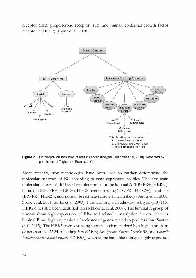
in situ carcinomas and invasive (infiltrating) carcinomas (Figure 2). In situ carcinoma
is a pre-invasive BC in which malignant cells are confined within the site of origin
and which can transform into an invasive cancer over a few years or even decades
(Barnes et al, 2012). However, only a subset of in situ cancers become invasive, and
recent studies do not support the notion that in situ carcinoma is an obligate
precursor of invasive BC (To et al, 2014). In situ carcinomas account for
approximately 20% of all diagnosed BCs (To et al, 2014). Moreover, in situ
carcinomas are further divided into ductal and lobular based on the origin of the
cancer cells (Figure 2). Ductal carcinoma in situ is a more common and
heterogeneous group of tumors than lobular carcinoma in situ, which presents low
histological variation (Figure 2). Similarly to in situ tumors, invasive carcinomas are
classified into subtypes, with the major subtypes being infiltrating ductal, invasive
lobular, ductal/lobular, mucinous, tubular, medullary, and papillary (Figure 2). Of
these, infiltrating ductal carcinoma and lobular carcinoma are the most common
subtypes, accounting for approximately 50-80% and 5-15% of all breast carcinomas,
respectively (Weigelt & Reis-Filho, 2009). In addition to histological type,
histological grade (grades 1-3) is used to classify breast carcinomas. Grade is an
assessment of the degree of differentiation and proliferative activity of a tumor and
reflects its aggressiveness (Weigelt et al, 2010). Moreover, several molecular markers
are used to further classify invasive carcinomas to determine which patients are likely
to respond to targeted therapies. The most commonly used markers include estrogen
24
receptor (ER), progesterone receptor (PR), and human epidermal growth factor
receptor 2 (HER2) (Payne et al, 2008).
Figure 2. Histological classification of breast cancer subtypes (Malhotra et al, 2010). Reprinted by permission of Taylor and Francis LLC.
More recently, new technologies have been used to further differentiate the
molecular subtypes of BC according to gene expression profiles. The five main
molecular classes of BC have been determined to be luminal A (ER/PR+, HER2-),
luminal B (ER/PR+, HER2+), HER2-overexpressing (ER/PR-, HER2+), basal-like
(ER/PR-, HER2-), and normal breast-like tumors (unclassified) (Perou et al, 2000,
Sorlie et al, 2001, Sorlie et al, 2003). Furthermore, a claudin-low subtype (ER/PR-,
HER2-) has also been identified (Herschkowitz et al, 2007). The luminal A group of
tumors show high expression of ERα and related transcription factors, whereas
luminal B has high expression of a cluster of genes related to proliferation (Santos
et al, 2015). The HER2-overexpressing subtype is characterized by a high expression
of genes at 17q22.24, including Erb-B2 Receptor Tyrosine Kinase 2 (ERBB2) and Growth
Factor Receptor-Bound Protein 7 (GRB7), whereas the basal-like subtype highly expresses

25
laminin, and keratins 5 and 17 (Santos et al, 2015). Normal breast-like tumors have
high expression levels of adipose tissue genes. The claudin-low subtype is
characterized by low expression of claudin genes, which are involved in tight
junctions and cell-cell adhesion, and high expression of vimentin, N-cadherin, and
immune system response genes (Santos et al, 2015). Basal-like, HER2-
overexpressing, and claudin-low tumors are the most aggressive and are associated
with a poor survival. In contrast, the luminal A type is the class of least aggressive
tumors (Santos et al, 2015). Both basal-like and HER2-overexpressing subtypes are
associated with a high frequency of TP53 mutations, whereas the basal-like subtype
is associated with only BRCA1 mutations (Cancer Genome Atlas Network, 2012).
Basal-like tumors are often referred to as triple-negative BCs (TNBCs) because most
are negative for ER, PR and HER2 (Cancer Genome Atlas Network, 2012).
In addition to molecular classification of BC, a functional classification system
based on the BC stem cells is emerging (Malhotra et al, 2010). In this system, BCs
are classified based on the tumor-initiating cells, and there are currently two
hypotheses in this regard. One suggests that BC heterogeneity arises from distinct
mammary stem/progenitor cells at various levels within the mammary cell hierarchy,
whereas the other hypothesis is that BC originates from a single mammary
stem/progenitor cell that is transformed by various oncogenes to give rise to various
types of cancer (Malhotra et al, 2010). Several markers for this classification system
have been identified, including CD44/CD24 and Aldehyde Dehydrogenase 1
Family, Member A1 (ALDH1) (Malhotra et al, 2010).

26
2 Ovarian cancer
2.1 Ovaries overview
The ovaries are a pair of organs in the female reproductive system and are located in
the pelvis, one on each side of the uterus (Figure 3). The main function of the ovaries
is to produce egg cells and the female hormones, progesterone and estrogen.
Figure 3. Female reproductive anatomy. SEER Cancer Statistics Factsheets: Ovary Cancer (National Cancer Institute, Surveillance, Epidemiology, and End Results Program).
2.2 Epidemiology
Ovarian cancer (OC) is a highly lethal gynecologic malignancy. It is the seventh most
common cancer and the eighth highest cause of death from cancer in women
worldwide, with an estimated 239,000 new cases (3.6% of all cancers) and 152,000
27
deaths (4.3% of all cancer deaths) in 2012 (Ferlay et al, 2015). The incidence rates of
OC are highest in more developed regions, with rates in these areas exceeding 7.5
per 100,000, whereas the lowest incidence is in Africa, with rates below 5 per 100,000
(Ferlay et al, 2015). Differences in incidence rates around the world are due to
ethnicity, genetic factors, socio-economically correlated environmental factors
related to lifestyle, nutrition, the use of exogenous hormones, reproduction, and
both diagnostic and medical treatment possibilities (La Vecchia, 2001, Lowe et al,
2013). The mortality rates of OC are higher in developed regions, such as North
America and Europe (from 5 to 6 per 100,000) than in less developed regions, such
as Eastern Asia (2 per 100,000) (Ferlay et al, 2015). More than 70% of women with
OC are diagnosed with advanced disease (Rauh-Hain et al, 2011). The five-year
survival rates for women with advanced disease vary from 20% to 30% (Rauh-Hain
et al, 2011). In Finland, 471 new OC cases were diagnosed in 2012 (Finnish Cancer
Registry), making OC the tenth most common cancer and the fifth highest cause of
death from cancer among women. In Finland, the incidence of OC is 8.6 per
100,000, the mortality rate is 4.8 per 100,000, and the five-year survival rate is 49%
(Finnish Cancer Registry).
2.3 Risk factors
Gender. OC is a sex-specific cancer, and being a female is a major risk factor.
Age. OC risk increases with age, and the majority of OCs are diagnosed in older
women. The median age at diagnosis is 63 years (National Cancer Institute,
Surveillance, Epidemiology, and End Results Program).
Race/Ethnicity. The highest incidence and mortality rates of OC are observed
among white females (National Cancer Institute, Surveillance, Epidemiology, and
End Results Program).
Family history of OC. A family history of OC is one of the strongest risk factors
for the disease. A woman with an OC-affected first-degree relative (mother, sister,
daughter) has a 5% risk, and a woman with two OC-affected first-degree relatives
has a 7% risk for developing the disease, whereas the lifetime risk for developing OC
in the general population is 1.6% (Prat et al, 2005). Additionally, a family history of
BC increases the risk of developing OC given that these two cancers comprise the
hereditary breast and ovarian cancer (HBOC) syndrome, which is caused by defects
in BRCA1 and BRCA2 (Lynch et al, 2009).
28
Genetics. It is estimated that inherited susceptibility can explain 5-15% of all
OCs (Lynch et al, 2009). Hereditary OC occurs in three different forms, site-specific
OC and as a component of two cancer syndromes, HBOC and Lynch syndrome
(also known as hereditary nonpolyposis colorectal cancer, or HNPCC syndrome)
(Prat et al, 2005). The causative genes for site-specific hereditary OC and HBOC
syndromes are two tumor suppressor genes, BRCA1 and BRCA2, whereas Lynch
syndrome is associated with defects in DNA mismatch repair genes, including
MLH1 and MSH2 (Prat et al, 2005). The majority (65-85%) of all hereditary OCs
are due to mutations in BRCA1 and BRCA2, and another 10-15% of hereditary cases
can be explained by Lynch syndrome gene mutations (Lynch et al, 2009). A carrier
of a BRCA1 or BRCA2 mutation, unselected for family history, has an average
cumulative lifetime risk of 39% and 11% for developing OC by the age of 70,
respectively (Antoniou et al, 2003). Moreover, the risk estimates for mutation carriers
are higher in HBOC families (Lynch et al, 2009).
Hormonal and reproductive factors. The ovarian epithelium responds strongly
to the local hormonal environment. Long-term exposure to elevated estrogen levels,
including early age of menarche, late age at natural menopause, and hormone
replacement therapy increase the risk of OC (Hunn & Rodriguez, 2012). Pregnancies
(especially after 35 years of age), having many children, breastfeeding, and the use of
oral contraceptives have protective effects (Hunn & Rodriguez, 2012).
Environmental factors. Several environmental factors related to lifestyle have
been reported to influence OC risk, but the study results are somewhat inconclusive
or controversial. Overweight and obesity (body mass index greater than 30) in early
adulthood have been reported to increase the risk of OC (Olsen et al, 2007). In
contrast, moderate physical exercise has been suggested to lower OC risk (Cannioto
& Moysich, 2015). Dietary factors, such as the intake of carbohydrates and dairy,
have been suggested to increase the risk of OC, whereas the consumption of green
leafy vegetables, vegetable oils and fish have been shown to have a protective effect
(Hunn & Rodriguez, 2012). Additionally, a high dietary intake of vitamin D has been
reported to be protective against specific histological subtypes of OC (Merritt et al,
2013). Smoking has been reported to increase the risk of mucinous subtype of OC,
but its effect on overall OC risk is uncertain (Collaborative Group on
Epidemiological Studies of Ovarian Cancer et al, 2012).
Inflammatory factors. Inflammatory factors have been reported to be involved
in ovarian carcinogenesis. Endometriosis has been shown to be associated with an
increased risk of ovarian carcinoma, especially endometrioid and clear cell types
(Prowse et al, 2006). Moreover, pelvic inflammatory disease and perineal talc
29
exposure have been shown contribute to OC risk, although the results for the latter
risk factor are somewhat inconclusive (Houghton et al, 2014, Huncharek et al, 2003,
Lin et al, 2011).
2.4 Clinical features and pathology
OC develops at a later age and is most typically observed in women over 60 years of
age. The hereditary form of OC is diagnosed about a decade earlier than non-
hereditary forms (Jazaeri, 2009). The symptoms of OC are unclear and can be easily
confused with those of common conditions, such as problems with digestion.
Symptoms may include abdominal bloating, pelvic and abdominal pain, feeling full
quickly, and difficulty eating (Goff, 2012). In the absence of clinically significant
symptoms and a lack of efficient screening methods, OC is primarily detected when
the disease is already at a late stage.
The origin and pathogenesis of OC are poorly understood making early detection
and new therapeutic approaches difficult. OC is a heterogeneous disease composed
of different types of tumors that have distinct features, behavior, and treatment
responses. Over 90% of the ovarian tumors are considered to originate from ovarian
surface epithelial cells, whereas other tumor types are considered to arise from germ
cells and sex-cord-stromal cells (Lynch et al, 2009). Based on the histopathology and
molecular genetic analyses, at least five main types of ovarian carcinomas are
identified: high-grade serous carcinoma (HGSC; 70%), endometrioid carcinoma
(EC; 10%), clear cell carcinoma (CCC; 10%), mucinous carcinoma (MC; 3%), and
low-grade serous carcinoma (LGSC; <5%), accounting for a total of 98% of ovarian
carcinomas (Prat, 2012). All these tumor types have different risk factors, precursor
lesions, patterns of spread, molecular events during oncogenesis, response to
chemotherapy, and prognosis (Table 1). HGSC is the most common ovarian
carcinoma and is detected at an advanced stage in approximately 80% of patients
(Prat, 2012). Thus, HGSC has the poorest prognosis of the different tumor types
(Table 1). Moreover, HGSC type particularly has been associated with germline
defects in BRCA1/2 genes (Risch et al, 2006), whereas EC and CCC types are
associated with endometriosis (Rosen et al, 2009). Relevant in this regard, EC is the
major HNPCC type (Prat, 2012). Moreover, OCs can be further classified according
to stages. The staging of OC (stages I-IV) is based on the FIGO nomenclature
(Heintz et al, 2006). Stage I disease is limited to the ovaries, whereas stage IV
indicates metastatic disease.

30
Based on the distinct morphologic and molecular genetic features, a dualistic
model of ovarian tumorigenesis has been proposed in which OCs can be divided
into two subgroups: Type I (low-grade pathway) and Type II (high-grade pathway)
(Kurman & Shih, 2010). Type I tumors consist of low-grade serous, low-grade
endometrioid, mucinous, clear cell and transitional (Brenner) carcinomas. Type II
tumors consist of high-grade serous carcinoma, undifferentiated carcinoma and
malignant mixed mesodermal tumors (carcinosarcomas). Type I tumors are generally
indolent, present in stage I (tumor confined to the ovary), and develop from well-
established precursors; these tumors are referred to as borderline tumors. Type I
tumors are relatively genetically stable and present specific mutations in certain
genes, such as Kirsten Rat Sarcoma Viral Oncogene Homolog (KRAS), B-Raf Proto-Oncogene,
Serine/Threonine Kinase (BRAF), ERBB2, Catenin (Cadherin-Associated Protein), Beta 1, 88
kDa (CTNNB1), PTEN, and Phosphatidylinositol-4,5-Bisphosphate 3-Kinase, Catalytic
Subunit Alpha (PIK3CA); however, Type I tumors rarely exhibit TP53 mutations.
Type II tumors are aggressive, present in advanced stage, develop from
intraepithelial cells, are genetically highly unstable, and have a high frequency of
TP53 mutations.
Moreover, there is compelling evidence that tumors that have been traditionally
considered primary ovarian tumors (high-grade serous, clear cell, and endometrioid)
actually originate in other pelvic organs and involve the ovary secondarily.
Specifically, high-grade serous carcinomas are reported to arise from epithelial
lesions in the distal fimbriated end of the fallopian tube, and clear cell and
endometrioid carcinomas originate from ovarian endometriosis (Kurman & Shih,
2010, Prat, 2012)

31
Table 1. Ovarian carcinoma: clinical and molecular features of the five most common types. (Prat, 2012) by permission of Oxford University Press.
HGSC LGSC MC EC CCC
Risk factors BRCA1/2 ? ? HNPCC ?
Precursor lesions Tubal Serous Cystadenoma/ Atypical Atypical
intraepithelial carcinoma borderline tumor borderline tumor? endometriosis endometriosis
Pattern of spread Very early transcoelomic spread Transcoelomic spread Usually confined to ovary Usually confined to pelvis Usually confined to pelvis
Molecular abnormalities BRCA, p53 BRAF, KRAS KRAS, HER2 PTEN, ARID1A HNF1, ARID1A
Chemosensitivity High Intermediate Low High Low
Prognosis Poor Intermediate Favorable Favorable Intermediate
Abbreviations: CCC, clear cell carcinoma; EC, endometrioid carcinoma; HGSC, high-grade serous carcinoma; HNPCC, hereditary polyposis colorectal carcinoma; LGSC, low-grade serous carcinoma; MC, mucinous carcinoma.

32
3 DNA Damage Response (DDR) pathway
The maintenance of genome integrity ensures the transmission of correct genetic
material across generations. Genetic material is continuously threatened by
spontaneous damages, such as occurs during DNA metabolism, and by damaging
agents coming from outside (exogenic) or inside (endogenic) the cell. Exogenic
threats include ultraviolet light, ionizing radiation, and chemicals, whereas endogenic
threats include reactive oxygen species, which are side products of normal cellular
metabolism. To protect genetic material from such damage, cells use a DNA damage
response (DDR) system that detects DNA damage and promotes the appropriate
cellular response, such as senescence, cell cycle checkpoint activation, DNA repair,
apoptosis, or tolerance (Figure 4). If the DDR machinery does not work properly,
the genome becomes unstable, which may result in uncontrolled behavior of the cell
and, eventually, cancer development. These processes are reviewed in (Bartek et al,
2007, Ciccia & Elledge, 2010, Harper & Elledge, 2007, Zannini et al, 2014)
Figure 4. The DNA damage response promotes the appropriate cellular response. (Zannini et al, 2014) by permission of Oxford University Press.
33
3.1 Different DDR repair mechanisms
DNA damage can be either single-stranded or double-stranded, and different repair
mechanisms are activated depending on the type of damage. Mispaired DNA bases
are changed to correct bases by mismatch repair, and small chemical alterations of
DNA bases and single-strand breaks are corrected by base excision repair. More
complex single-strand errors, such as pyrimidine dimers and intrastrand crosslinks,
are repaired by nucleotide excision repair. For the interstrand crosslinks, interstrand
crosslink repair is used. DNA double strand breaks (DSB) are the most deleterious
form of DNA damage and are repaired by at least by four independent mechanisms:
nonhomologous end joining (NHEJ), homologous recombination (HR), alternative
NHEJ, and single strand annealing. Of these processes, NHEJ and HR are the two
major mechanisms. Depending on the extent of DNA end processing, different
mechanisms are used to repair DSBs. HR is considered the most error-free
mechanism as it utilizes sister chromatids as a template for the synthesis of new
DNA. These processes are reviewed in (Ciccia & Elledge, 2010, Lord & Ashworth,
2012).
3.2 Key proteins in DDR
The DDR machinery is a complex network comprising numerous pathways,
proteins, and protein complexes that function in a well-coordinated manner. The
DDR is involved in all steps of this process, from DNA damage detection to the
activation of a cellular response to the damage. In basic terms, the DDR cascade
consists of proteins termed sensors, apical kinases, mediators, downstream kinases,
and effectors (Figure 5). The major regulators of DDR are the phosphoinositide 3-
kinase (PI3K)-related proteins kinases, ataxia-telangiectasia mutated (ATM), and
ATM and RAD3-related (ATR), which share many biochemical and functional
similarities. ATM primarily functions in response to DSBs, whereas ATR is primarily
activated in response to replication stress. However, both ATM and ATR target an
overlapping set of substrates in the DDR cascade. DNA lesions are recognized by
sensor proteins that vary with the different DDR regulators. For ATM, damage is
recognized by the MRN complex, which consists of Meiotic Recombination 11
Homolog A (S. Cerevisiae) (MRE11), RAD50 Homologue (S. Cerevisiae) (RAD50),
and Nijmegen Breakage Syndrome 1 (NBS1). For ATR, the damage-sensing proteins
include replication protein A (RPA) and the 9-1-1 complex bound by ATR
34
interacting protein (ATRIP). The 9-1-1 complex consists of RAD9 Homolog A (S.
Pompe) (RAD9), RAD1 Checkpoint DNA Exonuclease (RAD1), and HUS1 Checkpoint
Homolog (S. Pomple) (HUS1). Following detection of the DNA damage, ATM and
ATR initially phosphorylate mediator proteins, which can amplify the DDR by acting
both as recruiters of ATM/ATR substrates and as scaffolds upon which to assemble
complexes. At the site of DNA damage, phosphorylation of histone variant H2A
Histone Family, Member X (H2AX) on Serine 139 by ATM and ATR kinases is
required to recruit mediators, such as Mediator of DNA-Damage Checkpoint 1
(MDC1). Other mediator proteins include, for example, Tumor Protein P53 Binding
Protein 1 (53BP1), BRCA1, Topoisomerase (DNA) II Binding Protein (TopBP1),
and Claspin. Two kinases, CHK2 for ATM and Checkpoint Kinase 1 (CHK1) for
ATR, are involved in spreading the DNA damage signal through a phosphorylation
cascade. Along with ATM and ATR, CHK1 and CHK2 also phosphorylate effector
proteins, such as p53 and Cell Division Cycle 25 (Cdc25), which execute DDR
cellular responses. Additionally, a large number of other proteins are known to
participate in the DDR cascade. The DDR has also been discovered to play a role in
variety of different pathways, including RNA splicing, chromatin remodeling,
transcription, ubiquitination, and circadian rhythms. These findings are reviewed in
(Ciccia & Elledge, 2010, Cimprich & Cortez, 2008, Harper & Elledge, 2007, Sulli et
al, 2012).

35
Figure 5. Key proteins in the DNA damage response. Reprinted by permission from Macmillan Publisher Ltd: [NATURE REVIEWS CANCER] (Sulli et al, 2012), copyright (2012).
3.3 DDR and cancer
The DDR plays a central role in human physiology. Hereditary defects in genes
encoding key proteins in the DDR contribute to various human diseases, including
neurological disorders, infertility, immune deficiency, premature aging, and cancer.
Cancer, in particular, is driven by genomic instability. Several DDR-related cancer
syndromes have been recognized. The DDR syndromes, which can include breast
and ovarian cancer, include HNPCC syndrome, familial breast cancer syndrome,
Fanconi anemia (FA), Ataxia-telangiectasia (A-T), and Li-Fraumeni syndrome (LFS).
Of these, HNPCC is related to defects in mismatch repair genes such as MLH1,
MSH2, MutS Homolog 6 (MSH6), and Postmeiotic Segregation Increased (S. Cerevisiae) 2
(PMS2). Familial breast cancer syndrome is related to defects in homologous

36
recombination and damage signaling, and the causative genes include ATM, BRCA1,
BRCA2, BRIP1, CHK2, NBS1, PALB2, RAD50, and RAD51C. Moreover, FA is
related to defects in interstrand crosslink repair and homologous recombination, and
several FA genes have been recognized, including Fanconi Anemia, Complementation
Group D1 (FANCD1 or BRCA2), Fanconi Anemia, Complementation Group J (FANCJ
or BRIP1), and Fanconi Anemia, Complementation Group N (FANCN or PALB2).
Furthermore, A-T and Li-Fraumeni syndromes are associated with defects in DNA
damage signaling and DSB repair; causative genes for these conditions include ATM
and TP53, respectively. Reviewed in (Ciccia & Elledge, 2010, Jackson & Bartek,
2009)
37
4 Genetics of cancer
4.1 Hallmarks of cancer and mutation signature
Cancer can be considered a genetic disease. Cancer develops from a single somatic
cell that acquires changes in its DNA sequence throughout its lifespan and thereby
gains a growth advantage compared to other cells. Six alterations in cancer cell
physiology are considered essential for malignant growth, including self-sufficiency
in growth signals, insensitivity to growth-inhibitory signals, evasion of programmed
cell death, limitless replicative potential, sustained angiogenesis, and tissue invasion
and metastasis (Hanahan & Weinberg, 2000). Somatic mutations in the cancer cell
genome may encompass several distinct classes of DNA sequence changes, including
the substitution of one base by another, insertions or deletions of small or large
segments of DNA, inter- or intrachromosomal rearrangements, and copy-number
changes (Stratton et al, 2009). In solid tumors, such as those derived from the breast,
colon, or pancreas, an average of 33 to 65 genes display somatic mutations, and
approximately 95% of these mutations are single base substitutions (Vogelstein et al,
2013). Additionally, epigenetic changes, which alter chromatin structure and gene
expression, the acquisition of new DNA from exogenous sources (e.g., from viruses)
and defects in the mitochondrial genome contribute to cancer development (Stratton
et al, 2009).
Somatic mutations in cancer cell can be classified into driver and passenger
mutations according to their consequences for cancer development (Stratton et al,
2009). Driver mutations directly or indirectly confer a selective growth advantage to
the cell in which they occur. The other mutations that accumulate in the cell but do
not confer selective growth advantage are considered passengers. Typically, two to
eight driver mutations are necessary for cancer development (Vogelstein et al, 2013).
Two main types of genes participate cancer development; tumor suppressor genes
and oncogenes. Specifically, cancer-inhibiting tumor suppressor genes are
inactivated and cancer-promoting oncogenes are activated by mutations.

38
4.2 Tumor suppressor genes
Tumor suppressor genes encode proteins whose normal function is to inhibit cell
transformation. The proteins participate in a variety of critical and highly conserved
cell function, including regulation of the cell cycle and apoptosis, differentiation,
surveillance of genomic integrity, repair of DNA errors, signal transduction, and cell
adhesion (Oliveira et al, 2005). Tumor suppressor genes can be divided into three
classes, caretakers, gatekeepers, and landscapers, based on different properties
(Kinzler & Vogelstein, 1997, Kinzler & Vogelstein, 1998). Caretaker genes encode
DNA repair proteins that act as caretakers of the genome. The inactivation of
caretaker gene results in a greatly increased mutation rate and genomic instability.
Gatekeepers prevent cancer through direct control of cell growth. The inactivation
of gatekeeper gene contributes directly to the neoplastic growth of the tumor.
Landscaper genes encode proteins that affect the microenvironment of the tumor.
According to Knudson’s two hit hypothesis, two mutational events are required to
inactivate the both alleles in tumor suppressor genes (Knudson, 1971). In the
dominantly inherited form, one mutation is inherited via germinal cells and the
second occurs in somatic cells. In the nonhereditary form, both mutations occur in
the somatic cell. One of the most commonly known tumor suppressor genes is TP53,
which is mutated in more than half of all human cancers (Vousden & Lu, 2002). In
BC, BRCA1 and BRCA2 are by far the two most commonly known tumor
suppressor genes (Miki et al, 1994, Wooster et al, 1994).
4.3 Oncogenes
Oncogenes encode proteins that promote cell proliferation. These genes can be
categorized into six groups: transcription factors, chromatin remodelers, growth
factors, growth factor receptors, signal transducers, and apoptosis regulators (Croce,
2008). Oncogenes are derived from proto-oncogenes by point mutations,
amplifications, or chromosomal rearrangements (Croce, 2008). Compared to tumor
suppressor genes, an activating somatic mutation in one allele of an oncogene is
generally sufficient to confer a selective growth advantage on the cell (Vogelstein &
Kinzler, 2004). One of the most commonly known oncogenes is V-Myc Avian
Myelocytomatosis Viral Oncogene Homolog (MYC) (Dang, 2010). Moreover, the
amplification of the ERBB2/HER2 oncogene in BC is a well-known biological

39
marker with therapeutic value. Amplification of this gene is seen in approximately
20% of BCs and is associated with an aggressive disease (Sauter et al, 2009).
40
5 The genetic predisposition to breast and ovarian cancer: susceptibility genes
The genetic predisposition to breast and ovarian cancer has been well established.
Up to 10% of all BCs and up to 15% of all OCs are caused by inherited genetic
defects (Claus et al, 1996, Lynch et al, 2009). Less than half of the genetic
predisposition to BC has been resolved (Figure 6). BC predisposition factors can be
classified into three categories according to the risk associated with the disease: high-
risk genes (>10-fold elevated risk), moderate-risk genes (2-4-fold elevated risk) and
low-risk genes (<1.5-fold elevated risk) (Turnbull & Rahman, 2008). Rare mutations
in two major high-risk genes, BRCA1 and BRCA2, explain the majority 15% of BC
familial relative risk (i.e., the ratio of the risk of disease for a relative of an affected
individual to that for the general population) (Figure 6). Additionally, rare mutations
in high-to-moderate risk genes that are associated with cancer syndromes (e.g., TP53,
PTEN, Liver Kinase B (LKB1), and CDH1) and in moderate-risk genes (e.g., CHEK2,
ATM, and PALB2) add another 3% and 4% to the BC familial relative risk,
respectively (Figure 6). The remaining known genetic predisposition to BC is due to
common single nucleotide polymorphisms (SNPs) in low-risk genes, such as
Fibroblast Growth Factor Receptor 2 (FGRF2), Caspase 8, Apoptosis-Related Cysteine
Peptidase (CASP8), and TOX High Mobility Group Box Family Member 3 (TOX3) (Figure
6). In epithelial ovarian cancer, approximately 90% of the genetic predisposition is
explained by gene defects in the high-penetrance genes BRCA1 and BRCA2,
whereas the remaining 10% of the genetic predisposition is attributable to defects in
HNPCC syndrome genes (Prat et al, 2005). Additionally, moderate-to-high risk
alleles in genes such as BRIP1 and RAD51C have been determined to contribute to
a fraction of OC predisposition (Pelttari et al, 2011, Rafnar et al, 2011)

41
Figure 6. Genetic variants that predispose to breast cancer. From (Couch et al, 2014). Reprinted with the permission from AAAS.
5.1 High-risk genes: BRCA1 and BRCA2
5.1.1 BRCA1
Gene. The Breast Cancer 1, Early Onset (BRCA1) gene is located on the long arm of
chromosome 17 at 17q21. It was detected in the early-90s as a strong candidate gene
for breast and ovarian cancer susceptibility by linkage and positional cloning
methods (Miki et al, 1994). BRCA1 is a large gene, spanning an 81 kb region of
genomic DNA, consisting of 24 exons, and encoding a protein of 1,863 amino acids
(Miki et al, 1994, Smith et al, 1996). Exon 1 is non-coding, and exon 11 is the largest
exon, encoding over 60% of the BRCA1 protein (Miki et al, 1994, Thakur et al,
1997). The majority of clinically relevant mutations are in exon 11 (National Human
Genome Research Institute, Breast Cancer Information Core Database).
Protein structure and function. The two most important regions of the BRCA1
protein are a RING domain in the amino terminus and two BRCT domains in the
carboxyl terminus (Figure 7a). Additionally, in the middle of the protein are nuclear
localization signals (NLS) and a coiled-coil domain (Figure 7a). BRCA1 has
phosphorylation sites for ATM/ATR and CHK2 kinases and has a critical
interaction with several proteins, including BRCA1 Associated RING Domain 1
(BARD1), PALB2, Abraxas, Retinoblastoma Binding Protein 8 (CtIP), and BRIP1
(Figure 7a).
42
BRCA1 has vital role in normal cellular development, and BRCA1 deficiency
leads to early embryonic lethality in mice (Hakem et al, 1996, Ludwig et al, 1997).
BRCA1’s key role is to maintain genomic stability and function as a tumor
suppressor (Wang, 2012). This protein acts as central mediator of the cellular
response to DNA damage, regulating the activities of multiple repair and checkpoint
pathways (Wang, 2012). BRCA1 is a substrate of the central DNA damage response
kinases ATM/ATR, which control the DDR (Wang, 2012). The highly conserved
zinc-binding RING domain (residues 20-68) has DNA-binding properties and is an
interaction site for BRCA1-associated RING domain (BARD1) (Wu et al, 1996).
BRCA1 and BARD1 form a heterodimeric RING finger complex that has ubiquitin
E3 ligase activity. The enzymatic activity of the BRCA1-BARD1 complex has been
suggested to be critical in DNA double strand break repair and the tumor
suppression function of BRCA1, but the exact role of this enzymatic activity is not
well understood (Hashizume et al, 2001, Morris & Solomon, 2004, Reid et al, 2008).
Another protein that binds to the BRCA1 RING domain is BRCA1-associated
Protein-1 (BAP1), which is a nuclear-localized deubiquitinating enzyme that has
been suggested to have tumor suppressor properties (Eletr & Wilkinson, 2011,
Jensen et al, 1998). Two tandem and highly conserved BRCA1 BRCT domains
(residues 1699-1736 and 1818-1855) are essential for the tumor suppressor function
of this protein (Glover, 2006). These BRCT domains are phospho-peptide-binding
domains that recognize a phospho-SPxF motif (S, serine; P, proline; × varies; F,
phenylalanine) (Wang, 2012). Similar BRCT domains are present similar in a wide
variety of proteins and are involved in cell cycle regulation and DNA repair (Glover,
2006). BRCT-interacting proteins include Abraxas, BRIP1, and CtIP, which directly
interact with BRCT domains through their phospho-SPxF motifs in a
phosphorylation-dependent manner (Wang, 2012). Additionally, the BRCA1 coiled-
coil domain is an interacting site for PALB2 and BRCA2 (Wang, 2012). Moreover,
the tumor suppression function of BRCA1 has been shown to occur via
heterochromatin-mediated silencing (Zhu et al, 2011).

43
Figure 7. BRCA1 and BRCA2 functional domains and interacting proteins. Adapted and reprinted by permission from Macmillan Publishers Ltd: [NATURE REVIEWS CANCER] (Roy et al, 2011), copyright (2011).
5.1.2 BRCA2
Gene. Breast Cancer 2, Early Onset (BRCA2) is located on chromosome 13 at 13q12.
This gene was detected as another major breast and ovarian cancer susceptibility
gene by linkage and positional cloning methods in the 90s, immediately following
BRCA1 identification (Tavtigian et al, 1996, Wooster et al, 1994). BRCA2 is a large
gene, spanning approximately 70 kb of genomic DNA, consisting of 27 exons, and
encoding a protein of 3,418 amino acids (Wooster et al, 1994). Similar to BRCA1,
exon 1 is non-coding and exon 11 is the largest exon, encoding over half of the
protein (Tavtigian et al, 1996). The majority of clinically relevant BRCA2 mutations
are located in exon 11 (National Human Genome Research Institute, Breast Cancer
Information Core Database).
Protein structure and function. The BRCA2 protein consists of a DNA
binding domain, eight BRC repeats, and nuclear localization signals on the C-
terminus (Figure 7b). A critical phosphorylation site for cyclin dependent kinase 2
(CDK2) is located on the C-terminus (Figure 7b). BRCA2 has many interaction sites
for different proteins, including RAD51 Recombinase (RAD51), PALB2, and

44
Deleted In Split-Hand/Foot 1 (DSS1); these sites are critical for BRCA2’s proper
function (Figure 7b).
BRCA2 maintains genome stability and has a key role in the HR pathway in DNA
double-strand break repair. The HR pathway is dependent on the recombinase
function of RAD51, which is an important interacting partner of BRCA2. BRCA2
binds directly to RAD51 through its C-terminus and conserved BRC repeats.
BRCA2 is recruit RAD51 to the nucleus and to the site of DNA damage by utilizing
its nuclear localization signals, which are lacking in RAD51. Moreover, BRCA2
regulates RAD51 function during recombination events. The N-terminal portion of
BRCA2 interacts with PALB2, physically linking it to BRCA1. The BRCA1-PALB2-
BRCA2 complex is needed for the recruitment of RAD51 to the site of DNA
damage. DSS1 interacts with DNA-binding domain of BRCA2, and this interaction
is critical for the proper functioning of BRCA2. On the C-terminus of BRCA2 is a
CDK2 phosphorylation site (S3291), the phosphorylation status of which modulates
BRCA2’s C-terminal interaction with RAD51. These findings are reviewed in
(Gudmundsdottir & Ashworth, 2006, Roy et al, 2011)
5.1.3 Contribution of germline BRCA1/2 mutations to hereditary breast and ovarian cancer
Monoallelic germline BRCA1 and BRCA2 mutations predispose to hereditary breast
and ovarian cancers in a highly penetrant manner. Pathogenic BRCA1/2 mutations
appear to account for approximately 30% of cases of these cancers in high-risk
families (Couch et al, 2014). Most deleterious mutations are small insertions or
deletions that result in the translation of a truncated protein (Mavaddat et al, 2013).
The frequency of BRCA1/2 mutations varies depending on the population in
question. The frequencies are highest in founder populations, such as Ashkenazi
Jews (Levy-Lahad et al, 1997). According to a study of breast/ovarian cancer patients
who were unselected for family history, the average cumulative risk in BRCA1- and
BRCA2-mutation carriers by age 70 years was 65% and 45% for breast cancer and
39% and 11% for ovarian cancer, respectively (Antoniou et al, 2003). The risk
estimates for mutation carriers in multiple-case high-risk families are even higher
(Antoniou et al, 2003). Moreover, risk estimates vary by age at diagnosis of the index
case, the type and site of the cancer, and the site of the mutation (Mavaddat et al,
2013). In addition, several genetic modifiers of BRCA1 and BRCA2 contribute to
cancer risk in mutation carriers (Antoniou et al, 2008). Biallelic mutations in both of

45
these genes are rare. Specifically, biallelic BRCA1 mutations are extremely rare and
have been reported only in one early-onset OC patient (Domchek et al, 2013).
Moreover, a recent study reported a biallelic BRCA1 mutation in one early-onset BC
patient who presented with a Fanconi anemia-like disorder (Sawyer et al, 2015).
Biallelic BRCA2 mutations have been associated with Fanconi anemia subtypes
(Howlett et al, 2002). Additionally, a recent study reported a presence of biallelic
BRCA2 mutations in single family presenting with early-onset colorectal cancer in
the absence of Fanconi anemia features (Degrolard-Courcet et al, 2014).
BRCA1- and BRCA2-related breast tumors differ from each other and from
sporadic cancers. Typically, compared to sporadic tumors, BRCA1-related tumors
are poorly differentiated ductal carcinomas of higher grade and higher mitotic index.
Such tumors also exhibit a greater degree of nuclear polymorphism, less tubule
formation, and may exhibit features of medullary carcinoma (Mavaddat et al, 2013).
BRCA1-related tumors are often negative for estrogen and progesterone receptors
and HER2 and are more likely to be p53-positive (Lakhani et al, 2002). Moreover,
BRCA1-related tumors resemble basal subtype of BC (Sorlie et al, 2003). The
specific phenotype for BRCA2-related tumors has not been consistently described
(Phillips, 2000).
5.1.4 Contribution of germline BRCA1/2 mutations to other cancers
BRCA1 mutations are associated with an increased prostate cancer risk in male
carriers (Leongamornlert et al, 2012). BRCA2 mutations have been shown to
contribute to prostate, pancreatic, gallbladder, bile duct, and stomach cancers, as well
as melanoma (Breast Cancer Linkage Consortium, 1999). Both BRCA1 and BRCA2
mutations increase the male BC risk, and the risk is higher in BRCA2 mutation
carriers (Tai et al, 2007).
5.1.5 The germline BRCA1/2 mutation spectrum in Finnish hereditary breast and/or ovarian cancer families
In Finland, germline BRCA1/2-mutations contribute approximately 20% to familial
breast and ovarian cancer cases (Hartikainen et al, 2007, Vehmanen et al, 1997a). In
the Finnish breast and ovarian cancer studies, several BRCA1 and BRCA2
deleterious mutations have been reported, and total of fourteen mutations, seven in
each gene, are considered founder mutations. These mutations are designated as
46
such given that they are recurrent and account for the majority of all detected
BRCA1/2 mutation in Finland (Hartikainen et al, 2007, Huusko et al, 1998,
Sarantaus et al, 2000, Vehmanen et al, 1997a, Vehmanen et al, 1997b). The most
frequently observed Finnish founder mutations include 3604delA,
3744delT/3745delT, 4216-2ntA>G, 4446C>T, 5370C>T in BRCA1 and 999del5,
7708C>T, 8555T>G, and 9346-2ntA>T in BRCA2 (Sarantaus et al, 2000). There
are geographical differences in the distribution of founder mutations across Finland.
For example, in the Northern and Eastern part of Finland, the mutation spectrum is
scarce and specific, whereas in Southern Finland, almost all founder mutations have
been detected (Hartikainen et al, 2007, Huusko et al, 1998, Sarantaus et al, 2000).
Moreover, one mutation, 4088insA in BRCA2, has been detected uniquely in the
Eastern part of Finland (Hartikainen et al, 2007). Large genomic rearrangements in
BRCA1/2 are rare and only one study has reported a large BRCA1 deletion in a
Finnish family with a history of OC (Pylkas et al, 2008).
5.2 High-to-moderate-risk genes involved in cancer syndromes
5.2.1 TP53 and Li-Fraumeni syndrome
Tumor protein p53 (TP53) gene is located on chromosome 17 at 17p13. This gene
spans approximately 20 kb of genomic DNA and consists of 11 exons, encoding a
393-amino acid protein (i.e., p53) (McBride et al, 1986). Protein p53 is a tumor
suppressor protein whose main function is to maintain cell integrity by controlling
cell survival, proliferation, and death (Vousden & Prives, 2009). Protein p53 is
activated by diverse cell stress signals, such as DNA damage, oxidative stress,
hyperproliferative signals and hypoxia (Bieging et al, 2014). In its active form, p53
triggers the transcription of various target genes that further induce responses such
as cell cycle arrest, DNA repair, senescence, and apoptosis. These effects ultimately
either promote the repair and survival or the permanent removal of damaged cells
(Vousden & Prives, 2009). In addition, p53 regulates processes such as cellular
metabolism, stem cell function, invasion and metastasis, and cell-cell communication
within the tumor microenvironment; these effects may also contribute to tumor
suppression (Bieging et al, 2014). Protein p53 is inactive in more than half of all
human cancers due either to mutations in the TP53 gene or defects in signaling

47
pathways upstream or downstream of p53 (Vousden & Lu, 2002). Over 80% of
TP53 mutations in human tumors localize to the central region of the gene, which
encodes a highly conserved, sequence-specific DNA-binding domain (exons 5-8)
(Bieging et al, 2014, Vousden & Lu, 2002).
Germline mutations in TP53 contribute to Li-Fraumeni Syndrome (LFS) and its
variant, Li-Fraumeni Like-Syndrome (LFL). Both of these conditions are rare
autosomal cancer predisposition syndromes characterized by familial clustering of a
wide spectrum of tumors in children and young adults. These tumors are
predominantly breast tumors, bone and soft-tissue sarcomas, brain tumors,
leukemia, and adrenocortical tumors (Birch et al, 1994, Malkin et al, 1990). LFL
resembles LSF but is defined by less stringent classification criteria (Birch et al, 1994,
Eeles, 1995). Germline TP53 mutations are observed in 71-77% and 22-40% of LFS
and LFL families, respectively (Varley et al, 1997, Varley, 2003). In LFS families,
nearly half of the individuals with germline TP53 mutations develop cancer by the
age of 30 years, and the lifetime cancer risk is 73% in males and almost 100% in
females. This latter high-penetrance is primarily due to early-onset BC (McBride et
al, 2014). BC is the most common cancer type among female TP53 mutation carriers
in LFS/LFL families, accounting for 27% of all cancers (McBride et al, 2014).
Moreover, germline TP53 mutations have been reported to contribute up to 8% of
early-onset BC cases with family history of the disease (Lalloo et al, 2006, Lee et al,
2012, Mouchawar et al, 2010).
In Finland, germline TP53 mutations are very rare in the general BC population
but explain a small fraction of the hereditary BC cases (Rapakko et al, 2001).
Pathogenic TP53 mutations occurring in the conserved region of TP53 have been
reported in 3/108 (2.8%) Finnish BRCA1/2-negative hereditary BC families that
also exhibit features of either LFS or LFL (Huusko et al, 1999, Rapakko et al, 2001).
5.2.2 ATM and Ataxia-telangiectasia
Ataxia Telangiectasia Mutated (ATM) gene is located at locus 11q22-23 and consists of
66 exons, encoding a large protein of 3,056 amino acids (Gatti et al, 1988, Savitsky
et al, 1995, Uziel et al, 1996). The encoded protein belongs to a family of PI3K-
related kinases that are involved in various cellular responses to genotoxic stress.
These functions are carried out via phosphorylation of key proteins in cellular
response pathways (Shiloh & Ziv, 2013). The protein kinase ATM has a central role
as an activator of the DNA damage response cascade after DNA double-strand
48
breaks (Shiloh & Ziv, 2013). ATM exists as a homodimer that dissociates into active
monomers upon activation, which occurs via autophosphorylation at Ser1981 after
DNA damage (Shiloh & Ziv, 2013). In its active form, ATM is recruited to the DNA
damage site, where it activates a signaling cascade that includes direct and indirect
phosphorylation events of a large number of proteins. These phosphorylation events
lead to the activation of cell-cycle checkpoints and the initiation of DNA repair
(Shiloh & Ziv, 2013). Downstream targets of ATM include also proteins that are
encoded by the known BC susceptibility genes TP53, BRCA1, and CHEK2 (Ahmed
& Rahman, 2006).
Homozygous germline defects or compound heterozygosity in ATM are a cause
of Ataxia-telangiectasia (A-T), a rare autosomal recessive disorder characterized by
cerebellar degeneration, immunodeficiency, chromosomal instability, cancer
predisposition, radiation sensitivity, and cell cycle abnormalities (Savitsky et al, 1995,
Swift et al, 1986). Moreover, heterozygous A-T-causing germline mutations have
been shown to increase BC risk from 2-to-9-fold in female relatives of A-T patients
and in hereditary BC patients without a family history of A-T in different populations
(Broeks et al, 2000, Olsen et al, 2005, Renwick et al, 2006, Swift et al, 1987,
Thompson et al, 2005, Thorstenson et al, 2003). However, despite the central role
of ATM in DNA repair pathway, the contribution of ATM mutations to the risk of
other cancers is not clearly understood (Thompson et al, 2005). Although ATM has
been considered a moderate penetrance BC gene, various studies have been
inconsistent regarding the degree of BC risk related to heterozygous ATM variants
(Ahmed & Rahman, 2006, Prokopcova et al, 2007).
In Finland, the contribution of germline ATM variants to BC predisposition has
been studied in different geographical BC cohorts. A common heterozygous ATM
polymorphism, 5557G>A with IVS38-8T>C, was associated with bilateral BC (odds
ratio (OR) = 10.2; 95% confidence interval (CI) = 3.1-33.8; p = 0.001) in a cohort
of 185 breast or breast-ovarian cancer patients from 121 Northern Finnish families
(Heikkinen et al, 2005). However, the findings were not supported by further case-
control association analysis utilizing an extensive cohort of Southern Finnish familial
and unselected BC patients (Tommiska et al, 2006a). Moreover, two pathogenic
mutations, 7570G>C and 6903insA, that were originally identified in Finnish A-T
families were observed in 3/162 (1.9%) BC families of Central and Northern Finnish
origin (Allinen et al, 2002). The apparent yet overall limited role of heterozygous
ATM variants in hereditary BC predisposition was confirmed by the identification
of two pathogenic mutations, 7570G>C and 6903insA, and one A-T-causative
mutation (8734A>G) in 6/541 (1.1%) familial (P = 0.006, OR 12.4, 95% CI 1.5-

49
103.3) and 7/1124 (0.6%) unselected BC cases of Finnish origin (P = 0.07, OR 6.9,
95% CI 0.9-56.4); in population controls, the prevalence of these mutations was
1/1107 (0.1%) (Pylkas et al, 2007).
5.2.3 PTEN and Cowden syndrome
Phosphatase And Tensin Homolog gene (PTEN) is located on the long arm of
chromosome 10 at 10q23 and encodes a 403-amino acid protein (Li et al, 1997).
PTEN is a dual specificity protein and lipid phosphatase that blocks the PI3K/Akt
signaling pathway and thereby inhibits cell survival, growth, and proliferation
(Hopkins et al, 2014). Moreover, PTEN is known to act in a PI3K-independent
manner, inhibiting migration and affecting genomic stability, gene expression, and
the cell cycle (Hopkins et al, 2014).
PTEN is frequently altered in a variety of human cancers, including the brain,
breast, and prostate. To date, more than 2700 PTEN mutations have been identified
in different tumor types (Hopkins et al, 2014). PTEN has been detected as a
causative gene for Cowden syndrome, a rare autosomal dominant familial cancer
syndrome with a high risk of breast and thyroid cancer and the presence of benign
hamartomas in several tissue, including the skin, breast, thyroid, oral mucosa, and
intestinal epithelium (Hopkins et al, 2014, Nelen et al, 1996, Nelen et al, 1997). The
lifetime risk for female BC among PTEN mutation carriers is as high as 85%, even
higher than the BC risk estimates for BRCA1/2-mutation carriers (Antoniou et al,
2003, Tan et al, 2012). Germline PTEN mutations have been reported to be rare in
high-risk non-BRCA1/2 BC families (Blanco et al, 2010, Guenard et al, 2007).
However, a Finnish hereditary BC study reported that germline PTEN promoter
region variants affect tumor progression and the gene expression profile in BC
(Heikkinen et al, 2011).
5.2.4 STK11 and Peutz-Jeghers syndrome
Serine/Threonine Kinase 11 gene (STK11, also known as Liver Kinase B1, LKB1) is
located on the telomeric end of chromosome 19 at 19p13 and encodes a
serine/threonine kinase (STK11) of 433 amino acids (Hemminki et al, 1998). STK11
is tumor suppressor protein involved in several cellular responses, such as energy
metabolism, cellular polarity and cellular growth. STK11 regulates these processes
primarily via AMPK/mTOR signaling (Hemminki et al, 1998, Korsse et al, 2013).

50
Moreover, STK11 is involved in cell cycle regulation and apoptosis, and one of its
interacting partners is the tumor suppressor protein p53 (Korsse et al, 2013).
STK11 is a causative gene for Peutz-Jeghers Syndrome (PJS), a rare autosomal
dominant disorder characterized by hamartomatous polyps of the gastrointestinal
tract, mucocutaneous melanin deposits, and an increased risk of benign and
malignant neoplasms of the intestinal tract, pancreas, ovaries, testis, breast, and
uterus (Giardiello et al, 1987, Giardiello & Trimbath, 2006, Hemminki et al, 1998,
Jenne et al, 1998). Patients with PJS have a cumulative risk of 37-93% for all cancers
and of 32-54% for BC by the age of 70 (van Lier et al, 2010). The BC risk among
PJS patients is comparable to the BC risk associated with germline BRCA1/2
mutations (Antoniou et al, 2003). Germline STK11 mutations have been reported to
be rare in high-risk non-BRCA1/2 BC families (Guenard et al, 2010).
5.2.5 CDH1 and hereditary diffuse gastric cancer syndrome
Cadherin 1, type 1, E-cadherin (epithelial) (CDH1) gene is located on chromosome 16 at
16q22 and encodes a calcium-dependent cell-cell adhesion molecule of
approximately 728 amino acids (Berx et al, 1995). CDH1 is a transmembrane
glycoprotein that plays an important role in the formation and maintenance of the
normal architecture and function of epithelial cells (Berx et al, 1998). The adhesive
function of CDH1 is dependent on the interaction of its cytoplasmic domain with
catenin proteins, including β-catenin, which is a proto-oncogene that plays an
important role in Wnt-signaling (Behrens, 1999, Berx et al, 1998). The CDH1 protein
is a well-known growth and invasion suppressor in epithelial cells and acts through
complex mechanism, including the promotion of tissue organization and inhibition
of apoptosis (van Roy, 2014). The loss of CDH1 expression and/or its abnormal
function play an important role in human cancer development (Paredes et al, 2012).
Germline mutations in CDH1 have been identified as the genetic cause of
hereditary diffuse gastric cancer (HDGC) syndrome, which is characterized by 1) the
familial aggregation of diffuse gastric cancer cases among first or second degree
relatives and 2) an increased risk of other cancers, including lobular breast, colon,
prostate, and ovarian cancer (Fitzgerald et al, 2010, Pinheiro et al, 2014).
Approximately 25-30% of families fulfilling the HDGC criteria harbor inactivating
mutations in the CDH1 gene region (Fitzgerald et al, 2010). Germline CDH1
mutations are of high-penetrance, conferring high lifetime risks of developing gastric
and lobular BCs (over 80% and 60% by 80 years of age, respectively) (Fitzgerald et

51
al, 2010). However, germline CDH1 mutations are generally rare in lobular BC
patients without a family history of HDGC (McVeigh et al, 2014, Schrader et al,
2011, Xie et al, 2011).
In Finland, germline CDH1 alterations have been studied only in Finnish prostate
cancer patients who also presented with gastric cancer in their families. One CDH1
missense mutation has been identified to have a higher frequency in both familial
and unselected prostate cancer cases than in controls in a large-scale population-
based survey (Ikonen et al, 2001).
5.3 Moderate-risk genes
5.3.1 CHEK2
The Checkpoint Kinase 2 (CHEK2) gene is located on the long arm of chromosome
22 at 22q12 and encodes a serine/threonine-protein kinase that functions as a key
mediator of the DNA damage response, cell cycle control and DNA repair (Bartek
et al, 2001). The CHEK2 protein is activated via phosphorylation by the upstream
kinase ATM in response to DNA double strand breaks (Bartek et al, 2001). After
activation, CHEK2 kinase phosphorylates downstream effectors, including the
tumor suppressor proteins BRCA1 and p53, the transcription factor E2F1, the
PI3K, and the phosphatases Cell Division Cycle 25A (Cdc25A) and Cell Division
Cycle 25C (Cdc25C). These effects thereby activate different cellular responses, such
as damage-induced transcription, DNA repair, cell cycle arrest/delay, and apoptosis
(Bartek et al, 2001, Bartek & Lukas, 2003). The CHEK2 gene consists of 14 exons,
and protein structure shows three characteristic domains: 1) an amino-terminal
SQ/TQ regulatory domain with putative phosphorylation sites by ATM kinase, 2) a
forkhead-associated domain for binding to other phosphorylated proteins, and 3) a
carboxyl terminal serine/threonine kinase domain that has activation loop structure
and shows structural homology with other serine/threonine kinases (Bartek et al,
2001, Nevanlinna & Bartek, 2006) Defects in the CHEK2 protein contribute to the
development both hereditary and sporadic human cancers, and its role as a candidate
tumor suppressor has been suggested (Bartek et al, 2001).
Germline CHEK2 mutations were originally identified in Li-Fraumeni Syndrome,
which is a highly penetrant familial cancer phenotype usually associated with
inherited mutations in TP53 (Bell et al, 1999). However, germline CHEK2 variants
52
were subsequently identified in familial and unselected BC patients without Li-
Fraumeni or Li-Fraumeni-Like Syndrome, as well as in healthy population controls.
These results suggest that CHEK2 is a low-to-intermediate penetrance BC
susceptibility gene (Vahteristo et al, 2002). Two germline mutations are the most
studied CHEK2 variants with respect to hereditary BC and other cancers in different
populations. These mutations include 1) a c.1100delC deletion in the kinase domain
in exon 10, which results in a truncated protein; and 2) a missense mutation
(c.470T>C (I157T)) in the forkhead-associated domain in exon 3, which disrupts
critical interactions with its binding partners, such as BRCA1 (Nevanlinna & Bartek,
2006). The c.1100delC variant results in an approximately two-to-three-fold increase
in the risk of BC in women without BRCA1 and BRCA2 mutations (Meijers-
Heijboer et al, 2002). Similar observations for the c.1100delC variant were reported
in Finnish BRCA1/2-negative hereditary BC patients, with observed frequencies
5.5% (28/507) in cases and 1.4% (26/1,885) in controls (Vahteristo et al, 2002).
Additionally, a large international collaborative study by the CHEK2 Breast Cancer
Consortium found the c.1100delC variant in 1.9% (201/10,860) of BC cases and of
0.7% (64/9,065) controls (estimated odds ratio 2.34; 95% CI 1.72-3.20). These data
confirm that c.1100delC variant confers an increased risk (approximately two-fold)
of BC, and this risk is apparent in women unselected for family history (CHEK2
Breast Cancer Case-Control Consortium, 2004). Moreover, trends have been
observed towards an early age of onset and a bilateral form of BC among c.1100delC
variant carriers (CHEK2 Breast Cancer Case-Control Consortium, 2004, Oldenburg
et al, 2003, Vahteristo et al, 2002). Additionally, a higher frequency of c.1100delC
variant carriers has been reported in cases with an affected first-degree relative
(Vahteristo et al, 2002). As the c.1100delC is a low-to moderate risk variant, it has
been suggested that it acts as a modifier in combination with other gene variants,
thereby multiplying the BC risk in BRCA1/2-negative families (Antoniou et al, 2002,
Oldenburg et al, 2003). Compared to c.1100delC, I157T is a lower risk variant and
has been observed in different populations with varying frequencies. The highest
frequencies have been observed in Finnish (7.4%), Polish (6.7%), German (2.2%)
and Byelorussian (5.7%) populations (Bogdanova et al, 2005, Cybulski et al, 2004,
Kilpivaara et al, 2004). A recent meta-analysis confirmed the I157T variant to be
associated with a 1.5-fold increased BC risk both in familial and unselected BC cases
and a 4-fold increased BC risk in lobular BC cases (Liu et al, 2012).
Moreover, CHEK2 variants increase the risk of other cancers, including male
breast, colon, prostate, thyroid, and kidney cancers (Cybulski et al, 2004, Meijers-
Heijboer et al, 2002). The contribution of the CHEK2 variants to hereditary prostate

53
and sporadic and familial colorectal cancers has also been observed in the Finnish
population (Kilpivaara et al, 2003, Kilpivaara et al, 2006, Seppala et al, 2003).
5.3.2 PALB2
The Partner and localizer of BRCA2 (PALB2) gene is located on the short arm of
chromosome 16 at 16p12 and consists of 13 exons that encode a 1186-residue
polypeptide. PALB2 binds to the N-terminus of BRCA2 and ensures its proper
function in DNA double-strand break repair (Xia et al, 2006). The interaction of
PALB2 with BRCA2 occurs via its WD40 domain in the C-terminus (Xia et al, 2006).
PALB2 colocalizes with BRCA2 in nuclear foci, promotes its localization and
stability in nuclear structures. In addition, PALB2 enables BRCA2’s recombinational
repair and checkpoint functions, as well as its tumor suppression activity (Rahman
et al, 2007, Xia et al, 2006). Moreover, PALB2 has an important role as a BRCA1-
interacting partner, being part of the BRCA1-PALB2-BRCA2-complex, which plays
a critical role in homologous recombination repair (Sy et al, 2009, Zhang et al, 2009).
The interaction of PALB2 with BRCA1 occurs through the coiled-coil domain in
the N-terminus of PALB2 (Sy et al, 2009). Moreover, PALB2 directly interacts
through its WD40 domain with several other proteins that function in both cellular
responses to DNA damage and homologous recombination DNA repair. These
PALB2-interacting proteins include RAD51, RAD51C, X-Ray Repair
Complementing Defective Repair In Chinese Hamster Cells 3 (XRCC3), and polη
(Park et al, 2014c). The central portion of PALB2 interacts with Mortality Factor 4
Like 1 (MRG15), a component of histone modifying complexes; this interaction is
important in regulating homologous recombination (Park et al, 2014c). Furthermore,
PALB2 interacts with Kelch-Like ECH-Associated Protein 1 (KEAP1), a regulator
of the response to oxidative stress, and plays a role in cellular redox homeostasis
(Park et al, 2014c). PALB2 also functionally interacts with two tumor suppressors,
Receptor Associated Protein 80 (RAP80) and Abraxas (Park et al, 2014c).
Biallelic germline defects in PALB2 gene predispose to Fanconi anemia type N,
a subtype of a rare, recessive chromosomal instability disorder characterized by
growth retardation, congenital malformations, progressive bone marrow failure,
cancer predisposition, and cellular DNA hypersensitivity to cross-linking agents
(Reid et al, 2007). The Fanconi anemia type N subtype is similar to the D1 subtype,
which is caused by biallelic mutations in BRCA2, and a common feature of both
subtypes is a high risk of childhood cancer (Reid et al, 2007). Furthermore,
54
monoallelic germline loss-of-function (LOF) mutations in PALB2 have been
associated with an increased risk of BC, and PALB2 is considered a strong BC
susceptibility gene (Antoniou et al, 2014, Erkko et al, 2007, Rahman et al, 2007,
Southey et al, 2010, Tischkowitz et al, 2012). In contrast, the contribution of other
types of PALB2 mutations, such as missense variants, to BC risk is uncertain
(Tischkowitz et al, 2012). Germline PALB2 LOF mutations are rare but have been
identified in many populations, and the carrier frequencies vary from 0.1% to 3.0%
in familial BC cases (Southey et al, 2013). Population-specific PALB2 LOF founder
mutations have been reported in Finland, Poland, Canada, UK, Australia, and USA,
and these recurrent mutations show high enrichment in familial BC cases compared
to healthy controls (Southey et al, 2013). For example, a Finnish founder mutation,
c.1592delT, was reported in 6/2,501 (0.2%) healthy controls but in 3/313 (2.7%)
Northern Finnish hereditary BC patients and in 18/1,918 (0.9%) BC patients
unselected for family history (P = 0.005; OR 11.3; 95% CI 1.8–57.8 and P = 0.003,
OR 3.94, 95% CI 1.5–12.1, respectively) (Erkko et al, 2007). Another Finnish study
using a cohort of Southern Finnish BC patients identified the c.1592delT mutation
in 2/1,079 (0.2%) healthy controls, 19/947 (2.0%) familial BC cases, and 8/1,274
(0.6%) sporadic BC cases (P<0.0001; OR 11.03; 95% CI 2.65-97.78 and P=0.1207;
OR 3.40; 95% CI 0.68-32.95, respectively) (Heikkinen et al, 2009). The mean lifetime
risk of BC among females with germline PALB2 LOF mutation has been estimated
to be 35% (95% CI, 26 to 46) by 70 years of age (Antoniou et al, 2014). The BC risk
in PALB2 mutation carrier is high for cases with a family history of BC, being 58%
(95% CI, 50 to 66) compared to carriers with no affected relatives, for whom the
rate is 33% (95% CI, 25 to 44) (Antoniou et al, 2014). For the Finnish founder
mutation, c.1592delT, the BC risk has been estimated to be 40% (95% CI, 17-77) by
70 years of age (Erkko et al, 2008). As the BC risk associated with germline PALB2
LOF mutations in the familial setting is comparable to germline BRCA2 mutations
(Antoniou et al, 2003), the clinical relevance and usefulness of PALB2 mutation
screening in hereditary BC families negative for BRCA1/2 mutations has been
suggested by a growing number of studies (Antoniou et al, 2014, Casadei et al, 2011,
Haanpaa et al, 2013, Janatova et al, 2013, Southey et al, 2013, Teo et al, 2013).
Additionally, the contribution of PALB2 variants to the risk for several other
cancers has been studied, with the results indicating an increased risk of male BC
and familial pancreatic cancer (Jones et al, 2009, Vietri et al, 2015); in contrast, there
is little or no evidence showing an association with an increased risk of hereditary
prostate (Erkko et al, 2007, Tischkowitz et al, 2008) or ovarian cancer (Dansonka-
Mieszkowska et al, 2010, Prokofyeva et al, 2012).

55
5.3.3 BRIP1
The BRCA1-interacting protein C-terminal helicase 1 (BRIP1, also known as BACH1) gene
is located on chromosome 17 at 17q22 and encodes a 1249-amino acid protein.
BRIP1 is a member of the DEAH helicase family and is a binding partner of BRCA1
(Cantor et al, 2001). BRIP1 binds directly to the BRCT repeats of BRCA1 and
contributes its DNA repair and tumor suppressor functions and checkpoint control
(Cantor et al, 2001, Yu et al, 2003).
Biallelic germline mutations in BRIP1 predispose to Fanconi anemia subtype J
(Levitus et al, 2005), whereas rare monoallelic germline BRIP1 mutations have been
reported to confer susceptibility to BC (Cantor et al, 2004, Seal et al, 2006). A British
study identified truncating germline BRIP1 mutations among BC patients from
BRCA1/2-negative families, and the estimated the relative BC risk is approximately
twofold (Seal et al, 2006). Additionally, germline missense variants in BRIP1 have
been reported in BRCA1/2-negative breast or ovarian cancer patients from Jewish
high cancer risk families (Catucci et al, 2012). In addition to rare variants, common
polymorphisms in BRIP1 have been reported to contribute to the BC risk, but the
results are inconclusive (Pabalan et al, 2013, Ren et al, 2013, Sigurdson et al, 2004).
In Finland, BRIP1 mutation screening analyses in the Finnish breast/ovarian
cancer families have shown that germline mutations are very rare and their
contribution to familial BC seems marginal (Karppinen et al, 2003, Solyom et al,
2010, Vahteristo et al, 2006). Moreover, germline BRIP1 mutations have been
observed to confer a high risk of OC in Iceland and Spain, and BRIP1 has been
identified as a tumor suppressor gene for OC (Rafnar et al, 2011). Additionally,
BRIP1 gene polymorphisms have been reported to contribute to an individual’s
predisposition to cervical cancer in the Chinese population (Ma et al, 2013).
5.3.4 RAD50
The RAD50 Homolog (S. Cerevisiae) gene is located on chromosome 5 at 5q31 and
encodes a protein of 1,312 amino acids (Dolganov et al, 1996). RAD50 is part of
RAD50-MRE11-NBS1 (MRN, also known as MRE11) protein complex, which
plays an important role in maintaining genomic integrity. This complex acts by
governing the activation of the central transducing kinase ATM, thereby enabling
the detection of DNA double-strand breaks and controlling the DNA damage
response (Stracker & Petrini, 2011). Moreover, the MRN complex has a role in DNA
double-strand break metabolism, telomere homeostasis, meiosis, apoptosis, and
56
immune system development (Stracker & Petrini, 2011). The RAD50 protein shows
both sequence and structural homology to structural maintenance of chromosomes
family members, which control chromatin structure and dynamics (Lamarche et al,
2010). Similarly to other structural maintenance of chromosomes proteins, RAD50
contains N-terminal Walker A and C-terminal Walker B nucleotide-binding motifs
that associate with one another. This association forms a bipartite ATP (adenosine
triphosphate)-binding cassette (ABC)-type ATPase domain, which binds and
unwinds double-stranded DNA termini (Lamarche et al, 2010). The amino acids
between the N- and C-termini form an anti-parallel coiled-coil that terminates with
a zinc-hook (CxxC) domain (Lamarche et al, 2010). Both the coiled-coil and hook
domains of RAD50 are important for MRN complex function (Lamarche et al, 2010,
Stracker & Petrini, 2011). In the MRN complex, RAD50 directly interacts with
MRE11 through the coiled-coil region, whereas the interaction with NBS1 is
mediated through MRE11 (Lamarche et al, 2010). Moreover, MRE11 possesses both
endonuclease and exonuclease activity and is primarily responsible, together with
RAD50, for DNA binding (Stracker & Petrini, 2011). NBS1 stimulates the DNA
binding and nuclease activities of RAD50 and MRE11 and mediates protein-protein
interactions (e.g., with ATM) at DNA breakage sites (Lamarche et al, 2010). In
addition, the MRN complex functions as a part of multi-subunit genomic
surveillance complex together with BRCA1 and CtIP (Wang, 2012).
Homozygous germline mutations in any of the MRN complex genes predispose
to genomic instability disorders and severe cancer phenotypes. Biallelic mutations in
NBS1 are associated with a rare recessive autosomal Nijmegen breakage syndrome
characterized by microcephaly, growth retardation, immunodeficiency,
radiosensitivity, and cancer predisposition (Digweed & Sperling, 2004). Similarly, the
phenotype associated with biallelic RAD50 mutations resembles the Nijmegen
breakage syndrome disorder (Waltes et al, 2009). Moreover, biallelic mutations in
MRE11 are associated with ataxia-telangiectasia-like disorder (Stewart et al, 1999).
Additionally, mouse studies have shown that homozygous MRN gene knockouts are
lethal (Stracker & Petrini, 2011). Heterozygous mutations in MRN genes have been
reported to predispose to common types of cancer, including gastrointestinal,
prostate, and breast cancer (Ebi et al, 2007, Heikkinen et al, 2006, Zuhlke et al, 2012).
Specifically, truncating variants and missense substitutions that occur in key
functional domains in the MRN genes have been associated with an intermediate
(two- to four-fold) BC risk in certain populations (Damiola et al, 2014, Heikkinen et
al, 2006, Zhang et al, 2012).

57
In Finland, two pathogenic RAD50 mutations, c.687delT and IVS3-1G>A, have
been observed among Northern Finnish BC patients unselected for family history
(Heikkinen et al, 2006). The c.687delT mutation results in a stop codon at 234 and
is a relatively common low-risk Finnish founder mutation. This mutation has been
identified in 8/317 (2.5%) BC patients compared to 6/1000 (0.6%) healthy controls
(P = 0.008, OR 4.3, 95% CI 1.5–12.5). The IVS3-1G>A mutation has been observed
in 1/317 (0.3%) BC patients but in none of the 1000 healthy controls (Heikkinen et
al, 2006). Additionally, the c.687delT has been identified in 3/590 (0.5%) familial BC
patients of Southern Finnish origin compared to 1/560 (0.2%) healthy controls
(Tommiska et al, 2006b).
5.3.5 RAD51C
The RAD51 recombinase C (RAD51C) gene, one of five RAD51 paralogs, is located
on chromosome 17 at 17q23 and encodes a 376-amino-acid protein that is involved
in the recombinational repair of DNA damage (Dosanjh et al, 1998, Vaz et al, 2010).
RAD51C interacts with other RAD51 paralogs to form two complexes. The first of
these complexes consists of RAD51 Paralog B (RAD51B), RAD51C, RAD51
Paralog D (RAD51D), and X-Ray Repair Complementing Defective Repair In
Chinese Hamster Cells 2 (XRCC2); the second consists of RAD51C and XRCC3.
Both complexes work at different stages of BRCA2-RAD51-dependent homologous
recombination (Chun et al, 2013). RAD51C plays a role in recruiting RAD51 to sites
of DNA damage, regulates the resolution of recombination intermediates, and is
required for CHEK2 activation and checkpoint function (Somyajit et al, 2010).
Moreover, RAD51C interacts with the RAD18 E3 Ubiquitin Protein Ligase
(RAD18), which has a key role in transmitting the DNA damage signal to elicit
homologous recombination repair (Huang et al, 2009). It has recently been shown
that RAD51C interacts with BRCA2 and PALB2 to form a functionally important
complex in homologous recombination (Park et al, 2014b).
Originally, biallelic germline RAD51C mutations were associated with a Fanconi
anemia-like disorder (Vaz et al, 2010). Subsequently, rare pathogenic monoallelic
germline RAD51C mutations were identified in German families with both breast
and ovarian cancer but not BC alone. These results indicated the involvement of the
RAD51C variants in the predisposition to breast and ovarian cancer in a high-
penetrance manner (Meindl et al, 2010). Further studies have revealed that RAD51C
is actually a moderate-to-high risk ovarian cancer susceptibility gene but that

58
mutations do not affect or only slightly increase BC risk (Loveday et al, 2012, Pelttari
et al, 2011, Thompson et al, 2012a). However, recent studies in Spanish and Pakistani
populations have supported RAD51C’s role as a breast and ovarian cancer
susceptibility gene. These reports identified germline RAD51C mutations in
BRCA1/2-negative families with breast and ovarian cancer and BC only (Blanco et
al, 2014, Rashid et al, 2014).
In Finland, two recurrent deleterious RAD51C mutations, c.93delG and
c.837+1G>A, were observed in 4/277 (1.4%) Southern Finnish breast or ovarian
cancer families. Further screening of these two mutations in a series of breast or
ovarian cancer patient cohorts from the Helsinki and Tampere regions revealed an
association of these mutations with a 14-, 213- and 6-fold increased risk of familial
breast and ovarian cancer, familial OC in the absence of BC, and with unselected
OC, respectively (Pelttari et al, 2011). Another Finnish study confirmed the
contribution of germline RAD51C mutations to hereditary breast and ovarian cancer
susceptibility by identifying a rare novel pathogenic variant, c.-13_14del27, in 1/147
(0.7%) Northern Finnish breast and ovarian cancer families; in contrast, the variant
was absent in 990 unselected BC patients and in 852 healthy controls (Vuorela et al,
2011). Germline RAD51C variants have not been implicated in an increased risk for
other cancers, such as prostate or colorectal cancer in the Finnish population
(Pelttari et al, 2012).
5.3.6 FAM175A
The Family With Sequence Similarity 175, Member A (FAM175A) gene (also known as
ABRA1, CCDC98 and Abraxas) is located at genomic position 4q21 and encodes a
protein of 409 amino acids (Liu et al, 2007, Wang et al, 2007a). The Abraxas protein
interacts with BRCA1 and is required for genomic stability and tumor suppression
(Castillo et al, 2014). Abraxas binds directly to BRCA1 BRCT repeats through its
phosphorylated SPxF motif at the C-terminus, forming part of the BRCA1-A
complex (Kim et al, 2007, Liu et al, 2007, Wang et al, 2007a, Wang, 2012). The
BRCA1-A complex also includes four other proteins, RAP80, BRISC And BRCA1
Complex Member 1 (BABAM1), Brain And Reproductive Organ-Expressed
(TNFRSF1A Modulator) (BRE), and BRCA1/BRCA2-Containing Complex,
Subunit 3 (BRCC36) (Wang, 2012). Abraxas interacts with RAP80, BABAM1, and
BRE through its MPN domain at the N-terminus and with BRCC36 through its
coiled-coil domain located in the middle of the protein (Castillo et al, 2014). Abraxas

59
is a central organizer adaptor protein in the BRCA1-A complex, mediating
interactions between BRCA1 and other complex proteins and bridging interactions
between each member of the complex (Wang, 2012). The main function of the
BRCA1-A complex is to mediate DNA damage-induced ubiquitin signaling for
recruitment of BRCA1 to the site of DNA double-strand breaks (Wang, 2012). The
down-regulation any component of the BRCA1-A complex leads to increased cell
sensitivity to ionizing radiation and an inability of cells to both arrest the cell cycle
and mediate homologous recombination-dependent repair in response to DNA-
damage (Wang, 2012). Defects in BRCA1-A complex proteins have been reported
to contribute to breast tumor development (Wang, 2012).
Abraxas has been shown to be essential for DNA repair and tumor suppression
in an in vivo mouse model (Castillo et al, 2014). Moreover, reduced expression, gene
copy loss, and mutations in Abraxas are observed in multiple human tumors,
including breast and ovarian cancer (Castillo et al, 2014). A germline Abraxas
mutation that results in abrogated protein nuclear localization and DNA damage
response functions has been reported in 2.4% (3/125) of Northern Finnish BC
families and in 0.1% (1/991) BC patients unselected for family history; in contrast,
this mutation was found in 0% (0/868) of healthy female controls (Solyom et al,
2012).
5.3.7 FANCM
The Fanconi Anemia Complementation Group M (FANCM) gene is located at 14q21 and
encodes a 2,048-residue protein. FANCM belongs to a group of Fanconi anemia
(FA) proteins, the main role of which is to repair DNA interstrand crosslinks (Deans
& West, 2011). Currently, 17 genes encoding FA proteins have been identified. Many
of these genes, including FANCD1 (BRCA2), FANCJ (BRIP1), FANCN (PALB2),
and Fanconi Anemia, Complementation Group O (FANCO or RAD51C), are known
breast/ovarian cancer susceptibility genes (Schneider et al, 2015). FANCM plays a
particularly central role in the FA network. FANCM recognizes interstrand
crosslinks and recruits the FA core complex to the site of damage, ultimately leading
to damage signaling, the recruitment of repair proteins, and checkpoint activation
(Deans & West, 2011). In these crucial processes, FANCM has critical interactions
with Fanconi Anemia-Associated Protein Of 24 KDa (FAAP24) and Apoptosis-
inducing, TAF9-like Domain 1 and 2 proteins (also known as MHF1 and MHF2)
60
(Deans & West, 2011). The FANCM protein contains a helicase domain and DNA
translocase activity, which are important for its proper function (Xue et al, 2008).
A heterozygous FANCM nonsense mutation, c.5101C>T, was originally
associated with increased BC risk in the Finnish population (Kiiski et al, 2014). By
screening a total of 3,079 Finnish BC patients, the mutation was found to be
particularly enriched in patients with triple-negative BC (OR = 3.56; 95% CI = 1.81-
6.98; P =0.0002). Moreover, a recent study identified a loss-of-function variant,
c.5791C>T (p.Arg1931*), to be associated with familial BC risk in a multinational
cohort of familial BC patients (N = 8,635; OR = 3.93; 95% CI = 1.28-12.11; P =
0.017). These results further verify the role of FANCM as BC susceptibility gene
(Peterlongo et al, 2015).
5.4 Low-risk genes
Currently, common genetic variants in at least 79 loci have been identified to be
associated with low BC risk, and these variants together explain approximately 14%
of the inherited risk of the disease (Michailidou et al, 2015). These loci have been
detected through GWAS, which use a massive number of BC patients from the
general population and healthy controls. The detected common variants in these
studies typically confer a less than 1.5-fold elevated BC risk, and the risk varies
between estrogen receptor-positive and -negative disease (Garcia-Closas et al, 2013,
Michailidou et al, 2013). One of the most strongly BC-associated common SNPs is
located at the 10q26 locus in the Fibroblast Growth Factor Receptor 2 (FGFR2) gene
region. The FGFR2 risk locus predominantly predisposes individuals to estrogen
receptor-positive BC (Meyer et al, 2013). Other well-known low-risk loci are, for
example, 2q33 (CASP8), 2q35, 5q11 (Mitogen-Activated Protein Kinase Kinase Kinase 1,
E3 Ubiquitin or MAPK3K1), 8q24, 11p15 (Lymphocyte-Specific Protein 1 or LSP1), and
16q12 (Trinucleotide Repeat Containing 9 or TNRC9) (Cox et al, 2007, Easton et al, 2007,
Stacey et al, 2007). A recent GWAS also highlighted two novel candidate loci, 1q21.1
(PDZ Domain Containing 1 or PDZK1) and 18q12.3 (SET Binding Protein 1 or SETBP1),
in BC susceptibility (Michailidou et al, 2015).

61
6 Approaches for novel breast and ovarian cancer susceptibility gene identification
In the past decades, three principal experimental designs have been used in the
molecular identification of genetic breast/ovarian cancer susceptibility factors:
genome-wide linkage analysis, mutational screening of candidate genes, and
association studies (Turnbull & Rahman, 2008). In recent years, NGS techniques
have revolutionized cancer research. NGS technology provides high-throughput and
cost-effective applications that are well suited for disease gene identification and thus
provide a fruitful approach to further reveal genetic factors that contribute to breast
and ovarian cancer susceptibility.
6.1 Linkage analysis
Linkage analysis is based on mapping the disease locus by studying the co-
segregation of genomic markers with disease phenotype utilizing multiple members
of large high-risk families (Turnbull & Rahman, 2008). The region of the linked
marker is further analyzed by positional cloning to identify the likely causative gene.
Linkage analyses are suitable for mapping only high-penetrance genes. The two
major high-penetrance BC genes, BRCA1 and BRCA2, were identified using this
approach in the mid-90s (Miki et al, 1994, Wooster et al, 1994).
6.2 Mutational screening of candidate genes
Candidate gene studies have focused on mutational screening of genes that encode
BRCA1 and BRCA2-interacting partners or genes that function in the same DDR
pathway as BRCA1/2 (Turnbull & Rahman, 2008). Usually, the whole coding region
of the gene is screened by methods such as direct (Sanger) sequencing or
conformation-sensitive gel electrophoresis in a cohort of breast/ovarian cancer
patients. The deleterious mutations are then further analyzed in a large number of
cases and controls (Erkko et al, 2007). Since the identification of BRCA1 and

62
BRCA2, mutational screening of candidate genes has revealed CHEK2, PALB2, and
BRIP1, which are low-to-moderate breast/ovarian cancer risk genes (Erkko et al,
2007, Seal et al, 2006, Vahteristo et al, 2002).
6.3 Genome-wide association studies
GWASs are based on the analysis of variant frequency in a large number of cases
and controls (Turnbull & Rahman, 2008). GWASs utilize linkage disequilibrium-
based markers, and the association of the marker is primarily measured indirectly
(i.e., the associated marker itself is not a causative variant) (Hirschhorn & Daly,
2005). GWASs are the most suitable approach for the detection of variants that are
common in the population (allele frequency >5%). Since early 00s, GWASs have
become large multinational collaborations that analyze millions of genetic markers
in tens of thousands of cases and controls. For example, one such collaboration
effort is the Collaborative Oncological Gene-environment Study, which uses the
largest dataset ever seen in cancer research (239,832 individuals from 167 research
groups all over the world) (Collaborative Oncological Gene-environment Study).
This study aims to identify genetic factors contributing to breast, ovarian, and
prostate cancer. Currently, GWASs have identified more than 70 and 10 loci that are
associated with BC and serous epithelial OC, respectively (Kar et al, 2015,
Michailidou et al, 2015).
6.4 Next-generation sequencing
Next-generation sequencing has become one of the primary tools for identifying
defects that underlie genetic disorders. Whole exome sequencing (WES) (sequencing
of the whole protein-coding region of the genome) has become a particularly widely
used approach for the identification of novel disease genes. Such advances have
primarily been made in Mendelian diseases and more recently, complex diseases have
been examined (Cruchaga et al, 2014, Do et al, 2015, Ng et al, 2009, Ng et al, 2010).
Novel BC susceptibility genes have been identified through WES of multiple-case
BC families (Kiiski et al, 2014, Park et al, 2014a, Thompson et al, 2012b). The exome
represents only 1-2% of the genome but contains approximately 85% of disease-
causing mutations (Ng et al, 2009). Therefore, it is a more cost-effective method in
disease gene identification than whole genome sequencing. When NGS techniques

63
develop further and costs decrease, the whole genome sequencing will likely be the
ideal method for revealing the rest of the genetic predisposition to cancer, which
cannot be explained by defects in the coding region. Interestingly, NGS applications
have been widely adopted in the clinical setting as well. For instance, NGS has been
demonstrated to be an efficient screening method in the molecular diagnosis of
hereditary breast and ovarian cancer when using multigene panels of well-known
candidate genes (Castera et al, 2014, Trujillano et al, 2015).
64
AIMS OF THE STUDY
The aim of this study was to provide new information about the genetic factors that
predispose to hereditary breast and/or ovarian cancer (HBOC) in high-risk Finnish
BRCA1/2 founder mutation-negative families. The specific aims were the following:
1) To identify additional germline variants contributing to HBOC susceptibility
in seven known hereditary breast cancer-associated genes (I).
2) To analyze the role of germline copy number variations in HBOC
susceptibility (II).
3) To identify HBOC susceptibility genes and gene variants through exome
sequencing (III).
65
MATERIALS AND METHODS
1 Study subjects
1.1 High-risk HBOC individuals from the Tampere region (I-III)
The study subjects were recruited from the Tampere University Hospital Genetics
Outpatient Clinic (Tampere, Finland). The hospital records and pedigree
information of breast and/or ovarian cancer individuals who obtained genetic
counseling between 1997 and May 2008 (n=350) were reviewed. Those individuals
who 1) had strong family history of breast and/or ovarian cancer, 2) fulfilled the
high-risk hereditary BC criteria, and 3) previously tested negative for BRCA1/2
mutations were selected for inclusion (n=120). Negative mutation individuals were
identified by minisequencing of the previously known 28 Finnish BRCA1/2
mutations and by protein truncation tests (PTTs) of exon 11 for BRCA1 and exons
10 and 11 for BRCA2. The high-risk hereditary BC criteria for selecting study
subjects were as follows: 1) the individual or her first-degree relative (only female
family members were included when defining first-degree relatives) was diagnosed
with breast or ovarian cancer before reaching 30 years of age; or 2) two first-degree
relatives in the family were diagnosed with breast and/or ovarian cancer and at least
one of the cancers had been diagnosed at younger than 40 years of age; or 3) three
first-degree relatives in the family had breast and/or ovarian cancer and at least one
of the cancers had been diagnosed at younger than 50 years of age; or 4) four or
more first-degree relatives had breast and/or ovarian cancer at any age; or 5) the
same individual had breast and ovarian cancer; or 6) male BC was observed in the
family. Individuals with bilateral BC were considered to have two separate cancers.
Selected individuals were informed of the study and were asked to give a written
consent to participate, to use their existing DNA samples and medical records, and
to provide additional blood samples. Initially, one individual from each family

66
(referred to as the index individuals) who tested negative for BRCA1/2 mutations
was recruited into the study. In total, index individuals from 87 of the 120 recruited
high-risk families gave written consent and new blood samples. Subsequently
(between May 2013 and March 2014), recruitment was extended to affected and
healthy relatives of a subset of high-risk families. In total, 81 relatives from 14 high-
risk families participated in this study. Cancer diagnoses of index individuals and
their family members were confirmed from the hospital records and/or from the
Finnish Cancer Registry. Additionally, Population Registry Centre data were used to
confirm pedigree structures of the families. The numbers of analyzed high-risk
HBOC individuals and sample types in studies I-III are presented in Table 2.
1.2 High-risk HBOC individuals from the Turku region (II-III)
The study subjects were recruited from the Turku University Hospital Department
of Clinical Genetics (Turku, Finland) between May 2013 and February 2015. The
subjects were selected according to the high-risk hereditary BC criteria as described
in the previous section. In addition, these patients had been previously tested to be
BRCA1/2 mutation-negative according to the protocol designed by the Turku
University Hospital Department of Clinical Genetics. A similar recruitment protocol
was applied for the study subjects as described in the previous section. Originally,
one individual per family was recruited into this study and, subsequently, recruitment
was extended to affected and healthy relatives of a subset of high-risk families.
Written consent and permission to use medical records and existing DNA samples
and to collect new blood samples were obtained from all participants. The cancer
diagnoses of the index individuals and family members were confirmed from the
hospital records. The numbers of analyzed HBOC individuals and sample types in
studies II and III are shown in Table 2.
1.3 Breast or breast and ovarian cancer patients (III)
Breast or breast and ovarian cancer patients were BRCA1/2-negative females of
Finnish origin. Formalin-fixed paraffin-embedded (FFPE) breast tissue block
samples of breast or breast and ovarian cancer patients were obtained from the Auria
Biobank (Turku, Finland). FFPE tissue samples from 31 breast or breast and ovarian

67
cancer patients were utilized in study III (Table 2). Both tumor and normal tissue
samples were analyzed in parallel.
1.4 Male breast cancer patients (III)
Forty-four male BC patients included in this study belonged to a previously
described cohort of male BC patients (Syrjakoski et al, 2003, Syrjakoski et al, 2004).
Five additional male BC patients were recruited from the Turku University Hospital
Department of Clinical Genetics (Turku, Finland), as described in the previous
section. In total, 49 male BC patients were utilized in study III (Table 2).
1.5 Population controls (I-III)
Population controls were female or male blood donors whose blood samples were
obtained from the Finnish Red Cross. The donors’ blood samples had been collected
from the Tampere, Turku, and Kuopio regions during the years 1997-1998. An
anonymous, voluntary blood donor is between 18 and 65 years of age and healthy at
the time of blood draw. Up to 989 female population control samples were utilized
in studies I-III, and 909 male population control samples were utilized in study III
(Table 2).
Table 2. Study subjects and sample types used in studies I-III.
Subjects and sample types Study I Study II Study III
Tampere HBOC individuals
Germline DNA 82 81 81
FFPE tissue DNA - - 1
Turku HBOC individuals
Germline DNA - 20 66
Breast or breast and ovarian cancer patients
FFPE tissue DNA - - 31
Male breast cancer patients
Germline DNA - - 49
Female population controls
Germline DNA 384 905 989
Male population controls
Germline DNA - - 909
68
1.6 Ethical aspects (I-III)
Permission to collect data from high-risk HBOC families and to use data from the
Finnish Cancer Registry and Population Register Centre was granted by the National
Institute for Health and Welfare on January 12th, 2009 (license number
16/5.05.00/2009). Permission to collect and use blood samples and clinical data
from high-risk HBOC families who visited the Tampere University Hospital
Genetics Outpatient Clinic (Tampere, Finland) was received from the Ethical
Committee of Tampere University Hospital on August 13th, 2007 (latest extension
on April 9th, 2013) (license number R07121). Additionally, permission to carry out
the research project at the Department of Pediatrics Tampere University Hospital
was obtained on October 4th, 2007. Permission to use blood and clinical tissue
samples of deceased individuals for medical research purposes was obtained from
the National Authority for Medicolegal Affairs on January 28th, 2008 (license number
6107/04/046/07). For collaborative studies, the Ethical Committee of Turku
University Hospital granted permission on June 20th, 2012 (license number
T67/2012) to collect and use blood samples and clinical data from high-risk HBOC
families who visited the Department of Clinical Genetics, Turku University Hospital
(Turku, Finland). Additionally, the Auria Biobank (Turku, Finland) provided
permission to use their samples in the study (license number AB14-9588). All
individuals participating in this study were informed of the analyses and gave written
consent to use their samples and medical records in the study.
69
2 Methods
2.1 DNA extraction (I-III)
Genomic DNA was extracted from peripheral blood leukocytes using Puregene™
kits (Gentra Systems, Inc., Minneapolis, MN, USA) and Wizard® Genomic DNA
Purification Kit (Promega Corporation, Madison, WI, USA). DNA concentration
and purity was measured using a NanoDrop® ND-1000 Spectrophotometer and
NanoDrop 1000 V3.7.1 software (NanoDrop Technlogies Inc., Wilmington, DE,
USA). DNA from the FFPE breast tissue block was extracted using the Arrow DNA
kit and NorDiag Arrow Automated Magnetic Bead-Based Nucleic Acid Extraction
System (DiaSorin, Saluggia (Vercelli), Italy) according to the manufacturer’s
instructions at Fimlab Laboratories (Tampere, Finland) (one Tampere HBOC
individual sample). The DNA extraction of FFPE blocks obtained from the Auria
Biobank was performed using a GeneRead DNA FFPE Kit (Qiagen Inc., Valencia,
CA, USA) at the Department of Medical Biochemistry and Genetics, University of
Turku (Turku, Finland). Prior to DNA extraction, the FFPE block was reviewed by
a pathologist to confirm the presence of tumor cells.
2.2 Sanger sequencing (I, III)
Sanger sequencing was utilized to screen sequence variants from genomic DNA. In
study I, the entire coding region and exon-intron boundaries were analyzed for
BRCA1 (excluding previously analyzed exon 11), BRCA2 (excluding previously
analyzed exons 10 and 11), PALB2, BRIP1, RAD50, and CDH1. In study III, 18
variants were detected by exome sequencing and confirmed by Sanger sequencing
for the following genes: V-Akt Murine Thymoma Viral Oncogene Homolog 2 (AKT2),
ATM, BRCA1, Cyclin-Dependent Kinase Inhibitor 2A (CDKN2A), MYC, Nuclear Receptor
Coactivator 3 (NCOA3), Plasminogen Activator, Urokinase (PLAU), RAD1 Checkpoint
DNA Exonuclease (RAD1), RAD50, RAD52 Homolog (S. Cerevisiae) (RAD52),
Retinoblastoma-Like 2 (RBL2), RPA2, Ribonucleotide Reductase M2B (TP53 Inducible)
(RRM2B), Wingless-Type MMTV Integration Site Family, Member 3 (WNT3), and

70
Wingless-Type MMTV Integration Site Family, Member 10A (WNT10A). Additionally,
AKT2 and RRM2B gene variants were screened in cohorts of cancer patients and
healthy controls by Sanger sequencing. Primers for BRCA1, BRCA2, CDH1, AKT2,
ATM, CDKN2A, MYC, NCOA3, PLAU, RAD52, RBL2, RPA2, RRM2B, WNT3,
and WNT10A were designed with Primer3 software (Koressaar & Remm, 2007,
Untergasser et al, 2012), whereas primer sequences for PALB2, BRIP1, and RAD50
have been reported previously (Erkko et al, 2007, Tommiska et al, 2006b, Vahteristo
et al, 2006). All primer sequences used for Sanger sequencing are presented in Table
3. The PCR products were purified using rAPid Alkaline Phosphatase (Roche
Diagnostics GmbH, Mannheim, Germany) and Exonuclease I (New England
Biolabs, Ipswich, MA, USA) according to the ExoSAP-protocol (Nucleics,
Woollahra, Australia). Sequencing was carried out using the ABI PRISM® BigDye™
Terminator v3.1 Cycle Sequencing Kit and the Applied Biosystems 3130xl Genetic
Analyzer according to the manufacturer’s instructions (Life Technologies
Corporation, Carlsbad, CA, USA). The sequences were analyzed with Sequencher
software (different versions) (Gene Codes Corporation, Ann Arbor, MI, USA).
Table 3. Primer sequences used in Sanger sequencing.
Gene_exon Forward (5'->3') Reverse (5'->3')
Study I
BRCA1_exon 3 atgaagttgtcattttataaacctttt ggtcaattctgttcatttgcat
BRCA1_exon 4 cagttcctgacacagcagaca tggagccacataacacattca
BRCA1_exon 5 ggctcttaagggcagttgtg gtggttgcttccaacctagc
BRCA1_exon 6 cacttgctgagtgtgtttctca tggacagcacttgagtgtca
BRCA1_exon 7 gcatacatagggtttctcttggtt aaaattagcctggcatggtg
BRCA1_exon 8 gccaacaattgcttgactgtt gctgcctaccacaaatacaaa
BRCA1_exon 9 cccatgcctttaaccacttc tgcacatacatccctgaacc
BRCA1_exon 10 ttggtcatttgacagttctgc tgggttgtaaaggtcccaaa
BRCA1_exon 12 aatccagtcctgccaatgag aatgcaaaggacaccacaca
BRCA1_exon 13a catgggcattaattgcatga tttggccaacaatacacacc
BRCA1_exon 13b cagcaggaaatggctgaact gagcagggacaagaaccaag
BRCA1_exon 14 tgaattatcactatcagaacaaagca gatgtcagataccacagcatcttt
BRCA1_exon 15a attggcaggcaacatgaatc tgtttgttccaatacagcagatg
BRCA1_exon 15b cgatggttttctccttccat gagctatttttctaaagtgggctta
BRCA1_exon 16a ttggatttccaccaacactg ccctgctcacactttcttcc
BRCA1_exon 16b aagacagagccccagagtca cacagaactgtgattgttttctagatt
BRCA1_exon 17 tgtagaacgtgcaggattgc tttatgcagcagatgcaagg
BRCA1_exon 18 tccagattgatcttgggagtg taaagggaggaggggagaaa
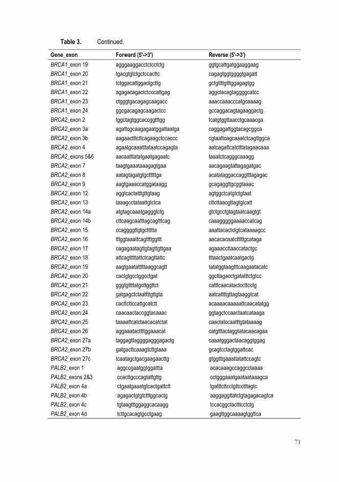
71
Table 3. Continued.
Gene_exon Forward (5'->3') Reverse (5'->3')
BRCA1_exon 19 agggaaggacctctcctctg ggtgcattgatggaaggaag
BRCA1_exon 20 tgacgtgtctgctccacttc cagagtggtggggtgagatt
BRCA1_exon 21 tctggacattggactgcttg gctgttttgtttggagagtgg
BRCA1_exon 22 agagacagactctcccattgag aggctacagtaggggcatcc
BRCA1_exon 23 ctgggtgacagagcaagacc aaaccaaacccatgcaaaag
BRCA1_exon 24 ggcgacagagcaagactcc gccaggacagtagaaggactg
BRCA2_exon 2 tggctagtggcaccggtttgg tcatgtggttaacctgcaaacga
BRCA2_exon 3a agattcgcaagagaatggattaatga caggagattggtacagcggca
BRCA2_exon 3b aagaactttcttcagaagctccaccc cgtaattcagcaaatctcagttggca
BRCA2_exon 4 agaatgcaaatttataatccagagta aatcagattcatctttatagaacaaa
BRCA2_exons 5&6 aacaatttatatgaatgagaatc taaatctcagggcaaagg
BRCA2_exon 7 taagtgaaataaagagtgaa aacagaagtattagagatgac
BRCA2_exon 8 aatagtagatgtgctttttga acatataggaccaggtttagagac
BRCA2_exon 9 aagtgaaaccatggataagg gcagaggttgcggtaaac
BRCA2_exon 12 aggtcactatttgttgtaag agtggctcatgtctgtaat
BRCA2_exon 13 taaagcctataattgtctca cttcttaacgttagtgtcatt
BRCA2_exon 14a atgtagcaaatgagggtctg gtctgcctgtagtaatcaagtgt
BRCA2_exon 14b cttcaagcaatttagcagtttcag caaagggggaaaaccatcag
BRCA2_exon 15 ccaggggttgtgcttttta aaattacactctgtcataaaagcc
BRCA2_exon 16 tttggtaaattcagttttggttt aacacacaatctttttgcataga
BRCA2_exon 17 cagagaatagttgtagttgttgaa agaaaccttaaccatactgc
BRCA2_exon 18 attcagtttttattctcagttattc tttaactgaatcaatgactg
BRCA2_exon 19 aagtgaatatttttaaggcagtt tatatggtaagtttcaagaatacatc
BRCA2_exon 20 cactgtgcctggcctgat ggcttagacctgatatttctgtcc
BRCA2_exon 21 gggtgttttatgcttggttct catttcaacatactccttcctg
BRCA2_exon 22 gatgagctctaattttgttgta aatcattttgttagtaaggtcat
BRCA2_exon 23 cacttcttccattgcatctt acaaaacaaaaattcaacatatgg
BRCA2_exon 24 caacaactaccggtacaaac ggtagctccaactaatcataaga
BRCA2_exon 25 taaaattcatctaacacatctat caactatccaatttgtataaaag
BRCA2_exon 26 aggaaatacttttggaaacat catgtttactaggtatacaacagaa
BRCA2_exon 27a taggagttaggggagggagactg caaatgggactaacaggtggag
BRCA2_exon 27b gatgacttcaaagtcttgtaaa gcagtcctagtggattcac
BRCA2_exon 27c tcaatagctgacgaagaacttg gtggtttgaaattatattccagtc
PALB2_exon 1 aggccgaatggtggattta acacaaagccaggcctaaaa
PALB2_exons 2&3 ccacttgcccagtattgttg cctgggaaatgaataataaagca
PALB2_exon 4a ctgaatgaaatgtcactgattctt tgatttcttcctgttcctttagtc
PALB2_exon 4b agagactgtgtctttggcactg aaggaggttatctgtagagacagtca
PALB2_exon 4c tgtaagtttggaggcacaagg tccacggctactttcctctg
PALB2_exon 4d tcttgcacagtgcctgaag gaagttggcaaaagtggttca

72
Table 3. Continued.
Gene_exon Forward (5'->3') Reverse (5'->3')
PALB2_exon 4e actgaagataatgacttgtctaggaa gtatgctggctttgcgagtt
PALB2_exon 5a tgttgggttttgttactattttgtg gttcctggcgggacagagt
PALB2_exon 5b cgcatggatacagaaatgg ggcaaatagtaattgttaactttcatc
PALB2_exon 5c actaaacaattcgacagttcagg acattcactaaggcatttcattc
PALB2_exon 6 acaatacgaagtagacattttgatga caagaagctatatgactgaattctttt
PALB2_exon 7 tgctttgcataaaacagcact tggtaagctgcccatctaca
PALB2_exon 8 attatccttgtacagtgagaatacaaa tgcacttaaaaccagctgaca
PALB2_exon 9 aaaaggttactcctcacatcacc cagcacagaaaaacgagatcc
PALB2_exon 10 cagttcaacaatgcggagaa tcttcacaacaaccctgtaaaa
PALB2_exon 11 tttccctggtcacctcctaa tgcttatgacttactgctctcactt
PALB2_exon 12 tcctgacatactcttgacagtc tccattcttctaagtgacacaaaaa
PALB2_exon 13 tggatatgtaatctgaattatatcttc cctaaaaccctttttctcaaagt
BRIP1_exon 2 ctgtttccagatttctccc gtgaacccagaaaatattctcc
BRIP1_exon 3 ccctggagtgcaatctcact tagcgacagcatggctgaa
BRIP1_exon 4 cctgggtgaactgggctgtag ttttaaaatcattccagagaaactaga
BRIP1_exon 5 ttgcctacctgtaagttatttatg accatgttcagctgtaactaactg
BRIP1_exon 6 gagctgttttggcctttgaga ctgagtgggttgctactgtcct
BRIP1_exon 7 gttctgattccatgtgaggtt gtacatataaaacacatactgagt
BRIP1_exon 8 gatgttcctcaaattctgagataa catctaaaagcttttacattcaac
BRIP1_exon 9 gcctatagtgtgaattttaaaatg cctagttaaccaaagtttactaac
BRIP1_exon 10 gatcaacgcatgacaataatgatg gggttactcactagatttaatctg
BRIP1_exon 11 gcatgttttgttgggtttcattgt ggtatgtattaaacacatgctagc
BRIP1_exon 12 gtaccagctctttcaaatgag ctatctttaaaagagtcaaccac
BRIP1_exon 13 acaggtgtgagccactttgc ggcacttcaggtatcttctaacttg
BRIP1_exon 14 cttgttgcttgatcttttatgtac ctaggaagcttactgtggtaa
BRIP1_exon 15 acagctctatgagatatattg tcataggagaacaagtacaat
BRIP1_exon 16 ttttcaaaatgacaagaataagcaa aagcaaagcgcaataaaatga
BRIP1_exon 17 ctgttagaagttaatatgatg gaatacataccagttcctatg
BRIP1_exon 18 ccaattttctgtctgtcccac gatagtagagctcatgttatgtg
BRIP1_exon 19 cttcactagaaaaagcaagtg ccaccatatttaaggaattaatc
BRIP1_exon 20a acctagcaattatgttagct tctgtatcttcaggatcgta
BRIP1_exon 20b attgatgccacccttacta taacataagcatgatgacata
RAD50_exon 1 ttgcttcggcctcagttaag ccgaaagatgtagcgacctg
RAD50_exon 2 tttatggtaaacttctgtggttctc ttttccagtgccaagttttct
RAD50_exon 3 tgcctttttctcagaaccaac gaaaacaaccatcaacttacagacc
RAD50_exon 4 gccatttctaacgggatagg gcatccaaattgcaaacaca
RAD50_exon 5 agtgacagcataatatcccactg ttgatttagccagtccacga
RAD50_exon 6 tgcctggacctggagtatct ggatggcaaaatggattcaa
RAD50_exon 7 tgaaggatattgaataaggtttggtt ttcactcagcataagtccttgg

73
Table 3. Continued.
Gene_exon Forward (5'->3') Reverse (5'->3')
RAD50_exon 8 cgtgaatctgcagctatctcaa atgccaaaatggagtccaac
RAD50_exon 9 tcttcagtgtacatatattccttttgc aaaatgctataatcttaggatcaaaaa
RAD50_exon 10 aattcttgcacaaaatatataacacc tgagtgcaaggtaggctatttta
RAD50_exon 11 tttttgttcttgatataatgtggagat aaacaattaaacaggtaagcatgaaa
RAD50_exon 12 tttgcctactcaaattttcaaac atgaaaattcaaaggtgtcaaag
RAD50_exon 13 agaaaaagatacaaccgtattcaga aggcatgagatgggtacctt
RAD50_exon 14 cctcagtctaactgtgagaaacatc caagccaccatggaacaag
RAD50_exon 15 tttgctaaaattgtatctagaaatgg ggaaggaacttcgttgtcca
RAD50_exon 16 ccgataaaaatgggaagaatga gatcgtgctactgcactcca
RAD50_exon 17 tgacccctaaagtaagataatggaa agtgtccgacgtggtgctat
RAD50_exon 18 ctgtgctctcctgttatgtgc tctttgtgtttctcgcattca
RAD50_exon 19 ttcaagatgggaaagacgac gcatttctattcaatggatcttct
RAD50_exon 20 tgcctgttacagatttcatgtt ccaggctgaggctaaaattc
RAD50_exon 21 tgacttttccacttcaggttgtt tgcaataagaaaatccccagtc
RAD50_exon 22 accattgaaaatattgaggaagtt ggtcataaggggaagagctg
RAD50_exon 23 caaaaggctacagagcataggtt tctctctttcagttacttgggtga
RAD50_exon 24 gcctgccatgagatgagaag cctgtccccaaatgtgagat
RAD50_exon 25 caaatcaaagaaggggttatgc cactttctgaggacctacatttct
CDH1_exon 1 gtgaaccctcagccaatcag gacccgaactttcttggaag
CDH1_exon 2 ggtcttgagggggtgactc ggtgtgggagtgcaatttct
CDH1_exon 3 gttcgctctttggagaagga ggctgagaaacctggattaga
CDH1_exon 4 gtctggctaggttggactgt cactgggtcttttccctttc
CDH1_exon 5 ggatttggcagagaagtacc gttaagctcctcatgtgttcag
CDH1_exon 6 ctcagagcctaggaaggtgtg gttacacaacctttgggctt
CDH1_exon 7 gtcccctcctttatccctca gacaactggcctagcaggat
CDH1_exon 8 ctggttccatgtgttgggc ccatgagcagtggtgacactt
CDH1_exon 9 cctttagccccctgagact gccaaagcgaatctacttct
CDH1_exon 10 gaaagtcatggcagaaacca ggtcttgtacagacaaatgaca
CDH1_exon 11 gaagaagcgcttaagccgtt gaagctcttccctccaaaagaa
CDH1_exon 12 ctgaagagccaggacaagat gtatcaatggaaggggtgac
CDH1_exon 13 cttgcgggtgtctttagttc ggagtctctttcccacatca
CDH1_exon 14 ctgtgatagctgctgcttct gctgtttcaaatgcctacct
CDH1_exon 15 ggcatcatccaaccataatc gctcaggcaagctgaaac
CDH1_exon 16 ccacaagtctgggtgcatt gaaactcatctcaagggaagg
Study III
AKT2_exon 3 gtggtaccccttgtgagtca gctaacaaggagggagagca
ATM_exon 17 tgaccgtggagaagtagaatca tgaggcctcttatactgcca
ATM_exon 22 aggcatctaacaaaggagagga tgtaagacattctactgccatct

74
Table 3. Continued.
Gene_exon Forward (5'->3') Reverse (5'->3')
ATM_exon 29 aattcttcttgccatatgtgagc tgggcggacagagtgagt
ATM_exon 37 tgtcaacggggcatgaaaat gttgtggatcggctcgtttg
BRCA1_exon 9 acacccaggatcctttcttg tgtaaaatgtgctccccaaaag
CDKN2A_exon 3 agcccatacgcaacgagatt gccagcttgcgataaccaaa
MYC_exon 2 actcaagactgcctcccg gaagggagaagggtgtgac
NCOA3_exon 18 gtagagaagtgatgcctggc ctcatgatgttggctcgtgg
PLAU_exon 2 gagaggagagagacagctgg cccgtggttctcaagcct
RAD1_exon 4 tggaattctctacacacaactgt tcactcgtcatatccaattcaga
RAD50_exon 3 acctgatctcctaatgatgctga acaaccatcaacttacagacct
RAD52_exon 6 gtctgccgttttcccacttt ctgcgtgactgtgtaactacg
RBL2_exon 13 ggaagagcggctgtttcaaa cacaaacctcttcacatgtagga
RPA2_exon 2 gaaggttagggagactcggg acaggaggtggaaaatttggc
RRM2B_exon 1 ggatgaggtaaatgttgctgttg aactccttgtcaccatccca
WNT3A_exon 2 tgttgggccacagtattcct aggctcagtcctgcttcag
WNT10A_exon 2 ccaatgacattctggacctcc aagaggaaggagagcagcag
2.3 Multiplex Ligation dependent Probe Amplification (MLPA) (I, II)
The MLPA method was used to detect large genomic rearrangements in BRCA1 and
BRCA2. The following SALSA® MLPA® kits were used according to the
manufacturer’s instructions: P002-B1 (lot 0508) for BRCA1 (study I), P090-A2 (lot
0808) for BRCA2 (study I), and POO2 probemix and EK1 reagent kit (lot C2-0811)
for BRCA1 (study II) (MRC-Holland, Amsterdam, the Netherlands). The analysis
was performed with Applied Biosystems 3130xl Genetic Analyzer and with Applied
Biosystems Genemapper® v.4.0 and Peak Scanner™ v1.0 software (Life
Technologies), as indicated by the manufacturer. In study II, the National Genetics
Reference Laboratory (Manchester, UK) Spreadsheet was utilized for data analysis
according to the manufacturer’s instructions.
2.4 High-Resolution Melt (HRM) analysis (I)
High-Resolution Melt (HRM) analysis was used to screen for genomic DNA
sequence variants in CHEK2. The primers were designed with Beacon Designer™
software (PREMIER Biosoft, Palo Alto, CA, USA). The primer sequences are
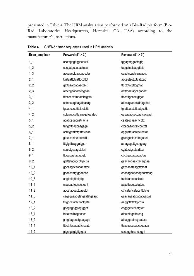
75
presented in Table 4. The HRM analysis was performed on a Bio-Rad platform (Bio-
Rad Laboratories Headquarters, Hercules, CA, USA) according to the
manufacturer’s instructions.
Table 4. CHEK2 primer sequences used in HRM analysis.
Exon_amplicon Forward (5’ -> 3’) Reverse (5’ -> 3’)
1_1 acctttgttgttggacacttt tggagtttggcatcgtg
1_2 cacgatgccaaactcca taggctcctcaggttctt
1_3 aagaacctgaggagccta caactccaatcagaacct
2_1 tgataattctgattgccttct accagtagttgtcattcac
2_2 gtgtgaatgacaactact ttgctgtatgttcggtat
2_3 ataccgaacatacagcaa actttgaatagcagagattt
3_1 tttcccactataaatctctgcta ttccattgccactgtgat
3_2 catacatagaagatcacagt attccagtaaccataagataa
4_1 tgaaacccatttctactcttt tgtattcatctcttaatgcctta
4_2 cctaaggcattaagagatgaatac gagaaaccaccaatcacaaat
5_1 acattcagacaatcacta caatagcaaacttccttt
5_2 tattggttcagcaagaga ctcacaaattcatccatcta
6_1 actctgttattctgtttatcaaa aggctttatactcttctcatat
7_1 gtttctcactactttcccttt gcaagcctacattagattct
8_1 tttgtgtttcaggatgga aatagagcttgcaggtag
8_2 ctacctgcaagctctatt cgatttctgcctaattca
9_1 ttggagaatatggttgtg cttcttgagatgacagtaa
9_2 gtattatacaccgtgactta gaacaagaatctacaggaa
10_1 ggcaagttcaacattattcc gttccacataaggttctcat
10_2 gaaccttatgtggaaccc caacagaaacaagaacttcag
10_3 aagttcttgtttctgttg tcatctaatcacctccta
11_1 ctgagaatgccacttgatt acacttgagtcctatgct
11_2 agcataggactcaagtgt ctttcatattcatacctttctctg
11_3 cagagaaaggtatgaatatgaaag gaacagaattgacaggagaa
12_1 tctggcatactcttactgata aaggcttcttctgtcgta
12_2 gaagttgttggtagtggat cagggcttcccatgtatt
13_1 tattatccttcagacaca atcatctttgcttatcag
13_2 gatgaagacatgaagaga atcaggaatacgaatacc
14_1 ttttcttttgaacatttctccatt ttcacaacacagcagcaca
14_2 gtgctgctgtgttgtgaa cccaggttccatcaggtt

76
2.5 SNP genotyping (I, III)
SNP genotyping was one of the methods used to determine the frequencies of the
SNPs identified in population controls in study I and in patient cohorts and
population controls in study III. The genotyping was performed with Applied
Biosystems TaqMan® SNP Genotyping Assays, with ABI PRISM® 7900HT
Sequence Detection System and SDS v2.2.2 software (Life Technologies) according
to the manufacturer’s instructions. Pre-designed and functionally tested assays were
used for three BRCA2 SNPs (rs28897749, rs11571833, rs1801426), and for one
PALB2 SNP (rs152451) in study I. For four SNPs, c.72A > T (BRCA2), c.814G >
A (PALB2), c.1000T > G (PALB2), and rs45551636 (PALB2), no pre-designed
assays were available and assays were designed using Applied Biosystems Custom
TaqMan® Assay Design Tool (Life Technologies) according to the manufacturer's
instructions (study I). In study III, the following assays were used: C_176281813_10
(CDKN2A c.C496T), C___1244825_20 (RAD52 c.G538A), C___2283286_20
(ATM c.T2572C), C__45273750_10 (ATM c.C3161G), C_166902853_10, (RPA2
c.C122T), C__30585831_10 (ATM c.A5558T), C__63879305_20 (ATM c.A4424G),
C__32333045_10 (RAD50 c.A280C), C_153129907_10 (BRCA1 c.A3904C),
C__32376001_10, (MYC n.A77G), C_102161615_10 (RBL2 c.G1723C),
C_167350917_10, (NCOA3 c.A3353C), C__15956024_20, (PLAU c.G43T),
C__15760210_10 (RAD1 c.G341A), C_168146154_10 (WNT10A c.C337T), and
C_190555357_10 (WNT3A c.G277A).
2.6 Copy number variation (CNV) analysis (II)
SNP array protocol. A genome-wide SNP genotyping to identify CNVs from
genomic DNA was performed at the Technology Centre, Institute for Molecular
Medicine Finland, University of Helsinki (Helsinki, Finland). For this analysis, an
Illumina HumanCytoSNP-12 v2.1 Beadchip, an iScan system, and standard reagents
and protocols provided by Illumina Inc. (San Diego, CA, USA) were used. Illumina’s
HumanCytoSNP-12 v2.1 Beadchip contains approximately 300,000 markers
distributed throughout the genome and has been optimized to target regions of
cytogenetic importance.
Data-analysis. The genotypes were visualized and analyzed with
GenomeStudio™ v2010.2 software, as indicated by the manufacturer (Illumina). The
call rates were greater than 99.5%. It was ensured that the samples met high quality
77
criteria (Wang et al, 2007b). CNV calling was performed with the PennCNV
(2009Aug27) program (Wang et al, 2007b). In addition, the QuantiSNP v2.3 (Colella
et al, 2007) and cnvPartition v3.1.6 (Illumina) programs were utilized to confirm the
PennCNV results when selecting CNVs for validation. All three programs were used
with default parameters. CNVs spanning less than three SNPs were filtered out.
Identified CNVs were queried against the Database of Genomic Variants (Database
of Genomic Variants) using the NCBI Genome Build 36 (hg 18). CNVs were
annotated using the NCBI RefSeq gene database (NCBI Reference Sequence
Database) to identify genes and exons overlapping the observed CNV loci. For
intergenic CNVs, the loci were expanded upstream and downstream of the CNV to
identify neighboring genes. CNV-affected genes were further studied by enrichment
analyses, including Gene Ontology terms, KEGG pathways, Pathway Commons,
and Wikipathways to reveal common functions of the gene products using the Web-
based Gene Set Analysis Toolkit V2 (WebGestalt2) (Zhang et al, 2005). Additionally,
CNV-affected genes were queried against the NCBI Online Mendelian Inheritance
in Man database (NCBI OMIM Database) and the Genetic Association Database
(Genetic Association Database) to determine whether the genes have previously
been associated with the disease. The Encyclopedia of DNA Elements database
(Encyclopedia of DNA elements at UCSC) was utilized to search possible functional
elements in the CNV region
CNV validation and genotyping. Selected CNVs were validated, and their
frequencies were determined in an additional cohort of HBOC patients and
population controls using TaqMan® Copy Number Assays, an ABI PRISM®
7900HT Real-Time PCR system, and SDS v2.2.2 software, according to the
manufacturer’s instructions (Life Technologies). The following pre-designed
TaqMan® Copy Number Assays were used: Hs04703682_cn (2q34),
Hs03458738_cn (3p11.1), Hs03253932_cn (5q15), Hs06178677_cn (8p23.2),
Hs02640223_cn (17q21.31), and Hs04482315_cn (19q13.41). As an internal
standard, a TaqMan® RNaseP Reference Assay was run with the TaqMan® Copy
Number Assays in a duplex, real-time PCR reaction following the manufacturer’s
guidelines (Life Technologies). The analysis was performed with Copy Caller v1.0
software, and copy numbers were calculated using the comparative CT method, as
indicated by the manufacturer (Life Technologies).
78
2.7 Exome sequencing (III)
Exome capture and next-generation sequencing protocol. Exome capture and
next-generation sequencing was performed at the BGI Tech Solutions (Hong Kong)
Co. Ltd using a SureSelect Human All Exon 51M kit (Agilent Technologies, Inc.,
Santa Clara, CA) and HiSeq 2000 technology (Illumina). Protocols provided by
Agilent, Illumina, and the BGI were followed. The genome sequencing coverage
depth was 50x per sample.
Data-analysis. The reads were aligned with Bowtie2 against the human reference
genome (hg19) using default parameters (Langmead & Salzberg, 2012). The quality
control was performed by FastQC (Babraham Bioinformatics FastQC). As a
preprocessing step for variant calling, PCR duplicates and reads with mapping quality
less than 10 were filtered out using SAMtools (Li et al, 2009). Variant calling was
performed using a bioinformatics toolkit (Pypette) that was developed in-house
(GitHub annalam/pypette). Variant annotation was conducted with Annovar (Wang
et al, 2010) using the Refseq genes as the reference gene set. Several filtering steps
were used to reduce the number of candidate variants for downstream analyses.
Based on the variant annotation, functional variants (non-synonymous single
nucleotide variants (SNVs), splicing site SNVs/indels, stop gain/loss variants, and
indels inducing frameshift) were prioritized. Furthermore, rare variants (minor allele
frequency ≤ 0.05 were selected by screening the variants against the data from the
1000 Genomes Project (1000 Genomes Project Consortium et al, 2012) (August
2014 version) and the Exome Sequencing Project 6500 (included in Annovar)
(NHLBI Exome Sequencing Project). Additionally, the variants were screened
against the Sequencing Initiative Suomi database to obtain information about allele
frequencies in the Finnish population (Sequencing Initiative Suomi). From the
remaining set of variants, only those variants for which the host genes participate in
the DNA damage response pathway were selected. The pathway data were retrieved
from the IntPath-database, which includes data integrated from the following several
sources: KEGG, Wikipathways and BioCyc (Zhou et al, 2012). To further reduce
the number of candidate variants, the deleteriousness of the variants was evaluated
by utilizing a precompiled set of pathogenicity predictions included in the Annovar's
ljb26_all dataset. The dataset covers predictions of the deleteriousness of all possible
SNVs for 10 pathogenicity predictor methods and 3 methods evaluating the
evolutionary conservation of the variant locus. Only variants that were predicted to
be deleterious by at least one of the predictors or were frameshift indels, were
selected for further assessment. Moreover, variants present only in healthy relatives

79
were excluded. Furthermore, variants that had a reliable genotype call rate and were
shared between affected family members or between families were prioritized. Based
on the above-mentioned filtering criteria, candidate variants were selected for further
validation experiments (described in sections 2.2 and 2.5). Moreover, variants
present in only early-onset patients were analyzed in detail. Similar filtering criteria
based on functionality, rarity, and predicted pathogenicity were used, as described
above. However, variants targeting any pathway genes were considered.
2.8 Statistical analyses (I-III)
Association of the identified variants with breast/ovarian cancer was tested using
Fisher’s exact test, and the chi-square test (R v2.15.2 (The R Project for Statistical
Computing) (R Core Team, R Foundation for Statistical Computing, Vienna,
Austria)) and PLINK v1.07 (Purcell et al, 2007). CNV distribution and median
lengths were compared between HBOC individuals and controls using the Wilcoxon
test (R v2.15.2) (study II). When Fisher’s exact test resulted a non-numerical value
for the enrichment analysis, A VCD package was implemented in R to estimate the
numerical values of the odds ratios (Meyer et al, 2012) (study II). P-values were two-
sided. P<0.05 was considered statistically significant.
2.9 Bioinformatics tools (I, III)
A Basic Local Alignment Search Tool (NCBI BLAST) tool was utilized to determine
whether the identified novel variants were located in genomic regions that are
conserved across different species (study I). The pathogenicity of missense variants
was predicted using the Pathogenic-or-Not-Pipeline (PON-P) (Thusberg & Vihinen,
2009) (study I). PON-P integrates amino-acid tolerance predictors (PolyPhen
version 2, Sift, PhD-SNP, SNAP) and a protein stability predictor (I-Mutant version
3) to predict the probability that missense variants affect protein function. ESEfinder
was used to predict the effects of variants on exonic splicing enhancer elements
(Cartegni et al, 2003) (study III).

80
2.10 MicroRNA database search (I)
A microRNA target site search of the microRNA database was performed for the
genomic positions of the novel variants (miRBase).

81
RESULTS
1 Germline sequence variants in BRCA1, BRCA2, CHEK2, PALB2, BRIP1, RAD50, and CDH1, and their contribution to HBOC susceptibility in high-risk families (I)
In study I, coding region and exon-intron boundaries of seven known BC
susceptibility genes were analyzed to identify additional variants that predispose to
inherited forms of breast and ovarian cancer. Index individuals from 82 of the high-
risk hereditary breast and/or ovarian cancer families analyzed in this study were
previously screened to be negative for 28 Finnish BRCA1 and BRCA2 founder
mutations by minisequencing. These individuals also tested negative for protein-
truncating variants in the largest exons (exon 11 for BRCA1 and exons 10 and 11
for BRCA2), suggesting that variants other than these already tested defects
contribute to breast and/or ovarian cancer susceptibility in these families. By
thoroughly screening 1) BRCA1 and BRCA2 using MLPA and Sanger sequencing
(with the exception of the previously analyzed largest exons); 2) PALB2, BRIP1,
RAD50, and CDH1 by Sanger sequencing; and 3) CHEK2 using the HRM method,
54 different variants in 82 HBOC individuals were identified. The variant spectrum
was as follows: 19 non-synonymous coding variants, 11 synonymous coding variants,
21 intronic variants, and 3 5’-untranslated region variants. All the detected variants
were single nucleotide changes or indels. In total, 14/54 (25.9%) of the identified
variants were novel.
Based on the variant type, novelty, observed frequency in HBOC individuals, and
available information in the databases, the most promising variants were selected
(n=18), and their frequency was determined in 384 controls. The primary focus was
on non-synonymous coding variants as these are most likely to be disease-causing.
82
Variant frequencies between HBOC individuals and controls were compared using
Fisher’s exact test, but no statistically significant difference was observed for any of
the variants. This result was probably due to the rareness of the variants and the
limited sample size used in this study. All the identified non-synonymous variants
and their association with breast and/or ovarian cancer risk are presented in Table
5. Three previously known intermediate breast/ovarian cancer risk variants, BRCA1
Arg1699Trp, CHEK2 Ile157Thr, and CHEK2 1100delC (bold in Table 5) were
observed in 11/82 (13.4%) HBOC individuals. One individual carried both of the
CHEK2 variants. The contribution of the two CHEK2 variants to BC risk was
notably high in the analyzed HBOC families, 10/82 (12.2%) (Table 5). The effects
of novel missense variants (n=5) on protein function was predicted using the PON-
P program: of the analyzed variants, only one possible pathogenic variant (BRCA2
Leu24Phe) was predicted. The substitution of the nonpolar leucine by the nonpolar
phenylalanine with an aromatic ring structure in the side chain at position 24 was
predicted significantly alter the properties of the side chain; this substitution was
predicted to be untolerated. The BRCA2 Leu24Phe variant was observed in one BC
patient (1/82, 1.2%) but in none of the 380 healthy controls (Table 5). The BC
patient had been diagnosed with ductal, grade III disease with ER+, PR- and
HER2+ status at the age of 53 years. Additionally, the patient had two BC-affected
first-degree relatives.
The clinical characteristics of the HBOC individuals carrying the identified non-
synonymous variants were reviewed, but no clear trend was observed among
individuals carrying the same variants. However, one interesting case was an early-
onset (diagnosed at the age of 26 years) BC patient carrying both of the BC-
associated CHEK2 variants, Ile157Thr and 1100delC. Thus, the combination of the
two CHEK2 variants was reported to contribute to early disease onset. Novel variant
genomic positions were queried against the known microRNA target sites from the
miRBase, but no hits were found. Moreover, a Basic Local Alignment Search Tool
was utilized to determine whether the identified novel variants were located in
conserved genomic positions, which may indicate a pathogenic role for the variant.
Sequence similarities between different species were observed for several novel
variant genomic positions, including BRCA2 Leu24Phe.
Detailed analysis of the BRCA1 and BRCA2 genes (Sanger sequencing and
MLPA, excluding the previously analyzed largest exons) revealed additional variants
that were not detected by the genetic testing protocol that targeted only the Finnish
founder-mutations. One such variant was Arg1699Trp in BRCA1, which is a known
intermediate breast/ovarian cancer risk variant.

83
Table 5. Identified non-synonymous variants in BRCA1, BRCA2, CHEK2, PALB2, BRIP1, and RAD50, and their association with hereditary breast and/or ovarian cancer risk.
Carrier frequency (%)
Gene, variant HBOC individuals Controls OR 95%CI P
BRCA1, Ser1613Gly 52/82 (63.4%) - - - -
BRCA1, Met1628Thr 4/82 (4.9%) 6/367 (1.6%) 3.09 0.85-11.19 0.090
BRCA1, Arg1699Trp 1/82 (1.2%) - - - -
BRCA2, Leu24Phe* 1/82 (1.2%) 0/380 (0%) - - 0.177
BRCA2, Val2728Ile 1/82 (1.2%) 1/378 (0.3 %) 4.65 0.29-75.19 0.325
BRCA2, Lys3326Stop 1/82 (1.2%) 11/378 (2.9%) 0.41 0.05-3.24 0.702
BRCA2, Ile3412Val 1/82 (1.2%) 8/379 (2.1%) 0.57 0.07-4.64 1.000
CHEK2, Ile157Thr 8/81 (9.8%) 21/381 (5.5%) 1.88 0.80-4.41 0.203
CHEK2, 1100delC 3/82 (3.7%) 6/380 (1.6%) 2.37 0.58-9.67 0.203
CHEK2, Val455Ile* 79/81 (97.5%) 373/382 (97.6%) 0.95 0.20-4.50 1.000
PALB2, Glu272Lys* 1/82 (1.2%) 0/372 (0%) - - 0.181
PALB2, Tyr334Asp* 1/82 (1.2%) 4/380 (1.1%) 1.16 0.13-10.52 1.000
PALB2, Leu337Ser 6/82 (7.3%) - - - -
PALB2, Gln559Arg 10/82 (12.2%) 64/371 (17.3%) 0.67 0.33-1.36 0.323
PALB2, Val932Met 3/82 (3.7%) - - - -
PALB2, Gly998Glu 1/82 (1.2%) 14/372 (3.8%) 0.32 0.04-2.44 0.491
BRIP1, Leu195Pro 2/82 (2.4%) - - - -
BRIP1, Pro919Ser 32/82 (39.0%) - - - -
RAD50, Asp515Gly* 1/82 (1.2%) 4/384 (1.0%) 1.17 0.13-10.63 1.000
Abbreviations: CI, confidence interval; HBOC, hereditary breast and/or ovarian cancer; OR, odds ratio.
*Novel variant.
Bolded variants are known breast/ovarian cancer risk variants.

84
2 Germline copy number variations and their contribution to HBOC susceptibility (II)
In study II, a genome-wide SNP genotyping was performed to identify germline
CNVs that confer susceptibility to HBOC. SNP genotyping was performed for index
individuals from 84 HBOC families and 36 healthy controls. After applying strict
sample quality criteria, 81 HBOC individuals and 35 controls were included in the
data analysis. In total, 545 autosomal CNVs, 300 deletions and 245 duplications,
were identified at 273 separate genomic regions in the analyzed HBOC individuals
and controls. The primary focus was on the CNVs that affected potential or known
genes in breast/ovarian cancer predisposition. Additionally, clinical features of the
CNV carriers were inspected. After several filtering steps, 6 CNVs were selected for
further validation by quantitative PCR. Five of these CNVs were genotyped in an
additional cohort of 20 HBOC patients and in up to 869 additional controls by
quantitative PCR (Table 6). The carrier frequencies of the validated CNVs were
compared between 101 HBOC individuals and up to 899 controls (Table 6).
Validated CNVs showed enrichment in HBOC cases compared to controls (Table
6). No statistically significant difference was observed, an effect that was probably
due to the limited sample size used in this study. Therefore, the results require
verification in a larger sample set.
Three of the validated CNVs were intronic deletions affecting Erb-B2 Receptor
Tyrosine Kinase 4 (ERBB4), EPH Receptor A3 (EPHA3), and CUB and Sushi multiple
domains 1 (CSMD1). ERBB4 and EPHA3 encode proteins that are important
regulators in signaling pathways, whereas CSMD1 is a tumor suppressor gene. A
novel ERBB4-affecting deletion was slightly more frequent in HBOC individuals
(5/101, 5.0%) than in controls (12/358, 3.4%) (OR=1.49 95%CI= 0.52-4.28) (Table
6). The EPHA3-affecting deletion was nearly twice as common in HBOC
individuals (12/101, 11.9%) as in controls (27/432, 6.3%) (OR=1.96 95%CI=0.97-
3.94) (Table 6). A novel CSMD1-affecting deletion was observed in one BC patient
(1/101, 1.0%), but the variant was very rare in controls (1/436, 0.2%) (OR=4.33
95%CI=0.27-69.57) (Table 6). One of the validated CNVs was an intergenic deletion
at a 5q15 region where regulatory elements were reported to be present according to
the The Encyclopedia of DNA Elements database. The 5q15 deletion was observed
85
in a homozygous form in one BC patient (1/101, 1.0%) who exhibited an interesting
clinical feature. Specifically, the patient had been diagnosed and died of BC at the
age of 29. The 5q15 homozygous variant was very rare in controls (1/899, 0.1%)
(Table 6). Two of the selected CNVs were exonic; one deletion affected BRCA1,
Neighbor Of BRCA1 Gene 1 (NBR1) and Neighbor Of BRCA1 Gene 2 (NBR2) and one
duplication affected Endogenous Retrovirus Group V, Member 2 (ERVV-2). Large
deletions affecting BRCA1 are known to predispose to hereditary breast and ovarian
cancer, and the deletion was identified in one BC patient (1/101, 1.0%) (Table 6)
with a family history of OC. ERVV-2 belongs to the endogenous retrovirus group
of proteins, and it has been implicated that in human cancer. The ERVV-2-affecting
duplication was observed to be slightly more frequent in HBOC individuals (11/101,
10.9%) than in controls (34/334, 10.2%) (OR=1.37 95%CI 0.73-2.55) (Table 6).
However, homozygous ERVV-2-affecting duplications were over four times more
common in HBOC individuals (4/101, 4.0%) than in controls (3/334, 0.9%) (Table
6).
Some findings were made with respect to the clinical features of the HBOC
individuals with the identified CNVs. For instance, a ductal tumor type was a
common feature for all 3p11.1 deletion (EPHA3 locus) carriers with BC (n=11). Of
these tumors, 9/11 were ER and PR positive. Similarly, the ductal tumor type
predominated in BC-affected females with a 19q13.41 duplication at the ERVV-2
locus (10/11 carriers). Interestingly, 3/4 homozygous 19q13.41 duplication carriers
had ductal high-grade tumors (grade III), implying a more aggressive disease. Two
of five 2q34 deletion (ERBB4 locus) carriers had a bilateral form of BC diagnosed at
≤43 years of age. Cosegregation of the novel 2q34 deletion with breast and/or
ovarian cancer was studied in one family in which an intermediate breast and ovarian
cancer risk variant, BRCA1 Arg1699Trp, was identified (study I). The 2q34 deletion
was identified in one OC-affected and two BC-affected relatives, some of whom
were carriers of the BRCA1 variant and some of whome were not.

86
Table 6. Association of the validated CNVs with hereditary breast and/or ovarian cancer risk.
CNV position, affected Carrier frequency
gene, type and size HBOC individuals Controls OR; 95%CI P
2q34
ERBB4 intronic deletiona 5/101 (5.0%) 12/358 (3.4%) 1.49; 0.52-4.28 0.457
28.7-59.0 kb
3p11.1
EPHA3 intronic deletion 12/101 (11.9%) 27/432 (6.3%) 1.96; 0.97-3.94 0.055
14.6 kb
5q15
intergenic deletion 5/101 (5.0%)b 57/899 (6.3%)c 0.92; 0.39-2.16 0.845
49.8 kb
8p23.2
CSMD1 intronic deletiona 1/101 (1.0%) 1/436 (0.2%) 4.33; 0.27-69.57 0.259
10.8 kb
17q21.31
BRCA1, NBR1, NBR2 exonic 1/101 (1.0%) 0/35) (0%) - 0.555
deletiond, 99.0 kb
19q13.41
ERVV-2 exonic duplication 11/101 (10.9%)e 34/334 (10.2%)f 1.37; 0.73-2.55 0.322
15.8-26.9 kb
Abbreviations: CI, confidence interval; CNV, copy number variation; HBOC, hereditary breast and/or ovarian cancer; OR, odds ratio.
a Novel variant.
b Homozygous in 1/101 (1.0%) of the HBOC individuals.
c Homozogyous in 1/899 (0.1%) of the controls.
d Large BRCA1-affecting deletions are known to be associated with HBOC susceptibility and there was no need to genotype additional controls.
e Homozygous in 4/101 (4.0%) of the HBOC individuals.
f Homozygous in 3/334 (0.9%) of the controls.

87
3 Identification of HBOC susceptibility genes and gene variants by exome sequencing (III)
In study III, with the aim of identifying gene variants that contribute to HBOC
susceptibility, whole exome sequencing was performed for 37 individuals from 13
high-risk BRCA1/2-negative HBOC families. Of the studied individuals, 23 were
female breast or breast and ovarian cancer patients, one was a male BC patient, and
13 were healthy relatives. Of note, six of the BC patients were diagnosed at a very
early age (≤ 29 years). In total, 736,963 sequence variants were detected in 37
individuals. Several filtering steps were used to reduce the number of candidate
variants for downstream analyses. The primary focus was on DDR pathway gene
variants that were shared between affected family members or families. Eighteen
candidate variants were selected for further analyses, genotyping of these variants
was performed in a cohort of 129 female HBOC cases and up to 989 healthy female
controls (Table 7). Additionally, two of the variants, which were detected in a male
BC patient, were also screened in a cohort of 49 male BC patients and 909 healthy
male controls (Table 7). Carrier frequencies of the variants were compared between
the cancer cases and controls (Table 7).
Five variants, ATM D1853V, MYC N26S, PLAU V15L, RAD1 G114D, and
RRM2B R71fs, were enriched in female HBOC cases compared to controls (OR
1.16-2-16) implying that the variants may be low-to-moderate risk alleles (Table 7).
A BRCA1 T1302P variant, detected in a two affected females in a single BC family
by exome sequencing, was absent in both female HBOC patients and female controls
(Table 7). This result implies that BRCA1 T1302P is an extremely rare BC
susceptibility variant. The RAD50 I94L variant, which was detected in a male BC
patient, was absent in a cohort of male BC cases and extremely rare in male controls
(1/909, 0.1%) (Table 7), suggesting it may contribute to male BC risk. The other
variant detected in a male BC patient, ATM Y1475C, was absent both in male BC
cases and male controls but was detected in female controls (Table 7). This results
suggests that ATM Y1475C is not specific for male BC. No statistically significant
association was reached for any of the variants, a result that can likely be explained
by the rareness of the variants and the limited sample size. Therefore, the results
need further verification. Eighteen variants were also screened from breast tumor
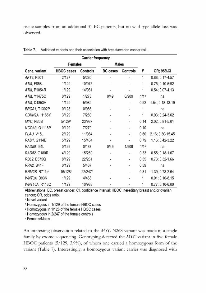
88
tissue samples from an additional 31 BC patients, but no wild type allele loss was
observed.
Table 7. Validated variants and their association with breast/ovarian cancer risk.
Carrier frequency
Females Males
Gene, variant HBOC cases Controls BC cases Controls P OR; 95%CI
AKT2, P50T 2/127 5/280 - - 1 0.88; 0.17-4.57
ATM, F858L 1/129 10/975 - - 1 0.75; 0.10-5.92
ATM, P1054R 1/129 14/981 - - 1 0.54; 0.07-4.13
ATM, Y1475C 0/129 1/278 0/49 0/909 1/1e na
ATM, D1853V 1/129 5/989 - - 0.52 1.54; 0.18-13.19
BRCA1, T1302P 0/128 0/986 - - 1 na
CDKN2A, H166Y 3/129 7/280 - - 1 0.93; 0.24-3.62
MYC, N26S 5/129b 23/987 - - 0.14 2.02; 0.81-5.01
NCOA3, Q1118P 0/129 7/279 - - 0.10 na
PLAU, V15L 2/129 11/984 - - 0.60 2.16; 0.30-15.45
RAD1, G114D 5/129 15/464 - - 0.79 1.16; 0.42-3.22
RAD50, I94L 0/129 0/187 0/49 1/909 1/1e na
RAD52, G180R 4/129 15/269 - - 0.33 0.55; 0.18-1.67
RBL2, E575Q 8/129 22/261 - - 0.55 0.73; 0.32-1.66
RPA2, S41F 0/129 5/467 - - 0.59 na
RRM2B, R71fsa 16/128c 22/247d - - 0.31 1.39; 0.73-2.64
WNT3A, D93N 1/129 4/468 - - 1 0.91; 0.10-8.15
WNT10A, R113C 1/129 10/988 - - 1 0.77; 0.10-6.00
Abbreviations: BC, breast cancer; CI, confidence interval; HBOC, hereditary breast and/or ovarian cancer; OR, odds ratio. a Novel variant b Homozygous in 1/129 of the female HBOC cases c Homozygous in 1/128 of the female HBOC cases d Homozygous in 2/247 of the female controls e Females/Males
An interesting observation related to the MYC N26S variant was made in a single
family by exome sequencing. Genotyping detected the MYC variant in five female
HBOC patients (5/129, 3.9%), of whom one carried a homozygous form of the
variant (Table 7). Interestingly, a homozygous variant carrier was diagnosed with

89
triple-negative BC at the age of 61 years and a family history of multiple cancers
(three BCs, colon cancer, and possibly throat and stomach cancers). Further analysis
identified the homozygous MYC variant in the index’s healthy sister (current age 81
years) and in heterozygous form in the index’s healthy young relatives.
Unfortunately, no DNA samples of BC-affected relatives were available at that time.
Of note, the homozygous form of the MYC variant was not detected among 987
healthy controls. A novel frameshift-inducing duplication in RRM2B was found to
be a very common variant in both female HBOC patients and healthy controls
(Table 7), although it was originally considered rare based on the allele frequencies
in the databases. A homozygous form of the RRM2B variant was also detected, but
the frequencies were similar both in female HBOC cases and controls (Table 7).
Another interesting observation related to the ATM D1853V variant. The ATM
D1853V was reported to affect splicing. The clinical features of the variant carriers
were inspected. One interesting finding was that one of the PLAU V15L variant
carriers, detected by genotyping, exhibited a mucinous subtype of BC (grade 2,
ER+/PR+/HER2-) that was diagnosed at the age of 55. The mucinous subtype of
BC is rare and is associated with a good prognosis.
Specific attention was also given to six early-onset BC patients (diagnosed ≤ 29
years) in the exome sequencing cohort. The aim of this analysis was to identify
variants that were present only in these early-onset patients and could explain the
drastic clinical outcome. After several filtering steps, variants of host genes that
participate in certain pathways were considered good candidates for BC
susceptibility. These pathways and functions included the cell cycle, proliferation,
apoptosis, adhesion, different signaling pathways, and the DNA damage response.
The candidate variants are presented in Table 8. Of these, 50 were non-synonymous
SNVs, 4 were stop-gains, and 3 were frameshift indels. The majority of the variants
were detected in a single patient only. Nonsynonymous variants in genes such as
Cyclin-Dependent Kinase 2 Interacting Protein (CINP), Focadhesin (FOCAD), Laminin,
Alpha 5 (LAMA5), Phospholipase D1, Phosphatidylcholine-Specific (PLD1), RBL2 and
Sphingosine-1-Phosphate Receptor 5 (S1PR5) were identified in two of six patients (Table
8). Additionally, enrichment was observed in certain pathway gene variants in the
same individual. For instance, enrichment was observed for extracellular matrix
(ECM)-receptor interaction and focal adhesion pathway genes. Additionally, of
special interest were variants that 1) induced a premature stop codon in 4 genes,
including DENN/MADD Domain Containing 2D (DENND2D), EF-Hand Calcium
Binding Domain 13 (EFCAB13), Epithelial Stromal Interaction 1 (Breast) (EPSTI1), and
TOPBP1-Interacting Checkpoint And Replication Regulator (TICRR); and 2) caused
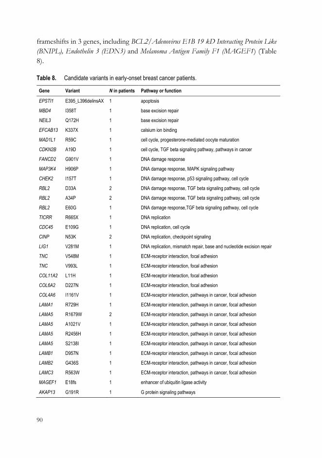
90
frameshifts in 3 genes, including BCL2/Adenovirus E1B 19 kD Interacting Protein Like
(BNIPL), Endothelin 3 (EDN3) and Melanoma Antigen Family F1 (MAGEF1) (Table
8).
Table 8. Candidate variants in early-onset breast cancer patients.
Gene Variant N in patients Pathway or function
EPSTI1 E395_L396delinsAX 1 apoptosis
MBD4 I358T 1 base excision repair
NEIL3 Q172H 1 base excision repair
EFCAB13 K337X 1 calsium ion binding
MAD1L1 R59C 1 cell cycle, progesterone-mediated oocyte maturation
CDKN2B A19D 1 cell cycle, TGF beta signaling pathway, pathways in cancer
FANCD2 G901V 1 DNA damage response
MAP3K4 H906P 1 DNA damage response, MAPK signaling pathway
CHEK2 I157T 1 DNA damage response, p53 signaling pathway, cell cycle
RBL2 D33A 2 DNA damage response, TGF beta signaling pathway, cell cycle
RBL2 A34P 2 DNA damage response, TGF beta signaling pathway, cell cycle
RBL2 E60G 1 DNA damage response,TGF beta signaling pathway, cell cycle
TICRR R665X 1 DNA replication
CDC45 E109G 1 DNA replication, cell cycle
CINP N53K 2 DNA replication, checkpoint signaling
LIG1 V281M 1 DNA replication, mismatch repair, base and nucleotide excision repair
TNC V548M 1 ECM-receptor interaction, focal adhesion
TNC V993L 1 ECM-receptor interaction, focal adhesion
COL11A2 L11H 1 ECM-receptor interaction, focal adhesion
COL6A2 D227N 1 ECM-receptor interaction, focal adhesion
COL4A6 I1161V 1 ECM-receptor interaction, pathways in cancer, focal adhesion
LAMA1 R729H 1 ECM-receptor interaction, pathways in cancer, focal adhesion
LAMA5 R1679W 2 ECM-receptor interaction, pathways in cancer, focal adhesion
LAMA5 A1021V 1 ECM-receptor interaction, pathways in cancer, focal adhesion
LAMA5 R2456H 1 ECM-receptor interaction, pathways in cancer, focal adhesion
LAMA5 S2138I 1 ECM-receptor interaction, pathways in cancer, focal adhesion
LAMB1 D957N 1 ECM-receptor interaction, pathways in cancer, focal adhesion
LAMB2 G436S 1 ECM-receptor interaction, pathways in cancer, focal adhesion
LAMC3 R563W 1 ECM-receptor interaction, pathways in cancer, focal adhesion
MAGEF1 E18fs 1 enhancer of ubiquitin ligase activity
AKAP13 G191R 1 G protein signaling pathways
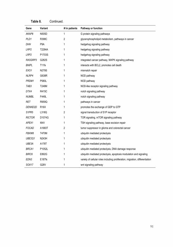
91
Table 8. Continued.
Gene Variant N in patients Pathway or function
AKAP8 N505D 1 G protein signaling pathways
PLD1 R398C 2 glycerophospholipid metabolism, pathways in cancer
DHH P9A 1 hedgehog signaling pathway
LRP2 T2284A 1 hedgehog signaling pathway
LRP2 P1703S 1 hedgehog signaling pathway
RASGRP3 G282S 1 integrated cancer pathway, MAPK signaling pathway
BNIPL T11fs 1 interacts with BCL2, promotes cell death
EXO1 N279S 1 mismatch repair
NLRP4 G638R 1 NOD pathway
PRDM1 P580L 1 NOD pathway
TAB3 T248M 1 NOD-like receptor signaling pathway
DTX4 R415C 1 notch signaling pathway
NUMBL F449L 1 notch signaling pathway
RET R959Q 1 pathways in cancer
DENND2D R16X 1 promotes the exchange of GDP to GTP
S1PR5 L318Q 2 signal transduction of S1P receptor
RICTOR D1074G 1 TOR signaling, mTOR signaling pathway
APEX1 I64V 1 TSH signaling pathway, base excision repair
FOCAD A1683T 2 tumor suppressor in glioma and colorectal cancer
FBXW8 T470M 1 ubiquitin mediated proteolysis
UBE2Q1 N243H 1 ubiquitin mediated proteolysis
UBE3A A178T 1 ubiquitin mediated proteolysis
BRCA1 P1052L 1 ubiquitin mediated proteolysis, DNA damage response
BIRC6 E892G 1 ubiquitin mediated proteolysis, apoptosis modulation and signaling
EDN3 E187fs 1 variety of cellular roles including proliferation, migration, differentiation
SOX17 G28V 1 wnt signaling pathway
92
DISCUSSION
1 Contribution of variants in well-known breast cancer susceptibility genes to high-risk Finnish HBOC families (I, II, III)
Mutations in well-known BC susceptibility genes, such as BRCA1, BRCA2, ATM,
PALB2, and CHEK2, explain approximately 20-30% of the genetic predisposition
to BC (Couch et al, 2014, Mavaddat et al, 2010, Turnbull & Rahman, 2008). BRCA1
and BRCA2 make a major contribution to this predisposition and in Finland
contribute to BC in approximately 20% of HBOC families (Hartikainen et al, 2007,
Vehmanen et al, 1997a). Additionally, Finnish founder mutations have been
identified in CHEK2, PALB2, or RAD50 and contribute to a fraction of BC cases
in Finnish HBOC families (Erkko et al, 2007, Heikkinen et al, 2006, Vahteristo et al,
2002). However, the genetic predisposition factors remain unknown in majority of
the high-risk families. Well-established clinical management strategies exist for
BRCA1 and BRCA2 mutation carriers (Couch et al, 2014). However, the clinical
management of high-risk BRCA1/2-negative HBOC patients is problematic.
Therefore, new information of breast and ovarian cancer predisposing factors is
urgently needed to improve the clinical assessment of high-risk families and to widen
the genetic testing and guidance protocol for other genes and mutations as well.
Additional variants were identified in BRCA1 and BRCA2 genes in HBOC
individuals who had been tested to be founder mutation-negative for both of these
genes. The detected variants in these two genes were reported to contribute to BC
in a fraction of the high-risk Finnish HBOC families. The BRCA1 Arg1699Trp
variant, which was detected in study I in three BC-affected females in a single HBOC
family, has been classified as clinically significant variant in the Breast Cancer
Information Core database. This variant was further confirmed to be an intermediate

93
breast and ovarian cancer risk variant by functional analyses (Spurdle et al, 2012).
Additionally, two other nonsynonymous variants, BRCA1 Met1628Thr and BRCA2
Val2728Ile, which were detected in study I, showed enrichment in HBOC families
compared to controls. This result implies that these variants may be low-to-moderate
risk alleles. Although the clinical significance of both variants is unknown
(Deffenbaugh et al, 2002, Ostrow et al, 2004, Phelan et al, 2005), further study is
clearly warranted. Additionally, a rare and possibly pathogenic variant in BRCA1,
T1302P, was detected in study III in two BC-affected relatives in a single family. The
role of this variant in BC predisposition is suspected given that it was not observed
among healthy controls. Additionally, one novel possibly pathogenic BRCA2 variant
(Leu24Phe) was detected in a single HBOC family in study I. The novel variant was
particularly interesting as it is located in the N-terminal portion of BRCA2, which is
an interaction site for PALB2. PALB2 is essential for key BRCA2 functions, and
sequence variants disrupting this interaction have been reported to play a role in
cancer predisposition (Xia et al, 2006). Thus, additional analyses of the Leu24Phe
variant are warranted. Moreover, a large deletion affecting BRCA1 was detected in
study II in a BC patient with family history of ovarian cancer. The same deletion,
which removes most of the gene, including the promoter, and prevents transcription
of BRCA1, has been reported in the previous Finnish study of ovarian cancer family
(Pylkas et al, 2008). Large genomic rearrangements in both BRCA1 and BRCA2 are
rare, but the findings confirm the contribution of BRCA1 rearrangements to a
fraction of the HBOC families in the presence of multiple cases of ovarian cancer.
An important finding in study I was that the two known BC-associated CHEK2
variants, Ile157Thr, and CHEK2 1100delC, contributed to BC predisposition in the
Finnish BRCA1/2-negative HBOC families to a remarkable degree (10/82, 12.2%);
these results suggest the clinical relevance of these variants. Among heterozygous
1100delC carriers with a family history of BC, the cumulative risk of BC by age 70 is
estimated to be 37%, which is comparable to the BC risk among BRCA1 and BRCA2
mutation carriers (Weischer et al, 2008). Moreover, 1100delC carriers seem to have
poorer disease-free and overall survival than non-carriers and an increased risk of
developing a second, primarily contralateral, BC (Ripperger et al, 2009). Therefore,
genotyping of the 1100delC variant for clinical assessment of BC risk has been
suggested particularly in Northern and Eastern European populations in which the
CHEK2 variants are observed with high frequencies (Weischer et al, 2008). The
1100delC increases the BC risk approximately two-fold, whereas the Ile157Thr is the
lower risk variant (CHEK2 Breast Cancer Case-Control Consortium, 2004,
Kilpivaara et al, 2004). Thus, the CHEK2 variants are suggested to act in

94
combination with other susceptibility alleles to multiply the BC risk (i.e., a polygenic
risk model) (CHEK2 Breast Cancer Case-Control Consortium, 2004). Here, a
particularly interesting finding was that both of the CHEK2 variants, 1100delC and
Ile157Thr, were observed in the same family, and a female patient carrying both of
the variants exhibited a dramatic clinical outcome (BC at the age of 26 years). This
set of results suggest a multiplicative effect of these two variants. Moreover, bilateral
BC was diagnosed in one of the three patients with 1100delC variant, which is in line
with previous observations. However, due to unclear clinical consequences related
to the incomplete segregation of the CHEK2 variants with BC and the fact that these
variants are very rare in most populations, genetic testing of CHEK2 variants has
not been justified in the clinical practice (Ripperger et al, 2009). Here, the results
favor a more profound segregation analyses of the two CHEK2 variants, 1100delC
and Ile157Thr, in the high-risk Finnish BRCA1/2-negative hereditary BC families.
ATM has been considered a moderate-risk BC gene, and heterozygous mutations
have been reported to confer an approximately two-fold elevated BC risk (Ahmed
& Rahman, 2006). An ATM D1853V (c.A5558T) variant was reported to contribute
a low-to-moderate BC predisposition in study III. The variant was identified
altogether in four HBOC families. Interestingly, a common ATM polymorphism
(c.G5557A) was observed among two of the c.A5558T variant carriers at the adjacent
nucleotide position. The c.G5557A variant is a common polymorphism. The
association of this variant with BC has been widely studied and has been associated
with bilateral BC in the Finnish study (Heikkinen et al, 2005). In line with these
results, one of the BC patients carrying both of the ATM variants, c.G5557A and
c.A5558T, had been diagnosed with bilateral disease. Furthermore, the c.G5557A
variant has been reported to have an effect on splicing (Thorstenson et al, 2003); a
similar observation was seen for the c.A5558T variant, which it was predicted to
create a new splicing site. Splicing mutations have been reported to be particularly
common in ATM (Thorstenson et al, 2003), which makes the c.A5558T variant an
intriguing candidate. Therefore, further studies are necessary to fully analyze the role
of the c.A5558T variant in splicing, either alone or in combination with the
c.G5557A variant.
BC-associated Finnish founder mutations have been reported in PALB2 and
RAD50 genes (Erkko et al, 2007, Heikkinen et al, 2006), but these founders were
not detected in the present studies. This difference, is likely explained by the rarity
of the mutations and the limited number of the studied families. Instead, a RAD50
I94L variant was reported to contribute to male BC in study III, but this finding
warrants further confirmation. There was no evidence that the identified coding

95
variants in the well-known BC susceptibility genes PALB2, BRIP1, RAD50, and
CDH1 contributed to female breast or ovarian cancer risk in the analyzed HBOC
families.

96
2 Contribution of germline copy number variations to HBOC susceptibility and the identification of candidate genes (II)
Copy number variation is defined as situation in which a segment of DNA (1 kb or
larger) shows an altered copy number compared to the reference genome (Feuk et
al, 2006). CNVs are known to play a role in cancer predisposition, but their role in
HBOC is largely unexplored (Krepischi et al, 2012a, Krepischi et al, 2012b). By
studying CNV disrupted genes, defective biological processes and novel candidate
genes underlying breast/ovarian cancer predisposition can be detected. A previous
Finnish study provided evidence of rare CNVs that contribute to BC susceptibility
by identifying disrupted genes in estrogen signaling and the TP53 tumor suppressor
network (Pylkas et al, 2012).
The current study identified CNV-affected genes as good candidates for HBOC
susceptibility. Of particular interest were ERBB4 at 2q34, EPHA3 at 3p11.1,
CSMD1 at 8p23.2, and ERVV-2 at 19q13.41.
ERBB4 encodes an epidermal growth factor receptor tyrosine kinase subfamily
member that functions in several cellular processes, including proliferation, survival,
migration, and differentiation (Sundvall et al, 2008). ERBB4 has a role in mammary
carcinogenesis and has been suggested to have both tumor-promoting and tumor-
suppressing functions (Sundvall et al, 2008). An ERBB4-affecting deletion was
observed in 5% (5/101) of the HBOC families. Segregation analysis of the novel
ERBB4-affecting intronic deletion in one HBOC family suggested that the deletion
may be a low-risk variant and may modify cancer risk when occurring in combination
with the intermediate breast and ovarian cancer risk variant, BRCA1 Arg1699Trp,
which was identified in study I.
EPHA3 encodes a protein that functions in signaling pathways. EPHA3 belongs
to the ephrin receptor subfamily of the receptor tyrosine kinase family, which has
central roles in normal cell physiology and disease pathogenesis (Pasquale, 2008).
Ephrin receptor signaling and ephrin ligands regulate tumor growth in variety of
cancers, including BC (Pasquale, 2010). Additionally, altered expression levels of
EPHA3 have been associated with gastric and colorectal cancers, and CNVs in the

97
EPHA3 region have been reported in hematological malignancies (Guan et al, 2011,
Xi & Zhao, 2011, Xi et al, 2012). Moreover, CNVs at the EPHA3 region have been
implicated in predisposition to hereditary prostate cancer (Laitinen et al, 2015),
making this CNV a plausible candidate also for HBOC susceptibility. Here, the
EPHA3-disrupting deletion occurred in 11.9% (12/101) of the HBOC families.
CSMD1 is a known tumor suppressor gene. The loss of CSMD1 has been
primarily associated with head and neck squamous cell carcinoma but has also been
observed in several epithelial cancers, including BC (Ma et al, 2009). Furthermore,
the decreased expression of CSMD1 has been associated with a high-tumor grade
and poor survival in invasive ductal breast carcinoma; this gene has also been used
as a prognostic marker (Kamal et al, 2010). Therefore, CSMD1 is an excellent
candidate gene for HBOC susceptibility. Here, the CSMD1-affecting deletion was
rare and was observed in a single (1/101, 1.0%) BC family
ERVV-2 encodes a protein in the human endogenous retrovirus (HERV) group.
HERVs and related genetic elements occupy approximately 8% of the human
genome, but their role in human cells is poorly understood (Suntsova et al, 2015).
HERVs encode active retroviral proteins that are likely involved in important
physiological functions and may be involved in the progression of cancer and several
other human diseases (Suntsova et al, 2015). Moreover, HERVs regulate the
expression of neighboring host genes and modify the genomic regulatory landscape
(e.g., transcription factor binding sites) (Suntsova et al, 2015). Therefore, ERVV-2
is a very interesting candidate gene in HBOC susceptibility, providing an intriguing
link between breast and ovarian cancer predisposition and endogenous retroviruses.
Here, the ERVV-2-affecting duplication was observed in 10.9% (11/101) of the
HBOC families. A particularly interesting observation was that the homozygous
form of the duplication (observed in 4/101, 4.0% of the HBOC families) was over
four times more common in HBOC cases compared to controls and seemed to
correlate with high-grade tumors.
One interesting CNV was detected at the non-genic 5q15 region. The CNV was
considered interesting due to clinical outcome of the single HBOC patient
(diagnosed and died of BC at the age of 29) carrying the homozygous form of the
CNV. The homozygous deletion at 5q15 was extremely rare in controls (1/899,
0.1%), implying that it may be disease-related and may have clinical significance for
families with early-onset BC cases. This intergenic deletion may disrupt the
transcriptional control of target gene expression, and this hypothesis was supported
by the finding that regulatory element activity is present at the 5q15 deletion region.
Gene expression regulation is a complex process, and regulatory elements can be far

98
from the target genes (Kleinjan & van Heyningen, 2005). An interesting observation
was that Repulsive Guidance Molecule Family Member B (RGMB), the aberrant expression
of which has been found in BC (Li et al, 2011), was observed to be the nearest
neighboring gene in the 5q15 deletion region (1.0 Mb). Further studies are necessary
to identify the target gene of the possible regulatory elements that are disrupted by
the deletion at 5q15.

99
3 Identification of novel candidate genes and gene variants in high-risk HBOC families (III)
3.1 DNA damage response pathway
DDR pathway is one of the central pathways in breast and ovarian cancer
predisposition. Several susceptibility genes from this pathway have been recognized
including BRCA1, BRCA2, ATM, PALB2, BRIP1, CHEK2, and RAD50 (Ciccia &
Elledge, 2010, Jackson & Bartek, 2009). Since the DDR pathway is a complex
network consisting of numerous proteins and complexes, there are likely unknown
candidates in this pathway that contribute to breast and ovarian cancer susceptibility.
In study III, the exome sequencing approach was utilized to analyze 13 high-risk
HBOC families, and the primary focus was in DDR pathway gene variants that were
shared between affected family members or between families. Family-specific
enrichments of multiple rare DDR pathway gene variants were observed, confirming
the central role of the DDR pathway defects in HBOC families. Moreover, different
combinations of possible low-to-moderate risk variants in critical pathway genes
were implicated in cancer predisposition and phenotypic variation (e.g., disease
onset) in the analyzed families. One such interesting combination was observed in a
family in which three BC-affected females were exome sequenced. Two females who
had been diagnosed with BC at a later age (>50 years) carried variants in both
CDKN2A (H166Y) and AKT2 (P50T), whereas the third female who had early-onset
disease (diagnosed at age 28) carried a variant (G901V) in Fanconi Anemia,
Complementation Group D2 (FANCD2). AKT2 is a serine threonine kinase that
functions in a key signaling pathway in mammary gland development and cancer
(Wickenden & Watson, 2010). CDKN2A is a tumor suppressor gene in melanoma.
Germline defects in CDKN2A have been reported in female BC patients with
melanoma (Nagore et al, 2009). Of note, melanoma was not observed among BC
patients in the family in which the CDKN2A variant was observed but one
unconfirmed melanoma case was reported in their male relative. Moreover,
FANCD2 belongs to the Fanconi Anemia complementation gene group, in which
biallelic mutations predispose to Fanconi Anemia (Schneider et al, 2015).
Heterozygous mutations in several Fanconi Anemia genes, such as FANCD1

100
(BRCA2), FANCN (PALB2), and FANCM, have been associated with BC
predisposition (Erkko et al, 2007, Kiiski et al, 2014, Wooster et al, 1994). These
results make FANCD2 an interesting candidate gene to further study in terms of BC
susceptibility, particularly in early-onset cases.
The most promising candidate variants (n=18) in DDR genes were observed in
additional cohorts of female HBOC cases, male BC cases, breast tumor samples, and
controls. Due to the rarity of these variants and the limited number of analyzed cases,
no statistically significant association with the disease was reached. In addition, no
wild type allele loss was observed for any of the variants in breast tumor samples.
However, five variants, ATM D1853V, MYC N26S, PLAU V15L, RAD1 G114D,
and RRM2B F71fs, were enriched in HBOC cases compared to controls (OR 1.16-
2-16). These results suggest that the variants may confer low-to-moderate risk for
breast and ovarian cancer and may contribute to a fraction of the cases in HBOC
families. However, further confirmative studies for the variants are warranted. All
five genes in which the variants occur play important roles in the DDR pathway,
making them good candidates genes for disease susceptibility. In addition to ATM,
which is a well-known BC susceptibility gene, MYC is an established oncogene that
plays a role in a variety of human cancers (Hoffman & Liebermann, 2008). PLAU
encodes a protein that plays a role in cancer metastasis (Moquet-Torcy et al, 2014).
RAD1 encodes a component of the cell cycle checkpoint complex that participates
in cell cycle checkpoint activation and DNA repair (Xu et al, 2009), and RRM2B
encodes a protein involved in the p53 checkpoint for the repair of damaged DNA
(Tanaka et al, 2000).
3.2 Other pathways
Pathways related to cell cycle, proliferation, apoptosis, signaling, and adhesion are
interesting candidate pathways for breast and ovarian cancer susceptibility, although
these pathways are primarily unexplored. By focusing on six exomes from early-
onset BC patients (diagnosed ≤ 29 years), rare pathogenic variants in these potential
pathway genes were explored. For instance, deleterious frameshift variants were
detected in BNIPL, EDN3, and MAGEF1, and variants that induce a premature
stop codon were detected in DENND2D, EFCAB13, EPST11, and TICRR. Of
these, BNIPL encodes an apoptosis-associated protein that interacts with B-Cell
CLL/Lymphoma 2 (BCL2) and promotes the invasion and metastasis of human
hepatocellular carcinoma cells (Qin et al, 2003, Xie et al, 2007), whereas EDN3 is an
101
important signaling molecule that participates in several key cellular processes, such
as proliferation, migration, and differentiation (Levin, 1995). Furthermore,
DENND2D is involved in cell signaling, and altered expression levels of this gene
have been reported in cancers (Hibino et al, 2014). TICRR encodes a protein that is
a crucial regulator of DNA replication and cell cycle checkpoints (Sansam et al,
2010). Furthermore, non-synonymous variants were observed in CINP, FOCAD,
and Exonuclease 1 (EXO1), which are also of great interest. CINP is involved in DNA
replication and checkpoint signaling (Lovejoy et al, 2009). FOCAD is a tumor
suppressor gene that has been associated with polyposis and colorectal cancer
development (Weren et al, 2015). Furthermore, EXO1 plays a role in mismatch
repair, and its role in susceptibility to BC has been reported (Michailidou et al, 2015).
The novel findings encourage the study of other pathways in breast/ovarian cancer
predisposition and provide information on potential candidate genes. These data can
be used an excellent foundation for future studies.

102
4 Limitations of the study
Breast and ovarian cancers are genetically heterogeneous diseases in which numerous
genetic factors play multiplicative roles. Genetic predisposition factors are often rare
and family specific, and there is phenotypic variation within families, making the
identification of predisposition factors challenging. Sample selection is one of the
critical steps when identifying disease-causing genetic factors. In the current study,
breast and/or ovarian cancer families were selected according to strict high-risk
hereditary BC criteria. There criteria were chosen to select families with similar
characteristics and that are most likely to share common hereditary defects. Although
well-selected high-risk HBOC families were used in this study, the number of
analyzed patients was somewhat limited and the statistical analyses lacked significant
association results. For these reasons, these findings require further confirmation in
a larger sample sets. Moreover, the availability of samples from both affected and
healthy relatives was a limiting step in the segregation analyses of certain variants.
Thus, there is a need to continue recruiting more high-risk HBOC families and more
relatives from the studied families. Additionally, tumor samples from these patients
would be essential to further study the deleteriousness of the variants identified in
the germline. When using large sample cohorts, clinically different subtypes (e.g.,
tumors with hormone receptor status) can be utilized. For instance, a previous
Finnish study identified a novel BC susceptibility gene to be particularly associated
with triple-negative BC (Kiiski et al, 2014). Here, for example, the results implicated
that copy number variation at 3p11.1 would be more common in BC patients with
ductal and estrogen- and progesterone-receptor positive tumor. Obviously, this
result should be confirmed in the larger cohort of BC patients to see the trend more
clearly. Overall, the Finnish population, which is a homogenous group, provides an
excellent basis for genetic studies. Although, it should be noted that there can be
great variation in the mutation spectrum between different parts of Finland. For
instance, the prevalence of BRCA1 and BRCA2 founder mutations varies across the
country (Hartikainen et al, 2007, Huusko et al, 1998, Sarantaus et al, 2000).
Since the identification of the two major breast and ovarian cancer susceptibility
genes, BRCA1 and BRCA2, a variety of different methodological approaches have
been utilized in susceptibility gene studies. Each of the approaches have their

103
strengths and limitations. In the current study, three different approaches were
utilized, including a candidate gene approach, genome-wide copy number variation
analysis and exome sequencing. The mutational screening of candidate genes is
accurate but often costly and time-consuming when using, for example, direct
sequencing. Genome-wide approaches provide a cost-effective and rapid approach
for the detection of a massive number of genetic variants in a single run. When using
genome-wide approaches, the challenge is to use appropriate filtering strategies to
trim down the number of candidate variants. If using filtering criteria that are too
stringent, disease-causing variants can be excluded already in the data-analysis phase.
Most commonly, variants are filtered based on allele frequency, predicted
pathogenicity, and function. Therefore, it is possible that causative variants were
missed in this study during these filtering steps. Moreover, exome sequencing detects
only variants that are located in the protein-coding region. Therefore, causative
variants that are located outside the coding region and affect, for example,
transcription, may have been missed. Additionally, focusing on certain pathway
genes (study III) might be a useful step in reducing the number of candidate variants.
However, variants in other pathways remain unexplored. Therefore, a subset of
exome-sequenced patients (study III) that included early-onset BC cases were re-
analyzed. In this analysis, variants in all pathways were considered potential
candidates when identifying novel candidate genes and pathways that likely play a
role in BC pathogenesis.

104
5 Future prospects
There is still a very large gap in knowledge concerning genetic predisposition factors
in high-risk HBOC families that do not carry defects in BRCA1 or BRCA2. There
is an immense need to obtain novel information of additional breast and ovarian
cancer susceptibility genes and gene defects to identify at-risk individuals as early as
possible. In this way, these individuals and their family members can be provided
with more efficient prevention, screening strategies, and therapeutic options. For
instance, patients with defective genes in the DNA-damage repair pathway, including
BRCA1 and BRCA2 mutation carriers, benefit from treatment with Poly (ADP-
ribose) polymerases (PARP) inhibitors (Livraghi & Garber, 2015).
Novel next-generation sequencing technologies provide fast and cost-effective
applications for the detection of genetic predisposition factors. Whole exome
sequencing, which focuses on only the protein-coding region of the genome, is
currently an attractive method for identifying novel susceptibility genes by (Ng et al,
2009). When additional technologies are developed and costs are reduced, whole
genome sequencing will be a major method for revealing the remaining genetic
defects that underlie these diseases. NGS technologies have been applied in clinical
settings as well. For instance, NGS has been shown to be an efficient tool in HBOC
diagnostics (Castera et al, 2014, Trujillano et al, 2015). However, the major challenge
related to NGS lies in transferring sequencing data into medical diagnoses. There is
a need to develop appropriate and validated guidelines related to data-analysis and
the interpretation of variants that are of unknown significance (Rehm et al, 2013).
Additionally, the amount of data will increase notably for whole genome sequencing,
requiring appropriate storage and handling strategies in the clinical setting.
Furthermore, when the number of candidate variants discovered through NGS
increases, there will be a need to functionally validate the findings and design novel
clinical assessment guidelines.
Moreover, multinational collaborations, such as the Collaborative Oncological
Gene-environment Study (Collaborative Oncological Gene-environment Study), are
likely to play an essential role in identifying predisposition factors for breast and
ovarian cancer. These types of consortia are able to study a massive number of
patients from a variety of populations. Additionally, new technologies have made it

105
possible to collect variety of different sample types from patients. The collected
samples and specific clinical information of the patients are increasingly being stored
in biobanks, providing an excellent source for research purposes. Moreover,
sequencing efforts have led to the creation of several databases, such as the 1000
Genomes (1000 Genomes Project Consortium et al, 2012) and The Cancer Genome
Atlas (The Cancer Genome Atlas). These provide large datasets that can be freely
used in cancer genetic studies. It is likely that the available information in these and
other databases will increase massively in the coming years and will provide
unprecedented possibilities in terms of integrating data from different sources.
Constantly increasing genetic information will enable healthcare to move towards
well-tailored medical care. In the future, it will likely be possible to design
personalized screening, prevention, and therapeutic strategies and thereby optimize
the clinical management of hereditary breast and ovarian cancer.
106
CONCLUSIONS
The current study was conducted to provide novel information about the genetic
factors predisposing to HBOC in high-risk BRCA1/2 founder mutation-negative
Finnish families.
The major findings of this study were following:
1. Previously known breast cancer-associated mutations in BRCA1 and CHEK2
contributed to 13.4% of the HBOC families. Proportion of the CHEK2
mutations was remarkable and clinically relevant. A novel possibly
pathogenic variant was identified in BRCA2.
2. Copy number variations at 3p11.1, 5q15, 8p23.2, and 19q13.41 were of
special interest and their role in HBOC predisposition was indicated.
Deletions at 3p11.1 and 8p23.2 affected intronic regions of EPHA3 and
CSMD1 genes, whereas duplication at 19q13.41 disrupted the coding region
of ERVV-2 gene. Additionally, deletion at 5q15 occurred at intergenic region
and was reported to affect regulatory elements.
3. Five variants in DNA damage response pathway genes ATM, MYC, PLAU,
RAD1, and RRM2B may act as low-to-moderate risk alleles. A rare variant
that may have clinical relevance was detected in BRCA1. Additionally, a rare
variant in RAD50 was suggested to predispose to male breast cancer.
Variants in novel candidate genes targeting DNA repair and replication,
signaling, apoptosis, and cell cycle were observed in early-onset breast cancer
patients. Novel candidate genes included, for example DENND2D, TICRR,
BNIPL, EDN3, and FOCAD.
107
ACKNOWLEDGEMENTS
This study was carried out in the Laboratory of Cancer Genetics, BioMediTech,
University of Tampere and Tampere University Hospital during the years 2007-2016.
I wish to thank the former director of IBT, Professor Olli Silvennoinen, M.D.,
Ph.D., the current director of the BioMediTech, Dr. Hannu Hanhijärvi, DDS, Ph.D.,
and Docent Erkki Seppälä, M.D., Ph.D., Medical Director of the Fimlab
Laboratories, for providing excellent research facilities. Tampere Graduate Program
in Biomedicine and Biotechnology (TGPBB) is acknowledged for the graduate
school position, travel grants, and excellent courses.
I sincerely thank my supervisors Professor Johanna Schleutker, Ph.D., and
Docent Satu-Leena Laasanen, M.D., Ph.D., for their help, guidance, and support
during this thesis. Johanna provided me the opportunity to join her research group
and guided me in the scientific aspects of the study. Satu-Leena provided me the
access to patient material and assisted me in the clinical aspects of the study.
I wish to thank my thesis committee members, Professor Anne Kallioniemi,
M.D., Ph.D., and Docent Ritva Karhu, Ph.D., for their help during the years.
The official reviewers of the thesis manuscript, Docent Outi Monni, Ph.D., and
Docent Jukka Moilanen, M.D., Ph.D., are warmly thanked for their comments and
feedback.
I warmly thank all the co-authors for their valuable contribution and professional
help. Specifically, I want to acknowledge Professor Mauno Vihinen, Ph.D., Professor
Matti Nykter, Ph.D., Minna Kankuri-Tammilehto, M.D., Ph.D., Tommi Rantapero,
M.Sc., Anna Lindström, M.Sc., Aleksandra Bebel, M.Sc., and Oyediran Akinrinade,
M.Sc.
I would like to thank all the present and former members of Genetic
Predisposition to Cancer study group. Spesicifically, Tiina Wahlfors, Ph.D., Henna
Mattila, Ph.D., Sanna Siltanen, Ph.D., Virpi Laitinen, M.Sc., CMG, Riikka Nurminen,
M.Sc., and Ms. Riina Kylätie are warmly thanked for their help, support, and
friendship both inside and outside the office and the lab. Additionally, Ms. Linda
Enroth, Ms. Riitta Vaalavuo, Ms. Kirsi Rouhento, Sanna Pakkanen, M.D., Ph.D.,
Martin Schindler, Ph.D., Jarkko Isotalo, Ph.D., Daniel Fischer, M.Sc., Ha Nati,
M.Sc., Elisa Vuorinen, M.Sc., Mimmi Patrikainen, Ph.D., Sanna-Kaisa Harjula,

108
M.Sc., Sanna Ränsi, M.Sc., Ville Rimpilä, M.Sc., and Ms. Aune Aho are warmly
acknowledged. Moreover, other co-workers and friends at BioMediTech, and the
personnel of the Tampere University Hospital Genetics Outpatien clinic are greatly
acknowledged. Special thanks to IT designer Jukka Lehtiniemi for skillful technical
assistance. Additionally, thank you RaiRai-group. I have shared many great moments
with you
Most importantly, I sincerely thank my closest family members and friends for
endless love, support, and encouragement. No tree can grow to heaven unless its
roots are firmly in the ground ♥
I sincerely thank all the cancer patients and their family members for participating
in this study.
This study was financially supported by the TGPBB, the Competitive Research
Funding of the Tampere University Hospital (grants 9K119, ML16, and 9M094),
Biocenter of Finland, Sigrid Juselius Foundation, the Academy of Finland (grant
251074), the Finnish Cultural Foundation, the Finnish Breast Cancer Group, the
Emil Aaltonen Foundation, the Ida Montini Foundation, the Orion-Farmos
Research Foundation, the Scientific Foundation of the City of Tampere, and the
Finnish Cancer Organizations.
Tampere, January 2016
Kirsi Määttä

109
REFERENCES
1000 Genomes Project Consortium, Abecasis GR, Auton A, Brooks LD, DePristo MA, Durbin RM, Handsaker RE, Kang HM, Marth GT, & McVean GA (2012) An integrated map of genetic variation from 1,092 human genomes. Nature 491: 56-65
Ahmed M & Rahman N (2006) ATM and breast cancer susceptibility. Oncogene 25: 5906-5911
Allinen M, Launonen V, Laake K, Jansen L, Huusko P, Kaariainen H, Borresen-Dale AL, & Winqvist R (2002) ATM mutations in Finnish breast cancer patients. J Med Genet 39: 192-196
Antoniou A, Pharoah PD, Narod S, Risch HA, Eyfjord JE, Hopper JL, Loman N, Olsson H, Johannsson O, Borg A, Pasini B, Radice P, Manoukian S, Eccles DM, Tang N, Olah E, Anton-Culver H, Warner E, Lubinski J, Gronwald J et al (2003) Average risks of breast and ovarian cancer associated with BRCA1 or BRCA2 mutations detected in case Series unselected for family history: a combined analysis of 22 studies. Am J Hum Genet 72: 1117-1130
Antoniou AC, Casadei S, Heikkinen T, Barrowdale D, Pylkas K, Roberts J, Lee A, Subramanian D, De Leeneer K, Fostira F, Tomiak E, Neuhausen SL, Teo ZL, Khan S, Aittomaki K, Moilanen JS, Turnbull C, Seal S, Mannermaa A, Kallioniemi A et al (2014) Breast-cancer risk in families with mutations in PALB2. N Engl J Med 371: 497-506
Antoniou AC, Pharoah PD, McMullan G, Day NE, Stratton MR, Peto J, Ponder BJ, & Easton DF (2002) A comprehensive model for familial breast cancer incorporating BRCA1, BRCA2 and other genes. Br J Cancer 86: 76-83
Antoniou AC, Spurdle AB, Sinilnikova OM, Healey S, Pooley KA, Schmutzler RK, Versmold B, Engel C, Meindl A, Arnold N, Hofmann W, Sutter C, Niederacher D, Deissler H, Caldes T, Kampjarvi K, Nevanlinna H, Simard J, Beesley J, Chen X et al (2008) Common breast cancer-predisposition alleles are associated with breast cancer risk in BRCA1 and BRCA2 mutation carriers. Am J Hum Genet 82: 937-948
Babraham Bioinformatics FastQC http://www.bioinformatics.bbsrc.ac.uk/projects/fastqc/, accessed on January 2015.
Barnes NL, Ooi JL, Yarnold JR, & Bundred NJ (2012) Ductal carcinoma in situ of the breast. BMJ 344: e797
Bartek J, Bartkova J, & Lukas J (2007) DNA damage signalling guards against activated oncogenes and tumour progression. Oncogene 26: 7773-7779
Bartek J, Falck J, & Lukas J (2001) CHK2 kinase--a busy messenger. Nat Rev Mol Cell Biol 2: 877-886
Bartek J & Lukas J (2003) Chk1 and Chk2 kinases in checkpoint control and cancer. Cancer Cell 3: 421-429
Behrens J (1999) Cadherins and catenins: role in signal transduction and tumor progression. Cancer Metastasis Rev 18: 15-30

110
Bell DW, Varley JM, Szydlo TE, Kang DH, Wahrer DC, Shannon KE, Lubratovich M, Verselis SJ, Isselbacher KJ, Fraumeni JF, Birch JM, Li FP, Garber JE, & Haber DA (1999) Heterozygous germ line hCHK2 mutations in Li-Fraumeni syndrome. Science 286: 2528-2531
Berx G, Becker KF, Hofler H, & van Roy F (1998) Mutations of the human E-cadherin (CDH1) gene. Hum Mutat 12: 226-237
Berx G, Staes K, van Hengel J, Molemans F, Bussemakers MJ, van Bokhoven A, & van Roy F (1995) Cloning and characterization of the human invasion suppressor gene E-cadherin (CDH1). Genomics 26: 281-289
Bieging KT, Mello SS, & Attardi LD (2014) Unravelling mechanisms of p53-mediated tumour suppression. Nat Rev Cancer 14: 359-370
Birch JM, Hartley AL, Tricker KJ, Prosser J, Condie A, Kelsey AM, Harris M, Jones PH, Binchy A, & Crowther D (1994) Prevalence and diversity of constitutional mutations in the p53 gene among 21 Li-Fraumeni families. Cancer Res 54: 1298-1304
Blanco A, Grana B, Fachal L, Santamarina M, Cameselle-Teijeiro J, Ruiz-Ponte C, Carracedo A, & Vega A (2010) Beyond BRCA1 and BRCA2 wild-type breast and/or ovarian cancer families: germline mutations in TP53 and PTEN. Clin Genet 77: 193-196
Blanco A, Gutierrez-Enriquez S, Santamarina M, Montalban G, Bonache S, Balmana J, Carracedo A, Diez O, & Vega A (2014) RAD51C germline mutations found in Spanish site-specific breast cancer and breast-ovarian cancer families. Breast Cancer Res Treat 147: 133-143
Bogdanova N, Enssen-Dubrowinskaja N, Feshchenko S, Lazjuk GI, Rogov YI, Dammann O, Bremer M, Karstens JH, Sohn C, & Dork T (2005) Association of two mutations in the CHEK2 gene with breast cancer. Int J Cancer 116: 263-266
Bray F, McCarron P, & Parkin DM (2004) The changing global patterns of female breast cancer incidence and mortality. Breast Cancer Res 6: 229-239
Breast Cancer Linkage Consortium (1999) Cancer risks in BRCA2 mutation carriers. J Natl Cancer Inst 91: 1310-1316
Broeks A, Urbanus JH, Floore AN, Dahler EC, Klijn JG, Rutgers EJ, Devilee P, Russell NS, van Leeuwen FE, & van 't Veer LJ (2000) ATM-heterozygous germline mutations contribute to breast cancer-susceptibility. Am J Hum Genet 66: 494-500
Cancer Genome Atlas Network (2012) Comprehensive molecular portraits of human breast tumours. Nature 490: 61-70
Cannioto RA & Moysich KB (2015) Epithelial ovarian cancer and recreational physical activity: A review of the epidemiological literature and implications for exercise prescription. Gynecol Oncol 137: 559-573
Cantor S, Drapkin R, Zhang F, Lin Y, Han J, Pamidi S, & Livingston DM (2004) The BRCA1-associated protein BACH1 is a DNA helicase targeted by clinically relevant inactivating mutations. Proc Natl Acad Sci U S A 101: 2357-2362
Cantor SB, Bell DW, Ganesan S, Kass EM, Drapkin R, Grossman S, Wahrer DC, Sgroi DC, Lane WS, Haber DA, & Livingston DM (2001) BACH1, a novel helicase-like protein, interacts directly with BRCA1 and contributes to its DNA repair function. Cell 105: 149-160
Cartegni L, Wang J, Zhu Z, Zhang MQ, & Krainer AR (2003) ESEfinder: A web resource to identify exonic splicing enhancers. Nucleic Acids Res 31: 3568-3571
Casadei S, Norquist BM, Walsh T, Stray S, Mandell JB, Lee MK, Stamatoyannopoulos JA, & King MC (2011) Contribution of inherited mutations in the BRCA2-interacting protein PALB2 to familial breast cancer. Cancer Res 71: 2222-2229

111
Castera L, Krieger S, Rousselin A, Legros A, Baumann JJ, Bruet O, Brault B, Fouillet R, Goardon N, Letac O, Baert-Desurmont S, Tinat J, Bera O, Dugast C, Berthet P, Polycarpe F, Layet V, Hardouin A, Frebourg T, & Vaur D (2014) Next-generation sequencing for the diagnosis of hereditary breast and ovarian cancer using genomic capture targeting multiple candidate genes. Eur J Hum Genet 22: 1305-1313
Castillo A, Paul A, Sun B, Huang TH, Wang Y, Yazinski SA, Tyler J, Li L, You MJ, Zou L, Yao J, & Wang B (2014) The BRCA1-interacting protein Abraxas is required for genomic stability and tumor suppression. Cell Rep 8: 807-817
Catucci I, Milgrom R, Kushnir A, Laitman Y, Paluch-Shimon S, Volorio S, Ficarazzi F, Bernard L, Radice P, Friedman E, & Peterlongo P (2012) Germline mutations in BRIP1 and PALB2 in Jewish high cancer risk families. Fam Cancer 11: 483-491
CHEK2 Breast Cancer Case-Control Consortium (2004) CHEK2*1100delC and susceptibility to breast cancer: a collaborative analysis involving 10,860 breast cancer cases and 9,065 controls from 10 studies. Am J Hum Genet 74: 1175-1182
Chun J, Buechelmaier ES, & Powell SN (2013) Rad51 paralog complexes BCDX2 and CX3 act at different stages in the BRCA1-BRCA2-dependent homologous recombination pathway. Mol Cell Biol 33: 387-395
Ciccia A & Elledge SJ (2010) The DNA damage response: making it safe to play with knives. Mol Cell 40: 179-204
Cimprich KA & Cortez D (2008) ATR: an essential regulator of genome integrity. Nat Rev Mol Cell Biol 9: 616-627
Claus EB, Schildkraut JM, Thompson WD, & Risch NJ (1996) The genetic attributable risk of breast and ovarian cancer. Cancer 77: 2318-2324
Colella S, Yau C, Taylor JM, Mirza G, Butler H, Clouston P, Bassett AS, Seller A, Holmes CC, & Ragoussis J (2007) QuantiSNP: an Objective Bayes Hidden-Markov Model to detect and accurately map copy number variation using SNP genotyping data. Nucleic Acids Res 35: 2013-2025
Coleman MP, Quaresma M, Berrino F, Lutz JM, De Angelis R, Capocaccia R, Baili P, Rachet B, Gatta G, Hakulinen T, Micheli A, Sant M, Weir HK, Elwood JM, Tsukuma H, Koifman S, E Silva GA, Francisci S, Santaquilani M, Verdecchia A et al (2008) Cancer survival in five continents: a worldwide population-based study (CONCORD). Lancet Oncol 9: 730-756
Collaborative Group on Epidemiological Studies of Ovarian Cancer, Beral V, Gaitskell K, Hermon C, Moser K, Reeves G, & Peto R (2012) Ovarian cancer and smoking: individual participant meta-analysis including 28,114 women with ovarian cancer from 51 epidemiological studies. Lancet Oncol 13: 946-956
Collaborative Group on Hormonal Factors in Breast Cancer (2002) Breast cancer and breastfeeding: collaborative reanalysis of individual data from 47 epidemiological studies in 30 countries, including 50302 women with breast cancer and 96973 women without the disease. Lancet 360: 187-195
Collaborative Group on Hormonal Factors in Breast Cancer (1996) Breast cancer and hormonal contraceptives: collaborative reanalysis of individual data on 53 297 women with breast cancer and 100 239 women without breast cancer from 54 epidemiological studies. Lancet 347: 1713-1727
Collaborative Oncological Gene-environment Study http://www.cogseu.org/, accessed on June 2015.
Couch FJ, Nathanson KL, & Offit K (2014) Two decades after BRCA: setting paradigms in personalized cancer care and prevention. Science 343: 1466-1470
112
Cox A, Dunning AM, Garcia-Closas M, Balasubramanian S, Reed MW, Pooley KA, Scollen S, Baynes C, Ponder BA, Chanock S, Lissowska J, Brinton L, Peplonska B, Southey MC, Hopper JL, McCredie MR, Giles GG, Fletcher O, Johnson N, dos Santos Silva I et al (2007) A common coding variant in CASP8 is associated with breast cancer risk. Nat Genet 39: 352-358
Croce CM (2008) Oncogenes and cancer. N Engl J Med 358: 502-511 Cruchaga C, Karch CM, Jin SC, Benitez BA, Cai Y, Guerreiro R, Harari O, Norton J, Budde
J, Bertelsen S, Jeng AT, Cooper B, Skorupa T, Carrell D, Levitch D, Hsu S, Choi J, Ryten M, UK Brain Expression Consortium, Hardy J et al (2014) Rare coding variants in the phospholipase D3 gene confer risk for Alzheimer's disease. Nature 505: 550-554
Cybulski C, Gorski B, Huzarski T, Masojc B, Mierzejewski M, Debniak T, Teodorczyk U, Byrski T, Gronwald J, Matyjasik J, Zlowocka E, Lenner M, Grabowska E, Nej K, Castaneda J, Medrek K, Szymanska A, Szymanska J, Kurzawski G, Suchy J et al (2004) CHEK2 is a multiorgan cancer susceptibility gene. Am J Hum Genet 75: 1131-1135
Damiola F, Pertesi M, Oliver J, Le Calvez-Kelm F, Voegele C, Young EL, Robinot N, Forey N, Durand G, Vallee MP, Tao K, Roane TC, Williams GJ, Hopper JL, Southey MC, Andrulis IL, John EM, Goldgar DE, Lesueur F, & Tavtigian SV (2014) Rare key functional domain missense substitutions in MRE11A, RAD50, and NBN contribute to breast cancer susceptibility: results from a Breast Cancer Family Registry case-control mutation-screening study. Breast Cancer Res 16: R58
Dang CV (2010) Enigmatic MYC Conducts an Unfolding Systems Biology Symphony. Genes Cancer 1: 526-531
Dansonka-Mieszkowska A, Kluska A, Moes J, Dabrowska M, Nowakowska D, Niwinska A, Derlatka P, Cendrowski K, & Kupryjanczyk J (2010) A novel germline PALB2 deletion in Polish breast and ovarian cancer patients. BMC Med Genet 11: 20-2350-11-20
Database of Genomic Variants http://dgv.tcag.ca/, accessed on September 2011. Deans AJ & West SC (2011) DNA interstrand crosslink repair and cancer. Nat Rev Cancer 11:
467-480 Deffenbaugh AM, Frank TS, Hoffman M, Cannon-Albright L, & Neuhausen SL (2002)
Characterization of common BRCA1 and BRCA2 variants. Genet Test 6: 119-121 Degrolard-Courcet E, Sokolowska J, Padeano MM, Guiu S, Bronner M, Chery C, Coron F,
Lepage C, Chapusot C, Loustalot C, Jouve JL, Hatem C, Ferrant E, Martin L, Coutant C, Baurand A, Couillault G, Delignette A, El Chehadeh S, Lizard S et al (2014) Development of primary early-onset colorectal cancers due to biallelic mutations of the FANCD1/BRCA2 gene. Eur J Hum Genet 22: 979-987
Digweed M & Sperling K (2004) Nijmegen breakage syndrome: clinical manifestation of defective response to DNA double-strand breaks. DNA Repair (Amst) 3: 1207-1217
Do R, Stitziel NO, Won HH, Jorgensen AB, Duga S, Angelica Merlini P, Kiezun A, Farrall M, Goel A, Zuk O, Guella I, Asselta R, Lange LA, Peloso GM, Auer PL, NHLBI Exome Sequencing Project, Girelli D, Martinelli N, Farlow DN, DePristo MA et al (2015) Exome sequencing identifies rare LDLR and APOA5 alleles conferring risk for myocardial infarction. Nature 518: 102-106
Dolganov GM, Maser RS, Novikov A, Tosto L, Chong S, Bressan DA, & Petrini JH (1996) Human Rad50 is physically associated with human Mre11: identification of a conserved multiprotein complex implicated in recombinational DNA repair. Mol Cell Biol 16: 4832-4841
113
Domchek SM, Tang J, Stopfer J, Lilli DR, Hamel N, Tischkowitz M, Monteiro AN, Messick TE, Powers J, Yonker A, Couch FJ, Goldgar DE, Davidson HR, Nathanson KL, Foulkes WD, & Greenberg RA (2013) Biallelic deleterious BRCA1 mutations in a woman with early-onset ovarian cancer. Cancer Discov 3: 399-405
Dosanjh MK, Collins DW, Fan W, Lennon GG, Albala JS, Shen Z, & Schild D (1998) Isolation and characterization of RAD51C, a new human member of the RAD51 family of related genes. Nucleic Acids Res 26: 1179-1184
Drooger JC, Hooning MJ, Seynaeve CM, Baaijens MH, Obdeijn IM, Sleijfer S, & Jager A (2015) Diagnostic and therapeutic ionizing radiation and the risk of a first and second primary breast cancer, with special attention for BRCA1 and BRCA2 mutation carriers: a critical review of the literature. Cancer Treat Rev 41: 187-196
Easton DF, Pooley KA, Dunning AM, Pharoah PD, Thompson D, Ballinger DG, Struewing JP, Morrison J, Field H, Luben R, Wareham N, Ahmed S, Healey CS, Bowman R, SEARCH collaborators, Meyer KB, Haiman CA, Kolonel LK, Henderson BE, Le Marchand L et al (2007) Genome-wide association study identifies novel breast cancer susceptibility loci. Nature 447: 1087-1093
Ebi H, Matsuo K, Sugito N, Suzuki M, Osada H, Tajima K, Ueda R, & Takahashi T (2007) Novel NBS1 heterozygous germ line mutation causing MRE11-binding domain loss predisposes to common types of cancer. Cancer Res 67: 11158-11165
Eeles RA (1995) Germline mutations in the TP53 gene. Cancer Surv 25: 101-124 Eletr ZM & Wilkinson KD (2011) An emerging model for BAP1's role in regulating cell
cycle progression. Cell Biochem Biophys 60: 3-11 Encyclopedia of DNA elements at UCSC https://genome.ucsc.edu/ENCODE/, accessed
on September 2011. Erkko H, Dowty JG, Nikkila J, Syrjakoski K, Mannermaa A, Pylkas K, Southey MC, Holli
K, Kallioniemi A, Jukkola-Vuorinen A, Kataja V, Kosma VM, Xia B, Livingston DM, Winqvist R, & Hopper JL (2008) Penetrance analysis of the PALB2 c.1592delT founder mutation. Clin Cancer Res 14: 4667-4671
Erkko H, Xia B, Nikkila J, Schleutker J, Syrjakoski K, Mannermaa A, Kallioniemi A, Pylkas K, Karppinen S, Rapakko K, Miron A, Sheng Q, Li G, Mattila H, Bell DW, Haber DA, Grip M, Reiman M, Jukkola-Vuorinen A, Mustonen A et al (2007) A recurrent mutation in PALB2 in Finnish cancer families. Nature 446: 316-319
Ferlay J, Soerjomataram I, Dikshit R, Eser S, Mathers C, Rebelo M, Parkin DM, Forman D, & Bray F (2015) Cancer incidence and mortality worldwide: Sources, methods and major patterns in GLOBOCAN 2012. Int J Cancer 136: E359-86
Feuk L, Carson AR, & Scherer SW (2006) Structural variation in the human genome. Nat Rev Genet 7: 85-97
Finnish Cancer Registry www.cancerregistry.fi, accessed on June 2015. Fitzgerald RC, Hardwick R, Huntsman D, Carneiro F, Guilford P, Blair V, Chung DC,
Norton J, Ragunath K, Van Krieken JH, Dwerryhouse S, Caldas C, & International Gastric Cancer Linkage Consortium (2010) Hereditary diffuse gastric cancer: updated consensus guidelines for clinical management and directions for future research. J Med Genet 47: 436-444
Garcia-Closas M, Couch FJ, Lindstrom S, Michailidou K, Schmidt MK, Brook MN, Orr N, Rhie SK, Riboli E, Feigelson HS, Le Marchand L, Buring JE, Eccles D, Miron P, Fasching PA, Brauch H, Chang-Claude J, Carpenter J, Godwin AK, Nevanlinna H et al (2013) Genome-wide association studies identify four ER negative-specific breast cancer risk loci. Nat Genet 45: 392-8, 398e1-2
114
Gatti RA, Berkel I, Boder E, Braedt G, Charmley P, Concannon P, Ersoy F, Foroud T, Jaspers NG, & Lange K (1988) Localization of an ataxia-telangiectasia gene to chromosome 11q22-23. Nature 336: 577-580
Genetic Association Database http://geneticassociationdb.nih.gov/, accessed on September 2011.
Giardiello FM & Trimbath JD (2006) Peutz-Jeghers syndrome and management recommendations. Clin Gastroenterol Hepatol 4: 408-415
Giardiello FM, Welsh SB, Hamilton SR, Offerhaus GJ, Gittelsohn AM, Booker SV, Krush AJ, Yardley JH, & Luk GD (1987) Increased risk of cancer in the Peutz-Jeghers syndrome. N Engl J Med 316: 1511-1514
GitHub annalam/pypette https://github.com/annalam/pypette, accessed on January 2015. Glover JN (2006) Insights into the molecular basis of human hereditary breast cancer from
studies of the BRCA1 BRCT domain. Fam Cancer 5: 89-93 Goff B (2012) Symptoms associated with ovarian cancer. Clin Obstet Gynecol 55: 36-42 Guan M, Liu L, Zhao X, Wu Q, Yu B, Shao Y, Yang H, Fu X, Wan J, & Zhang W (2011)
Copy number variations of EphA3 are associated with multiple types of hematologic malignancies. Clin Lymphoma Myeloma Leuk 11: 50-53
Gudmundsdottir K & Ashworth A (2006) The roles of BRCA1 and BRCA2 and associated proteins in the maintenance of genomic stability. Oncogene 25: 5864-5874
Guenard F, Labrie Y, Ouellette G, Beauparlant CJ, Bessette P, Chiquette J, Laframboise R, Lepine J, Lesperance B, Pichette R, Plante M, Durocher F, & INHERIT BRCAs (2007) Germline mutations in the breast cancer susceptibility gene PTEN are rare in high-risk non-BRCA1/2 French Canadian breast cancer families. Fam Cancer 6: 483-490
Guenard F, Pedneault CS, Ouellette G, Labrie Y, Simard J, INHERIT, & Durocher F (2010) Evaluation of the contribution of the three breast cancer susceptibility genes CHEK2, STK11, and PALB2 in non-BRCA1/2 French Canadian families with high risk of breast cancer. Genet Test Mol Biomarkers 14: 515-526
Haanpaa M, Pylkas K, Moilanen JS, & Winqvist R (2013) Evaluation of the need for routine clinical testing of PALB2 c.1592delT mutation in BRCA negative Northern Finnish breast cancer families. BMC Med Genet 14: 82-2350-14-82
Hakem R, de la Pompa JL, Sirard C, Mo R, Woo M, Hakem A, Wakeham A, Potter J, Reitmair A, Billia F, Firpo E, Hui CC, Roberts J, Rossant J, & Mak TW (1996) The tumor suppressor gene Brca1 is required for embryonic cellular proliferation in the mouse. Cell 85: 1009-1023
Hanahan D & Weinberg RA (2000) The hallmarks of cancer. Cell 100: 57-70 Hanf V & Gonder U (2005) Nutrition and primary prevention of breast cancer: foods,
nutrients and breast cancer risk. Eur J Obstet Gynecol Reprod Biol 123: 139-149 Harper JW & Elledge SJ (2007) The DNA damage response: ten years after. Mol Cell 28: 739-
745 Hartikainen JM, Kataja V, Pirskanen M, Arffman A, Ristonmaa U, Vahteristo P, Ryynanen
M, Heinonen S, Kosma VM, & Mannermaa A (2007) Screening for BRCA1 and BRCA2 mutations in Eastern Finnish breast/ovarian cancer families. Clin Genet 72: 311-320
Hashizume R, Fukuda M, Maeda I, Nishikawa H, Oyake D, Yabuki Y, Ogata H, & Ohta T (2001) The RING heterodimer BRCA1-BARD1 is a ubiquitin ligase inactivated by a breast cancer-derived mutation. J Biol Chem 276: 14537-14540

115
Heikkinen K, Rapakko K, Karppinen SM, Erkko H, Knuutila S, Lundan T, Mannermaa A, Borresen-Dale AL, Borg A, Barkardottir RB, Petrini J, & Winqvist R (2006) RAD50 and NBS1 are breast cancer susceptibility genes associated with genomic instability. Carcinogenesis 27: 1593-1599
Heikkinen K, Rapakko K, Karppinen SM, Erkko H, Nieminen P, & Winqvist R (2005) Association of common ATM polymorphism with bilateral breast cancer. Int J Cancer 116: 69-72
Heikkinen T, Greco D, Pelttari LM, Tommiska J, Vahteristo P, Heikkila P, Blomqvist C, Aittomaki K, & Nevanlinna H (2011) Variants on the promoter region of PTEN affect breast cancer progression and patient survival. Breast Cancer Res 13: R130
Heikkinen T, Karkkainen H, Aaltonen K, Milne RL, Heikkila P, Aittomaki K, Blomqvist C, & Nevanlinna H (2009) The breast cancer susceptibility mutation PALB2 1592delT is associated with an aggressive tumor phenotype. Clin Cancer Res 15: 3214-3222
Heintz AP, Odicino F, Maisonneuve P, Quinn MA, Benedet JL, Creasman WT, Ngan HY, Pecorelli S, & Beller U (2006) Carcinoma of the ovary. FIGO 26th Annual Report on the Results of Treatment in Gynecological Cancer. Int J Gynaecol Obstet 95 Suppl 1: S161-92
Hemminki A, Markie D, Tomlinson I, Avizienyte E, Roth S, Loukola A, Bignell G, Warren W, Aminoff M, Hoglund P, Jarvinen H, Kristo P, Pelin K, Ridanpaa M, Salovaara R, Toro T, Bodmer W, Olschwang S, Olsen AS, Stratton MR et al (1998) A serine/threonine kinase gene defective in Peutz-Jeghers syndrome. Nature 391: 184-187
Herschkowitz JI, Simin K, Weigman VJ, Mikaelian I, Usary J, Hu Z, Rasmussen KE, Jones LP, Assefnia S, Chandrasekharan S, Backlund MG, Yin Y, Khramtsov AI, Bastein R, Quackenbush J, Glazer RI, Brown PH, Green JE, Kopelovich L, Furth PA et al (2007) Identification of conserved gene expression features between murine mammary carcinoma models and human breast tumors. Genome Biol 8: R76
Hibino S, Kanda M, Oya H, Takami H, Shimizu D, Nomoto S, Hishida M, Niwa Y, Koike M, Yamada S, Nishikawa Y, Asai M, Nakayama G, Fujii T, Sugimoto H, Fujiwara M, & Kodera Y (2014) Reduced expression of DENND2D through promoter hypermethylation is an adverse prognostic factor in squamous cell carcinoma of the esophagus. Oncol Rep 31: 693-700
Hirschhorn JN & Daly MJ (2005) Genome-wide association studies for common diseases and complex traits. Nat Rev Genet 6: 95-108
Hoffman B & Liebermann DA (2008) Apoptotic signaling by c-MYC. Oncogene 27: 6462-6472
Hopkins BD, Hodakoski C, Barrows D, Mense SM, & Parsons RE (2014) PTEN function: the long and the short of it. Trends Biochem Sci 39: 183-190
Houghton SC, Reeves KW, Hankinson SE, Crawford L, Lane D, Wactawski-Wende J, Thomson CA, Ockene JK, & Sturgeon SR (2014) Perineal powder use and risk of ovarian cancer. J Natl Cancer Inst 106: 10.1093/jnci/dju208. Print 2014 Sep
Howlett NG, Taniguchi T, Olson S, Cox B, Waisfisz Q, De Die-Smulders C, Persky N, Grompe M, Joenje H, Pals G, Ikeda H, Fox EA, & D'Andrea AD (2002) Biallelic inactivation of BRCA2 in Fanconi anemia. Science 297: 606-609
Huang J, Huen MS, Kim H, Leung CC, Glover JN, Yu X, & Chen J (2009) RAD18 transmits DNA damage signalling to elicit homologous recombination repair. Nat Cell Biol 11: 592-603

116
Huncharek M, Geschwind JF, & Kupelnick B (2003) Perineal application of cosmetic talc and risk of invasive epithelial ovarian cancer: a meta-analysis of 11,933 subjects from sixteen observational studies. Anticancer Res 23: 1955-1960
Hunn J & Rodriguez GC (2012) Ovarian cancer: etiology, risk factors, and epidemiology. Clin Obstet Gynecol 55: 3-23
Huusko P, Castren K, Launonen V, Soini Y, Paakkonen K, Leisti J, Vahakangas K, & Winqvist R (1999) Germ-line TP53 mutations in Finnish cancer families exhibiting features of the Li-Fraumeni syndrome and negative for BRCA1 and BRCA2. Cancer Genet Cytogenet 112: 9-14
Huusko P, Paakkonen K, Launonen V, Poyhonen M, Blanco G, Kauppila A, Puistola U, Kiviniemi H, Kujala M, Leisti J, & Winqvist R (1998) Evidence of founder mutations in Finnish BRCA1 and BRCA2 families. Am J Hum Genet 62: 1544-1548
Ikonen T, Matikainen M, Mononen N, Hyytinen ER, Helin HJ, Tommola S, Tammela TL, Pukkala E, Schleutker J, Kallioniemi OP, & Koivisto PA (2001) Association of E-cadherin germ-line alterations with prostate cancer. Clin Cancer Res 7: 3465-3471
Jackson SP & Bartek J (2009) The DNA-damage response in human biology and disease. Nature 461: 1071-1078
Janatova M, Kleibl Z, Stribrna J, Panczak A, Vesela K, Zimovjanova M, Kleiblova P, Dundr P, Soukupova J, & Pohlreich P (2013) The PALB2 gene is a strong candidate for clinical testing in BRCA1- and BRCA2-negative hereditary breast cancer. Cancer Epidemiol Biomarkers Prev 22: 2323-2332
Jazaeri AA (2009) Molecular profiles of hereditary epithelial ovarian cancers and their implications for the biology of this disease. Mol Oncol 3: 151-156
Jemal A, Center MM, DeSantis C, & Ward EM (2010) Global patterns of cancer incidence and mortality rates and trends. Cancer Epidemiol Biomarkers Prev 19: 1893-1907
Jenne DE, Reimann H, Nezu J, Friedel W, Loff S, Jeschke R, Muller O, Back W, & Zimmer M (1998) Peutz-Jeghers syndrome is caused by mutations in a novel serine threonine kinase. Nat Genet 18: 38-43
Jensen DE, Proctor M, Marquis ST, Gardner HP, Ha SI, Chodosh LA, Ishov AM, Tommerup N, Vissing H, Sekido Y, Minna J, Borodovsky A, Schultz DC, Wilkinson KD, Maul GG, Barlev N, Berger SL, Prendergast GC, & Rauscher FJ,3rd (1998) BAP1: a novel ubiquitin hydrolase which binds to the BRCA1 RING finger and enhances BRCA1-mediated cell growth suppression. Oncogene 16: 1097-1112
Jones S, Hruban RH, Kamiyama M, Borges M, Zhang X, Parsons DW, Lin JC, Palmisano E, Brune K, Jaffee EM, Iacobuzio-Donahue CA, Maitra A, Parmigiani G, Kern SE, Velculescu VE, Kinzler KW, Vogelstein B, Eshleman JR, Goggins M, & Klein AP (2009) Exomic sequencing identifies PALB2 as a pancreatic cancer susceptibility gene. Science 324: 217
Kamal M, Shaaban AM, Zhang L, Walker C, Gray S, Thakker N, Toomes C, Speirs V, & Bell SM (2010) Loss of CSMD1 expression is associated with high tumour grade and poor survival in invasive ductal breast carcinoma. Breast Cancer Res Treat 121: 555-563
Kar SP, Tyrer JP, Li Q, Lawrenson K, Aben KK, Anton-Culver H, Antonenkova N, Chenevix-Trench G, Australian Cancer Study, Australian Ovarian Cancer Study Group, Baker H, Bandera EV, Bean YT, Beckmann MW, Berchuck A, Bisogna M, Bjorge L, Bogdanova N, Brinton L, Brooks-Wilson A et al (2015) Network-Based Integration of GWAS and Gene Expression Identifies a HOX-Centric Network Associated with Serous Ovarian Cancer Risk. Cancer Epidemiol Biomarkers Prev 24: 1574-1584

117
Karppinen SM, Vuosku J, Heikkinen K, Allinen M, & Winqvist R (2003) No evidence of involvement of germline BACH1 mutations in Finnish breast and ovarian cancer families. Eur J Cancer 39: 366-371
Khandekar MJ, Cohen P, & Spiegelman BM (2011) Molecular mechanisms of cancer development in obesity. Nat Rev Cancer 11: 886-895
Kiiski JI, Pelttari LM, Khan S, Freysteinsdottir ES, Reynisdottir I, Hart SN, Shimelis H, Vilske S, Kallioniemi A, Schleutker J, Leminen A, Butzow R, Blomqvist C, Barkardottir RB, Couch FJ, Aittomaki K, & Nevanlinna H (2014) Exome sequencing identifies FANCM as a susceptibility gene for triple-negative breast cancer. Proc Natl Acad Sci U S A 111: 15172-15177
Kilpivaara O, Alhopuro P, Vahteristo P, Aaltonen LA, & Nevanlinna H (2006) CHEK2 I157T associates with familial and sporadic colorectal cancer. J Med Genet 43: e34
Kilpivaara O, Laiho P, Aaltonen LA, & Nevanlinna H (2003) CHEK2 1100delC and colorectal cancer. J Med Genet 40: e110
Kilpivaara O, Vahteristo P, Falck J, Syrjakoski K, Eerola H, Easton D, Bartkova J, Lukas J, Heikkila P, Aittomaki K, Holli K, Blomqvist C, Kallioniemi OP, Bartek J, & Nevanlinna H (2004) CHEK2 variant I157T may be associated with increased breast cancer risk. Int J Cancer 111: 543-547
Kim H, Huang J, & Chen J (2007) CCDC98 is a BRCA1-BRCT domain-binding protein involved in the DNA damage response. Nat Struct Mol Biol 14: 710-715
Kinzler KW & Vogelstein B (1998) Landscaping the cancer terrain. Science 280: 1036-1037 Kinzler KW & Vogelstein B (1997) Cancer-susceptibility genes. Gatekeepers and caretakers.
Nature 386: 761, 763 Kleinjan DA & van Heyningen V (2005) Long-range control of gene expression: emerging
mechanisms and disruption in disease. Am J Hum Genet 76: 8-32 Knudson AG,Jr (1971) Mutation and cancer: statistical study of retinoblastoma. Proc Natl
Acad Sci U S A 68: 820-823 Koressaar T & Remm M (2007) Enhancements and modifications of primer design program
Primer3. Bioinformatics 23: 1289-1291 Korsse SE, Peppelenbosch MP, & van Veelen W (2013) Targeting LKB1 signaling in cancer.
Biochim Biophys Acta 1835: 194-210 Krepischi AC, Achatz MI, Santos EM, Costa SS, Lisboa BC, Brentani H, Santos TM,
Goncalves A, Nobrega AF, Pearson PL, Vianna-Morgante AM, Carraro DM, Brentani RR, & Rosenberg C (2012a) Germline DNA copy number variation in familial and early-onset breast cancer. Breast Cancer Res 14: R24
Krepischi AC, Pearson PL, & Rosenberg C (2012b) Germline copy number variations and cancer predisposition. Future Oncol 8: 441-450
Kurman RJ & Shih I (2010) The origin and pathogenesis of epithelial ovarian cancer: a proposed unifying theory. Am J Surg Pathol 34: 433-443
La Vecchia C (2001) Epidemiology of ovarian cancer: a summary review. Eur J Cancer Prev 10: 125-129
Laitinen VH, Akinrinade O, Rantapero T, Tammela TL, Wahlfors T, & Schleutker J (2015) Germline copy number variation analysis in Finnish families with hereditary prostate cancer. Prostate
Lakhani SR, Van De Vijver MJ, Jacquemier J, Anderson TJ, Osin PP, McGuffog L, & Easton DF (2002) The pathology of familial breast cancer: predictive value of immunohistochemical markers estrogen receptor, progesterone receptor, HER-2, and p53 in patients with mutations in BRCA1 and BRCA2. J Clin Oncol 20: 2310-2318

118
Lalloo F, Varley J, Moran A, Ellis D, O'dair L, Pharoah P, Antoniou A, Hartley R, Shenton A, Seal S, Bulman B, Howell A, & Evans DG (2006) BRCA1, BRCA2 and TP53 mutations in very early-onset breast cancer with associated risks to relatives. Eur J Cancer 42: 1143-1150
Lamarche BJ, Orazio NI, & Weitzman MD (2010) The MRN complex in double-strand break repair and telomere maintenance. FEBS Lett 584: 3682-3695
Langmead B & Salzberg SL (2012) Fast gapped-read alignment with Bowtie 2. Nat Methods 9: 357-359
Lee DS, Yoon SY, Looi LM, Kang P, Kang IN, Sivanandan K, Ariffin H, Thong MK, Chin KF, Mohd Taib NA, Yip CH, & Teo SH (2012) Comparable frequency of BRCA1, BRCA2 and TP53 germline mutations in a multi-ethnic Asian cohort suggests TP53 screening should be offered together with BRCA1/2 screening to early-onset breast cancer patients. Breast Cancer Res 14: R66
Leongamornlert D, Mahmud N, Tymrakiewicz M, Saunders E, Dadaev T, Castro E, Goh C, Govindasami K, Guy M, O'Brien L, Sawyer E, Hall A, Wilkinson R, Easton D, UKGPCS Collaborators, Goldgar D, Eeles R, & Kote-Jarai Z (2012) Germline BRCA1 mutations increase prostate cancer risk. Br J Cancer 106: 1697-1701
Levin ER (1995) Endothelins. N Engl J Med 333: 356-363 Levitus M, Waisfisz Q, Godthelp BC, de Vries Y, Hussain S, Wiegant WW, Elghalbzouri-
Maghrani E, Steltenpool J, Rooimans MA, Pals G, Arwert F, Mathew CG, Zdzienicka MZ, Hiom K, De Winter JP, & Joenje H (2005) The DNA helicase BRIP1 is defective in Fanconi anemia complementation group J. Nat Genet 37: 934-935
Levy-Lahad E, Catane R, Eisenberg S, Kaufman B, Hornreich G, Lishinsky E, Shohat M, Weber BL, Beller U, Lahad A, & Halle D (1997) Founder BRCA1 and BRCA2 mutations in Ashkenazi Jews in Israel: frequency and differential penetrance in ovarian cancer and in breast-ovarian cancer families. Am J Hum Genet 60: 1059-1067
Li H, Handsaker B, Wysoker A, Fennell T, Ruan J, Homer N, Marth G, Abecasis G, Durbin R, & 1000 Genome Project Data Processing Subgroup (2009) The Sequence Alignment/Map format and SAMtools. Bioinformatics 25: 2078-2079
Li J, Ye L, Mansel RE, & Jiang WG (2011) Potential prognostic value of repulsive guidance molecules in breast cancer. Anticancer Res 31: 1703-1711
Li J, Yen C, Liaw D, Podsypanina K, Bose S, Wang SI, Puc J, Miliaresis C, Rodgers L, McCombie R, Bigner SH, Giovanella BC, Ittmann M, Tycko B, Hibshoosh H, Wigler MH, & Parsons R (1997) PTEN, a putative protein tyrosine phosphatase gene mutated in human brain, breast, and prostate cancer. Science 275: 1943-1947
Lin HW, Tu YY, Lin SY, Su WJ, Lin WL, Lin WZ, Wu SC, & Lai YL (2011) Risk of ovarian cancer in women with pelvic inflammatory disease: a population-based study. Lancet Oncol 12: 900-904
Liu C, Wang Y, Wang QS, & Wang YJ (2012) The CHEK2 I157T variant and breast cancer susceptibility: a systematic review and meta-analysis. Asian Pac J Cancer Prev 13: 1355-1360
Liu Y, Colditz GA, Rosner B, Berkey CS, Collins LC, Schnitt SJ, Connolly JL, Chen WY, Willett WC, & Tamimi RM (2013) Alcohol intake between menarche and first pregnancy: a prospective study of breast cancer risk. J Natl Cancer Inst 105: 1571-1578
Liu Z, Wu J, & Yu X (2007) CCDC98 targets BRCA1 to DNA damage sites. Nat Struct Mol Biol 14: 716-720
Livraghi L & Garber JE (2015) PARP inhibitors in the management of breast cancer: current data and future prospects. BMC Med 13: 188-015-0425-1
119
Lord CJ & Ashworth A (2012) The DNA damage response and cancer therapy. Nature 481: 287-294
Loveday C, Turnbull C, Ruark E, Xicola RM, Ramsay E, Hughes D, Warren-Perry M, Snape K, Breast Cancer Susceptibility Collaboration (UK), Eccles D, Evans DG, Gore M, Renwick A, Seal S, Antoniou AC, & Rahman N (2012) Germline RAD51C mutations confer susceptibility to ovarian cancer. Nat Genet 44: 475-6; author reply 476
Lovejoy CA, Xu X, Bansbach CE, Glick GG, Zhao R, Ye F, Sirbu BM, Titus LC, Shyr Y, & Cortez D (2009) Functional genomic screens identify CINP as a genome maintenance protein. Proc Natl Acad Sci U S A 106: 19304-19309
Lowe KA, Chia VM, Taylor A, O'Malley C, Kelsh M, Mohamed M, Mowat FS, & Goff B (2013) An international assessment of ovarian cancer incidence and mortality. Gynecol Oncol 130: 107-114
Ludwig T, Chapman DL, Papaioannou VE, & Efstratiadis A (1997) Targeted mutations of breast cancer susceptibility gene homologs in mice: lethal phenotypes of Brca1, Brca2, Brca1/Brca2, Brca1/p53, and Brca2/p53 nullizygous embryos. Genes Dev 11: 1226-1241
Lynch BM, Neilson HK, & Friedenreich CM (2011) Physical activity and breast cancer prevention. Recent Results Cancer Res 186: 13-42
Lynch ED, Ostermeyer EA, Lee MK, Arena JF, Ji H, Dann J, Swisshelm K, Suchard D, MacLeod PM, Kvinnsland S, Gjertsen BT, Heimdal K, Lubs H, Moller P, & King MC (1997) Inherited mutations in PTEN that are associated with breast cancer, cowden disease, and juvenile polyposis. Am J Hum Genet 61: 1254-1260
Lynch HT, Casey MJ, Snyder CL, Bewtra C, Lynch JF, Butts M, & Godwin AK (2009) Hereditary ovarian carcinoma: heterogeneity, molecular genetics, pathology, and management. Mol Oncol 3: 97-137
Ma C, Quesnelle KM, Sparano A, Rao S, Park MS, Cohen MA, Wang Y, Samanta M, Kumar MS, Aziz MU, Naylor TL, Weber BL, Fakharzadeh SS, Weinstein GS, Vachani A, Feldman MD, & Brose MS (2009) Characterization CSMD1 in a large set of primary lung, head and neck, breast and skin cancer tissues. Cancer Biol Ther 8: 907-916
Ma XD, Cai GQ, Zou W, Huang YH, Zhang JR, Wang DT, & Chen BL (2013) BRIP1 variations analysis reveals their relative importance as genetic susceptibility factor for cervical cancer. Biochem Biophys Res Commun 433: 232-236
Malhotra GK, Zhao X, Band H, & Band V (2010) Histological, molecular and functional subtypes of breast cancers. Cancer Biol Ther 10: 955-960
Malkin D, Li FP, Strong LC, Fraumeni JF,Jr, Nelson CE, Kim DH, Kassel J, Gryka MA, Bischoff FZ, & Tainsky MA (1990) Germ line p53 mutations in a familial syndrome of breast cancer, sarcomas, and other neoplasms. Science 250: 1233-1238
Martin AM & Weber BL (2000) Genetic and hormonal risk factors in breast cancer. J Natl Cancer Inst 92: 1126-1135
Masciari S, Larsson N, Senz J, Boyd N, Kaurah P, Kandel MJ, Harris LN, Pinheiro HC, Troussard A, Miron P, Tung N, Oliveira C, Collins L, Schnitt S, Garber JE, & Huntsman D (2007) Germline E-cadherin mutations in familial lobular breast cancer. J Med Genet 44: 726-731
Mavaddat N, Antoniou AC, Easton DF, & Garcia-Closas M (2010) Genetic susceptibility to breast cancer. Mol Oncol 4: 174-191
Mavaddat N, Peock S, Frost D, Ellis S, Platte R, Fineberg E, Evans DG, Izatt L, Eeles RA, Adlard J, Davidson R, Eccles D, Cole T, Cook J, Brewer C, Tischkowitz M, Douglas F, Hodgson S, Walker L, Porteous ME et al (2013) Cancer risks for BRCA1 and

120
BRCA2 mutation carriers: results from prospective analysis of EMBRACE. J Natl Cancer Inst 105: 812-822
McBride KA, Ballinger ML, Killick E, Kirk J, Tattersall MH, Eeles RA, Thomas DM, & Mitchell G (2014) Li-Fraumeni syndrome: cancer risk assessment and clinical management. Nat Rev Clin Oncol 11: 260-271
McBride OW, Merry D, & Givol D (1986) The gene for human p53 cellular tumor antigen is located on chromosome 17 short arm (17p13). Proc Natl Acad Sci U S A 83: 130-134
McPherson K, Steel CM, & Dixon JM (2000) ABC of breast diseases. Breast cancer-epidemiology, risk factors, and genetics. BMJ 321: 624-628
McVeigh TP, Choi JK, Miller NM, Green AJ, & Kerin MJ (2014) Lobular breast cancer in a CDH1 splice site mutation carrier: case report and review of the literature. Clin Breast Cancer 14: e47-51
Meijers-Heijboer H, van den Ouweland A, Klijn J, Wasielewski M, de Snoo A, Oldenburg R, Hollestelle A, Houben M, Crepin E, van Veghel-Plandsoen M, Elstrodt F, van Duijn C, Bartels C, Meijers C, Schutte M, McGuffog L, Thompson D, Easton D, Sodha N, Seal S et al (2002) Low-penetrance susceptibility to breast cancer due to CHEK2(*)1100delC in noncarriers of BRCA1 or BRCA2 mutations. Nat Genet 31: 55-59
Meindl A, Hellebrand H, Wiek C, Erven V, Wappenschmidt B, Niederacher D, Freund M, Lichtner P, Hartmann L, Schaal H, Ramser J, Honisch E, Kubisch C, Wichmann HE, Kast K, Deissler H, Engel C, Muller-Myhsok B, Neveling K, Kiechle M et al (2010) Germline mutations in breast and ovarian cancer pedigrees establish RAD51C as a human cancer susceptibility gene. Nat Genet 42: 410-414
Merritt MA, Cramer DW, Vitonis AF, Titus LJ, & Terry KL (2013) Dairy foods and nutrients in relation to risk of ovarian cancer and major histological subtypes. Int J Cancer 132: 1114-1124
Meyer D, Zeileis A, & Hornik K (2012) vcd: Visualizing Categorical Data. R package version 1.2-13
Meyer KB, O'Reilly M, Michailidou K, Carlebur S, Edwards SL, French JD, Prathalingham R, Dennis J, Bolla MK, Wang Q, de Santiago I, Hopper JL, Tsimiklis H, Apicella C, Southey MC, Schmidt MK, Broeks A, Van 't Veer LJ, Hogervorst FB, Muir K et al (2013) Fine-scale mapping of the FGFR2 breast cancer risk locus: putative functional variants differentially bind FOXA1 and E2F1. Am J Hum Genet 93: 1046-1060
Miao H, Verkooijen HM, Chia KS, Bouchardy C, Pukkala E, Laronningen S, Mellemkjaer L, Czene K, & Hartman M (2011) Incidence and outcome of male breast cancer: an international population-based study. J Clin Oncol 29: 4381-4386
Michailidou K, Beesley J, Lindstrom S, Canisius S, Dennis J, Lush MJ, Maranian MJ, Bolla MK, Wang Q, Shah M, Perkins BJ, Czene K, Eriksson M, Darabi H, Brand JS, Bojesen SE, Nordestgaard BG, Flyger H, Nielsen SF, Rahman N et al (2015) Genome-wide association analysis of more than 120,000 individuals identifies 15 new susceptibility loci for breast cancer. Nat Genet 47: 373-380
Michailidou K, Hall P, Gonzalez-Neira A, Ghoussaini M, Dennis J, Milne RL, Schmidt MK, Chang-Claude J, Bojesen SE, Bolla MK, Wang Q, Dicks E, Lee A, Turnbull C, Rahman N, Breast and Ovarian Cancer Susceptibility Collaboration, Fletcher O, Peto J, Gibson L, Dos Santos Silva I et al (2013) Large-scale genotyping identifies 41 new loci associated with breast cancer risk. Nat Genet 45: 353-61, 361e1-2

121
Miki Y, Swensen J, Shattuck-Eidens D, Futreal PA, Harshman K, Tavtigian S, Liu Q, Cochran C, Bennett LM, & Ding W (1994) A strong candidate for the breast and ovarian cancer susceptibility gene BRCA1. Science 266: 66-71
miRBase http://www.mirbase.org/, accessed on August 2010. Molina-Montes E, Perez-Nevot B, Pollan M, Sanchez-Cantalejo E, Espin J, & Sanchez M
(2014) Cumulative risk of second primary contralateral breast cancer in BRCA1/BRCA2 mutation carriers with a first breast cancer: A systematic review and meta-analysis. Breast 23: 721-742
Moquet-Torcy G, Tolza C, Piechaczyk M, & Jariel-Encontre I (2014) Transcriptional complexity and roles of Fra-1/AP-1 at the uPA/Plau locus in aggressive breast cancer. Nucleic Acids Res 42: 11011-11024
Morris JR & Solomon E (2004) BRCA1 : BARD1 induces the formation of conjugated ubiquitin structures, dependent on K6 of ubiquitin, in cells during DNA replication and repair. Hum Mol Genet 13: 807-817
Mouchawar J, Korch C, Byers T, Pitts TM, Li E, McCredie MR, Giles GG, Hopper JL, & Southey MC (2010) Population-based estimate of the contribution of TP53 mutations to subgroups of early-onset breast cancer: Australian Breast Cancer Family Study. Cancer Res 70: 4795-4800
Nagore E, Montoro A, Garcia-Casado Z, Botella-Estrada R, Insa A, Lluch A, Lopez-Guerrero JA, & Guillen C (2009) Germline mutations in CDKN2A are infrequent in female patients with melanoma and breast cancer. Melanoma Res 19: 211-214
National Cancer Institute, Surveillance, Epidemiology, and End Results Program www.seer.cancer.gov, accessed on June 2015.
National Human Genome Research Institute, Breast Cancer Information Core Database http://research.nhgri.nih.gov/bic/, accessed on June 2015.
NCBI BLAST http://blast.ncbi.nlm.nih.gov/Blast.cgi, accessed on August 2010. NCBI OMIM Database http://www.ncbi.nlm.nih.gov/omim, accessed on September 2011. NCBI Reference Sequence Database http://www.ncbi.nlm.nih.gov/refseq/, accessed on
September 2011. Nelen MR, Padberg GW, Peeters EA, Lin AY, van den Helm B, Frants RR, Coulon V,
Goldstein AM, van Reen MM, Easton DF, Eeles RA, Hodgsen S, Mulvihill JJ, Murday VA, Tucker MA, Mariman EC, Starink TM, Ponder BA, Ropers HH, Kremer H et al (1996) Localization of the gene for Cowden disease to chromosome 10q22-23. Nat Genet 13: 114-116
Nelen MR, van Staveren WC, Peeters EA, Hassel MB, Gorlin RJ, Hamm H, Lindboe CF, Fryns JP, Sijmons RH, Woods DG, Mariman EC, Padberg GW, & Kremer H (1997) Germline mutations in the PTEN/MMAC1 gene in patients with Cowden disease. Hum Mol Genet 6: 1383-1387
Nevanlinna H & Bartek J (2006) The CHEK2 gene and inherited breast cancer susceptibility. Oncogene 25: 5912-5919
Ng SB, Bigham AW, Buckingham KJ, Hannibal MC, McMillin MJ, Gildersleeve HI, Beck AE, Tabor HK, Cooper GM, Mefford HC, Lee C, Turner EH, Smith JD, Rieder MJ, Yoshiura K, Matsumoto N, Ohta T, Niikawa N, Nickerson DA, Bamshad MJ et al (2010) Exome sequencing identifies MLL2 mutations as a cause of Kabuki syndrome. Nat Genet 42: 790-793
Ng SB, Turner EH, Robertson PD, Flygare SD, Bigham AW, Lee C, Shaffer T, Wong M, Bhattacharjee A, Eichler EE, Bamshad M, Nickerson DA, & Shendure J (2009)
122
Targeted capture and massively parallel sequencing of 12 human exomes. Nature 461: 272-276
NHLBI Exome Sequencing Project https://esp.gs.washington.edu/drupal/, accessed on January 2015.
Oldenburg RA, Kroeze-Jansema K, Kraan J, Morreau H, Klijn JG, Hoogerbrugge N, Ligtenberg MJ, van Asperen CJ, Vasen HF, Meijers C, Meijers-Heijboer H, de Bock TH, Cornelisse CJ, & Devilee P (2003) The CHEK2*1100delC variant acts as a breast cancer risk modifier in non-BRCA1/BRCA2 multiple-case families. Cancer Res 63: 8153-8157
Oliveira AM, Ross JS, & Fletcher JA (2005) Tumor suppressor genes in breast cancer: the gatekeepers and the caretakers. Am J Clin Pathol 124 Suppl: S16-28
Olsen CM, Green AC, Whiteman DC, Sadeghi S, Kolahdooz F, & Webb PM (2007) Obesity and the risk of epithelial ovarian cancer: a systematic review and meta-analysis. Eur J Cancer 43: 690-709
Olsen JH, Hahnemann JM, Borresen-Dale AL, Tretli S, Kleinerman R, Sankila R, Hammarstrom L, Robsahm TE, Kaariainen H, Bregard A, Brondum-Nielsen K, Yuen J, & Tucker M (2005) Breast and other cancers in 1445 blood relatives of 75 Nordic patients with ataxia telangiectasia. Br J Cancer 93: 260-265
Ostrow KL, McGuire V, Whittemore AS, & DiCioccio RA (2004) The effects of BRCA1 missense variants V1804D and M1628T on transcriptional activity. Cancer Genet Cytogenet 153: 177-180
Pabalan N, Jarjanazi H, & Ozcelik H (2013) Association between BRIP1 (BACH1) polymorphisms and breast cancer risk: a meta-analysis. Breast Cancer Res Treat 137: 553-558
Palmer JR, Wise LA, Hatch EE, Troisi R, Titus-Ernstoff L, Strohsnitter W, Kaufman R, Herbst AL, Noller KL, Hyer M, & Hoover RN (2006) Prenatal diethylstilbestrol exposure and risk of breast cancer. Cancer Epidemiol Biomarkers Prev 15: 1509-1514
Pan H, He Z, Ling L, Ding Q, Chen L, Zha X, Zhou W, Liu X, & Wang S (2014) Reproductive factors and breast cancer risk among BRCA1 or BRCA2 mutation carriers: results from ten studies. Cancer Epidemiol 38: 1-8
Paredes J, Figueiredo J, Albergaria A, Oliveira P, Carvalho J, Ribeiro AS, Caldeira J, Costa AM, Simoes-Correia J, Oliveira MJ, Pinheiro H, Pinho SS, Mateus R, Reis CA, Leite M, Fernandes MS, Schmitt F, Carneiro F, Figueiredo C, Oliveira C et al (2012) Epithelial E- and P-cadherins: role and clinical significance in cancer. Biochim Biophys Acta 1826: 297-311
Park DJ, Tao K, Le Calvez-Kelm F, Nguyen-Dumont T, Robinot N, Hammet F, Odefrey F, Tsimiklis H, Teo ZL, Thingholm LB, Young EL, Voegele C, Lonie A, Pope BJ, Roane TC, Bell R, Hu H, Shankaracharya, Huff CD, Ellis J et al (2014a) Rare mutations in RINT1 predispose carriers to breast and Lynch syndrome-spectrum cancers. Cancer Discov 4: 804-815
Park JY, Singh TR, Nassar N, Zhang F, Freund M, Hanenberg H, Meetei AR, & Andreassen PR (2014b) Breast cancer-associated missense mutants of the PALB2 WD40 domain, which directly binds RAD51C, RAD51 and BRCA2, disrupt DNA repair. Oncogene 33: 4803-4812
Park JY, Zhang F, & Andreassen PR (2014c) PALB2: the hub of a network of tumor suppressors involved in DNA damage responses. Biochim Biophys Acta 1846: 263-275
Pasquale EB (2010) Eph receptors and ephrins in cancer: bidirectional signalling and beyond. Nat Rev Cancer 10: 165-180
123
Pasquale EB (2008) Eph-ephrin bidirectional signaling in physiology and disease. Cell 133: 38-52
Payne SJ, Bowen RL, Jones JL, & Wells CA (2008) Predictive markers in breast cancer--the present. Histopathology 52: 82-90
Pelttari LM, Heikkinen T, Thompson D, Kallioniemi A, Schleutker J, Holli K, Blomqvist C, Aittomaki K, Butzow R, & Nevanlinna H (2011) RAD51C is a susceptibility gene for ovarian cancer. Hum Mol Genet 20: 3278-3288
Pelttari LM, Nurminen R, Gylfe A, Aaltonen LA, Schleutker J, & Nevanlinna H (2012) Screening of Finnish RAD51C founder mutations in prostate and colorectal cancer patients. BMC Cancer 12: 552-2407-12-552
Perou CM, Sorlie T, Eisen MB, van de Rijn M, Jeffrey SS, Rees CA, Pollack JR, Ross DT, Johnsen H, Akslen LA, Fluge O, Pergamenschikov A, Williams C, Zhu SX, Lonning PE, Borresen-Dale AL, Brown PO, & Botstein D (2000) Molecular portraits of human breast tumours. Nature 406: 747-752
Peterlongo P, Catucci I, Colombo M, Caleca L, Mucaki E, Bogliolo M, Marin M, Damiola F, Bernard L, Pensotti V, Volorio S, Dall'Olio V, Meindl A, Bartram C, Sutter C, Surowy H, Sornin V, Dondon MG, Eon-Marchais S, Stoppa-Lyonnet D et al (2015) FANCM c.5791C>T nonsense mutation (rs144567652) induces exon skipping, affects DNA repair activity and is a familial breast cancer risk factor. Hum Mol Genet 24: 5345-5355
Pharoah PD, Day NE, Duffy S, Easton DF, & Ponder BA (1997) Family history and the risk of breast cancer: a systematic review and meta-analysis. Int J Cancer 71: 800-809
Phelan CM, Dapic V, Tice B, Favis R, Kwan E, Barany F, Manoukian S, Radice P, van der Luijt RB, van Nesselrooij BP, Chenevix-Trench G, kConFab, Caldes T, de la Hoya M, Lindquist S, Tavtigian SV, Goldgar D, Borg A, Narod SA, & Monteiro AN (2005) Classification of BRCA1 missense variants of unknown clinical significance. J Med Genet 42: 138-146
Phillips KA (2000) Immunophenotypic and pathologic differences between BRCA1 and BRCA2 hereditary breast cancers. J Clin Oncol 18: 107S-12S
Pinheiro H, Oliveira C, Seruca R, & Carneiro F (2014) Hereditary diffuse gastric cancer - Pathophysiology and clinical management. Best Pract Res Clin Gastroenterol 28: 1055-1068
Prat J (2012) New insights into ovarian cancer pathology. Ann Oncol 23 Suppl 10: x111-7 Prat J, Ribe A, & Gallardo A (2005) Hereditary ovarian cancer. Hum Pathol 36: 861-870 Prokofyeva D, Bogdanova N, Bermisheva M, Zinnatullina G, Hillemanns P, Khusnutdinova
E, & Dork T (2012) Rare occurrence of PALB2 mutations in ovarian cancer patients from the Volga-Ural region. Clin Genet 82: 100-101
Prokopcova J, Kleibl Z, Banwell CM, & Pohlreich P (2007) The role of ATM in breast cancer development. Breast Cancer Res Treat 104: 121-128
Prowse AH, Manek S, Varma R, Liu J, Godwin AK, Maher ER, Tomlinson IP, & Kennedy SH (2006) Molecular genetic evidence that endometriosis is a precursor of ovarian cancer. Int J Cancer 119: 556-562
Purcell S, Neale B, Todd-Brown K, Thomas L, Ferreira MA, Bender D, Maller J, Sklar P, de Bakker PI, Daly MJ, & Sham PC (2007) PLINK: a tool set for whole-genome association and population-based linkage analyses. Am J Hum Genet 81: 559-575
Pylkas K, Erkko H, Nikkila J, Solyom S, & Winqvist R (2008) Analysis of large deletions in BRCA1, BRCA2 and PALB2 genes in Finnish breast and ovarian cancer families. BMC Cancer 8: 146-2407-8-146
124
Pylkas K, Tommiska J, Syrjakoski K, Kere J, Gatei M, Waddell N, Allinen M, Karppinen SM, Rapakko K, Kaariainen H, Aittomaki K, Blomqvist C, Mustonen A, Holli K, Khanna KK, Kallioniemi OP, Nevanlinna H, & Winqvist R (2007) Evaluation of the role of Finnish ataxia-telangiectasia mutations in hereditary predisposition to breast cancer. Carcinogenesis 28: 1040-1045
Pylkas K, Vuorela M, Otsukka M, Kallioniemi A, Jukkola-Vuorinen A, & Winqvist R (2012) Rare Copy Number Variants Observed in Hereditary Breast Cancer Cases Disrupt Genes in Estrogen Signaling and TP53 Tumor Suppression Network. PLoS Genet 8: e1002734
Qin W, Hu J, Guo M, Xu J, Li J, Yao G, Zhou X, Jiang H, Zhang P, Shen L, Wan D, & Gu J (2003) BNIPL-2, a novel homologue of BNIP-2, interacts with Bcl-2 and Cdc42GAP in apoptosis. Biochem Biophys Res Commun 308: 379-385
Rafnar T, Gudbjartsson DF, Sulem P, Jonasdottir A, Sigurdsson A, Jonasdottir A, Besenbacher S, Lundin P, Stacey SN, Gudmundsson J, Magnusson OT, le Roux L, Orlygsdottir G, Helgadottir HT, Johannsdottir H, Gylfason A, Tryggvadottir L, Jonasson JG, de Juan A, Ortega E et al (2011) Mutations in BRIP1 confer high risk of ovarian cancer. Nat Genet 43: 1104-1107
Rahman N, Seal S, Thompson D, Kelly P, Renwick A, Elliott A, Reid S, Spanova K, Barfoot R, Chagtai T, Jayatilake H, McGuffog L, Hanks S, Evans DG, Eccles D, Breast Cancer Susceptibility Collaboration (UK), Easton DF, & Stratton MR (2007) PALB2, which encodes a BRCA2-interacting protein, is a breast cancer susceptibility gene. Nat Genet 39: 165-167
Rapakko K, Allinen M, Syrjakoski K, Vahteristo P, Huusko P, Vahakangas K, Eerola H, Kainu T, Kallioniemi OP, Nevanlinna H, & Winqvist R (2001) Germline TP53 alterations in Finnish breast cancer families are rare and occur at conserved mutation-prone sites. Br J Cancer 84: 116-119
Rashid MU, Muhammad N, Faisal S, Amin A, & Hamann U (2014) Deleterious RAD51C germline mutations rarely predispose to breast and ovarian cancer in Pakistan. Breast Cancer Res Treat 145: 775-784
Rauh-Hain JA, Krivak TC, Del Carmen MG, & Olawaiye AB (2011) Ovarian cancer screening and early detection in the general population. Rev Obstet Gynecol 4: 15-21
Rehm HL, Bale SJ, Bayrak-Toydemir P, Berg JS, Brown KK, Deignan JL, Friez MJ, Funke BH, Hegde MR, Lyon E, & Working Group of the American College of Medical Genetics and Genomics Laboratory Quality Assurance Commitee (2013) ACMG clinical laboratory standards for next-generation sequencing. Genet Med 15: 733-747
Reid LJ, Shakya R, Modi AP, Lokshin M, Cheng JT, Jasin M, Baer R, & Ludwig T (2008) E3 ligase activity of BRCA1 is not essential for mammalian cell viability or homology-directed repair of double-strand DNA breaks. Proc Natl Acad Sci U S A 105: 20876-20881
Reid S, Schindler D, Hanenberg H, Barker K, Hanks S, Kalb R, Neveling K, Kelly P, Seal S, Freund M, Wurm M, Batish SD, Lach FP, Yetgin S, Neitzel H, Ariffin H, Tischkowitz M, Mathew CG, Auerbach AD, & Rahman N (2007) Biallelic mutations in PALB2 cause Fanconi anemia subtype FA-N and predispose to childhood cancer. Nat Genet 39: 162-164
Ren LP, Xian YS, Diao DM, Chen Y, Guo Q, & Dang CX (2013) Further evidence for the contribution of the BRCA1-interacting protein-terminal helicase 1 (BRIP1) gene in breast cancer susceptibility. Genet Mol Res 12: 5793-5801

125
Renehan AG, Tyson M, Egger M, Heller RF, & Zwahlen M (2008) Body-mass index and incidence of cancer: a systematic review and meta-analysis of prospective observational studies. Lancet 371: 569-578
Renwick A, Thompson D, Seal S, Kelly P, Chagtai T, Ahmed M, North B, Jayatilake H, Barfoot R, Spanova K, McGuffog L, Evans DG, Eccles D, Breast Cancer Susceptibility Collaboration (UK), Easton DF, Stratton MR, & Rahman N (2006) ATM mutations that cause ataxia-telangiectasia are breast cancer susceptibility alleles. Nat Genet 38: 873-875
Ripperger T, Gadzicki D, Meindl A, & Schlegelberger B (2009) Breast cancer susceptibility: current knowledge and implications for genetic counselling. Eur J Hum Genet 17: 722-731
Risch HA, McLaughlin JR, Cole DE, Rosen B, Bradley L, Fan I, Tang J, Li S, Zhang S, Shaw PA, & Narod SA (2006) Population BRCA1 and BRCA2 mutation frequencies and cancer penetrances: a kin-cohort study in Ontario, Canada. J Natl Cancer Inst 98: 1694-1706
Rosen DG, Yang G, Liu G, Mercado-Uribe I, Chang B, Xiao XS, Zheng J, Xue FX, & Liu J (2009) Ovarian cancer: pathology, biology, and disease models. Front Biosci (Landmark Ed) 14: 2089-2102
Roy R, Chun J, & Powell SN (2011) BRCA1 and BRCA2: different roles in a common pathway of genome protection. Nat Rev Cancer 12: 68-78
Sansam CL, Cruz NM, Danielian PS, Amsterdam A, Lau ML, Hopkins N, & Lees JA (2010) A vertebrate gene, ticrr, is an essential checkpoint and replication regulator. Genes Dev 24: 183-194
Santos C, Sanz-Pamplona R, Nadal E, Grasselli J, Pernas S, Dienstmann R, Moreno V, Tabernero J, & Salazar R (2015) Intrinsic cancer subtypes--next steps into personalized medicine. Cell Oncol (Dordr) 38: 3-16
Sarantaus L, Huusko P, Eerola H, Launonen V, Vehmanen P, Rapakko K, Gillanders E, Syrjakoski K, Kainu T, Vahteristo P, Krahe R, Paakkonen K, Hartikainen J, Blomqvist C, Lopponen T, Holli K, Ryynanen M, Butzow R, Borg A, Wasteson Arver B et al (2000) Multiple founder effects and geographical clustering of BRCA1 and BRCA2 families in Finland. Eur J Hum Genet 8: 757-763
Sauter G, Lee J, Bartlett JM, Slamon DJ, & Press MF (2009) Guidelines for human epidermal growth factor receptor 2 testing: biologic and methodologic considerations. J Clin Oncol 27: 1323-1333
Savitsky K, Bar-Shira A, Gilad S, Rotman G, Ziv Y, Vanagaite L, Tagle DA, Smith S, Uziel T, Sfez S, Ashkenazi M, Pecker I, Frydman M, Harnik R, Patanjali SR, Simmons A, Clines GA, Sartiel A, Gatti RA, Chessa L et al (1995) A single ataxia telangiectasia gene with a product similar to PI-3 kinase. Science 268: 1749-1753
Sawyer SL, Tian L, Kahkonen M, Schwartzentruber J, Kircher M, University of Washington Centre for Mendelian Genomics, FORGE Canada Consortium, Majewski J, Dyment DA, Innes AM, Boycott KM, Moreau LA, Moilanen JS, & Greenberg RA (2015) Biallelic mutations in BRCA1 cause a new Fanconi anemia subtype. Cancer Discov 5: 135-142
Schneider M, Chandler K, Tischkowitz M, & Meyer S (2015) Fanconi anaemia: genetics, molecular biology, and cancer - implications for clinical management in children and adults. Clin Genet 88: 13-24
Schrader KA, Masciari S, Boyd N, Salamanca C, Senz J, Saunders DN, Yorida E, Maines-Bandiera S, Kaurah P, Tung N, Robson ME, Ryan PD, Olopade OI, Domchek SM,

126
Ford J, Isaacs C, Brown P, Balmana J, Razzak AR, Miron P et al (2011) Germline mutations in CDH1 are infrequent in women with early-onset or familial lobular breast cancers. J Med Genet 48: 64-68
Seal S, Thompson D, Renwick A, Elliott A, Kelly P, Barfoot R, Chagtai T, Jayatilake H, Ahmed M, Spanova K, North B, McGuffog L, Evans DG, Eccles D, Breast Cancer Susceptibility Collaboration (UK), Easton DF, Stratton MR, & Rahman N (2006) Truncating mutations in the Fanconi anemia J gene BRIP1 are low-penetrance breast cancer susceptibility alleles. Nat Genet 38: 1239-1241
Seppala EH, Ikonen T, Mononen N, Autio V, Rokman A, Matikainen MP, Tammela TL, & Schleutker J (2003) CHEK2 variants associate with hereditary prostate cancer. Br J Cancer 89: 1966-1970
Sequencing Initiative Suomi http://www.sisuproject.fi/, accessed on January 2015. Shiloh Y & Ziv Y (2013) The ATM protein kinase: regulating the cellular response to
genotoxic stress, and more. Nat Rev Mol Cell Biol 14: 197-210 Sigurdson AJ, Hauptmann M, Chatterjee N, Alexander BH, Doody MM, Rutter JL, &
Struewing JP (2004) Kin-cohort estimates for familial breast cancer risk in relation to variants in DNA base excision repair, BRCA1 interacting and growth factor genes. BMC Cancer 4: 9
Smith TM, Lee MK, Szabo CI, Jerome N, McEuen M, Taylor M, Hood L, & King MC (1996) Complete genomic sequence and analysis of 117 kb of human DNA containing the gene BRCA1. Genome Res 6: 1029-1049
Solyom S, Aressy B, Pylkas K, Patterson-Fortin J, Hartikainen JM, Kallioniemi A, Kauppila S, Nikkila J, Kosma VM, Mannermaa A, Greenberg RA, & Winqvist R (2012) Breast cancer-associated Abraxas mutation disrupts nuclear localization and DNA damage response functions. Sci Transl Med 4: 122ra23
Solyom S, Pylkas K, & Winqvist R (2010) Screening for large genomic rearrangements of the BRIP1 and CHK1 genes in Finnish breast cancer families. Fam Cancer 9: 537-540
Somyajit K, Subramanya S, & Nagaraju G (2010) RAD51C: a novel cancer susceptibility gene is linked to Fanconi anemia and breast cancer. Carcinogenesis 31: 2031-2038
Sorlie T, Perou CM, Tibshirani R, Aas T, Geisler S, Johnsen H, Hastie T, Eisen MB, van de Rijn M, Jeffrey SS, Thorsen T, Quist H, Matese JC, Brown PO, Botstein D, Lonning PE, & Borresen-Dale AL (2001) Gene expression patterns of breast carcinomas distinguish tumor subclasses with clinical implications. Proc Natl Acad Sci U S A 98: 10869-10874
Sorlie T, Tibshirani R, Parker J, Hastie T, Marron JS, Nobel A, Deng S, Johnsen H, Pesich R, Geisler S, Demeter J, Perou CM, Lonning PE, Brown PO, Borresen-Dale AL, & Botstein D (2003) Repeated observation of breast tumor subtypes in independent gene expression data sets. Proc Natl Acad Sci U S A 100: 8418-8423
Southey MC, Teo ZL, Dowty JG, Odefrey FA, Park DJ, Tischkowitz M, Sabbaghian N, Apicella C, Byrnes GB, Winship I, Baglietto L, Giles GG, Goldgar DE, Foulkes WD, Hopper JL, & kConFab for the Beast Cancer Family Registry (2010) A PALB2 mutation associated with high risk of breast cancer. Breast Cancer Res 12: R109
Southey MC, Teo ZL, & Winship I (2013) PALB2 and breast cancer: ready for clinical translation! Appl Clin Genet 6: 43-52
Spurdle AB, Whiley PJ, Thompson B, Feng B, Healey S, Brown MA, Pettigrew C, kConFab, Van Asperen CJ, Ausems MG, Kattentidt-Mouravieva AA, van den Ouweland AM, Dutch Belgium UV Consortium, Lindblom A, Pigg MH, Schmutzler RK, Engel C, Meindl A, German Consortium of Hereditary Breast and Ovarian Cancer, Caputo S
127
et al (2012) BRCA1 R1699Q variant displaying ambiguous functional abrogation confers intermediate breast and ovarian cancer risk. J Med Genet 49: 525-532
Stacey SN, Manolescu A, Sulem P, Rafnar T, Gudmundsson J, Gudjonsson SA, Masson G, Jakobsdottir M, Thorlacius S, Helgason A, Aben KK, Strobbe LJ, Albers-Akkers MT, Swinkels DW, Henderson BE, Kolonel LN, Le Marchand L, Millastre E, Andres R, Godino J et al (2007) Common variants on chromosomes 2q35 and 16q12 confer susceptibility to estrogen receptor-positive breast cancer. Nat Genet 39: 865-869
Stewart GS, Maser RS, Stankovic T, Bressan DA, Kaplan MI, Jaspers NG, Raams A, Byrd PJ, Petrini JH, & Taylor AM (1999) The DNA double-strand break repair gene hMRE11 is mutated in individuals with an ataxia-telangiectasia-like disorder. Cell 99: 577-587
Stracker TH & Petrini JH (2011) The MRE11 complex: starting from the ends. Nat Rev Mol Cell Biol 12: 90-103
Stratton MR, Campbell PJ, & Futreal PA (2009) The cancer genome. Nature 458: 719-724 Stratton MR & Rahman N (2008) The emerging landscape of breast cancer susceptibility.
Nat Genet 40: 17-22 Sulli G, Di Micco R, & d'Adda di Fagagna F (2012) Crosstalk between chromatin state and
DNA damage response in cellular senescence and cancer. Nat Rev Cancer 12: 709-720 Sundvall M, Iljin K, Kilpinen S, Sara H, Kallioniemi OP, & Elenius K (2008) Role of ErbB4
in breast cancer. J Mammary Gland Biol Neoplasia 13: 259-268 Suntsova M, Garazha A, Ivanova A, Kaminsky D, Zhavoronkov A, & Buzdin A (2015)
Molecular functions of human endogenous retroviruses in health and disease. Cell Mol Life Sci 72: 3653-3675
Swift M, Morrell D, Cromartie E, Chamberlin AR, Skolnick MH, & Bishop DT (1986) The incidence and gene frequency of ataxia-telangiectasia in the United States. Am J Hum Genet 39: 573-583
Swift M, Reitnauer PJ, Morrell D, & Chase CL (1987) Breast and other cancers in families with ataxia-telangiectasia. N Engl J Med 316: 1289-1294
Sy SM, Huen MS, & Chen J (2009) PALB2 is an integral component of the BRCA complex required for homologous recombination repair. Proc Natl Acad Sci U S A 106: 7155-7160
Syrjakoski K, Hyytinen ER, Kuukasjarvi T, Auvinen A, Kallioniemi OP, Kainu T, & Koivisto PA (2003) Androgen receptor gene alterations in Finnish male breast cancer. Breast Cancer Res Treat 77: 167-170
Syrjakoski K, Kuukasjarvi T, Waltering K, Haraldsson K, Auvinen A, Borg A, Kainu T, Kallioniemi OP, & Koivisto PA (2004) BRCA2 mutations in 154 finnish male breast cancer patients. Neoplasia 6: 541-545
Tai YC, Domchek S, Parmigiani G, & Chen S (2007) Breast cancer risk among male BRCA1 and BRCA2 mutation carriers. J Natl Cancer Inst 99: 1811-1814
Tan MH, Mester JL, Ngeow J, Rybicki LA, Orloff MS, & Eng C (2012) Lifetime cancer risks in individuals with germline PTEN mutations. Clin Cancer Res 18: 400-407
Tanaka H, Arakawa H, Yamaguchi T, Shiraishi K, Fukuda S, Matsui K, Takei Y, & Nakamura Y (2000) A ribonucleotide reductase gene involved in a p53-dependent cell-cycle checkpoint for DNA damage. Nature 404: 42-49
Tavtigian SV, Simard J, Rommens J, Couch F, Shattuck-Eidens D, Neuhausen S, Merajver S, Thorlacius S, Offit K, Stoppa-Lyonnet D, Belanger C, Bell R, Berry S, Bogden R, Chen Q, Davis T, Dumont M, Frye C, Hattier T, Jammulapati S et al (1996) The

128
complete BRCA2 gene and mutations in chromosome 13q-linked kindreds. Nat Genet 12: 333-337
Teo ZL, Park DJ, Provenzano E, Chatfield CA, Odefrey FA, Nguyen-Dumont T, kConFab, Dowty JG, Hopper JL, Winship I, Goldgar DE, & Southey MC (2013) Prevalence of PALB2 mutations in Australasian multiple-case breast cancer families. Breast Cancer Res 15: R17
Terry PD & Rohan TE (2002) Cigarette smoking and the risk of breast cancer in women: a review of the literature. Cancer Epidemiol Biomarkers Prev 11: 953-971
Thakur S, Zhang HB, Peng Y, Le H, Carroll B, Ward T, Yao J, Farid LM, Couch FJ, Wilson RB, & Weber BL (1997) Localization of BRCA1 and a splice variant identifies the nuclear localization signal. Mol Cell Biol 17: 444-452
The Cancer Genome Atlas http://cancergenome.nih.gov/, accessed on June 2015. The R Project for Statistical Computing https://www.r-project.org/, accessed on September
2011. Thompson D, Duedal S, Kirner J, McGuffog L, Last J, Reiman A, Byrd P, Taylor M, &
Easton DF (2005) Cancer risks and mortality in heterozygous ATM mutation carriers. J Natl Cancer Inst 97: 813-822
Thompson ER, Boyle SE, Johnson J, Ryland GL, Sawyer S, Choong DY, kConFab, Chenevix-Trench G, Trainer AH, Lindeman GJ, Mitchell G, James PA, & Campbell IG (2012a) Analysis of RAD51C germline mutations in high-risk breast and ovarian cancer families and ovarian cancer patients. Hum Mutat 33: 95-99
Thompson ER, Doyle MA, Ryland GL, Rowley SM, Choong DY, Tothill RW, Thorne H, kConFab, Barnes DR, Li J, Ellul J, Philip GK, Antill YC, James PA, Trainer AH, Mitchell G, & Campbell IG (2012b) Exome sequencing identifies rare deleterious mutations in DNA repair genes FANCC and BLM as potential breast cancer susceptibility alleles. PLoS Genet 8: e1002894
Thorstenson YR, Roxas A, Kroiss R, Jenkins MA, Yu KM, Bachrich T, Muhr D, Wayne TL, Chu G, Davis RW, Wagner TM, & Oefner PJ (2003) Contributions of ATM mutations to familial breast and ovarian cancer. Cancer Res 63: 3325-3333
Thusberg J & Vihinen M (2009) Pathogenic or not? And if so, then how? Studying the effects of missense mutations using bioinformatics methods. Hum Mutat 30: 703-714
Tice JA, O'Meara ES, Weaver DL, Vachon C, Ballard-Barbash R, & Kerlikowske K (2013) Benign breast disease, mammographic breast density, and the risk of breast cancer. J Natl Cancer Inst 105: 1043-1049
Tischkowitz M, Capanu M, Sabbaghian N, Li L, Liang X, Vallee MP, Tavtigian SV, Concannon P, Foulkes WD, Bernstein L, WECARE Study Collaborative Group, Bernstein JL, & Begg CB (2012) Rare germline mutations in PALB2 and breast cancer risk: a population-based study. Hum Mutat 33: 674-680
Tischkowitz M, Sabbaghian N, Ray AM, Lange EM, Foulkes WD, & Cooney KA (2008) Analysis of the gene coding for the BRCA2-interacting protein PALB2 in hereditary prostate cancer. Prostate 68: 675-678
To T, Wall C, Baines CJ, & Miller AB (2014) Is carcinoma in situ a precursor lesion of invasive breast cancer? Int J Cancer 135: 1646-1652
Tommiska J, Jansen L, Kilpivaara O, Edvardsen H, Kristensen V, Tamminen A, Aittomaki K, Blomqvist C, Borresen-Dale AL, & Nevanlinna H (2006a) ATM variants and cancer risk in breast cancer patients from Southern Finland. BMC Cancer 6: 209
Tommiska J, Seal S, Renwick A, Barfoot R, Baskcomb L, Jayatilake H, Bartkova J, Tallila J, Kaare M, Tamminen A, Heikkila P, Evans DG, Eccles D, Aittomaki K, Blomqvist C,
129
Bartek J, Stratton MR, Nevanlinna H, & Rahman N (2006b) Evaluation of RAD50 in familial breast cancer predisposition. Int J Cancer 118: 2911-2916
Trujillano D, Weiss ME, Schneider J, Koster J, Papachristos EB, Saviouk V, Zakharkina T, Nahavandi N, Kovacevic L, & Rolfs A (2015) Next-generation sequencing of the BRCA1 and BRCA2 genes for the genetic diagnostics of hereditary breast and/or ovarian cancer. J Mol Diagn 17: 162-170
Turnbull C & Rahman N (2008) Genetic predisposition to breast cancer: past, present, and future. Annu Rev Genomics Hum Genet 9: 321-345
Untergasser A, Cutcutache I, Koressaar T, Ye J, Faircloth BC, Remm M, & Rozen SG (2012) Primer3--new capabilities and interfaces. Nucleic Acids Res 40: e115
Uziel T, Savitsky K, Platzer M, Ziv Y, Helbitz T, Nehls M, Boehm T, Rosenthal A, Shiloh Y, & Rotman G (1996) Genomic Organization of the ATM gene. Genomics 33: 317-320
Vahteristo P, Bartkova J, Eerola H, Syrjakoski K, Ojala S, Kilpivaara O, Tamminen A, Kononen J, Aittomaki K, Heikkila P, Holli K, Blomqvist C, Bartek J, Kallioniemi OP, & Nevanlinna H (2002) A CHEK2 genetic variant contributing to a substantial fraction of familial breast cancer. Am J Hum Genet 71: 432-438
Vahteristo P, Yliannala K, Tamminen A, Eerola H, Blomqvist C, & Nevanlinna H (2006) BACH1 Ser919Pro variant and breast cancer risk. BMC Cancer 6: 19
van Lier MG, Wagner A, Mathus-Vliegen EM, Kuipers EJ, Steyerberg EW, & van Leerdam ME (2010) High cancer risk in Peutz-Jeghers syndrome: a systematic review and surveillance recommendations. Am J Gastroenterol 105: 1258-64; author reply 1265
van Roy F (2014) Beyond E-cadherin: roles of other cadherin superfamily members in cancer. Nat Rev Cancer 14: 121-134
Varley JM (2003) Germline TP53 mutations and Li-Fraumeni syndrome. Hum Mutat 21: 313-320
Varley JM, McGown G, Thorncroft M, Santibanez-Koref MF, Kelsey AM, Tricker KJ, Evans DG, & Birch JM (1997) Germ-line mutations of TP53 in Li-Fraumeni families: an extended study of 39 families. Cancer Res 57: 3245-3252
Vaz F, Hanenberg H, Schuster B, Barker K, Wiek C, Erven V, Neveling K, Endt D, Kesterton I, Autore F, Fraternali F, Freund M, Hartmann L, Grimwade D, Roberts RG, Schaal H, Mohammed S, Rahman N, Schindler D, & Mathew CG (2010) Mutation of the RAD51C gene in a Fanconi anemia-like disorder. Nat Genet 42: 406-409
Vehmanen P, Friedman LS, Eerola H, McClure M, Ward B, Sarantaus L, Kainu T, Syrjakoski K, Pyrhonen S, Kallioniemi OP, Muhonen T, Luce M, Frank TS, & Nevanlinna H (1997a) Low proportion of BRCA1 and BRCA2 mutations in Finnish breast cancer families: evidence for additional susceptibility genes. Hum Mol Genet 6: 2309-2315
Vehmanen P, Friedman LS, Eerola H, Sarantaus L, Pyrhonen S, Ponder BA, Muhonen T, & Nevanlinna H (1997b) A low proportion of BRCA2 mutations in Finnish breast cancer families. Am J Hum Genet 60: 1050-1058
Vietri MT, Caliendo G, Casamassimi A, Cioffi M, De Paola ML, Napoli C, & Molinari AM (2015) A novel PALB2 truncating mutation in an Italian family with male breast cancer. Oncol Rep 33: 1243-1247
Vogelstein B & Kinzler KW (2004) Cancer genes and the pathways they control. Nat Med 10: 789-799
Vogelstein B, Papadopoulos N, Velculescu VE, Zhou S, Diaz LA,Jr, & Kinzler KW (2013) Cancer genome landscapes. Science 339: 1546-1558
130
Vousden KH & Lu X (2002) Live or let die: the cell's response to p53. Nat Rev Cancer 2: 594-604
Vousden KH & Prives C (2009) Blinded by the Light: The Growing Complexity of p53. Cell 137: 413-431
Vuorela M, Pylkas K, Hartikainen JM, Sundfeldt K, Lindblom A, von Wachenfeldt Wappling A, Haanpaa M, Puistola U, Rosengren A, Anttila M, Kosma VM, Mannermaa A, & Winqvist R (2011) Further evidence for the contribution of the RAD51C gene in hereditary breast and ovarian cancer susceptibility. Breast Cancer Res Treat 130: 1003-1010
Waltes R, Kalb R, Gatei M, Kijas AW, Stumm M, Sobeck A, Wieland B, Varon R, Lerenthal Y, Lavin MF, Schindler D, & Dork T (2009) Human RAD50 deficiency in a Nijmegen breakage syndrome-like disorder. Am J Hum Genet 84: 605-616
Wang B (2012) BRCA1 tumor suppressor network: focusing on its tail. Cell Biosci 2: 6-3701-2-6
Wang B, Matsuoka S, Ballif BA, Zhang D, Smogorzewska A, Gygi SP, & Elledge SJ (2007a) Abraxas and RAP80 form a BRCA1 protein complex required for the DNA damage response. Science 316: 1194-1198
Wang K, Li M, Hadley D, Liu R, Glessner J, Grant SF, Hakonarson H, & Bucan M (2007b) PennCNV: an integrated hidden Markov model designed for high-resolution copy number variation detection in whole-genome SNP genotyping data. Genome Res 17: 1665-1674
Wang K, Li M, & Hakonarson H (2010) ANNOVAR: functional annotation of genetic variants from high-throughput sequencing data. Nucleic Acids Res 38: e164
Weigelt B, Geyer FC, & Reis-Filho JS (2010) Histological types of breast cancer: how special are they? Mol Oncol 4: 192-208
Weigelt B & Reis-Filho JS (2009) Histological and molecular types of breast cancer: is there a unifying taxonomy? Nat Rev Clin Oncol 6: 718-730
Weischer M, Bojesen SE, Ellervik C, Tybjaerg-Hansen A, & Nordestgaard BG (2008) CHEK2*1100delC genotyping for clinical assessment of breast cancer risk: meta-analyses of 26,000 patient cases and 27,000 controls. J Clin Oncol 26: 542-548
Weren RD, Venkatachalam R, Cazier JB, Farin HF, Kets CM, de Voer RM, Vreede L, Verwiel ET, van Asseldonk M, Kamping EJ, Kiemeney LA, Neveling K, Aben KK, Carvajal-Carmona L, Nagtegaal ID, Schackert HK, Clevers H, van de Wetering M, Tomlinson IP, Ligtenberg MJ et al (2015) Germline deletions in the tumour suppressor gene FOCAD are associated with polyposis and colorectal cancer development. J Pathol 236: 155-164
Wickenden JA & Watson CJ (2010) Key signalling nodes in mammary gland development and cancer. Signalling downstream of PI3 kinase in mammary epithelium: a play in 3 Akts. Breast Cancer Res 12: 202
Wooster R, Neuhausen SL, Mangion J, Quirk Y, Ford D, Collins N, Nguyen K, Seal S, Tran T, & Averill D (1994) Localization of a breast cancer susceptibility gene, BRCA2, to chromosome 13q12-13. Science 265: 2088-2090
Wu LC, Wang ZW, Tsan JT, Spillman MA, Phung A, Xu XL, Yang MC, Hwang LY, Bowcock AM, & Baer R (1996) Identification of a RING protein that can interact in vivo with the BRCA1 gene product. Nat Genet 14: 430-440
Xi HQ, Wu XS, Wei B, & Chen L (2012) Aberrant expression of EphA3 in gastric carcinoma: correlation with tumor angiogenesis and survival. J Gastroenterol 47: 785-794
131
Xi HQ & Zhao P (2011) Clinicopathological significance and prognostic value of EphA3 and CD133 expression in colorectal carcinoma. J Clin Pathol 64: 498-503
Xia B, Sheng Q, Nakanishi K, Ohashi A, Wu J, Christ N, Liu X, Jasin M, Couch FJ, & Livingston DM (2006) Control of BRCA2 cellular and clinical functions by a nuclear partner, PALB2. Mol Cell 22: 719-729
Xie L, Qin W, Li J, He X, Zhang H, Yao G, Shu H, Yao M, Wan D, & Gu J (2007) BNIPL-2 promotes the invasion and metastasis of human hepatocellular carcinoma cells. Oncol Rep 17: 605-610
Xie ZM, Li LS, Laquet C, Penault-Llorca F, Uhrhammer N, Xie XM, & Bignon YJ (2011) Germline mutations of the E-cadherin gene in families with inherited invasive lobular breast carcinoma but no diffuse gastric cancer. Cancer 117: 3112-3117
Xu M, Bai L, Gong Y, Xie W, Hang H, & Jiang T (2009) Structure and functional implications of the human rad9-hus1-rad1 cell cycle checkpoint complex. J Biol Chem 284: 20457-20461
Xue Y, Li Y, Guo R, Ling C, & Wang W (2008) FANCM of the Fanconi anemia core complex is required for both monoubiquitination and DNA repair. Hum Mol Genet 17: 1641-1652
Yu X, Chini CC, He M, Mer G, & Chen J (2003) The BRCT domain is a phospho-protein binding domain. Science 302: 639-642
Zannini L, Delia D, & Buscemi G (2014) CHK2 kinase in the DNA damage response and beyond. J Mol Cell Biol 6: 442-457
Zhang B, Kirov S, & Snoddy J (2005) WebGestalt: an integrated system for exploring gene sets in various biological contexts. Nucleic Acids Res 33: W741-8
Zhang F, Fan Q, Ren K, & Andreassen PR (2009) PALB2 functionally connects the breast cancer susceptibility proteins BRCA1 and BRCA2. Mol Cancer Res 7: 1110-1118
Zhang ZH, Yang LS, Huang F, Hao JH, Su PY, & Sun YH (2012) Current evidence on the relationship between two polymorphisms in the NBS1 gene and breast cancer risk: a meta-analysis. Asian Pac J Cancer Prev 13: 5375-5379
Zhou H, Jin J, Zhang H, Yi B, Wozniak M, & Wong L (2012) IntPath--an integrated pathway gene relationship database for model organisms and important pathogens. BMC Syst Biol 6 Suppl 2: S2-0509-6-S2-S2. Epub 2012 Dec 12
Zhu Q, Pao GM, Huynh AM, Suh H, Tonnu N, Nederlof PM, Gage FH, & Verma IM (2011) BRCA1 tumour suppression occurs via heterochromatin-mediated silencing. Nature 477: 179-184
Zuhlke KA, Johnson AM, Okoth LA, Stoffel EM, Robbins CM, Tembe WA, Salinas CA, Zheng SL, Xu J, Carpten JD, Lange EM, Isaacs WB, & Cooney KA (2012) Identification of a novel NBN truncating mutation in a family with hereditary prostate cancer. Fam Cancer 11: 595-600

RESEARCH ARTICLE Open Access
Screening for BRCA1, BRCA2, CHEK2, PALB2, BRIP1,RAD50, and CDH1 mutations in high-risk FinnishBRCA1/2-founder mutation-negative breast and/orovarian cancer individualsKirsi M Kuusisto1,2, Aleksandra Bebel1, Mauno Vihinen1, Johanna Schleutker1,2, Satu-Leena Sallinen3*
Abstract
Introduction: Two major high-penetrance breast cancer genes, BRCA1 and BRCA2, are responsible forapproximately 20% of hereditary breast cancer (HBC) cases in Finland. Additionally, rare mutations in several othergenes that interact with BRCA1 and BRCA2 increase the risk of HBC. Still, a majority of HBC cases remainunexplained which is challenging for genetic counseling. We aimed to analyze additional mutations in HBC-associated genes and to define the sensitivity of our current BRCA1/2 mutation analysis protocol used in geneticcounseling.
Methods: Eighty-two well-characterized, high-risk hereditary breast and/or ovarian cancer (HBOC) BRCA1/2-foundermutation-negative Finnish individuals, were screened for germline alterations in seven breast cancer susceptibilitygenes, BRCA1, BRCA2, CHEK2, PALB2, BRIP1, RAD50, and CDH1. BRCA1/2 were analyzed by multiplex ligation-dependent probe amplification (MLPA) and direct sequencing. CHEK2 was analyzed by the high resolution melt(HRM) method and PALB2, RAD50, BRIP1 and CDH1 were analyzed by direct sequencing. Carrier frequenciesbetween 82 (HBOC) BRCA1/2-founder mutation-negative Finnish individuals and 384 healthy Finnish populationcontrols were compared by using Fisher’s exact test. In silico prediction for novel missense variants effects wascarried out by using Pathogenic-Or-Not -Pipeline (PON-P).
Results: Three previously reported breast cancer-associated variants, BRCA1 c.5095C > T, CHEK2 c.470T > C, andCHEK2 c.1100delC, were observed in eleven (13.4%) individuals. Ten of these individuals (12.2%) had CHEK2 variants,c.470T > C and/or c.1100delC. Fourteen novel sequence alterations and nine individuals with more than one non-synonymous variant were identified. One of the novel variants, BRCA2 c.72A > T (Leu24Phe) was predicted to belikely pathogenic in silico. No large genomic rearrangements were detected in BRCA1/2 by multiplex ligation-dependent probe amplification (MLPA).
Conclusions: In this study, mutations in previously known breast cancer susceptibility genes can explain 13.4% ofthe analyzed high-risk BRCA1/2-negative HBOC individuals. CHEK2 mutations, c.470T > C and c.1100delC, make aconsiderable contribution (12.2%) to these high-risk individuals but further segregation analysis is needed toevaluate the clinical significance of these mutations before applying them in clinical use. Additionally, we identifiednovel variants that warrant additional studies. Our current genetic testing protocol for 28 Finnish BRCA1/2-foundermutations and protein truncation test (PTT) of the largest exons is sensitive enough for clinical use as a primaryscreening tool.
* Correspondence: [email protected] of Pediatrics, Genetics Outpatient Clinic, Tampere UniversityHospital, Biokatu 8, Tampere, 33520, FinlandFull list of author information is available at the end of the article
Kuusisto et al. Breast Cancer Research 2011, 13:R20http://breast-cancer-research.com/content/13/1/R20
© 2011 Kuusisto et al.; licensee BioMed Central Ltd. This is an open access article distributed under the terms of the Creative CommonsAttribution License (http://creativecommons.org/licenses/by/2.0), which permits unrestricted use, distribution, and reproduction inany medium, provided the original work is properly cited.

IntroductionBreast cancer (BrCa) is the most common cancer amongwomen in Finland, with about 4,000 cases diagnosedyearly (Finnish Cancer Registry). It has been estimatedthat a monogenic trait accounts for 5 to 10% of all BrCacases [1]. The two major high-penetrance BrCa genes,BRCA1 (breast cancer 1) and BRCA2 (breast cancer 2),are responsible for 30% of hereditary breast cancer(HBC) cases worldwide, but only for about 20% in Fin-land [2-4]. BRCA2 mutations have been found to bemore common in the Finnish population than BRCA1[5]. In addition to BRCA1 and BRCA2 mutations, thereare certain hereditary cancer syndromes, such as Li-Fraumeni, Cowden, Peutz-Jeghers and diffuse gastriccancer syndromes, associated with a high risk of BrCa[6-9]. However, these syndromes very seldom explainHBC.BRCA1 and BRCA2 have many DNA damage response
functions in the cell [10]. Therefore, it has beenhypothesized that genes coding for proteins that interactwith BRCA1 or BRCA2 or act in the same DNA repairpathway would be likely candidate genes for HBC sus-ceptibility. As expected, CHEK2 (checkpoint kinase 2),PALB2 (partner and localizer of BRCA2), BRIP1(BRCA1-interacting protein 1), and RAD50 (humanhomolog of Saccharomyces cerevisiae RAD50) have beenshown to have rare, moderate-risk BrCa-associated var-iants, which have also been studied in the Finnish popu-lation [11-14]. In addition, BrCa-associated variants havebeen reported in the CDH1 (cadherin-1) [15].Although mutations in many genes have been found
to predispose an individual to BrCa, approximately 75 to80% of HBC cases remain unexplained [16]. It is likelythat additional BrCa susceptibility gene mutationsremain unidentified, especially in the category of moder-ate- to low-penetrance gene variants that individuallyconfer only minimal risk but that, through multiplicativeand/or cumulative effects, can cause relatively high riskfor the carriers [17]. Genome-wide association studies(GWAs) have revealed multiple low penetrance, singlenucleotide polymorphisms (SNPs) in many genes andchromosomal loci with increased risk of BrCa. Forexample, SNPs in the fibroblast growth factor receptor 2(FGFR2) gene have shown significant associationwith increased risk among BrCa cases with strong familyhistory [18].To address the problem of heterogeneous HBC in
genetic counseling, we wanted to investigate possibleadditional mutations in HBC-associated genes. The aimof this study was to screen seven known BrCa suscept-ibility genes for additional mutations in 82 well-charac-terized, Finnish, high-risk hereditary breast and/orovarian cancer (HBOC) individuals tested to be BRCA1/2-founder mutation negative. In addition, the sensitivity
of our current BRCA1/2 mutation analysis protocol wasdefined for genetic counseling purposes.
Materials and methodsPatients and controlsIndex individuals of 82 high-risk Finnish HBOC familieswere screened for germline alterations in BrCa-asso-ciated genes. All individuals had been detected to befounder mutation-negative by minisequencing of thepreviously known 28 Finnish BRCA1/2 mutations andby protein truncation test (PTT) of exon 11 for BRCA1and exons 10 and 11 for BRCA2. Study material hadbeen collected from the individuals, who visited theTampere University Hospital Genetics Outpatient Clinicbetween January 1997 and May 2008. The hospital dis-trict, in the area of Pirkanmaa, consists of over 20%(1.23 million) of the Finnish population. Individualswere chosen to be included in this study according tothe following criteria of high-risk HBC: (a) the indivi-dual or her first-degree relative (only female familymembers were included when defining first-degree rela-tives) had BrCa or ovarian cancer (OvCa) at youngerthan 30 years of age; or (b) two first-degree relatives inthe family had BrCa and/or OvCa and at least one ofthe cancers had been diagnosed at younger than 40years of age; or (c) three first-degree relatives in thefamily had BrCa and/or OvCa and at least one of thecancers had been diagnosed at younger than 50 years ofage; or (d) four or more first-degree relatives had BrCaand/or OvCa at any age; or (e) the same individual hadBrCa and OvCa. Patient with bilateral BrCa was consid-ered to have two separate cancers. According to thesecriteria, our study material also included 11 non-affectedfemales in addition to 71 BrCa and/or OvCa patients.We were also able to get blood samples from twoaffected relatives in 2 out of 11 separate families withhealthy index. These relatives with BrCa were screenedfor the same variant as that identified in the index. Theclinical data of the studied individuals are presented inTable 1. As controls, 384 blood samples from anon-ymous healthy females, collected from the Finnish RedCross, were used. All individuals have been informed ofthe analyses, and they have given written consent to usetheir already existing DNA samples. Permission for theresearch project has been received from the EthicalCommittee of Tampere University Hospital and theNational Authority for Medicolegal Affairs.
Mutation detectionDNA samples of the individuals were kindly receivedfrom the Tampere University Hospital Genetics Outpati-ent Clinic. Mutation screening for BRCA1, BRCA2,PALB2, BRIP1, RAD50, and CDH1 was performed bydirect sequencing. Whole-coding regions and exon-
Kuusisto et al. Breast Cancer Research 2011, 13:R20http://breast-cancer-research.com/content/13/1/R20
Page 2 of 13
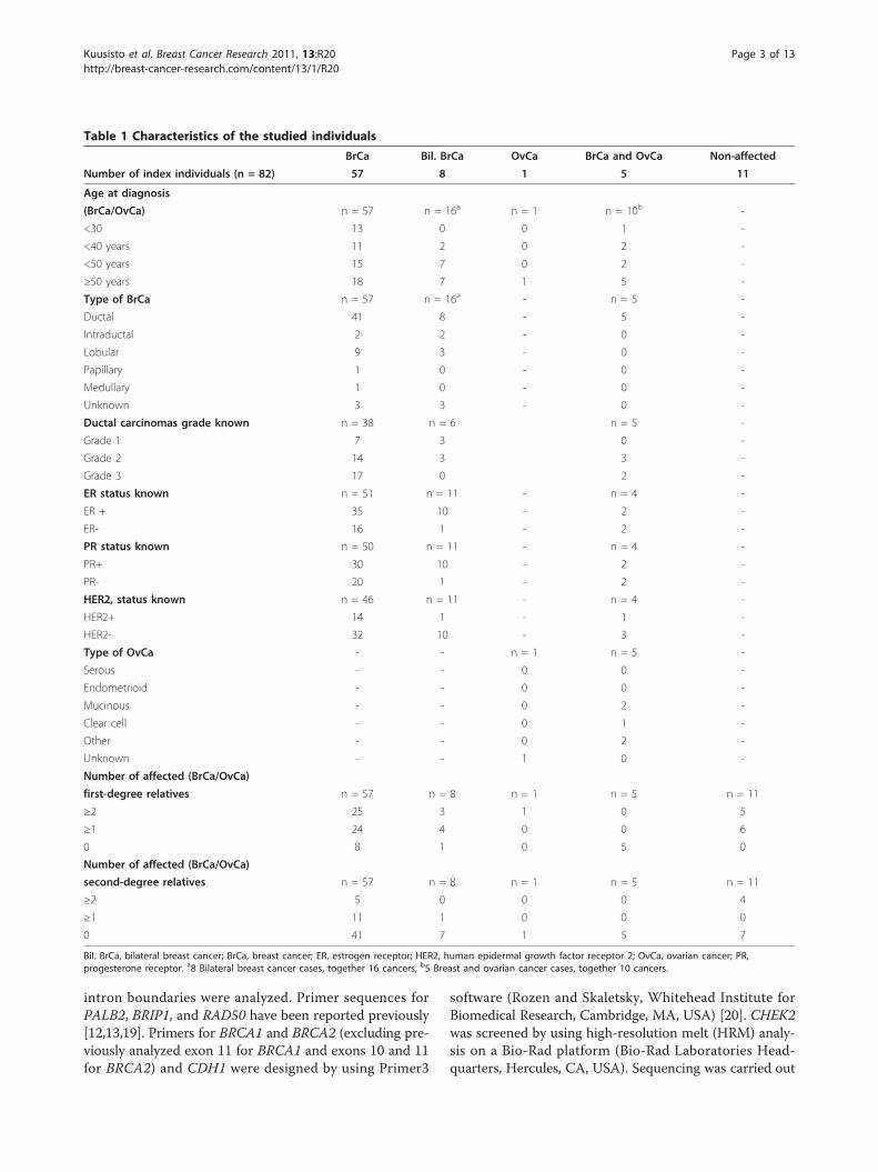
intron boundaries were analyzed. Primer sequences forPALB2, BRIP1, and RAD50 have been reported previously[12,13,19]. Primers for BRCA1 and BRCA2 (excluding pre-viously analyzed exon 11 for BRCA1 and exons 10 and 11for BRCA2) and CDH1 were designed by using Primer3
software (Rozen and Skaletsky, Whitehead Institute forBiomedical Research, Cambridge, MA, USA) [20]. CHEK2was screened by using high-resolution melt (HRM) analy-sis on a Bio-Rad platform (Bio-Rad Laboratories Head-quarters, Hercules, CA, USA). Sequencing was carried out
Table 1 Characteristics of the studied individuals
BrCa Bil. BrCa OvCa BrCa and OvCa Non-affected
Number of index individuals (n = 82) 57 8 1 5 11
Age at diagnosis
(BrCa/OvCa) n = 57 n = 16a n = 1 n = 10b -
<30 13 0 0 1 -
<40 years 11 2 0 2 -
<50 years 15 7 0 2 -
≥50 years 18 7 1 5 -
Type of BrCa n = 57 n = 16a - n = 5 -
Ductal 41 8 - 5 -
Intraductal 2 2 - 0 -
Lobular 9 3 - 0 -
Papillary 1 0 - 0 -
Medullary 1 0 - 0 -
Unknown 3 3 - 0 -
Ductal carcinomas grade known n = 38 n = 6 n = 5 -
Grade 1 7 3 0 -
Grade 2 14 3 3 -
Grade 3 17 0 2 -
ER status known n = 51 n = 11 - n = 4 -
ER + 35 10 - 2 -
ER- 16 1 - 2 -
PR status known n = 50 n = 11 - n = 4 -
PR+ 30 10 - 2 -
PR- 20 1 - 2 -
HER2, status known n = 46 n = 11 - n = 4 -
HER2+ 14 1 - 1 -
HER2- 32 10 - 3 -
Type of OvCa - - n = 1 n = 5 -
Serous - - 0 0 -
Endometrioid - - 0 0 -
Mucinous - - 0 2 -
Clear cell - - 0 1 -
Other - - 0 2 -
Unknown - - 1 0 -
Number of affected (BrCa/OvCa)
first-degree relatives n = 57 n = 8 n = 1 n = 5 n = 11
≥2 25 3 1 0 5
≥1 24 4 0 0 6
0 8 1 0 5 0
Number of affected (BrCa/OvCa)
second-degree relatives n = 57 n = 8 n = 1 n = 5 n = 11
≥2 5 0 0 0 4
≥1 11 1 0 0 0
0 41 7 1 5 7
Bil. BrCa, bilateral breast cancer; BrCa, breast cancer; ER, estrogen receptor; HER2, human epidermal growth factor receptor 2; OvCa, ovarian cancer; PR,progesterone receptor. a8 Bilateral breast cancer cases, together 16 cancers, b5 Breast and ovarian cancer cases, together 10 cancers.
Kuusisto et al. Breast Cancer Research 2011, 13:R20http://breast-cancer-research.com/content/13/1/R20
Page 3 of 13
using the Big Dye Terminator v.3.1 Cycle Sequencing Kitand ABIPRISM 3130 × l Genetic Analyzer (Applied Bio-systems, Foster City, CA, USA). Sequences were analyzedwith Sequencher v.4.7 software (Gene Codes Corporation,Ann Arbor, MI, USA). Primer sequences, detailed HRMand PCR reaction conditions are available upon request.Control frequencies were determined for 18 variants
by HRM (CHEK2 variants), direct sequencing (BRCA1c.4883T > C and RAD50 c.1544A > G) and TaqMan®
SNP genotyping assays (Applied Biosystems, Foster City,CA, USA) and with an ABI7900 instrument (AppliedBiosystems, Foster City, CA, USA). Assays were alreadydesigned and functionally tested for the following SNPs:c.8182G > A (rs28897749), c.9976A > T (rs11571833),c.10234A > G (rs1801426), and c.1676A > G (rs152451).As for the c.72A > T, c.814G > A, c.1000T > G, andc.2993G > A (rs45551636) variants, assays were designedby Custom TaqMan® Assay Design Tool (Applied Bio-systems, Foster City, CA, USA) according to manufac-turer’s instructions.The multiplex ligation-dependent probe amplification
(MLPA) analysis was performed for BRCA1 and BRCA2(SALSA MLPA kit P002-B1 for BRCA1 (lot 0508) andkit P090-A2 for BRCA2 (lot 0808), MRC-Holland,Amsterdam, the Netherlands) according to manufac-turer’s instructions and analyzed with ABIPRISM 3130xlGenetic Analyzer and Genemapper® v.4.0 software(Applied Biosystems, Foster City, CA, USA).
Statistical analysesCarrier frequencies between 82 studied individuals and384 population controls were compared by using Fish-er’s exact test [21]. All P-values were two sided. Oddsratios (OR) were generated by two-by-two table.
In silico prediction of novel missense variants effectsThe effects of five novel-coding missense variants,BRCA2 c.72A > T (Leu24Phe), CHEK2 c.1363G > A(Val455Ile), PALB2 c.814G > A (Glu272Lys), PALB2c.1000T > G (Tyr334Asp), and RAD50 c.1544A > G(Asp515Gly), were predicted with a number of tools byusing Pathogenic-Or-Not-Pipeline (PON-P) [22]. Thepredictions included those for amino acid tolerance(programs PolyPhen version 2, Sift, PhD-SNP, SNAP)and protein stability (I-Mutant version 3). PON-P allowssimultaneous submission of a number of variations andproteins to selected predictors. PON-P utilizes machinelearning to combine results from several individualpredictions.
MicroRNA database and BLAST search for novel variantsMicroRNA (miRNA) target site search was performedfor the novel variant genomic positions from the micro-RNA database (miRBase) [23]. Also BLAST search [24]
was performed for the novel human variant genomicpositions to see if these sites are conserved among dif-ferent organisms including mouse, rat, cow, andchicken.
ResultsIndex individuals of 82 high-risk HBOC families werescreened for germline alterations in BRCA1, BRCA2,CHEK2, PALB2, BRIP1, RAD50, and CDH1 genes.Detailed clinical information of analyzed individuals isshown in Table 1. All of the identified 54 sequence var-iants with their observed genotype frequencies and rs-numbers are presented in Supplementary Table S1 inAdditional file 1. All of the identified non-synonymousand novel sequence alterations are summarized in Table 2.Table 2 variants are presented in Table 3 with index indi-vidual and family cancer history. In addition, as our studymaterial also included healthy index individuals from 11families, we made an effort to get blood samples from twoaffected relatives in 2 out of 11 separate families. Theserelatives with BrCa were screened for the same variant asthat identified in the healthy index. Analysis was per-formed for the new cases in family 112 (CHEK2 c.470T >C and PALB2 c.1676A > G variants) and family 231(BRCA1 c.4883T > C variant; Table 3). In family 112, thecase proved to have the same PALB2 c.1676A > G variantas the index individual but in family 231, the affected rela-tive did not carry the BRCA1 c.4883T > C variant (datanot shown). To further evaluate the impact of these 11healthy index cases, we recalculated the frequencies with-out these 11 individuals for those variants accepted to bemeaningful for BrCa risk. Supplementary Table S2 inAdditional file 2 shows these re-calculated frequencies forBRCA1 c.5095C > T, CHEK2 c.470T > C, and CHEK2c.1100delC variants. No statistically significant effect wasseen for exclusion of the 11 cases.
BRCA1 and BRCA2 mutation analysisAnalysis of BRCA1 and BRCA2 revealed altogether 16 dif-ferent sequence variants, seven in BRCA1 and nine inBRCA2 [see Supplementary Table S1 in Additional file 1].All but two of the identified variants in BRCA1, c.4883T >C and c.5095C > T, have been reported to be neutral inthe databases. Heterozygous c.4883T > C variant wasobserved in 4 of 82 (4.9%) women of which three hadBrCa and one had a family history of breast, cervix andskin cancers (Tables 2 and 3). In population controls, thefrequency of the c.4883T > C variant was 6 of 367 (1.6%).The c.5095C > T variant has been classified as a deleter-ious mutation in the Breast Cancer Information Core(BIC) database. The heterozygous c.5095C > T mutationwas observed in 1 of 82 (1.2%) women. The mutation car-rying woman had BrCa diagnosed at the age of 42 yearsand a strong family history of cancer (Tables 2 and 3,
Kuusisto et al. Breast Cancer Research 2011, 13:R20http://breast-cancer-research.com/content/13/1/R20
Page 4 of 13

Figure 1, Family 249). Additional mutation analysis alsorevealed two other affected women carrying the c.5095C >T mutation in the same family. In BRCA2, three of thenine identified variants were novel, c.68-80insT, c.72A > T,and c.793 + 34T > G (Tables 2 and 3.). The heterozygousmissense variant c.72A > T (Leu24Phe), was observed in 1of 82 (1.2%) women but not in population controls. Thec.72A > T variant carrying woman had BrCa diagnosed atthe age of 53 years. She had also two affected first-degree
relatives (mother and sister). Protein predictions by PON-Psuggested that substitution of leucine by phenylalanine inposition 24 changes significantly the properties of the sidechain and the substitution would not be tolerated. All theother identified variants in BRCA2 have been reported pre-viously and they are either neutral or the clinical signifi-cance of the variants is yet uncertain especially with thethree missense variants, c.8182G > A, c.9976A > T andc.10234A > G (Tables 2 and 3). No deletions or duplication
Table 2 Identified non-synonymous and novel sequence alterations
Carrier frequency
Gene/Nucleotide changea Effect on protein rs Numberb Individuals Controls P-values OR; 95%CI Status
BRCA1
4837A > G Ser1613Gly rs1799966 0.634 (52/82)g na - - Reportedc, d
4883T > C Met1628Thr rs4986854 0.049 (4/82) 0.016 (6/367) 0.090 3.09; 0.85-11.19 Reportedc, d
5095C > T Arg1699Trp rs55770810 0.012 (1/82) na - - Reportedc, d
BRCA2
68-80insTf - - 0.012 (1/82) na - - Novel
72A > T Leu24Phe - 0.012 (1/82) 0 (0/380) 0.177 na Novel
793 + 34T > G - - 0.012 (1/82) na - - Novel
8182G > A Val2728Ile rs28897749 0.012 (1/82) 0.003 (1/378) 0.325 4.65; 0.29-75.19 Reportedc, d
9976A > T Lys3326Stop rs11571833 0.012 (1/82) 0.029 (11/378) 0.702 0.41; 0.05-3.24 Reportedc, d
10234A > G Ile3412Val rs1801426 0.012 (1/82) 0.021 (8/379) 1.000 0.57; 0.07-4.64 Reportedc, d
CHEK2
444 + 85T > A - - 0.012 (1/82) 0.005 (2/364) 0.457 2.23; 0.20-24.94 Novel
470T > C Ile157Thr - 0.098 (8/81) 0.055 (21/381) 0.203 1.88; 0.80-4.41 Reportede
792 + 39C > T - - 0.012 (1/82) 0.021 (8/375) 1.000 0.57; 0.07-4.60 Novel
1100delCf Fs, stop at codon 381 - 0.037 (3/82) 0.016 (6/380) 0.203 2.37; 0.58-9.67 Reportede
1290T > C His430His - 0.951 (77/81)g 0.974 (372/382) 0.281 0.52; 0.16-1.69 Novel
1314T > C Asp438Asp - 0.951 (77/81)g 0.974 (372/382) 0.281 0.52; 0.16-1.69 Novel
1363G > A Val455Ile - 0.975 (79/81)g 0.976 (373/382) 1.000 0.95; 0.20-4.50 Novel
PALB2
814G > A Glu272Lys - 0.012 (1/82) 0 (0/372) 0.181 na Novel
1000T > G Tyr334Asp - 0.012 (1/82) 0.011 (4/380) 1.000 1.16; 0.13-10.52 Novel
1010T > C Leu337Ser rs45494092 0.073 (6/82) na - - Reportedc
1676A > G Gln559Arg rs152451 0.122 (10/82) 0.173 (64/371) 0.323 0.67; 0.33-1.36 Reportedc
2205A > G Pro735Pro - 0.012 (1/82) na - - Novel
2794G > A Val932Met rs45624036 0.037 (3/82) na - - Reportedc
2993G > A Gly998Glu rs45551636 0.012 (1/82) 0.038 (14/372) 0.491 0.32; 0.04-2.44 Reportedc
BRIP1
584T > C Leu195Pro rs4988347 0.024 (2/82) na - - Reportedc
2755C > T Pro919Ser rs4986764 0.390 (32/82)g na - - Reportedc
RAD50
1544A > G Asp515Gly - 0.012 (1/82) 0.010 (4/384) 1.000 1.17; 0.13-10.63 Novel
2398-32A > G - - 0.012 (1/82) na - - Novel
3475 + 33C > G - - 0.012 (1/82) na - - Novel
CI, confidence interval; Fs, frameshift; na, not analyzed; OR, odds ratio. aThe reference nucleotide sequencies were obtained from the UCSC Genome Browser [44]and the accession numbers were following: BRCA1: [UCSC Genome Browser:NM_007295.2], BRCA2: [UCSC Genome Browser:NM_000059.3], CHEK2: [UCSC GenomeBrowser:NM_007194.3], PALB2: [UCSC Genome Browser:NM_024675.3], BRIP1: [UCSC Genome Browser:NM_032043.1], RAD50: [UCSC Genome Browser:NM_005732.3], and CDH1: [UCSC Genome Browser:NM_004360.3]. The accession numbers for the protein sequencies obtained from the Swiss-Prot Proteinknowledgebase [45] were following: BRCA1: [Swiss-Prot:P38398], BRCA2: [Swiss-Prot:P51587], CHEK2: [Swiss-Prot:O96017], PALB2: [Swiss-Prot:Q86YC2], BRIP1: [Swiss-Prot:Q9BX63], RAD50: [Swiss-Prot:Q92878], and CDH1: [Swiss-Prot:P12830]. bThe RefSNP number, obtained from the NCBI Single Nucleotide Polymorphismdatabase (dbSNP) [46]. cThe NCBI dbSNP [46]. dThe Breast Cancer Information Core database [47]. eReported in the Finnish population by Vahteristo et al. [11].fHeterozygous deletion or insertion.gDue to the high frequency of the variant observed in analyzed individuals, variant is not presented in Table 3.
Kuusisto et al. Breast Cancer Research 2011, 13:R20http://breast-cancer-research.com/content/13/1/R20
Page 5 of 13

Table 3 Identified variants in the studied individuals
Family id Gene and variant Type of cancer BrCa/OvCa Histology,Grade Receptor status Other cancer cases in the family(Age at diagnosis if available)
202 BRCA1, 4883T > C Br (26) Ductal, 3 ER-, PR-, HER2- Skin (54)
206 BRCA1, 4883T > C Br (53) Ductal, 1 ER-, PR-, HER2- Bil. Ov (64), Br (49)
231 BRCA1, 4883T > C - Br x2 (33, 46), Cer (60), Skin (73)
232 BRCA1, 4883T > C Br (34) Ductal, na ER +, PR +, HER2 na Br (39)
249 (Figure 1) BRCA1, 5095C > T Br (42) Medullary, na na Br x5 (35, 44, 57, 67, 71, ),
Co (78), Kid (67), Mel (63), Ov (45),
Skin, To (51), Ute (39)
115 BRCA2, 68-80insT Br (59) Lobular, 2 ER+, PR+, HER2- Br x3 (<50), Br x2 (60, 60)
240 BRCA2, 72A > T Br (53) Ductal, 3 ER+, PR-, HER2+ Br x2 (42, 62)
207 BRCA2, 793 + 34T > G Br (38) na na Bil. Br (64)
5 (Figure 8) BRCA2, 8182G > A - Bil. Br x2 (43, 48 and 54, 76),
BRCA2, 10234A > G Br (43), Brain (75), Lip (45), Lung (81),Skin (75), Sto (56)
4 BRCA2, 9976A > T - Bil. Br x2 (53, 69 and <70),
CHEK2, 470T > C Br (<70)
212 CHEK2, 444 + 85T > A Bil. Br (43) Ductal, 2 and na ER+, PR+, HER2- and Br (52)
PALB2, 2794G > A na
110 (Figure 3) CHEK2, 792 + 39C > T Br (26) Ductal, 2 ER+, PR+, HER2+ Br (48), Ca (84), Pr (64)
CHEK2, 470T > C
CHEK2, 1100delC
RAD50, 2398-32A > G
112 CHEK2, 470T > C - Br x3 (35, 43, 83), Skin (76),
PALB2, 1676A > G Lung (71)
120 CHEK2, 470T > C - Br (64), Ov (72)
122 CHEK2, 470T > C Br (25) Ductal, 2 ER-, PR-, HER2+ Brain (66, Ca (83),Cer (31),
Pr (93), Re (73), Skin (87)
126 CHEK2, 470T > C Br (48) Ductal, na ER, PR, HER2 na Bil. Br, Br x2 (51, <53)
129 (Figure 2) CHEK2, 470T > C Skin (70), Lobular, 2 and ER-, PR-, HER2- and Bil. Br (59), Co (58), Skin (48)
PALB2, 1676A > G Bil. Br (78), Ductal, 1 ER+, PR+, HER2-
Sto (82)
262 (Figure 6) CHEK2, 470T > C Bil. Br Intraductal, na and ER+, PR+, HER2+ and Br (57), Panc (83), Si (79)
PALB2, 1000T > G (45, 58) Ductal, na ER+, PR+, HER2-
264 (Figure 4) CHEK2, 1100delC Bil. Br (44) Lobular, 2 ER+, PR+, HER2- Br x2 (44, 52)
265 (Figure 5) CHEK2, 1100delC Br (45) Ductal, 3 ER+, PR+, HER2+ Br (38)
PALB2, 1676A > G
237 PALB2, 814G > A Br (28) Ductal, 2 ER+, PR+, HER2+ -
133 PALB2, 1010T > C Br (48) Ductal, 2 ER+, PR+, HER2- Int, Br x2 (73, 79), Skin (60)
235 PALB2, 1010T > C Br (52) na na Bil. Br (28), Br (56)
239 PALB2, 1010T > C Br (37) Ductal, 2 ER+, PR+, HER2- Br ( > 90)
250 PALB2, 1010T > C Br (24) Ductal, 3 ER+, PR+, HER2+ Cer (30), Ov (83)
260 PALB2, 1010T > C Br (29) Ductal, 3 ER-, PR-, HER2- Br (58), Lung (60)
267 PALB2, 1010T > C Br (48) Ductal, 1 ER+, PR+, HER2 na Br x2 (51, 58)
113 PALB2, 1676A > Ga Br (51), Ductal, 3 ER-, PR-, HER2+ Br (35)
Skin (55)
131 (Figure 7) PALB2, 1676A > G Bil. Br (54) Intraductal, na and na Bil. Br (46), Br (48)
BRIP1, 584T > C Ductal, 2
229 PALB2, 1676A > G Bil. Br (68) na na Bil. Br (50, 70), Br x2 (45, 50)
236 PALB2, 1676A > G Br (29) Intraductal, na na Br (52)
246 PALB2, 1676A > G Thy (30), Ductal, 3 ER-, PR-, HER2+ Br x2 (49, 54), Re (61)
Cer (33),
Br (39)
Kuusisto et al. Breast Cancer Research 2011, 13:R20http://breast-cancer-research.com/content/13/1/R20
Page 6 of 13

were identified either in BRCA1 or BRCA2 by multiplexligation-dependent probe amplification (MLPA).
CHEK2 mutation analysisIn CHEK2, two previously reported BrCa-associated var-iants in the Finnish population, c.470T > C andc.1100delC, were identified in 10 of 82 (12.1%) individuals(Tables 2 and 3). The heterozygous c.470T > C variantwas observed in eight women of which three were healthy.Two of the c.470T > C variant carriers had bilateral BrCaand they carried also PALB2 missense variants (an exam-ple of the family pedigree of the index individual carryingthe both variants is presented in Figure 2, Family 129).
The heterozygous c.1100delC variant was detected inthree women (Tables 2 and 3). One woman carryingc.1100delC with an early-onset disease of 26 years of agealso carried the c.470T > C and the novel c.792 + 39C > TCHEK2 variants as well as the RAD50, c.2398-32A > Gvariant (Figure 3, Family 110). A second patient with thec.1100delC variant had bilateral BrCa at the age of 44years and two other affected individuals in her family(mother and father’s sister; Figure 4, Family 264). A thirdpatient with the c.1100delC variant had BrCa diagnosed atthe age of 45 years and one affected individual (mother) inher family. This woman carried also the PALB2 c.1676A >G variant (Figure 5, Family 265). In addition to c.470T > C
Table 3 Identified variants in the studied individuals (Continued)
268 PALB2, 1676A > G Br (62) Papillary, na ER+, PR+, HER2- Br x2 (36, 38)
PALB2, 2205A > G
271 PALB2, 1676A > G Thy (62), Lobular, 2 ER+, PR+, HER2- Br x2 (43, 44)
Br (65)
102 PALB2, 2794G > A Br (29) Lobular, na ER-, PR-, HER2+ Br (72)
BRIP1, 584T > C
244 PALB2, 2794G > A Br (45) Ductal, 2 ER+, PR+, HER2- Bil. Br (<45), Br x2 (<35, 46),
Brain (67)
270 PALB2, 2993G > A Br (66) Ductal, 3 ER+, PR+, HER2- Br x2 (48, <66)
257 RAD50, 1544A > G Br (39) Lobular, 2 ER+, PR+, HER2- Br (69)
225 RAD50, 3475+33C > G Br (43) Ductal, 1 ER+, PR+, HER2- Br x2 (52, 77), Kid (64)
ER, estrogen receptor; PR, progesterone receptor; HER2, human epidermal growth factor receptor 2; na, not available; Bil, Bilateral; Br, breast; Ca, cancer withunknown primary site; Cer, cervix; Co, colon; Int, intestines; Kid, kidney; Mel, melanoma; Ov, ovary; Panc, pancreas; Pr, prostate; Re, rectum; Si, Sigma; Sto,stomach; Thy, thyroid; To, tongue; Ute, uterus. aHomozygous variant. Cancers diagnosed in the paternal side of the family are presented in italics. Cancers diagnosedin siblings or their children of the index patients are underlined. Cancers diagnosed in the children of the index patients are presented in bold.
Br 42
Br 44
Br 71Co 78
Skin Mel 63 Br 57 Ute 39Br 67Kid 67
To 51Br 35Ov45 BRCA1, c.5095C>T
BRCA1, c.5095C>T
72BRCA1, c.5095C>T
Family 249
Figure 1 Family 249 pedigree. Family pedigree of the index individual with the identified BRCA1 c.5095C > T variant (same variant was alsoidentified in the daughter of the index individual and in the daughter of the index individual’s paternal uncle). Individuals with breast or ovariancancer with age at diagnosis are marked with black circles. Other cancers are marked in grey and accompanied by age at diagnosis, if known.Index individual is marked with an arrow. Deceased individuals are indicated with a slash. Current ages of healthy females are marked if known.Br, breast cancer; Co, colon; Kid, kidney; Mel, melanoma; Ov, ovarian cancer; To, tongue; Ute, uterus.
Kuusisto et al. Breast Cancer Research 2011, 13:R20http://breast-cancer-research.com/content/13/1/R20
Page 7 of 13

and c.1100delC, five novel variants (Table 2) and one com-mon polymorphism [see Supplementary Table S1 in Addi-tional file 1] were identified in CHEK2. The novel non-synonymous variant, c.1363G > A (Val455Ile), is based onthe computational predictions, and is likely benign.
PALB2 mutation analysisIn PALB2, altogether nine different variants, includingthree novel ones, were identified [see SupplementaryTable S1 in Additional file 1]. Only one of the identifiedvariants reported previously, c.2586 + 58C > T, has been
associated with a 36% increase of BrCa risk (odds ratio(OR): 1.36; 95% confidence intervals (CIs), 1.13-1.64; P =0.001) in a Chinese population [25]. We identified thec.2586 + 58C > T variant in 36 of 82 (43.9%) women. Anovel heterozygous c.814G > A variant was identified in
Skin 48Co 58
Skin 70Bil. Br 78Sto 82
Bil. Br 59, 64
CHEK2, c.470T>CPALB2, c.1676A>G
Family 129
Figure 2 Family 129 pedigree. Family pedigree of the indexindividual with the identified CHEK2 c.470T > C and PALB2 c.1676A> G variants. Individuals with breast cancer with age at diagnosisare marked with black circles. Other cancers are marked in grey andaccompanied by age at diagnosis, if known. Index individual ismarked with an arrow. Deceased individuals are indicated with aslash. Bil. Br, bilateral breast cancer; Co, colon; Sto, stomach.
Br 26
Ca84
Pr 64
Br 26
Ca84
Pr 64
Br 48
CHEK2, c.470T>CCHEK2, c.792+39C>TCHEK2, c.1100delCRAD50, c.2398-32A>G
37
595353
Family 110
Figure 3 Family 110 pedigree. Family pedigree of the indexindividual with the identified CHEK2 c.470T > C, c.792 + 39C > T,c.1100delC, and RAD50 c. 2398-32A > G variants. Individuals withbreast cancer with age at diagnosis are marked with black circles.Other cancers are marked in grey and accompanied by age atdiagnosis, if known. Index individual is marked with an arrow.Deceased individuals are indicated with a slash. Current ages ofhealthy females are marked if known. Br, breast cancer; Ca, cancerwith unknown primary site; Pr, prostate.
Bil. Br 44
Br 44Br 52
CHEK2,c.1100delC
17
Bil. Br 44
Br 44Br 52
CHEK2,c.1100delC
17
52
Family 264
Figure 4 Family 264 pedigree. Family pedigree of the indexindividual with the identified CHEK2 c.1100delC variant. Individualswith breast cancer with age at diagnosis are marked with blackcircles. Index individual is marked with an arrow. Deceasedindividuals are indicated with a slash. Current ages of healthy femalesare marked if known. Bil. Br, bilateral breast cancer; Br, breast cancer.
Br 45
Br 38
CHEK2, c.1100delCPALB2, c.1676A>G
22
Family 265
Figure 5 Family 265 pedigree . Family pedigree of the indexindividual with the identified CHEK2 c.1100delC and PALB2 c.1676A >G variants. Individuals with breast cancer with age at diagnosis aremarked with black circles. Index individual is marked with an arrow.Current ages of healthy females are marked if known. Br, breast cancer.
Kuusisto et al. Breast Cancer Research 2011, 13:R20http://breast-cancer-research.com/content/13/1/R20
Page 8 of 13
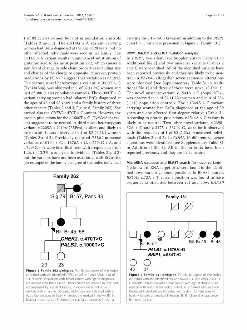
1 of 82 (1.2%) women but not in population controls(Tables 2 and 3). The c.814G > A variant carryingwoman had BrCa diagnosed at the age of 28 years, but noother affected individuals were seen in her family. Thec.814G > A variant results in amino acid substitution ofglutamic acid to lysine at position 272, which causes asignificant change to side chain properties including sizeand change of the charge to opposite. However, proteinpredictions by PON-P suggest that variation is neutral.The second novel heterozygous variant, c.1000T > G(Tyr334Asp), was observed in 1 of 82 (1.2%) women andin 4 of 380 (1.1%) population controls. The c.1000T > Gvariant carrying woman had bilateral BrCa diagnosed atthe ages of 45 and 58 years and a family history of threeother cancers (Tables 2 and 3, Figure 6, Family 262). Shecarried also the CHEK2 c.470T > C variant. However, theprotein predictions for the c.1000T > G (Tyr334Asp) var-iant suggest it to be neutral. A third novel heterozygousvariant, c.2205A > G (Pro735Pro), is silent and likely tobe neutral. It was observed in 1 of 82 (1.2%) women(Tables 2 and 3). Previously reported PALB2 missensevariants, c.1010T > C, c.1676A > G, c.2794G > A, andc.2993G > A were identified here with frequencies from1.2% to 12.2% in analyzed individuals (Tables 2 and 3)but the variants have not been associated with BrCa risk(an example of the family pedigree of the index individual
carrying the c.1676A > G variant in addition to the BRIP1c.584T > C variant is presented in Figure 7, Family 131).
BRIP1, RAD50, and CDH1 mutation analysisIn BRIP1, two silent [see Supplementary Table S1 inAdditional file 1] and two missense variants (Tables 2and 3) were identified. All of the identified variants havebeen reported previously and they are likely to be neu-tral. In RAD50, altogether seven sequence alterationswere observed [see Supplementary Table S1 in Addi-tional file 1] and three of these were novel (Table 2).The novel missense variant, c.1544A > G (Asp515Gly),was observed in 1 of 82 (1.2%) women and in 4 of 384(1.1%) population controls. The c.1544A > G variantcarrying woman had BrCa diagnosed at the age of 39years and one affected first-degree relative (Table 3).According to protein predictions, c.1544A > G variant islikely to be neutral. Two other novel variants, c.2398-32A > G and c.3475 + 33C > G, were both observedwith the frequency of 1 of 82 (1.2%) in analyzed indivi-duals (Tables 2 and 3). In CDH1, 10 different sequencealterations were identified [see Supplementary Table S1in Additional file 1]. All of the variants have beenreported previously and they are likely neutral.
MicroRNA database and BLAST search for novel variantsNo known miRNA target sites were found in the identi-fied novel variant genomic positions. In BLAST search,BRCA2 c.72A > T variant position was found to havesequence similarities between rat and cow. RAD50
Bil. Br 45, 58
Br 57, Panc 83Sig 79
29 23
CHEK2, c.470T>CPALB2, c.1000T>G
Bil. Br 45, 58
Br 57, Sig 79
29 23
CHEK2, c.4PALB2, c.1
Family 262
Figure 6 Family 262 pedigree. Family pedigree of the indexindividual with the identified CHEK2 c.470T > C and PALB2 c.1000T> G variants. Individuals with breast cancer with age at diagnosisare marked with black circles. Other cancers are marked in grey andaccompanied by age at diagnosis, if known. Index individual ismarked with an arrow. Deceased individuals are indicated with aslash. Current ages of healthy females are marked if known. Bil. Br,bilateral breast cancer; Br, breast cancer, Panc, pancreas; Si, sigma.
Bil. Br 54 Bil. Br 46 Br 4885
43 37
PALB2, c.1676A>GBRIP1, c.584T>C
Family 131
Figure 7 Family 131 pedigree. Family pedigree of the indexindividual with the identified PALB2 c.1676A > G and BRIP1 c.584T >C variants. Individuals with breast cancer with age at diagnosis aremarked with black circles. Index individual is marked with an arrow.Deceased individuals are indicated with a slash. Current ages ofhealthy females are marked if known. Bil. Br, bilateral breast cancer;Br, breast cancer.
Kuusisto et al. Breast Cancer Research 2011, 13:R20http://breast-cancer-research.com/content/13/1/R20
Page 9 of 13
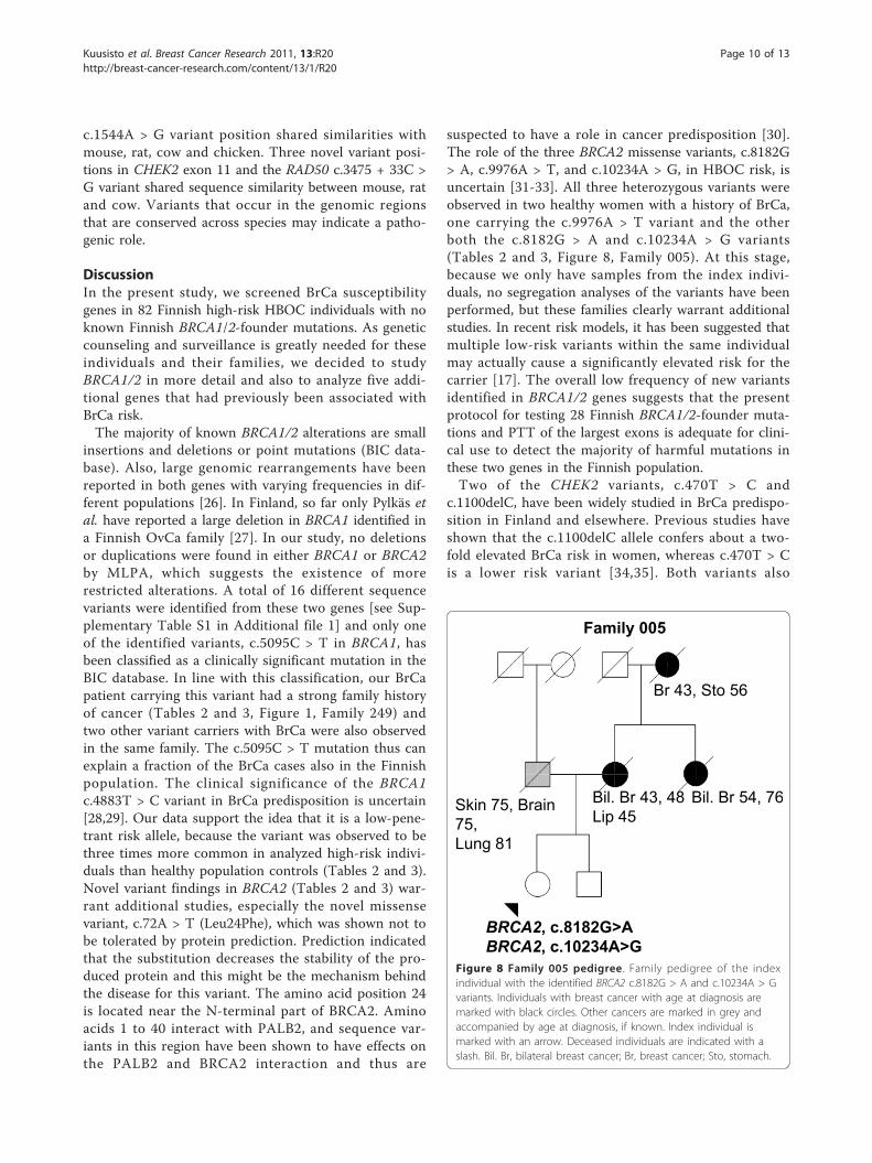
c.1544A > G variant position shared similarities withmouse, rat, cow and chicken. Three novel variant posi-tions in CHEK2 exon 11 and the RAD50 c.3475 + 33C >G variant shared sequence similarity between mouse, ratand cow. Variants that occur in the genomic regionsthat are conserved across species may indicate a patho-genic role.
DiscussionIn the present study, we screened BrCa susceptibilitygenes in 82 Finnish high-risk HBOC individuals with noknown Finnish BRCA1/2-founder mutations. As geneticcounseling and surveillance is greatly needed for theseindividuals and their families, we decided to studyBRCA1/2 in more detail and also to analyze five addi-tional genes that had previously been associated withBrCa risk.The majority of known BRCA1/2 alterations are small
insertions and deletions or point mutations (BIC data-base). Also, large genomic rearrangements have beenreported in both genes with varying frequencies in dif-ferent populations [26]. In Finland, so far only Pylkäs etal. have reported a large deletion in BRCA1 identified ina Finnish OvCa family [27]. In our study, no deletionsor duplications were found in either BRCA1 or BRCA2by MLPA, which suggests the existence of morerestricted alterations. A total of 16 different sequencevariants were identified from these two genes [see Sup-plementary Table S1 in Additional file 1] and only oneof the identified variants, c.5095C > T in BRCA1, hasbeen classified as a clinically significant mutation in theBIC database. In line with this classification, our BrCapatient carrying this variant had a strong family historyof cancer (Tables 2 and 3, Figure 1, Family 249) andtwo other variant carriers with BrCa were also observedin the same family. The c.5095C > T mutation thus canexplain a fraction of the BrCa cases also in the Finnishpopulation. The clinical significance of the BRCA1c.4883T > C variant in BrCa predisposition is uncertain[28,29]. Our data support the idea that it is a low-pene-trant risk allele, because the variant was observed to bethree times more common in analyzed high-risk indivi-duals than healthy population controls (Tables 2 and 3).Novel variant findings in BRCA2 (Tables 2 and 3) war-rant additional studies, especially the novel missensevariant, c.72A > T (Leu24Phe), which was shown not tobe tolerated by protein prediction. Prediction indicatedthat the substitution decreases the stability of the pro-duced protein and this might be the mechanism behindthe disease for this variant. The amino acid position 24is located near the N-terminal part of BRCA2. Aminoacids 1 to 40 interact with PALB2, and sequence var-iants in this region have been shown to have effects onthe PALB2 and BRCA2 interaction and thus are
suspected to have a role in cancer predisposition [30].The role of the three BRCA2 missense variants, c.8182G> A, c.9976A > T, and c.10234A > G, in HBOC risk, isuncertain [31-33]. All three heterozygous variants wereobserved in two healthy women with a history of BrCa,one carrying the c.9976A > T variant and the otherboth the c.8182G > A and c.10234A > G variants(Tables 2 and 3, Figure 8, Family 005). At this stage,because we only have samples from the index indivi-duals, no segregation analyses of the variants have beenperformed, but these families clearly warrant additionalstudies. In recent risk models, it has been suggested thatmultiple low-risk variants within the same individualmay actually cause a significantly elevated risk for thecarrier [17]. The overall low frequency of new variantsidentified in BRCA1/2 genes suggests that the presentprotocol for testing 28 Finnish BRCA1/2-founder muta-tions and PTT of the largest exons is adequate for clini-cal use to detect the majority of harmful mutations inthese two genes in the Finnish population.Two of the CHEK2 variants, c.470T > C and
c.1100delC, have been widely studied in BrCa predispo-sition in Finland and elsewhere. Previous studies haveshown that the c.1100delC allele confers about a two-fold elevated BrCa risk in women, whereas c.470T > Cis a lower risk variant [34,35]. Both variants also
Br 43, Sto 56
Bil. Br 43, 48Lip 45
Bil. Br 54, 76Skin 75, Brain 75,Lung 81
BRCA2, c.8182G>ABRCA2, c.10234A>G
Family 005
Figure 8 Family 005 pedigree. Family pedigree of the indexindividual with the identified BRCA2 c.8182G > A and c.10234A > Gvariants. Individuals with breast cancer with age at diagnosis aremarked with black circles. Other cancers are marked in grey andaccompanied by age at diagnosis, if known. Index individual ismarked with an arrow. Deceased individuals are indicated with aslash. Bil. Br, bilateral breast cancer; Br, breast cancer; Sto, stomach.
Kuusisto et al. Breast Cancer Research 2011, 13:R20http://breast-cancer-research.com/content/13/1/R20
Page 10 of 13

associate with other cancers in the Finnish population[36-38]. In our study, two of the CHEK2 variants,c.470T > C and c.1100delC, were identified in 10 out of82 analyzed individuals (12.2%) suggesting that the con-tribution of the two CHEK2 variants to BrCa risk isremarkable in the high-risk Finnish BRCA1/2-foundermutation-negative individuals. However, clinical screen-ing of the CHEK2 variants has not yet been justified dueto unclear clinical consequences related to incompletesegregation of the variants with BrCa in the high-riskBrCa families [39,40]. Based on the findings of thisstudy, we agree that interpretation of the CHEK2 muta-tion analysis results is very difficult, because many othergene variants were also identified in individuals witheither c.470T > C or c.1100delC variants and some ofthe variant carriers had not (yet) been diagnosed withBrCa. Thus profound segregation analysis of the c.470T> C and c.1100delC variants for BRCA1/2-foundermutation-negative families would be needed to furtherstudy clinical significance of these variants. Also thenovel variants identified in CHEK2 should be furtheranalyzed.PALB2 has been associated with BrCa predisposition
in Finland by Erkko et al. [12] and the c.1592delT var-iant was classified as a Finnish founder mutation. In thisstudy the founder deletion was not found, which isprobably explained by the limited number of analyzedhigh-risk HBOC individuals. We identified two novelPALB2 missense variants, c.814G > A (Glu272Lys) andc.1000T > G (Tyr334Asp), in affected individuals (Tables2 and 3). Protein predictions suggested a non-patho-genic role of these substitutions but further studies areneeded to confirm these findings. None of the four pre-viously reported PALB2 missense variants, c.1010T > C,c.1676A > G, c.2794G > A, and c.2993G > A, have beenassociated with BrCa risk [12,41]. Interestingly, thesevariants were identified also together with other variantsin analyzed individuals (Tables 2 and 3). One of theidentified intronic variants, c.2586 + 58C > T, has beenassociated with an increase of BrCa risk in a Chinesepopulation [25] but there is no evidence of that in theFinnish population.BRIP1 and RAD50 genes have been shown to have rare
BrCa associated variants in familial BrCa patients [14,42].Here, BRIP1 mutation analysis revealed only previouslyreported likely neutral variants. Whereas analysis ofRAD50 identified three novel sequence alterations,including one missense variant, c.1544A > G (Asp515Gly)(Tables 2 and 3). To further study the role of these novelvariants, additional analyses are needed. Germline muta-tions in CDH1 have been previously found to associatewith hereditary diffuse gastric cancer syndrome, butmutations have been also identified in familial invasivelobular BrCa patients without hereditary diffuse gastric
cancer [15,43]. Here, only neutral variants were identi-fied, and all of them have been reported earlier [see Sup-plementary Table S1 in Additional file 1]. No clearresults were found that any of the identified genetic var-iants in BRIP1, RAD50, or CDH1 would increase theBrCa/OvCa risk in the analyzed high-risk Finnish HBOCindividuals.
ConclusionsIn this study, 13.4% of the analyzed, high-risk BRCA1/2-founder mutation-negative HBOC cases can beexplained by previously reported mutations in BrCa sus-ceptibility genes. CHEK2 mutations, c.470T > C andc.1100delC, make a considerable contribution (12.2%) tothese high-risk individuals but further segregation analy-sis are needed to evaluate the clinical significance ofthese mutations before applying them in clinical use.Novel variant findings warrant additional studies withspecial interest in the novel missense variant, BRCA2c.72A > T (Leu24Phe), which was predicted to bearuntolerated mutations and to destabilize the protein.The complex nature of HBOC addresses the need forgenome-wide approaches to further study these indivi-duals and to create new tools for genetic counseling.This study also confirmed that our current genetic test-ing protocol for the 28 Finnish BRCA1/2-founder muta-tions and PTT of the largest exons is sensitive enoughfor clinical use in the majority of Finnish HBC/HBOCindividuals.
Additional material
Additional file 1: Supplementary Table S1. All of the identified 54sequence alterations. Supplementary Table S1 include detailedinformation about all of the identified sequence alterations.
Additional file 2: Supplementary Table S2. Identified breast cancerassociated variants in affected 71 individuals. Supplementary TableS2 includes re-calculated frequencies for BRCA1 c.5095C > T, CHEK2c.470T > C, and CHEK2 c.1100delC variants in affected 71 indexindividuals (11 unaffected index individuals excluded).
AbbreviationsBRCA1: breast cancer 1 gene; BRCA2: breast cancer 2 gene; BrCa: breastcancer; BRIP1: BRCA1-interacting protein 1 gene; CDH1: cadherin-1 gene;CHEK2: checkpoint kinase 2 gene; CI: confidence interval; FGFR2: fibroblastgrowth factor receptor 2 gene; GWAs: genome-wide association studies;HBC: hereditary breast cancer; HBOC: high-risk hereditary breast and/orovarian cancer; HRM: high resolution melt; miRNA: microRNA; MLPA:multiplex ligation-dependent probe amplification; OR: odds ratio; OvCa:ovarian cancer; PALB2: Partner and localizer of BRCA2; PCR: polymerase chainreaction; PTT: protein truncation test; RAD50: human homolog of S.cerevisiae RAD50 gene; SNP: single nucleotide polymorphism.
AcknowledgementsWe thank all the patients for their participation into this study. We alsothank personnel of the Tampere University Hospital Genetics OutpatientClinic and JS’s research group for all the help. Special thanks are given toMs. Linda Enroth for skillful technical assistance. Also Ms. Ekaterina Slitikova
Kuusisto et al. Breast Cancer Research 2011, 13:R20http://breast-cancer-research.com/content/13/1/R20
Page 11 of 13
and Ms. Aune Aho are greatly acknowledged for their contribution. Funding:This work was supported by the Competitive Research Funding of theTampere University Hospital (9K119, ML16), Biocentre Finland and SigridJuselius Foundation. The Finnish Cultural Foundation and the Finnish BreastCancer Group have financially supported author KMK.
Author details1Institute of Biomedical Technology, University of Tampere, Biokatu 8,Tampere, 33520, Finland. 2Centre for Laboratory Medicine, TampereUniversity Hospital, Biokatu 4, Tampere, 33520, Finland. 3Department ofPediatrics, Genetics Outpatient Clinic, Tampere University Hospital, Biokatu 8,Tampere, 33520, Finland.
Authors’ contributionsKMK participated patient collection, carried out the sequencing, MLPA andstatistical analysis and drafted the manuscript. AB carried out and interpretedthe HRM analysis of the CHEK2 gene and helped to draft the methodssection of the manuscript. MV performed and interpreted protein predictionanalysis in silico and helped to draft the manuscript. JS and S-L Sparticipated in the study design and coordination, and helped to draft themanuscript. S-L S also participated in patient collection and was responsiblefor genetic counseling of patients. All authors read and approved the finalmanuscript.
Competing interestsThe authors declare that they have no competing interests.
Received: 9 August 2010 Revised: 14 December 2010Accepted: 28 February 2011 Published: 28 February 2011
References1. Claus EB, Schildkraut JM, Thompson WD, Risch NJ: The genetic attributable
risk of breast and ovarian cancer. Cancer 1996, 77(11):2318-2324.2. Miki Y, Swensen J, Shattuck-Eidens D, Futreal PA, Harshman K, Tavtigian S,
Liu Q, Cochran C, Bennett LM, Ding W: A strong candidate for the breastand ovarian cancer susceptibility gene BRCA1. Science 1994,266(5182):66-71.
3. Tavtigian SV, Simard J, Rommens J, Couch F, Shattuck-Eidens D,Neuhausen S, Merajver S, Thorlacius S, Offit K, Stoppa-Lyonnet D,Belanger C, Bell R, Berry S, Bogden R, Chen Q, Davis T, Dumont M, Frye C,Hattier T, Jammulapati S, Janecki T, Jiang P, Kehrer R, Leblanc JF, Mitchell JT,McArthur-Morrison J, Nguyen K, Peng Y, Samson C, Schroeder M,Snyder SC, et al: The complete BRCA2 gene and mutations inchromosome 13q-linked kindreds. Nat Genet 1996, 12:333-337.
4. Vehmanen P, Friedman LS, Eerola H, McClure M, Ward B, Sarantaus L,Kainu T, Syrjakoski K, Pyrhonen S, Kallioniemi OP, Muhonen T, Luce M,Frank TS, Nevanlinna H: Low proportion of BRCA1 and BRCA2 mutationsin Finnish breast cancer families: evidence for additional susceptibilitygenes. Hum Mol Genet 1997, 6:2309-2315.
5. Syrjakoski K, Vahteristo P, Eerola H, Tamminen A, Kivinummi K, Sarantaus L,Holli K, Blomqvist C, Kallioniemi OP, Kainu T, Nevanlinna H: Population-based study of BRCA1 and BRCA2 mutations in 1035 unselected Finnishbreast cancer patients. J Natl Cancer Inst 2000, 92:1529-1531.
6. Li FP, Fraumeni JF Jr: Rhabdomyosarcoma in children: epidemiologicstudy and identification of a familial cancer syndrome. J Natl Cancer Inst1969, 43:1365-1373.
7. Starink TM, van der Veen JP, Arwert F, de Waal LP, de Lange GG, Gille JJ,Eriksson AW: The Cowden syndrome: a clinical and genetic study in 21patients. Clin Genet 1986, 29:222-233.
8. Giardiello FM, Brensinger JD, Tersmette AC, Goodman SN, Petersen GM,Booker SV, Cruz-Correa M, Offerhaus JA: Very high risk of cancer in familialPeutz-Jeghers syndrome. Gastroenterology 2000, 119:1447-1453.
9. Keller G, Vogelsang H, Becker I, Hutter J, Ott K, Candidus S, Grundei T,Becker KF, Mueller J, Siewert JR, Hofler H: Diffuse type gastric and lobularbreast carcinoma in a familial gastric cancer patient with an E-cadheringermline mutation. Am J Pathol 1999, 155:337-342.
10. Welcsh PL, Owens KN, King MC: Insights into the functions of BRCA1 andBRCA2. Trends Genet 2000, 16:69-74.
11. Vahteristo P, Bartkova J, Eerola H, Syrjakoski K, Ojala S, Kilpivaara O,Tamminen A, Kononen J, Aittomaki K, Heikkila P, Holli K, Blomqvist C,Bartek J, Kallioniemi OP, Nevanlinna H: A CHEK2 genetic variant
contributing to a substantial fraction of familial breast cancer. Am J HumGenet 2002, 71:432-438.
12. Erkko H, Xia B, Nikkila J, Schleutker J, Syrjakoski K, Mannermaa A,Kallioniemi A, Pylkas K, Karppinen S, Rapakko K, Miron A, Sheng Q, Li G,Mattila H, Bell DW, Haber DA, Grip M, Reiman M, Jukkola-Vuorinen A,Mustonen A, Kere J, Aaltonen LA, Kosma V, Kataja V, Soini Y, Drapkin RI,Livingston DM, Winqvist R: A recurrent mutation in PALB2 in Finnishcancer families. Nature 2007, 446:316-319.
13. Vahteristo P, Yliannala K, Tamminen A, Eerola H, Blomqvist C, Nevanlinna H:BACH1 Ser919Pro variant and breast cancer risk. BMC Cancer 2006, 6:19.
14. Heikkinen K, Rapakko K, Karppinen SM, Erkko H, Knuutila S, Lundan T,Mannermaa A, Borresen-Dale AL, Borg A, Barkardottir RB, Petrini J,Winqvist R: RAD50 and NBS1 are breast cancer susceptibility genesassociated with genomic instability. Carcinogenesis 2006, 27:1593-1599.
15. Guilford P, Hopkins J, Harraway J, McLeod M, McLeod N, Harawira P,Taite H, Scoular R, Miller A, Reeve AE: E-cadherin germline mutations infamilial gastric cancer. Nature 1998, 392:402-405.
16. Easton DF: How many more breast cancer predisposition genes arethere? Breast Cancer Res 1999, 1:14-17.
17. Antoniou AC, Easton DF: Models of genetic susceptibility to breastcancer. Oncogene 2006, 25:5898-5905.
18. Easton DF, Pooley KA, Dunning AM, Pharoah PD, Thompson D,Ballinger DG, Struewing JP, Morrison J, Field H, Luben R, Wareham N,Ahmed S, Healey CS, Bowman R, SEARCH collaborators, Meyer KB,Haiman CA, Kolonel LK, Henderson BE, Le Marchand L, Brennan P,Sangrajrang S, Gaborieau V, Odefrey F, Shen CY, Wu PE, Wang HC, Eccles D,Evans DG, Peto J, et al: Genome-wide association study identifies novelbreast cancer susceptibility loci. Nature 2007, 447:1087-1093.
19. Tommiska J, Seal S, Renwick A, Barfoot R, Baskcomb L, Jayatilake H,Bartkova J, Tallila J, Kaare M, Tamminen A, Heikkila P, Evans DG, Eccles D,Aittomaki K, Blomqvist C, Bartek J, Stratton MR, Nevanlinna H, Rahman N:Evaluation of RAD50 in familial breast cancer predisposition. Int J Cancer2006, 118:2911-2916.
20. Rozen S, Skaletsky H: Primer3 on the www for general users and forbiologist programmers. Methods Mol Biol 2000, 132:365-386.
21. Two by two table analysis online. Including chi-square, odds ratio, riskratio. [http://www.quantitativeskills.com/sisa/statistics/twoby2.htm].
22. Thusberg J, Vihinen M: Pathogenic or not? And if so, then how? Studyingthe effects of missense mutations using bioinformatics methods. HumMutat 2009, 30:703-714.
23. miRBase. [http://www.mirbase.org/].24. BLAST: Basic Local Alignment Search Tool. [http://blast.ncbi.nlm.nih.gov/
Blast.cgi].25. Chen P, Liang J, Wang Z, Zhou X, Chen L, Li M, Xie D, Hu Z, Shen H,
Wang H: Association of common PALB2 polymorphisms with breastcancer risk: a case-control study. Clin Cancer Res 2008, 14:5931-5937.
26. Mazoyer S: Genomic rearrangements in the BRCA1 and BRCA2 genes.Hum Mutat 2005, 25:415-422.
27. Pylkas K, Erkko H, Nikkila J, Solyom S, Winqvist R: Analysis of largedeletions in BRCA1, BRCA2 and PALB2 genes in Finnish breast andovarian cancer families. BMC Cancer 2008, 8:146.
28. Ostrow KL, McGuire V, Whittemore AS, DiCioccio RA: The effects of BRCA1missense variants V1804D and M1628T on transcriptional activity. CancerGenet Cytogenet 2004, 153:177-180.
29. Phelan CM, Dapic V, Tice B, Favis R, Kwan E, Barany F, Manoukian S,Radice P, van der Luijt RB, van Nesselrooij BP, Chenevix-Trench G, kConFab ,Caldes T, de la Hoya M, Lindquist S, Tavtigian SV, Goldgar D, Borg A,Narod SA, Monteiro AN: Classification of BRCA1 missense variants ofunknown clinical significance. J Med Genet 2005, 42:138-146.
30. Xia B, Sheng Q, Nakanishi K, Ohashi A, Wu J, Christ N, Liu X, Jasin M,Couch FJ, Livingston DM: Control of BRCA2 cellular and clinical functionsby a nuclear partner, PALB2. Mol Cell 2006, 22:719-729.
31. Deffenbaugh AM, Frank TS, Hoffman M, Cannon-Albright L, Neuhausen SL:Characterization of common BRCA1 and BRCA2 variants. Genet Test 2002,6:119-121.
32. Wagner TM, Hirtenlehner K, Shen P, Moeslinger R, Muhr D, Fleischmann E,Concin H, Doeller W, Haid A, Lang AH, Mayer P, Petru E, Ropp E,Langbauer G, Kubista E, Scheiner O, Underhill P, Mountain J, Stierer M,Zielinski C, Oefner P: Global sequence diversity of BRCA2: analysis of 71breast cancer families and 95 control individuals of worldwidepopulations. Hum Mol Genet 1999, 8:413-423.
Kuusisto et al. Breast Cancer Research 2011, 13:R20http://breast-cancer-research.com/content/13/1/R20
Page 12 of 13

33. Mazoyer S, Dunning AM, Serova O, Dearden J, Puget N, Healey CS,Gayther SA, Mangion J, Stratton MR, Lynch HT, Goldgar DE, Ponder BA,Lenoir GM: A polymorphic stop codon in BRCA2. Nat Genet 1996,14:253-254.
34. CHEK2 Breast Cancer Case-Control Consortium: CHEK2*1100delC andsusceptibility to breast cancer: a collaborative analysis involving 10,860breast cancer cases and 9,065 controls from 10 studies. Am J Hum Genet2004, 74:1175-1182.
35. Kilpivaara O, Vahteristo P, Falck J, Syrjakoski K, Eerola H, Easton D,Bartkova J, Lukas J, Heikkila P, Aittomaki K, Holli K, Blomqvist C,Kallioniemi OP, Bartek J, Nevanlinna H: CHEK2 variant I157T may beassociated with increased breast cancer risk. Int J Cancer 2004,111:543-547.
36. Seppala EH, Ikonen T, Mononen N, Autio V, Rokman A, Matikainen MP,Tammela TL, Schleutker J: CHEK2 variants associate with hereditaryprostate cancer. Br J Cancer 2003, 89:1966-1970.
37. Kilpivaara O, Laiho P, Aaltonen LA, Nevanlinna H: CHEK2 1100delC andcolorectal cancer. J Med Genet 2003, 40:e110.
38. Kilpivaara O, Alhopuro P, Vahteristo P, Aaltonen LA, Nevanlinna H: CHEK2I157T associates with familial and sporadic colorectal cancer. J MedGenet 2006, 43:e34.
39. Nevanlinna H, Bartek J: The CHEK2 gene and inherited breast cancersusceptibility. Oncogene 2006, 25:5912-5919.
40. Ripperger T, Gadzicki D, Meindl A, Schlegelberger B: Breast cancersusceptibility: current knowledge and implications for geneticcounselling. Eur J Hum Genet 2009, 17:722-731.
41. Rahman N, Seal S, Thompson D, Kelly P, Renwick A, Elliott A, Reid S,Spanova K, Barfoot R, Chagtai T, Jayatilake H, McGuffog L, Hanks S,Evans DG, Eccles D, Breast Cancer Susceptibility Collaboration (UK),Easton DF, Stratton MR: PALB2, which encodes a BRCA2-interactingprotein, is a breast cancer susceptibility gene. Nat Genet 2007,39:165-167.
42. Cantor SB, Bell DW, Ganesan S, Kass EM, Drapkin R, Grossman S, Wahrer DC,Sgroi DC, Lane WS, Haber DA, Livingston DM: BACH1, a novel helicase-likeprotein, interacts directly with BRCA1 and contributes to its DNA repairfunction. Cell 2001, 105:149-160.
43. Masciari S, Larsson N, Senz J, Boyd N, Kaurah P, Kandel MJ, Harris LN,Pinheiro HC, Troussard A, Miron P, Tung N, Oliveira C, Collins L, Schnitt S,Garber JE, Huntsman D: Germline E-cadherin mutations in familial lobularbreast cancer. J Med Genet 2007, 44:726-731.
44. UCSC Genome Browser Home. [http://genome.ucsc.edu/].45. ExPASy - UniProt Knowledgebase: Swiss-Prot and TrEMBL. [http://au.
expasy.org/sprot/].46. SNP Home. [http://www.ncbi.nlm.nih.gov/snp].47. Breast Cancer Information Core. [http://research.nhgri.nih.gov/bic/].
doi:10.1186/bcr2832Cite this article as: Kuusisto et al.: Screening for BRCA1, BRCA2, CHEK2,PALB2, BRIP1, RAD50, and CDH1 mutations in high-risk Finnish BRCA1/2-founder mutation-negative breast and/or ovarian cancer individuals.Breast Cancer Research 2011 13:R20.
Submit your next manuscript to BioMed Centraland take full advantage of:
• Convenient online submission
• Thorough peer review
• No space constraints or color figure charges
• Immediate publication on acceptance
• Inclusion in PubMed, CAS, Scopus and Google Scholar
• Research which is freely available for redistribution
Submit your manuscript at www.biomedcentral.com/submit
Kuusisto et al. Breast Cancer Research 2011, 13:R20http://breast-cancer-research.com/content/13/1/R20
Page 13 of 13

Copy Number Variation Analysis in Familial BRCA1/2-Negative Finnish Breast and Ovarian CancerKirsi M. Kuusisto1, Oyediran Akinrinade1, Mauno Vihinen2, Minna Kankuri-Tammilehto3, Satu-
Leena Laasanen4, Johanna Schleutker1,5*
1 Institute of Biomedical Technology/BioMediTech, University of Tampere and Fimlab Laboratories, Tampere, Finland, 2Department of Experimental Medical Science,
Lund University, Lund, Sweden, 3Department of Clinical Genetics, Turku University Hospital, Turku, Finland, 4Department of Pediatrics, Genetics Outpatient Clinic, and
Department of Dermatology, Tampere University Hospital, Tampere, Finland, 5Department of Medical Biochemistry and Genetics, Institute of Biomedicine, University of
Turku, Turku, Finland
Abstract
Background: Inherited factors predisposing individuals to breast and ovarian cancer are largely unidentified in a majority offamilies with hereditary breast and ovarian cancer (HBOC). We aimed to identify germline copy number variations (CNVs)contributing to HBOC susceptibility in the Finnish population.
Methods: A cohort of 84 HBOC individuals (negative for BRCA1/2-founder mutations and pre-screened for the mostcommon breast cancer genes) and 36 healthy controls were analysed with a genome-wide SNP array. CNV-affecting geneswere further studied by Gene Ontology term enrichment, pathway analyses, and database searches to reveal genes withpotential for breast and ovarian cancer predisposition. CNVs that were considered to be important were validated andgenotyped in 20 additional HBOC individuals (6 CNVs) and in additional healthy controls (5 CNVs) by qPCR.
Results: An intronic deletion in the EPHA3 receptor tyrosine kinase was enriched in HBOC individuals (12 of 101, 11.9%)compared with controls (27 of 432, 6.3%) (OR= 1.96; P= 0.055). EPHA3 was identified in several enriched molecular functionsincluding receptor activity. Both a novel intronic deletion in the CSMD1 tumor suppressor gene and a homozygousintergenic deletion at 5q15 were identified in 1 of 101 (1.0%) HBOC individuals but were very rare (1 of 436, 0.2% and 1 of899, 0.1%, respectively) in healthy controls suggesting that these variants confer disease susceptibility.
Conclusion: This study reveals new information regarding the germline CNVs that likely contribute to HBOC susceptibility inFinland. This information may be used to facilitate the genetic counselling of HBOC individuals but the preliminary resultswarrant additional studies of a larger study group.
Citation: Kuusisto KM, Akinrinade O, Vihinen M, Kankuri-Tammilehto M, Laasanen S-L, et al. (2013) Copy Number Variation Analysis in Familial BRCA1/2-NegativeFinnish Breast and Ovarian Cancer. PLoS ONE 8(8): e71802. doi:10.1371/journal.pone.0071802
Editor: Syed A. Aziz, Health Canada and University of Ottawa, Canada
Received March 13, 2013; Accepted July 3, 2013; Published August 13, 2013
Copyright: � 2013 Kuusisto et al. This is an open-access article distributed under the terms of the Creative Commons Attribution License, which permitsunrestricted use, distribution, and reproduction in any medium, provided the original author and source are credited.
Funding: This research was funded in part by the Academy of Finland (grant 251074) and the Competitive Research Funding of the Tampere University Hospital(grant 9M094). The Finnish Cultural Foundation has supported author KMK as well. The funders had no role in study design, data collection and analysis, decisionto publish, or preparation of the manuscript.
Competing Interests: The authors have declared that no competing interests exist.
* E-mail: [email protected]
Introduction
Breast cancer (BC) is the most common cancer among women
in western countries, including Finland. Inherited BC risk is
known to be associated with rare, highly penetrant variants,
mainly single nucleotide polymorphisms (SNPs) and small
insertions and deletions (indels) in BRCA1 and BRCA2, which
account for nearly 20% of hereditary breast and/or ovarian
cancer (HBOC) cases in Finland [1–3]. Additionally, variants in
other BRCA1/2 interacting genes, including CHEK2, PALB2,
RAD51C, and Abraxas, are known to account for a low proportion
of HBOC susceptibility in the Finnish population [4–7].
In addition to SNPs and small indels, copy number variations
(CNVs) contribute to susceptibility to complex diseases and
disorders [8]. A CNV is a segment of DNA (1 kb or larger) that
presents an altered copy number compared with the reference
genome [9]. Depending on the location, CNVs may affect target
gene expression through a dosage effect or by disrupting gene
regulatory elements [10]. CNVs were initially associated with
neurological disorders, but studies have demonstrated the role of
CNVs also in other diseases, including several cancers [11–15].
Despite the fact that several heritable risk factors for breast and
ovarian cancer have been recognised, in the majority (up to 80%)
of HBOC families, inherited risk is likely explained by yet
unknown factors, which makes genetic counselling and clinical
surveillance challenging. The contribution of rare germline CNVs
to breast and ovarian cancer susceptibility has also been
established in the Finnish population, but their role is mostly
unexplored [16,17]. Therefore, new information regarding germ-
line CNVs and their role in HBOC predisposition is needed to
identify CNVs that may be used clinically to facilitate the genetic
counselling of HBOC families.
To determine additional genetic factors contributing to HBOC
susceptibility in the Finnish population and gain new information
PLOS ONE | www.plosone.org 1 August 2013 | Volume 8 | Issue 8 | e71802

for genetic counselling, we analysed germline CNVs in a cohort of
84 well-characterised HBOC BRCA1/2-founder mutation-nega-
tive Finnish individuals who have been pre-screened for the most
common high- and moderate-penetrant genes [18].
Materials and Methods
Study MaterialIndex individuals from 84 HBOC families were collected from
the Tampere University Hospital Genetics Outpatient Clinic
between January 1997 and May 2008. Individuals were selected
according to previously reported high-risk hereditary BC criteria
[18]. All individuals had been determined to be founder mutation-
negative by minisequencing the 28 previously known Finnish
BRCA1/2 mutations and a protein truncation test (PTT) for
BRCA1 exon 11 and BRCA2 exons 10 and 11. Eighty-one of the
individuals included in this study have previously been charac-
terised and screened for germline alterations in seven known BC-
associated genes [18]. In addition, the index individuals from three
additional HBOC families were included (described in File S1).
For CNV validation analysis, index individuals from 20 additional
HBOC families, collected from Turku University Hospital
Genetics Outpatient Clinic between 2007 and 2011 were utilised.
Clinical characteristics of the 20 additional HBOC individuals
(negative for BRCA1/2-mutations) are described in File S2. As
controls, 905 DNA samples from anonymous healthy females,
collected from the Finnish Red Cross, were used. All of the HBOC
individuals studied have been informed of the analyses, and they
have given written consent to use their existing DNA samples.
Permission for the research project has been received from the
Ethical Committees of Tampere and Turku University Hospitals
and the National Authority for Medicolegal Affairs.
Copy Number Variation AnalysisThe DNA samples from 84 HBOC individuals and 36 controls
were genotyped by using the genome-wide SNP array HumanCy-
toSNP-12 v.2.1 Beadchip (Illumina, Inc, San Diego, CA, USA),
which targets regions of known cytogenetic importance. Sample
preparation was performed according to the Infinium II assay
protocol (Illumina, Inc, San Diego, CA, USA) at the Institute for
Molecular Medicine, Finland. Log R Ratios (LRRs), B Allele
frequencies (BAF), and X and Y channel intensities for each
sample were exported from normalised Illumina data using
GenomeStudio software (GSGTv1.7.4) to perform CNV calling.
All of the samples had call rates greater than 99.5%. High sample
quality was ensured by applying previously reported quality
criteria [19]. Thus, 81 HBOC individuals and 35 controls were
suitable for analysis. CNV calling was performed with the
PennCNV (2009Aug27) program [19]. Additionally, two other
programs, QuantiSNP v2.3 [20] and cnvPartition v3.1.6 (Illumina
Inc, San Diego, CA, USA) were used to confirm the PennCNV
results when selecting CNVs for validation. Programs were used
with default parameters. CNVs spanning less than three SNPs
were filtered out.
Statistical AnalysesCNV distribution and median lengths were compared between
HBOC individuals and controls using the Wilcoxon test (R
v2.15.2, R Development Core Team, R Foundation for Statistical
Computing, Vienna, Austria). CNV carrier frequencies between
HBOC individuals and controls were compared with the Fisher’s
exact or x2 tests (R v2.15.2 and PLINK v.1.07 [21]). All P-values
were two-sided. A P-value,0.05 was considered statistically
significant. Furthermore, a VCD package was implemented in R
to estimate the numerical values of the odds ratios for enrichment
analysis in case a non-numerical value was returned from the
Fisher’s exact test [22].
CNV Validation and Genotyping by Quantitative Real-time PCR (qPCR)Selected CNVs were validated (6 CNVs) and genotyped in 20
additional HBOC individuals (6 CNVs) and in 299–869 additional
healthy female controls (5 CNVs) by TaqManH Copy Number
Assays and TaqManH real-time PCR, respectively, on an ABI
PRISM 7900 sequence detection system (Applied Biosystems,
Foster City, CA, USA). The following pre-designed TaqManHCopy Number Assays were used: Hs04703682_cn (2q34),
Hs03458738_cn (3p11.1), Hs03253932_cn (5q15),
Hs06178677_cn (8p23.2), Hs02640223_cn (17q21.31), and
Hs04482315_cn (19q13.41). As an internal standard, a TaqManHRNaseP Reference Assay (Applied Biosystems, Part Number
4403326) was run with the pre-designed TaqManH Copy Number
Assays in a duplex, real-time PCR reaction (see File S3 for more
details).
Data AnalysisIdentified CNVs were queried for overlap with the Database of
Genomic Variants (DGV), Toronto (http://projects.tcag.ca/
variation/) using NCBI Genome Build 36 (hg 18). A CNV locus
was considered novel if it did not overlap with any of the
established CNV loci in the DGV. CNVs were annotated using
NCBI RefSeq genes (http://www.ncbi.nlm.nih.gov/RefSeq/) to
identify genes/exons overlapping the observed CNV loci. For
intergenic CNVs, the loci were expanded upstream and
downstream of the CNV to identify neighbouring genes.
Enrichment analyses, including Gene Ontology (GO) terms,
KEGG pathways, Pathway Commons, and Wikipathways, were
performed for CNV-affecting genes to reveal common functions of
the gene products using the Web-based Gene Set Analysis Toolkit
V2 (WebGestalt2) [23]. Furthermore, CNV-affected genes were
queried for overlap against genes listed in the NCBI Online
Mendelian Inheritance in Man (OMIM) database (http://www.
ncbi.nlm.nih.gov/omim) to identify genomic loci associated with
genetic disorders. In addition, a Genetic Association Database
(GAD) (http://geneticassociationdb.nih.gov/) search was per-
formed to identify genes analysed in previous association studies
for complex diseases and disorders.
Results
We performed genome-wide CNV analysis with a SNP array
targeting regions of known cytogenetic importance for individuals
from 84 Finnish HBOC families and 36 healthy controls. After
applying the quality control criteria, 81 HBOC individuals and 35
controls (n = 116) were included in the data analysis. The aim of
this study was to identify germline CNVs contributing to HBOC
susceptibility in Finnish families.
The PennCNV program was used to detect 545 autosomal
CNVs at 273 different genomic regions in HBOC individuals and
controls (n = 116). All of the identified CNVs are presented in
detail in Table S1. A summary of the CNVs identified by
PennCNV are shown in Table 1. The most important observa-
tions are that the average number of CNVs was slightly higher in
HBOC individuals compared with controls, and deletions were
more frequent in HBOC individuals. There was no statistically
significant difference in the median size the CNVs between the
HBOC individuals and controls (52.3 kb vs. 50.5 kb; P=0.90).
However, the median deletion size in HBOC individuals was
Copy Number Variations in Breast/Ovarian Cancer
PLOS ONE | www.plosone.org 2 August 2013 | Volume 8 | Issue 8 | e71802

smaller compared with the controls (39.2 kb vs. 56.8 kb; P=0.07).
In contrast, the median duplication size was significantly larger
(P=0.01) in HBOC individuals compared with controls (68.7 kb
vs. 47.5 kb).
Annotation of all of the 545 CNVs against the genes in the
NCBI RefSeq database revealed 313 (57.4%) gene-affecting CNVs
(Table 1). Most importantly, gene-affecting deletions were more
common in HBOC individuals compared with controls (Table 1).
The identified CNVs were compared with healthy control sample
data collected in the Database of Genomic Variants. The main
observation was that the proportion of novel deletions to all
deletions in HBOC individuals was nearly three times larger
compared with controls (Table 1). In contrast, novel duplications
in HBOC individuals were observed less frequently compared with
controls (Table 1).
In this study, we focused on CNVs with the following
characteristics: they were enriched in HBOC individuals com-
pared with controls and 1) affected known or potential genes
contributing to HBOC predisposition (3 CNVs); or 2) they were
homozygous, and carriers presented with interesting clinical
outcomes (1 CNV); or 3) they were not reported in the Database
of Genomic Variants and affected genes related to BC (2 CNVs).
CNVs of interest were confirmed by another program (Quan-
tiSNP or cnvPartition). In total, six CNVs were selected for further
validation by qPCR, and they were genotyped in additional cohort
of index individuals from 20 HBOC families and five of the CNVs
were genotyped in 299–869 additional healthy female controls
(Table 2). The CNVs were correlated with clinical data from the
HBOC individuals (Table 3).
All six validated CNVs listed in Table 2 are located in genomic
regions related to BC. CNVs in the intronic regions of ERBB4 and
EPHA3 were enriched in HBOC individuals compared with
controls (Table 2). EPHA3 and ERBB4 encode proteins that are
involved in important signalling pathways. A homozygous deletion
in the 5q15 locus was identified in one BC patient (1 out of the
101, 1.0%) with drastic clinical characteristics (Tables 2 and 3).
This homozygous deletion was observed only in 1 out of the 899
(0.1%) healthy controls (Table 2). Deletions affecting the intronic
region of the CSMD1 tumor suppressor gene and exonic regions of
the highly penetrant BRCA1 were observed only in 1 out of the 101
(1.0%) HBOC individuals and CSMD1 deletion was identified in 1
out of the 436 (0.2%) controls (Table 2). Because large deletions in
BRCA1 are known to predispose to HBOC, there was no need to
screen for the deletion in additional controls. A duplication
affecting the coding region of the ERVV-2 gene, which belongs to
endogenous retroviruses, was more commonly homozygous in
HBOC individuals compared with controls (Table 2).
The clinical characteristics and family cancer history for
individuals with HBOC with the six validated CNVs are presented
in Table 3 (only CNVs identified in our original cohort of 81
HBOC individuals first analysed in the SNP array are presented).
Most importantly, 2 out of the 5 individuals with HBOC with a
novel deletion at the 2q34 ERBB4 locus had bilateral BC that was
diagnosed at #43 years of age (Table 3; families 221 and 212). We
were able to analyse the segregation of the 2q34 deletion in family
249 (Table 3) in which a deleterious BRCA1 variant was previously
identified in three individuals (Figure 1) [18]. The 2q34 deletion
was identified in the index’s mother (homozygous) and two
paternal cousins (heterozygous) (Figure 1). However, the index’s
daughter did not carry the deletion (Figure 1). A common feature
for all of the 3p11.1 deletion (at EPHA3 locus) carriers was ductal
BC diagnosed at #50 years and positive hormone receptor status
(6 out of the 8 carriers) in the cohort of 81 HBOC individuals
(Table 3). In the second cohort of 20 additional HBOC
individuals, 3p11.1 deletion was identified in two BC patients,
one ovarian cancer patient and a patient who had both breast and
ovarian cancer (File S2). Interestingly, all three patients with BC
presented ductal form of the cancer and estrogen and progesterone
receptor positive status (File S2). Intergenic deletion in the 5q15
region was of great interest because it was found as a homozygous
deletion in a BC patient who was diagnosed at age 29 years and
died of BC at the same age (Table 3, family 123). Additionally, one
heterozygous 5q15 deletion carrier had BC diagnosed at an early
age (24 years) and the other had thyroid and cervical cancers in
addition to BC diagnosed before age 40 years (Table 3; families
250 and 246). A novel deletion of high interest at 8p23.2, which
affects the CSMD1 intronic region, was identified in a patient with
ductal grade 2, hormone receptor positive BC diagnosed at a
relatively early age (36 years) with a paternal family history of BC
(Table 3, family 128 and Figure 2). A deletion affecting BRCA1,
Table 1. Summary of the identified copy number variations (CNVs) by PennCNV in 81 hereditary breast and/or ovarian cancer(HBOC) individuals and 35 controls.
Average noper sample Median size (kb) Gene-affecting (%) Novel CNVs (%)
All CNVs (n =545)
HBOC individuals 392/81 (4.8) 52.3 228/392 (0.58) 37/392 (0.09)
Controls 153/35 (4.4) 50.5 85/153 (0.56) 11/153 (0.07)
HBOC individuals only 215/81 (2.7) 52.5 141/215 (0.66) 36/215 (0.17)
Deletions (n =300)
HBOC individuals 222/81 (2.7) 39.2 109/222 (0.56) 30/222 (0.14)
Controls 78/35 (2.2) 56.8 34/78 (0.44) 4/78 (0.05)
HBOC individuals only 116/81 (1.4) 34.6 72/116 (0.62) 29/116 (0.25)
Duplications (n=245)
HBOC individuals 170/81 (2.1) 68.7 119/170 (0.70) 7/170 (0.04)
Controls 75/245 (2.1) 47.5 51/75 (0.68) 7/75 (0.09)
HBOC individuals only 99/245 (1.2) 60.8 69/99 (0.70) 7/99 (0.07)
Abbreviations: no =number.doi:10.1371/journal.pone.0071802.t001
Copy Number Variations in Breast/Ovarian Cancer
PLOS ONE | www.plosone.org 3 August 2013 | Volume 8 | Issue 8 | e71802

NBR1, and NBR2 at 17q21.31 was identified in a patient with
hormone receptor-negative BC with a family history of ovarian
cancer (Table 3, family 252).
Enrichment analysis was performed for the CNV-affecting
genes to identify common functions of the gene products. EPHA3,
ERBB4 and BRCA1 were identified in several GO term categories
and pathways that were significantly overrepresented (P,0.05)
(presented in detail in Table S2). Both EPHA3 and ERBB4 were
identified to have molecular functions related to receptor activity,
transmembrane receptor activity, molecular transducer activity,
and signal transducer activity. In contrast, BRCA1 was identified in
several pathways related to DNA double-strand breaks and repair.
In addition, Online Mendelian Inheritance in Man (OMIM) and
The Genetic Association database searches revealed the role of
CSMD1 in BC.
Discussion
In this study, we aimed to identify CNVs contributing to HBOC
susceptibility in Finland and obtain new information for the
genetic counselling of HBOC families. We utilised a cohort of
well-characterised BRCA1/2-founder mutation-negative individu-
als from 84 Finnish hereditary breast and/or ovarian cancer
families who had been previously screened for variations in seven
known BC genes [18].
Here, we identified more gene-disrupting deletions in HBOC
individuals compared with controls suggesting that altered
function of their protein products, particularly in critical pathways,
could explain pathogenic events in HBOC individuals. Addition-
ally, a proportion of novel gene-affecting deletions, which were not
reported in healthy controls in the database, was higher in HBOC
individuals compared with controls, suggesting that these novel
CNVs are more likely to be disease -related.
We focused on CNVs that were enriched in HBOC individuals
compared with controls and affected genes that likely play a role in
HBOC predisposition. In addition, one intergenic deletion was
also included for further validation based on the homozygous form
of the aberration and notably poor clinical characteristics of the
carrier. Thus, six CNVs were considered to be the most relevant
for further validation. Because our sample number in the SNP
array was limited, we also genotyped the six CNVs in a cohort of
20 additional HBOC individuals. Furthermore, five of the CNVs
were genotyped in 299–869 additional healthy controls. Because
clinical characteristics of the additional cohort of 20 HBOC
individuals were comparable to our original cohort of 81 HBOC
individuals, we combined the observed frequencies of the CNVs in
both cohorts in Table 2. Additionally, we performed segregation
analysis of one family to determine how the CNV co-segregated
with the disease and another BC-associated variant. The CNVs
were compared with the clinical data of the HBOC individuals.
In this study, the most frequently observed aberration in HBOC
individuals was a deletion disrupting the EPHA3 intronic region
(Table 2). EPHA3 belongs to the ephrin receptor subfamily of the
receptor tyrosine kinase (RTK) family, which plays an important
role in normal cell physiology and disease pathogenesis [24].
Ephrin receptor signalling together with ephrin-ligands is known
to regulate both tumour growth and suppression in several
different cancers including BC [25]. According to recent studies,
altered EPHA3 expression is associated with gastric and colorectal
cancers, and CNVs in the EPHA3 region have been found to be
associated with haematologic malignancies [26–28]. However,
haematologic malignancies were not observed in EPHA3 deletion
carriers in this study. Our data suggest that an intronic deletion
may disrupt the EPHA3 regulatory elements, thus leading to
altered protein function and pathogenic BC events. Thus,
considering the important role of EPHA3 in signalling pathways,
the segregation of the intronic deletion should be studied in the
families and the deletion should be further screened in a larger
sample set.
The intergenic 5q15 deletion, particularly as a homozygous
deletion, is highly interesting from a clinical perspective. This
deletion was identified in a patient who had been diagnosed with
Table 2. Validated copy number variations.
Carrier frequency
Cytobanda Gene(s) Type Size (kb)b HBOC indc Controlsd P-values OR; 95%CI Statuse
2q34 ERBB4 intronic deletion 28.7–59.0 0.050 (5/101) 0.034 (12/358) 0.457 1.49; 0.52–4.28 Novel
3p11.1 EPHA3 intronic deletion 14.6 0.119 (12/101) 0.063 (27/432) 0.055 1.96; 0.97–3.94 Reported
5q15 – intergenic deletion 49.8 0.050 (5/101)f 0.063 (57/899) 0.845 0.92; 0.39–2.16 Reported
8p23.2 CSMD1 intronic deletion 10.8 0.010 (1/101) 0.002 (1/436) 0.259 4.33; 0.27–69.57 Novel
17q21.31 BRCA1, NBR1, NBR2 exonic deletion 99.0 0.010 (1/101) 0 (0/35) 0.555 na Reported
19q13.41 ERVV-2 exonic duplication 15.8–26.9 0.109 (11/101)g 0.102 (34/334) 0.322 1.37; 0.73–2.55 Reported
Abbreviations: CI = confidence interval; na = not available; OR = odds ratio.aAccording to the NCBI Genome Build 36.1 (hg 18). Exact start and end positions of the CNVs are provided in Table S1.bSize reported in HBOC individuals analysed in the SNP array (may vary between individuals).cCombined frequencies of original cohort of 81 HBOC individuals (analysed in the SNP array) and cohort of 20 additional HBOC individuals (genotyped by TaqManHCopy Number Assays). CNVs in the 2q34, 5q15, 8p23.2, and 17q21.31 regions were not observed in additional cohort of 20 HBOC individuals. Heterozygous deletion(copy number 1) in the 3p11.1 region was also identified in 4 out of the 20 additional HBOC individuals (File S2). Heterozygous duplication (copy number 3) in the19q13.41 region was also identified in 3 out of the 20 additional HBOC individuals (File S2). Homozygous duplication (copy number 4) in the 19q13.41 region wasidentified in 1 out of the 20 additional HBOC individuals (File S2).dThirty-five controls were first analyzed in the SNP array. CNVs were also screened in additional controls by TaqManH Copy Number Assays (excluding BRCA1 affectingCNV since large deletions in BRCA1 coding regions are known to associate with breast and ovarian cancer susceptibility).eSearch against the Database of Genomic Variants (DGV).fDeletion in the 5q15 region was homozygous (copy number 0) in 1 out of the 101 (0.010) HBOC individuals and in 1 out of the 899 (0.001) controls and heterozygous(copy number 1) in 4 out of the 101 (0.040) HBOC individuals and in 56 out of the 899 (0.062) controls.gDuplication in the 19q13.41 region was homozygous (copy number 4) in 4 out of the 101 (0.040) HBOC individuals and in 3 out of the 334 (0.009) controls andheterozygous (copy number 3) in 7 out of the 101 (0.069) HBOC individuals and in 31 out of the 334 (0.093) controls.doi:10.1371/journal.pone.0071802.t002
Copy Number Variations in Breast/Ovarian Cancer
PLOS ONE | www.plosone.org 4 August 2013 | Volume 8 | Issue 8 | e71802

BC at age 29 and died of the disease at the same age. Homozygous
deletion of the 5q15 locus was extremely rare in healthy controls (1
out of the 899, 0.1%) (Table 2), which emphasises the importance
of the variation. Moreover, it is possible that a fraction of the
anonymous controls may develop breast or ovarian cancer later in
life although they were healthy at the time of the blood draw. The
5q15 deletion may affect the transcriptional control of target gene
expression. Regulatory elements of the target gene can extend to
long distances outside of the transcription unit [29], which makes
gene expression regulation a complex process. Interestingly,
aberrant expression of the nearest neighbouring gene (1.0 Mb
distance), RGMB, has been implicated in BC [30]. Additional
analysis is needed to determine whether RGMB regulatory
elements exist in the 5q15 deletion locus. Moreover, a previous
copy number study of breast tumours-associated aberrations in the
5q15–5q21 locus with p53 status and patient survival suggests that
the 5q15 region may be important in BC predisposition [31].
Furthermore, to reveal possible functional elements located in the
deletion region, the Encyclopedia of DNA Elements (ENCODE)
(http://genome.ucsc.edu/ENCODE/) was utilised. Preliminary
analysis revealed enhancer and promotor-associated histone mark
(H3K4Me1) activity and DNase hypersensitivity, which indicate
that regulatory elements are active in this genomic region. Thus,
the 5q15 homozygous deletion requires special attention because it
may have clinical significance for screening families with BC with
early disease onset. Interestingly, two heterozygous 5q15 loss
carriers with lobular BC (Table 3, families 264 and 129) were
previously found to carry BC-associated CHEK2 variants [18].
The novel 8p23.2 deletion affects an intronic region in the
CSMD1 tumour suppressor gene. CSMD1 has mainly been
Table 3. The clinical characteristics and family cancer history for HBOC individuals analysed in the SNP array with the six validatedcopy number variations.
Family Variation Cancer (age at dg)Br/Ov Cahistology/grade Receptor Status
Ca cases in the family(age at dg if known)
221 2q34 del Bil. Br (39, 42) duct, gr 1 and ER+, PR+, HER22 and Br (51), Panc (54)
duct, gr 2 ER+, PR+, HER22
212 2q34 del Bil. Br (43) duct, gr na and na ER+, PR+, HER22 and na Br (52)
263 2q34 del Ov (69), Br (72) duct, gr 3 ER2, PR2, HER22 –
249 2q34 del Br (42) medullary, na na Br (35, 44, 57, 67, 71), Ute (39), Kid (67), Mel (63)
Ov (45), Skin, To (51), Co (78)
132 2q34 del Br (47) duct, gr 1 ER+, PR+, HER2 na Br (38)
232 3p11.1 del Br (34) duct, na ER+, PR+, HER2 na Br (39)
244 3p11.1 del Br (45) duct, gr 2 ER+, PR+, HER22 Bil. Br (,45), Br (,35, 46), Brain (67)
121 3p11.1 del Br (50) duct, gr 3 ER2, PR2, HER2+ 4xBr (36, 39, 40, 48)
207 3p11.1 del Br (38) duct, gr 3 na Bil.Br (64)
230 3p11.1 del Br (33), Kid (37) duct, gr 1 ER+, PR+, HER22 Br (70)
118 3p11.1 del Ov (32), Br (40), Mel (41) Mucinous and ER+, PR+, HER22 –
19q13.41 dup duct, gr 2
269 3p11.1 del Br (36) duct, gr 1 ER+, PR+, HER22 3xBr (52, 70, 72), Skin (66)
225 3p11.1 del Br (43) duct, gr 1 ER+, PR+, HER22 2xBr (52, 77), Kid (64)
123 5q15 del* Br (29) duct, gr 2 ER+, PR2, HER22 Br (65), Eso (73)
250 5q15 del Br (24) duct, gr 3 ER+, PR+, HER2+ Cer (30), Ov (83)
264 5q15 del Bil. Br (44) lob, gr 2 ER+, PR+, HER22 Br (44, 52)
19q13.41 dup
129 5q15 del BCC (70), Bil. Br (78), left: lob, gr 2, left: ER2, PR2, HER22, Bil. Br (59), BCC (48), Co (58)
Sto (82) right: duct, gr 1 right: ER+, PR+, HER22
246 5q15 del Thy (30), Cer (33), Br (39) duct, gr 3 ER2, PR2, HER2+ 2xBr (49, 54), Rectum (61)
128 8p23.2 del Br (36) duct, gr 2 ER+, PR+, HER22 2x Br (45, 58), GI (57), Mel (69)
252 17q21.31 del Br (46) duct, gr 3 ER2, PR2, HER22 Bil. Ov (46), Ov (44)
240 19q13.41 dup* Br (53) duct, gr 3 ER+, PR2, HER2+ 2xBr (42, 62)
206 19q13.41 dup Br (53) duct, gr 1 ER2, PR2, HER22 Bil. Br (64), Br (49)
133 19q13.41 dup Br (48) duct, gr 2 ER+, PR+, HER22 2xBr (73, 79), Int, BCC (60)
113 19q13.41 dup* Br (51), BCC (55) duct, gr 3 ER2, PR2, HER2+ Br (35)
239 19q13.41 dup* Br (37) duct, gr 2 ER+, PR+, HER22 Br (.90), Co
*Homozygous CNV.Abbreviations: BCC = Basal-cell carsinoma; Bil. Br = bilateral breast; Br = breast; Ca = cancer; Cer = cervix in situ carsinoma/cervix carsinoma; Co= colon; Dg = diagnosis;Del = deletion; Duct = ductal; Dup= duplication; Eso = esophagus; GI = gastrointestinal; gr = grade; Int = intestine; Kid = kidney; Lob = lobular; Mel =melanoma; na = notavailable; Ov = ovary; Panc =pancreatic; Sto = stomach; Thy = thyroid; To = tongue; Ute = ute. Cancers diagnosed in the paternal side of the family are presented in italics.Cancers diagnosed in siblings or their children of the index patients are underlined. Cancers diagnosed in the children of the index patients are presented in bold.doi:10.1371/journal.pone.0071802.t003
Copy Number Variations in Breast/Ovarian Cancer
PLOS ONE | www.plosone.org 5 August 2013 | Volume 8 | Issue 8 | e71802

Figure 1. Family 249 pedigree. Index individual carries a novel 59.0 kb deletion in the 2q34 locus. The deletion affects intronic region of theERBB4 gene, which encodes a receptor tyrosine kinase family member that plays an important role in several cellular signalling pathways. Thedeletion was also identified in index’s mother and two paternal cousins. Mother carried homozygous deletion (indicated with an asterisk). Index’sdaughter was tested to be negative for the deletion. Additionally, deleterious BRCA1 c.5095C.T variant has been previously identified in threeindividuals in the family. Females are marked with circles and males are marked with squares. Index individual is marked with an arrow. Breast andovarian cancers are marked with black circles with the age at diagnosis. Other cancers are marked with grey and specified with the age at diagnosis(Br: breast, Co: colon, Kid: kidney, Mel: melanoma, Ov: ovarian, To: tongue, Ute: uterus). Deceased individuals are marked with a slash. Current age ofindex’s healthy sister is indicated. Generations are marked with the Roman numerals on the left. The pedigree figure has been modified from Kuusistoet al, 2011 [18].doi:10.1371/journal.pone.0071802.g001
Figure 2. Family 128 pedigree. Index individual carries a novel 10.8 kb deletion in the 8p23.2. The deletion affects intronic region of the CSMD1tumor suppressor gene. Females are marked with circles and males are marked with squares. Number in circle or squares indicates descendants.Index individual is marked with an arrow. Breast cancers are marked with black circles with the age at diagnosis. Other cancers are marked with greyand specified with the age at diagnosis (Br: breast, GI: gastrointestinal, Mel: melanoma). Deceased individuals are marked with a slash. Current ages ofhealthy females are presented in the paternal side of the family. In addition, the current age of index’s healthy daughter is indicated. Generations aremarked with the Roman numerals on the left.doi:10.1371/journal.pone.0071802.g002
Copy Number Variations in Breast/Ovarian Cancer
PLOS ONE | www.plosone.org 6 August 2013 | Volume 8 | Issue 8 | e71802

associated with head and neck squamous cell carcinoma, but
CSMD1 losses is also reported to contribute to the tumourigenesis
of several other epithelial cancers, including BC [32]. In addition,
CSMD1 deletions and aberrant splicing have been shown to
contribute to altered CSMD1 function in vivo [32]. Moreover,
decreased CSMD1 expression has been associated with high
tumour grade and the poor survival of invasive ductal breast
carcinoma, and the role of CSMD1 expression as a potential BC
prognostic marker has been suggested [33]. In this study, the
CSMD1-affecting intronic deletion was identified in the index
individual for one BC family (1 out of the 101, 1.0%) (family 128,
Figure 2 and Table 3). In this family, the index patient and her
paternal aunt and grandmother had been diagnosed with BC at
ages 36, 45, and 58 years, respectively (Figure 2). In addition,
gastrointestinal cancer was diagnosed on the paternal side of the
family (father) (Figure 2). Interestingly, the CSMD1-affecting
deletion was observed only in 1 out of the 436 (0.2%) healthy
controls, suggesting that this rare variant likely predisposes
individuals to BC. We are currently seeking DNA samples from
the other family members (family 128) to determine whether the
variation co-segregates with BC in the family. In addition,
although the deletion should be screened for in larger sample
set, the CSMD1 gene is a potential candidate for the further study
of HBOC susceptibility in Finnish families.
A novel deletion at 2q34 affects the intronic region of the
ERBB4 gene, which is known to play a role in BC [34]. ERBB4
encodes an epidermal growth factor RTK subfamily member that
regulates several cellular processes and plays an important role in
cancer [35]. We found that the aberration in ERBB4 is 1.5 times
more common in HBOC individuals compared with controls
suggesting that it may be a disease-related low-risk variant
(Table 2). In addition, the clinical features of the ERBB4 deletion
carriers were interesting because two of the HBOC individuals
had bilateral BC diagnosed at a relatively early age (Table 3). To
further analyse the deletion, we were able to perform a segregation
analysis in one family in which a deleterious BRCA1 c.5095C.T
variant was previously recognised (Figure 1) [18]. Thus, three BC
cases in the family (index, index’s daughter and paternal cousin)
are explained by the paternally inherited high-penetrant BRCA1
variant. The ERBB4 deletion was observed on the maternal and
paternal sides of the family (Figure 1). However, in the mother,
who had BC diagnosed at an older age, the ERBB4 deletion was
homozygous, suggesting that the deletion could contribute to BC
development at an older age, particularly in its homozygous form.
Thus, it would be interesting to screen for the deletion in other BC
cases diagnosed at an older age on the mother’s side of the family
as well. Additionally, an ovarian cancer patient who was negative
for the highly -penetrant BRCA1 variant was found to carry a
heterozygous form of the 2q34 deletion, suggesting that the
deletion may also contribute to ovarian cancer risk to some extent
(Figure 1).
BRCA1 deletions are known to predispose to breast/ovarian
cancer [36]. In this study, a large deletion overlapping exons 1A-
13 of BRCA1 was observed in one individual with BC diagnosed at
age 46 years and with ovarian cancers diagnosed in her mother
and half-sister (Table 3, family 252). In our previous analysis, the
sample was excluded from the MLPA analysis due to a low sample
quality value [18]. The BRCA1 deletion encompassing exons 1A-
13 has been reported in a Finnish breast/ovarian cancer family
[37]. Here, the deletion was found to affect also the neighbouring
genes NBR1 (entire gene) and NBR2 (exons 1–10) according to the
PennCNV, QuantiSNP and cnvPartition programs. Similar
findings have been reported worldwide in a few studies [38,39].
Because the BRCA1 deletion is known to be clinically relevant,
MLPA analysis was performed to validate the BRCA1 deletion
(Figure S1). Genetic counselling was offered for the deletion carrier
patient.
The duplication identified at 19q13.41 affects exon 1 of the
ERVV-2 gene. ERVV-2 belongs to the human endogenous
retrovirus (ERV) family and the involvement of ERVs in the
pathogenesis of human cancer has been suggested but their roles in
biological disease processes are poorly understood [40]. Because
19q13 genomic region has been previously associated with BC
[41], this prompted us to further examine the duplication affecting
the ERVV-2 coding region. Screening for the duplication in
additional controls revealed that it was as common in controls
compared with HBOC individuals (Table 2). However, the
homozygous form of the variation was 4.4 times more common
in HBOC individuals compared with controls (Table 2), suggesting
that the aberration may contribute to breast and ovarian cancer
risk to some extent, but further studies are needed to confirm the
findings. Of interest, one of the homozygous duplication carriers
(Table 3, family 240) had been reported to carry a novel BRCA2
variant predicted to be pathogenic [18].
In conclusion, this study is a continuation of our previous work
with the aim of elucidating genetic factors contributing to HBOC
susceptibility in Finland. We have identified several potential
CNVs that likely increase the risk of HBOC susceptibility that may
thus explain a fraction of breast and ovarian cancer cases. The
aberrations at 3p11.1, 5q15, and 8p23.2 regions require special
attention because they may be utilised for the genetic counselling
of HBOC families, but more studies are needed to confirm the
preliminary findings.
Supporting Information
Figure S1 BRCA1 deletion (exons 1A-13) confirmationby MLPA.
(PDF)
Table S1 All of the identified 545 copy number varia-tions (CNVs) at 273 different genomic regions (listedaccording to P-values).
(PDF)
Table S2 Enriched GO term categories and pathways(P-value less than 0.05) involving EPHA3, ERBB4 andBRCA1.
(PDF)
File S1 Clinical characteristics of three additionalindividuals.
(PDF)
File S2 Clinical characteristics of 20 additional HBOCindividuals utilised for CNV validation analysis.
(PDF)
File S3 Copy number variation validation protocol byquantitative RT-PCR.
(PDF)
Acknowledgments
We thank all of the patients and their family members for participation in
this study. We also thank the personnel of the Tampere University Hospital
Genetics Outpatient Clinic and JS’s research group for all of their help. In
addition, we greatly acknowledge the Finnish Institute for Molecular
Medicine Technology Centre for genomic services.
Copy Number Variations in Breast/Ovarian Cancer
PLOS ONE | www.plosone.org 7 August 2013 | Volume 8 | Issue 8 | e71802
Author Contributions
Conceived and designed the experiments: KMK MV SLL JS. Performed
the experiments: KMK OA. Analyzed the data: KMK OA. Contributed
reagents/materials/analysis tools: MKT MV SLL JS. Wrote the paper:
KMK OA SLL JS. Revised the manuscript: MKT MV.
References
1. Miki Y, Swensen J, Shattuck-Eidens D, Futreal PA, Harshman K, et al. (1994) A
strong candidate for the breast and ovarian cancer susceptibility gene BRCA1.
Science 266: 66–71.
2. Tavtigian SV, Simard J, Rommens J, Couch F, Shattuck-Eidens D, et al. (1996)
The complete BRCA2 gene and mutations in chromosome 13q-linked kindreds.
Nat Genet 12: 333–337.
3. Vehmanen P, Friedman LS, Eerola H, McClure M, Ward B, et al. (1997) Low
proportion of BRCA1 and BRCA2 mutations in finnish breast cancer families:
Evidence for additional susceptibility genes. Hum Mol Genet 6: 2309–2315.
4. Vahteristo P, Bartkova J, Eerola H, Syrjakoski K, Ojala S, et al. (2002) A
CHEK2 genetic variant contributing to a substantial fraction of familial breast
cancer. Am J Hum Genet 71: 432–438.
5. Erkko H, Xia B, Nikkila J, Schleutker J, Syrjakoski K, et al. (2007) A recurrent
mutation in PALB2 in finnish cancer families. Nature 446: 316–319.
6. Pelttari LM, Heikkinen T, Thompson D, Kallioniemi A, Schleutker J, et al.
(2011) RAD51C is a susceptibility gene for ovarian cancer. Hum Mol Genet 20:
3278–3288.
7. Solyom S, Aressy B, Pylkas K, Patterson-Fortin J, Hartikainen JM, et al. (2012)
Breast cancer-associated abraxas mutation disrupts nuclear localization and
DNA damage response functions. Sci Transl Med 4: 122ra23.
8. Stankiewicz P, Lupski JR (2010) Structural variation in the human genome and
its role in disease. Annu Rev Med 61: 437–455.
9. Feuk L, Carson AR, Scherer SW (2006) Structural variation in the human
genome. Nat Rev Genet 7: 85–97.
10. Stranger BE, Forrest MS, Dunning M, Ingle CE, Beazley C, et al. (2007)
Relative impact of nucleotide and copy number variation on gene expression
phenotypes. Science 315: 848–853.
11. Walsh T, McClellan JM, McCarthy SE, Addington AM, Pierce SB, et al. (2008)
Rare structural variants disrupt multiple genes in neurodevelopmental pathways
in schizophrenia. Science 320: 539–543.
12. Pinto D, Pagnamenta AT, Klei L, Anney R, Merico D, et al. (2010) Functional
impact of global rare copy number variation in autism spectrum disorders.
Nature 466: 368–372.
13. Diskin SJ, Hou C, Glessner JT, Attiyeh EF, Laudenslager M, et al. (2009) Copy
number variation at 1q21.1 associated with neuroblastoma. Nature 459: 987–
991.
14. Liu W, Sun J, Li G, Zhu Y, Zhang S, et al. (2009) Association of a germ-line
copy number variation at 2p24.3 and risk for aggressive prostate cancer. Cancer
Res 69: 2176–2179.
15. Venkatachalam R, Verwiel ET, Kamping EJ, Hoenselaar E, Gorgens H, et al.
(2011) Identification of candidate predisposing copy number variants in familial
and early-onset colorectal cancer patients. Int J Cancer 129: 1635–1642.
16. Krepischi AC, Achatz MI, Santos EM, Costa SS, Lisboa BC, et al. (2012)
Germline DNA copy number variation in familial and early-onset breast cancer.
Breast Cancer Res 14: R24.
17. Pylkas K, Vuorela M, Otsukka M, Kallioniemi A, Jukkola-Vuorinen A, et al.
(2012) Rare copy number variants observed in hereditary breast cancer cases
disrupt genes in estrogen signaling and TP53 tumor suppression network. PLoS
Genet 8: e1002734.
18. Kuusisto KM, Bebel A, Vihinen M, Schleutker J, Sallinen SL (2011) Screening
for BRCA1, BRCA2, CHEK2, PALB2, BRIP1, RAD50, and CDH1 mutations
in high-risk finnish BRCA1/2-founder mutation-negative breast and/or ovarian
cancer individuals. Breast Cancer Res 13: R20.
19. Wang K, Li M, Hadley D, Liu R, Glessner J, et al. (2007) PennCNV: An
integrated hidden markov model designed for high-resolution copy number
variation detection in whole-genome SNP genotyping data. Genome Res 17:
1665–1674.
20. Colella S, Yau C, Taylor JM, Mirza G, Butler H, et al. (2007) QuantiSNP: An
objective bayes hidden-markov model to detect and accurately map copynumber variation using SNP genotyping data. Nucleic Acids Res 35: 2013–2025.
21. Purcell S, Neale B, Todd-Brown K, Thomas L, Ferreira MA, et al. (2007)PLINK: A tool set for whole-genome association and population-based linkage
analyses. Am J Hum Genet 81: 559–575.
22. Meyer D, Zeileis A, Hornik K (2012) Vcd: Visualizing categorical data.R package version 1.2–13.
23. Zhang B, Kirov S, Snoddy J (2005) WebGestalt: An integrated system forexploring gene sets in various biological contexts. Nucleic Acids Res 33: W741–
8.
24. Pasquale EB (2008) Eph-ephrin bidirectional signaling in physiology and disease.Cell 133: 38–52.
25. Pasquale EB (2010) Eph receptors and ephrins in cancer: Bidirectional signallingand beyond. Nat Rev Cancer 10: 165–180.
26. Xi HQ, Wu XS, Wei B, Chen L (2012) Aberrant expression of EphA3 in gastriccarcinoma: Correlation with tumor angiogenesis and survival. J Gastroenterol.
27. Xi HQ, Zhao P (2011) Clinicopathological significance and prognostic value of
EphA3 and CD133 expression in colorectal carcinoma. J Clin Pathol 64: 498–503.
28. Guan M, Liu L, Zhao X, Wu Q, Yu B, et al. (2011) Copy number variations ofEphA3 are associated with multiple types of hematologic malignancies. Clin
Lymphoma Myeloma Leuk 11: 50–53.
29. Kleinjan DA, van Heyningen V (2005) Long-range control of gene expression:Emerging mechanisms and disruption in disease. Am J Hum Genet 76: 8–32.
30. Li J, Ye L, Mansel RE, Jiang WG (2011) Potential prognostic value of repulsiveguidance molecules in breast cancer. Anticancer Res 31: 1703–1711.
31. Jain AN, Chin K, Borresen-Dale AL, Erikstein BK, Eynstein Lonning P, et al.
(2001) Quantitative analysis of chromosomal CGH in human breast tumorsassociates copy number abnormalities with p53 status and patient survival. Proc
Natl Acad Sci U S A 98: 7952–7957.32. Ma C, Quesnelle KM, Sparano A, Rao S, Park MS, et al. (2009)
Characterization CSMD1 in a large set of primary lung, head and neck, breastand skin cancer tissues. Cancer Biol Ther 8: 907–916.
33. Kamal M, Shaaban AM, Zhang L, Walker C, Gray S, et al. (2010) Loss of
CSMD1 expression is associated with high tumour grade and poor survival ininvasive ductal breast carcinoma. Breast Cancer Res Treat 121: 555–563.
34. Sundvall M, Iljin K, Kilpinen S, Sara H, Kallioniemi OP, et al. (2008) Role ofErbB4 in breast cancer. J Mammary Gland Biol Neoplasia 13: 259–268.
35. Hynes NE, Lane HA (2005) ERBB receptors and cancer: The complexity of
targeted inhibitors. Nat Rev Cancer 5: 341–354.36. Woodward AM, Davis TA, Silva AG, Kirk JA, Leary JA, et al. (2005) Large
genomic rearrangements of both BRCA2 and BRCA1 are a feature of theinherited breast/ovarian cancer phenotype in selected families. J Med Genet 42:
e31.37. Pylkas K, Erkko H, Nikkila J, Solyom S, Winqvist R (2008) Analysis of large
deletions in BRCA1, BRCA2 and PALB2 genes in finnish breast and ovarian
cancer families. BMC Cancer 8: 146.38. Garcia-Casado Z, Romero I, Fernandez-Serra A, Rubio L, Llopis F, et al. (2011)
A de novo complete BRCA1 gene deletion identified in a spanish woman withearly bilateral breast cancer. BMC Med Genet 12: 134.
39. Gad S, Bieche I, Barrois M, Casilli F, Pages-Berhouet S, et al. (2003)
Characterisation of a 161 kb deletion extending from the NBR1 to the BRCA1genes in a french breast-ovarian cancer family. Hum Mutat 21: 654.
40. Ruprecht K, Mayer J, Sauter M, Roemer K, Mueller-Lantzsch N (2008)Endogenous retroviruses and cancer. Cell Mol Life Sci 65: 3366–3382.
41. Nexo BA, Vogel U, Olsen A, Nyegaard M, Bukowy Z, et al. (2008) Linkagedisequilibrium mapping of a breast cancer susceptibility locus near RAI/
PPP1R13L/iASPP. BMC Med Genet 9: 56-2350-9-56.
Copy Number Variations in Breast/Ovarian Cancer
PLOS ONE | www.plosone.org 8 August 2013 | Volume 8 | Issue 8 | e71802
Related Documents