This is the author’s version of a work that was submitted/accepted for pub- lication in the following source: Azzam, O., Yambao, M.L.M., Muhsin, M., McNally, K.L., & Umadhay, K.M.L. (2000) Genetic diversity of rice tungro spherical virus in tungro- endemic provinces of the Philippines and Indonesia. Archives of Virology, 145 (6), pp. 1183-1197. This file was downloaded from: c Copyright 2000 Springer The original publication is available at SpringerLink http://www.springerlink.com Notice: Changes introduced as a result of publishing processes such as copy-editing and formatting may not be reflected in this document. For a definitive version of this work, please refer to the published source: http://dx.doi.org/10.1007/s007050070118

Welcome message from author
This document is posted to help you gain knowledge. Please leave a comment to let me know what you think about it! Share it to your friends and learn new things together.
Transcript

This is the author’s version of a work that was submitted/accepted for pub-lication in the following source:
Azzam, O., Yambao, M.L.M., Muhsin, M., McNally, K.L., & Umadhay,K.M.L. (2000) Genetic diversity of rice tungro spherical virus in tungro-endemic provinces of the Philippines and Indonesia. Archives of Virology,145(6), pp. 1183-1197.
This file was downloaded from: http://eprints.qut.edu.au/39813/
c© Copyright 2000 Springer
The original publication is available at SpringerLinkhttp://www.springerlink.com
Notice: Changes introduced as a result of publishing processes such ascopy-editing and formatting may not be reflected in this document. For adefinitive version of this work, please refer to the published source:
http://dx.doi.org/10.1007/s007050070118

Genetic diversity of rice tungro spherical virus in tungro-endemicprovinces of the Philippines and Indonesia∗
O. Azzam1, M. L. M. Yambao1, M. Muhsin 2, K. L. McNally 1, and K. M. L. Umadhay 1
1International Rice Research Institute, Makati City, The Philippines2Research Institute for Food Crops Biotechnology, Bogor, Indonesia
Summary.The two adjacent genes of coat protein 1 and 2 of rice tungro sphericalvirus (RTSV) were amplifie from total RNA extracts of serologically indistin-guishable fiel isolates from the Philippines and Indonesia, using reverse tran-scriptase polymerase chain reaction (RT-PCR). Digestion withHindIII andBstYIrestriction endonucleases differentiated the amplifie DNA products into eightdistinct coat protein genotypes. These genotypes were then used as indicators ofvirus diversity in the field Inter- and intra-site diversities were determined overthree cropping seasons. At each of the sites surveyed, one or two main genotypesprevailed together with other related minor or mixed genotypes that did not re-place the main genotype over the sampling time. The cluster of genotypes foundat the Philippines sites was significantl different from the one at the Indonesiasites, suggesting geographic isolation for virus populations. Phylogenetic studiesbased on the nucleotide sequences of 38 selected isolates confir the spatial dis-tribution of RTSV virus populations but show that gene fl w may occur betweenpopulations. Under the present conditions, rice varieties do not seem to exert se-lective pressure on the virus populations. Based on the selective constraints inthe coat protein amino acid sequences and the virus genetic composition per site,a negative selection model followed by random-sampling events due to vectortransmissions is proposed to explain the inter-site diversity observed.
Introduction
The fact that populations ofRNAviral genomes are genetically heterogeneous andhave potential for rapid adaptability and evolution is well studied and reviewed
∗GenBank accession numbers of the sequences reported in this paper are AF113796-AF113812 for RTSV-CP1 and AF113813-AF113829 for RTSV-CP2.
[6, 7, 15–17, 27] but, there are few studies on how these populations have evolvedand how they are related to natural selection. Fraile et al. [11] analyzed a collec-tion of 53 isolates of tobacco mild green mosaic tobamovirus (TMGMV) fromNicotiana glaucafrom four countries and showed that evolutionary mechanisms,such as isolation and geographic-specifi selection, were the main forces drivingthe variability of TMGMV isolates. Their work also showed that recent colo-nization of new areas or host plants might have different effects on the TMGMVgeographic subpopulations. On the other hand, analysis of the temporal variationin pepper mild mottle virus (PMMV), another tobamovirus, showed that, in theareas newly colonized by the virus, there was strong selection for a single variantthat was genetically stable for 4 years [19].
After high-yielding rice varieties were introduced in the early 1960s through-out South and Southeast Asian countries and double-rice cropping and staggeredplanting became more common in irrigated areas, tungro became an increasinglyimportant disease [13]. Until the mid-1970s, it was believed that this diseasewas associated with only spherical particles. Saito et al. [24], however, showedevidence that it is associated with two viruses: rice tungro spherical waikavirus(RTSV): family Sequiviridae; and rice tungro bacilliform virus (RTBV): fam-ily Caulimoviridae[18]. Rice tungro has become endemic in most intensiverice-producing areas of the Philippines and Indonesia. Occasional large-scaleoutbreaks occur, but they are difficul to predict and farmers face difficul man-agement decisions.
Rice varieties with resistance to the vector have been deployed to control thedisease, although many of them sustained severe damage within a few years ofrelease [14]. Reliable management strategies vary among producing areas. Wewere interested in studying the temporal and spatial genetic diversity of bothtungro viruses in intensive rice irrigated ecosystems in an attempt to understandthe evolutionary mechanisms that favor a more durable resistance and aid in thedesign of more effective management strategies in such ecosystems. In this study,we shall discuss only RTSV.
In the tungro complex,RTSV is semi-persistently transmittedbygreen leafhop-per vectors and facilitates plant-to-plant transmission of RTBV. RTSV has apolyadenylated single-stranded RNA genome of about 12 kb encapsidated withinisometric particles [25]. The capsid comprises three coat protein species that areexpressed as a polyprotein at the 5′ end of viral RNAgenome, and they are cleavedat glutamine carboxy termini that are recognized by 3C-type proteases [8, 33].Isolates of RTSV in the Philippines vary in their virulence on specifi rice vari-eties, and two variants (RTSV-A and RTSV-Vt6) show differential virulence onrice variety TKM6 [3]. In the field plants infected with RTSV alone show noapparent symptoms, except for occasional stunting [5]. In addition, all RTSV iso-lates described so far are indistinguishable serologically using polyclonal antiseraagainst purifie virus preparations.
Using various molecular tools, several studies have been performed to differ-entiate tungro viruses. Cabauatan et al. [2] differentiated four biological variantsof RTBV by DNA fingerprintin using three restriction enzymes. Arboleda and

Genetic diversity of rice tungro spherical virus
Azzam [1], using this method, attempted a preliminary mapping of RTBV fielpopulations in tungro-endemic provinces of the Philippines. Fan et al. [9] de-scribed two distinct strains of RTBV, one from the Indian subcontinent and theother from Southeast Asia, using Southern blot hybridization and sequencing.
Similarly, reverse transcriptase polymerase chain reaction (RT-PCR), South-ern blot hybridization, and sequencing were used to differentiate the Philippineand Malaysian RTSV isolates from the Bangladesh and Indian isolates [32]. TheIndian RTSV isolate was also differentiated from the Philippine, Malaysian, andThailand isolates by differential electrophoretic mobility of its CP3 and the dif-ferential response of this protein to cellulolytic enzymes [8]. The two biologicalvariants of RTSV maintained at IRRI were differentiated using RT-PCR, South-ern blot hybridization, and restriction analysis of the coat protein species 1 and 2(CP1 and CP2) [31]. This methodology was used to examine the genetic diversityof RTSV fiel populations in the Philippines and Indonesia during three croppingseasons and to determine the intra- and inter-site diversities. In this paper, weshow that RTSV populations exist as groups of variants each with one or twodominant genotypes, and other related minor or mixed genotypes do not replacethe main genotypes over three seasons. For that period, IRRI or other varietieshad no effect on the genetic composition of the virus population per site. Instead,the virus populations in the Philippines and Indonesia seem to be geographicallyisolated although migration may occur. Sequence analysis of the coat protein andthe stable virus genetic composition per site imply a negative selection evolu-tionary model followed by random-sampling events for the inter-site diversity ofRTSV populations.
Materials and methods
Collection of tungro-infected plants
In the Philippines, three main rice-producing provinces (Nueva Ecija, Bicol, and North Cota-bato) were selected and they are >200 km apart. In Indonesia, the main rice-producingprovinces in West Java (Subang and Bogor) and Bali island where rice is grown throughoutthe year, were selected. Bogor is 200 km from Subang while Bali is >2000 km from theother two provinces. Tungro is endemic at all the sites. Randomly chosen sites were sampledin Nueva Ecija, Bicol, and Bogor during the 1996 and 1997 wet seasons, 96W and 97W,respectively (Table 1). Three sites were sampled in North Cotabato in the dry and wet seasonsof 1997, 97D, and 97W, respectively. Also, three sites were sampled in Bali in 97W and97D and one site in Subang in 97W, 97D, and the wet season of 1998 (98W). Samples werecollectedmainly from the tillering and early booting stages of the rice crop andmost exhibitedtungro-like symptoms. Each sample was catalogued and tested serologically against RTBVand RTSV antisera by enzyme-linked immunosorbent assay (ELISA) [4]. Because of limitedleaf size, all samples collected from Bali in 97W were directly analyzed by RT-PCR with-out any serological test. Approximately 0.1 g of leaf sample was used in serological testingand those samples that were ELISA-positive for RTSV (whether they were RTBV-positiveor-negative) were subsequently processed for total RNA extraction and RT-PCR. An isolaterepresents a single field-infecte plant.

Genetic diversity of rice tungro spherical virus
Total RNA extraction, RT-PCR, and restriction analysis
Total RNA was extracted from each of the RTSV isolates and a complementary DNA tothe two adjacent coat protein genes 1 and 2 was synthesized using RSCP2C3607 primer(RSCP2 = RTSV coat protein 2, V or C = viral or complementary sense, and number =nucleotide position according to the published sequence by Shen et al. [25]). Amplificatio ofone region containing both CP1 and CP2 was done using RSCP1V2453 and RSCP2C3607primers [31]. About 7.5ml of RT-PCR products were separated on 1% agarose gels with1×TBE (Tris-Borate-EDTA) at 100 volts for 3 h. Gels were stained with ethidium bromidefor 15–20 min, viewed under UV light, and photographed. RT-PCR products (7–12ml) weredigested with 10–20U ofHindIII and BstYI (XhoII) restriction endonucleases in a volume of20ml. These enzymes were selected because they showed polymorphism for the two RTSVbiological variants, A and Vt6. The mixture was incubated at 37◦ C overnight. Undigestedand digested products were loaded in 1% agarose gels along with Biomarker high and low(BioVentures, Inc.) with 1× TBE buffer, and run at 100 volts. Each gel was then stained withethidium bromide for 5min and photographed, and the sizes of digested DNA fragmentswere estimated. To eliminate confusion resulting from digestions, selected samples per sitewere processed 2–3 times and digestion patterns were confirmed
Sequence analysis
To verify the classificatio of coat protein genotypes, four RT-PCR products for each ofgenotypes I, II, and VIII, two products for genotypes III, IV, and VII, seven products forgenotype V, and another four products representing Mix genotypes were selected for fur-ther sequence analysis (Fig. 2). Sequencing reactions were performed directly on the PCRproducts using the following four primers: RSCP1V2453, RSCP1V2872, RSCP2C3607, andRSCP2C3067. Sequencing protocols were those of Tracy and Mulcahy [30] and Rosenthaland Charnock-Jones [23]. Data were analyzed using the PE-Biosystems version 3.0 of Se-quencing Analysis. In addition, the following sequences were included: PgA (RTSV-A, anisolate from IRRI greenhouse, Philippines, that was cloned and sequenced in 1989, EMBLaccession numberM95497 [25]), Pg09VI andPg05V (twoRTSVvariants from the same IRRIgreenhouse isolate that were purifie in 1995 and 1996, respectively), Pg08V and Pg16VI (thecurrent RTSV isolates at IRRI), PgVt6III (RTSV-Vt6, a resistance-breaking isolate represent-ing genotype III, from Mindanao, Philippines), M70III (a Malaysian RTSV isolate, EMBLaccession number U70989 [32]), and P71III (another RTSV isolate from IRRI, Philippines,EMBL accession number U71440 [33]).
Phylogenetic analysis
Protein sequences of the CP1 and CP2 species of the 38RTSV isolates and maize chloroticdwarf waikavirus (MCDV = EMBL accession number U67839 [21]): family Sequiviridaewere aligned using CLUSTALW [29], followed by manual refinemen of the alignments us-ing SeqPup 0.6f [12]. The alignment for CP1 consisted of 196 amino acids (aa) (the firs12 aa were missing) while that for CP2 had 169 aa (the last 35 aa were missing). Using theprotein alignment as a framework, nucleotide sequences were arranged such that each codonwas aligned to the corresponding amino acid. Phylogenetic reconstruction was accomplishedby neighbor-joining and parsimony methods using programs from PHYLIP 3.5c [10]. SE-QBOOT was used to create 500 bootstrap-resampled data sets for each alignment. For thenucleotide alignments, DNADIST was used to calculate matrices using the maximum like-lihood method while, for protein alignments, PROTDIST was used with the Dayhoff PAMmodel for substitution rates. Each set of distance matrices was subjected to neighbor-joiningtree construction using NEIGHBOR, with the input order of sequences randomized 10 times.

DNAPARS was used to construct trees from the bootstrap-resampled nucleotide data basedon the ordinary parsimony model with input orders randomized 10 times. Using the universalgenetic code with input orders randomized 10 times, bootstrap-resampled protein alignmentswere analyzed by the ordinary parsimony model of PROTPARS. Maximum likelihood anal-ysis was accomplished for the nucleotide alignments of a subset of 24 isolates andMCDV bythe quartet puzzling method of PUZZLE 4.0 [28], using default parameters. A PERL scriptwas used to automate the process: a random seed was generated and a parameter fil createdfor SEQBOOT; one bootstrap-resampled alignment was generated by SEQBOOT; this datafil was used to calculate the quartet tree. Output file were appended and the process wasrepeated 500 times. For each method, the set of 500 trees (or more if tied trees were found)served as an input for determining the majority rule consensus tree by CONSENSE. For theneighbor-joining and parsimony analyses, branch lengths were estimated by FITCH, sup-plying the consensus tree as a user-define tree along with the appropriate distance matrixcalculated from the non-bootstrap-resampled alignment. For the quartet puzzling method,branch lengths for consensus trees were determined by supplying the consensus tree alongwith the non-bootstrap-resampled alignment to PUZZLE. Graphics of the trees were drawnusing TREEVIEW 1.5 [20]. For all analyses, the MCDV sequence served as an outgroup toroot the tree, but was pruned from the tree for fina display.
Insect transmission
To evaluate the biological properties ofRTSV fiel populations, the 90 collected samples fromNorth Cotabato in 97D were insect-transmitted individually on the susceptible rice variety,TN1. Three green leafhopper adults (virus-free), which were maintained under greenhouseconditions, were fed on the detached leaf sample (inoculum) overnight and transferred to ahealthy 10-day-old TN1 seedling for overnight inoculation. The inoculated plants were thensprayed, kept in separate cages, and monitored by ELISA, Southern blot hybridization andRT-PCR for the presence or absence of tungro viruses at 3 weeks post-inoculation (wpi).Recovered isolates were re-transmitted on TN1 to increase the amount of inoculum andthen transmitted on 5 different RTSV-resistant rice hosts: Balimau Putih (IRGC accesssionnumber 17204, tolerant of tungro infection), Adday Selection (IRGC accession number 177,resistant to RTSV-A and Vt6), TKM6 (resistant to RTSV-A), TW5 (a near-isogenic linederived fromUtriMerah IRGCaccession number 16680 and resistant toRTSV-A), andTW16(a near-isogenic line derived from Utri Merah IRGC accession number 16682 and resistantto the two strains of RTSV). Virus infection was checked also by ELISA and RT-PCR at3wpi.
Results
RTSV isolates based on the restriction enzyme patterns of RT-PCR products
Three hundred fift RTSV isolates were differentiated by restriction enzyme anal-ysis of amplifie RT-PCR products from the virus coat protein species 1 and 2.Eight coat protein genotypes were identifie in the total population of the Philip-pines and Indonesia based on the size of the uncut RT-PCR product and thesizes of the HindIII and BstYI-digested fragments (Fig. 1 and Table 2). A visualcode from I to VIII was assigned to each distinct genotype. A Mix code wasgiven to those genotypes that showed variable and/or mixed enzyme restrictionpatterns.

Genetic diversity of rice tungro spherical virus
Fig. 1. Coat protein genotypes in natural rice tungro spherical virus (RTSV) populations inthe Philippines and Indonesia during three cropping seasons. Genotypeswere identifie basedon the estimated size fragments of their RT-PCR products when digested with HindIII andBstYI (XhoII) restriction enzymes (see Table 2) and later confirme by sequence analysis.MBiomarker high and low (BioVentures, Inc.), I-VIII distinct genotypes,U undigested RT-PCRproduct (1.15 kb), H HindIII-digested product, B BstYI-digested product. The Mix genotype
is a genotype with a variable and/or a mixed restriction pattern
Table 2. Estimated size fragments of RT-PCR products for the rice tungro sphericalvirus coat protein genotypes when digested with HindIII and BstYI (XhoII)
restriction enzymes and confirme by sequence analysis
Coat protein genotype Hindlll (kb) BstYI (Xholl) (kb)
I 1.15 (no site) 1.1 (1092)II 1.15 (no site) 1.00 (66, 1057)III (RTSV-Vt6)a,b 1.15 (no site) 0.70, 0.30, 0.20 (108, 815, 1099)IV 1.15 (no site) 0.68, 0.45 (416, 1065)V 0.58 (578 or 579) 1.15 (no site)VI 0.58 (535 or 480) 1.00 (991 or 56 and 1047)VII 0.58 0.70, 0.45VIII 0.58 (550 or 579) 0.45, 0.38, 0.25 (440, 806, 1090)Mix Mix Mix
aRTSV-Vt6 is a resistance-breaking strain onTKM6and sequence analysis confirmeits restriction pattern [31]
bNumbers in parentheses are the mapped positions of the restriction enzyme basedon the sequence data of coat protein species 1 and 2 from the different RT-PCR products.If the site was not found, it is still indicated between parentheses
Diversity of RTSV in the Philippines and Indonesia
The frequency of each coat protein genotype per site was calculated and usedas an indicator for the genetic structure and diversity of the virus at that site.Results showed that three to six genotypes could occur at one site, but onlyone or two genotypes were dominant (Table 1). Distinct genotypes could beidentifie in the two countries. In the Philippines, genotypes II, III, and VI were

detected in Nueva Ecija and Bicol provinces in 96W, while genotypes I-III, V,and VI were detected at North Cotabato sites during 97D and 97W. GenotypesIV, VII, and VIII were not detected at any site. Genotype II accounted for 56%of the genotypes in the Philippines, followed by genotype VI (15%), during threeseasons. Minor or mixed genotypes were also present but at a lower frequency.In Indonesia, the population structure was different. Genotypes I, II, IV, V, VII,and VIII were detected in Bali and Subang, but only genotypes VII and VIIIwere detected in Bogor and genotypes III and VI were not detected at any site.Based on the frequency distribution and Chi-square analysis, the genotypes andtheir frequencies vary more by location than by variety or time. In Bali, genotypeV accounted for 60% of the genotypes during both the 97D and 97W seasons.In Subang, genotypes IV and VIII were dominant (30% and 28%) during threeseasons, while in Bogor genotype VII was the most frequent (59%). The presenceof mixed infections was seen during both the dry and wet seasons and at mostsites and the mixtures consisted mainly of the predominant genotypes (data notshown).
Within M’Lang, P. Galak, Gianyar, and Tj. Siang sites, there were no signif-icant differences in the composition of the virus population over time (Table 1).Only within Tulunan were there significan differences between 97D and 97W.At the other sites, no sampling was done over time to verify the existence of atemporal pattern. The composition of the virus population per variety, mainlyIR64, was compared across sites and time. Results showed that virus populationsdo not differ according to rice variety. IR64, which was planted in the Nueva Ecija96W and M’Lang 97W, Philippines, had a significantl different virus popula-tion than IR64 at the Indonesia sites. Similar to IR64, each of the other varietieswas infected by at least two viral genotypes.
Nucleotide sequence analysis of coat protein species 1 and 2
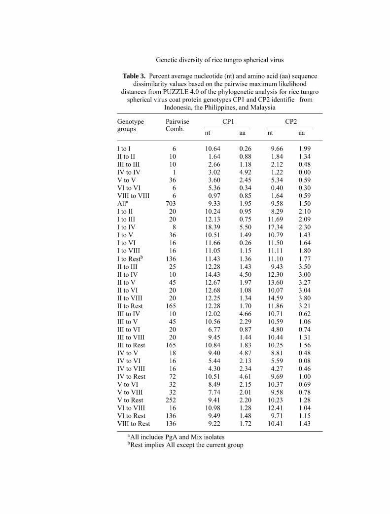
The two coat protein sequences of the selected 38 RTSV isolates that representmost of the coat protein genotype groups were aligned separately and percentnucleotide (nt) and aa sequence dissimilarity for both were calculated using themaximum likelihood distances (PUZZLE 4.0) of the phylogenetic analysis. Se-quence data from genotype VII were not clear, and therefore, this group was notincluded in the sequence analysis. For both CP1 and CP2 sequences, the per-cent average nt divergence between the genotype groups was significantl higherthan among members of the same genotype group except for genotype groups I(Table 3). The most divergent genotype groups were groups I and IV (18.39%sequence divergence in CP1 and 17.34% in CP2) whereas the most closely re-lated genotype groups were groups VIII and IV (4.30% and 4.27% for CP1 andCP2, respectively). The aa divergence was lower and not consistent with the ntdivergence except for the aa divergence between IV-IV comparisons for CP1,which was higher than the nt divergence. For CP2, the aa divergence was low andsimilar between the different genotype groups and among members of the samegroup.
Genetic diversity of rice tungro spherical virus
Table 3. Percent average nucleotide (nt) and amino acid (aa) sequencedissimilarity values based on the pairwise maximum likelihood
distances from PUZZLE 4.0 of the phylogenetic analysis for rice tungrospherical virus coat protein genotypes CP1 and CP2 identifie from
Indonesia, the Philippines, and Malaysia
Genotype Pairwise CP1 CP2groups Comb. nt aa nt aa
I to I 6 10.64 0.26 9.66 1.99II to II 10 1.64 0.88 1.84 1.34III to III 10 2.66 1.18 2.12 0.48IV to IV 1 3.02 4.92 1.22 0.00V to V 36 3.60 2.45 5.34 0.59VI to VI 6 5.36 0.34 0.40 0.30VIII to VIII 6 0.97 0.85 1.64 0.59Alla 703 9.33 1.95 9.58 1.50I to II 20 10.24 0.95 8.29 2.10I to III 20 12.13 0.75 11.69 2.09I to IV 8 18.39 5.50 17.34 2.30I to V 36 10.51 1.49 10.79 1.43I to VI 16 11.66 0.26 11.50 1.64I to VIII 16 11.05 1.15 11.11 1.80I to Restb 136 11.43 1.36 11.10 1.77II to III 25 12.28 1.43 9.43 3.50II to IV 10 14.43 4.50 12.30 3.00II to V 45 12.67 1.97 13.60 3.27II to VI 20 12.68 1.08 10.07 3.04II to VIII 20 12.25 1.34 14.59 3.80II to Rest 165 12.28 1.70 11.86 3.21III to IV 10 12.02 4.66 10.71 0.62III to V 45 10.56 2.29 10.59 1.06III to VI 20 6.77 0.87 4.80 0.74III to VIII 20 9.45 1.44 10.44 1.31III to Rest 165 10.84 1.83 10.25 1.56IV to V 18 9.40 4.87 8.81 0.48IV to VI 16 5.44 2.13 5.59 0.08IV to VIII 16 4.30 2.34 4.27 0.46IV to Rest 72 10.51 4.61 9.69 1.00V to VI 32 8.49 2.15 10.37 0.69V to VIII 32 7.74 2.01 9.58 0.78V to Rest 252 9.41 2.20 10.23 1.28VI to VIII 16 10.98 1.28 12.41 1.04VI to Rest 136 9.49 1.48 9.71 1.15VIII to Rest 136 9.22 1.72 10.41 1.43
aAll includes PgA and Mix isolatesbRest implies All except the current group
Phylogenies of RTSV coat protein species 1 and 2
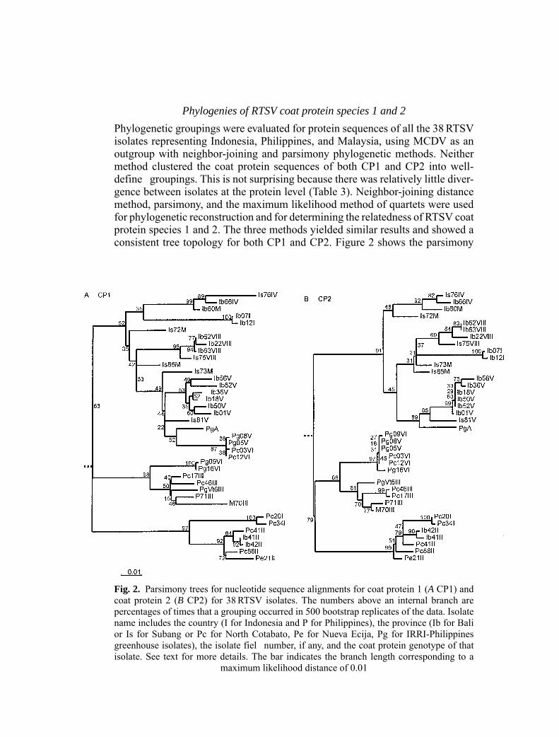
Phylogenetic groupings were evaluated for protein sequences of all the 38RTSVisolates representing Indonesia, Philippines, and Malaysia, using MCDV as anoutgroup with neighbor-joining and parsimony phylogenetic methods. Neithermethod clustered the coat protein sequences of both CP1 and CP2 into well-define groupings. This is not surprising because there was relatively little diver-gence between isolates at the protein level (Table 3). Neighbor-joining distancemethod, parsimony, and the maximum likelihood method of quartets were usedfor phylogenetic reconstruction and for determining the relatedness of RTSV coatprotein species 1 and 2. The three methods yielded similar results and showed aconsistent tree topology for both CP1 and CP2. Figure 2 shows the parsimony
Fig. 2. Parsimony trees for nucleotide sequence alignments for coat protein 1 (A CP1) andcoat protein 2 (B CP2) for 38RTSV isolates. The numbers above an internal branch arepercentages of times that a grouping occurred in 500 bootstrap replicates of the data. Isolatename includes the country (I for Indonesia and P for Philippines), the province (Ib for Balior Is for Subang or Pc for North Cotabato, Pe for Nueva Ecija, Pg for IRRI-Philippinesgreenhouse isolates), the isolate fiel number, if any, and the coat protein genotype of thatisolate. See text for more details. The bar indicates the branch length corresponding to a
maximum likelihood distance of 0.01

Genetic diversity of rice tungro spherical virus
tree. For CP1 (Fig. 2A), RTSV isolates were clustered together with their respec-tive genotype class except for genotypes I, VI and PgA. The Indonesia isolatesof genotype group I clustered together on a separate branch from the Philippinesisolates of the same genotype group. Similarly, Philippines fiel isolates of geno-type group VI clustered on a separate branch from the greenhouse isolates of thesame genotype group. Isolate PgA (the 1989 IRRI isolate that does not belong toany genotype group) clustered with members of genotype group V. For CP2 (Fig.2B), the same three major lineages were observed but with minor differences thatoccurred in the placement of Indonesia isolates from genotype group I and ofPhilippines isolates from genotype groups V and VI clustered now together onthe second major branch of CP2 tree.
Biological properties of RTSV field isolates from North Cotabato
Out of 90 attempted insect-transmissions, only fi e isolates were recovered onTN1 plants based on their positive reactions to both tungro viruses by ELISA(Table 4). Insect mortality rates were high when fed on detached leaves andtransmission rates, therefore, were low. The transmission on larger populationsof TN1 showed that variable symptoms could be induced by the different isolates.Isolates derived from theKabacan site could inducemild, typical tungro, or severesymptoms. Because most isolates were also infected with RTBV, it was difficulto assess the effect of RTSV on symptom expression. Further transmissions on thedifferent rice hosts, which carry sources of resistance to RTSV, were performed.Results showed that the recovered isolates had a different RTSV pathogenicityspectrum based on their differential reactions to the selected hosts as measuredby positive ELISA reactions and RT-PCR (Table 4). Isolate 17 (genotype III)was confirme after two transmission experiments on TN1, Balimau Putih, andTKM6 plants (Fig. 3). Isolates 32, 37, 38, and 83 (mix genotypes) were retainedas mixed genotypes, a single genotype, like genotype VI from isolate 37 that wasrecovered from Balimau Putih plants, or no amplificatio was obtained.
Table 4. ELISA and RT-PCR results for the recovered RTSV isolates from North Cotabatowhen insect-transmitted to TN1, the susceptible host (firs transmission), and to Balimau
Putih (B. P.), Adday Selection (A. S.), TKM6, TW5, and TW16, the resistant hosts to RTSV(second transmission)
Field Original % infection % infection on Recoveredisolate CP-genotype on TN1a B.P. A.S. TKM6 TW5 TW16 CP-genotype
17 III 91 37 0 5 0 0 III32 Mix 89 58 0 10 0 0 Mix37 Mix 20 9 0 0 0 0 VI38 Mix 100 47 0 0 0 0 No amp.83 Mix 100 30 0 0 0 0 No amp.
a% infection by rice tungro spherical virus using ELISA

Fig. 3. The distinct coat protein genotype III, as recovered from TN1, TKM6, and BalimauPutih (accession number 17204) infected plants with isolate 17 of M’Lang after three trans-mission experiments. M Biomarker high and low (BioVentures, Inc.), U undigested RT-PCRproduct, H HindIII-digested product, B BstYI-digested product. 17204= accession number
for Balimau Putih, a rice variety
Discussion
In this study, we have characterized the genetic diversity of RTSV fiel popu-lations using two approaches: (1) restriction analysis of a PCR-amplifie regionfor virus coat protein 1 and 2 species to examine the distribution of differentgenotypes in tungro endemic sites, and (2) sequence analysis of representativeisolates from the different coat protein genotype groups to determine virus popu-lation structure. Based on the restriction analysis, three to six RTSV coat proteingenotypes with different frequencies can be identifie per site and based on thesequence analysis, genotype groups may have an average nt sequence divergencebetween 4 to 18% for both CP1 and CP2 regions. Isolates within most of thegenotype groups are closely related. In addition, as shown by insect transmissionexperiments, isolates from a single site may have different biological properties.Therefore, our data show that RTSV populations in the tungro-endemic regions ofthe Philippines and Indonesia are diverse and exist as sub-populations of relatedbut not identical genetic and biological variants.
Phylogenetic analyses of both CP1 and CP2 sequences show that RTSV viruspopulations have threemajor lineages.Most of the Indonesia isolates are clusteredin the firs lineage with high significance found for the nodes in the bootstrapanalysis of parsimony tree (52% for CP1 tree and 91% for CP2 tree). The othertwo lineages have also high significance for nodes clustering mostly Philippinesisolates (98% and 97% for CP1 and 68% and 99% for CP2). These results confirthe geographical distribution of RTSV populations. However, the spatial patternmay not be discrete because two Indonesia isolates from genotype group II clus-tered in the third lineage along with the Philippines isolates from the same group.Similarly, the 1989 IRRI isolate, PgA, clustered with the Indonesia isolates in thefirs lineage along with genotype group V. Migration or gene fl w seems to occurbetween the populations in addition to the presence of location specifi isolatessuch as the isolates from genotype groups I, IV, VII, and VIII in Indonesia, andgroups I and III in the Philippines.

Genetic diversity of rice tungro spherical virus
At most of sites within each country, the genetic composition of the viruspopulation was not significantl different over the two or three cropping seasons.This result suggest that the geographically isolated populations are geneticallystable over the sampling time. The silentmutations inmost of the CP1 andCP2 se-quences show that a purifying negative selection mechanismmay be an importantfactor for the observed genetic stability. Negative selection eliminates deleteriousvariants from the population and evenmay defin the original founder population.The inter-site virus diversity could then result from negative selection followedby founder events such as vector transmissions that are the only means of spreadfor the virus. The semi-persistent mode of transmission, the size and biotype ofactive green leafhopper transmitters, and the fligh behavior of the vector [22]may impose constraints on the transmissibility and diversity of virus populations.
In intensive rice ecosystems, where rapid temporal and spatial fluctuationin the relative populations of different rice varieties and/or active transmittersoccur, it is reasonable to predict that RTSV has to undergo continuous adaptationand is forced through several bottlenecks. Although rice varieties, such as thevector resistant IR64, do not differ from other deployed varieties and do not seemto exert a selective pressure on the virus populations, the short duration of thestudy makes it difficul to observe an effect, if there is any. However, because theevolution of a virus population is rarely independent of the host, it is essential tokeep monitoring virus population structure and its virulence after the deploymentof new resistance genes (to the vector or to the virus).
Sequence comparisons of coat protein genotypes and the inconsistent cluster-ing of the Philippines fiel and greenhouse genotypes V and VI or the Indonesiagenotypes I and Mix in the CP1 and CP2 phylogenetic trees imply that otherevolutionary forces, such as recombination, may also be involved in the inter-sitevirus diversity and slow that the two coat protein genes evolve at different rates.Indeed, mixed infections were high, ranged between 0–38%, and consisted ofmixtures of two or three RTSV variants within a single plant, as shown by our re-striction analysis and transmission experiments. Therefore, recombination amongvariants can easily occur and if the recombinants are transmitted, they can alsochange the inter-site diversity of local virus populations. Further work is neededto confir that natural RTSV recombinants exist, but, in general, recombinationhas been demonstrated earlier for many plants RNA viruses [26].
The geographic isolation of RTSV populations has implications for the de-ployment of resistance to tungro disease. Although our data suggest that the de-ployed resistance has to take into account local virus variability, the appearance ofresistance-breaking strains in one geographic locationmay notmean that theywilleasily spread to other locations. Localized outbreaks could then be managed bytargeted deployment of relevant resistance genes to that particular environment.In summary, our data support the presence of heterogeneous RTSV populationsin the fiel and a less discrete spatial structure of these populations. Moreover,the results imply a purifying negative selection evolutionary model followed byfounder effects, mainly due to vector transmissions, to account for the inter-sitevariability observed.

Acknowledgements
The authors with to thank Drs. Ulrich Melcher, Oklahoma State University, USA, and Fer-nando Arenal-Garcia, Universidad Politecnica de Madrid, Spain, for critically reviewing themanuscript; the Biotechnology Center, University ofWisconsin-Madison, for sequencing thecoat protein PCR products; Ms. Lani Quintana for the statistical analysis; Joel Macatangayfor setting the server for the phylogenetic analyses; Ed Coloquio, Panfil Domingo, and Es-quirion A. Baguioso for their technical help; Roger Cabunagan and Truong Hoai Xuan fortheir assistance in collecting samples.
References
1. Arboleda M, Azzam O (2000) Inter-and Intra-site genetic diversity of natural fielpopulations of rice tungro bacilliform virus in the Philippines. Arch Virol 145: 275–289
2. Cabauatan PQ, Arboleda M, Azzam O (1998) Differentiation of rice tungro bacilliformvirus strains by restriction analysis andDNAhybridization. J VirolMethods 76: 121–126
3. Cabauatan PQ, Cabunagan RC, Koganezawa H (1995) Biological variants of rice tungroviruses in the Philippines. Phytopathology 85: 77–81
4. Cabauatan PQ, Hibino H (1988) Isolation, purification and serology of rice tungrobacilliform and rice tungro spherical viruses. Plant Dis 72: 526–528
5. Dahal G, Dasgupta I, Lee G, Hull R (1992) Comparative transmission of, and varietalreaction to, three isolates of rice tungro virus disease. Ann Appl Biol 120: 287–300
6. Domingo E, Holland JJ (1994) Mutation rates and rapid evolution of RNA viruses. In:Morse SS (ed) The evolutionary biology of viruses. Raven Press, NewYork, pp 161–184
7. Domingo E, Holland JJ, Biebricher C, EigenM (1995) Quasispecies: the concept and theword. In: Gibbs AJ, Calisher CH, Arenal-Garcia F (eds) Molecular basis of evolution.Cambridge University Press, Cambridge, pp 181–191
8. DrukaA, Burns T, Zhang S, Hull R (1996) Immunological characterization of rice tungrospherical virus coat proteins and differentiation of isolates from the Philippines and India.J Gen Virol 77: 1 975–1 983
9. Fan Z, Dahal G, Dasgupta I, Hay J, Hull R (1996) Variation in the genome of rice tungrobacilliform virus: molecular characterization of six isolates. J Gen Virol 77: 847–854
10. Felsenstein J (1993) PHYLIP (Phylogeny Inference Package) version 3.5c. Distributedby the author. Department of Genetics, University of Washington, Seattle
11. FraileA,Malpica JM,ArandaMA,Cerezo-RodriguezE,Arenal-Garcia F (1996)Geneticdiversity in tobacco mild green mosaic tobamovirus infecting the wild plant Nicotianaglauca. Virology 223: 148–155
12. Gilbert D (1996) SeqPup version 0.6f. Distributed by the author. Biology Department,Indiana University, Bloomington
13. Hibino H (1996) Biology and epidemiology of rice viruses. Annu Rev Phytopathol 34:249–274
14. Hibino H, Daquiog RD, Mesina EM, Aguiero VM (1990) Resistances in rice to tungro-associated viruses. Plant Dis 74: 923–926
15. Kurath G, Heick JA, Dodds JA (1993) RNase protection analyses show high geneticdiversity among fiel isolates of satellite tobacco mosaic virus. Virology 194: 414–418
16. Kurath G, Palukaitis P (1989) RNA sequence heterogeneity in natural populations ofthree satellite RNAs of cucumber mosaic virus. Virology 193: 231–240
17. McNeil J, French R, Hein GL, Stephen Baenziger PS, Eskridge KM (1996) Character-ization of genetic variability among natural populations of wheat streak mosaic virus.Phytopathology 86: 1 222–1 227

Genetic diversity of rice tungro spherical virus
18. Mayo MA, Pringle CR (1998) Virus taxonomy- 1997. J Gen Virol 79: 649–65719. Moya A, Garcia-Arenal F (1995) Population genetics of viruses. In: Gibbs AJ, Calisher
CH, Arenal-Garcia F (eds) Molecular basis of evolution. Cambridge University Press,Cambridge, pp 213–223
20. Page RDM 1996 TREEVIEW: An application to display phylogenetic trees on personalcomputers. Comput Appl Biosci 12: 357–358
21. Reddick BB, Habera LF, Law MD (1997) Nucleotide sequence and taxonomy of maizechlorotic dwarf virus within the family Sequiviridae. J Gen Virol 78: 1 165–1 174
22. Riley JR, Reynolds DR, Farrow RA (1987) The migration of Nilaparvata lugens(Stal.)(Delphacidae) and other Hemiptera associated with rice during the dry season in thePhilippines: a study using radar, visual observations, aerial netting and ground trapping.Bull Entomol Res 77: 145–169
23. Rosenthal A, Charnock-Jones DS (1992) New protocols for DNA sequencing with dyeterminators. DNA Sequ 3: 61–64
24. Saito Y, Hibino H, Omura T, Usugi T (1981) Transmission of rice tungro bacilliformvirus and rice tungro spherical virus by leafhopper vectors. Vth International Congressof Virology, 213 (Abstr.)
25. Shen P, Kaniewska M, Smith C, Beachy RN (1993) Nucleotide sequence and genomicorganization of rice tungro spherical virus. Virology 193: 621–630
26. Simon AE, Bujarski JJ (1994) RNA-RNA recombination and evolution in virus-infectedplants. Ann Rev Phytopathol 32: 337–362
27. Skotnicki ML, Mackenzie AM, Gibbs AJ (1996) Genetic variation in population ofkennedya yellow mosaic tymovirus. Arch Virol 141: 99–110
28. Strimmer K, Von Haeseler A (1996) Quartet puzzling: a quartet maximum likelihoodmethod for reconstructing tree topologies. Mol Biol Evol 13: 964–969
29. Thompson JD,HigginsDG,GibsonTJ (1994)CLUSTALW: improving the sensitivity ofprogressive multiple sequence alignment through sequence weighting, position specifigap penalties and weight matrix choice. Nucleic Acids Res 22: 4 673–4 680
30. Tracy TE, LSMulcahy (1991) A simple method for direct automated sequencing of PCRfragments. BioTechniques 11: 68–75
31. Yambao MLM, Cabauatan P, Azzam O (1998) Differentiation of rice tungro sphericalvirus variants by RT-PCR and RFLP. Int Rice Res Notes 23: 22–24
32. Zhang S, Lee G, Davies JW, Hull R (1997) Variation in coat protein genes among fi egeographically different isolates of rice tungro spherical virus. Arch Virol 142: 1 873–1 879
33. Zhang S, JonesMC, Barker P, Davies JW, Hull R (1993)Molecular cloning and sequenc-ing of coat protein-encoding cDNA of rice tungro spherical virus – a plant picornavirus.Virus Genes 7: 121–132
Authors’ address: Dr. O. Azzam, Biotech Center, University of Wisconsin-Madison, 425Henry Mall, Madison, WI 53706, U.S.A.
Related Documents