Cell Reports Report Genetic Dissection of Neurotrophin Signaling through the p75 Neurotrophin Receptor Ioannis Charalampopoulos, 1,2 Annalisa Vicario, 1 Iosif Pediaditakis, 2 Achille Gravanis, 2 Anastasia Simi, 1 and Carlos F. Iba ´n ˜ ez 1,3, * 1 Department of Neuroscience, Karolinska Institute, Stockholm 17177, Sweden 2 Department of Pharmacology, Faculty of Medicine, University of Crete, Heraklion, Crete 71003, Greece 3 Life Sciences Institute, Department of Physiology, National University of Singapore, Singapore 117456, Singapore *Correspondence: [email protected] http://dx.doi.org/10.1016/j.celrep.2012.11.009 SUMMARY Structural determinants underlying signaling speci- ficity in the tumor necrosis factor receptor super- family (TNFRSF) are poorly characterized, and it is unclear whether different signaling outputs can be genetically dissociated. The p75 neurotrophin receptor (p75 NTR ), also known as TNFRSF16, is a key regulator of trophic and injury responses in the nervous system. Here, we describe a genetic approach for dissecting p75 NTR signaling and deci- phering its underlying logic. Structural determinants important for regulation of cell death, NF-kB, and RhoA pathways were identified in the p75 NTR death domain (DD). Proapoptotic and prosurvival pathways mapped onto nonoverlapping epitopes, demon- strating that different signaling outputs can be genet- ically separated in p75 NTR . Dissociation of c-Jun kinase (JNK) and caspase-3 activities indicated that JNK is necessary but not sufficient for p75 NTR -medi- ated cell death. RIP2 recruitment and RhoGDI release were mechanistically linked, indicating that competi- tion for DD binding underlies crosstalk between NF- kB and RhoA pathways in p75 NTR signaling. These results provide insights into the logic of p75 NTR sig- naling and pave the way for a genetic dissection of p75 NTR function and physiology. INTRODUCTION Plasma membrane receptors relay extracellular signals by alter- ing the activities of multiple intracellular effectors and signaling pathways. Understanding how different receptor signaling out- puts interact with each other and contribute to changes in cell and animal physiology has been one of the main challenges in signal transduction research. In receptor tyrosine kinases, indi- vidual phosphotyrosine residues govern signaling output and specificity. Substitutions in specific intracellular tyrosines can generate receptor mutants that become uncoupled from indi- vidual signaling effectors, such as PI3 kinase, Grb2, or PLCg, providing unparalleled understanding of the physiological rele- vance of individual receptor signaling outputs. Aside from a handful of receptor tyrosine kinases, however, such level of understanding is either very limited or inexistent for other types of receptors. Members of the tumor necrosis factor receptor superfamily (TNFRSF) engage different signaling pathways, including NF- kB, c-Jun kinase (JNK), and caspase cascades, through pro- tein-protein interactions mediated by intracellular death domains (DDs), a six-helix bundle globular domain that is essential for TNFRSF signaling (Park et al., 2007a; Haase et al., 2008). How- ever, structural determinants underlying signaling specificity in the TNFRSF are poorly characterized, and it is unclear whether different signaling outputs can be genetically dissociated. p75 neurotrophin receptor (p75 NTR ), also known as TNFRSF16, is a transmembrane receptor for neurotrophic factors of the neurotrophin family, which includes nerve growth factor (NGF), brain-derived neurotrophic factor (BDNF), neurotrophin-3, and neurotrophin-4 (Dechant and Barde, 2002; Roux and Barker, 2002; Chao, 2003; Gentry et al., 2004; Underwood and Coulson, 2008). Neurotrophin binding to p75 NTR can lead to activation of NF-kB and cell survival (Carter et al., 1996; Khursigara et al., 2001), activation of JNK, caspases, and cell death (Yoon et al., 1998; Friedman, 2000), and inhibition of the small GTPase RhoA and axonal growth (Yamashita et al., 1999; Yamashita and Tohyama, 2003). In addition, activation of p75 NTR by unpro- cessed neurotrophins (proneurotrophins) together with the core- ceptor sortilin is thought to preferentially result in cell death (Lee et al., 2001; Nykjaer et al., 2004). Like other members of the TNRSF, p75 NTR lacks catalytic activity, and signaling proceeds via ligand-induced recruitment and release of cytoplasmic effec- tors to and from its intracellular domain. Numerous intracellular proteins have been identified by their ability to interact with p75 NTR (Dechant and Barde, 2002; Roux and Barker, 2002; Gentry et al., 2004), but, with a few exceptions, their identifica- tion has not clarified our understanding of p75 NTR function and physiology. How p75 NTR connects to different signaling path- ways and how these contribute to p75 NTR function remain key challenges in the field. In order to address these questions, we have undertaken a genetic approach to dissect p75 NTR signaling and decipher its underlying logic. A comprehensive structure-function analysis was performed on the p75 NTR DD (Liepinsh et al., 1997), thereby linking specific structural determinants to each of the three major signaling outputs of p75 NTR . Cell Reports 2, 1–8, December 27, 2012 ª2012 The Authors 1 Please cite this article in press as: Charalampopoulos et al., Genetic Dissection of Neurotrophin Signaling through the p75 Neurotrophin Receptor, Cell Reports (2012), http://dx.doi.org/10.1016/j.celrep.2012.11.009

Welcome message from author
This document is posted to help you gain knowledge. Please leave a comment to let me know what you think about it! Share it to your friends and learn new things together.
Transcript

Please cite this article in press as: Charalampopoulos et al., Genetic Dissection of Neurotrophin Signaling through the p75 Neurotrophin Receptor, CellReports (2012), http://dx.doi.org/10.1016/j.celrep.2012.11.009
Cell Reports
Report
Genetic Dissection of Neurotrophin Signalingthrough the p75 Neurotrophin ReceptorIoannis Charalampopoulos,1,2 Annalisa Vicario,1 Iosif Pediaditakis,2 Achille Gravanis,2 Anastasia Simi,1
and Carlos F. Ibanez1,3,*1Department of Neuroscience, Karolinska Institute, Stockholm 17177, Sweden2Department of Pharmacology, Faculty of Medicine, University of Crete, Heraklion, Crete 71003, Greece3Life Sciences Institute, Department of Physiology, National University of Singapore, Singapore 117456, Singapore
*Correspondence: [email protected]://dx.doi.org/10.1016/j.celrep.2012.11.009
SUMMARY
Structural determinants underlying signaling speci-ficity in the tumor necrosis factor receptor super-family (TNFRSF) are poorly characterized, and itis unclear whether different signaling outputs canbe genetically dissociated. The p75 neurotrophinreceptor (p75NTR), also known as TNFRSF16, isa key regulator of trophic and injury responses inthe nervous system. Here, we describe a geneticapproach for dissecting p75NTR signaling and deci-phering its underlying logic. Structural determinantsimportant for regulation of cell death, NF-kB, andRhoA pathways were identified in the p75NTR deathdomain (DD). Proapoptotic and prosurvival pathwaysmapped onto nonoverlapping epitopes, demon-strating that different signaling outputs can be genet-ically separated in p75NTR. Dissociation of c-Junkinase (JNK) and caspase-3 activities indicated thatJNK is necessary but not sufficient for p75NTR-medi-ated cell death. RIP2 recruitment andRhoGDI releasewere mechanistically linked, indicating that competi-tion for DD binding underlies crosstalk between NF-kB and RhoA pathways in p75NTR signaling. Theseresults provide insights into the logic of p75NTR sig-naling and pave the way for a genetic dissection ofp75NTR function and physiology.
INTRODUCTION
Plasma membrane receptors relay extracellular signals by alter-
ing the activities of multiple intracellular effectors and signaling
pathways. Understanding how different receptor signaling out-
puts interact with each other and contribute to changes in cell
and animal physiology has been one of the main challenges in
signal transduction research. In receptor tyrosine kinases, indi-
vidual phosphotyrosine residues govern signaling output and
specificity. Substitutions in specific intracellular tyrosines can
generate receptor mutants that become uncoupled from indi-
vidual signaling effectors, such as PI3 kinase, Grb2, or PLCg,
providing unparalleled understanding of the physiological rele-
vance of individual receptor signaling outputs. Aside from a
handful of receptor tyrosine kinases, however, such level of
understanding is either very limited or inexistent for other types
of receptors.
Members of the tumor necrosis factor receptor superfamily
(TNFRSF) engage different signaling pathways, including NF-
kB, c-Jun kinase (JNK), and caspase cascades, through pro-
tein-protein interactionsmediated by intracellular death domains
(DDs), a six-helix bundle globular domain that is essential for
TNFRSF signaling (Park et al., 2007a; Haase et al., 2008). How-
ever, structural determinants underlying signaling specificity in
the TNFRSF are poorly characterized, and it is unclear whether
different signaling outputs can be genetically dissociated. p75
neurotrophin receptor (p75NTR), also known as TNFRSF16, is
a transmembrane receptor for neurotrophic factors of the
neurotrophin family, which includes nerve growth factor (NGF),
brain-derived neurotrophic factor (BDNF), neurotrophin-3, and
neurotrophin-4 (Dechant and Barde, 2002; Roux and Barker,
2002; Chao, 2003; Gentry et al., 2004; Underwood and Coulson,
2008). Neurotrophin binding to p75NTR can lead to activation of
NF-kB and cell survival (Carter et al., 1996; Khursigara et al.,
2001), activation of JNK, caspases, and cell death (Yoon et al.,
1998; Friedman, 2000), and inhibition of the small GTPase
RhoA and axonal growth (Yamashita et al., 1999; Yamashita
and Tohyama, 2003). In addition, activation of p75NTR by unpro-
cessed neurotrophins (proneurotrophins) together with the core-
ceptor sortilin is thought to preferentially result in cell death (Lee
et al., 2001; Nykjaer et al., 2004). Like other members of the
TNRSF, p75NTR lacks catalytic activity, and signaling proceeds
via ligand-induced recruitment and release of cytoplasmic effec-
tors to and from its intracellular domain. Numerous intracellular
proteins have been identified by their ability to interact with
p75NTR (Dechant and Barde, 2002; Roux and Barker, 2002;
Gentry et al., 2004), but, with a few exceptions, their identifica-
tion has not clarified our understanding of p75NTR function and
physiology. How p75NTR connects to different signaling path-
ways and how these contribute to p75NTR function remain key
challenges in the field.
In order to address these questions, we have undertaken a
genetic approach to dissect p75NTR signaling and decipher its
underlying logic. A comprehensive structure-function analysis
was performed on the p75NTR DD (Liepinsh et al., 1997), thereby
linking specific structural determinants to each of the threemajor
signaling outputs of p75NTR.
Cell Reports 2, 1–8, December 27, 2012 ª2012 The Authors 1
Please cite this article in press as: Charalampopoulos et al., Genetic Dissection of Neurotrophin Signaling through the p75 Neurotrophin Receptor, CellReports (2012), http://dx.doi.org/10.1016/j.celrep.2012.11.009
RESULTS AND DISCUSSION
Alanine-Scanning Mutagenesis of the p75NTR DDAn arbitrary cutoff of 50% relative solvent accessibility identified
30 highly exposed amino acid residues in the p75NTR DD (Figures
S1A and S1B). Exposed residues are less likely to play structural
roles and represent candidate sites for interaction with down-
stream effector proteins. Three Gly residues playing important
structural roles in loops connecting a helices were excluded
from the analysis. The remaining 27 residues, as well as Glu363
(30% relative solvent accessibility), were targeted by alanine
(Ala)-scanning mutagenesis (Cunningham and Wells, 1989).
The four Alas in the native sequence were substituted by Asp.
With the exception of L342, T343, and A390, all other highly
exposed residues are conserved between the DDs of rat and
human p75NTR. A total of 22 p75NTR mutants were generated
carrying either individual substitutions or combinations. A dele-
tion construct lacking the entire DD (DDD), but retaining the jux-
tamembrane region and the C-terminal tail, was also generated
for comparison. All p75NTR mutants were expressed at compa-
rable levels in transfected cells and retained wild-type (WT)
activity in at least one, but often more, signaling pathways (see
below), indicating that the mutations preserved the structural
integrity of the domain.
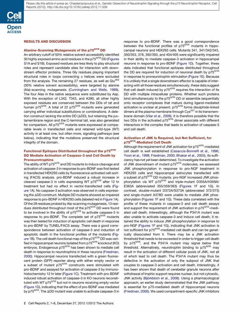
Functional Epitopes Distributed throughout the p75NTR
DD Mediate Activation of Caspase-3 and Cell Death byProneurotrophinsThe ability ofWT p75NTR and DDmutants to induce cleavage and
activation of caspase-3 in response to pro-BDNFwas first tested
in transfected HEK293 cells by fluorescence-activated cell-sort-
ing (FACS) analysis. pro-BDNF induced a robust increase in
cleaved caspase-3 in cells expressing WT p75NTR after 12 hr
treatment but had no effect in vector-transfected cells (Fig-
ure 1A). No caspase-3 activation was observed in cells express-
ing theDDD construct. Multiple DDmutants showed an impaired
response to pro-BDNF in HEK293 cells (labeled red in Figure 1A).
Of the 28 residues probed by Ala-scanningmutagenesis, 13 resi-
dues distributed throughout most of the p75NTR DD were found
to be involved in the ability of p75NTR to activate caspase-3 in
response to pro-BDNF. The complete set of p75NTR mutants
was then tested for induction of apoptotic cell death in response
to pro-BDNF by TUNEL/FACS assay. There was a good corre-
spondence between activation of caspase-3 and induction of
apoptotic death in the functional profiles of the mutants (Fig-
ure 1B). The cell death functional map of the p75NTR DDwas veri-
fied in hippocampal neurons isolated from p75NTR knockout (KO)
embryos. Endogenous p75NTR has been shown to mediate cell
death in response to neurotrophins in these neurons (Friedman,
2000). Hippocampal neurons transfected with a green fluores-
cent protein (GFP) reporter along with either empty vector or
a subset of mutant p75NTR constructs were stimulated with
pro-BDNF and assayed for activation of caspase-3 by immuno-
histochemistry 12 hr later (Figure 1C). Treatment with pro-BDNF
induced robust activation of caspase-3 in KO neurons reconsti-
tuted with WT p75NTR but not in neurons receiving empty vector
(Figure 1D), indicating that the effect of pro-BDNF was mediated
by p75NTR. The DDDmutant was unable to activate caspase-3 in
2 Cell Reports 2, 1–8, December 27, 2012 ª2012 The Authors
response to pro-BDNF. There was a good correspondence
between the functional profiles of p75NTR mutants in hippo-
campal neurons and HEK293 cells. Mutants 341, 341/342/343,
350/353, 378, 392/393, and 404/405 were significantly impaired
in their ability to mediate caspase-3 activation in hippocampal
neurons in response to pro-BDNF (Figure 1D). Together, these
data indicated that functional epitopes distributed throughout
the DD are required for induction of neuronal death by p75NTR
in response to proneurotrophin stimulation (Figure 1E). Because
it is unlikely that a single downstream effector is capable of inter-
acting with all those residues simultaneously, these data suggest
that cell death induced by p75NTR requires the interaction of its
DD with multiple intracellular proteins. Whether such proteins
bind simultaneously to the p75NTR DD or assemble sequentially
onto receptor complexes that mature during ligand-mediated
activation is unclear at present. p75NTR forms disulphide-linked
dimers at the plasmamembrane through Cys257 in its transmem-
brane domain (Vilar et al., 2009). It is therefore possible that the
two DDs in the activated p75NTR dimer associate with different
interactors in the complex that leads to activation of caspase-3
and cell death.
Activation of JNK Is Required, but Not Sufficient, forp75NTR-Mediated Cell DeathAlthough the requirement of JNK activation for p75NTR-mediated
cell death is well established (Casaccia-Bonnefil et al., 1996;
Yoon et al., 1998; Friedman, 2000; Bhakar et al., 2003), its suffi-
ciency has not yet been determined. To investigate the activation
of JNK downstream of mutant p75NTR molecules, we assessed
JNK phosphorylation in response to pro-NGF treatment in
HEK293 cells and hippocampal astrocytes transfected with
a subset of p75NTR DD mutants. pro-NGF increased JNK phos-
phorylation via WT p75NTR and triple-mutant D355A/H359A/
E363A (abbreviated 355/359/363) (Figures 1F and 1G). In
contrast, double-mutant D372A/S373A (abbreviated 372/373)
and single-mutant A378D were unable to mediate JNK phos-
phorylation (Figures 1F and 1G). These data correlated with the
profile of these mutants in caspase-3 and cell death assays
and support the requirement of JNK activation in p75NTR-medi-
ated cell death. Interestingly, although the P341A mutant was
also unable to activate caspase-3 and induce cell death, it re-
tained the ability to induce JNK phosphorylation in response to
pro-NGF (Figures 1F and 1G), indicating that JNK activation is
not sufficient for p75NTR-mediated cell death and can be genet-
ically dissociated from it. There may be a JNK activation
threshold that needs to be exceeded in order to trigger cell death
by p75NTR, and the P341A mutant may signal below that
threshold. Alternatively, neurotrophin binding to p75NTR may
result in the activation of different cellular pools of JNK, not all
of which lead to cell death. The P341A mutant may thus be
defective in the activation of only the subpool of JNK that
couples to caspase-3 activation and cell death. Interestingly, it
has been shown that death of cerebellar granule neurons after
withdrawal of trophic support requires nuclear, but not cytosolic,
JNK activity (Bjorkblom et al., 2008). Using a pharmacological
approach, an earlier study demonstrated that the JNK pathway
is essential for p75-mediated death of hippocampal neurons
(Friedman, 2000). We used compartment-specific JNK inhibitors
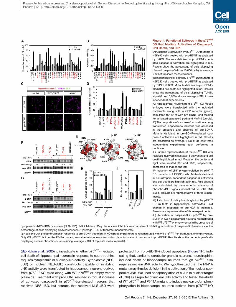
Figure 1. Functional Epitopes in the p75NTR
DD that Mediate Activation of Caspase-3,
Cell Death, and JNK
(A) Caspase-3 activation by p75NTR DD mutants in
HEKs93 cells treated with pro-BDNF as analyzed
by FACS. Mutants deficient in pro-BDNF-medi-
ated caspase-3 activation are highlighted in red.
Results show the percentage of cells displaying
cleaved caspase-3 (from 10,000 cells) as average
± SD of triplicate measurements.
(B) Induction of cell death by p75NTR DDmutants in
HEK293 cells treated with pro-BDNF as analyzed
by TUNEL/FACS. Mutants deficient in pro-BDNF-
mediated cell death are highlighted in red. Results
show the percentage of cells displaying TUNEL
signal (from 10,000 cells) as average ± SD of three
independent experiments.
(C) Hippocampal neurons from p75NTR KO mouse
embryos were transfected with the indicated
constructs along with a GFP reporter (green),
stimulated for 12 hr with pro-BDNF, and stained
for activated caspase-3 (red) and MAP-2 (purple).
(D) The proportion of caspase-3 activation among
transfected hippocampal neurons was assessed
in the presence and absence of pro-BDNF.
Mutants deficient in pro-BDNF-mediated cas-
pase-3 activation are highlighted in red. Results
are presented as average ± SD of at least three
independent experiments each performed in
duplicate.
(E) Surface representation of the p75NTR DD with
residues involved in caspase-3 activation and cell
death highlighted in red. Views on the center and
right were rotated 90� and 180�, respectively,
compared to that on the left.
(F) Induction of JNK phosphorylation by p75NTR
DD mutants in HEK293 cells. Mutants deficient
in neurotrophin-dependent caspase-3 activation
and cell death are highlighted in red. Fold change
was calculated by densitometric scanning of
phospho-JNK signals normalized to total JNK
levels. Results are representative of three experi-
ments.
(G) Induction of JNK phosphorylation by p75NTR
DD mutants in hippocampal astrocytes. Fold
change in response to pro-NGF is indicated.
Results are representative of three experiments.
(H) Activation of caspase-3 in p75NTR by pro-
BDNF in KO hippocampal neurons reconstituted
withWT p75NTR or empty vector in the presence of
cytoplasmic (NES-JBD) or nuclear (NLS-JBD) JNK inhibitors. Only the nuclear inhibitor was capable of inhibiting activation of caspase-3. Results show the
percentage of cells displaying cleaved caspase-3 (average ± SD of triplicate measurements).
(I) Nuclear c-Jun phosphorylation in response to pro-BDNF treatment in KO hippocampal neurons reconstituted withWT p75NTR, P341Amutant, or empty vector.
Only WT p75NTR, but not the P341A mutant, was able to induce nuclear c-Jun phosphorylation in response to pro-BDNF. Results show the percentage of cells
displaying nuclear phospho-c-Jun staining (average ± SD of triplicate measurements).
Please cite this article in press as: Charalampopoulos et al., Genetic Dissection of Neurotrophin Signaling through the p75 Neurotrophin Receptor, CellReports (2012), http://dx.doi.org/10.1016/j.celrep.2012.11.009
(Bjorkblom et al., 2005) to investigate whether p75NTR-mediated
cell death of hippocampal neurons in response to neurotrophins
requires cytoplasmic or nuclear JNK activity. Cytoplasmic (NES-
JBD) or nuclear (NLS-JBD) constructs capable of inhibiting
JNK activity were transfected in hippocampal neurons derived
from p75NTR KO mice along with WT p75NTR or empty vector
plasmids. Treatment with pro-BDNF resulted in robust increase
of activated caspase-3 in p75NTR-transfected neurons that
received NES-JBD, but neurons that received NLS-JBD were
protected from pro-BDNF-induced apoptosis (Figure 1H), indi-
cating that, similar to cerebellar granule neurons, neurotrophin-
induced death of hippocampal neurons through p75NTR also
requires nuclear JNK activity. We hypothesized that the P341A
mutant may thus be deficient in the activation of the nuclear sub-
pool of JNK. We used phosphorylation of c-Jun (a nuclear target
of JNK) as a reporter of nuclear JNK activity and tested the ability
of WT p75NTR and P341A mutant to induce nuclear c-Jun phos-
phorylation in hippocampal neurons derived from p75NTR KO
Cell Reports 2, 1–8, December 27, 2012 ª2012 The Authors 3
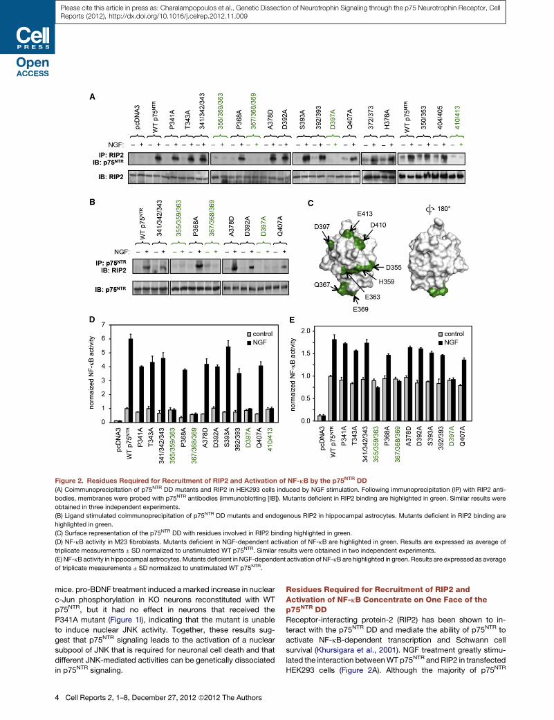
Figure 2. Residues Required for Recruitment of RIP2 and Activation of NF-kB by the p75NTR DD
(A) Coimmunoprecipitation of p75NTR DD mutants and RIP2 in HEK293 cells induced by NGF stimulation. Following immunoprecipitation (IP) with RIP2 anti-
bodies, membranes were probed with p75NTR antibodies (immunoblotting [IB]). Mutants deficient in RIP2 binding are highlighted in green. Similar results were
obtained in three independent experiments.
(B) Ligand stimulated coimmunoprecipitation of p75NTR DD mutants and endogenous RIP2 in hippocampal astrocytes. Mutants deficient in RIP2 binding are
highlighted in green.
(C) Surface representation of the p75NTR DD with residues involved in RIP2 binding highlighted in green.
(D) NF-kB activity in M23 fibroblasts. Mutants deficient in NGF-dependent activation of NF-kB are highlighted in green. Results are expressed as average of
triplicate measurements ± SD normalized to unstimulated WT p75NTR. Similar results were obtained in two independent experiments.
(E) NF-kB activity in hippocampal astrocytes. Mutants deficient in NGF-dependent activation of NF-kB are highlighted in green. Results are expressed as average
of triplicate measurements ± SD normalized to unstimulated WT p75NTR.
Please cite this article in press as: Charalampopoulos et al., Genetic Dissection of Neurotrophin Signaling through the p75 Neurotrophin Receptor, CellReports (2012), http://dx.doi.org/10.1016/j.celrep.2012.11.009
mice. pro-BDNF treatment induced amarked increase in nuclear
c-Jun phosphorylation in KO neurons reconstituted with WT
p75NTR, but it had no effect in neurons that received the
P341A mutant (Figure 1I), indicating that the mutant is unable
to induce nuclear JNK activity. Together, these results sug-
gest that p75NTR signaling leads to the activation of a nuclear
subpool of JNK that is required for neuronal cell death and that
different JNK-mediated activities can be genetically dissociated
in p75NTR signaling.
4 Cell Reports 2, 1–8, December 27, 2012 ª2012 The Authors
Residues Required for Recruitment of RIP2 andActivation of NF-kB Concentrate on One Face of thep75NTR DDReceptor-interacting protein-2 (RIP2) has been shown to in-
teract with the p75NTR DD and mediate the ability of p75NTR to
activate NF-kB-dependent transcription and Schwann cell
survival (Khursigara et al., 2001). NGF treatment greatly stimu-
lated the interaction betweenWT p75NTR and RIP2 in transfected
HEK293 cells (Figure 2A). Although the majority of p75NTR
Please cite this article in press as: Charalampopoulos et al., Genetic Dissection of Neurotrophin Signaling through the p75 Neurotrophin Receptor, CellReports (2012), http://dx.doi.org/10.1016/j.celrep.2012.11.009
mutants behaved as WT, four mutants (labeled green in Fig-
ure 2A) showed a dramatic impairment in NGF-dependent
recruitment of RIP2. Similar results were obtained for a subset
of the mutants in hippocampal astrocytes by assessing the
recovery of endogenously expressed RIP2 after immunoprecip-
itation of transfected p75NTR constructs (Figure 2B). A subset of
p75NTR DD mutants was also tested for their ability to enhance
NF-kB-mediated transcription after NGF stimulation in M23
fibroblasts and hippocampal astrocytes. There was total corre-
spondence between RIP2 binding and NF-kB activity profiles
in both cell types (Figures 2D and 2E), indicating that RIP2
is the main effector linking p75NTR to the NF-kB pathway. In
contrast to the caspase-3/cell death pathway, DD residues
that mediated RIP2 recruitment and NF-kB activation showed
a more restricted distribution, mapping onto one face of the
domain (Figure 2C). The caspase-recruitment domain (CARD)
at the C terminus of RIP2 has previously been shown to mediate
RIP2 binding to the p75NTR DD (Khursigara et al., 2001). DDs,
CARDs, and death-effector domains constitute a superfamily
of structurally related domains that mediate the formation of
large protein complexes between TNFRSF receptors, caspases
and a host of adaptor proteins (Park et al., 2007a). These
domains interact with each other through six different types of
topologically homologous interfaces related to those observed
in the crystal structures of the complexes between Tube and
Pelle DDs (Xiao et al., 1999), Apaf1 and caspase-9 CARDs (Qin
et al., 1999), and PIDD and RAIDD DDs (Park et al., 2007b),
respectively. Themutations that disrupted RIP2 binding mapped
on the type Ib ‘‘Tube-like’’ (residues 410 and 413) and type IIb
‘‘Apaf1-like’’ (residues 355, 359, 363, 367, and 369) interfaces
of the p75NTR DD (Weber and Vincenz, 2001; Park et al.,
2007b), suggesting that complex formation with RIP2 and
perhaps a third CARD- or DD-containing protein is required for
activation of the NF-kB pathway by p75NTR.
Different Structural Determinants in the p75NTR DDMediate RhoGDI Binding and ReleaseThe third major signaling pathway regulated by p75NTR involves
activation of the small GTPase RhoA by constitutive binding of
the p75NTR DD to Rho GDP dissociation inhibitor (RhoGDI),
thereby preventing RhoGDI from inhibiting RhoA (Yamashita
et al., 1999; Yamashita and Tohyama, 2003). NGF binding to
p75NTR releases RhoGDI resulting in RhoA inhibition (Yamashita
and Tohyama, 2003). As expected, WT p75NTR interacted with
RhoGDI and activated RhoA in HEK293 cells, whereas NGF
treatment decreased RhoGDI binding and RhoA activity (Figures
3A and 3B). Two types of phenotypes were identified in our
collection of p75NTR DD mutants. Double mutants K350A/
N353A (abbreviated 350/353) and D410A/E413A (abbreviated
410/413) were unable to bind RhoGDI and elevate RhoA activity
(Figures 3A and 3B, yellow). Neither these mutants nor DDD was
affected by NGF treatment. Residues 350/353 and 410/413
cluster close to each other forming a tight functional epitope
near the N and C termini of the domain (Figure 3C), suggesting
that they represent a RhoGDI binding site in the p75NTR DD.
This RhoGDI epitope showed good correspondence with one
of the two regions targeted by Pep5, a small peptide isolated
in a phage display screen for its ability to bind to the p75NTR
DD (Ilag et al., 1999). Pep5 has been shown to inhibit RhoGDI
binding to p75NTR (Yamashita and Tohyama, 2003) and to block
the ability of p75NTR to activate RhoA (Park et al., 2010). These
observations are consistent with the idea that RhoGDI and
Pep5 bind to the same epitope on the p75NTR DD.
RIP2 Is Required for the Release of RhoGDI from p75NTR
in Response to NGFA second set of mutants bound RhoGDI and enhanced RhoA
activity similar to WT p75NTR but failed to release RhoGDI and
decrease RhoA activity in response to NGF (Figures 3A and 3B,
green). Thiswasunexpectedbecause loss-of-functionmutations
normally result in weaker, not tighter, binding. Remarkably, this
was the same set of mutants that showed deficient RIP2 binding
(Figures 2A–2C). The fact that both RhoGDI and RIP2 required
residues 410/413 for binding to the p75NTR DD suggested
a competitive interaction between the two effectors, such that
recruitment of RIP2 mediates the release of RhoGDI. This notion
was investigated in mouse embryo fibroblasts (MEF) derived
from RIP2 KO mice (Kobayashi et al., 2002). MEF cells express
RIP2 and RhoGDI endogenously but lack p75NTR. As expected,
endogenous RhoGDI was found associatedwith p75NTR in trans-
fectedWTMEF cells, and this interaction was decreased by NGF
treatment (Figure 3D, left). RhoGDI was also coimmunoprecipi-
tated with p75NTR in RIP2 KO MEF cells, but in contrast to WT
cells, NGF treatment had no effect on RhoGDI binding to
p75NTR (Figure 3D, right). Importantly, theability ofNGF treatment
to release RhoGDI from the receptor was restored after transfec-
tion of RIP2 in KO MEF cells (Figure 3E). These data support the
idea that RIP2 and RhoGDI compete for binding to the p75NTR
DD. They also bring a note of caution on the use of Pep5 to
displace RhoGDI from p75NTR because this peptide could also
be interferingwith the regulation of theNF-kBpathway. Together,
our results reveal an unexpected mechanistic link between the
NF-kB and RhoA pathways in p75NTR signaling.
ConclusionsThe three major pathways activated by neurotrophin binding to
p75NTR have so far been studied in isolation, and it has been
unclear whether—or how—they are mechanistically linked at
the level of the receptor. The mechanisms by which p75NTR
couples to these pathways and how they contribute to p75NTR
function have been outstanding questions in the field. We have
elucidated a structure-function map of the p75NTR DD linking
individual residues to distinct interactors and downstream sig-
naling pathways (Figures 4A and 4B). This study demonstrates
that the major signaling outputs of p75NTR are genetically sepa-
rable at the level of the receptor and that, in a way analogous to
receptor tyrosine kinases, it is possible to generate p75NTR
mutants that are selectively deficient in one pathway but not
others. We present examples of how this knowledge can be
used to reveal the underlying logic of p75NTR signaling. This
structure-function map can now serve as a conceptual and
technical framework for clarifying the physiological relevance
of each of the major signaling pathways regulated by p75NTR
and other TNFRSF receptors and for aiding in the discovery of
new strategies for inhibiting p75NTR signaling in nervous system
injury and degeneration.
Cell Reports 2, 1–8, December 27, 2012 ª2012 The Authors 5
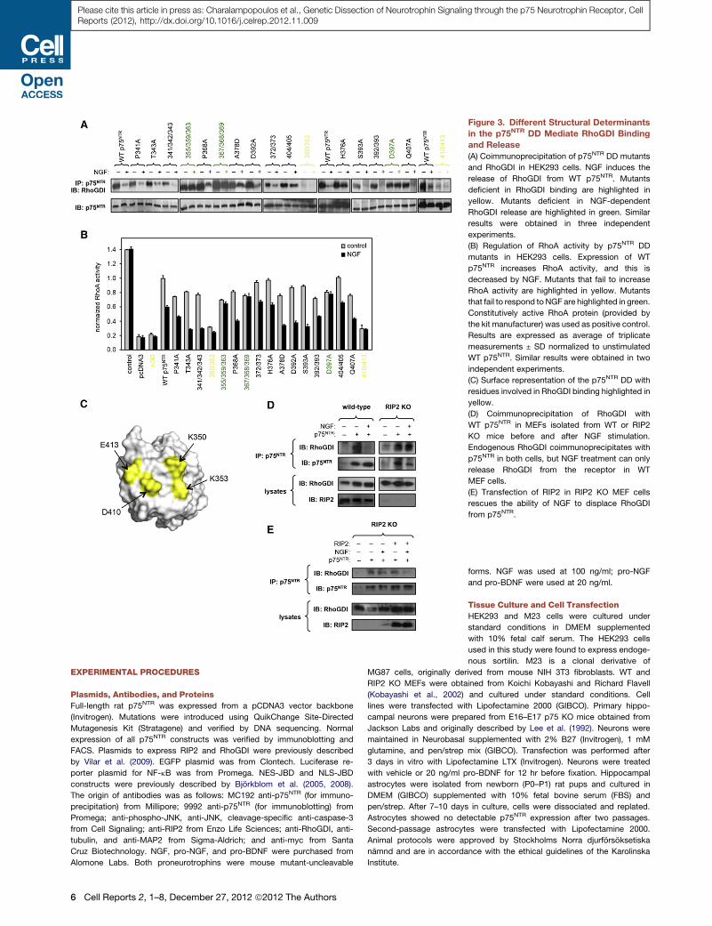
Figure 3. Different Structural Determinants
in the p75NTR DD Mediate RhoGDI Binding
and Release
(A) Coimmunoprecipitation of p75NTR DD mutants
and RhoGDI in HEK293 cells. NGF induces the
release of RhoGDI from WT p75NTR. Mutants
deficient in RhoGDI binding are highlighted in
yellow. Mutants deficient in NGF-dependent
RhoGDI release are highlighted in green. Similar
results were obtained in three independent
experiments.
(B) Regulation of RhoA activity by p75NTR DD
mutants in HEK293 cells. Expression of WT
p75NTR increases RhoA activity, and this is
decreased by NGF. Mutants that fail to increase
RhoA activity are highlighted in yellow. Mutants
that fail to respond to NGF are highlighted in green.
Constitutively active RhoA protein (provided by
the kit manufacturer) was used as positive control.
Results are expressed as average of triplicate
measurements ± SD normalized to unstimulated
WT p75NTR. Similar results were obtained in two
independent experiments.
(C) Surface representation of the p75NTR DD with
residues involved in RhoGDI binding highlighted in
yellow.
(D) Coimmunoprecipitation of RhoGDI with
WT p75NTR in MEFs isolated from WT or RIP2
KO mice before and after NGF stimulation.
Endogenous RhoGDI coimmunoprecipitates with
p75NTR in both cells, but NGF treatment can only
release RhoGDI from the receptor in WT
MEF cells.
(E) Transfection of RIP2 in RIP2 KO MEF cells
rescues the ability of NGF to displace RhoGDI
from p75NTR.
Please cite this article in press as: Charalampopoulos et al., Genetic Dissection of Neurotrophin Signaling through the p75 Neurotrophin Receptor, CellReports (2012), http://dx.doi.org/10.1016/j.celrep.2012.11.009
EXPERIMENTAL PROCEDURES
Plasmids, Antibodies, and Proteins
Full-length rat p75NTR was expressed from a pCDNA3 vector backbone
(Invitrogen). Mutations were introduced using QuikChange Site-Directed
Mutagenesis Kit (Stratagene) and verified by DNA sequencing. Normal
expression of all p75NTR constructs was verified by immunoblotting and
FACS. Plasmids to express RIP2 and RhoGDI were previously described
by Vilar et al. (2009). EGFP plasmid was from Clontech. Luciferase re-
porter plasmid for NF-kB was from Promega. NES-JBD and NLS-JBD
constructs were previously described by Bjorkblom et al. (2005, 2008).
The origin of antibodies was as follows: MC192 anti-p75NTR (for immuno-
precipitation) from Millipore; 9992 anti-p75NTR (for immunoblotting) from
Promega; anti-phospho-JNK, anti-JNK, cleavage-specific anti-caspase-3
from Cell Signaling; anti-RIP2 from Enzo Life Sciences; anti-RhoGDI, anti-
tubulin, and anti-MAP2 from Sigma-Aldrich; and anti-myc from Santa
Cruz Biotechnology. NGF, pro-NGF, and pro-BDNF were purchased from
Alomone Labs. Both proneurotrophins were mouse mutant-uncleavable
6 Cell Reports 2, 1–8, December 27, 2012 ª2012 The Authors
forms. NGF was used at 100 ng/ml; pro-NGF
and pro-BDNF were used at 20 ng/ml.
Tissue Culture and Cell Transfection
HEK293 and M23 cells were cultured under
standard conditions in DMEM supplemented
with 10% fetal calf serum. The HEK293 cells
used in this study were found to express endoge-
nous sortilin. M23 is a clonal derivative of
MG87 cells, originally derived from mouse NIH 3T3 fibroblasts. WT and
RIP2 KO MEFs were obtained from Koichi Kobayashi and Richard Flavell
(Kobayashi et al., 2002) and cultured under standard conditions. Cell
lines were transfected with Lipofectamine 2000 (GIBCO). Primary hippo-
campal neurons were prepared from E16–E17 p75 KO mice obtained from
Jackson Labs and originally described by Lee et al. (1992). Neurons were
maintained in Neurobasal supplemented with 2% B27 (Invitrogen), 1 mM
glutamine, and pen/strep mix (GIBCO). Transfection was performed after
3 days in vitro with Lipofectamine LTX (Invitrogen). Neurons were treated
with vehicle or 20 ng/ml pro-BDNF for 12 hr before fixation. Hippocampal
astrocytes were isolated from newborn (P0–P1) rat pups and cultured in
DMEM (GIBCO) supplemented with 10% fetal bovine serum (FBS) and
pen/strep. After 7–10 days in culture, cells were dissociated and replated.
Astrocytes showed no detectable p75NTR expression after two passages.
Second-passage astrocytes were transfected with Lipofectamine 2000.
Animal protocols were approved by Stockholms Norra djurforsoksetiska
namnd and are in accordance with the ethical guidelines of the Karolinska
Institute.
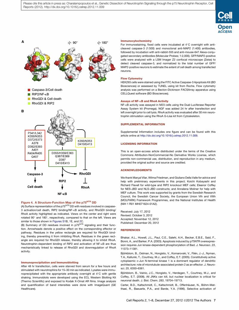
Figure 4. A Structure-Function Map of the p75NTR DD
(A) Surface representation of the p75NTR DDwith residues involved in caspase-
3 activation/cell death, RIP2 binding/NF-kB activity, and RhoGDI binding/
RhoA activity highlighted as indicated. Views on the center and right were
rotated 90� and 180�, respectively, compared to that on the left. Views are
similar to those shown in Figures S1B, 1E, and 2C.
(B) Summary of DD residues involved in p75NTR signaling and their func-
tion. Arrowheads denote a positive effect on the corresponding effector or
pathway. Residues in the yellow rectangle are required for RhoGDI bind-
ing, thereby preventing it from inhibiting RhoA. Residues in the green rect-
angle are required for RhoGDI release, thereby allowing it to inhibit RhoA.
Neurotrophin-dependent binding of RIP2 and activation of NF-kB are thus
mechanistically linked to release of RhoGDI and downregulation of RhoA
activity.
Please cite this article in press as: Charalampopoulos et al., Genetic Dissection of Neurotrophin Signaling through the p75 Neurotrophin Receptor, CellReports (2012), http://dx.doi.org/10.1016/j.celrep.2012.11.009
Immunoprecipitation and Immunoblotting
After 48 hr transfection, cells were starved from serum for a few hours and
stimulated with neurotrophins for 15–30 min as indicated. Lysates were immu-
noprecipitated with the appropriate antibody overnight at 4�C with gentle
shaking. Immunoblots were developed using the ECL Western Blotting Kit
(Thermo Scientific) and exposed to Kodak X-Omat AR films. Image analysis
and quantification of band intensities were done with ImageQuant (GE
Healthcare).
Immunocytochemistry
For immunostaining, fixed cells were incubated at 4�C overnight with anti-
cleaved caspase-3 (1:500) and monoclonal anti-MAP2 (1:400) antibodies,
followed by incubation with anti-rabbit-555 and anti-mouse-647 Alexa-conju-
gated secondary antibodies (Molecular Probes; 1:2,000). GFP/MAP2-positive
cells were analyzed with a LSM Imager Z2 confocal microscope (Zeiss) to
detect cleaved caspase-3, and normalized to the total number of GFP/
MAP2-positive neurons to estimate the extent of cell death among transfected
neurons.
Flow Cytometry
HEK293 cells were stained using the FITC Active Caspase-3 Apoptosis Kit (BD
Biosciences) or assessed by TUNEL using kit from Roche. Flow cytometry
analysis was performed on a Becton-Dickinson FACSArray apparatus using
CELLQuest software (BD Biosciences).
Assays of NF-kB and RhoA Activity
NF-kB activity was assayed in M23 cells using the Dual-Luciferase Reporter
Assay System kit (Promega). NGF was added 24 hr after transfection and
left overnight prior to cell lysis. RhoA activity was evaluated after 30min neuro-
trophin stimulation using the RhoA G-Lisa kit from Cytoskeleton.
SUPPLEMENTAL INFORMATION
Supplemental Information includes one figure and can be found with this
article online at http://dx.doi.org/10.1016/j.celrep.2012.11.009.
LICENSING INFORMATION
This is an open-access article distributed under the terms of the Creative
Commons Attribution-NonCommercial-No Derivative Works License, which
permits non-commercial use, distribution, and reproduction in any medium,
provided the original author and source are credited.
ACKNOWLEDGMENTS
We thankMarcal Vilar, Wilma Friedman, andGiulianoDella Valle for advice and
help with preliminary experiments in this project; Koichi Kobayashi and
Richard Flavell for wild-type and RIP2 knockout MEF cells; Eleanor Coffey
for NES-JBD and NLS-JBD constructs; and Annalena Moliner for help with
MEF culture. This work was supported by grants from the Swedish Research
Council, the Swedish Cancer Society, the European Union Vth and VIIth
(MOLPARK) Framework Programmes, and the National Institutes of Health
(NIH 1 R01 MH071624-01A2).
Received: July 17, 2012
Revised: October 3, 2012
Accepted: November 12, 2012
Published: December 20, 2012
REFERENCES
Bhakar, A.L., Howell, J.L., Paul, C.E., Salehi, A.H., Becker, E.B.E., Said, F.,
Bonni, A., and Barker, P.A. (2003). Apoptosis induced by p75NTR overexpres-
sion requires Jun kinase-dependent phosphorylation of Bad. J. Neurosci. 23,
11373–11381.
Bjorkblom, B., Ostman, N., Hongisto, V., Komarovski, V., Filen, J.-J., Nyman,
T.A., Kallunki, T., Courtney, M.J., and Coffey, E.T. (2005). Constitutively active
cytoplasmic c-Jun N-terminal kinase 1 is a dominant regulator of dendritic
architecture: role of microtubule-associated protein 2 as an effector. J. Neuro-
sci. 25, 6350–6361.
Bjorkblom, B., Vainio, J.C., Hongisto, V., Herdegen, T., Courtney, M.J., and
Coffey, E.T. (2008). All JNKs can kill, but nuclear localization is critical for
neuronal death. J. Biol. Chem. 283, 19704–19713.
Carter, B.D., Kaltschmidt, C., Kaltschmidt, B., Offenhauser, N., Bohm-Mat-
thaei, R., Baeuerle, P.A., and Barde, Y.A. (1996). Selective activation of
Cell Reports 2, 1–8, December 27, 2012 ª2012 The Authors 7
Please cite this article in press as: Charalampopoulos et al., Genetic Dissection of Neurotrophin Signaling through the p75 Neurotrophin Receptor, CellReports (2012), http://dx.doi.org/10.1016/j.celrep.2012.11.009
NF-kappa B by nerve growth factor through the neurotrophin receptor p75.
Science 272, 542–545.
Casaccia-Bonnefil, P., Carter, B.D., Dobrowsky, R.T., and Chao, M.V. (1996).
Death of oligodendrocytes mediated by the interaction of nerve growth factor
with its receptor p75. Nature 383, 716–719.
Chao, M.V. (2003). Neurotrophins and their receptors: a convergence point for
many signalling pathways. Nat. Rev. Neurosci. 4, 299–309.
Cunningham, B.C., and Wells, J.A. (1989). High-resolution epitope mapping of
hGH-receptor interactions by alanine-scanning mutagenesis. Science 244,
1081–1085.
Dechant, G., and Barde, Y.A. (2002). The neurotrophin receptor p75(NTR):
novel functions and implications for diseases of the nervous system. Nat.
Neurosci. 5, 1131–1136.
Friedman, W.J. (2000). Neurotrophins induce death of hippocampal neurons
via the p75 receptor. J. Neurosci. 20, 6340–6346.
Gentry, J.J., Barker, P.A., and Carter, B.D. (2004). The p75 neurotrophin
receptor: multiple interactors and numerous functions. Prog. Brain Res. 146,
25–39.
Haase, G., Pettmann, B., Raoul, C., and Henderson, C.E. (2008). Signaling by
death receptors in the nervous system. Curr. Opin. Neurobiol. 18, 284–291.
Ilag, L.L., Rottenberger, C., Liepinsh, E., Wellnhofer, G., Rudert, F., Otting, G.,
and Ilag, L.L. (1999). Selection of a peptide ligand to the p75 neurotrophin
receptor death domain and determination of its binding sites by NMR.
Biochem. Biophys. Res. Commun. 255, 104–109.
Khursigara, G., Bertin, J., Yano, H., Moffett, H., DiStefano, P.S., and Chao,
M.V. (2001). A prosurvival function for the p75 receptor death domain medi-
ated via the caspase recruitment domain receptor-interacting protein 2.
J. Neurosci. 21, 5854–5863.
Kobayashi, K., Inohara, N., Hernandez, L.D., Galan, J.E., Nunez, G., Janeway,
C.A., Medzhitov, R., and Flavell, R.A. (2002). RICK/Rip2/CARDIAK mediates
signalling for receptors of the innate and adaptive immune systems. Nature
416, 194–199.
Lee, K.F., Li, E., Huber, L.J., Landis, S.C., Sharpe, A.H., Chao, M.V., and Jae-
nisch, R. (1992). Targeted mutation of the gene encoding the low affinity NGF
receptor p75 leads to deficits in the peripheral sensory nervous system. Cell
69, 737–749.
Lee, R., Kermani, P., Teng, K.K., and Hempstead, B.L. (2001). Regulation of
cell survival by secreted proneurotrophins. Science 294, 1945–1948.
8 Cell Reports 2, 1–8, December 27, 2012 ª2012 The Authors
Liepinsh, E., Ilag, L.L., Otting, G., and Ibanez, C.F. (1997). NMR structure of the
death domain of the p75 neurotrophin receptor. EMBO J. 16, 4999–5005.
Nykjaer, A., Lee, R., Teng, K.K., Jansen, P., Madsen, P., Nielsen, M.S., Jacob-
sen, C., Kliemannel, M., Schwarz, E., Willnow, T.E., et al. (2004). Sortilin is
essential for proNGF-induced neuronal cell death. Nature 427, 843–848.
Park, H.H., Lo, Y.C., Lin, S.-C., Wang, L., Yang, J.K., and Wu, H. (2007a). The
death domain superfamily in intracellular signaling of apoptosis and inflamma-
tion. Annu. Rev. Immunol. 25, 561–586.
Park, H.H., Logette, E., Raunser, S., Cuenin, S., Walz, T., Tschopp, J., andWu,
H. (2007b). Death domain assembly mechanism revealed by crystal structure
of the oligomeric PIDDosome core complex. Cell 128, 533–546.
Park, K.J., Grosso, C.A., Aubert, I., Kaplan, D.R., and Miller, F.D. (2010).
p75NTR-dependent, myelin-mediated axonal degeneration regulates neural
connectivity in the adult brain. Nat. Neurosci. 13, 559–566.
Qin, H., Srinivasula, S.M., Wu, G., Fernandes-Alnemri, T., Alnemri, E.S., and
Shi, Y. (1999). Structural basis of procaspase-9 recruitment by the apoptotic
protease-activating factor 1. Nature 399, 549–557.
Roux, P.P., and Barker, P.A. (2002). Neurotrophin signaling through the p75
neurotrophin receptor. Prog. Neurobiol. 67, 203–233.
Underwood, C.K., and Coulson, E.J. (2008). The p75 neurotrophin receptor.
Int. J. Biochem. Cell Biol. 40, 1664–1668.
Vilar, M., Charalampopoulos, I., Kenchappa, R.S., Simi, A., Karaca, E., Re-
versi, A., Choi, S., Bothwell, M., Mingarro, I., Friedman, W.J., et al. (2009).
Activation of the p75 neurotrophin receptor through conformational rearrange-
ment of disulphide-linked receptor dimers. Neuron 62, 72–83.
Weber, C.H., and Vincenz, C. (2001). The death domain superfamily: a tale of
two interfaces? Trends Biochem. Sci. 26, 475–481.
Xiao, T., Towb, P., Wasserman, S.A., and Sprang, S.R. (1999). Three-dimen-
sional structure of a complex between the death domains of Pelle and Tube.
Cell 99, 545–555.
Yamashita, T., and Tohyama, M. (2003). The p75 receptor acts as a displace-
ment factor that releases Rho from Rho-GDI. Nat. Neurosci. 6, 461–467.
Yamashita, T., Tucker, K.L., and Barde, Y.A. (1999). Neurotrophin binding to
the p75 receptor modulates Rho activity and axonal outgrowth. Neuron 24,
585–593.
Yoon, S.O., Casaccia-Bonnefil, P., Carter, B.D., and Chao, M.V. (1998).
Competitive signaling between TrkA and p75 nerve growth factor receptors
determines cell survival. J. Neurosci. 18, 3273–3281.
Supplemental Information
SUPPLEMENTAL REFERENCE
DeLano, W.L. (2002). The PyMOL Molecular Graphics System, version 1.5.0.4, Schrodinger, LLC. http://www.pymol.org/.
Cell Reports 2, 1563–1570, December 27, 2012 ª2012 The Authors S1
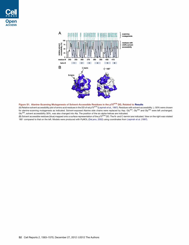
Figure S1. Alanine-Scanning Mutagenesis of Solvent-Accessible Residues in the p75NTR DD, Related to Results
(A) Relative solvent accessibility plot of amino acid residues in the DD of rat p75NTR (Liepinsh et al., 1997). Residues with solvent accessibilityR 50%were chosen
for alanine-scanning mutagenesis as indicated. Solvent-exposed Alanine side chains were replaced by Asp. Gly354, Gly362 and Gly365 were left unchanged.
Glu363, solvent accessibility 30%, was also changed into Ala. The position of the six alpha-helices are indicated.
(B) Solvent accessible residues (blue) mapped onto a surface representation of the p75NTR DD. The N- and C-termini are indicated. View on the right was rotated
180� compared to that on the left. Models were produced with PyMOL (DeLano, 2002) using coordinates from Liepinsh et al. (1997).
S2 Cell Reports 2, 1563–1570, December 27, 2012 ª2012 The Authors
Related Documents


