Genetic disruption of calpain correlates with loss of membrane blebbing and differential expression of RhoGDI-1, cofilin and tropomyosin Anna K. Larsen*, René Lametsch*, John S. Elce†, Jørgen K. Larsen‡, Bo Thomsen§, Martin R. Larsen||, Moira A. Lawson*, Peter A. Greer¶ and Per Ertbjerg*. * Department of Food Science, Faculty of Life Sciences, University of Copenhagen, Denmark † Department of Biochemistry, Queen’s University, Kingston, Ontario, Canada ‡ Finsen Laboratory, Finsen Center, Rigshospitalet, Copenhagen, Denmark § Dept. of Genetics and Biotechnology, Faculty of Agricultural Science, University of Aarhus, Denmark || Department of Biochemistry and Molecular Biology, University of Southern Denmark, Denmark ¶ Queen’s Cancer Research Institute, Queen’s University, Kingston, Ontario, Canada Corresponding author: Anna Karina Larsen, Dept. of Food Science, Faculty of Life Sciences, University of Copenhagen Rolighedsvej 30, DK-1958 Frederiksberg C, Denmark, Phone: + 45 35 33 31 84 Fax: + 45 35 33 33 41 e-mail: [email protected] Page heading: Calpain is required for membrane blebbing Keywords: m-calpain, GFP, proteomics, RhoGDI-1, cofilin, tropomyosin Stage 2(a) POST-PRINT Biochemical Journal Immediate Publication. Published on 12 Dec 2007 as manuscript BJ20070522 THIS IS NOT THE FINAL VERSION - see doi:10.1042/BJ20070522 Licenced copy. Copying is not permitted, except with prior permission and as allowed by law. © 2007 The Authors Journal compilation © 2007 Biochemical Society

Welcome message from author
This document is posted to help you gain knowledge. Please leave a comment to let me know what you think about it! Share it to your friends and learn new things together.
Transcript

Genetic disruption of calpain correlates with loss of membrane blebbing and differential expression of RhoGDI-1, cofilin and tropomyosin
Anna K. Larsen*, René Lametsch*, John S. Elce†, Jørgen K. Larsen‡, Bo Thomsen§,
Martin R. Larsen||, Moira A. Lawson*, Peter A. Greer¶ and Per Ertbjerg*.
* Department of Food Science, Faculty of Life Sciences, University of Copenhagen, Denmark
† Department of Biochemistry, Queen’s University, Kingston, Ontario, Canada
‡ Finsen Laboratory, Finsen Center, Rigshospitalet, Copenhagen, Denmark
§ Dept. of Genetics and Biotechnology, Faculty of Agricultural Science, University of Aarhus, Denmark
|| Department of Biochemistry and Molecular Biology, University of Southern Denmark, Denmark
¶ Queen’s Cancer Research Institute, Queen’s University, Kingston, Ontario, Canada
Corresponding author: Anna Karina Larsen, Dept. of Food Science, Faculty of Life Sciences, University of Copenhagen Rolighedsvej 30, DK-1958 Frederiksberg C, Denmark, Phone: + 45 35 33 31 84 Fax: + 45 35 33 33 41 e-mail: [email protected] Page heading: Calpain is required for membrane blebbing Keywords: m-calpain, GFP, proteomics, RhoGDI-1, cofilin, tropomyosin
Stag
e 2(
a) P
OST
-PR
INT
Biochemical Journal Immediate Publication. Published on 12 Dec 2007 as manuscript BJ20070522
TH
IS IS
NO
T T
HE
FIN
AL
VE
RS
ION
- s
ee d
oi:1
0.10
42/B
J200
7052
2
Licenced copy. Copying is not permitted, except with prior permission and as allowed by law.
© 2007 The Authors Journal compilation © 2007 Biochemical Society
List of complete address affiliations:
* Anna K. Larsen, René Lametsch, Moira A. Lawson and Per Ertbjerg: Department of Food Science, Faculty of Life Sciences, University of Copenhagen, Rolighedsvej 30, DK-1958 Frederiksberg C, Denmark. [email protected], [email protected] and [email protected] Tel: + 45 35 33 32 55 † John S. Elce: Department of Biochemistry, Queen’s University, Botterell Hall, Kingston, Ontario, Canada K7L 3N6, [email protected] ‡ Jørgen K. Larsen: Finsen Laboratory & Experimental Pathology Unit, Rigshospitalet, Copenhagen Biocenter, Ole Maaløes Vej 5, DK-2200 København N, Denmark. [email protected]: +45 35 45 60 82 § Bo Thomsen : Dept. of Genetics and Biotechnology, Faculty of Agricultural Sciences, University of Aarhus, Research Centre Foulum, Blichers Allé 20, DK-8830 Tjele. [email protected]: +45 89 99 19 00 || Martin R. Larsen: Department of Biochemistry and Molecular Biology, University of Southern Denmark, DK-5230 Odense, Denmark. [email protected]: +45 65 50 23 42 ¶ Peter A. Greer: Queen's University Cancer Research Institute, Botterell Hall, Room A309, Kingston, Ontario K7L 3N6, Canada. [email protected]: (613) 533 2813
Stag
e 2(
a) P
OST
-PR
INT
Biochemical Journal Immediate Publication. Published on 12 Dec 2007 as manuscript BJ20070522
TH
IS IS
NO
T T
HE
FIN
AL
VE
RS
ION
- s
ee d
oi:1
0.10
42/B
J200
7052
2
Licenced copy. Copying is not permitted, except with prior permission and as allowed by law.
© 2007 The Authors Journal compilation © 2007 Biochemical Society

ABBREVIATIONS
FACS: fluorescence-activated cell sorting
FAK: focal adhesion kinase
FN: fibronectin
GFP: green fluorescent protein
LIMK: Lin11, Isl1, Mec3 kinases
MALDI-TOF-MS: Matrix-assisted laser desorption/ionization time-of-flight mass spectrometry
MARCKS: myristylated alanine-rich C kinase substrate
MEF: mouse embryonic fibroblast
MLC: myosin light chain
MLCK: myosin light chain kinase
MLCP: myosin light chain phosphatase
PAK: p21-activated kinase
PKC: protein kinase C
RhoGDI: Rho GDP-dissociation inhibitor
ROCK: Rho-associated kinase
Tm2: Tropomyosin 1 (α), splice isoform 2
3
Stag
e 2(
a) P
OST
-PR
INT
Biochemical Journal Immediate Publication. Published on 12 Dec 2007 as manuscript BJ20070522
TH
IS IS
NO
T T
HE
FIN
AL
VE
RS
ION
- s
ee d
oi:1
0.10
42/B
J200
7052
2
Licenced copy. Copying is not permitted, except with prior permission and as allowed by law.
© 2007 The Authors Journal compilation © 2007 Biochemical Society

SYNOPSIS
Dynamic regulation of the actin-cytoskeleton is essential to cell motility, spreading, and the formation of
membrane surface extensions like lamellipodia, ruffles and blebs. The ubiquitous calpains contribute to
integrin-mediated cytoskeletal remodelling during cell migration and spreading, by cleavage of focal
adhesion components and signalling molecules. In this study, the live-cell morphology of calpain-knockout
and wild type cells was examined by time-lapse fluorescence microscopy and a role of calpain in mediating
formation of sporadic membrane blebs was established. Membrane blebbing was significantly reduced in
calpain-knockout cells, and genetic rescue fully restored the wild type phenotype in knockout cells.
Proteomic comparison of wild type and knockout cells identified decreased levels of RhoGDI-1 and cofilin
1, and increased levels of tropomyosin in calpain knockout cells, suggesting a role of calpain in regulating
membrane extensions involving these proteins. RhoGDI, cofilin and tropomyosin are known regulators of
actin filament dymamics and membrane extensions. The reduced levels of RhoGDI-1 in calpain-knockout
cells observed by proteome analysis were confirmed by immunoblotting. Genetic rescue of the calpain-
knockout cells enhanced RhoGDI-1-expression 2-fold above that normally present in wild type cells. These
data suggest a regulatory connection between calpain and RhoGDI-1 to promote formation of membrane
blebs.
(197 words)
4
Stag
e 2(
a) P
OST
-PR
INT
Biochemical Journal Immediate Publication. Published on 12 Dec 2007 as manuscript BJ20070522
TH
IS IS
NO
T T
HE
FIN
AL
VE
RS
ION
- s
ee d
oi:1
0.10
42/B
J200
7052
2
Licenced copy. Copying is not permitted, except with prior permission and as allowed by law.
© 2007 The Authors Journal compilation © 2007 Biochemical Society
INTRODUCTION
Active remodelling of the cytoskeleton is fundamental to most biological processes that involve
motility and changes in cell morphology, including cell migration, spreading, division and cell fusion.
Through their interactions with components of signalling pathways, focal adhesions and the cytoskeleton, the
ubiquitous calpains are regulators of such changes [1-3].
A range of cell surface protrusions, including lamellipodia, pseudopodia, filopodia, membrane ruffles
and blebs are integral phenomena of eukaryotic cell motility, that rely on the dynamic reorganization of the
actin cytoskeleton, mediated by small GTPases of the Rho family [4]. Rho, Rac and Cdc42 proteins are
essential regulators of diverse aspects of cell morphology and motility. They are tightly regulated at several
levels, including by the Rho GDP-dissociation inhibitors (RhoGDIs) [5]. The precise functional significance
of blebs, ruffles and lamellipodia, and the mechanisms leading to their formation are not fully understood;
but these phenomena all appear to relate to cell migration [6;7]. Membrane blebs are hyaline, typically
spherical cell-surface extensions that occur during apoptosis [8;9], cell division [10] and migration [11].
Blebs are very transient and dynamic in nature, and may transform into, or alternate with other extensions
like ruffles and lamellipodia [11]. Lamellipodia and ruffles are flat F-actin dense extensions that normally
protrude along the leading edge of migrating cells [4][12]. Membrane blebbing was initially associated with
apoptosis [8;9] and cell injury [13;14] and a retracted, round cell morphology associated with large, highly
dynamic membrane blebs are indeed key features of apoptosis. Non-apoptotic blebs that evolve during cell
division and migration are less studied, but have been reported to differ from apoptotic blebs with regards to
size, shape and F-actin distribution [9]. Blebbing reflects a general loss of adhesion strength between the
actin cytoskeleton and the plasma membrane [15] and is initiated by membrane detachment from the
underlying actin layer. Bleb expansion is a passive process, driven by a flow of fluid; and is independent on
actin polymerization, unlike ruffles or lamellipodia. Bleb retraction, however requires actin filament
assembly, myosin II-dependent contraction [16] and is controlled by the phosphorylation of myosin II
regulatory light chain (MLC) [8]. MLC phosphorylation is regulated by myosin light chain kinase (MLCK),
myosin light chain phosphatase (MLCP), the downstream Rho effector, ROCK (Rho-associated kinase) and
PAK (p21-activated kinase) [17]. Activation of ROCK appears to be crucial for bleb formation, and may
involve interaction with activated Rho, or occur by constitutive activation via caspase-3-mediated cleavage
of its auto-inhibitory domain [18;19]. Another ROCK-substrate involved in formation of blebs, ruffles and
cell migration includes the LIMKs (Lin11, Isl1 and Mec3 kinases). The phosphorylation of LIMK by ROCK or PAKs in turn increases LIMK-mediated phosphorylation of cofilin, which changes its F-actin modulating
activity, and facilitates actin cytoskeleton dynamics [20]. Cofilin is essential for the rapid turnover of actin
filaments, actin polymerization, and membrane protrusions, and has the capacity to determine the direction
of cell motility [21;22].
Calpains are intracellular Ca2+-dependent cysteine proteases involved in various cellular functions. A
range of structural and signalling proteins implicated in cytoskeletal remodelling, are cleaved by the
ubiquitous µ- and m-calpains, including spectrin, talin, paxillin, focal adhesion kinase (FAK) and β3-integrin
5
Stag
e 2(
a) P
OST
-PR
INT
Biochemical Journal Immediate Publication. Published on 12 Dec 2007 as manuscript BJ20070522
TH
IS IS
NO
T T
HE
FIN
AL
VE
RS
ION
- s
ee d
oi:1
0.10
42/B
J200
7052
2
Licenced copy. Copying is not permitted, except with prior permission and as allowed by law.
© 2007 The Authors Journal compilation © 2007 Biochemical Society

[3;23-25]. Calpains have also been reported to directly modulate PKC [26], Rho A [27], and some proteins
involved in bleb formation, including several ROCK substrates [17] and MLCK, in vitro [28]. The
ubiquitous µ- and m-calpains are heterodimers composed of distinct 80 kDa catalytic subunits (encoded by
capn1 and capn2, respectively) and a common 28 kDa regulatory subunit (encoded by capn4). Both subunits
undergo autolysis from their N-termini, as part of the activation/degradation mechanism, and plasma
membrane translocation is associated with µ-calpain activation in platelets and erythrocytes upon treatment
with the Ca2+-ionophores ionomycin and A23187 [29;30]. Localization of m-calpain has been investigated in
several cell types, but its membrane localization has not been resolved [23].
The physiological functions of µ- and m-calpain have been addressed by gene knockout studies in
mice [31-34]. Homozygous deletion of the capn4 gene abolished both µ- and m-calpain activities, resulting
in embryonic lethality [31][34][35]. Disruption of capn2 also resulted in pre-implantation embryonic
lethality [33]. In contrast, disruption of capn1, which eliminated only µ-calpain, resulted in viable and fertile
mice with a platelet aggregation defect [32]. Collectively these studies provide compelling genetic evidence
for an essential role for m-calpain in mammalian development. Mouse embryonic fibroblasts derived from
capn4 knockout mice (capn4-/- MEFs) represent powerful genetic model systems to study the physiological
functions of calpain [3;31]. The objective of the present study was to address the biological significance of
calpain with regard to cell morphology and motility. The capn4-/- MEFs were used to explore the role of
calpain in these phenomena [3;31]. Time-lapse fluorescence microscopy of cells cultured on un-coated glass-
surfaces revealed that the occurrence of sporadic, apparently non-apoptotic membrane blebs observed in wild
type MEFs, was significantly reduced in capn4 knockout relative to the wild type cells. Co-transfection of
capn4-/- MEFs with a plasmid encoding the 28 kDa small subunit and a GFP-tagged m-calpain 80 kDa
subunit (m80k-GFP) completely restored the blebbing behaviour, suggesting that calpain has a critical role in
sporadic membrane blebbing. Proteomic comparison of wild type and capn4-/- MEFs revealed significant
differences in the levels of RhoGDI-1, cofilin 1 and fibroblast tropomyosin isoform (Tm2). A connection
between RhoGDI-1 and calpain expression was further established by immunoblotting analysis of wild type,
capn4-/- MEFs and genetically rescued capn4-/- MEFs. Our data thus point to a calpain-dependent and
possibly RhoGDI-1-mediated mechanism contributing to formation of membrane blebs.
EXPERIMENTAL
Plasmids, constructs and primers
The m80k-pEGFP expression vector, encoding a GFP-tagged 80 kDa subunit of rat m-calpain (m80k-
GFP), was constructed by PCR amplification of the full length capn2 sequence from rat-m80k-CHis6-pET24
[36]. DNA was amplified for 30 cycles, using Vent® DNA Polymerase. Primer sequences were 5’-GTT
TAG CTA GCA GAA GGA GAT ATA CCA TG-3’ for the forward and 5’-GCG GCC GCA AGC TTA
CCT AG-3’ for the reverse primer, with restriction sites for NheI and HindIII endonucleases underlined in
the primers. The PCR amplified product was digested with NheI and HindIII, purified and ligated into
pEGFP-N1 (Clontech Laboratories, Inc.) encoding enhanced GFP (EGFP). All enzymes were from New
6
Stag
e 2(
a) P
OST
-PR
INT
Biochemical Journal Immediate Publication. Published on 12 Dec 2007 as manuscript BJ20070522
TH
IS IS
NO
T T
HE
FIN
AL
VE
RS
ION
- s
ee d
oi:1
0.10
42/B
J200
7052
2
Licenced copy. Copying is not permitted, except with prior permission and as allowed by law.
© 2007 The Authors Journal compilation © 2007 Biochemical Society
England Biolabs, Inc. The boundaries of the resulting m80k-EGFP fusion protein were as follows: (m80k)-
WLSFSVL-GKLRILQSTVPRARDPPVAT-MVSKGEE-(EGFP) where the letters in italics represent linker
peptide and letters in bold indicate the C- and N-termini of 80 kDa m-calpain and EGFP, respectively. The
expression vector rat-28k-pMSCV, similar to rat-28k-pSBC [3] encoding the 28 kDa subunit was obtained
by subcloning of the 28k cDNA sequence from rat-28k-pCR®2.1 (J. S. Elce, unpublished) into pMSCVpuro-
NotI-linker. A SalI-EcoRI fragment of rat-28k-pCR®2.1 was ligated into the XhoI and EcoRI-sites of
pMSCVpuro by means of XhoI- and EcoRI-digestion. Correctly cloned plasmids were identified by
restriction analysis followed by automated DNA sequencing by MWG Biotech AG, Ebersberg, Germany and
Cortec DNA Service Laboratories, Inc., Canada.
Cell culture and transfection
The capn4-/- and wild type mouse embryonic fibroblast (MEF) cell lines used in this study were
established by SV40 large T-antigen immortalization of the corresponding primary cells [3;31]. Cells were
maintained in Dulbecco’s Modified Eagles Medium, DMEM (Sigma) supplemented with 10% Fetal calf
serum (Invitrogen), 100 U/mL penicillin and 100 µg/mL streptomycin (Invitrogen), at 37°C and 5% CO2 in a
humid atmosphere.
Transient transfections with the various vector constructs were performed in 35 mm or 100 mm dishes
using the PolyFect® Transfection Reagent (Qiagen) according to the manufacturer’s instructions. To
increase the probability of achieving co-transfection with the two types of plasmid, a 1.5 fold mass excess of
the plasmid encoding the small subunit was used relative to that encoding m80k-GFP. Transfections for
time-lapse fluorescence microscopy were carried out in 35 mm MatTek™ (no.0) glass bottom dishes. On the
day before transfection 105 cells per well were seeded in 3 mL of culture medium. On the day of transfection,
1.5 µg m80k-pEGFP and 2.25 µg pSBC-28k (or 28k-pMSCV) DNA were mixed with 10 µL PolyFect
Reagent diluted to 100 µL final in serum free DMEM and added to the cells. Control cells were transfected
with 3.75 µg pEGFP-N1. Cell cultures were incubated for 24 hours to allow GFP expression. Transfection in
100 mm dishes was briefly as follows: 1.6 x 10⁶ cells were seeded in 8 mL of culture medium. Transfection
was with 4 µg m80k-pEGFP and 6 µg pSBC-28k plasmid DNA mixed with 25 µL PolyFect Reagent in 300
µL (final) serum free DMEM which was added to the cells. Cells subjected to FACS cell sorting were
transfected in 100 mm dish format as described above. Cells with high m80k-GFP expression (m-calpain-
GFP) were separated from non-transfected and low expressing cells using a BD FACS Vantage SE Diva cell
sorter with the following settings; 100 µm flow nozzle, Diva option and purity mode sorting.
Epifluorescence microscopy
For fluorescence microscopy, cells were transfected either in 35 mm MatTek™ glass bottom culture dishes
for live cell studies, or on 24x24 mm glass cover slips (no.1) in 6-well dishes (35 mm) for fixed samples.
Cells grown on cover slips were washed twice with PBS and fixed with 4% paraformaldehyde in PBS for 10
minutes at RT. After fixation, the cells were washed with PBS, incubated with TRITC-phalloidin (Sigma)
7
Stag
e 2(
a) P
OST
-PR
INT
Biochemical Journal Immediate Publication. Published on 12 Dec 2007 as manuscript BJ20070522
TH
IS IS
NO
T T
HE
FIN
AL
VE
RS
ION
- s
ee d
oi:1
0.10
42/B
J200
7052
2
Licenced copy. Copying is not permitted, except with prior permission and as allowed by law.
© 2007 The Authors Journal compilation © 2007 Biochemical Society

diluted in PBS, washed and mounted on microscope object glasses using DAKO® Fluorescent mounting
medium (DAKO A/S, Denmark). Time-lapse recordings of live cells were carried out in 2 mL of growth
medium, which was replaced at least 2 hours prior to the experiment. 35 mm MatTek glass bottom dishes
were placed on a 37°C heated stage (Tempcontrol 37-2 digital by PeCon; Leica, Germany) on an inverted
microscope. The microscope (Leica DM IRB) was equipped with an infinity corrected PL FLUOTAR 63x
/0.70 objective and a manual shutter. Green fluorescence was recorded with a FITC filter (Leica). Images
were recorded with an Evolution QE Monochrome CCD camera (Media Cybernetics Inc.) and image
recording was controlled by the ImagePro®Plus (version 4.5.1) software package (Media Cybernetics Inc.,
USA). Time-lapse series of 20 images per microscopic field were recorded over 400 to 600 seconds in 20-30
second intervals and variable exposure times, shutting off the light in between each exposure. Images were
sequentially observed to visualize cellular movements within the given time frame. Recordings of Ca2+
ionophore stimulated cells were carried out similarly, beginning with t = 0 at the addition of 5 µM A23187 to
the medium.
Immunoblotting and antibodies
For immunoblotting, cells were harvested, washed with ice-cold PBS and centrifuged at 1000g for 5
minutes at 4°C. The recovered fibroblasts were resuspended in lysis buffer (50 mM HEPES, pH 7.6, 150 mM
NaCl, 1% Triton X-100, 5 mM EDTA, 10 mM 2-mercaptoethanol, 0.1 mM PMSF, 10 µg/mL leupeptin),
incubated on ice for 5 minutes and centrifuged at 10000 x g for 10 minutes at 4°C. Protein concentration was
determined using the bicinchoninic acid (BCA) protein assay kit (Pierce, Chemical Company, Rockford,
USA). Protein samples of the soluble cell lysates were denatured with SDS sample buffer, resolved on 9%
Tris-Tricine SDS-PAGE gels followed by transfer at 30 V in Tris glycine transfer buffer (20% methanol,
88.8 mM glycine, 25 mM Tris-HCl pH 8.3) or using 4-12% Bis-Tris NuPAGE pre-cast gels (with MES
running buffer) and NuPAGE transfer buffer for blotting onto (0.2 µm) PVDF membranes (Invitrogen).
Membranes were blocked in 10 mM Tris-HCl pH 8.0, 150 mM NaCl, 0.12% Tween-20 (TBST) with 5% low
fat dry milk and incubated for 1 hour at RT with primary antibodies appropriately diluted in TBST with 1%
milk. Antibodies used for detection were rabbit anti-human m80k N-terminal peptide antibody (RP2) (Triple
Point Biologics, Inc. 1:10000), rabbit anti-GFP/EGFP Living Colors® Peptide Antibody (#8367-1)
(Clontech Laboratories, Inc. 1:200), a polyclonal rabbit antibody against rat m-calpain (80k/21k) [37] and
rabbit anti-RhoGDI (#A20, Santa Cruz Biotechnology, 1:5000). After incubation, membranes were
repeatedly washed in large volumes of TBST. Secondary antibodies were HRP-conjugated anti-rabbit IgG
antibody (#W4011, Promega Biosciences, Inc. and NA934V, Amersham Pharmacia Biotech) diluted 1:5000
in TBST 5% milk. After this incubation, membranes were thoroughly washed and the immunoblots were
developed using enhanced chemiluminescence kits (ECL+ from Amersham Pharmacia Biotech or
Renaissance, NEN Life Science Products).
8
Stag
e 2(
a) P
OST
-PR
INT
Biochemical Journal Immediate Publication. Published on 12 Dec 2007 as manuscript BJ20070522
TH
IS IS
NO
T T
HE
FIN
AL
VE
RS
ION
- s
ee d
oi:1
0.10
42/B
J200
7052
2
Licenced copy. Copying is not permitted, except with prior permission and as allowed by law.
© 2007 The Authors Journal compilation © 2007 Biochemical Society
Two-dimensional electrophoresis and protein identification
Cells were expanded in growth medium until 80-90% confluence. Before harvest, the cells were
washed twice with PBS and then scraped and pooled into ice-cold PBS. The cells were then centrifuged
1000g for 5 min at 4° C, resuspended in ice-cold lysis buffer (50 mM HEPES, pH 7.5, 5 mM EDTA, 1%
Triton X-100, 2 mM DTT) followed by brief sonication (8-10 bursts of ≤1 second each) and centrifugation at
20000g for 15 minutes at 4°C. The supernatants, containing the soluble cytosolic fraction, were divided into
aliquots and used for protein determination or mixed with glycerol to 50 %, for storage at -80°C until use.
Protein concentration was determined with the BCA assay (Pierce). Two-dimensional gel electrophoresis
was performed using the Criterion pre-cast gel system from Bio-Rad using 40 µg protein samples for
analytical gels, and 75 µg samples for preparative gels. Samples diluted into 200 µL rehydration buffer were
loaded onto 11 cm, pH 4-7 immobilized pH-gradient strips for the first dimension (isoelectric focusing). In
the second dimension, samples were run on 8-16% gels. Analytical gels were silver stained as described
previously [38] and preparative gels were stained using a reversible imidazole-zinc staining procedure. Three
analytical gels from wild type and capn4-/- MEF cell lysates, plus two mixed sample gels, were used for the
image analysis. The gels were scanned with a UMAX PowerLook 1120 scanner (UMAX Technologies) and
analysed using ImageMaster 2D Platinum software v5 (GE Healthcare). Differences between the two groups
were evaluated by Student’s t test with P < 0.05 indicating the threshold significance level. All significant
spots identified with the image analytical software were individually inspected and confirmed. Spots of
interest were excised from the preparative gels and digested with trypsin using an in-gel digestion protocol
[39]. Prior to mass spectrometry analysis, protein samples were desalted and concentrated using a porous
reverse phase R2 material of 20-30 µm bead size (PerSeptive Biosystems) packed in a constricted GELoader
column tip (Eppendorf). Peptides were eluted directly onto the target using 0.5 µL matrix solution (5 mg/mL
α-cyano-4-hydroxycinnamic acid in 70% acetonitrile). The proteins were identified with matrix-assisted laser
desorption/ionization time-of-flight mass spectrometry; MALDI-TOF-TOF-MS (4700 Proteomics analyzer,
Applied Biosystems) [39], using a peptide mass tolerance of ±50 ppm and fragment mass tolerance of ±0.5
kDa as standard settings. Protein identifications were performed with the Mascot database search program
and were evaluated with MS/MS as previously described [39].
Statistical methods
Time lapse data of the proportion (frequency) of blebbing cells were analysed by the Comparison of
Proportions method with the Analysis of Frequency procedure and evaluated with the Chi square test.
Morphology data by means of F-actin dense areas (base of blebs) was evaluated by Analysis of Frequency
with the Chi square and Cochran-Mantel-Haenszel test statistics, all using SAS software (version 8.2).
Immunoblots were evaluated by densitometric quantification of the protein bands, and differences were
evaluated using Student’s t-test
9
Stag
e 2(
a) P
OST
-PR
INT
Biochemical Journal Immediate Publication. Published on 12 Dec 2007 as manuscript BJ20070522
TH
IS IS
NO
T T
HE
FIN
AL
VE
RS
ION
- s
ee d
oi:1
0.10
42/B
J200
7052
2
Licenced copy. Copying is not permitted, except with prior permission and as allowed by law.
© 2007 The Authors Journal compilation © 2007 Biochemical Society

RESULTS
Calpain deficient cells displayed reduced membrane blebbing
Non-apoptotic membrane blebbing is commonly found in various types of cell cultures and accompany
physiological processes like cytokinesis and migration [7;9-11]. Adherent cells growing on un-coated glass
surfaces display continuous membrane blebbing in the absence of ECM components in the medium [40].
Wild type and capn4-/- MEFs expressing GFP were compared using time-lapse fluorescence microscopy to
test for the morphological differences in these cells when growing on un-coated glass surfaces. Wild type
cells displayed a dynamic protrusive morphology, particularly visible at the cell perimeter, characteristic of
sporadic membrane blebs. In contrast, capn4-/- MEFs displayed significantly less blebbing behaviour (Figure
1A and B). The fraction (percentage) of cells exhibiting membrane blebs within the capn4-/- and wild type
MEF populations was quantified by inspection of GFP expressing cells. Statistical evaluation of the
occurrence of membrane blebs (in percentage) in more than three independent experiments showed that the
fraction of wild type cells displaying sporadic blebs, was significantly higher than that of the capn4-/- cells
(Figure 1A). Supplementary Movies of the wild type and capn4-/- MEFs shown in Figure 1 are available as
SupplMovie1A and SupplMovie1B, respectively, in QuickTime format, and at
http://elearning.life.ku.dk/akl/SupplMovie1A and http://elearning.life.ku.dk/akl/SupplMovie1B.
Membrane blebs have been characterized by local actin-dense staining at the base of the blebs, using
rhodamine-phalloidin [9;10] and GFP-tagged actin [16]. Actin-dense areas were used to indirectly assess
(quantify) the amount of membrane blebs in capn4-/- and wild type MEFs by means of TRITC-phalloidin
staining of F-actin in fixed cells. Capn4-/- and wild type MEFs were scored and ranked by the occurrence of
actin-dense areas (Figure 1C), the presence of dense/extended cortical actin, actin foci and amount of stress
fibres (not shown). Actin-dense areas were defined as discrete actin patches, mainly at the cell perimeter,
with a local staining intensity higher than the cytoplasm, and an appearance different from both smaller actin
foci, extended cortical actin and stress fibres. The numbers of actin-dense areas within each cell were
counted and the cells were assigned to four categories based on having no (0), very few (1-3), moderate
numbers (4-10) or relatively high (>10) numbers of actin-dense areas per cell. The fraction of cells in each of
the four groups of the total number of observed cells are shown in Figure 1D. Statistical evaluation of
quantified actin-dense areas in the two cell populations indicated that the number of actin-dense areas was
higher in wild type MEFs than in capn4-/- MEFs. These observations, using a more indirect approach, thus
confirmed the phenotypic difference revealed by live cell analysis of GFP transfected cells and support the
live-cell data indicating that calpain is involved in plasma membrane blebbing.
Expression of m80k-GFP in calpain-deficient cells require co-expression of the small subunit
To allow a further investigation of the role of calpain in membrane blebbing and to achieve a genetic
rescue of calpain in capn4-/- MEFs, cells were co-transfected with a capn4 expression plasmid encoding the
small regulatory subunit, along with a plasmid encoding an m80k-GPF fusion protein. This approach
10
Stag
e 2(
a) P
OST
-PR
INT
Biochemical Journal Immediate Publication. Published on 12 Dec 2007 as manuscript BJ20070522
TH
IS IS
NO
T T
HE
FIN
AL
VE
RS
ION
- s
ee d
oi:1
0.10
42/B
J200
7052
2
Licenced copy. Copying is not permitted, except with prior permission and as allowed by law.
© 2007 The Authors Journal compilation © 2007 Biochemical Society

facilitated detection of successfully transfected cells, and also provided a direct means to monitor the real
time localization and the potential dynamic distribution of m-calpain in living cells.
Expression of the m80k-GFP fusion protein (107 kDa) in capn4-/- and wild type cells was assessed by
immunoblotting using two m-calpain specific antibodies (results with one antibody are shown in Figure 2A).
When the cells were transfected with the m80k-pEGFP plasmid alone, m80k-GFP was well expressed in
wild type cells, but not in capn4-/- MEFs (Figure 2A, lanes 2). However, when cells were co-transfected with
m80k-pEGFP and a plasmid encoding the small subunit (28k), expression of the m80k-GFP fusion protein
was clearly detected in both wild type and in capn4-/- cells (Figure 2A, lanes 3). In contrast, expression of
m80k-GFP in wild type fibroblasts was not affected by co-transfection with a plasmid encoding the small
subunit (Figure 2A, upper panel). These observations are consistent with the previous findings when
exogenous large and/or small calpain subunits, as well as m80k-GFP, were expressed into Eschericia coli or
capn4-/- MEFs [31;35;36;41;42;43]. The conclusions of these studies are that an intact small regulatory
subunit is required in order for the large catalytic subunits to have enzymatic activity; and co-expression of
the small subunit appears to stabilize large subunit expression. Despite the lack of detectable µ- and m-
calpain activities in capn4-/- MEFs [3;31] the endogenous m-calpain 80 kDa subunit can still be detected by
immunoblotting in these cells, although at significantly lower levels than in wild type cells.
The cellular localization of m80k-GFP and GFP (control) was compared by fluorescence microscopy
in capn4-/- cells, wild type fibroblasts (Figure 2B and C) and in C2C12 myoblasts (not shown). The calpain-
GFP fusion protein consistently localized diffusely within the cell cytoplasm but was largely restricted from
the nucleus. In contrast, GFP localized throughout the cell, including the nucleus.
Genetic rescue of membrane blebbing in calpain - deficient fibroblasts
To determine if the reduced blebbing observed in capn4-/- MEFs was directly linked to calpain
deficiency, capn4-/- MEFs cells were co-transfected with a plasmid encoding the calpain small subunit (28k)
and m80k-pEGFP to express m-calpain-GFP, and examined using time-lapse fluorescence microscopy.
Membrane blebbing was restored to the wild type level by the genetic rescue of these capn4-/- MEFs both
under non-stimulated and Ca2+-stimulated conditions (Figure 3A). Representative cells can be seen in
Supplementary movie 2 (SupplMovie2; QuickTime) and at: http://elearning.life.ku.dk/akl/SupplMovie2.
In non-stimulated capn4-/- MEFs that co-expressed 28k and m80k-GFP, the fraction of cells exhibiting
blebs was approximately the same as in wild type cells expressing GFP and this was significantly higher (P <
0.0001) than capn4-/- cells expressing GFP alone (Figure 3A, black bars). The relationship between calpain
expression and formation of sporadic membrane blebs was then tested under conditions of elevated
intracellular levels of Ca2+. Time-lapse experiments were carried out in the presence of 5 µM Ca2+-ionophore
A23187 (Figure 3A, grey bars and Supplementary movie 2) and evaluated the same way as for the non-
stimulated cells. These experiments confirmed the important points found for non-stimulated cells. The
fraction of GFP-expressing cells displaying blebs was significantly higher in the wild type MEFs than in the
capn4-/- population (P < 0.0001). Co-expression of m80k-GFP and 28k, or GFP and 28k, increased the
11
Stag
e 2(
a) P
OST
-PR
INT
Biochemical Journal Immediate Publication. Published on 12 Dec 2007 as manuscript BJ20070522
TH
IS IS
NO
T T
HE
FIN
AL
VE
RS
ION
- s
ee d
oi:1
0.10
42/B
J200
7052
2
Licenced copy. Copying is not permitted, except with prior permission and as allowed by law.
© 2007 The Authors Journal compilation © 2007 Biochemical Society

fraction of blebbing cells to levels that were significantly higher (P < 0.0001 and P < 0.01, respectively) than
found in capn4-/- cells expressing GFP alone (Figure 3A, grey bars). Collectively, these data supported a
correlation between calpain expression and the formation of sporadic membrane blebs, and showed that re-
expression of the small subunit (28k) restored the blebbing phenotype in capn4-/- cells.
Use of the GFP-tagged (m80k) large subunit allowed us to examine any potential changes in the
cellular m-calpain distribution. No clear sign of m-calpain re-localization or trafficking was detected in the
time course of these experiments. The stability of m80k-GFP expressed in capn4-/- cells was assessed under
conditions identical to those used in the Ca2+-stimulated time-lapse experiments to verify that the GFP fusion
protein was not autolytically degraded during the time course of the experiments. Immunoblotting of Ca2+-
stimulated cells co-transfected with m80k-GFP and 28k showed that the fusion protein largely remained
intact within the 5 - 15 min of A23187-treatment (Supplementary Figure 1).
RhoGDI-1 is down-regulated in calpain-deficient cells
In order to gain insight into the cellular functions of calpain which might mediate membrane blebbing,
a comparison of the protein expression profiles in soluble cell fractions of wild type and capn4-/- MEFs was
next performed. Image analysis of proteins resolved by two-dimensional electrophoresis (2-DE) revealed
several statistically significant quantitative differences in many polypeptides; and fifteen of these were
selected for analysis by mass spectrometry (Table 1). This led to the identification of twelve distinct proteins
(Table 1, Figure 4A). These were: calpain small subunit (28k); cofilin 1; Rho-GDP dissociation inhibitor 1
(RhoGDI-1); tropomyosin 2 (Tm2: TPM-1, isoform 2); retinoic acid binding protein 1 (CRABP-1); alanyl-
tRNA-synthetase; mannose-6-phosphate receptor binding protein (MPR-BP); thioredoxin-dependent
peroxide reductase (AOP-1); elongation initiation factor 5A (eIF-5A); isocitrate dehydrogenase α (IDH3A);
endoplasmic reticulum protein precursor (ERp29) and heat-shock protein 70 (HSP-70). Two separate spots
for each of the proteins IDH 3A, ERp29 and HSP-70 were found to have changed intensities, and these were
subsequently confirmed by MS/MS. Detection of several spots assigned with the same polypeptide identity,
were assumed to be due to processing, post-transcriptional modification or the presence different isoforms of
these proteins. Three of the twelve proteins; RhoGDI-1, cofilin 1 and tropomyosin have previously been
implicated in the dynamic rearrangement of the actin cytoskeleton during membrane protrusion and
extensions of blebs and ruffles [5;12;21;22;44;45]. Comparison of the mean spot intensities revealed a 2-fold
decrease of RhoGDI-1 and a 5-fold increase in the level of fibroblast tropomyosin (Tm2) in capn4-/- MEFs
versus wild type cells (Figure 4 and Table 1). As expected, the calpain small (28 kDa) subunit was
undetectable in capn4-/- MEFs, and cofilin 1 was absent or at least below the detection limit in capn4-/- MEFs
cytoplasmic cell extracts (Figure 4B, C). These findings could reflect changes at the levels of gene
expression, protein stability, or possibly differences in the relative proportions of these proteins present in the
soluble and insoluble fractions of wild type and capn4-/- MEFs. Given the roles of cofilin, tropomyosin and
RhoGDI-1 in regulating the actin cytoskeleton, these data are consistent with a role of calpain in contributing
to the regulation of membrane blebbing.
12
Stag
e 2(
a) P
OST
-PR
INT
Biochemical Journal Immediate Publication. Published on 12 Dec 2007 as manuscript BJ20070522
TH
IS IS
NO
T T
HE
FIN
AL
VE
RS
ION
- s
ee d
oi:1
0.10
42/B
J200
7052
2
Licenced copy. Copying is not permitted, except with prior permission and as allowed by law.
© 2007 The Authors Journal compilation © 2007 Biochemical Society

To determine if the observed changes in RhoGDI-1 were directly linked to the lack of functional
calpain, the relative expression levels of RhoGDI-1 and endogenous m-calpain large subunit (m80k) in wild
type and capn4-/- cell lysates were next assessed by immunoblotting (Figure 5). Whole-cell lysates as well as
cytoplasmic and pellet (insoluble) fractions were investigated from independent cultures of wild type and
capn4-/- MEFs. Quantification of the immunoblots showed that the levels of RhoGDI-1 and m80k were
significantly lower in capn4-/- MEFs relative to wild type cells (Figure 5), although the difference in
RhoGDI-1 was not as great as the 2.1 fold reduction indicated by the proteomic analysis (Table 1). This
reduction was observed in whole cell lysates and in cytoplasmic lysates (Supplementary Figure 2). RhoGDI-
1 and m80k were nearly undetectable in the pellet fractions of wild type and capn4-/- MEFs (not shown),
suggesting that translocation to the in insoluble cytoskeletal fraction was not a plausible explanation for the
observed decrease of RhoGDI-1 in capn4-/- MEFs. Attempts to confirm the decrease in cofilin 1 by
immunoblotting were unsuccessful due to cross-reactivity of the tested cofilin 1 antibodies with cofilin 2,
and no attempts were made to confirm the increased level of Tm2 by immunoblotting.
If the reduction in RhoGDI-1 levels was a consequence of calpain deficiency in capn4-/- MEFs, it was
reasoned that restoring small subunit expression in these cells would also restore Rho-GDI-1 expression. To
test this, the levels of RhoGDI-1 and m80k were compared in whole cell lysates prepared from wild type,
capn4-/-, and capn4-/- MEFs transfected with plasmids encoding 28k and m80k-GFP. FACS-sorting was used
to enrich for the m80k-GFP expressing population in this last group (Figure 5). Knockout and wild type cell
lysates from three independent cultures were compared with those expressing m-calpain-GFP after being
subjected to FACS-sorting. The level of m80k-GFP was more than 10-fold higher than that of the
endogenous m80k subunit in the FACS-sorted transfected capn4-/- cells, and notably the endogenous m80k
subunit was not detectable in this cell population (Figure 5A). Co-expression of the small subunit and m80k-
GFP in capn4-/- MEFs resulted in a significant (P < 0.01) increase in RhoGDI-1 expression, to approximately
2-fold more than that detected in untransfected capn4-/- MEFs, and 1.7 fold more than that in wild type cells
(Figure 5B). Collectively these data provide strong evidence for a direct relationship between expression of
calpain and RhoGDI-1.
DISCUSSION
Owing to the key roles of calpains in integrin-mediated cell migration and spreading, the majority of reports
relating calpain to cell motility have focused on migration. In this study, we investigated the influence of
calpain on cell morphology and motility, and found that calpain is required for the formation of sporadic
membrane blebs. These spontaneously occurring, apparently non-apoptotic surface extensions were typically
smaller and more focal than apoptotic blebs, and often observed in well spread wild type fibroblasts, but
were much less frequent in the calpain-deficient cells. Calpain activity has previously been associated with
bleb formation during apoptosis and disturbances of Ca2+-homeostasis in oxidative stressed hepatocytes [46].
However, apoptotic blebs are usually much larger, more homogeneous in size, and are strongly associated
with cell retraction and a round morphology [9]. Rescue of calpain expression in capn4-/- MEFs completely
restored the blebbing phenotype, and increased the expression of RhoGDI-1 to approximately 2-fold above
13
Stag
e 2(
a) P
OST
-PR
INT
Biochemical Journal Immediate Publication. Published on 12 Dec 2007 as manuscript BJ20070522
TH
IS IS
NO
T T
HE
FIN
AL
VE
RS
ION
- s
ee d
oi:1
0.10
42/B
J200
7052
2
Licenced copy. Copying is not permitted, except with prior permission and as allowed by law.
© 2007 The Authors Journal compilation © 2007 Biochemical Society
the level of wild type cells. Our data indicate that calpain regulates formation of membrane blebs and
establishes a novel connection between calpain and RhoGDI-1, suggesting that these proteins may have
linked roles in formation of membrane blebs. The blebbing behaviour was not unambiguously stimulated by
calcium ionophore. This suggests that the influx of extracellular calcium associated with ionophore treatment
is not responsible for initiating the calpain activity associated with blebbing.
Calpain-associated blebbing was identified by live cell studies of capn4-/- and wild type MEFs cultured under
conditions of low integrin-dependent cell migration, with cells grown on non-coated glass surfaces. Previous
studies of these cells, performed under conditions of stimulated integrin-signalling using fibronectin-coated
(FN) surfaces demonstrated impaired migration rates, abnormal cytoskeletal organization, reduced numbers
of focal adhesions, and diminished cleavage of spectrin and talin [3]. Moreover, lamellipod protrusion was
improperly regulated in capn4-/- MEFs and in wild type cells which have had m-calpain knocked down,
compared to wild type MEFs [47]. These previous observations are consistent with our findings, and together
these data support the idea that small, transient membrane extensions like blebs and ruffles are involved in
cell migration, and may represent an integral part of the motile behaviour of certain cell types, regulated by
both extrinsic physical and intrinsic biochemical signals. The reduced blebbing found in capn4-/- cells was
restored by co-transfection with expression plasmids encoding the small subunit along with an m80k-GPF
expression plasmid to facilitate assessment of the transfected cell population. Incomplete calpain rescue in
capn4-/- MEFs transfected with the two 28k-expression vectors used in this study (pSBC-28k and pMSCV-
28k) is fully consistent with our earlier findings. It therefore seems likely that co-expression with m80k-GFP
enhanced the stability of 28k expression in the capn4-/- MEFs, and this resulted in a more complete
restoration of calpain activity and blebbing behaviour. The experiments done here do not distinguish between
the involvements of m- and µ-calpain in the blebbing phenotype. Rescue of small subunit expression has
been shown to restore both m80k and µ80k subunit expression, and restore both m- and µ-calpain activites to
capn4-/- MEFs [31]. RNA knockdown experiments showed that lamellipodial dynamics were disrupted in
cells deficient in the m80k, but not µ80k subunit [47]. While this supports a tentative conclusion that it is
loss of m-calpain that caused the blebbing defect observed here; we can not exclude the possible
involvement of µ-calpain. Proteome analysis of the calpain-deficient cells revealed significant alterations in the level of several
proteins, including the calpain 28 kDa subunit itself. The identification of RhoGDI-1, cofilin 1 and Tm2 was
of particular interest, because of their previously defined roles as regulators of the cytoskeleton during the
formation of lamellipodia, membrane ruffles and blebs [4;5;12;21;22;44;45;48;49]. The level of RhoGDI-1
was 2-fold reduced and cofilin 1 was virtually eliminated, whereas Tm2 was 5-fold increased, suggesting its
potential up-regulation or increased stability in the cytoplasm of calpain knockout cells. Tropomyosin and
cofilin both regulate actin filament turnover and network dynamics by their mutually exclusive binding to F-
actin [21]. Cofilin is essential for both actin filament turnover and polymerization. The severing activity of
14
Stag
e 2(
a) P
OST
-PR
INT
Biochemical Journal Immediate Publication. Published on 12 Dec 2007 as manuscript BJ20070522
TH
IS IS
NO
T T
HE
FIN
AL
VE
RS
ION
- s
ee d
oi:1
0.10
42/B
J200
7052
2
Licenced copy. Copying is not permitted, except with prior permission and as allowed by law.
© 2007 The Authors Journal compilation © 2007 Biochemical Society

cofilin, which creates free barbed ends, has been found to promote lamellipodium extension and to set the
direction of cell motility in rat mammary carcinoma (MTLn3) cells [22]. Tropomyosin antagonizes cofilin
and stabilizes F-actin [21;22]. Recent studies indicates a connection between the regulation of blebs or
lamellipodium formation, a disturbed tropomyosin expression [12;45] and tropomyosin phosphorylation
[49]. In addition, active non-phosphorylated cofilin (S3A-cofilin) was found co-localized with actin and
Arp2/3 in apoptotic blebs, suggesting a role of cofilin in formation of these structures [48]. However, the
precise mechanisms by which cofilin and tropomyosin contribute to blebbing remain to be established.
Interestingly, we observed reduced membrane blebbing and a concurrent decreased level of cofilin in the
capn4-/- MEFs. Our observations that changed levels of tropomyosin, cofilin and RhoGDI-1 coincided with
markedly reduced blebbing in calpain-deficient cells were intriguing, and may suggest a connection between
these proteins and calpain in cell motility. The finding that co-expression of 28k and m80k-GFP in capn4-/-
cells correlated with RhoGDI-1 over-expression suggests a regulatory relationship between calpain and
RhoGDI-1. The observed changes in cofilin 1 and tropomyosin expression were not further investigated
here; clearly this merits future studies. RhoGDIs (1, 2 and 3) are key regulators of Rho-GTPases through
their capacity to bind and maintain them in a cytoplasmic, inactive GDP-bound state [4;5]. RhoGDIs inhibit
the dissociation of GDP, regulate the GTPase membrane-cytosolic cycling, and prevent interaction of Rho-
GTPases with regulatory and effector molecules (e.g. ROCK). The phosphorylation of RhoGDI-1 by PKCα
and PAK has been reported to activate RhoA and Rac [50], and several PKC isoenzymes are also calpain
substrates in vitro [26]. These findings suggest a potential regulatory link between calpain and RhoGDI-1. In
addition, consistent with a role of calpain in promoting caspase-12, -9 and -3 mediated ER stress-induced
apoptosis [35], it is possible that activation of ROCK by caspase-3, which induces MLC-mediated blebbing
in apoptotic and non-apoptotic cells, could be calpain-regulated. Given the technical limitations of the
proteomic methods used in this study, we would not expect to observe and identify all the changes occurring
in the proteome as a result of calpain deficiency. Indeed, the fifteen proteins identified here is likely to be
just a subset of the total changes. The fact that the µ80k and m80k catalytic subunits were not identified as
differentially expressed proteins verifies these apparent limitations, since it has been previously shown by
immunoblotting analysis that the steady state levels of these two proteins are reduced in capn4-/- MEF
[31;35].
In summary, this study provides novel evidence linking calpain to regulation of membrane blebbing and
identifies RhoGDI-1, cofilin and tropomyosin as potential downstream mediators of this intriguing cellular
behaviour. The relationship between blebbing and cell migration is not well understood, but it is interesting
that calpain has now been implicated in the regulation of both phenomena. This observation, and the
identification of three actin regulators whose expression is linked to calpain, may lead to new insights about
the regulation of cytoskeletal remodelling during these processes.
15
Stag
e 2(
a) P
OST
-PR
INT
Biochemical Journal Immediate Publication. Published on 12 Dec 2007 as manuscript BJ20070522
TH
IS IS
NO
T T
HE
FIN
AL
VE
RS
ION
- s
ee d
oi:1
0.10
42/B
J200
7052
2
Licenced copy. Copying is not permitted, except with prior permission and as allowed by law.
© 2007 The Authors Journal compilation © 2007 Biochemical Society

ACKNOWLEDGMENTS
The authors gratefully acknowledge Dr. Bent Aasted, Department of Veterinary Pathobiology (Faculty of
Life Sciences, University of Copenhagen) for flow cytometry analysis of GFP transfectants. We also wish to
thank Dr. Peter Schjerling, Copenhagen Muscle Research Centre for the pEGFP-N1 vector and helpful
advice, Teresa B. Farré for excellent technical assistance and Dr. Ib Skovgaard, Department of Natural
Sciences (Faculty of Life Sciences, University of Copenhagen) for supervision on statistical analyses. This
work was supported by the Danish Agricultural and Veterinary Research Council and a grant from the
Canadian Institutes of Health Research.
16
Stag
e 2(
a) P
OST
-PR
INT
Biochemical Journal Immediate Publication. Published on 12 Dec 2007 as manuscript BJ20070522
TH
IS IS
NO
T T
HE
FIN
AL
VE
RS
ION
- s
ee d
oi:1
0.10
42/B
J200
7052
2
Licenced copy. Copying is not permitted, except with prior permission and as allowed by law.
© 2007 The Authors Journal compilation © 2007 Biochemical Society

Reference List
1 Huttenlocher, A., Palecek, S. P., Lu, Q., Zhang, W., Mellgren, R. L., Lauffenburger, D. A., Ginsberg, M. H. and Horwitz, A. F. (1997) Regulation of cell migration by the calcium-dependent protease calpain. J. Biol. Chem. 272, 32719-32722
2 Potter, D. A., Tirnauer, J. S., Janssen, R., Croall, D. E., Hughes, C. N., Fiacco, K. A., Mier, J. W., Maki, M. and Herman, I. M. (1998) Calpain regulates actin remodeling during cell spreading. J. Cell Biol. 141, 647-662
3 Dourdin, N., Bhatt, A. K., Dutt, P., Greer, P. A., Arthur, J. S. C., Elce, J. S. and Huttenlocher, A. (2001) Reduced cell migration and disruption of the actin cytoskeleton in calpain-deficient embryonic fibroblasts. Journal of Biological Chemistry 276, 48382-48388
4 Burridge, K. and Wennerberg, K. (2004) Rho and Rac take center stage. Cell 116, 167-179
5 DerMardirossian, C. and Bokoch, G. M. (2005) GDIs: central regulatory molecules in Rho GTPase activation. Trends Cell Biol. 15, 356-363
6 Grinnell, F. (1982) Migration of human neutrophils in hydrated collagen lattices. J. Cell Sci. 58, 95-108
7 Sahai, E. and Marshall, C. J. (2003) Differing modes of tumour cell invasion have distinct requirements for Rho/ROCK signalling and extracellular proteolysis. Nat. Cell Biol. 5, 711-719
8 Mills, J. C., Stone, N. L., Erhardt, J. and Pittman, R. N. (1998) Apoptotic membrane blebbing is regulated by myosin light chain phosphorylation. J. Cell Biol. 140, 627-636
9 Laster, S. M. and Mackenzie, J. M., Jr. (1996) Bleb formation and F-actin distribution during mitosis and tumor necrosis factor-induced apoptosis. Microsc. Res. Tech. 34, 272-280
10 Fishkind, D. J., Cao, L. G. and Wang, Y. L. (1991) Microinjection of the catalytic fragment of myosin light chain kinase into dividing cells: effects on mitosis and cytokinesis. J. Cell Biol. 114, 967-975
11 Trinkaus, J. P. (1973) Surface activity and locomotion of Fundulus deep cells during blastula and gastrula stages. Dev. Biol. 30, 69-103
12 Gupton, S. L., Anderson, K. L., Kole, T. P., Fischer, R. S., Ponti, A., Hitchcock-DeGregori, S. E., Danuser, G., Fowler, V. M., Wirtz, D., Hanein, D. and Waterman-Storer, C. M. (2005) Cell migration without a lamellipodium: translation of actin dynamics into cell movement mediated by tropomyosin. J. Cell Biol. 168, 619-631
13 Jewell, S. A., Bellomo, G., Thor, H., Orrenius, S. and Smith, M. (1982) Bleb formation in hepatocytes during drug metabolism is caused by disturbances in thiol and calcium ion homeostasis. Science 217, 1257-1259
14 Lemasters, J. J., DiGuiseppi, J., Nieminen, A. L. and Herman, B. (1987) Blebbing, free Ca2+ and mitochondrial membrane potential preceding cell death in hepatocytes. Nature 325, 78-81
15 Keller, H. and Eggli, P. (1998) Protrusive activity, cytoplasmic compartmentalization, and restriction rings in locomoting blebbing Walker carcinosarcoma cells are related to detachment of cortical actin from the plasma membrane. Cell Motil. Cytoskeleton 41, 181-193
17
Stag
e 2(
a) P
OST
-PR
INT
Biochemical Journal Immediate Publication. Published on 12 Dec 2007 as manuscript BJ20070522
TH
IS IS
NO
T T
HE
FIN
AL
VE
RS
ION
- s
ee d
oi:1
0.10
42/B
J200
7052
2
Licenced copy. Copying is not permitted, except with prior permission and as allowed by law.
© 2007 The Authors Journal compilation © 2007 Biochemical Society

16 Charras, G. T., Yarrow, J. C., Horton, M. A., Mahadevan, L. and Mitchison, T. J. (2005) Non-equilibration of hydrostatic pressure in blebbing cells. Nature 435, 365-369
17 Riento, K. and Ridley, A. J. (2003) Rocks: multifunctional kinases in cell behaviour. Nat. Rev. Mol. Cell Biol. 4, 446-456
18 Coleman, M. L., Sahai, E. A., Yeo, M., Bosch, M., Dewar, A. and Olson, M. F. (2001) Membrane blebbing during apoptosis results from caspase-mediated activation of ROCK I. Nat. Cell Biol. 3, 339-345
19 Sebbagh, M., Renvoize, C., Hamelin, J., Riche, N., Bertoglio, J. and Breard, J. (2001) Caspase-3-mediated cleavage of ROCK I induces MLC phosphorylation and apoptotic membrane blebbing. Nat. Cell Biol. 3, 346-352
20 Arber, S., Barbayannis, F. A., Hanser, H., Schneider, C., Stanyon, C. A., Bernard, O. and Caroni, P. (1998) Regulation of actin dynamics through phosphorylation of cofilin by LIM-kinase. Nature 393, 805-809
21 DesMarais, V., Ghosh, M., Eddy, R. and Condeelis, J. (2005) Cofilin takes the lead. J. Cell Sci. 118, 19-26
22 Ghosh, M., Song, X., Mouneimne, G., Sidani, M., Lawrence, D. S. and Condeelis, J. S. (2004) Cofilin promotes actin polymerization and defines the direction of cell motility. Science 304, 743-746
23 Beckerle, M. C., Burridge, K., Demartino, G. N. and Croall, D. E. (1987) Colocalization of Calcium-Dependent Protease-Ii and One of Its Substrates at Sites of Cell-Adhesion. Cell 51, 569-577
24 Bialkowska, K., Kulkarni, S., Du, X., Goll, D. E., Saido, T. C. and Fox, J. E. (2000) Evidence that beta3 integrin-induced Rac activation involves the calpain-dependent formation of integrin clusters that are distinct from the focal complexes and focal adhesions that form as Rac and RhoA become active. J. Cell Biol. 151, 685-696
25 Carragher, N. O., Levkau, B., Ross, R. and Raines, E. W. (1999) Degraded collagen fragments promote rapid disassembly of smooth muscle focal adhesions that correlates with cleavage of pp125(FAK), paxillin, and talin. J. Cell Biol. 147, 619-630
26 Kishimoto, A., Mikawa, K., Hashimoto, K., Yasuda, I., Tanaka, S., Tominaga, M., Kuroda, T. and Nishizuka, Y. (1989) Limited Proteolysis of Protein Kinase-C Subspecies by Calcium-Dependent Neutral Protease (Calpain). Journal of Biological Chemistry 264, 4088-4092
27 Kulkarni, S., Goll, D. E. and Fox, J. E. (2002) Calpain cleaves RhoA generating a dominant-negative form that inhibits integrin-induced actin filament assembly and cell spreading. J. Biol. Chem. 277, 24435-24441
28 Ito, M., Tanaka, T., Nunoki, K., Hidaka, H. and Suzuki, K. (1987) The Ca2+ -activated protease (calpain) modulates Ca2+/calmodulin dependent activity of smooth muscle myosin light chain kinase. Biochem. Biophys. Res. Commun. 145, 1321-1328
29 Ariyoshi, H., Shiba, E., Sakon, M., Kambayashi, J., Yoshida, K., Kawashima, S. and Mori, T. (1993) Translocation of human platelet calpain-I. Biochem. Mol. Biol. Int. 30, 63-72
18
Stag
e 2(
a) P
OST
-PR
INT
Biochemical Journal Immediate Publication. Published on 12 Dec 2007 as manuscript BJ20070522
TH
IS IS
NO
T T
HE
FIN
AL
VE
RS
ION
- s
ee d
oi:1
0.10
42/B
J200
7052
2
Licenced copy. Copying is not permitted, except with prior permission and as allowed by law.
© 2007 The Authors Journal compilation © 2007 Biochemical Society

30 Molinari, M., Anagli, J. and Carafoli, E. (1994) Ca(2+)-activated neutral protease is active in the erythrocyte membrane in its nonautolyzed 80-kDa form. J. Biol. Chem. 269, 27992-27995
31 Arthur, J. S., Elce, J. S., Hegadorn, C., Williams, K. and Greer, P. A. (2000) Disruption of the murine calpain small subunit gene, Capn4: calpain is essential for embryonic development but not for cell growth and division. Mol. Cell Biol. 20, 4474-4481
32 Azam, M., Andrabi, S. S., Sahr, K. E., Kamath, L., Kuliopulos, A. and Chishti, A. H. (2001) Disruption of the mouse mu-calpain gene reveals an essential role in platelet function. Mol. Cell Biol. 21, 2213-2220
33 Dutt, P., Croall, D. E., Arthur, S. C., De Veyra, T., Williams, K., Elce, J. S. and Greer, P. A. (2006) m-Calpain is required for preimplantation embryonic development in mice. BMC. Dev. Biol. 6, 3
34 Zimmerman, U. J., Boring, L., Pak, J. H., Mukerjee, N. and Wang, K. K. (2000) The calpain small subunit gene is essential: its inactivation results in embryonic lethality. IUBMB. Life 50, 63-68
35 Tan, Y., Dourdin, N., Wu, C., De Veyra, T., Elce, J. S. and Greer, P. A. (2006) Ubiquitous calpains promote caspase-12 and Jnk activation during ER stress-induced apoptosis. J. Biol. Chem.
36 Elce, J. S., Hegadorn, C., Gauthier, S., Vince, J. W. and Davies, P. L. (1995) Recombinant calpain II: improved expression systems and production of a C105A active-site mutant for crystallography. Protein Eng 8, 843-848
37 Samis, J. A., Back, D. W., Graham, E. J., DeLuca, C. I. and Elce, J. S. (1991) Constitutive expression of calpain II in the rat uterus during pregnancy and involution. Biochem. J. 276 ( Pt 2), 293-299
38 Lametsch, R. and Bendixen, E. (2001) Proteome analysis applied to meat science: characterizing postmortem changes in porcine muscle. J. Agric. Food Chem. 49, 4531-4537
39 Jensen, O. N., Larsen, M. R. and Roepstorff, P. (1998) Mass spectrometric identification and microcharacterization of proteins from electrophoretic gels: Strategies and applications. Proteins-Structure Function and Genetics 74-89
40 Sugrue, S. P. and Hay, E. D. (1981) Response of basal epithelial cell surface and Cytoskeleton to solubilized extracellular matrix molecules. J. Cell Biol. 91, 45-54
41 Larsen, A. K., De Veyra, T., Jia, Z. C., Wells, A., Dutt, P. and Elce, J. S. (2004) Expression of human, mouse, and rat m-calpains in Escherichia coli and in murine fibroblasts. Protein Expression and Purification 33, 246-255
42 Graham-Siegenthaler, K., Gauthier, S., Davies, P. L. and Elce, J. S. (1994) Active recombinant rat calpain II. Bacterially produced large and small subunits associate both in vivo and in vitro. J. Biol. Chem. 269, 30457-30460
43 Elce, J. S., Davies, P. L., Hegadorn, C., Maurice, D. H. and Arthur, J. S. (1997) The effects of truncations of the small subunit on m-calpain activity and heterodimer formation. Biochem. J. 326 ( Pt 1), 31-38
44 Aizawa, H., Sutoh, K. and Yahara, I. (1996) Overexpression of cofilin stimulates bundling of actin filaments, membrane ruffling, and cell movement in Dictyostelium. J. Cell Biol. 132, 335-344
19
Stag
e 2(
a) P
OST
-PR
INT
Biochemical Journal Immediate Publication. Published on 12 Dec 2007 as manuscript BJ20070522
TH
IS IS
NO
T T
HE
FIN
AL
VE
RS
ION
- s
ee d
oi:1
0.10
42/B
J200
7052
2
Licenced copy. Copying is not permitted, except with prior permission and as allowed by law.
© 2007 The Authors Journal compilation © 2007 Biochemical Society
45 Wong, K., Wessels, D., Krob, S. L., Matveia, A. R., Lin, J. L., Soll, D. R. and Lin, J. J. (2000) Forced expression of a dominant-negative chimeric tropomyosin causes abnormal motile behavior during cell division. Cell Motil. Cytoskeleton 45, 121-132
46 Nicotera, P., Hartzell, P., Davis, G. and Orrenius, S. (1986) The formation of plasma membrane blebs in hepatocytes exposed to agents that increase cytosolic Ca2+ is mediated by the activation of a non-lysosomal proteolytic system. FEBS Lett. 209, 139-144
47 Franco, S., Perrin, B. and Huttenlocher, A. (2004) Isoform specific function of calpain 2 in regulating membrane protrusion. Experimental Cell Research 299, 179-187
48 Mannherz, H. G., Gonsior, S. M., Gremm, D., Wu, X., Pope, B. J. and Weeds, A. G. (2005) Activated cofilin colocalises with Arp2/3 complex in apoptotic blebs during programmed cell death. Eur. J. Cell Biol. 84, 503-515
49 Houle, F., Rousseau, S., Morrice, N., Luc, M., Mongrain, S., Turner, C. E., Tanaka, S., Moreau, P. and Huot, J. (2003) Extracellular signal-regulated kinase mediates phosphorylation of tropomyosin-1 to promote cytoskeleton remodeling in response to oxidative stress: impact on membrane blebbing. Mol. Biol. Cell 14, 1418-1432
50 DerMardirossian, C., Schnelzer, A. and Bokoch, G. M. (2004) Phosphorylation of RhoGDI by Pak1 mediates, dissociation of Rac GTPase. Molecular Cell 15, 117-127
20
Stag
e 2(
a) P
OST
-PR
INT
Biochemical Journal Immediate Publication. Published on 12 Dec 2007 as manuscript BJ20070522
TH
IS IS
NO
T T
HE
FIN
AL
VE
RS
ION
- s
ee d
oi:1
0.10
42/B
J200
7052
2
Licenced copy. Copying is not permitted, except with prior permission and as allowed by law.
© 2007 The Authors Journal compilation © 2007 Biochemical Society

Table 1
spot
no.Protein name / protein ID
Swiss-Prot
Accession
numbers
Mw
(kDa)
Sequence
Coverage
(%)
No.
Match
peptides
No. of
signific.
MS/MS
peptides
Fold increase
( ) or
decrease ( )
relative to
wild type cells
1 calpain small subunit O88456 28 32 % 7 2 *
2 cofilin 1, non-muscle isoform: COF1 P18760 19 62 % 9 3 *
3 retinoic acid binding protein: CRABP-1 P02695 16 70 % 12 6 *
4 alanyl-tRNA- synthetase Q8BGQ7 108 42 % 27 7 2.6
5 mannose-6-phos. receptor binding protein:
MPR-BP
Q8BKV9 47 74 % 21 7 2.3
6 Rho-GDP dissociation inhibitor 1: RhoGDI-1 Q99PT1 23 67 % 11 6 2.1
7 Fibroblast tropomyosin, isoform 2: Tm2
(tropomyosin 1 ( ); TPM-1, isoform 2)
P58771 33 19 % 4 1 5.4
8 thioredoxin-depend. peroxide reductase:
AOP-1
P20108 28 38 % 7 1 2.4
9 elongation initiation factor 5A: eIF-5A P10159 17 21 % 5 2 1.8
10 isocitrate dehydrogenase , mit. precursor:
IDHA
Q28480 38 33 % 12 8 2.4
11 isocitrate dehydrogenase 3 : IDH 3A ** NP 083849 40 30 % 9 2 2.5
12 endoplasmic reticulum protein precursor:
ERp29
P57759 29 22 % 6 1 9.0
13 endoplasmic reticulum protein precursor:
ERp29
P57759 29 40 % 7 5 2.3
14 heat-shock protein 70: HSP70 P38647 73 37 % 17 7 6.3
15 heat-shock protein 70: HSP70 P38647 73 33 % 15 5 6.3
* Spots below the detection limit in capn4-/- cells
** NCBI accession number.
Table 1: Protein identification data from mass spectrometry and 2D-gels of capn4-/- and wild type cells.Proteins were identified based on the differential expression pattern in triplicate cytoplasmic lysates of
capn4-/- and capn4+/+ MEF cultures. Fifteen different spots were found significantly changed (P < 0.05) by
image analysis and twelve different proteins were correspondingly identified by mass spectrometry and the
Mascot database search engine. From the left side, spot numbers, protein names and Swiss-Prot ID numbers
are listed followed by predicted molecular weight, the sequence coverage, numbers of match peptides and
the number of peptides subjected to MS/MS, which were used for the identification are listed. The
approximate fold-change of each protein is listed in the far right column. Arrows pointing up or down
indicates fold increase or decrease, respectably of the given protein relative to wild type cells.
21Stag
e 2(
a) P
OST
-PR
INT
Biochemical Journal Immediate Publication. Published on 12 Dec 2007 as manuscript BJ20070522
TH
IS IS
NO
T T
HE
FIN
AL
VE
RS
ION
- s
ee d
oi:1
0.10
42/B
J200
7052
2
Licenced copy. Copying is not permitted, except with prior permission and as allowed by law.
© 2007 The Authors Journal compilation © 2007 Biochemical Society

FIGURE LEGENDS
Figure 1: Reduced membrane blebbing activity in calpain-deficient cells. Time-lapse fluorescence microscopy of (A) wild type (capn4+/+), and (B) calpain knockout (capn4-/-) immortalized mouse embryonic fibroblasts expressing GFP. Four frames of a twenty frame image series are shown. White arrows indicate sites of sporadic membrane blebs. Bars represent 10 µm. (C) Phallodin staining of actin-dense areas in single cells, which likely correspond to the base of blebs, was used to quantify the amount of blebbing (in %) in the wild type (left panel) and calpain knockout (right panel) MEF populations. (D) The percentages of cells with no (0), very few (1-3), moderate numbers (4-10) or high (>10) numbers of actin-dense sites per cell was evaluated. The numbers of actin-dense areas/sites per cell was significantly higher (P < 0.001) in wild type cells (n =156) than in calpain-deficient cells (n = 167). Figure 2: Ectopically expressed m80k-GFP is cytoplasmic and depends on co-expression with exogenous small subunit in calpain-deficient but not in wild type MEFs. (A) Immunoblotting analysis of wild type (upper panel) and capn4-/- (lower panel) cells expressing m80k-GFP in the presence or absence of exogenous small (28 kDa) subunit. Lane 1: Untransfected cells, Lane 2: Cells transfected with m80k-pEGFP, Lane 3: Cells co-transfected with m80k-pEGFP and 28k-pMSCV (RP2 antibody was used). Transfection efficiencies of wild type cells were generally slightly lower than capn4-/- cells. (B) Fluorescence microscopy of capn4-/- MEFs that co-express 28k and m80k-GFP and (C) GFP control. Bars represent 10 µm. The images were obtained 24 hrs post transfection. Figure 3: Reduced membrane blebbing in calpain-deficient cells is restored to wild type levels by ectopically co-expression of the calpain 28 k subunit and m80k-GFP. Membrane blebbing of non-stimulated and Ca2+-ionophore stimulated wild type and capn4-/- MEFs expressing GFP, co-expressing 28k and GFP or 28k and m80k-GFP (m-calpain-GFP) was monitored by time-lapse fluorescence microscopy. Quantification of membrane blebbing was evaluated as the fraction of cells displaying blebs of the total number (n) of observed cells (in %). (A) Black and grey columns, arranged pair wise represents the non-stimulated and Ca2+-ionophore treated cells, respectively. Columns 1 and 2: Wild type cells expressing GFP (n = 39 and n = 33); columns 3 and 4: Capn4-/- cells expressing GFP (n = 64 and n = 72); columns 5 and 6: Capn4-/- cells co-expressing 28k and GFP (n = 20 and n=41); column 7 and 8: Capn4-/- cells co-expressing m80k-GFP and 28k (n = 66 and n = 52). Bars represent standard errors. Columns with distinct letters (a, b, c) are significantly different; columns labelled a and c (P< 0.0001) and columns marked a and b (P< 0.01). (B) Live cell fluorescence microscopy of capn4-/- cells co-expressing m80k-GFP and 28k. Four frames of a twenty frame image series (recorded within 10 min) collected after addition of A23187 are shown. Bars represent 10 µm. Figure 4: Differential expression of RhoGDI-1, cofilin 1 and tropomyosin 2 in calpain-deficient versus wild type fibroblasts. (A) Two-dimensional gel electrophoresis of a mixed gel showing the location of the twelve distinct proteins with altered cytoplasmic expression patterns in capn4-/- versus wild type MEFs, identified by mass spectrometry. (B) RhoGDI-1, cofilin 1, tropomyosin (Tm2) and calpain small subunit (28k) spots were differentially expressed on the two representative gels with wild type and capn4-/- cell lysates. (C) Quantification of the relative spot intensities of RhoGDI-1, cofilin 1, Tm2 and calpain 28k were significantly different (P< 0.05) between the genotypes. Bars represent standard errors from three gels of each genotype.
22
Stag
e 2(
a) P
OST
-PR
INT
Biochemical Journal Immediate Publication. Published on 12 Dec 2007 as manuscript BJ20070522
TH
IS IS
NO
T T
HE
FIN
AL
VE
RS
ION
- s
ee d
oi:1
0.10
42/B
J200
7052
2
Licenced copy. Copying is not permitted, except with prior permission and as allowed by law.
© 2007 The Authors Journal compilation © 2007 Biochemical Society

Figure 5: Diminished RhoGDI-1 expression in calpain-deficient cells was rescued by co-expression ofcalpain 28 k subunit and m80k-GFP.Immunoblotting analysis to detect and quantify m-calpain and RhoGDI-1 in whole cell lysates of wild type
and capn4-/- triplicate cell cultures and a GFP-positive FACS-sorted population of capn4-/- MEFs after co-
transfection with plasmids encoding the calpain small subunit and m80k-GFP. (A) A representative
immunoblot showing endogenous m80k and exogenous m80k-GFP (RP2 antibody). Densitometry analysis
indicates that levels of the endogenous m80k subunit were about 2-fold higher in wild type (Lanes 1-3) than
in capn4-/- MEFs (Lanes 4-6). Comparison with FACS-sorted capn4-/- MEFs expressing m80k-GFP shows
that the level of m80k-GFP (Lanes 7-8) was at least 10-fold higher than the level of endogenous m80k in
both cell types. The faint faster migrating band in lanes 7 and 8 likely represents a cleavage product of the
m80k-GFP protein; and interestingly, the endogenous m80k protein was not detected in these cells. The
relevant lanes shown were cropped in sections (Lanes 1-3, 4-6 and 7-8) from a larger gel image. (B) Top:
The m-calpain immunoblot was stripped and reprobed to detect RhoGDI-1. A representative blot is shown,
with the same sections cropped as in (A). (B) Bottom: Relative RhoGDI-1 levels were assessed by
densitometry analysis of RhoGDI-1 band intensities in three independent experiments. Columns with distinct
letters (a, b, c) are significantly different (P< 0.01). Bars represent standard errors.
Stag
e 2(
a) P
OST
-PR
INT
Biochemical Journal Immediate Publication. Published on 12 Dec 2007 as manuscript BJ20070522
TH
IS IS
NO
T T
HE
FIN
AL
VE
RS
ION
- s
ee d
oi:1
0.10
42/B
J200
7052
2
Licenced copy. Copying is not permitted, except with prior permission and as allowed by law.
© 2007 The Authors Journal compilation © 2007 Biochemical Society
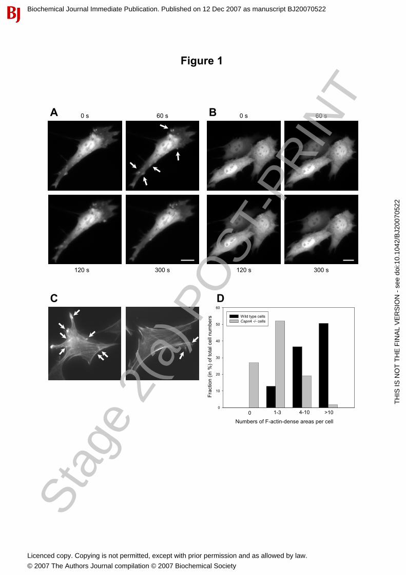
Figure 1
B
Fra
ctio
n(in
%)
of to
tal c
elln
umbe
rs
0
10
20
30
40
50
60
0 1-3 4-10 >10
DWild type cellsCapn4 -/- cells
Numbers of F-actin-dense areas per cell
A 0 s
120 s 300 s
60 s
120 s
0 s
300 s
60 s
C
Stag
e 2(
a) P
OST
-PR
INT
Biochemical Journal Immediate Publication. Published on 12 Dec 2007 as manuscript BJ20070522
TH
IS IS
NO
T T
HE
FIN
AL
VE
RS
ION
- s
ee d
oi:1
0.10
42/B
J200
7052
2
Licenced copy. Copying is not permitted, except with prior permission and as allowed by law.
© 2007 The Authors Journal compilation © 2007 Biochemical Society

Figure 2
A
GFP control
1 2 3
Capn4 +/+
105
105
Capn4 -/-
kDa
75
75
m80k-GFP
m80k-GFP
C
m-calpain-GFP
B
m80k
m80k
Stag
e 2(
a) P
OST
-PR
INT
Biochemical Journal Immediate Publication. Published on 12 Dec 2007 as manuscript BJ20070522
TH
IS IS
NO
T T
HE
FIN
AL
VE
RS
ION
- s
ee d
oi:1
0.10
42/B
J200
7052
2
Licenced copy. Copying is not permitted, except with prior permission and as allowed by law.
© 2007 The Authors Journal compilation © 2007 Biochemical Society

Figure 3
A%
cel
ls d
isp
layi
ng
mem
bra
ne
ble
bs
0
20
40
60
80
100
Capn4 +/+GFP
Capn4 -/-GFP
Capn4 -/-GFP + 28k
Capn4 -/-m80-GFP
+ 28k
a a
a
a
cc
b
c
120 s
300 s
60 s
180 s
B
Stag
e 2(
a) P
OST
-PR
INT
Biochemical Journal Immediate Publication. Published on 12 Dec 2007 as manuscript BJ20070522
TH
IS IS
NO
T T
HE
FIN
AL
VE
RS
ION
- s
ee d
oi:1
0.10
42/B
J200
7052
2
Licenced copy. Copying is not permitted, except with prior permission and as allowed by law.
© 2007 The Authors Journal compilation © 2007 Biochemical Society

Figure 4
AHSP70
HSP70
IDH3A
IDH3A
ERp29
ERp29
AOP-1
Cofilin 1
Alanyl-tRNA-synthetase
MPR-BP
Tropomyosin
Calpain 28k
RhoGDI-1
eIF-5A
CRABP-1
4.0 4.5 5.0 5.5 6.0 6.5 7.0pH
B Tropomyosin RhoGDI-1 Calpain 28k Cofilin 1
Wild type
Tropomyosin RhoGDI-1 Calpain 28k Cofilin 1
calpain 28k
Sp
ot
inte
nsi
ty
0,0
0,2
0,4
0,6
0,8
1,0
1,2Wild typeCapn4 -/-
RhoGDI-1 cofilin 1 tropomyosin
C
Capn4-/-
Stag
e 2(
a) P
OST
-PR
INT
Biochemical Journal Immediate Publication. Published on 12 Dec 2007 as manuscript BJ20070522
TH
IS IS
NO
T T
HE
FIN
AL
VE
RS
ION
- s
ee d
oi:1
0.10
42/B
J200
7052
2
Licenced copy. Copying is not permitted, except with prior permission and as allowed by law.
© 2007 The Authors Journal compilation © 2007 Biochemical Society
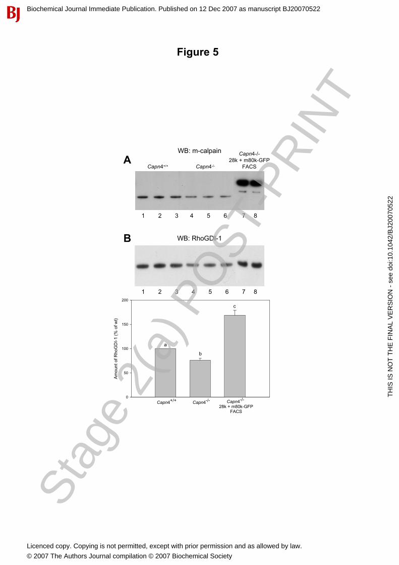
Figure 5
WB: m-calpain
Am
ount
of R
hoG
DI-
1(%
of w
t)
0
50
100
150
200
Capn4-/- Capn4-/-
28k + m80k-GFPFACS
Capn4+/+
a
b
c
ACapn4-/-Capn4+/+
Capn4-/-28k + m80k-GFP
FACS
B WB: RhoGDI-1
Stag
e 2(
a) P
OST
-PR
INT
Biochemical Journal Immediate Publication. Published on 12 Dec 2007 as manuscript BJ20070522
TH
IS IS
NO
T T
HE
FIN
AL
VE
RS
ION
- s
ee d
oi:1
0.10
42/B
J200
7052
2
Licenced copy. Copying is not permitted, except with prior permission and as allowed by law.
© 2007 The Authors Journal compilation © 2007 Biochemical Society
Related Documents