12 Genetic Disorders Associated with Gingival Enlargement Mostafa Ibrahim 1 , Maha Abouzaid 2 , Mennat Mehrez 3 , Heba Gamal El Din 4 and Ghada El Kamah 5 1,2,3,4 Oro-Dental Genetics Department, National research center, Cairo 5 Clinical Genetics Department, National research center, Cairo Egypt 1. Introduction A number of genetic disorders present with gingival manifestations which may be in the form of desquamative, ulcerative lesions or an enlargement of the gingiva. Gingival enlargement is a broad term that refers to gingival overgrowth without cause suggestion i.e. a strictly clinical description of the condition avoiding the flawed pathologic implications of terms used such as hypertrophic gingivitis or gingival hyperplasia. In this chapter we will summarize gingival enlargement that can be attributed to gene pathology. Gingival enlargement may present in some genetic disorders secondary to certain treatments not to actual gene expression e.g. Cystinosis secondary to treatment with cyclosporine-A, or epilepsy treated with phenytoin. This category of genetic disorders will not be discussed in this chapter, but should be considered in the differential diagnosis. Genetic disorders associated with gingival enlargement fall into four main categories according to etiology, clinical presentation and histopathological findings (Table 1). This classification is suggested as a guiding tool in differential diagnosis. The first category is Hereditary Gingival Fibromatosis (HGF), which represents a heterogeneous group of disorders characterized by progressive enlargement of the gingiva. HGF may appear as an isolated entity i.e. as autosomal dominant Gingival Fibromatosis or as part of a syndrome. These syndromes are rather rare but they all have gingival fibromatosis as a constant feature. The second category is Lysosomal Storage Disorders which are a group of disorders characterized by deposition of macromolecules anywhere in the body including the gingiva leading to gingival enlargement. Gingival enlargement in this category is not always a constant feature. It ranges from being common to being a rare feature. The third category is referred to as Vascular Disorders while the last category includes syndromes characterized by the presence of characteristic dental abnormalities. 2. Hereditary gingival fibromatosis Gingival enlargement may present as a specific entity, hereditary gingival fibromatosis (HGF), and may appear in an isolated form. However, there are several uncommon syndromes in which gingival fibromatosis can be a feature. www.intechopen.com

Welcome message from author
This document is posted to help you gain knowledge. Please leave a comment to let me know what you think about it! Share it to your friends and learn new things together.
Transcript

12
Genetic Disorders Associated with Gingival Enlargement
Mostafa Ibrahim1, Maha Abouzaid2, Mennat Mehrez3, Heba Gamal El Din4 and Ghada El Kamah5
1,2,3,4Oro-Dental Genetics Department, National research center, Cairo 5Clinical Genetics Department, National research center, Cairo
Egypt
1. Introduction
A number of genetic disorders present with gingival manifestations which may be in the form of desquamative, ulcerative lesions or an enlargement of the gingiva. Gingival enlargement is a broad term that refers to gingival overgrowth without cause suggestion i.e. a strictly clinical description of the condition avoiding the flawed pathologic implications of terms used such as hypertrophic gingivitis or gingival hyperplasia. In this chapter we will summarize gingival enlargement that can be attributed to gene pathology. Gingival enlargement may present in some genetic disorders secondary to certain treatments not to actual gene expression e.g. Cystinosis secondary to treatment with cyclosporine-A, or epilepsy treated with phenytoin. This category of genetic disorders will not be discussed in this chapter, but should be considered in the differential diagnosis. Genetic disorders associated with gingival enlargement fall into four main categories according to etiology, clinical presentation and histopathological findings (Table 1). This classification is suggested as a guiding tool in differential diagnosis. The first category is Hereditary Gingival Fibromatosis (HGF), which represents a heterogeneous group of disorders characterized by progressive enlargement of the gingiva. HGF may appear as an isolated entity i.e. as autosomal dominant Gingival Fibromatosis or as part of a syndrome. These syndromes are rather rare but they all have gingival fibromatosis as a constant feature. The second category is Lysosomal Storage Disorders which are a group of disorders characterized by deposition of macromolecules anywhere in the body including the gingiva leading to gingival enlargement. Gingival enlargement in this category is not always a constant feature. It ranges from being common to being a rare feature. The third category is referred to as Vascular Disorders while the last category includes syndromes characterized by the presence of characteristic dental abnormalities.
2. Hereditary gingival fibromatosis
Gingival enlargement may present as a specific entity, hereditary gingival fibromatosis (HGF), and may appear in an isolated form. However, there are several uncommon syndromes in which gingival fibromatosis can be a feature.
www.intechopen.com

Gingival Diseases – Their Aetiology, Prevention and Treatment
190
Hereditary Gingival Fibromatosis
Lysosomal Storage Disorders
Vascular Disorders
Disorders Associated with
Dental Abnormalities
Gingival fibromatosis (isolated)
Hurler syndrome Sturge Weber
syndrome Wilson syndrome
Zimmerman – Laband syndrome
Maroteaux-Lamy syndrome
Klippel-Trenaunay syndrome
Goltz syndrome
Ramon syndrome Scheie syndrome
Regional Odontodysplasia Systemic hyalinosis Hurler/ scheie
Jones syndrome Hunter syndrome
Rutherfurd syndrome Sly syndrome
Cross Syndrome I- Cell disease
Gingival fibromatosis, hypertrichosis and mental retardation.
Aspartylglucosaminuria
Neurofibromatosis type I
Alpha Mannosidosis
Schinzel – Giedion syndrome
Niemann – Pick disease
Costello syndrome Anderson – Fabry
disease
Menkes Kinky hair disease
Ligneous periodontitis
Cowden syndrome
Table 1. Classification of genetic disorders associated with gingival enlargement.
Clinically HGF develops as a slowly progressive, benign, localized or generalized
enlargement of keratinized gingiva that, in severe cases, may cover the crowns of the
teeth.Localized forms of HGF usually affect the maxillary tuberosities and the labial gingiva
around the mandibular molars. However, the symmetric generalized form of HGF that
affects the labial, lingual, and palatal gingiva is the most common (Baptista, 2002; Kelekis-
Cholakis et al., 2002). Males and females are equally affected. (Xiao et al., 2001; Ye et al.,
2005). Enlarged gingiva may be normal in color or erythematous and are firm and nodular
on palpation. Although the alveolar bone is usually unaffected, gingival enlargement results
in pseudo-pocketing and periodontal problems, due to difficulties in maintaining an
effective level of oral hygiene. The overgrowth may also result in functional and esthetic
concerns, create diastemas, impede or delay tooth eruption, and create changes in facial
appearance as a result of lip protrusion. Severe overgrowth can result in crowding of the
tongue, speech impediment, and difficulty with mastication, and can prevent normal
closure of lips (Lynch et al., 1994; Shafer, 1983).The onset of gingival overgrowth usually
coincides with the eruption of the permanent incisors, or, at times, with the eruption of the
primary dentition. In very rare cases; it can be also present at birth (Anderson et al., 1969).
www.intechopen.com

Genetic Disorders Associated with Gingival Enlargement
191
Since HGF has not been reported in edentulous patients, it appears that the presence of teeth
is necessary for overgrowth to develop. Histologically: HGF usually involves moderate hyperplasia of a dense, hyperkeratotic epithelium with elongated rete ridges (Araujo et al., 2003; Doufexi et al., 2005). Epithelial hyperplasia can also occur as a consequence of acanthosis, but this was found only in areas of chronic inflammation (Farrer-Brown et al., 1972; Raeste et al., 1978). HGF tissues show an increased amount of collagen fiber bundles running in all directions associated with few fibroblasts and blood vessels (Araujo et al., 2003; Doufexi et al., 2005; Martelli-Junior et al., 2000). Two populations of fibroblasts were identified in the lesions. One contains little cytoplasm around the nucleus, which is associated with dense collagen bundles. The other contains prominent cytoplasm with well developed organelles. Those fibroblasts have been considered inactive and active, respectively (Collan et al., 1982; Sakamoto et al., 2002). The connective tissue in HGF also exhibits an accumulation of elastic and oxytalan fibers (Baptista, 2002; Chavrier & Couble, 1979; Doufexi et al., 2005; Hart et al., 2000; Sakamoto et al., 2002). Although a rare finding, small osseous calcifications and abundant neurovascular bundles may also be present (Gunhan et al., 1995; Kelekis-Cholakis et al., 2002). HGF does not usually involve inflammation and local accumulation of inflammatory cells can be found only in cases where pseudo-pocketing resulted in plaque accumulation (Shafer, 1983).
Extracellular matrix production and degradation:
The hallmark of HGF is the accumulation of excess extracelluar matrix (ECM). Transforming growth factor (TGF) expression is up regulated in HGF (Häkkinen & Csiszar, 2007) . TGF can promote ECM accumulation by increasing ECM synthesis and can also inhibit ECM breakdown by down regulating matrix metalloproteinases (MMPs) expression and by increasing expression of tissue inhibitors of matrix metalloproteinases (Steffensen et al., 2001).
2.1 Isolated hereditary gingival fibromatosis Isolated hereditary gingival fibromatosis (OMIM #135300; Gene Map locus 2p21; other loci reported on chromosomes 5q & 11p) is mainly autosomal dominant (Fig.1), though autosomal recessive inheritance has been reported. The enlargement affects both deciduous and permanent dentition. The gingiva appears firm, non hemorrhagic and large enough to interfere with speech and, in some instances, with mouth closure (Ramakrishnan et al., 2010).
(a)Affected mother (b)Affected son
Fig. 1. Isolated autosomal dominant hereditary gingival fibromatosis.
www.intechopen.com

Gingival Diseases – Their Aetiology, Prevention and Treatment
192
2.2 Zimmerman – Laband syndrome Zimmerman – Laband syndrome or Laband syndrome (OMIM #135500; Gene Map locus
3p14.3) is an autosomal dominant disorder. Apart from gingival enlargement, it is
characterized by abnormal fingers, nails, nose, and ears. Other findings include splenomegaly,
hepatomegaly, and hyperextensible metacarpophalangelal joints (Hoogendijk et al., 2006).
2.3 Ramon Syndrome Ramon Syndrome (OMIM #266270) is characterized by cherubism, seizures, mental deficiency,
hypertrichosis, stunted growth and juvenile rheumatoid arthritis (Suhanya et al., 2010).
2.4 Systemic Hyalinosis Systemic Hyalinosis is an autosomal recessive systemic disorder due to mutation in CMG2, or ANTXR2 gene. It is characterized by widespread deposition of hyaline material in all body tissues. Some tend to classify this entity into infantile systemic hyalynosis (OMIM #236490, Gene Map locus 4q21) and juvenile hyaline fibromatosis (Murray-Puretic-Drescher syndrome OMIM #228600) according to age of onset & disease severity. Individuals usually present with painful joint contractures, diffuse thickening of the skin with pearly papules and fleshy nodules and failure to thrive. Gingival enlargement is a constant feature and other oral structures may also be enlarged. Histopathologic features are the deposition of amorphous, eosinophilic hyaline material (fig.2) (El-Kamah & Mostafa, 2009 ; El-Kamah et al., 2010).
2.5 Jones syndrome Jones syndrome (OMIM #135550) is autosomal dominant in inheritance. It is mainly characterized by gingival fibromatosis with progressive sensorineural deafness (Kasaboğlu et al., 2004).
2.6 Rutherfurd syndrome Rutherford syndrome (OMIM #180900) is usually autosomal dominant in inheritance. Its key features are corneal opacity, mental retardation and aggressive behavior. Gingival fibromatosis in this syndrome may be associated with failure of tooth eruption (Raja et al., 2008).
2.7 Cross syndrome Cross- McKusick- Breen syndrome or Kramer’s syndrome (OMIM #257800) is characterized by hypopigmentation, mental retardation and writhing movement of hands and legs (Witkop, 1971).
2.8 Gingival fibromatosis, hypertrichosis and mental retardation Gingival fibromatosis, hypertrichosis and mental retardation (OMIM #605400) is autosomal
recessive in inheritance. It is characterized by epilepsy, finger abnormalities, hirsutism,
bulbous short nose and abnormal ears (Gohlich-Ratmann et al., 2000).
2.9 Neurofibromatosis type I Neurofibromatosis or Von Recklinghausen disease (OMIM #162200, Gene Map locus
17q11.2) is an autosomal dominant neurocutaneous disorder caused by mutation in NF1
www.intechopen.com
Genetic Disorders Associated with Gingival Enlargement
193
(a) Gingival enlargement. (b) Fleshy nodules.
(c) Multiple nodules on joints. (d) 200X Hyalinosis in dermal papillae.
Fig. 2. Systemic Hyalinosis.
gene. It is characterized by cafe-au-lait spots, Lisch nodules in the eye, fibromatous tumors
of the skin with an increased risk of developing benign and malignant tumors. Orally, there
is gingival neurofibroma. Characteristic histopathologic features: neurofibroma cells can be
detected with their nuclei among the waved collagen fibers -beneath the oral mucosa.
Epithelium and numerous tumor cells and capillary vessels can be seen among waved
collagen fibers (El-Kamah et al., 2004).
2.10 Schinzel-Giedion syndrome Schinzel-Giedion syndrome or Schinzel-Giedion mid-face retraction syndrome (OMIM
#269150, Gene Map locus 18q21.1) is an autosomal recessive malformation syndrome
characterized by severe mid-face retraction referred to as ‘figure-of-eight' appearance,
severe mental retardation and congenital heart defect. Neither the etiology nor detailed
clinical course is known since most of the patients affected with Schinzel-Giedion syndrome
die before the age of ten. However, a long-lived patient showed gingival hyperplasia that
was progressive even after gingivectomy. Histopathologic examination revealed fibrous
hyperplasia of the gingiva with mucoid depositions and no inflammatory changes (Kondoh
et al., 2001).
www.intechopen.com
Gingival Diseases – Their Aetiology, Prevention and Treatment
194
2.11 Costello syndrome Costello syndrome or Noonan like syndrome with nasal papillomata (OMIM #218040) is a
rare disease characterized by fetal and neonatal macrostomia with slow postnatal growth
due to the severe feeding difficulties, distinctive coarse facial dysmorphism and mental
retardation. The most striking cutaneous feature is redundant skin of the neck, hands and
feet. Nasal and perioral papillomas are also common between the ages of 2 and 15. Oral
examination is important as Costello syndrome patients develop gingival hyperplasia
usually within the first years of life and is considered as a quite distinct feature that can also
aid in its differential diagnosis from Noonan syndrome and Cardiofaciocutaneous
syndrome that phenotypically overlap with Costello syndrome (Digilio et al., 2008).
3. Lysosomal storage disorders
Lysosomal storage diseases are a heterogeneous group of disorders caused by lysosomal
enzyme dysfunction including mucopolysaccharidosis, mucolipidosis and others.
Individually they are very rare, but this group as a whole has a prevalence of more than
1:8,000 live births (Manger, 2010). The majority of lysosomal storage disorders (LSDs) result
from defective lysosomal acid hydrolysis of endogenous macromolecules and their
consequent accumulation. Over 40 disorders have been described. They tend to be
multisystemic and are always progressive, although the rate of progression may vary. There
are several potential ways in which accumulated substrate might cause the disease. The
most obvious is enlargement of the affected cell, resulting in enlargement of the respective
organ such as hepatosplenomegaly, cardiomyopathy etc. (Vellodi, 2005). The buildup of
undigested material, secondary to lysosomal enzyme dysfunction, results in the formation
of typical histochemical and ultrastructural changes. Light microscopy often reveals
engorged macrophages with a characteristic appearance, such as that of ‘sea-blue
histiocytes’ in Niemann–Pick disease (Vanier et al., 1988).
3.1 Mucopolysaccharidosis Mucopolysaccharidosis (MPS) are a family of lysosomal storage disorders resulting from the
partial catabolism of several glycosaminoglycans (GAGs). Depending on which particular
enzyme is deficient, the MPS syndromes are defined into groups MPS I through VII, with
several subgroups for a total of 10 disorders. In humans, clinical features include
dysmorphic features, hepatosplenomegaly, hypertelorism, macroglossia, hypoplastic and
irregularly shaped teeth, hyperplastic lips and gingiva, facial dysmorphia, corneal clouding,
and mental retardation. Gingival enlargement is considered as one of the main oral
manifestations of Maroteaux-Lamy syndrome, and a common feature in Hurler syndrome.
It is rarely reported with Scheie syndrome, Hurler/ scheie compound syndrome, Hunter's
syndrome and Sly syndrome. Gingival enlargement may not be previously reported with
Sanfilippo syndrome and Morquio syndrome (Sheridan et al., 1994).
3.1.1 Hurler syndrome Hurler syndrome (Mucopolysaccharidosis IH, OMIM #607014, Gene Map locus 4p16.3) is
an autosomal recessive disorder caused by a mutation in the gene encoding for the enzyme
alpha-L-iduronidase leading to deficiency of the enzyme and accumulation of
glycosaminoglycans (heparan sulphate and dermatan sulphate) in various tissues (Hingston
www.intechopen.com
Genetic Disorders Associated with Gingival Enlargement
195
et al., 2006). Hurler sundrome is characterized by mental retardation, dwarfism, coarse facial
features, flexion contractures, hepatosplenomegaly, hernias, corneal clouding (Leroy &
Croeker, 1966; McKusick et al., 1965), respiratory infections and cardiac complications
(Mckusick &Neufeld, 1983). Gingival hyperplasia is a common feature in this disorder.
Other intraoral features include macroglossia, short mandibular rami with abnormal
condyles consistent with limited opening of the mouth, spaced hypoplastic peg-shaped
teeth with retarded eruption, and localized dentigerous cyst-like radiolucencies (Gardner,
1971; Keith & Weidmann, 1990; Thomas &Tandon, 2000; Worth, 1966). Histopathological
reports showed Hurler cells in the gingival tissue (Gardner, 1968).
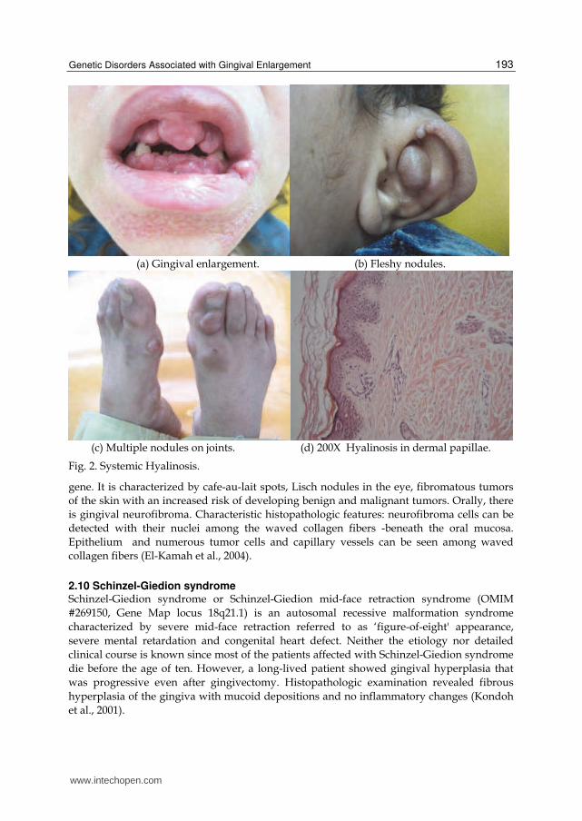
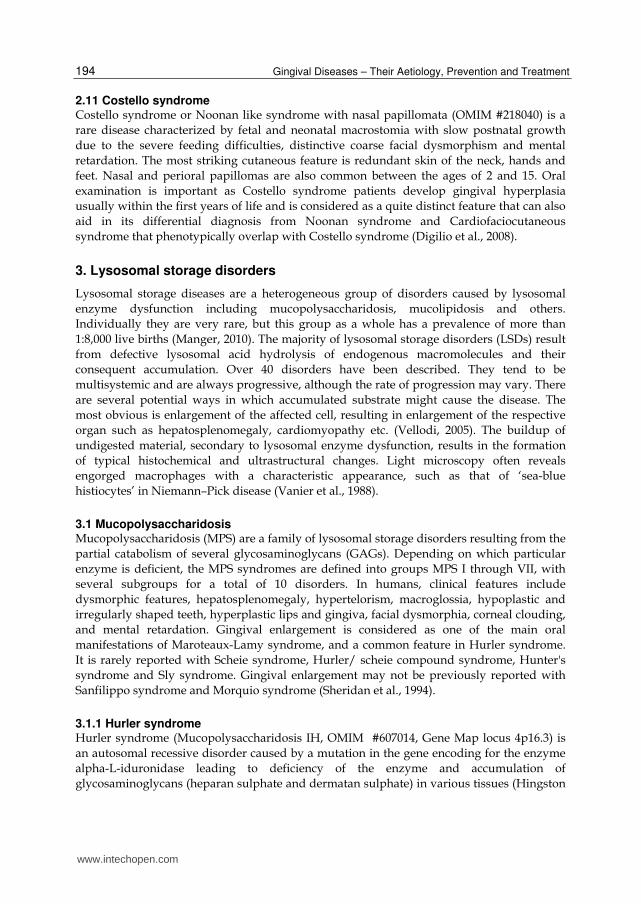
3.1.2 Maroteaux-lamy syndrome Maroteaux-lamy syndrome or Mucopolysaccharidosis typeVI (OMIM #253200, Gene Map locus 5q13) is a lysosomal storage disorder inherited as an autosomal recessive trait. It is due to deficiency of arylsulphatase B enzyme which results in accumulation of dermatan sulphate in tissues and its excretion in urine. It is characterized by growth retardation, enlargement of the skull with a long anteroposterior dimension and corneal opacities. Presence of normal intelligence as well as metachromatic inclusions in leukocytes distinguishes it from other mucopolysaccharidosis (Fig 3,a). The oral findings include short or stubby, malformed, peg-shaped, poorly formed and calcified teeth with delayed eruption. Gingival hyperplasia and hypertrophy of the maxillary alveolar ridges are often mentioned as the main oral manifestations of the Maroteaux-lamy syndrome (Fig 3,b). Also, the anterior teeth may present an open-bite relationship in association with macroglossia. (Alpoz et al., 2006; Guimaraes et al.,2010).
(a) MPS VI (usually with normal intellectual
development). (b) Gingival hyperplasia and hypertrophy of
the maxillary alveolar ridges.
Fig. 3. Maroteaux-lamy syndrome.
3.1.3 Scheie and Hurler /Sheie syndrome Scheie syndrome (Mucoploysaccharidosis IS, OMIM #607016, Gene Map locus 4p16.3) represents the mildest form of mucoplysaccharidosis. An intermediate phenotype lying in between these two variants of mucopolysaccharidosis I is the Hurler /Sheie compound
www.intechopen.com
Gingival Diseases – Their Aetiology, Prevention and Treatment
196
syndrome (Mucoploysaccharidosis I H/S, OMIM #607015, Gene Map locus 4p16.3) (Kelly, 1976).
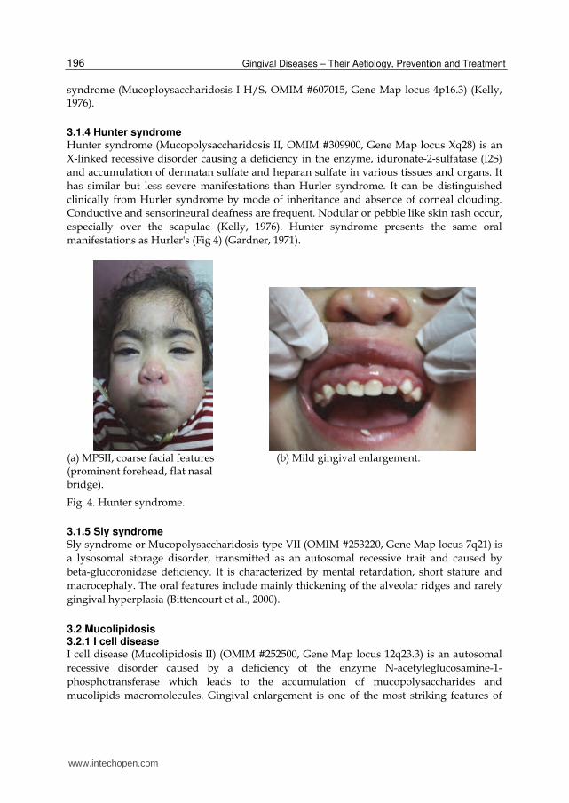
3.1.4 Hunter syndrome Hunter syndrome (Mucopolysaccharidosis II, OMIM #309900, Gene Map locus Xq28) is an
X-linked recessive disorder causing a deficiency in the enzyme, iduronate-2-sulfatase (I2S)
and accumulation of dermatan sulfate and heparan sulfate in various tissues and organs. It
has similar but less severe manifestations than Hurler syndrome. It can be distinguished
clinically from Hurler syndrome by mode of inheritance and absence of corneal clouding.
Conductive and sensorineural deafness are frequent. Nodular or pebble like skin rash occur,
especially over the scapulae (Kelly, 1976). Hunter syndrome presents the same oral
manifestations as Hurler's (Fig 4) (Gardner, 1971).
(a) MPSII, coarse facial features (prominent forehead, flat nasal bridge).
(b) Mild gingival enlargement.
Fig. 4. Hunter syndrome.
3.1.5 Sly syndrome Sly syndrome or Mucopolysaccharidosis type VII (OMIM #253220, Gene Map locus 7q21) is
a lysosomal storage disorder, transmitted as an autosomal recessive trait and caused by
beta-glucoronidase deficiency. It is characterized by mental retardation, short stature and
macrocephaly. The oral features include mainly thickening of the alveolar ridges and rarely
gingival hyperplasia (Bittencourt et al., 2000).
3.2 Mucolipidosis 3.2.1 I cell disease I cell disease (Mucolipidosis II) (OMIM #252500, Gene Map locus 12q23.3) is an autosomal
recessive disorder caused by a deficiency of the enzyme N-acetyleglucosamine-1-
phosphotransferase which leads to the accumulation of mucopolysaccharides and
mucolipids macromolecules. Gingival enlargement is one of the most striking features of
www.intechopen.com

Genetic Disorders Associated with Gingival Enlargement
197
this syndrome and the patient’s lower face has a fish-like profile. It is referred to as I cell
disease based on the histopathologic features because the macromolecules that accumulate
inside the cell form characteristic cytoplasmic inclusions (Mahfouz et al., 2010).
3.3 Miscellaneous lysosomal storage 3.3.1 Aspartylglucosaminuria Aspartylglucosaminuria or AGU (OMIM #208400, Gene Map locus 4q33-4q35) is an
autosomal recessive lysosomal storage disorder caused by deficiency of
aspartylglucosaminidase leading to the accumulation of glycoasparagines in lysosomes. The
main symptom is progressive mental retardation where the patients are only able to learn
new skills and abilities up to the age of 16 years. They then undergo gradual somatic and
mental deterioration. The facial features coarsen with age with characteristic sagging of the
facial skin. Dysmorphic orofacial features include macroglossia, malocclusions, limited
mouth opening as well as thick lips. Edematous buccal mucosa (leukoedema) and gingival
fibromatosis are common in AGU patients. The gingival overgrowths were diagnosed
histologically as fibroepithelial hyperplasia (Arvio et al., 1999).
3.3.2 Alpha Mannosidosis Alpha Mannosidosis (OMIM #248500, Gene Map locus 19q13-19q12), is a rare lysosomal
storage disorder, transmitted as an autosomal recessive trait, and is due to deficient activity
of alpha mannosidase, resulting in an abnormal accumulation of mannose-containing
residues. It is characterized by growth and mental retardation, coarse facial features and
muscular hypotonia. The oral findings include macroglossia, widely spaced teeth and firm
hyperplastic nodules of the gingiva which upon histologic examination reveals infiltration
with foamy histiocytes. Blood smears show cytoplasmic vacuolization of lymphocytes and
monocytes (Ischigami et al., 1995).
3.3.3 Niemann-Pick disease Niemann-Pick disease (OMIM #257200, Gene Map locus 18q11-18q12 type C, 11p15), an
autosomal recessive disorder caused by deficiency of a specific enzyme activity ‘acid
sphingomyelinase’ with subsequent accumulation of sphingolipids in cells, throughout the
body. Oral findings include thick lips, macroglossia and widely spaced teeth. Although
gingival enlargement is not considered a constant feature, a case was presented with
generalized grade III gingival enlargement, which recurred even after excision and
thorough maintenance implying that there is a link between the disease and the gingival
enlargement. Gingival biopsy upon histologic examination revealed infiltration with foamy
histiocytes. Blood smear showed cytoplasmic vacuolization of lymphocytes and monocytes
(Kaisare, 2007).
3.3.4 Anderson Fabry disease Anderson Fabry disease or Angiokeratoma Corporis Diffusum (OMIM #301500, Gene Map
locus Xq21-Xq22) is an X-linked recessively inherited disease due to deficiency of the
enzyme ceramide trihexosidase, that results in intracellular accumulation of the glycolipid
ceramide trihexoside in vascular endothelial cells, pericytes, fibroblasts, macrophages, and
other cells of the body. The disease is characterized by painful crises involving the
www.intechopen.com

Gingival Diseases – Their Aetiology, Prevention and Treatment
198
extremities and the abdomen as well as angiokeratomas of the skin that may also involve
the oral mucous membrane, mainly the labial mucosa followed by the buccal mucosa and
the gingiva. Gingival enlargement may be present secondary to dilantin therapy. Young et
al. (1978) presented a case with Fabry disease where granulomatous gingivitis has been
described. Histologically, angiokeratoma of the gingiva shows ceramide inclusions not only
in the connective tissue, but also in the oral epithelial cells.
3.3.5 Menkes Kinky hair disease Menkes Kinky hair disease or Menkes Steely hair syndrome (OMIM #309400, Gene Map locus Xq13) is a rare X-linked recessive neurodegenerative disorder caused by a defect of copper transport and metabolism. It is characterized by brittle, sparse and twisted hair, and generalized depigmentation of the hair. The oral findings include delayed dentition and gingival hyperplasia (McKusick, 2011).
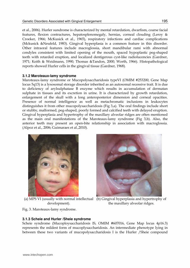
3.3.6 Ligneous periodontitis Ligneous periodontitis, Plasminogen deficiency or Ligneous conjunctivitis (OMIM #217090, Gene Map locus 6q22) is an autosomal recessive disorder in PLG gene. Plasminogen deficiency is characterized by gingival swelling involving both the maxillary and mandibular arches, pinkish waxy painless masses that have no tendency to bleed with palpation and hyperplastic gingival papillae concealing most of the teeth. Areas of the gingiva covered with tough yellowish white membrane, thin pseudomembrane, that could be wiped away, overlay the tough part of the membrane (Fig.5,a). Other disease manifestations include; ligneous conjunctivitis (Fig.5,b), Corneal involvement that may lead to blindness in 30% of cases. Other system involvement such as laryngeal and tracheobronchial involvement resulting in voice change and obstructive pulmonary disease have been described. Characteristic histopathologic manifestations shown in (Fig.5,c) are epithelial hyperplasia and fibrin deposition underneath the epithelium and around the blood vessels. The dermis shows edema and perivascular mixed cellular infiltrate; mostly plasma cells, polymorphonuclear leukocytes, few lymphocytes, and mast cells. Amorphous hyaline-like eosinophilic material of the pseudomembranes, which resembles amyloid but negative for Congo red stain, that contains fibrin (El-Darouti et al., 2009).
3.3.7 Cowden syndrome Cowden syndrome or Multiple Hamartomas (OMIM #158350, Gene Map locus 10q23.31) is an autosomal dominant inherited disorder. In 80% of cases it is due to mutation in the PTEN tumor suppressor gene. Others may have mutations in certain subunits of succinate dehydrogenase, mitochondrial enzyme (Ni et al., 2008). Recently, methylation of the KILLIN gene has also been reported in patients with similar clinical features. Oral manifestations include cobblestone-like papules of the gingiva and buccal mucosa. However, the disease is characterized by learning disabilities, autism, and/or mental retardation, macrocephaly and multiple hamartomatous lesions, especially of the skin, mucous membranes, breast and thyroid. Verrucous skin lesions of the face and limbs, and multiple facial trichilemmomas are common findings. Hamartomatous polyps of the gastrointestinal tract, mucocutaneous lesions, and increased risk of developing neoplasms have been reported (Tan et al., 2011).
www.intechopen.com

Genetic Disorders Associated with Gingival Enlargement
199
(a) Yellowish white pseudo-membrane
covering most of the hypertrophic gingiva.
(b) Yellowish white pseudo-membrane affecting the tarsal conjunctiva.
(c) Fibrin deposition around dermal blood vessels associated with perivascular and
interstitial mixed infiltrate.
(d) Panoramic radiograph showing floating teeth with severe alveolar bone loss.
Fig. 5. Clinical and histopathological characteristics in Ligneous periodontitis and Ligneous conjunctivitis.
4. Vascular disorders
4.1 Sturge Weber syndrome Sturge Weber syndrome or encephalofacial angiomatosis (OMIM #185300) is almost always
a sporadic disease. However, there have been reports of cases with autosomal recessive and
dominant inheritance. It has four main features; unilateral cutaneous nevi along trigeminal
nerve sensory distribution (Fig.6,a), unilateral vascular hyperplasia of oral mucosa and
gingiva, neurological manifestations and ocular complications (Pereira de Godoy et al., 2010;
Zhou et al., 2010). Sturge-Weber syndrome is characterized by an intracranial vascular
anomaly and calcification, leptomeningeal angiomatosis, most often involving the occipital
and posterior parietal lobes (Fig.6b).
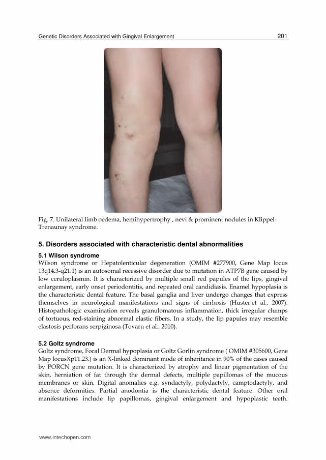
4.2 Klippel-Trenaunay syndrome Klippel-Trenaunay syndrome or Angioosteohypertrophy syndrome (OMIM #%149000,
Gene Map locus 8q22.3) has a paradominant inheritance (Happle, 1993), It is characterized
by a triad of features, namely, vascular nevi, venous varicosities, and hyperplasia of hard
and soft tissues in the affected area (Fig.7). Despite its rarity, Klippel-Trenaunay Syndrome
www.intechopen.com

Gingival Diseases – Their Aetiology, Prevention and Treatment
200
should be considered in the differential diagnosis of gingival enlargement. (Anand
& Roshna, 2006). Gingival capillary hemangiomas, gingival fibroma, Gingival fibromatosis,
gingival hyperplasia. Other oral manifestations include high arched palate, unilateral
hypertrophy, or increase in size of periodontal tissues, tongue capillary hemangiomas,
unilateral macroglossia, increase in size of fungi-form papillae, unilateral increase in lips
size, teeth malformation, diastema formation, premature eruption of teeth on affected side,
delayed exfoliation of primary teeth, early mineralization of roots on affected side,
accelerated growth of teeth, anterior open bite, cross bite and floor of mouth capillary
hemangiomas (Fakir et al., 2009; McKusick, 2011).
(a) Congenital large port wine stain involving the right side of the face and scalp extending
to the left side.
(b) CT scan showing intracranial calcification.
Fig. 6. Sturge Weber syndrome.
www.intechopen.com
Genetic Disorders Associated with Gingival Enlargement
201
Fig. 7. Unilateral limb oedema, hemihypertrophy , nevi & prominent nodules in Klippel-Trenaunay syndrome.
5. Disorders associated with characteristic dental abnormalities
5.1 Wilson syndrome Wilson syndrome or Hepatolenticular degeneration (OMIM #277900, Gene Map locus
13q14.3-q21.1) is an autosomal recessive disorder due to mutation in ATP7B gene caused by
low ceruloplasmin. It is characterized by multiple small red papules of the lips, gingival
enlargement, early onset periodontitis, and repeated oral candidiasis. Enamel hypoplasia is
the characteristic dental feature. The basal ganglia and liver undergo changes that express
themselves in neurological manifestations and signs of cirrhosis (Huster et al., 2007).
Histopathologic examination reveals granulomatous inflammation, thick irregular clumps
of tortuous, red-staining abnormal elastic fibers. In a study, the lip papules may resemble
elastosis perforans serpiginosa (Tovaru et al., 2010).
5.2 Goltz syndrome Goltz syndrome, Focal Dermal hypoplasia or Goltz Gorlin syndrome ( OMIM #305600, Gene
Map locusXp11.23.) is an X-linked dominant mode of inheritance in 90% of the cases caused
by PORCN gene mutation. It is characterized by atrophy and linear pigmentation of the
skin, herniation of fat through the dermal defects, multiple papillomas of the mucous
membranes or skin. Digital anomalies e.g. syndactyly, polydactyly, camptodactyly, and
absence deformities. Partial anodontia is the characteristic dental feature. Other oral
manifestations include lip papillomas, gingival enlargement and hypoplastic teeth.
www.intechopen.com

Gingival Diseases – Their Aetiology, Prevention and Treatment
202
Characteristic histopathologic features showed deposits of fat cells or adipose tissue in the
dermis (Maas et al., 2009; McKusick, 2011).
5.3 Odontodysplasia Odontodysplasia is an uncommon condition that can affect both primary and permanent dentitions. Both enamel and dentine are defected. Clinically, the teeth are mutilated in shape, pitted and yellowish to brownish in colour with excessive wear. Radiographically, enamel & dentine show lack of contrast, with decreased radiopacity rendering the tooth a ghost like appearance. The pulp chambers are wide and with open apices (Fig.8 a & b)
(a) Affected teeth are mutilated in shape, pitted and yellowish to brownish in color (arrow) with gingival enlargement in the
affected side.
(b) Panoramic radiograph showing ghost like appearance of the affected teeth.
(c) Gingival biopsy showing odontogenic tissue in the epithelium and intramesenchymalcalcifications.
Fig. 8. Regional maxillary odontodysplasia.
www.intechopen.com
Genetic Disorders Associated with Gingival Enlargement
203
(Hamdan et al., 2004). It commonly presents as regional odontodysplasia where one or few
teeth may be involved. One or more quadrants may be involved but generalized
involvement is extremely rare (Shah and Gupta, 1998). Gingival enlargement is frequently
reported with regional type. It may present as an isolated or associating epidermal nevus/
Schimmelpenning-Feuerstein-Mims syndrome (OMIM #%163200) (McKusick, 2011;
Murakami et al., 1999). The exact etiology of odontodysplasia is still unknown. Genetic
predisposition has been proposed but the presence of local irritating factors during tooth
development has been more advocated. Gingival biopsy examination showed odontogenic
tissue in the epithelium and intramesenchymal calcifications (Fig.8c).
6. Conclusion
Gingival enlargement is an important feature in many genetic disorders. It can be one of the
main diagnostic features in some of these disorders e.g. ligneous periodontitis. In others
gingival enlargement coupled with other clinical features direct the physician to further
investigations. Accordingly, metabolic analysis, enzymatic essay, molecular analysis to
detect the candidate genes and histopathological studies may be requested. Histopatological
findings are considered of diagnostic value in a limited number of cases. They may become
pathonemonic when coupled with clinical examination e.g. hyaline material in hyalinosis,
fat deposits in Focal Dermal hypoplasia, odondogenic cells in odontodysplasia ……etc.
7. Acknowledgement
We would like to thank our colleagues in the departments of Oro-dental Genetics, Clinical
Genetics and Limb anomalies Clinic in the National Research Centre, Cairo, Egypt. Authors
would like to dedicate this work to the Egyptian revolution of January 25th 2011.
8. References
Alpöz, A.; Coker, M.; Celen, E.; Ersin, N.; Gökçen, D.; van Diggelenc, O. & Huijmansc, J.
(2006). The oral manifestations of Maroteaux-Lamy syndrome
(mucopolysaccharidosis VI); A case report, Oral Surg Oral Med Oral Pathol Oral
Radiol Endod, 101:632-7, ISSN: 1528-395X
Anand, PS. & Roshna, T. (2006). Rare instance of gingival enlargement in Klippel-Trenaunay
syndrome: a case report, J Contemp Dent Pract, 7:92-8, ISSN: 1526-3711
Anderson, J.; Cunliffe, WJ.; Roberts, DF. & Close, H. (1969). Hereditary gingival
fibromatosis, Br Med J, 3:218-219, ISSN: 0267-0623
Araujo, CS.; Graner, E.; Almeida, OP.; Sauk, JJ. & Coletta, RD. (2003). Histomorphometric
characteristics and expression of epidermal growth factor and its receptor by
epithelial cells of normal gingiva and hereditary gingival fibromatosis, J Periodontal
Res, 38:237-241, ISSN: 1600-0765
Baptista, IP. (2002). Hereditary gingival fibromatosis: a case report, J Clin Periodontol, 29:871-
874, ISSN: 1600-051X
www.intechopen.com

Gingival Diseases – Their Aetiology, Prevention and Treatment
204
Bittencourt, L.; Campos, V.; Moliterno, L.; Ribeiro, D.& Sampaio, R. (2000). Hereditary
gingival fibromatosis: review of the literature and a case report, Quintessence Int,
31(6):415-8, ISSN: 1936-7163
Chavrier, C. & Couble, ML. (1979). Ultrastructure of the connective corium in hereditary
gingival hyperplasia, J Biol Buccale, 7:191-203, ISSN: 0301-3952
Collan, Y.; Ranta, H.; Vartio, T.; Perheentupa, J. & Raeste, AM. (1982). Histohemical and
biochemical study of hereditary fibrous hyperplasia of the gingiva, Scand J Dent
Res, 90:20-28, ISSN: 0029-845X
Digilio, M.; Sarkozy, A.; Capolino, R.; Chiarini Testa, M.; Esposito, G.; de Zorzi, A.; Cutrera,
R.; Marino, B.& Dallapiccola, B. (2007). Costello syndrome: clinical diagnosis in the
first year of life, Eur J Pediatr, 167:621-628, ISSN: 1432-1076
Doufexi, A.; Mina, M. & Ioannidou, E. (2005). Gingival overgrowth in children:
epidemiology, pathogenesis, and complications. A literature review, J Periodontol,
76:3-10, ISSN: 1943-3670
El-Darouti, M.; Zayed, A.; El-Kamah, Gh. & Mostafa, M. (2009). Ligneous Conjunctivitis
with oral mucous membrane involvement and decreased plasminogen level, Pediatr
Dermatol, 26: 448–451, ISSN: 1525-1470
El-Kamah, Gh.; Abd-El-salam, GH.; Hassan, NA.; El-Darouti, M. & Temtamy, SA. (2004).
Neurofibromatosis 1: unusual clinical associations, Kasr El-Eini medical Journal,
10:45-56
El-Kamah, Gh.; & Mostafa, M. (2009). Heterogeneity and atypical presentation in infantile
systemic hyalinosis with severe labio-gingival enlargement: first Egyptian report,
Dermatol Online J, 15:6, ISSN: 1087-2108
El-Kamah, Gh.; Fong, K.; El-Ruby, M.; Affifi, HH.; Clements, SE.; Amr, K.; El-Darouti, M.&
McGrath, JA. (2010). Spectrum of mutations in the ANTXR2 (CMG2) gene in
infantile systemic hyalinosis and juvenile hyaline fibromatosis, Br J Dermatol .
163(1):213-215, ISSN: 1365-2133
Fakir, E.; Roberts, T.; Stephen, L. & Beighton, P. (2009). Klippel-Trenaunay-Weber
syndrome: orodental manifestations and management considerations, Oral Surg
Oral Med Oral Pathol Oral Radiol Endod, 107:754-758, ISSN: 1528-395X
Farrer-Brown, G.; Lucas, RB. & Winstock, D. (1972). Familial gingival fibromatosis: an unusual pathology, J Oral Pathol, 1:76-83, ISSN: 0300-9777
Gardner, DG. (1968). Metachromatic cells in the gingiva in Hurler's syndrome, Oral Surg
Oral Med Oral Pathol, 26:782-9, ISSN: 0030-4220
Gardner, DG. (1971). The oral manifestations of Hurler’s syndrome, Oral Surg Oral Med Oral
Pathol Oral Radiol Endod, 32:46-57, ISSN: 1528-395X
Gohlich-Ratmann, G.; Lackner, A.; Schaper, J.; Voit, T. & Gillessen-Kaesbach, G. (2000).
Syndrome of gingival hypertrophy, hirsutism, mental retardation and
brachymetacarpia in two sisters: specific entity or variant of a described condition?,
Am J Med Genet, 95: 241-246, ISSN: 1096-8628
Guimarães Mdo, C.; de Farias, SM.; Costa, AM.; de Amorim, RF. (2010). Maroteaux-Lamy
syndrome: Oro-facial features after treatment by Bone Marrow Transplant, Oral
Health Prev Dent; 8: 139-142, ISSN: 1602-1622
www.intechopen.com

Genetic Disorders Associated with Gingival Enlargement
205
Gunhan, O.; Gardner, DG.; Bostanci, H. & Gunhan, M. (1995). Familial gingival
fibromatosis with unusual histologic findings, J Periodontol, 66:1008-1011, ISSN:
1943-3670
Häkkinen, L. & Csiszar, A. (2007). Hereditary gingival fibromatosis: characteristics
and novel putative pathogenic mechanisms, J Dent Res, 86:25-34, ISSN: 1544-
0591
Hamdan, MA.; Sawair, FA.; Rajab, LD.; Hamdan, AM. & Al-Omari, IK. (2004). Regional
odontodysplasia: a review of the literature and report of a case, Int J Paediatr Dent,
14:363-7, ISSN: 1365-263X
Happle, R. (1993). Klippel-Trenaunay syndrome: is it a paradominant trait?, Br J
Dermatol, 128:465-466, ISSN: 1365-2133
Hart, TC.; Pallos, D.; Bowden, DW.; Bolyard. J.; Pettenati, MJ. & Cortelli, JR. (1998). Genetic
linkage of hereditary gingival fibromatosis to chromosome 2p21, Am J Hum Genet,
62:876-883, ISSN: 1537-6605
Hart, TC.; Pallos, D.; Bozzo, L.; Almeida, OP.; Marazita, ML. & O'Connell, JR. (2000).
Evidence of genetic heterogeneity for hereditary gingival fibromatosis, J Dent Res,
79:1758-1764, ISSN: 1544-0591
Hingston, EJ.; Hunter, ML.; Hunter, B. & Drage, N. (2006) . Hurler’s syndrome: dental
findings in a case treated with bone marrow transplantation in infancy, Int J
Paediatr Dent, 16:207-12, ISSN: 1365-263X
Hoogendijk, CF.; Marx, J.; Honey, EM.; Pretorius, E. & Christianson, AL. (2006).
Ultrastructural investigation of Zimmermann-Laband syndrome, Ultrastruct Pathol,
30:423-6, ISSN: 1521-0758
Huster, D.; Purnat, TD.; Burkhead, JL.; Ralle, M.; Fiehn, O.; Stuckert, F.; Olson, NE.;
Teupser, D. & Lutsenko, S. (2007). High copper selectively alters lipid metabolism
and cell cycle machinery in the mouse model of Wilson disease, J Biol Chem,
282:8343-8355, ISSN: 1083-351X
Ishigami, T.; Schmidt-Westhausen, A.; Philipsen, HP.; Baiborodin, SI.; Gelderblom, H.&
Reichart, PA. (1995). Oral manifestations of alpha-mannosidosis: report of a case
with ultrastructural findings, J Oral Pathol Med, 24:85-8, ISSN: 1600-0714
Kaisare, S. (2007). Gingival enlargement in Niemann-Pick disease: a coincidence or link?
Report of a unique case, Oral Surg Oral Med Oral Pathol Oral Radiol Endod, 104:e35-
e39, ISSN: 1528-395X
Kasaboğlu, O.; Tümer, C. & Balci, S.(2004). Hereditary gingival fibromatosis and
sensorineural hearing loss in a 42-year-old man with Jones syndrome, Genet Couns,
15:213-8, ISSN: 1015-8146
Keith, O.; Scully, C. & Weidmann, GM. (1990). Orofacial features of Scheie (Hurler-Scheie)
syndrome (alpha-L-iduronidase deficiency), Oral Surg Oral Med Oral Pathol Oral
Radiol Endod, 70:70-4, ISSN: 1528-395X
Kelekis-Cholakis, A.; Wiltshire, WA. & Birek, C. (2002). Treatment and long term follow-up
of a patient with hereditary gingival fibromatosis: a case report, J Can Dent Assoc,
68:290-294, ISSN: 1488-2159
Kelly, TE. (1976). The mucopolysaccharidoses and mucolipidoses, Clin Orthop Relat Res,
114:116-33, ISSN: 1528-1132
www.intechopen.com

Gingival Diseases – Their Aetiology, Prevention and Treatment
206
Leroy, JG. & Croeker, AC. (1966). Clinical definition of Hurler-Hunter phenotypes, Amer J
Dis Child, 112: 518-530, ISSN: 0002-922X
Kondoh, T.; Kamimura, N.; Tsuru, A.; Matsumoto, T.; Matsuzaka, T. & Moriuchi, H.
(2010). A case of Schinzel-Giedion syndrome complicated with progressive
severe gingival hyperplasia and progressive brain atrophy, Pediatr int, 43:181-
184, ISSN: 1442-200X
Lynch, M.; Brightman, VJ. & Greenberg, MS. (1994). Burket's oral medicine: diagnosis and
treatment, 9th ed., J.B. Lippincott, ISBN: 1550093266, Philadelphia
Maas, SM.; Lombardi, MP.; van Essen, AJ.; Wakeling, EL.; Castle, B.; Temple, IK.; Kumar,
VK.; Writzl, K. & Hennekam, RC. (2009). Phenotype and genotype in 17 patients
with Goltz-Gorlin syndrome, J Med Genet, 46:716-20, ISSN: 1468-6244
M Mahfouz, AK.; George, G.; Al-Bahlani, SS. & Al Nabhani, MZ. (2010). Difficult
intubation management in a child with I-cell disease, Saudi J Anaesth, 4:105-7,
ISSN: 0975-3125
Manger, B. (2010). Lysosomal storage diseases, Z Rheumatol, 69:527-38, ISSN: 1435-1250
Martelli-Junior, H.; Bolzani, G.; Graner, E.; Bozzo, L. & Coletta, RD. (2000). Microscopic and
proliferative comparison of gingival fibroblasts from patients with normal gingiva
and with hereditary gingival fibromatosis, Pesqui Odontol Bras, 14:123-129, ISSN:
1517-7491
McKusick, VA. Online Mendelian Inheritance in Man (OMIM), Accessed last on: 19/4/2011,
Available at: http://www.ncbi.nlm.nih.gov/omim/
McKusick, VA.; Kaplan, D.; Wise, D.; Hanley, WB.; Suddarth, SB.; Sevick, ME. & Maumanee,
AE. (1965). The genetic mucopolysaceharidoses, Medicine (Baltimore), 44: 445-483,
ISSN: 1536-5964
McKusick, VA. & Neufeld, EF. (1983). The mucopolysaccharide storage diseases. In:
Stanbury JB, Wyngaarden JB, Fredrickson DS, Goldstein JL & Brown MS (eds): The
Metabolic Basis of Inherited Disease, pp 751-777, 5th ed., McGraw-Hill
Murakami, A.; Skovby, F.; Andreasen, JO.; Cohen, MM Jr.; Jensen, BL. & Kreiborg, S. (1999).
Oral manifestations of Schimmelpenning syndrome: case report and review of
literature, Ann Acad Med Singapore, 28:744-8, ISSN: 0304-4602
Ni, Y.; Zbuk, KM.; Sadler, T.; Patocs, A.; Lobo, G.; Edelman, E.; Platzer, P.; Orloff, MS.;
Waite, KA. & Eng, C. (2008). Germline mutations and variants in the succinate
dehydrogenase genes in Cowden and Cowden-like syndromes, Am J Hum Genet,
83: 261, ISSN: 1537-6605
Pereira de Godoy, JM. & Fett-Conte, AC. (2010). Dominant inheritance and intra-familial
variations in the association of Sturge-Weber and Klippel-Trenaunay-Weber
syndromes, Indian J Hum Genet, 16:26-7, ISSN: 1998-362X
Raeste, AM.; Collan, Y. & Kilpinen, E. (1978). Hereditary fibrous hyperplasia of the gingiva
with varying penetrance and expressivity, Scand J Dent Res, 86:357-365, ISSN: 0029-
845X
Raja, TA.; Albadri, S. & Hood, C. (2008). Case report: Rutherfurd syndrome associated with
Marfan syndrome, Eur Arch Paediatr Dent, 9:138-41, ISSN: 0029-845X
www.intechopen.com

Genetic Disorders Associated with Gingival Enlargement
207
Ramakrishnan, T. & Kaur, M. (2010). Multispeciality approach in the management of patient
with hereditary gingival fibromatosis: 1-year follow up: a case report, Int J Dent,
2010:575979, ISSN: 1687-8736
Sakamoto, R.; Nitta, T.; Kamikawa, Y.; Kono, S.; Kamikawa, Y. & Sugihara, K. (2002).
Histochemical, immunohistochemical, and ultrastructural studies of gingival
fibromatosis: a case report, Med Electron Microsc, 35:248-254, ISSN: 0918-4287
Shafer, WG. (1983). A textbook of oral pathology. 4th ed., W.B. Saunders, Philadelphia
Shah, N. & Gupta, YK. (1998). Generalized odontodysplasia- a case report, J Indian Soc Pedod
Prev Dent, 16:40-3, ISSN: 1998-3905
Sheridan, O.; Wortman, J.; Harvey, C.; Hayden, J. & Haskins, M. (1994). Craniofacial
abnormalities in animal models of mucopolysaccharidoses I, VI, and VII, J Craniofac
Genet Dev Biol, 14(1):7-15, ISSN: 0270-4145
Steffensen, B.; Häkkinen, L. & Larjava, H. (2001). Proteolytic events of woundhealing
coordinated interactions among matrix metalloproteinases (MMPs), integrins,
and extracellular matrix molecules, Crit Rev Oral Biol Med, 12:373-398, ISSN:
1544-1113
Suhanya, J.; Aggarwal, C.; Mohideen, K.; Jayachandran, S. & Ponniah, I. (2010). Cherubism
combined with epilepsy, mental retardation and gingival fibromatosis (Ramon
syndrome): a case report, Head Neck Pathol, 4:126-31, ISSN: 1936-0568
Tan, MH.; Mester, J.; Peterson, C.; Yang, Y.; Chen, JL.; Rybicki, LA.; Milas, K.; Pederson, H.;
Remzi, B.; Orloff, MS. & Eng, C. (2011). A clinical scoring system for selection of
patients for PTEN mutation testing is proposed on the basis of a prospective study
of 3042 subjects, Am J Hum Genet, 88: 42–56, ISSN: 1537-6605
Thomas, S. & Tandon S. (2000). Hurler syndrome: a case report, J Clin Pediatr Dent, 24:335-8,
ISSN: 1557-5268
Tovaru, S.; Parlatescu, I.; Dumitriu, AS.; Bucur, A. & Kaplan§I, I. (2010). Oral complications
associated with D-penicillamine treatment for Wilson disease: A clinicopathologic
report, J Periodontol, 81: 1231-1236, ISSN: 1943-3670
Vanier, M.; Wenger, D.; Comly, M.; Rousson, R.; Brady, R. & Pentchev, PG. (1988).
Niemann-Pick disease group C: clinical variability and diagnosis based on
defective cholesterol esterification. A collaborative study on 70 patients, Clin Genet,
33:331–348, ISSN: 1399-0004
Vellodi, A. (2005). Lysosomal storage disorders, Br J Haematol, 128:413-31, ISSN: 1365-2141.
Witkop, CJ Jr. (1971). Heterogeneity in gingival fibromatosis, Birth Defects Orig Artic Ser,
7:210-21, ISSN: 0547-6844
Worth, HM. (1966). Hurler’s syndrome. A study of radiologic appearances in the jaws, Oral
Surg Oral Med Oral Pathol Oral Radiol Endod, 22:21-35, ISSN: 1528-395X
Xiao, S.; Bu, L.; Zhu, L.; Zheng, G.; Yang, M. & Qian, M. (2001). A new locus for hereditary
gingival fibromatosis (GINGF2) maps to 5q13-q22, Genomics, 74:180-185, ISSN:
1089-8646
Ye, X.; Shi, L.; Cheng, Y.; Peng, Q.; Huang, S. & Liu, J. (2005). A novel locus for autosomal
dominant hereditary gingival fibromatosis, GINGF3, maps to chromosome 2p22.3-
p23.3, Clin Genet, 68:239-244, ISSN: 1399-0004
www.intechopen.com

Gingival Diseases – Their Aetiology, Prevention and Treatment
208
Young, W.; Sauk, JJ.; Pihlstrom, B. & Fish, A. (1978). Histopathology and electron and
immunofluorescence microscopy of gingivitis granulomatosa associated with
glossitis and chellitis in a case of Anderson-Fabry disease, Oral Surg Oral Med Oral
Pathol, 46:540-54, ISSN: 1528-395X
Zhou, J.; Li, NY.; Zhou, XJ.; Wang, JD.; Ma, HH. & Zhang, RS. (2010). Sturge-Weber
syndrome: a case report and review of literatures, Chin Med J, 123:117-21, ISSN:
0366-6999
www.intechopen.com
Gingival Diseases - Their Aetiology, Prevention and TreatmentEdited by Dr. Fotinos Panagakos
ISBN 978-953-307-376-7Hard cover, 230 pagesPublisher InTechPublished online 22, September, 2011Published in print edition September, 2011
InTech EuropeUniversity Campus STeP Ri Slavka Krautzeka 83/A 51000 Rijeka, Croatia Phone: +385 (51) 770 447 Fax: +385 (51) 686 166www.intechopen.com
InTech ChinaUnit 405, Office Block, Hotel Equatorial Shanghai No.65, Yan An Road (West), Shanghai, 200040, China
Phone: +86-21-62489820 Fax: +86-21-62489821
Gingival diseases are a family of distinct pathological entities that involve the gingival tissues. These signs andsymptoms of these diseases are so prevalent in populations around the world that they are often considered tobe “normal†features. The diseases are now classified into two main groups namely: Plaque-Induced andNon-Plaque Induced Gingival Diseases. This book provides dentists, dental hygienists, dental therapists andstudents with a comprehensive review of gingival diseases, their aetiology and treatment.
How to referenceIn order to correctly reference this scholarly work, feel free to copy and paste the following:
Mostafa Ibrahim, Maha Abouzaid, Mennat Mehrez, Heba Gamal El Din and Ghada El Kamah (2011). GeneticDisorders Associated with Gingival Enlargement, Gingival Diseases - Their Aetiology, Prevention andTreatment, Dr. Fotinos Panagakos (Ed.), ISBN: 978-953-307-376-7, InTech, Available from:http://www.intechopen.com/books/gingival-diseases-their-aetiology-prevention-and-treatment/genetic-disorders-associated-with-gingival-enlargement

© 2011 The Author(s). Licensee IntechOpen. This chapter is distributedunder the terms of the Creative Commons Attribution-NonCommercial-ShareAlike-3.0 License, which permits use, distribution and reproduction fornon-commercial purposes, provided the original is properly cited andderivative works building on this content are distributed under the samelicense.
Related Documents