REPORT Genetic Defects in Surfactant Protein A2 Are Associated with Pulmonary Fibrosis and Lung Cancer Yongyu Wang, 1 Phillip J. Kuan, 1,2 Chao Xing, 1 Jennifer T. Cronkhite, 1 Fernando Torres, 2 Randall L. Rosenblatt, 2 J. Michael DiMaio, 3 Lisa N. Kinch, 4 Nick V. Grishin, 4,5 and Christine Kim Garcia 1,2, * Idiopathic pulmonary fibrosis (IPF) is a lethal scarring lung disease that affects older adults. Heterozygous rare mutations in the genes encoding telomerase are found in ~15% of familial cases. We have used linkage to map another disease-causing gene in a large family with IPF and adenocarcinoma of the lung to a 15.7 Mb region on chromosome 10. We identified a rare missense mutation in a candidate gene, SFTPA2, within the interval encoding surfactant protein A2 (SP-A2). Another rare mutation in SFTPA2 was identified in another family with IPF and lung cancer. Both mutations involve invariant residues in the highly conserved carbohydrate-recognition domain of the protein and are predicted to disrupt protein structure. Recombinant proteins carrying these mutations are retained in the endo- plasmic reticulum and are not secreted. These data are consistent with SFTPA2 germline mutations that interfere with protein trafficking and cause familial IPF and lung cancer. Idiopathic pulmonary fibrosis (IPF, MIM 178500) is a scar- ring lung disease that presents in older adults with short- ness of breath and cough. The mean length of time from disease diagnosis to death is only 3 years. IPF is distin- guished by its progressive course, radiographic features, and pathologic evidence of patchy injury with foci of repli- cating fibroblasts at the interface of normal and scarred lung tissue. 1 Approximately 2% of subjects with IPF have a familial disease form characterized by an autosomal- dominant pattern of inheritance with incomplete pene- trance. 2 In ~15% of these families, the disease is caused by mutations in the genes encoding the protein (TERT, MIM 187270) or RNA component (TERC, MIM 602322) of telomerase, 3,4 an enzyme that maintains the integrity of the chromosomal ends. An additional 25% of individ- uals who have either familial or sporadic pulmonary fibrosis but who do not have a mutation in TERT or TERC have evidence of telomere shortening of circulating leukocytes. 5 Although evidence of telomerase dysfunction is present in a large proportion of IPF cases, other genetic defects probably cause disease. We have 59 kindreds who have familial pulmonary fibrosis and no identifiable mutations in the coding regions of TERT or TERC. To identify other genes that are defective in IPF, we performed a whole-genome linkage study in one of the largest of these families in our collec- tion. The study was approved by the Institutional Review Board of the University of Texas Southwestern Medical Center at Dallas. Kindreds were described previously. 5 Written informed consent was obtained from all subjects. Genomic DNA was isolated from either circulating leuko- cytes or paraffin-embedded archived tissue. 4 In family F27, multiple members have early-onset pulmonary fibrosis and lung cancer cosegregating in an autosomal-dominant pattern, suggesting that these two diseases share a common etiology. The proband of the family, subject IV:8 (Figure 1), is a 51-year-old white man who has ten relatives with pulmonary fibrosis; five of the ten died before age 50. Four individuals (III:8, III:12, IV:2, and IV:7) also had adenocarcinoma of the lung with features of bronchioloalveolar cell carcinoma (BAC). Three other related individuals had pulmonary adenocarcinoma or BAC in the absence of known fibrosis. Photomicro- graphs of hematoxylin and eosin-stained slides of resected lung tissue from affected family members are shown in Figure 2. Seven individuals, including the proband, were evaluated with pulmonary function testing and high-reso- lution CT scans of the chest; medical records or death certificates were obtained when possible. Additional clin- ical information is provided in Table S1. We performed a whole genome linkage analysis of 29 family members by using the Illumina Linkage IVb SNP panel of >6,000 SNPs. Call rates varied from 97.2% to 98.5% for Autopure-purified DNA and from 56.4% to 73.9% for whole-genome-amplified DNA extracted from archived samples. Individuals with pulmonary fibrosis and/or lung cancer were classified as ‘‘affected,’’ and all others were assigned an unknown affectation status. We used the software MERLIN 6 to screen the entire genome by using multipoint model-free linkage analysis, 7 and we then evaluated the regions with the highest signals by using a model-based method. Figure S1 in the Supple- mental Data shows the analysis of family F27 with the highest peak on chromosome 10; the model-free LOD 1 Eugene McDermott Center for Human Growth and Development, 2 Department of Internal Medicine, 3 Department of Cardiovascular and Thoracic Surgery, 4 Department of Biochemistry, 5 Howard Hughes Medical Institute, University of Texas Southwestern Medical Center, Dallas, TX, 75390, USA. *Correspondence: [email protected] DOI 10.1016/j.ajhg.2008.11.010. ª2009 by The American Society of Human Genetics. All rights reserved. 52 The American Journal of Human Genetics 84, 52–59, January 9, 2009

Welcome message from author
This document is posted to help you gain knowledge. Please leave a comment to let me know what you think about it! Share it to your friends and learn new things together.
Transcript

REPORT
Genetic Defects in Surfactant Protein A2Are Associated with Pulmonary Fibrosisand Lung Cancer
Yongyu Wang,1 Phillip J. Kuan,1,2 Chao Xing,1 Jennifer T. Cronkhite,1 Fernando Torres,2
Randall L. Rosenblatt,2 J. Michael DiMaio,3 Lisa N. Kinch,4 Nick V. Grishin,4,5
and Christine Kim Garcia1,2,*
Idiopathic pulmonary fibrosis (IPF) is a lethal scarring lung disease that affects older adults. Heterozygous rare mutations in the genes
encoding telomerase are found in ~15% of familial cases. We have used linkage to map another disease-causing gene in a large family
with IPF and adenocarcinoma of the lung to a 15.7 Mb region on chromosome 10. We identified a rare missense mutation in a candidate
gene, SFTPA2, within the interval encoding surfactant protein A2 (SP-A2). Another rare mutation in SFTPA2 was identified in another
family with IPF and lung cancer. Both mutations involve invariant residues in the highly conserved carbohydrate-recognition domain of
the protein and are predicted to disrupt protein structure. Recombinant proteins carrying these mutations are retained in the endo-
plasmic reticulum and are not secreted. These data are consistent with SFTPA2 germline mutations that interfere with protein trafficking
and cause familial IPF and lung cancer.
Idiopathic pulmonary fibrosis (IPF, MIM 178500) is a scar-
ring lung disease that presents in older adults with short-
ness of breath and cough. The mean length of time from
disease diagnosis to death is only 3 years. IPF is distin-
guished by its progressive course, radiographic features,
and pathologic evidence of patchy injury with foci of repli-
cating fibroblasts at the interface of normal and scarred
lung tissue.1 Approximately 2% of subjects with IPF have
a familial disease form characterized by an autosomal-
dominant pattern of inheritance with incomplete pene-
trance.2 In ~15% of these families, the disease is caused
by mutations in the genes encoding the protein (TERT,
MIM 187270) or RNA component (TERC, MIM 602322)
of telomerase,3,4 an enzyme that maintains the integrity
of the chromosomal ends. An additional 25% of individ-
uals who have either familial or sporadic pulmonary
fibrosis but who do not have a mutation in TERT or
TERC have evidence of telomere shortening of circulating
leukocytes.5 Although evidence of telomerase dysfunction
is present in a large proportion of IPF cases, other genetic
defects probably cause disease.
We have 59 kindreds who have familial pulmonary
fibrosis and no identifiable mutations in the coding
regions of TERT or TERC. To identify other genes that are
defective in IPF, we performed a whole-genome linkage
study in one of the largest of these families in our collec-
tion. The study was approved by the Institutional Review
Board of the University of Texas Southwestern Medical
Center at Dallas. Kindreds were described previously.5
Written informed consent was obtained from all subjects.
Genomic DNA was isolated from either circulating leuko-
cytes or paraffin-embedded archived tissue.4
52 The American Journal of Human Genetics 84, 52–59, January 9, 2
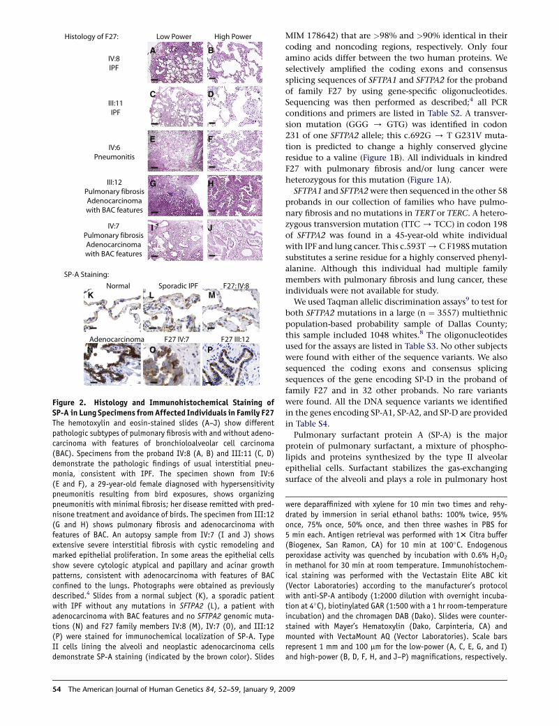
In family F27, multiple members have early-onset
pulmonary fibrosis and lung cancer cosegregating in an
autosomal-dominant pattern, suggesting that these two
diseases share a common etiology. The proband of the
family, subject IV:8 (Figure 1), is a 51-year-old white man
who has ten relatives with pulmonary fibrosis; five of the
ten died before age 50. Four individuals (III:8, III:12, IV:2,
and IV:7) also had adenocarcinoma of the lung with
features of bronchioloalveolar cell carcinoma (BAC). Three
other related individuals had pulmonary adenocarcinoma
or BAC in the absence of known fibrosis. Photomicro-
graphs of hematoxylin and eosin-stained slides of resected
lung tissue from affected family members are shown in
Figure 2. Seven individuals, including the proband, were
evaluated with pulmonary function testing and high-reso-
lution CT scans of the chest; medical records or death
certificates were obtained when possible. Additional clin-
ical information is provided in Table S1.
We performed a whole genome linkage analysis of 29
family members by using the Illumina Linkage IVb SNP
panel of >6,000 SNPs. Call rates varied from 97.2% to
98.5% for Autopure-purified DNA and from 56.4% to
73.9% for whole-genome-amplified DNA extracted from
archived samples. Individuals with pulmonary fibrosis
and/or lung cancer were classified as ‘‘affected,’’ and all
others were assigned an unknown affectation status. We
used the software MERLIN6 to screen the entire genome
by using multipoint model-free linkage analysis,7 and we
then evaluated the regions with the highest signals by
using a model-based method. Figure S1 in the Supple-
mental Data shows the analysis of family F27 with the
highest peak on chromosome 10; the model-free LOD
1Eugene McDermott Center for Human Growth and Development, 2Department of Internal Medicine, 3Department of Cardiovascular and Thoracic
Surgery, 4Department of Biochemistry, 5Howard Hughes Medical Institute, University of Texas Southwestern Medical Center, Dallas, TX, 75390, USA.
*Correspondence: [email protected]
DOI 10.1016/j.ajhg.2008.11.010. ª2009 by The American Society of Human Genetics. All rights reserved.
009

rs877783
rs4869
D10S
580
D10S
1730
D10S
201
D10S
1786
D10S
1686
D10S
5523
72Mb 89Mb
81.7Mb80.9Mb
SFTPDSFTPA1SFTPA2
20 100
Collagen-like region Neck
134 248
Carbohydrate recognition domain
G231V
GKG CAG TGG AATGATACATACY T D G/V Q W N
B
72
36
41 56 57 53 50
52 82 49 54 73 79 51 47 56
85 72 38 61
66
48 56 455341
I.
II.
III.
IV.
(1) (2)
(1)
(1)
1
(2)
(2)
(3) (4) (5) (6) (7) (8)
6
(3) (5) (7) (8) (9) 10 11 12 13 14
2 3 4 5 6 7 8 9 10
65 62
+ +
+ +
+ + + + +
F27: c.692G>T G231V
Pulmonary Fibrosis Undefined Lung Disease
Lung Cancer
(+) (+)
(+) (+)
(+)
(+)
(4) 6
4649 55
70 68
(1) (2)
4
(3) (4)
(3)
55 35
CKG810: c.593T>C F198S
(2)
(1) (2)
68
(5)
51 35
68
(5)
A
+
TYC CGC TAC TCAGACCCT GGAP G D F/S R Y S
F198S
C
F198
G231
D
64
Neck
CRD
Human SFTPA2 185 AYVGLTEGPSPGDFRYSDGTPVNYTNWYRGEPAGRGKEQCVEMYTDGQWNDRNCLYSRLTICDF 248 Human SFTPA1 AYVGLTEGPSPGDFRYSDGTPVNYTNWYRGEPAGRGKEQCVEMYTDGQWNDRNCLYSRLTICEF Chimpanzee SFTPA2 AYVGLTEGPSPGDFRYSDGTPVNYTNWYRGEPAGRGKEQCVEMYTDGQWNDRNCLYSRLTICEF Chimpanzee SFTPA1 AYVGLTEGPSPGDFRYSDGTPVNYTNWYRGEPAGRGKEQCVEMYTDGQWNDRNCLYSRLTICEF Rhesus monkey SFTPA2 AYVGLTEGPSPGDFRYSDGTPVNYTNWYPGEPAGRGTEQCVEMYTDGRWNDRNCLYNRLTICEF Rhesus monkey SFTPA1 AYVGLTEGPSPGDFRYSDGTPVNYTNWYPGEPAGRGTEQCVEMYTDGRWNDRNCLYNRLTICEF Lemur AYLGLAEGPTPGDFRYLDGAPVNYSNWYPGEPGGRGKEKCVEMYADGQWNDKNCLQYRLAICEF Elephant AYLGLAEGSTPGEFYYLDGAPVNYTNWYLGEPRGLGKERCVEMYTDGQWNDKNCLQHRLTICEF Armadillo AYLGLAEGPTPGEFRQLDGAPVNYTNWYPGEPRGRGKERCVDMYTDGQWNDKNCLQYRLAICEF Cow AYLGLVEGPTAGDFYYLDGAPVNYTNWYPGEPRGRGKEKCVEIYTDGQWNDKNCLQYRLAICEF Sheep AYLGLAEGPTAGDFYYLDGAPVNYTNWYPGEPRGRGKEKCVEIYTDGQWNDKNCLQYRLAICEF Horse AYLGLEEGPTAGDFYYLDGAPVNYTNWYPGEPRGRGKEKCVEMYTDGQWNDRSCLQYRLAICEF Pig AYLGLVEGPTAGDFFYLDGTPVNYTNWYPGEPRGRGKEKCVEMYTDGQWNDRNCQQYRLAICEF Cat AYLGLEEGPSPGDFCYLDGAPVNYTNWYPGEPRGGGKEKCVEMYTDGQWNNKNCLQYRLAICEF Dog AYLGLVESPDSGDFQYMDGAPVNYTNWYPGEPRGRGKEQCVEMYTDGQWNNKNCLQYRLAICEF Rabbit AYLGLAEGPTAGDFYYLDGDPVNYTNWYPGEPRGQGREKCVEMYTDGKWNDKNCLQYRLVICEF Bat AYLGLSESPTPGVFHYLDDTPVNYTKWYPGEPRGRGKEKCVEMYTDGQWNDKSCHQYRLTVCEF E shrew AYLGLEEGSEPGDFYSIDGTQVNYTNWYPGEPRGRGKEKCVEMYTDGKWNDKNCLQYRLTICEF Mouse PYLGVIEGQTPGDFHYLDGASVNYTNWYPGEPRGRGKEKCVEMYTDGKWNDKGCLQYRLAICEF Rat VYLGMIEDQTPGDFHYLDGASVNYTNWYPGEPRGQGKEKCVEMYTDGTWNDRGCLQYRLAVCEF Guinea pig SYLGLTEGHTPGDFHYLDGSPLNYTNWYPGEPRGRGKEKCAEMYLDGTWNDKNCLQSRLTICEF Ground squirrel AYLGLVESNTPGDFRYLDETPVNYTNWYPGEPRGRGREKCVEMYTDGTWNDRNCLQYRLAICEF E hedgehog AYLGLEEGSTLGDFYNLDGAPLNYTNWYPGEPSGQGKEKCVEMYTDGKWNDKGCLQYRLAICEF M hedgehog AYLGLTEGTTPGEFNDLDRTPVNYTNWYPGEPKGMGTERCVEMYTNGQWNDRNCLQYRLTICEF Short-tailed opossum AYLGLTEGKTPGKFYHLNGSPMNYTNWYSGEPAGKGREPCVEMYKDGTWNDKSCLQYRLTVCEF Brushtail opossum AFLGLREGKTPGKFYYLDGSPVNYTNWYEGSPDGGGTENCVEMYTDGTWNDRYCYKSRLAVCEF Platypus AYLGIVEGKDPGKFYYLDETPVNYTNWYQREPRGGGKENCVEMYTDGTWNDKNCQQYRLTICEF Chicken AYLGIKESLIPGTFQFLNGGELSYTNWYSHEPSGKGEEECVEMYTDGTWNDRRCNQNRLVVCQF Chicken AYLGIKESDTAGQFKYVNNQPLNYTSWQQYEPNGKGTEKCVEMYTDGNWKDRKCNLYRLTVCEY Frog AYLGIKVGDIADQFHYLDGTSVNYTNWGKNEPSGKGKEPCVEIYTDGHWNDKVCNQYRLTVCEF Frog AYLGIKVGDIADQFHYLNGTSVNYTNWGNNEPSSKGKEPCVEMYTDGHWNDKVCNQYRLTVCEF
F198 G231
(1)
Figure 1. Mutations in SFTPA2 Segregate with Familial Lung Cancer and Pulmonary Fibrosis(A) Abridged pedigrees are shown. The arrow indicates the index case. Circles represent females; squares represent males. Symbols witha slash through them indicate deceased subjects. Individuals with pulmonary fibrosis, lung cancer, or undefined lung disease are indi-cated by blue, red, and green symbols, respectively. Numbers below each symbol correspond to the individuals described in Table S1;numbers in parentheses indicate individuals for whom no DNA sample was available. The presence or absence of a mutation in SFTPA2is indicated by plus or minus signs, respectively. When the mutation was inferred based upon the pattern of inheritance, the plussign is placed in parentheses. The current age or the age at death is indicated to the upper right of each symbol. The SFTPA2 mutationsand predicted amino acid changes are listed above each family.(B) A region of chromosome 10 cosegregates with pulmonary fibrosis and lung cancer. The genes that encode paralogs of both surfactantprotein A and surfactant protein D lie within the linked region. The DNA sequence of a segment of SFTPA2 exon 6 is shown for the hetero-zygous CKG810 (left) and the proband of family F27 (right). A single base substitution (arrow) changes the wild-type thymidine to cyto-sine and leads to the predicted substitution of phenylalanine to serine at codon 198 (left); substitution of the wild-type guanine tothymidine leads to the predicted substitution of glycine for valine at codon 231 (right). Both mutations affect the coding region ofthe carbohydrate-recognition domain of surfactant protein A2.(C) The terminal portion of the amino acid sequence of the carbohydrate-recognition domain of surfactant A proteins is shown fromdiverse vertebrate species. This domain is highly conserved. Both the phenylalanine and glycine at positions 198 and 231, respectively,are conserved in all species shown. All sequences were obtained from NCBI; the comparison of the different proteins was based on a Clus-talW-generated alignment.(D) The ribbon diagram of the crystal structure of the monomeric carbohydrate recognition domain (CRD) and neck domains of rat surfac-tant protein A is shown; the positions of amino acids corresponding to codons 198 and 231 of the human sequence are indicated byarrows.
score is 3.22 (p value ¼ 6.0 3 10�5), and the model-based
LOD score is 2.74 (p value < 1.8 3 10�3) as determined
in a dominant genetic model with a penetrance of 0.95.
All affected family members share an identical-by-descent
15.7 Mb region of chromosome 10 (bounded by markers
rs877783 and rs4869, Figure S2). Additional polymorphic
microsatellite markers within this region were genotyped,
and haplotypes were determined by MERLIN.
The A
Approximately 118 annotated genes, including the
genes encoding surfactant proteins A and D, reside within
the linked interval. These two genes were considered excel-
lent candidates because they are both expressed at high
levels in the lung and play key roles in maintaining the
integrity of the air spaces (alveoli) of the lung. Surfactant
A has two isoforms, SP-A1 and SP-A2, which are encoded
by adjacent genes (SFTPA1, MIM 178630, and SFTPA2,
merican Journal of Human Genetics 84, 52–59, January 9, 2009 53
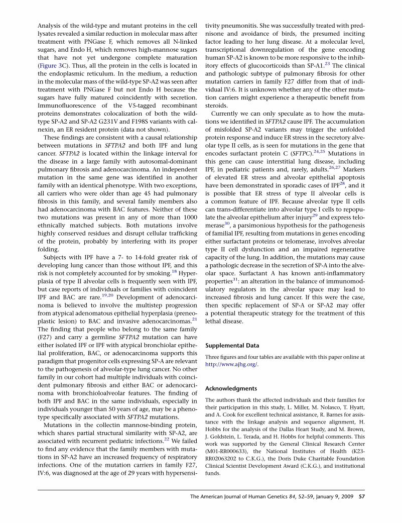
Histology of F27: Low Power High Power
IV:6Pneumonitis
IV:7Pulmonary fibrosisAdenocarcinomawith BAC features
III:12Pulmonary fibrosisAdenocarcinomawith BAC features
IV:8IPF
III:11IPF
SP-A Staining:
F27 IV:7
Normal Sporadic IPF
Adenocarcinoma
F27: IV:8
F27 III:12
K L M
N O P
A B
C D
E F
G H
I J
Figure 2. Histology and Immunohistochemical Staining ofSP-A in Lung Specimens from Affected Individuals in Family F27The hemotoxylin and eosin-stained slides (A–J) show differentpathologic subtypes of pulmonary fibrosis with and without adeno-carcinoma with features of bronchioloalveolar cell carcinoma(BAC). Specimens from the proband IV:8 (A, B) and III:11 (C, D)demonstrate the pathologic findings of usual interstitial pneu-monia, consistent with IPF. The specimen shown from IV:6(E and F), a 29-year-old female diagnosed with hypersensitivitypneumonitis resulting from bird exposures, shows organizingpneumonitis with minimal fibrosis; her disease remitted with pred-nisone treatment and avoidance of birds. The specimen from III:12(G and H) shows pulmonary fibrosis and adenocarcinoma withfeatures of BAC. An autopsy sample from IV:7 (I and J) showsextensive severe interstitial fibrosis with cystic remodeling andmarked epithelial proliferation. In some areas the epithelial cellsshow severe cytologic atypical and papillary and acinar growthpatterns, consistent with adenocarcinoma with features of BACconfined to the lungs. Photographs were obtained as previouslydescribed.4 Slides from a normal subject (K), a sporadic patientwith IPF without any mutations in SFTPA2 (L), a patient withadenocarcinoma with BAC features and no SFTPA2 genomic muta-tions (N) and F27 family members IV:8 (M), IV:7 (O), and III:12(P) were stained for immunochemical localization of SP-A. TypeII cells lining the alveoli and neoplastic adenocarcinoma cellsdemonstrate SP-A staining (indicated by the brown color). Slides
54 The American Journal of Human Genetics 84, 52–59, January 9, 2
MIM 178642) that are >98% and >90% identical in their
coding and noncoding regions, respectively. Only four
amino acids differ between the two human proteins. We
selectively amplified the coding exons and consensus
splicing sequences of SFTPA1 and SFTPA2 for the proband
of family F27 by using gene-specific oligonucleotides.
Sequencing was then performed as described;4 all PCR
conditions and primers are listed in Table S2. A transver-
sion mutation (GGG / GTG) was identified in codon
231 of one SFTPA2 allele; this c.692G / T G231V muta-
tion is predicted to change a highly conserved glycine
residue to a valine (Figure 1B). All individuals in kindred
F27 with pulmonary fibrosis and/or lung cancer were
heterozygous for this mutation (Figure 1A).
SFTPA1 and SFTPA2 were then sequenced in the other 58
probands in our collection of families who have pulmo-
nary fibrosis and no mutations in TERT or TERC. A hetero-
zygous transversion mutation (TTC / TCC) in codon 198
of SFTPA2 was found in a 45-year-old white individual
with IPF and lung cancer. This c.593T / C F198S mutation
substitutes a serine residue for a highly conserved phenyl-
alanine. Although this individual had multiple family
members with pulmonary fibrosis and lung cancer, these
individuals were not available for study.
We used Taqman allelic discrimination assays9 to test for
both SFTPA2 mutations in a large (n ¼ 3557) multiethnic
population-based probability sample of Dallas County;
this sample included 1048 whites.8 The oligonucleotides
used for the assays are listed in Table S3. No other subjects
were found with either of the sequence variants. We also
sequenced the coding exons and consensus splicing
sequences of the gene encoding SP-D in the proband of
family F27 and in 32 other probands. No rare variants
were found. All the DNA sequence variants we identified
in the genes encoding SP-A1, SP-A2, and SP-D are provided
in Table S4.
Pulmonary surfactant protein A (SP-A) is the major
protein of pulmonary surfactant, a mixture of phospho-
lipids and proteins synthesized by the type II alveolar
epithelial cells. Surfactant stabilizes the gas-exchanging
surface of the alveoli and plays a role in pulmonary host
were deparaffinized with xylene for 10 min two times and rehy-drated by immersion in serial ethanol baths: 100% twice, 95%once, 75% once, 50% once, and then three washes in PBS for5 min each. Antigen retrieval was performed with 13 Citra buffer(Biogenex, San Ramon, CA) for 10 min at 100�C. Endogenousperoxidase activity was quenched by incubation with 0.6% H2O2
in methanol for 30 min at room temperature. Immunohistochem-ical staining was performed with the Vectastain Elite ABC kit(Vector Laboratories) according to the manufacturer’s protocolwith anti-SP-A antibody (1:2000 dilution with overnight incuba-tion at 4�C), biotinylated GAR (1:500 with a 1 hr room-temperatureincubation) and the chromagen DAB (Dako). Slides were counter-stained with Mayer’s Hematoxylin (Dako, Carpinteria, CA) andmounted with VectaMount AQ (Vector Laboratories). Scale barsrepresent 1 mm and 100 mm for the low-power (A, C, E, G, and I)and high-power (B, D, F, H, and J–P) magnifications, respectively.
009

defense by binding, aggregating and opsonizing various
microorganisms.10,11 SP-A belongs to a structurally homol-
ogous family of innate-immune defense proteins that are
known as collectins and contain a collagen-like N-terminal
region that is involved in trimerization of the protein and
a C-terminal carbohydrate recognition (CRD) lectin
domain.12,13 It assembles into 18-subunit oligomers that
are composed of six disulfide-linked trimers assembled in
a ‘‘flower bouquet’’ pattern. Mice that lack SP-A are suscep-
tible to pulmonary infections from multiple organisms
and lack tubular myelin, the ordered lattice-like array of in-
tersecting membranes and proteins in the alveolar space.14
Both of the mutations we identified in SP-A2 involve
amino acids in the CRD, which is highly conserved among
all surfactant orthologs from Xenopus to humans
(Figure 1C). Both mutations are predicted to disrupt the
tertiary structure of the CRD domain (Figure 1D). The
bulky aromatic ring of phenylalanine at position 198 is
located in the hydrophobic core of the protein, and substi-
tution of a serine at this position would be predicted to
destabilize the protein on the basis of the crystal structure
of the rat protein.15 The glycine at position 231 lies adja-
cent to an alpha helix, and the substitution of a larger
residue such as a valine at this position would be predicted
to result in disruptive steric clashes.
Immunodetectable SP-A was detected in the type II alve-
olar cells via a rabbit polyclonal antibody raised against
purified human SP-A (a kind gift from C. R. Mendelson);
this antibody does not distinguish between SP-A1 and
SP-A2. The staining of type II cells in the affected indi-
vidual IV:8 in family F27 was similar to the staining of
normal lung or lung tissue from a patient with sporadic
IPF and no mutations in SFTPA2. In both individuals IV:7
and III:12, the adenocarcinoma cells lining the air spaces
demonstrate expression of SP-A (Figure 2), which is charac-
teristic of this type of lung cancer.16
To determine what effect the sequence variations identi-
fied in SP-A2 in this study have on the synthesis and secre-
tion of the protein, we expressed the wild-type and the
variant proteins in A549 cells, an immortalized human
alveolar epithelial cell line derived from a lung adenocarci-
noma. High-fidelity DNA polymerases were used for
cloning, and all subclones were confirmed by sequence
analysis. Full-length human SFTPA1 cDNA was PCR ampli-
fied from clone LIFESEQ90096303 (Open Biosystems,
Waltham, MA) and subcloned into pcDNA3 (Invitrogen,
Carlsbad, CA) downstream of the CMV promoter. We
used two partial IMAGE cDNA clones, 5184888 and
841707 (Invitrogen), to construct a full-length SFTPA2
cDNA. Site-directed mutagenesis (QuickChange, Strata-
gene) was utilized so that the DNA sequence of wild-type
SP-A1 and SP-A2 clones exactly matched NM_005411.3
and NM_006926.2, respectively. In a parallel set of experi-
ments, we used expression constructs containing an in-
frame 14 amino acid V5-tag immediately after the glutamic
acid at position 21 by primer extension mutagenesis and
zipper PCR.
The A
A549 cells were transfected with the expression plasmids
as follows: 350,000 cells were plated on 35 mm wells in
2 ml complete medium (Ham’s F12 with 10% FBS and
1% P/S) and transfected on day 1 with 1–2 mg DNA and
with 3 ml FuGENE HD Transfection Reagent (Roche, Basel,
Switzerland) per mg DNA according to the manufacturer’s
protocol. The cells were refed with complete medium on
day 2. Seventy-two hours after transfection, we examined
the cell lysate and medium by immunoblotting to assay
for the presence of SP-A. One milliliter of cultured medium
was removed from each well and centrifuged at 16,000 3 g
for 10 min at 4�C. The cells were washed once with 2 ml ice-
cold PBS, harvested in 300 ml of RIPA lysis buffer (150 mM
NaCl, 50 mM Tris-HCl [pH 8], 1 mM EDTA [pH 8], 1%
NP-40, 0.1% SDS, and 0.1% deoxycholate with 1 tablet of
protease cocktail [Roche] per 10 ml buffer), sonicated for
10 s., and centrifuged at 16,000 3 g at 4�C for 10 min. The
protein concentration of the medium and cellular lysates
was determined with the BCA Assay (Pierce, Rockford, IL).
Protein aliquots were eletrophoresed on 10% SDS-PAGE
Bio-Rad minigels and transferred to nitrocellulose Protran
membranes (Whatman, Dassel, Germany). The blots
were incubated for 1 hr at room temperature in blocking
buffer (5% dried milk in TBST [150 mM NaCl, 10 mM
Tris (pH 8), 0.1% Tween-20]), incubated with primary anti-
body overnight at a dilution of 1:10,000 (anti-SP-A or anti-
V5 mAb R960-25 from Invitrogen) in blocking buffer,
washed four times in TBST for 5 min each, incubated
with secondary antibody (Southern Biotech; Birmington,
AL) at 1:20,000 in blocking buffer for 1 hr at room temper-
ature, washed four times in TBST for 5 min each, and devel-
oped with SuperSignal West Pico Chemiluminescent
substrate (Pierce) according to the manufacturer’s protocol.
In cells expressing either wild-type SP-A or one of the SP-
A variants with an allele frequency >5%, immunoreactive
protein was present in both the cell lysate and the medium
(Figure 3A). In contrast, the SP-A2 G231V and F198S vari-
ants were poorly expressed in the cells, and no protein
was detected in the culture medium (Figure 3A). When
a V5-specific antibody was used in a parallel set of experi-
ments to analyze cells expressing the V5-tagged constructs,
the protein was readily detected in the cell lysates but not
in the medium. Another rare variant identified in this
study, SP-A2 L12W, was expressed and secreted in amounts
comparable to the wild-type protein. This variant failed to
cosegregate with pulmonary fibrosis in the family in which
the mutation was identified.
We also examined all the other rare and common SP-A1
variants that were identified in this study. All of these
forms of SP-A1 had an expression pattern that was similar
to the wild-type protein (Figure S3).
To determine whether the wild-type SP-A1 or SP-A2 in-
teracted with the G231V and F198S SP-A2 variants, we
coexpressed an in-frame Myc-tagged wild-type SP-A1 or
SP-A2 (three copies of EQKLISEEDLN engineered after
the signal peptide as described above) with the V5-tagged
SP-A2 variants and immunoprecipitated the cellular lysates
merican Journal of Human Genetics 84, 52–59, January 9, 2009 55

B
MMM M M M M
1 2 3 4 5 6 7 8 9 10 11 12 13 14
C C C C C C C
Tran
sfec
ted
cDN
A
SP-A1 SP-A2
V5-taggedSP-A2 w
t
myc-taggedSP-A
Imm
unob
lots
anti-V5
Mock
Mock
IP:
anti-
myc IB: anti-myc
IB: anti-V5
anti-myc
A
37
25
MMM M M M M M M
1 2 3 4 5 6 7 8 9 10 11 12 13 14 15 16 17 18
C C C C C C C C C
Moc
k
SP
-A2
wild
-typ
e
L12W
F198
SG
231V
T9N
V50L
A91P
Q22
3K
Rare Variants Common Variants
37
25
V5-taggedconstructs
Untaggedconstructs
Surfactant SP-A2
kD
++ +
+++
37
25
Endo HPNGase F
+
+
M M M M
2 3 4 5 6 7 8 9 10 11 12 13 141
C C C C C CC M C C
Wild-type F198SG231V
kD
V5-SP-A2
G23
1VF1
98S
wt
G23
1VF1
98S
C
Figure 3. The Effects of the G231V and F198S SFTPA2 Muta-tions on Protein Expression, Secretion, Interactions, and Endo-glycosidase Sensitivity in Transiently Transfected A549 Cells(A) A549 cells were transiently transfected with vector plasmid(Mock), the wild-type SP-A2 expression construct and variousuntagged SP-A2 constructs (upper panels) or V5-tagged constructs(lower panels). Rare variants are those that were found ata frequency of <5%. Aliquots of cellular lysates (C, 40 mg) andmedium (M, 80 mg) were analyzed via immunoblotting with eithera rabbit polyclonal that was raised against purified human SP-A(upper panel) or a mouse monoclonal antibody that recognizesthe V5 epitope (lower panel). Blots were exposed for 1–20 minor 1–20 s for the anti-SP-A and anti-V5 antibodies, respectively.
56 The American Journal of Human Genetics 84, 52–59, January 9, 2
by using a monoclonal antibody directed against the c-Myc
epitope. 48 hr after transfection, A549 cells were washed
with ice-cold PBS, scraped on ice in 0.5 ml of freshly
prepared lysis buffer (100 mM NaCl, 50 mM HEPES [pH
7.4], 1.5 mM MgCl2, and 0.5% NP-40 with 1 tablet of
protease cocktail (Roche) per 10 ml buffer), and centrifuged
16,000 3 g at 4�C for 15 min. The supernatants were trans-
ferred to a new tube and incubated with 2 mg of 9E10 mAb
(Santa Cruz Biotechnology) directed against the c-Myc
epitope and 200 mg (20 ml) of a protein A-agarose slurry (Cal-
biochem, San Diego, CA) for 16 hr at 4�C. Protein A-bound
antibodies and proteins were separated from the superna-
tant by centrifugation (800 3 g, 15 s) and washed twice
for 10 min in 800 ul of the lysis buffer at 4�C. The agarose
pellet was resuspended in 23 Laemli buffer and boiled 5
min prior to SDS-PAGE. Immunoblot analysis of reserved
aliquots of cellular lysate and medium was performed
with anti-Myc (1:1000 dilution of 9E10) or anti-V5 anti-
bodies as described above.
Immunoprecipitation of Myc-tagged wild-type SP-A1 re-
sulted in the coprecipitation of wild-type SP-A2 from both
the cell lysate and the medium (Figure 3B). In contrast to
these results, no V5-tagged SP-A2 G231V or F198S coim-
muniprecipitated with wild-type SP-A1 in the medium,
although they were detected in the cellular lysate. Similar
results were obtained when the Myc-tagged wild-type SP-
A2 was used. Thus, the variants were expressed and inter-
acted with both wild-type SP-A1 and SP-A2 in the cells
but were not secreted into the medium.
To determine whether the mutant forms of SP-A2
(G231V and F198S) were retained in the endoplasmic retic-
ulum (ER), we subjected aliquots of cellular lysates (25 mg)
or medium (80 mg) to treatment with either Endoglycosi-
dase H or PNGase F (New England Biolabs, Ipswich, MA)
for 1 hr at 37�C in the provided buffers. These endoglyco-
sidases differ in their specificities according to the degree of
maturation of the N-linked sugar chain attached to SP-A2.
Removal of the N-linked sugar results in an ~8 kDa reduc-
tion in the apparent molecular mass of the protein.17
(B) A549 cells transfected with empty vector (Mock), plasmid ex-pressing the Myc-tagged SP-A1 or SP-A2, and the V5-taggedwild-type or variant SP-A2. Forty-eight hours after transfectionthe cell lysates (C) and medium (M) were split into two aliquots.The Myc-tagged SP-A proteins were immunoprecipitated from thecellular lysates and media, subjected to SDS-PAGE, transferred tonitrocellulose membrane, and immunoblotted with antibodiesdirected against the Myc or V5 epitopes (upper panels). The arrowand arrowhead indicate the position of the Myc-tagged SP-A andV5-tagged SP-A2, respectively. Aliquots of cell lysates (C) andmedium (M) (40 mg) were subjected to SDS-PAGE, transferred tonitrocellulose membranes, and immunobloted with antibodiesdirected against the Myc or V5 epitopes (lower panels).(C) A549 cells were transiently transfected with V5-tagged SP-A2wild-type, G231V, or F198S constructs. Aliquots of lysates (C) ormedia (M) were treated with the indicated endoglycosidases andsubjected to immunoblot analysis. Untreated samples were treatedin parallel tubes without the addition of enzyme.
009
Analysis of the wild-type and mutant proteins in the cell
lysates revealed a similar reduction in molecular mass after
treatment with PNGase F, which removes all N-linked
sugars, and Endo H, which removes high-mannose sugars
that have not yet undergone complete maturation
(Figure 3C). Thus, all the protein in the cells is located in
the endoplasmic reticulum. In the medium, a reduction
in the molecular mass of the wild-type SP-A2 was seen after
treatment with PNGase F but not Endo H because the
sugars have fully matured coincidently with secretion.
Immunofluorescence of the V5-tagged recombinant
proteins demonstrates colocalization of both the wild-
type SP-A2 and SP-A2 G231V and F198S variants with cal-
nexin, an ER resident protein (data not shown).
These findings are consistent with a causal relationship
between mutations in SFTPA2 and both IPF and lung
cancer. SFTPA2 is located within the linkage interval for
the disease in a large family with autosomal-dominant
pulmonary fibrosis and adenocarcinoma. An independent
mutation in the same gene was identified in another
family with an identical phenotype. With two exceptions,
all carriers who were older than age 45 had pulmonary
fibrosis in this family, and several family members also
had adenocarcinoma with BAC features. Neither of these
two mutations was present in any of more than 1000
ethnically matched subjects. Both mutations involve
highly conserved residues and disrupt cellular trafficking
of the protein, probably by interfering with its proper
folding.
Subjects with IPF have a 7- to 14-fold greater risk of
developing lung cancer than those without IPF, and this
risk is not completely accounted for by smoking.18 Hyper-
plasia of type II alveolar cells is frequently seen with IPF,
but case reports of individuals or families with coincident
IPF and BAC are rare.19,20 Development of adenocarci-
noma is believed to involve the multistep progression
from atypical adenomatous epithelial hyperplasia (preneo-
plastic lesion) to BAC and invasive adenocarcinomas.21
The finding that people who belong to the same family
(F27) and carry a germline SFTPA2 mutation can have
either isolated IPF or IPF with atypical bronchiolar epithe-
lial proliferation, BAC, or adenocarcinoma supports this
paradigm that progenitor cells expressing SP-A are relevant
to the pathogenesis of alveolar-type lung cancer. No other
family in our cohort had multiple individuals with coinci-
dent pulmonary fibrosis and either BAC or adenocarci-
noma with bronchioloalveolar features. The finding of
both IPF and BAC in the same individuals, especially in
individuals younger than 50 years of age, may be a pheno-
type specifically associated with SFTPA2 mutations.
Mutations in the collectin mannose-binding protein,
which shares partial structural similarity with SP-A2, are
associated with recurrent pediatric infections.22 We failed
to find any evidence that the family members with muta-
tions in SP-A2 have an increased frequency of respiratory
infections. One of the mutation carriers in family F27,
IV:6, was diagnosed at the age of 29 years with hypersensi-
The
tivity pneumonitis. She was successfully treated with pred-
nisone and avoidance of birds, the presumed inciting
factor leading to her lung disease. At a molecular level,
transcriptional downregulation of the gene encoding
human SP-A2 is known to be more responsive to the inhib-
itory effects of glucocorticoids than SP-A1.23 The clinical
and pathologic subtype of pulmonary fibrosis for other
mutation carriers in family F27 differ from that of indi-
vidual IV:6. It is unknown whether any of the other muta-
tion carriers might experience a therapeutic benefit from
steroids.
Currently we can only speculate as to how the muta-
tions we identified in SFTPA2 cause IPF. The accumulation
of misfolded SP-A2 variants may trigger the unfolded
protein response and induce ER stress in the secretory alve-
olar type II cells, as is seen for mutations in the gene that
encodes surfactant protein C (SFTPC).24,25 Mutations in
this gene can cause interstitial lung disease, including
IPF, in pediatric patients and, rarely, adults.26,27 Markers
of elevated ER stress and alveolar epithelial apoptosis
have been demonstrated in sporadic cases of IPF28, and it
is possible that ER stress of type II alveolar cells is
a common feature of IPF. Because alveolar type II cells
can trans-differentiate into alveolar type I cells to repopu-
late the alveolar epithelium after injury29 and express telo-
merase30, a parsimonious hypothesis for the pathogenesis
of familial IPF, resulting from mutations in genes encoding
either surfactant proteins or telomerase, involves alveolar
type II cell dysfunction and an impaired regenerative
capacity of the lung. In addition, the mutations may cause
a pathologic decrease in the secretion of SP-A into the alve-
olar space. Surfactant A has known anti-inflammatory
properties31: an alteration in the balance of immunomod-
ulatory regulators in the alveolar space may lead to
increased fibrosis and lung cancer. If this were the case,
then specific replacement of SP-A or SP-A2 may offer
a potential therapeutic strategy for the treatment of this
lethal disease.
Supplemental Data
Three figures and four tables are available with this paper online at
http://www.ajhg.org/.
Acknowledgments
The authors thank the affected individuals and their families for
their participation in this study, L. Miller, M. Nolasco, T. Hyatt,
and A. Cook for excellent technical assistance, R. Barnes for assis-
tance with the linkage analysis and sequence alignment, H.
Hobbs for the analysis of the Dallas Heart Study, and M. Brown,
J. Goldstein, L. Terada, and H. Hobbs for helpful comments. This
work was supported by the General Clinical Research Center
(M01-RR000633), the National Institutes of Health (K23-
RR02063202 to C.K.G.), the Doris Duke Charitable Foundation
Clinical Scientist Development Award (C.K.G.), and institutional
funds.
American Journal of Human Genetics 84, 52–59, January 9, 2009 57

Received: September 25, 2008
Revised: November 11, 2008
Accepted: November 19, 2008
Published online: December 18, 2008
Web Resources
The URLs for data presented herein are as follows:
Online Mendelian Inheritance in Man (OMIM), http://www.ncbi.
nlm.nih.gov/omim/
National Center for Biotechnology Information (NCBI), http://
www.ncbi.nlm.nih.gov/
ClustalW, www.ebi.ac.uk/clustalw
All SNPs have been submitted to the dbSNP database, http://www.
ncbi.nlm.nih.gov/SNP/
References
1. American Thoracic Society, and European Respiratory Society.
(2002). American Thoracic Society/European Respiratory
Society international multidisciplinary consensus classifica-
tion of the idiopathic interstitial pneumonias. This joint state-
ment of the American Thoracic Society (ATS), and the Euro-
pean Respiratory Society (ERS) was adopted by the ATS board
of directors, June 2001 and by the ERS Executive Committee,
June 2001. Am. J. Respir. Crit. Care Med. 165, 277–304.
2. Marshall, R.P., Puddicombe, A., Cookson, W.O., and Laurent,
G.J. (2000). Adult familial cryptogenic fibrosing alveolitis in
the United Kingdom. Thorax 55, 143–146.
3. Armanios, M.Y., Chen, J.J., Cogan, J.D., Alder, J.K., Ingersoll,
R.G., Markin, C., Lawson, W.E., Xie, M., Vulto, I., Phillips,
J.A. 3rd., et al. (2007). Telomerase mutations in families with
idiopathic pulmonary fibrosis. N. Engl. J. Med. 356, 1317–
1326.
4. Tsakiri, K.D., Cronkhite, J.T., Kuan, P.J., Xing, C., Raghu, G.,
Weissler, J.C., Rosenblatt, R.L., Shay, J.W., and Garcia, C.K.
(2007). Adult-onset pulmonary fibrosis caused by mutations
in telomerase. Proc. Natl. Acad. Sci. USA 104, 7552–7557.
5. Cronkhite, J.T., Xing, C., Raghu, G., Chin, K.M., Torres, F.,
Rosenblatt, R.L., and Garcia, C.K. (2008). Telomere shortening
in familial and sporadic pulmonary fibrosis. Am. J. Respir. Crit.
Care Med. 178, 729–737.
6. Abecasis, G.R., Cherny, S.S., Cookson, W.O., and Cardon, L.R.
(2002). Merlin—Rapid analysis of dense genetic maps using
sparse gene flow trees. Nat. Genet. 30, 97–101.
7. Kong, A., and Cox, N.J. (1997). Allele-sharing models: LOD
scores and accurate linkage tests. Am. J. Hum. Genet. 61,
1179–1188.
8. Victor, R.G., Haley, R.W., Willett, D.L., Peshock, R.M., Vaeth,
P.C., Leonard, D., Basit, M., Cooper, R.S., Iannacchione, V.G.,
Visscher, W.A., et al. (2004). The Dallas Heart Study: A popula-
tion-based probability sample for the multidisciplinary study
of ethnic differences in cardiovascular health. Am. J. Cardiol.
93, 1473–1480.
9. Cohen, J., Pertsemlidis, A., Kotowski, I.K., Graham, R., Garcia,
C.K., and Hobbs, H.H. (2005). Low LDL cholesterol in individ-
uals of African descent resulting from frequent nonsense
mutations in PCSK9. Nat. Genet. 37, 161–165.
10. Wright, J.R. (2005). Immunoregulatory functions of surfac-
tant proteins. Nat. Rev. Immunol. 5, 58–68.
58 The American Journal of Human Genetics 84, 52–59, January 9, 2
11. Mason, R.J., Greene, K., and Voelker, D.R. (1998). Surfactant
protein A and surfactant protein D in health and disease.
Am. J. Physiol. 275, L1–L13.
12. Gupta, G., and Surolia, A. (2007). Collectins: sentinels of
innate immunity. Bioessays 29, 452–464.
13. Drickamer, K. (1988). Two distinct classes of carbohydrate-
recognition domains in animal lectins. J. Biol. Chem. 263,
9557–9560.
14. Korfhagen, T.R., LeVine, A.M., and Whitsett, J.A. (1998).
Surfactant protein A (SP-A) gene targeted mice. Biochim. Bio-
phys. Acta 1408, 296–302.
15. Head, J.F., Mealy, T.R., McCormack, F.X., and Seaton, B.A.
(2003). Crystal structure of trimeric carbohydrate recognition
and neck domains of surfactant protein A. J. Biol. Chem. 278,
43254–43260.
16. Gazdar, A.F., Linnoila, R.I., Kurita, Y., Oie, H.K., Mulshine, J.L.,
Clark, J.C., and Whitsett, J.A. (1990). Peripheral airway cell
differentiation in human lung cancer cell lines. Cancer Res.
50, 5481–5487.
17. Whitsett, J.A., Ross, G., Weaver, T., Rice, W., Dion, C., and
Hull, W. (1985). Glycosylation and secretion of surfactant-
associated glycoprotein A. J. Biol. Chem. 260, 15273–15279.
18. Hubbard, R., Venn, A., Lewis, S., and Britton, J. (2000). Lung
cancer and cryptogenic fibrosing alveolitis. A population-
based cohort study. Am. J. Respir. Crit. Care Med. 161, 5–8.
19. Beaumont, F., Jansen, H.M., Elema, J.D., ten Kate, L.P., and
Sluiter, H.J. (1981). Simultaneous occurrence of pulmonary
interstitial fibrosis and alveolar cell carcinoma in one family.
Thorax 36, 252–258.
20. Jones, A.W. (1970). Alveolar cell carcinoma occurring in
idiopathic interstitial pulmonary fibrosis. Br. J. Dis. Chest 64,
78–84.
21. Wistuba, I.I., and Gazdar, A.F. (2006). Lung cancer preneopla-
sia. Annu. Rev. Pathol. 1, 331–348.
22. Sumiya, M., Super, M., Tabona, P., Levinsky, R.J., Arai, T.,
Turner, M.W., and Summerfield, J.A. (1991). Molecular basis
of opsonic defect in immunodeficient children. Lancet 337,
1569–1570.
23. McCormick, S.M., and Mendelson, C.R. (1994). Human SP-A1
and SP-A2 genes are differentially regulated during develop-
ment and by cAMP and glucocorticoids. Am. J. Physiol. 266,
L367–L374.
24. Bridges, J.P., Wert, S.E., Nogee, L.M., and Weaver, T.E. (2003).
Expression of a human surfactant protein C mutation associ-
ated with interstitial lung disease disrupts lung development
in transgenic mice. J. Biol. Chem. 278, 52739–52746.
25. Mulugeta, S., Nguyen, V., Russo, S.J., Muniswamy, M., and
Beers, M.F. (2005). A surfactant protein C precursor protein
BRICHOS domain mutation causes endoplasmic reticulum
stress, proteasome dysfunction, and caspase 3 activation.
Am. J. Respir. Cell Mol. Biol. 32, 521–530.
26. Nogee, L.M., Dunbar, A.E. 3rd, Wert, S.E., Askin, F., Hamvas,
A., and Whitsett, J.A. (2001). A mutation in the surfactant
protein C gene associated with familial interstitial lung
disease. N. Engl. J. Med. 344, 573–579.
27. Thomas, A.Q., Lane, K., Phillips, J. 3rd, Prince, M., Markin, C.,
Speer, M., Schwartz, D.A., Gaddipati, R., Marney, A., Johnson,
J., et al. (2002). Heterozygosity for a surfactant protein C gene
mutation associated with usual interstitial pneumonitis and
cellular nonspecific interstitial pneumonitis in one kindred.
Am. J. Respir. Crit. Care Med. 165, 1322–1328.
009

28. Korfei, M., Ruppert, C., Mahavadi, P., Henneke, I., Markart, P.,
Koch, M., Lang, G., Fink, L., Bohle, R.M., Seeger, W., et al.
(2008). Epithelial endoplasmic reticulum stress and apoptosis
in sporadic idiopathic pulmonary fibrosis. Am. J. Respir. Crit.
Care Med. 178, 838–846.
29. Evans, M.J., Cabral, L.J., Stephens, R.J., and Freeman, G.
(1975). Transformation of alveolar type 2 cells to type 1 cells
following exposure to NO2. Exp. Mol. Pathol. 22, 142–150.
The A
30. Driscoll, B., Buckley, S., Bui, K.C., Anderson, K.D., and War-
burton, D. (2000). Telomerase in alveolar epithelial develop-
ment and repair. Am. J. Physiol. Lung Cell. Mol. Physiol.
279, L1191–L1198.
31. Borron,P.,McIntosh, J.C.,Korfhagen, T.R.,Whitsett, J.A., Taylor,
J., and Wright, J.R. (2000). Surfactant-associated protein A
inhibits LPS-induced cytokine and nitric oxide production
invivo. Am. J. Physiol. Lung Cell. Mol. Physiol. 278, L840–L847.
merican Journal of Human Genetics 84, 52–59, January 9, 2009 59
Related Documents