1 3 Arch Toxicol (2016) 90:2337–2348 DOI 10.1007/s00204-016-1769-9 REVIEW ARTICLE Genetic damage in humans exposed to extremely low‑frequency electromagnetic fields A. Maes 1 · L. Verschaeve 1,2 Received: 26 May 2016 / Accepted: 15 June 2016 / Published online: 23 June 2016 © Springer-Verlag Berlin Heidelberg 2016 Introduction Extremely low-frequency magnetic fields (ELF-MF) have been classified by the International Agency for Research on Cancer (IARC) as ‘possible carcinogenic to humans’ (group 2B). This was essentially because of the observed asso- ciation with childhood leukaemia (IARC 2002). Although some scientists are in favour of a re-evaluation based on new analyses and recent less convincing study results (e.g. Leitgeb 2015a, b), this association is at present still accepted (SCENIHR 2015). However, a causal relation- ship between magnetic field exposures and childhood leu- kaemia was never established and laboratory investigations also did not provide convincing supportive evidence (EHC 2007; Schmiedel and Blettner 2010; SCENIHR 2015). For example, results from studies on ELF-MF-induced genetic effects are controversial and most scientists do not consider that the available data clearly point towards such effects. Because of the low energy levels in molecular interactions, it is physically highly implausible that ELF fields cause direct genetic damage. However, it has been theorised that ELF may enhance such damage from other sources (e.g. endogenous radicals) or that epigenetic (non-genotoxic) interference in signal transduction may enhance cancer formation. Yet, studies on the effects of ELF magnetic field exposure of cells did generally not show genotoxic effects at magnetic flux densities well above those found in daily life situations. There is, however, some evidence that ELF magnetic fields may interact with DNA-damaging agents and be co-genotoxic (Vijayalaxmi and Obe 2005; Bergqvist et al. 2003; EHC 2007; Udroiu et al. 2010; Mark- kanen 2009). It also should be stressed that, as pointed out by Udroiu et al. (2010), possible aneugenic effects of elec- tromagnetic fields did not get much attention so far despite the growing interest for the link between aneuploidy and Abstract The classification of extremely low-frequency magnetic fields by the International Agency for Research on Cancer in the group of ‘possible human carcinogens’ (group 2B) is essentially based on epidemiologic evidence showing an association between MF exposures and child- hood leukaemia. Despite many in vitro and in vivo inves- tigations, there is no established causal relationship yet. However, human cytogenetic biomonitoring studies that were conducted in the past show predominantly positive results, i.e. increased cytogenetic damage in peripheral blood lymphocytes or buccal cells of ELF-MF-exposed subjects. This is important given the established link between observed cytogenetic damage in cells of people and an increased cancer risk. We here conducted an evalua- tion of the published investigations and found that many of the studies clearly have shortcomings, which often prevent any firm conclusion. As a matter of fact, there are reasons to believe that effects are not that impressive. However, the totality of the studies cannot simply be disregarded war- ranting further caution and the application, to a certain extent, of the precautionary principle. Keywords Magnetic fields · ELF · Cytogenetic damage · Lymphocytes · Buccal cells · Workers * L. Verschaeve [email protected] 1 Scientific Institute of Public Health, O.D. Food, Medicines and Consumers Safety, J. Wytsmanstreet 14, 1050 Brussels, Belgium 2 Department of Biomedical Sciences, University of Antwerp, Antwerp, Belgium

Welcome message from author
This document is posted to help you gain knowledge. Please leave a comment to let me know what you think about it! Share it to your friends and learn new things together.
Transcript

1 3
Arch Toxicol (2016) 90:2337–2348DOI 10.1007/s00204-016-1769-9
REVIEW ARTICLE
Genetic damage in humans exposed to extremely low‑frequency electromagnetic fields
A. Maes1 · L. Verschaeve1,2
Received: 26 May 2016 / Accepted: 15 June 2016 / Published online: 23 June 2016 © Springer-Verlag Berlin Heidelberg 2016
Introduction
Extremely low-frequency magnetic fields (ELF-MF) have been classified by the International Agency for Research on Cancer (IARC) as ‘possible carcinogenic to humans’ (group 2B). This was essentially because of the observed asso-ciation with childhood leukaemia (IARC 2002). Although some scientists are in favour of a re-evaluation based on new analyses and recent less convincing study results (e.g. Leitgeb 2015a, b), this association is at present still accepted (SCENIHR 2015). However, a causal relation-ship between magnetic field exposures and childhood leu-kaemia was never established and laboratory investigations also did not provide convincing supportive evidence (EHC 2007; Schmiedel and Blettner 2010; SCENIHR 2015). For example, results from studies on ELF-MF-induced genetic effects are controversial and most scientists do not consider that the available data clearly point towards such effects. Because of the low energy levels in molecular interactions, it is physically highly implausible that ELF fields cause direct genetic damage. However, it has been theorised that ELF may enhance such damage from other sources (e.g. endogenous radicals) or that epigenetic (non-genotoxic) interference in signal transduction may enhance cancer formation. Yet, studies on the effects of ELF magnetic field exposure of cells did generally not show genotoxic effects at magnetic flux densities well above those found in daily life situations. There is, however, some evidence that ELF magnetic fields may interact with DNA-damaging agents and be co-genotoxic (Vijayalaxmi and Obe 2005; Bergqvist et al. 2003; EHC 2007; Udroiu et al. 2010; Mark-kanen 2009). It also should be stressed that, as pointed out by Udroiu et al. (2010), possible aneugenic effects of elec-tromagnetic fields did not get much attention so far despite the growing interest for the link between aneuploidy and
Abstract The classification of extremely low-frequency magnetic fields by the International Agency for Research on Cancer in the group of ‘possible human carcinogens’ (group 2B) is essentially based on epidemiologic evidence showing an association between MF exposures and child-hood leukaemia. Despite many in vitro and in vivo inves-tigations, there is no established causal relationship yet. However, human cytogenetic biomonitoring studies that were conducted in the past show predominantly positive results, i.e. increased cytogenetic damage in peripheral blood lymphocytes or buccal cells of ELF-MF-exposed subjects. This is important given the established link between observed cytogenetic damage in cells of people and an increased cancer risk. We here conducted an evalua-tion of the published investigations and found that many of the studies clearly have shortcomings, which often prevent any firm conclusion. As a matter of fact, there are reasons to believe that effects are not that impressive. However, the totality of the studies cannot simply be disregarded war-ranting further caution and the application, to a certain extent, of the precautionary principle.
Keywords Magnetic fields · ELF · Cytogenetic damage · Lymphocytes · Buccal cells · Workers
* L. Verschaeve [email protected]
1 Scientific Institute of Public Health, O.D. Food, Medicines and Consumers Safety, J. Wytsmanstreet 14, 1050 Brussels, Belgium
2 Department of Biomedical Sciences, University of Antwerp, Antwerp, Belgium

2338 Arch Toxicol (2016) 90:2337–2348
1 3
carcinogenesis. Some evidence of ELF-MF-induced aneu-ploidy was already published yet (Udroiu et al. 2006; Maes et al. 2016).
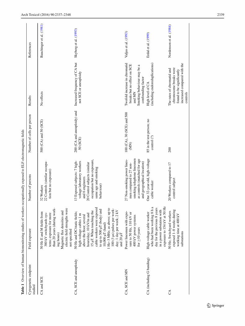
As genetic damage is very often a prerequisite for cancer, not only in vitro and in vivo animal studies were conducted but also several cytogenetic biomonitoring studies in people who were occupationally exposed to electric and magnetic fields. Most of these studies showed an increased frequency of genetic damage in the white blood or exfoliated buccal cells of the workers (Table 1). Despite above-mentioned uncertainties and lack of convincing evidence in in vitro and in vivo investigations, these human studies are often seen as alarming and supportive for an ELF-MF-induced cancer risk. However, a careful and critical investigation of these studies is needed to identify possible methodological shortcomings and hence better appreciate the validity of the studies. A previous critical evaluation was, for example, per-formed with respect to cytogenetic biomonitoring studies of subjects being exposed to radiofrequency fields, and this revealed the presence of many shortcomings preventing any clear conclusion, even when the majority of studies showed genetic damage in the blood and buccal cells of the exposed subjects (Verschaeve 2009). The same might be true when ELF (electro)magnetic fields are considered. We here pre-sent an evaluation of cytogenetic biomonitoring studies on ELF-(electro)magnetic field (ELF-EMF)-exposed subjects as published in the scientific literature.
Cytogenetic investigations of human subjects occupationally exposed to ELF‑EM fields
Several but yet relatively few studies were published on the cytogenetic damage in cells from ELF-EMF-exposed persons. Most investigations were on peripheral blood lym-phocytes. In some of the studies also buccal epithelial cells were investigated. A short overview of these studies and their conclusions is given here (Table 1).
Bauchinger et al. (1981) investigated structural chromo-some aberrations and sister chromatid exchanges (SCE) in blood cells of subjects following long-term exposure to elec-tric and magnetic fields. The chromosome analysis was car-ried out in the lymphocytes of 32 workers who were occupa-tionally exposed for more than 20 years to 50 Hz alternating electric and magnetic fields from 380 kV switchyards. As a control group, 22 workers of a similar age were included. Their occupation was also similar but did not coincide with ELF-EM exposure. There was no difference in the frequencies of chromosome aberrations and SCE between both groups.
Skyberg et al. (1993) investigated 13 laboratory employ-ees exposed to electromagnetic fields. From them, seven were high-voltage (up to 200 kV) laboratory cable splic-ers and six engineers exposed to static, alternating or pulsed
electric and magnetic fields. Matched controls consisted of 20 subjects with a similar job description (but no exposure), age and smoking behaviour. The alternating 50 Hz mag-netic fields were usually 5–10 µT but may occasionally have reached much higher values (±500 µT whole-body exposure; ±10,000 µT at the level of the hands). Chromosome aberra-tions, SCEs and aneuploidy (numerical chromosome aber-rations) were studied in the subject’s peripheral white blood cells. In this study, an increased frequency in structural chro-mosome aberrations was found but not in SCEs or aneuploidy.
Valjus et al. (1993) examined lymphocytes from power line inspectors and maintenance personnel who had a more than 10-year exposure to electromagnetic fields. They found a twofold increase in the incidence of chromatid breaks compared with unexposed controls, but no differ-ence with respect to SCEs and micronuclei.
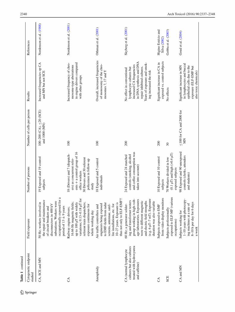
Nordenson et al. (1984, 1988, 2001) performed several cytogenetic biomonitoring studies on occupationally exposed subjects. A study of chromosome aberrations in peripheral blood lymphocytes of 20 switchyard workers at 400 kV substations revealed increased frequencies of such aberra-tions compared with the controls (Nordenson et al. 1984). In a follow-up study, 38 employees of electric power com-panies were studied; amongst them, 19 of the subjects were involved in the repair and maintenance of circuit breakers and disconnectors in 400 kV substations. The other 19 individu-als served as controls and were only exposed to normal envi-ronmental electromagnetic fields. The frequency of cells with chromosomal aberrations and micronuclei was significantly increased compared with the frequencies in the control cells. SCEs were not increased (Nordenson et al. 1988). Another study of Nordenson et al. (2001) was conducted on train engine drivers, train dispatchers, office workers and police-men. The drivers were exposed to magnetic fields ranging from a few µT to more than 100 µT. Chromosome aberrations were again investigated in peripheral lymphocytes. A pilot study of 18 engine drivers revealed a significant four times higher frequency of cells with chromosome aberrations com-pared with a control group of 16 office workers. A follow-up study of another 30 engine drivers and 30 policemen (used a controls) again showed a significant increase in the frequency of cells with chromosome-type aberrations.
A study by Othman et al. (2001) was specifically devoted to aneuploidy and involved 18 male traffic controllers and engineers exposed to electromagnetic fields. They had a sta-tistically increased frequency of monosomy of chromosome 7 and 17 and loss of the Y chromosome compared with a matched control population of five male individuals. The numerical chromosome aberrations were investigated with fluorescence in situ hybridisation (FISH) techniques.
Another investigation of Skyberg et al. (2001) was again on high-voltage laboratory workers exposed to electromag-netic fields and mineral oil. The study population consisted
2339Arch Toxicol (2016) 90:2337–2348
1 3
Tabl
e 1
Ove
rvie
w o
f hu
man
bio
mon
itori
ng s
tudi
es o
f w
orke
rs o
ccup
atio
nally
exp
osed
to E
LF-
elec
trom
agne
tic fi
elds
Cyt
ogen
etic
end
poin
t st
udie
dFi
eld
expo
sure
Num
ber
of p
erso
nsN
umbe
r of
cel
ls p
er p
erso
nR
esul
tsR
efer
ence
s
CA
and
SC
E50
Hz
E a
nd M
fiel
ds f
rom
38
0 kV
sw
itchy
ards
sys
-te
ms;
exp
osur
e fo
r m
ore
than
20
year
s (d
urin
g w
ork-
ing
hour
s)M
agne
tic fl
ux d
ensi
ties
and
elec
tric
fiel
d st
reng
ths
wer
e no
t spe
cifie
d
32 W
orke
rs22
Con
trol
s (s
imila
r oc
cupa
-tio
n bu
t no
expo
sure
)
500
(CA
) an
d 50
(SC
E)
No
effe
cts
Bau
chin
ger
et a
l. (1
981)
CA
, SC
E a
nd a
neup
loid
y50
Hz
and
DC
/sta
tic fi
elds
; hi
gh-v
olta
ge c
able
s 1
m
abov
e th
e flo
or; s
ever
al
hour
s/da
y; 1
0 kV
/m a
nd
15 µ
T. W
hen
touc
hing
the
cabl
e w
orke
rs w
ere
expo
sed
to u
p to
500
µT
(bo
dy)
and
10,0
00 µ
T (
hand
)1
Hz–
3 M
Hz;
as
abov
e; u
p to
10
0 (3
µs)
pul
ses
per
wor
k-da
y; 1
day
per
wee
k; 2
kV
an
d 20
µT
13 E
xpos
ed s
ubje
cts:
7 h
igh-
volta
ge la
bora
tory
wor
kers
an
d 6
engi
neer
s20
Con
trol
sub
ject
s (s
imila
r oc
cupa
tion
but n
o ex
posu
re,
sam
e ag
e an
d sm
okin
g be
havi
our)
200
(CA
and
ane
uplo
idy)
and
50
(SC
E)
Incr
ease
d fr
eque
ncy
of C
A b
ut
not S
CE
or
aneu
ploi
dySk
yber
g et
al.
(199
3)
CA
, SC
E a
nd M
NPo
wer
line
smen
with
exp
o-su
re to
50
Hz,
110
kV
or
400
kV p
ower
sys
tem
s fo
r ≥
10 y
ears
27 N
on-s
mok
ing
pow
er li
nes-
men
com
pare
d to
27
non-
smok
ing
tele
phon
e lin
esm
en
as c
ontr
ols
(mat
ched
for
age
an
d ge
ogra
phic
al lo
catio
n)
100
(CA
), 3
0 (S
CE
) an
d 50
0 (M
N)
Twof
old
incr
ease
in c
hrom
atid
br
eaks
but
no
effe
ct o
n SC
E
and
MN
Smok
ing
beha
viou
r m
ay b
e a
conf
ound
ing
fact
or
Val
jus
et a
l. (1
993)
CA
(in
clud
ing
G b
andi
ng)
Hig
h-vo
ltage
cab
le w
orke
r w
ho h
ad b
een
wor
king
8 h
a
day
for
the
prev
ious
8 y
ears
in
a p
ower
sub
stat
ion
with
ex
posu
re to
154
kV
at 5
0 H
z
One
32-
year
-old
, hig
h-vo
ltage
ca
ble
wor
ker
85 in
the
test
per
son;
no
cont
rol (
?)H
igh
leve
l of
CA
(inc
ludi
ng e
ndor
edup
licat
ions
)E
rdal
et a
l. (1
999)
CA
50 H
z; S
witc
hyar
d w
orke
rs
expo
sed
1–8
wee
ks d
urin
g w
orki
ng ti
me
at 4
00 k
V
subs
tatio
ns
20 W
orke
rs c
ompa
red
to 1
7 co
ntro
l sub
ject
200
The
rat
es o
f ch
rom
atid
and
ch
rom
osom
e br
eaks
wer
e fo
und
to b
e si
gnifi
cant
ly
incr
ease
d co
mpa
red
with
the
cont
rols
Nor
dens
on e
t al.
(198
4)
2340 Arch Toxicol (2016) 90:2337–2348
1 3
Tabl
e 1
con
tinue
d
Cyt
ogen
etic
end
poin
t st
udie
dFi
eld
expo
sure
Num
ber
of p
erso
nsN
umbe
r of
cel
ls p
er p
erso
nR
esul
tsR
efer
ence
s
CA
, SC
E a
nd M
N50
Hz;
wor
kers
invo
lved
in
the
repa
ir a
nd m
aint
enan
ce
of c
ircu
it br
eake
rs a
nd
disc
onne
ctor
s in
400
kV
su
bsta
tions
. Wor
kers
wer
e oc
cupa
tiona
lly e
xpos
ed f
or a
pe
riod
of
13 ±
9 y
ears
19 E
xpos
ed a
nd 1
9 co
ntro
l su
bjec
ts10
0–20
0 (C
A),
≤20
(SC
E)
and
1000
(M
N)
Incr
ease
d fr
eque
ncie
s op
CA
an
d M
N b
ut n
ot S
CE
Nor
dens
on e
t al.
(198
8)
CA
Rai
lway
eng
ine
driv
ers;
16
.66
Hz
mag
netic
fiel
ds;
up to
100
µT
with
wid
ely
vari
atio
ns; 0
.13–
0.18
µT
for
re
fere
nt tr
ain
disp
atch
ers;
ex
posu
re c
ontin
uous
for
w
hole
wor
king
day
18 (
Dri
vers
) an
d 7
(dis
patc
h-er
s) a
s co
ncur
rent
ref
er-
ents
+ a
con
trol
gro
up o
f 16
of
fice
wor
kers
30 D
rive
rs a
nd 3
0 re
fere
nt
polic
emen
in f
ollo
w-u
p st
udy
100
Incr
ease
d fr
eque
ncy
of c
hro-
mos
ome-
type
abe
rrat
ions
in
eng
ine
driv
ers
com
pare
d w
ith o
ther
gro
ups
Nor
dens
on e
t al.
(200
1)
Ane
uplo
idy
Air
traf
fic c
ontr
olle
rs a
nd
engi
neer
s be
ing
expo
sed
to E
MF-
field
s fr
om r
adar
sc
reen
s, a
nten
nae,
sat
el-
lite
inst
alla
tions
, etc
. for
10
–27
year
s. E
xpos
ure
is
thus
not
onl
y to
EL
F-E
MF!
!
18 E
xpos
ed a
nd 5
con
trol
in
divi
dual
s10
0O
vera
ll, in
crea
sed
freq
uenc
ies
of m
onos
omy
of th
e ch
ro-
mos
omes
7, 1
7 an
d Y
Oth
man
et a
l. (2
001)
CA
(no
rmal
lym
phoc
yte
cultu
res
but a
lso
cultu
res
trea
ted
with
hyd
roxy
urea
an
d ca
ffei
ne)
60 H
z; a
gen
erat
or s
olde
r-in
g an
d tr
ansf
orm
er te
ster
s gr
oup
wor
king
in h
igh-
volt-
age
labo
rato
ries
. Exp
osur
es
wer
e to
dif
fere
nt m
agne
tic
and
elec
tric
fiel
d st
reng
ths
(e.g
. 6 µ
T–7
mT
). E
xpos
ure
dura
tion
was
als
o va
riab
le
24 E
xpos
ed a
nd 2
4 m
atch
ed
cont
rols
(sm
okin
g, a
lcoh
ol
and
coff
ee c
onsu
mpt
ion
was
ta
ken
into
acc
ount
)
200
No
effe
ct in
con
vent
iona
l ly
mph
ocyt
e cu
lture
s bu
t in
crea
sed
CA
fre
quen
cies
in
DN
A s
ynth
esis
and
DN
A
repa
ir in
hibi
ted
cultu
res.
Yea
rs o
f ex
posu
re a
nd s
mok
-in
g in
crea
sed
the
risk
Skyb
erg
et a
l. (2
001)
CA
Subj
ects
exp
osed
to E
MF
from
vid
eo d
ispl
ay m
onito
rs10
Exp
osed
and
10
cont
rol
subj
ects
200
Sign
ifica
nt in
crea
se in
CA
in
expo
sed
vs. c
ontr
ol s
ubje
cts
Hig
ino
Est
écio
and
Si
lva
(200
2)
SCE
Subj
ects
pro
fess
iona
lly
expo
sed
to E
LF-
MF
(var
ious
oc
cupa
tions
)
70 W
orke
rs d
ivid
ed in
low
(0
.1 µ
T)
and
high
(>
0.4
µT)
expo
sed
subj
ects
30N
o ef
fect
sG
obba
et a
l. (2
003)
CA
and
MN
Subj
ects
wor
king
for
1–
10 y
ears
with
pho
toco
py-
ing
mac
hine
s at
a r
ate
of
8–10
h p
er d
ay f
or 6
day
s a
wee
k
98 E
xpos
ed a
nd 9
0 un
expo
sed
cont
rols
(cl
erks
, atte
nder
s an
d st
uden
ts)
≤10
0 fo
r C
A a
nd 2
000
for
MN
Sign
ifica
nt in
crea
se in
MN
in
lym
phoc
ytes
and
buc
cal
epith
elia
l cel
ls d
ue to
the
expo
sure
(E
LF-
EM
F bu
t al
so to
xic
chem
ical
s)
Gou
d et
al.
(200
4)

2341Arch Toxicol (2016) 90:2337–2348
1 3
Tabl
e 1
con
tinue
d
Cyt
ogen
etic
end
poin
t st
udie
dFi
eld
expo
sure
Num
ber
of p
erso
nsN
umbe
r of
cel
ls p
er p
erso
nR
esul
tsR
efer
ence
s
MN
Subj
ects
occ
upat
iona
lly
expo
sed
to E
M fi
elds
fro
m
vide
o di
spla
y m
onito
rs.
Ave
rage
wor
king
tim
e w
as
14 ±
7.4
4 ye
ars
20 E
xpos
ed a
nd 2
0 un
expo
sed
(?)
cont
rol s
ubje
cts
(mat
ched
fo
r ag
e an
d se
x)
2000
Incr
ease
d fr
eque
ncy
of M
N
and
brok
en e
gg c
ells
in e
xfo-
liate
d bu
ccal
cel
ls f
rom
the
expo
sed
subj
ects
Car
bona
ri e
t al.
(200
5)
CA
and
MN
50 H
z; W
orke
rs w
orki
ng f
or
19 ±
7 y
ears
in tr
ans-
form
er a
nd d
istr
ibut
ion
line
stat
ions
(15
4–38
0 kV
).
Ele
ctri
c an
d m
agne
tic fi
eld
stre
ngth
s re
sp. b
etw
een
130–
1500
V/m
and
0.2
5–17
A/m
32 T
rans
form
er w
orke
rs a
nd
23 o
ffice
wor
kers
17 C
ontr
ol s
ubje
cts
50 (
CA
), 1
000
(MN
)Si
gnifi
cant
ly h
ighe
r fr
eque
n-ci
es o
f bo
th C
A a
nd M
N in
th
e ‘e
lect
rica
l wor
kers
’. C
A
incr
ease
d w
ith th
e ye
ars
of
expo
sure
Cel
ikle
r et
al.
(200
9)
CA
and
SC
EE
lect
ric
trai
n en
gine
dri
vers
. E
xpos
ure
‘ass
umed
’ to
be
high
15 E
lect
ric
trai
n en
gine
dri
vers
an
d 15
con
trol
s co
nsis
ting
of
trai
n gu
ards
(sa
me
age
and
soci
o-ec
onom
ic s
tatu
s)
100
(CA
) an
d ±
30 (
SCE
)N
o ef
fect
(an
d no
indi
catio
ns
of s
yner
gist
ic e
ffec
ts w
ith
mito
myc
in C
)
Gad
hia
et a
l. (2
010)
MN
and
SC
E50
Hz;
wel
ders
exp
osed
to
EM
F vi
a el
ectr
ic a
rc
wel
ding
app
arat
us d
urin
g th
e w
orki
ng s
hift
(7
am to
5
pm).
Mag
netic
flux
den
sity
of
0.0
3–34
5.06
µT
(m
ean
valu
e =
7.8
1 µT
)
21 W
elde
rs a
nd 2
1 no
n-ex
pose
d co
ntro
ls (
mat
ched
fo
r ag
e, r
esid
ence
and
sm
ok-
ing
habi
t)
2000
for
MN
and
100
for
SC
EM
icro
nucl
ei f
requ
ency
in
the
expo
sed
wor
kers
was
si
gnifi
cant
ly h
ighe
r bu
t the
si
ster
chr
omat
id e
xcha
nge
freq
uenc
y w
as s
igni
fican
tly
low
er in
exp
osed
sub
ject
s co
mpa
red
with
the
cont
rols
Dom
inic
i et a
l. (2
011)
‘DN
A c
omet
s’Sa
me
as D
omin
ici e
t al.
(201
1)Id
50D
NA
dam
age
(tai
l int
ensi
ty
and
tail
mom
ent)
was
si
gnifi
cant
ly lo
wer
in th
e ex
pose
d gr
oup
com
pare
d to
th
e co
ntro
l gro
up
Vill
arin
i et a
l. 20
15)
CA
and
MN
50–6
0 H
z; e
lect
rica
l em
ploy
-ee
s in
tran
sfor
mer
s an
d po
wer
line
(di
rect
exp
o-su
re)
and
offic
e w
orke
rs in
pl
aces
adj
acen
t to
elec
tric
su
pply
sub
stat
ions
(in
dire
ct
expo
sure
). E
xpos
ure
dura
-tio
n fr
om 2
0 ±
4.7
(di
rect
) an
d 23
± 6
yea
rs (
indi
rect
).
Ele
ctri
c fie
ld s
tren
gth
300–
1500
V/m
; mag
netic
fie
ld s
tren
gth
0.25
–17
A/m
).
As
Cel
ikle
r et
al.
(200
9)?
50 E
xpos
ed a
nd 2
0 co
ntro
l su
bjec
ts10
0 fo
r C
A a
nd ≥
1000
for
M
NSi
gnifi
cant
incr
ease
in b
oth
CA
and
MN
in e
xpos
ed v
s.
cont
rol s
ubje
cts
Bal
amur
alik
rish
nan
et a
l. (2
012)

2342 Arch Toxicol (2016) 90:2337–2348
1 3
Tabl
e 1
con
tinue
d
Cyt
ogen
etic
end
poin
t st
udie
dFi
eld
expo
sure
Num
ber
of p
erso
nsN
umbe
r of
cel
ls p
er p
erso
nR
esul
tsR
efer
ence
s
‘DN
A c
omet
s’W
orke
rs o
ccup
atio
nally
ex
pose
d fo
r 2–
30 y
ears
(m
ean =
9 y
ears
) to
EM
F fr
om 1
32 k
V s
ubst
atio
ns
142
Exp
osed
sub
ject
s an
d 15
1 co
ntro
ls (
mat
ched
for
age
, so
cio-
econ
omic
sta
tus
and
life-
styl
e fa
ctor
s)
200
Tend
ency
tow
ards
incr
ease
d D
NA
dam
age
and
incr
ease
d ox
idat
ive
stre
ss p
aram
eter
s
Tiw
ari e
t al.
(201
5)
CA
and
SC
E50
Hz;
wor
kers
occ
upat
iona
lly
expo
sed
for
3–19
yea
rs)
to
elec
trom
agne
tic fi
eld
from
a
132–
230
kV e
lect
ric
supp
ly
subs
tatio
n
15 W
orke
rs a
nd 8
con
trol
s20
0 fo
r C
A &
25
for
SCE
Incr
ease
d fr
eque
ncy
of C
A b
ut
not o
f SC
E. C
ell p
rolif
era-
tion
indi
ces
and
the
mito
tic
inde
x w
ere
low
er in
the
expo
sed
subj
ects
Kha
lil e
t al.
(199
3)
CA
, SC
E a
nd M
NSu
bjec
ts p
rofe
ssio
nally
ex
pose
d to
EL
F-M
F (m
ean
expo
sure
= 0
.35
µT)
109
Exp
osed
sub
ject
s. 3
1 w
ork-
ers
expo
sed
to m
agne
tic fl
ux
dens
ities
exc
eedi
ng 1
µT
wer
e re
-eva
luat
ed
App
roxi
mat
ely
200
met
a-ph
ases
for
CA
; not
men
-tio
ned
for
SCE
and
MN
No
diff
eren
ces
seen
bet
wee
n lo
w (
<0.
µT
), m
oder
ate
(>0.
2 µT
) an
d hi
gh (
>1
µT)
expo
sure
s
Scar
ingi
et a
l. (2
007)
‘DN
A c
omet
s’ a
nd M
N60
Hz;
Hum
an v
olun
teer
s ex
pose
d fo
r 4
h to
mag
netic
flu
x de
nsity
of
200
µT
20 E
xpos
ed a
nd 1
0 co
ntro
l su
bjec
ts50
for
DN
A c
omet
s &
25
for
SCE
No
effe
cts
Alb
ert e
t al.
(200
9)
CA
50 H
z; s
ubje
cts
livin
g cl
ose
to
pow
er li
nes
or p
rofe
ssio
nally
ex
pose
d vi
a vi
deo
disp
lay
units
No
expo
sure
ass
essm
ent d
one
24 V
DU
wor
kers
and
10
resi
-de
ntia
l exp
osur
es17
Con
trol
sub
ject
s
200
No
effe
cts
Mae
s (1
998)
CA
chr
omos
ome
aber
ratio
ns, SCE
sis
ter
chro
mat
id e
xcha
nges
, MN
mic
ronu
clei

2343Arch Toxicol (2016) 90:2337–2348
1 3
of 24 individuals who were compared to 24 matched con-trols. The exposed group included employees from the high-voltage laboratory and generator soldering depart-ment. Due to their activities, they were exposed to both electric and magnetic fields as well as oil mist and vapour. The authors did not find excessive cytogenetic damage in the exposed subjects compared with the unexposed con-trols but found indications that the electromagnetic fields in combination with mineral oil exposure may produce chro-mosomal aberrations.
Higino Estécio and Silva (2002) found a significant higher frequency of aberrant metaphases and anomalies per cell in individuals exposed to radiation from video display monitors. Ten occupationally exposed individuals were studied, and the results were compared to these obtained in ten control subjects. The frequency of chromatid breaks was higher in the blood cells from EMF-exposed subjects compared with the controls.
Gobba et al. (2003) performed an investigation on peripheral blood lymphocytes from 70 workers exposed to various levels of ELF-EMF covering different occupations without the (known) involvement of exposure to mutagens and carcinogens. SCE frequencies, high-frequency cells (HFC) and the number of SCEs in HFC were investigated. No genotoxic effects were found at exposure levels of approximately 2 µT (the exposure levels currently found in most workplaces).
Goud et al. (2004) performed a micronucleus test in blood cells from subjects who regularly used photocopying machines and who were therefore exposed to toxic com-ponents of toners, toxic gazes as ozone, volatile organic components (VOCs) and extremely low-frequency elec-tromagnetic fields. A total of 98 workers were included in this study as well as 90 age- and sex-matched controls. The workers had an increased frequency of both chromo-some aberrations and micronuclei in their white blood cells. Increased micronucleus frequencies were also found in their buccal epithelial cells. Due to exposure to chemical agents as well and smoking as a confounding factor, it is very difficult to ascribe the results to the electromagnetic fields only.
Carbonari et al. (2005) found increased micronucleus frequencies as a result of exposure to electromagnetic fields from computer cathode ray tube video display moni-tors. Exposure was for at least 5 years and thus involved extremely low and very low electromagnetic fields. In this study, ten male and ten female occupational users of micro-computers were involved. The control population consisted of 20 unexposed subjects matched for age and gender. They were selected from the general population living in the same city. The frequency of micronuclei was studied in exfoliated buccal cells. Cells from EMF-exposed individu-als had a higher frequency of micronuclei compared with
the frequency in control cells. The effect was also signifi-cantly more pronounced in female individuals.
Another study was on occupational exposure to electric and magnetic fields involving 55 workers in transformer and distribution line stations in the Bursa province of Turkey (Celikler et al. 2009). The experimental group consisted of 32 technicians working inside the transformers and 23 office workers (outside the transformers). There were 17 control subjects who were working in different workplaces or were retired, housewives and students. Chromosome aberrations and micronucleus frequencies in peripheral lymphocytes were higher in the exposed ‘electrical’ workers. The fre-quency of chromosome aberrations furthermore increased with the years of exposure.
A cytogenetic investigation on railway engine drivers who were exposed to ELF-EMF was conducted by Gadhia et al. (2010). In this study, sister chromatid exchanges and structural chromosome aberrations were investigated. It was assumed that the engine drivers were exposed to rela-tively high magnetic field densities whereas their exposure to other (chemical) agents was assumed low and usually negligible. This study did not show any increased cytoge-netic damage in the ELF-EMF-exposed subject and hence did not support the hypothesis that ELF-EMFs are geno-toxic. This study involved a total of 15 railway engine driv-ers as the exposed population and 15 train guards as unex-posed controls. Both groups matched with respect to age, habits and socio-economic conditions.
Welders are exposed to ELF magnetic field intensi-ties that are 2–200 times higher than the exposure in most ‘electrical occupations’ and in households. The subjects who participated in the study of Dominici et al. (2011) were exposed to 0.03 µT up to a few hundred µT from elec-tric arc welding apparatus. Exposure was, however, always lower than the 2004 European unit action value of 500 µT. In this study, cytogenetic effects were examined by means of the micronucleus and SCE test in the lymphocytes of 21 welders who were enrolled in two different welding com-panies in central Italy. The control population consisted of 21 non-exposed blood donors matched for age, residence and smoking habit. The exposed group showed ‘dose-dependent’ and significantly higher frequencies of micro-nuclei compared with the control group. On the other hand, there was a significant decrease in the frequency of SCEs.
Results of the alkaline comet assay in peripheral blood lymphocytes of the same welders and controls were pub-lished separately (Villarini et al. 2015). Data were pre-sented for comet tail length, tail intensity and tail moment. According to the authors, there was significant less DNA damage (tail intensity and tail moment) in the blood cells of exposed welders compared with the unexposed probands.
Balamuralikrishnan et al. (2012) studied 70 Indian subjects from whom 50 were occupationally exposed to

2344 Arch Toxicol (2016) 90:2337–2348
1 3
low-frequency electromagnetic fields and 20 were unex-posed controls. The 50 exposed subjects were subdivided into a group of 28 power line and transformer workers (direct exposure) and 22 electrical board office workers (indirect exposures). Lymphocytes from exposed subjects had higher frequencies of structural chromosome aberra-tions and micronuclei compared with the frequencies in cells from the control subjects. Chromosome aberrations and micronuclei frequencies increased with age in both exposed and non-exposed subjects, but this was statistically significant only in the EMF-exposed subjects. According to the authors, chronic occupational exposure to EMFs may lead to an increased risk of genetic damage among the elec-trical workers.
Tiwari et al. (2015) used the alkaline comet assay to investigate DNA damage in cells from workers at 132 kV substations who were exposed to ELF-EMFs for more than 2 years. Blood sample of 142 exposed subjects and 151 non-exposed individuals was analysed. A ‘tendency’ towards increased DNA damage was found in the exposed subjects compared with non-exposed controls, but statisti-cal significance was not stated.
Khalil et al. (1993) investigated workers from a 132–230 kV supply station and found increased frequencies of chromosomal aberrations but not of sister chromatid exchanges.
Scaringi et al. (2007) briefly described the results of a cytogenetic investigation on ELF-MF exposed subjects (no precision). They found no difference between workers with low (<0.2 µT) and higher exposure levels (>0.2 µT and >1 µT). It was not clear how many cells were investigated per individual (especially for SCE and MN).
Other than professional exposures to ELF‑MFs
All above studies were on occupationally exposed subjects. To our knowledge, there were only two investigations on other ELF-EMF-exposed persons. Albert et al. (2009) found no cytogenetic effects in human volunteers exposed for 4 h to magnetic flux densities of 200 µT, whereas Maes (1998) studied chromosomal aberrations in VDU workers and residentially (power line) exposed individuals. Here also, no cytogenetic effects could be attributed to the expo-sure. However, this was only a limited pilot experiment lacking any data on exposure levels or possible confound-ing factors.
Discussion
We now know that a high frequency of structural chro-mosomal aberrations in lymphocytes is predictive of an
increased cancer risk, irrespective of the cause of the aber-rations (Bonassi et al. 1995, 2000, 2007, 2008; Hagmar et al. 1998, 2004). The chromosome aberration test is there-fore predictive for cancer at least at the level of a popu-lation. It is not predictive at the individual level as many factors may be responsible for an increased chromosome aberration frequency (recent illness or viral infection, etc.). Recent studies also provided evidence that an increased micronucleus frequency in peripheral lymphocytes is asso-ciated with an increased risk of cancer and other age-related degenerative diseases (Bonassi et al. 2007, 2011; Murgia et al. 2008; Migliore et al. 2011; Andreassi et al. 2011). Previous studies (e.g. Hagmar et al. 1998) did not find such an association, but the size of the cohort was too small and the material too heterogeneous to provide reliable findings. Moreover, most of the data were not obtained by using the more sensitive ex vivo/in vitro cytokinesis-block method-ology (Mateuca et al. 2006). A high(er) micronucleus fre-quency in blood cells of a given population thus indicates that this population has a higher cancer risk. As for struc-tural chromosome aberrations, this holds true at the level of the population but not at the individual level.
Sister chromatid exchanges and ‘DNA comets’ can be used as indicator tests for DNA damage and biomarkers of exposure rather than as biomarkers of effect as they do not necessarily correspond to an increased mutation risk. SCEs actually detect symmetrical or asymmetrical exchanges between sister chromatids of a single chromosome which are probably related to recombinational repair. The alka-line comet assay on the other hand detects single and dou-ble DNA breaks and alkali labile site that may or may not result in mutagenesis. Although both tests are well-known genotoxicity tests and hence related to carcinogenesis, the link with carcinogenesis in humans is no established yet. The tests, however, remain important.
Because of the association between genetic effects and cancer (at least in many instances), several studies were carried out on possible (cyto)genetic effects in subjects who were occupationally exposed to extremely low-frequency electromagnetic fields. Most of these studies showed increased genetic damage and hence overall the conclusion might be rather alarming. However, these studies need to be carefully examined. According to Gobba et al. (2003), no firm conclusions could be drawn yet with respect to pos-sible ELF-induced genotoxicity in occupational exposed persons. This conclusion was amongst others based on the controversial data and lack of replication studies. We also noted the increased chromosomal aberrations in cable splicers (Skyberg et al. 1993), but when all the 13 employ-ees of the study were compared with job-matched refer-ents, no statistically significant differences were found. From the seven cable splicers, actually only three subjects were recently exposed and the other four had been on sick

2345Arch Toxicol (2016) 90:2337–2348
1 3
leave and were transferred to other departments within the company. Statistically significant increases in chromosome breaks were found only in the three subjects which is a far too low study population to base any conclusion on, espe-cially as smoking was also identified as a confounder. The more recent study of Skyberg et al. (2001) in welders was furthermore not able to find any increased cytogenetic dam-age. From the 24 exposed subjects, 12 were working in the high-voltage laboratory and 12 were employed in the gen-erator soldering department where exposure was also to oil mist and vapours. Differences in response with regard to different genetic endpoints (e.g. increased levels of struc-tural chromosome aberrations but not of micronuclei and sister chromatid exchanges; Valjus et al. 1993) may con-tribute to the confusion although this may, at least partly, be ascribed to other measured endpoints and hence other mechanisms of action. It is, for example, well known that ionising radiations produce structural chromosome aberra-tions but much less SCEs (Evans 1977). The same was seen with radiofrequency fields (Verschaeve et al. 2010), and this was apparently also confirmed in the investigations on ELF-MFs.
Other studies were performed since but final conclusions yet remain difficult to draw, for example as a result of other contradictory findings as shown by the study by Gadhia et al. (2010) in train engine drivers whose results were in contradiction with the findings of Nordenson et al. (2001). As a matter of fact, we identified a number of shortcoming or discussion points that may hinder a proper evaluation of ELF-EMF-induced genotoxicity in humans and explain the present lack of any clear answer with respect to genotoxic effects of ELF-EMF in humans:
• To start with, most studies were not accompanied by robust dosimetric evaluations (see Table 1). Often only a very superficial job description was given as the only estimate of a ‘higher’ exposure level compared with the control population (e.g. Bauchinger et al. 1981; Valjus et al. 1993; Nordenson et al. 1984, 1988; Higino Esté-cio and Silva 2002; Gadhia et al. 2010). When meas-urements of electric and/or magnetic fields were done, the overall exposure of involved subjects yet remain uncertain due to job variations (e.g. variable exposure durations, engine drivers switching from one engine to another, no information on ‘other’ potential exposures as for example from computer screens in subject sup-posed to be exposed to other ELF-EMF sources as the main exposure, etc.).
• Most of the studies mention the use of ‘matched’ control populations, but often it is not clear what this means. For example, they may be matched with respect to age, gender and life style, but other factors may be important as well but were largely ignored. Bauch-
inger et al. (1981) mentioned that control subjects had a similar occupation than the 380 kV switchyard work-ers, but it is not clear what this actually means. Car-bonari et al. (2005) indicated that they used a protocol published by the International Commission for Pro-tection against Environmental Mutagens and Carcino-gens (Carrano 1988) to obtain necessary information on ‘life styles and personal factors’, but little is done with that information. In their study, exposure of video display workers was quantified as the number of work-ing years (14.45 on the average) but apparently also the controls that they have designated as ‘unexposed’ had an average working time with video display monitors of 11.7 years. It is difficult then to understand in what both exposed and unexposed populations actually dif-fered.
• Othman et al. (2001) supposedly investigated ELF-EMF-exposed subjects, but exposure was to EMF-fields from radar screens, antennae, satellite installations and closed circuit televisions. Exposure was therefore also, and essentially, to other forms of ‘non-ionising radia-tions’ (radiofrequencies). It is not clear from the paper what exposure was prevailing. As a matter of fact, all studies dating from later than the early 1990s should preferentially also consider exposure to radiofrequency electromagnetic fields as from mobile phones and other wireless communication devices, but no study actually did. This might be important as IARC also classified radiofrequency (RF) electromagnetic fields (as from mobile phones) into class 2B (possible carcinogenic to humans; IARC 2013), and the RF exposure might, at least in some of the studies, be more important than the ELF-MF exposures that were supposedly investigated. The study of Skyberg et al. (2001) also involved expo-sures to other agents than electromagnetic fields (min-eral oil). The same holds true for the investigations on welders (Dominici et al. 2011; Villarini et al. 2015) and frequent users of photocopying machines (Goud et al. 2004). In welders, welding fumes were possible impor-tant confounders. Dominici et al. (2011) and Villarini et al. (2015) reported higher frequencies of micronucle-ated cells but lower frequencies of SCEs and DNA dam-age according to the comet assay. They highlighted the fact that reduced SCE frequencies were already reported as a result of exposure to chromium and/or nickel pre-sent in the welding fumes and may be explained by a reduced DNA repair capacity. The results in the comet assay were explained by different chromium and/or nickel (or other metals) exposure levels, which lead to DNA–protein cross-links at lower concentrations. Goud et al. (2004) showed increased micronucleus levels in white blood cells and buccal cells of frequent users of photocopying machines, but exposure was also to toxic

2346 Arch Toxicol (2016) 90:2337–2348
1 3
VOCs and other compounds. Smoking could also be an important confounder.
• The study of Celikler et al. (2009) and Balamura-likrishnan et al. (2012) involved power line and trans-former workers. Although both studies reported higher frequencies of chromosomal aberrations and micronu-clei, there are reasons for concern. For example, espe-cially the Indian study reported very low micronucleus frequencies compared with historical values in most if not all of the laboratories worldwide. Frequencies of 1.32 ± 1.12 and 1.18 ± 0.73 per 1000 cells were found in exposed subjects compared with 0.45 ± 0.60 per 1000 cells in the controls. Even the frequencies in the exposed population were much lower than what is nor-mally reported in unexposed control cells. Bonassi et al. (2001), for example, reported an overall median micro-nucleus frequency in non-exposed (i.e. normal) subjects of 6.5 per thousand and an interquartile range between 3 and 12 per thousand. These values were based on a database of nearly 7000 subjects. Another example is provided by Rastkhah et al. (2016) who reported from 6 to 21 micronuclei per 1000 binucleated cells as the average baseline frequency. There are numerous other examples in the scientific literature.
• The study of Tiwari et al. (2015) only reported a ten-dency to higher DNA damage levels in substation work-ers reflecting over interpretation of the data rather than a real effect.
• The value of cytogenetic biomonitoring studies is, amongst others, largely dependent on two important parameters, i.e. the number of investigated cells per person and the number of individuals that were inves-tigated in as well the test population as their controls. The requested number of cells can be calculated with statistical tools. Statistical methods have demonstrated that, in order to detect a doubled chromosome aberra-tion frequency in a human biomonitoring study, one should investigate at least 200 metaphase figures per person and at least 20 persons per group (Whorton et al. 1979; Whorton 1985). This holds true only if confound-ers can be maximally excluded (no smokers or drug users, no medication or chronic diseases, same age dis-tribution between the groups, no expected exposure to other potential mutagens, etc.). If confounders cannot be sufficiently excluded, it is necessary to increase the sample size (cell number and/or number of individuals). Calculations of the number of cells and individuals that are needed in a cytogenetic study are, however, seldom done, and often a compromise is adopted between what is considered feasible in terms of time and work load and what is yet supposed to be enough. It is nevertheless assumed that one should at least investigate 200–500 metaphase figures per sample. That also the number of
involved persons is important is obvious. Not all per-sons react in the same way, and a representative sample of the population is needed (Verschaeve 2015). Scien-tists do not completely agree on the number of cells and subjects that should be investigated in order to conduct a well-designed and statistically robust cytogenetic bio-monitoring study, but generally speaking the numbers of 200 cells for chromosome aberrations, 50 for SCEs, 100 for analysis of ‘DNA comets’ and 2000 for analysis of micronuclei are considered to be minimal requirements, together with 20–50 subjects in both the test popula-tion and control group. It is clear that in the above-men-tioned studies (Table 1) these numbers were not always achieved. Many studies therefore provided results that were statistically not sufficiently robust.
• Many of the above reported studies which showed cytogenetic damage in the peripheral blood lympho-cytes or buccal cells of exposed subjects concern expo-sure levels which may be assumed higher than those of the ‘non-professionally exposed controls’, but exposure levels were yet usually not very high. Exposure lev-els were in many cases probably much lower than the exposure levels that were applied in in vitro and in vivo investigations. These experimental studies nevertheless largely produced negative findings. The same holds true for the study of Albert et al. (2009) where an exposure to 200 µT magnetic flux densities also did not induce genetic effects. Here one may argue yet that the expo-sure was limited in time (4 h only). According to a WHO report (EHC 2007), studies of the effects of ELF magnetic fields on cells have generally shown no induc-tion of genotoxicity at fields below 50 mT, although some more recent data show effects at 35 µT. According to SCENIHR (2015), positive effects may be expected above approximately 100 µT. Whatever the real value is, these exposure levels are still considerably higher than the alleged exposure levels in most of the professionally exposed subject investigated in the above-mentioned cytogenetic biomonitoring studies. It is therefore diffi-cult to believe that all reported cytogenetic effects are really due to the ELF-MF, rather than to other factors, as for example, exposure to other (genotoxic) agents, methodological shortcomings resulting in for example poor statistical power, over interpretation of data or, sometimes even bad science.
Above considerations show that there are many short-comings and reasons to minimise the scope of the findings. However, there yet is the fact that only five out of 22 studies (23 %) did not show cytogenetic damage in the investigated ELF-EMF-exposed subjects, and hence, this still is reason for concern. The evaluation of the investigations does not mean that exposures to extremely low-frequency magnetic

2347Arch Toxicol (2016) 90:2337–2348
1 3
fields are cleared from any suspicion and that no protective measures need to be taken by authorities in order to reason-ably apply the precautionary principle. Indeed, no consistent evidence of harm does not equal evidence of no harm, and we may not expect totally consistent results from scientific research when such a complex matter is concerned.
Conclusion
According to above investigations presenting a number of shortcomings and contradictions between the study results, no firm conclusion can be drawn with respect to alleged ELF-EMF induced genetic effects in exposed subjects. We still should be alert as some indications of induced genetic effects and carcinogenesis cannot be simply disregarded. Cytogenetic biomonitoring studies that were conducted so far did have important shortcomings. For this reason, we believe that more thorough and better controlled investiga-tions using the right genetic endpoints on adequate num-bers of cells and individuals still should be envisaged.
Acknowledgments This literature review was conducted as part of our activities within the Belgian BioElectroMagnetics Group (BBEMG).
References
Albert GC, McNamee JP, Marro L, Bellier PV, Prato FS, Thomas AW (2009) Assessment of genetic damage in peripheral blood of human volunteers exposed (whole-body) to a 200 muT, 60 Hz magnetic field. Int J Radiat Biol 85:144–152
Andreassi MG, Barale R, Iozzo P, Picano E (2011) The association of micronucleus frequency with obesity, diabetes and cardiovascu-lar disease. Mutagenesis 26:77–83
Balamuralikrishnan B, Balachandar V, Kumar SS, Stalin N, Varsha P, Devi SM, Arun M, Manikantan P, Venkatesan C, Sasikala K, Dharwadjar SN (2012) Evaluation of chromosomal alterations in electric workers occupationally exposed to low frequency of electromagnetic fields (EMFs) in Coimbatore population, India. Asian Pac J Cancer Prev 13:2961–2966
Bauchinger M, Hauf R, Schmid E, Dresp J (1981) Analysis of struc-tural chromosome changes and SCE after occupational long-term exposure to electric and magnetic fields from 380 kV-sys-tems. Radiat Environ Biophys 19:235–238
Bergqvist U, Brix J, de Gruijl F, de Seze R,Hietanen M, Jeffereys JGR, Lagroye I, Lotz GW, Owen RD, Repacholi MH, Saunders R, Tenforde TS, Verschaeve L, Veyret B (2003) Review of exper-imental investigations of EMF biological effects (0-100 kHz)—ICNIRP Standing committee II. In: Matthes R, McKinley A, Bernhardt J, Vecchia P, Veyret B (eds) Exposure to static and low frequency electromagnetic fields, biological effects and health consequences. ICNIRP13/2003, ISBN 3-934994-03-2
Bonassi S, Abbondandolo A, Camurri L, Dal PL, De FM, Degrassi F, Forni A, Lamberti L, Lando C, Padovani P (1995) Are chromo-some aberrations in circulating lymphocytes predictive of future cancer onset in humans? Preliminary results of an Italian cohort study. Cancer Genet Cytogenet 79:133–135
Bonassi S, Hagmar L, Stromberg U, Montagud AH, Tinnerberg H, Forni A, Heikkila P, Wanders S, Wilhardt P, Hansteen IL, Knud-sen LE, Norppa H (2000) Chromosomal aberrations in lympho-cytes predict human cancer independently of exposure to carcin-ogens. European Study Group on Cytogenetic Biomarkers and Health. Cancer Res 60:1619–1625
Bonassi S, Fenech M, Lando C, Lin YP, Ceppi M, Chang WP, Hol-land N, Kirsch-Volders M, Zeiger E, Ban S, Barale R, Bigatti MP, Bolognesi C, Jia C, Di Giorgio M, Ferguson LR, Fucic A, Lima OG, Hrelia P, Krishnaja AP, Lee TK, Migliore L, Mikha-levich L, Mirkova E, Mosesso P, Müller WU, Odagiri Y, Scarffi MR, Szabova E, Vorobtsova I, Vral A, Zijno A (2001) HUman MicroNucleus project: international database comparison for results with the cytokinesis-block micronucleus assay in human lymphocytes: I. Effect of laboratory protocol, scoring criteria, and host factors on the frequency of micronuclei. Environ Mol Mutagen 37:31–45
Bonassi S, Znaor A, Ceppi M, Lando C, Chang WP, Holland N, Kirsch-Volders M, Zeiger E, Ban S, Barale R, Bigatti MP, Bolognesi C, Cebulska-Wasilewska A, Fabianova E, Fucic A, Hagmar L, Joksic G, Martelli A, Migliore L, Mirkova E, Scarfi MR, Zijno A, Norppa H, Fenech M (2007) An increased micro-nucleus frequency in peripheral blood lymphocytes predicts the risk of cancer in humans. Carcinogenesis 28:625–631
Bonassi S, Norppa H, Ceppi M, Stromberg U, Vermeulen R, Znaor A, Cebulska-Wasilewska A, Fabianova E, Fucic A, Gundy S, Han-steen IL, Knudsen LE, Lazutka J, Rossner P, Sram RJ, Boffetta P (2008) Chromosomal aberration frequency in lymphocytes pre-dicts the risk of cancer: results from a pooled cohort study of 22 358 subjects in 11 countries. Carcinogenesis 29:1178–1183
Bonassi S, El-Zein R, Bolognesi C, Fenech M (2011) Micronuclei frequency in peripheral blood lymphocytes and cancer risk: evi-dence from human studies. Mutagenesis 26:93–100
Carbonari K, Gonçalves L, Roth D, Moreira P, Fernández R, Mar-tino-Roth MdG (2005) Increased micronucleated cell frequency related to exposure to radiation emitted by computer cathode ray tube video display monitors. Genet Mol Biol 28:469–474
Carrano AV (1988) Considerations for population monitoring using cytogenetic techniques. ICPEMC—International Commission For Protection Against Environmental Mutagens and Carcino-gens. Publication N. 14. Mutat Res 204:379–406
Celikler S, Aydemir N, Vatan O, Kurtuldu S, Bilaloglu R (2009) A biomonitoring study of genotoxic risk to workers of transform-ers and distribution line stations. Int J Environ Hyg Health Res 19:421–430
Dominici L, Villarini M, Fatigoni C, Monarca S, Moretti M (2011) Genotoxic hazard evaluation in welders occupationally exposed to extremely low-frequency magnetic fields (ELF-MF). Int J Hyg Environ Health 215:68–75
EHC (2007) Environmental health criteria 238: extremely low fre-quency fields. World Health Organization, Geneva. ISBN 978-92-4-157238-5
Erdal ME, Erdal N, Oğuzkan S (1999) Chromosomal aberrations in peripheral lymphocytes of a high-voltage power lineman exposed to electromagnetic fields. Turk J Med Sci 29:335–336
Evans HJ (1977) Molecular mechanisms in the induction of chromo-some aberrations. In: Scott D, Bridges BA, Sobels FH (eds) Pro-gress in genetic toxicology. Elsevier/North Holland Biomedical Press, Amsterdam, pp 57–74. ISBN 0-444-80014-X
Gadhia P, Chakraborty S, Pithawala M (2010) Cytogenetic studies on railway engine drivers exposed to extremely low frequency elec-tromagnetic fields (ELF-EMF). Int J Hum Genet 10:263–269
Gobba F, Rocatto L, Sinigaglia B, Temperani P (2003) Sister chroma-tid exchanges (SCE) and high frequency cells in workers occu-pationally exposed to extremely low frequency magnetic fields. Med Lav 94:450–458 (in Italian)

2348 Arch Toxicol (2016) 90:2337–2348
1 3
Goud KI, Hasan Q, Balakrishna N, Rao KP, Ahuja YR (2004) Geno-toxicity evaluation of individuals working with photocopying machines. Mutat Res 563:151–158
Hagmar L, Bonassi S, Stromberg U, Brøgge A, Knudsen LE, Norppa H, Reuterwall C (1998) Chromosomal aberrations in lympho-cytes predict human cancer: a report from the European Study Group on Cytogenetic Biomarkers and Health (ESCH). Cancer Res 58:4117–4121
Hagmar L, Stromberg U, Bonassi S, Hansteen IL, Knudsen LE, Lind-holm C, Norppa H (2004) Impact of types of lymphocyte chro-mosomal aberrations on human cancer risk: results from Nordic and Italian cohorts. Cancer Res 64:2258–2263
Higino Estécio MR, Silva AE (2002) Chromosome abnormalities caused by computer video display monitors’ radiation. Rev Saúde Pública 36:330–336 (in Portuguese)
IARC (2002) IARC monographs on the evaluation of carcinogenic risks to humans. Vol. 80, non-ionizing radiation, part 1: static and extremely low-frequency (ELF) electric and magnetic fields. IARC Press, Lyon. ISBN 92-832-1280-0
IARC (2013) IARC monographs on the evaluation of carcinogenic risks to humans. Vol. 102, non- ionizing radiation, part 2: radi-ofrequency electromagnetic fields. IARC Press, Lyon. ISBN 978-92-832-1325-3
Khalil AM, Quassem W, Amoura F (1993) Cytogenetic changes in human lymphocytes from workers occupationally exposed to high-voltage electromagnetic fields. Electro Magnetobiol 12:17–26
Leitgeb N (2015a) Synoptic analysis of epidemiologic evidence of glioma risk from mobile phones. J Electromagn Anal 7:233–243
Leitgeb N (2015b) Synoptic analysis clarifies childhood leukemia risk from ELF magnetic field exposure. J Electromagn Anal 7:245–258
Maes A (1998) Genetic effects of non-ionizing radiations, especially microwaves and extremely low frequency fields. PhD thesis, Free University of Brussels (VUB), Brussels (in Dutch)
Maes A, Anthonissen R, Wambacq S, Simons K, Verschaeve L (2016) The cytome assay as a tool to investigate the possible associa-tion between exposure to extreme low frequency magnetic fields and an increased risk for Alzheimer’s disease. J Alzheimer’s Dis 50:741–749
Markkanen A (2009) Effects of electromagnetic fields on cellular responses to agents causing oxidative stress and DNA damage. Kuopio University Publications C. Natural and Environmental Sciences vol 253, 59 p. ISBN 978-951-27-1191-8
Mateuca R, Lombaert N, Aka PV, Decordier I, Kirsch-Volders M (2006) Chromosomal changes: induction, detection meth-ods and applicability in human biomonitoring. Biochimie 88:1515–1531
Migliore L, Coppedè F, Fenech M, Thomas P (2011) Association of micronucleus frequency with neurodegenerative diseases. Mutagenesis 26:85–92
Murgia E, Ballardin M, Bonassi S, Rossi AM, Barale R (2008) Vali-dation of micronuclei frequency in peripheral blood lymphocytes as early cancer risk biomarker in a nested case-control study. Mutat Res 639:27–34
Nordenson I, Hansson Mild K, Nordström S, Sweins A, Birke E (1984) Clastogenic effects in human lymphocytes of power fre-quency electric fields: in vivo and in vitro studies. Radiat Envi-ron Biophys 23:191–201
Nordenson I, Hansson Mild K, Ostman U, Ljungberg H (1988) Chro-mosomal effects in lymphocytes of 400 kV-substation workers. Radiat Environ Biophys 27:39–47
Nordenson I, Hansson Mild K, Järventaus H, Hirvonen A, Sandström M, Wilén J, Blix N, Norppa H (2001) Chromosome aberrations in peripheral lymphocytes of train engine drivers. Bioelectro-magnetics 22:306–315
Othman EO, Aly MS, El Nahas SM (2001) Aneuploidy in workers occupationally exposed to electromagnetic fields detected by FISH. Cytologia 66:117–125
Rastkhah E, Zakeri F, Ghoranneviss M, Rajabpour MR, Farshidpour MR, Mianji F, Bayat M (2016) The cytokinesis-blocked micro-nucleus assay: dose–response calibration curve, background frequency in the population and dose estimation. Radiat Environ Biophys 55:41–51
Scaringi M, Temperani P, Rossi P, Bravo G, Gobba F (2007) Evalua-tion of the genotoxicity of the extremely low frequency-magnetic fields (ELF-MF) in workers exposed for professional reasons. Giorn It Med Lavoro Ergon 29:420–421 (in Italian)
SCENIHR (2015) Opinion on Potential health effects of exposure to electromagnetic fields (EMF) European Commission, DG Health and Food Safety. http://ec.europa.eu/health/scientific_commit-tees/emerging/docs/scenihr_o_041.pdf
Schmiedel S, Blettner M (2010) The association between extremely low-frequency electromagnetic fields and childhood leukaemia in epidemiology: enough is enough? Br J Cancer 103:931–932
Skyberg K, Hansteen I-L, Vistnes AM (1993) Chromosome aberra-tions in lymphocytes of high-voltage cable splicers exposed to electromagnetic fields. Scand J Work Environ Health 19:29–34
Skyberg K, Hansteen I-L, Vistnes AM (2001) Chromosomal aberra-tions in lymphocytes of employees in transformer and generator production exposed to electromagnetic fields and mineral oil. Bioelectromagnetics 22:150–160
Tiwari R, Lakshmi NK, Bhargava SC, Ahuja YR (2015) Epineph-rine, DNA integrity and oxidative stress in workers exposed to extremely low-frequency electromagnetic fields (ELF-EMFs) at 132 kV substations. Electromagn Biol Med 34:56–62
Udroiu I, Cristaldi M, Icradi LA (2006) Clastogenicity and ane-uploidy in newborn and adult mice exposed to 50 Hz magnetic fields. Int J Radiat Biol 82:561–567
Udroiu I, Giuliani L, Icradi LA (2010) Genotoxic properties of extremely low frequency electromagnetic fields. Eur J Oncol Libr 5:123–134
Valjus J, Norppa H, Järventaus H, Sorsa M, Nykyri E, Salomaa S, Järvinen P, Kjader J (1993) Analysis of chromosomal aberra-tions, sister chromatid exchanges and micronuclei among power linesmen with long-term exposure to 50-Hz electromagnetic fields. Radiat Environ Biophys 32:325–336
Verschaeve L (2009) Genetic damage in subjects exposed to radiofre-quency radiation. Mut Res 681:259–270
Verschaeve L (2015) Genetic toxicology: tests and applications. Uni-versity Press, Antwerp. ISBN 978-90-5718-283-9
Verschaeve L, Juutilainen J, Lagroye I, Miyakoshi J, van Rongen E, Saunders R, de Seze R, Tenforde T, Veyret B, Xu Z (2010) In vitro and in vivo genotoxicity of radiofrequency fields. Mut Res 705:252–268
Vijayalaxmi, Obe G (2005) Controversial cytogenetic observations in mammalian somatic cells exposed to extremely low frequency electromagnetic radiation: a review and future research recom-mendations. Bioelectromagnetics 26:412–430
Villarini M, Dominici L, Fatigoni C, Levorato S, Vannini S, Monarca S, Moretti M (2015) Primary DNA damage in welders occupa-tionally exposed to extremely-low-frequency magnetic fields (ELF-MF). Ann Ig 27:511–519
Whorton EB Jr (1985) Some experimental design and analysis con-siderations for cytogenetics studies. Environ Mutagen 7(Suppl 4):9–15
Whorton EB Jr, Bee DE, Kilian DJ (1979) Variations in the propor-tion of abnormal cells and required sample sizes for human cytogenetic studies. Mutat Res 64:79–86
Related Documents