Accepted Manuscript Title: Genetic characterization of a new astrovirus detected in dogs suffering from diarrhoea Authors: Anna Toffan, Christine Monceyron Jonassen, Cristian De Battisti, Eliana Schiavon, Tone Kofstad, Ilaria Capua, Giovanni Cattoli PII: S0378-1135(09)00248-X DOI: doi:10.1016/j.vetmic.2009.04.031 Reference: VETMIC 4432 To appear in: VETMIC Received date: 16-1-2009 Revised date: 17-4-2009 Accepted date: 28-4-2009 Please cite this article as: Toffan, A., Jonassen, C.M., De Battisti, C., Schiavon, E., Kofstad, T., Capua, I., Cattoli, G., Genetic characterization of a new astrovirus detected in dogs suffering from diarrhoea, Veterinary Microbiology (2008), doi:10.1016/j.vetmic.2009.04.031 This is a PDF file of an unedited manuscript that has been accepted for publication. As a service to our customers we are providing this early version of the manuscript. The manuscript will undergo copyediting, typesetting, and review of the resulting proof before it is published in its final form. Please note that during the production process errors may be discovered which could affect the content, and all legal disclaimers that apply to the journal pertain.

Welcome message from author
This document is posted to help you gain knowledge. Please leave a comment to let me know what you think about it! Share it to your friends and learn new things together.
Transcript

Accepted Manuscript
Title: Genetic characterization of a new astrovirus detected indogs suffering from diarrhoea
Authors: Anna Toffan, Christine Monceyron Jonassen,Cristian De Battisti, Eliana Schiavon, Tone Kofstad, IlariaCapua, Giovanni Cattoli
PII: S0378-1135(09)00248-XDOI: doi:10.1016/j.vetmic.2009.04.031Reference: VETMIC 4432
To appear in: VETMIC
Received date: 16-1-2009Revised date: 17-4-2009Accepted date: 28-4-2009
Please cite this article as: Toffan, A., Jonassen, C.M., De Battisti, C., Schiavon,E., Kofstad, T., Capua, I., Cattoli, G., Genetic characterization of a newastrovirus detected in dogs suffering from diarrhoea, Veterinary Microbiology (2008),doi:10.1016/j.vetmic.2009.04.031
This is a PDF file of an unedited manuscript that has been accepted for publication.As a service to our customers we are providing this early version of the manuscript.The manuscript will undergo copyediting, typesetting, and review of the resulting proofbefore it is published in its final form. Please note that during the production processerrors may be discovered which could affect the content, and all legal disclaimers thatapply to the journal pertain.

Page 1 of 18
Accep
ted
Man
uscr
ipt
1
Genetic characterization of a new astrovirus detected in dogs suffering from1
diarrhoea2
Anna Toffan°1, Christine Monceyron Jonassen°§2, Cristian De Battisti1, Eliana Schiavon3, Tone 3
Kofstad2, Ilaria Capua1, Giovanni Cattoli1*4
1 Istituto Zooprofilattico Sperimentale delle Venezie, Research and Development Department,5
Viale dell’Università 10, 35020, Legnaro, Padova, Italy6
2 National Veterinary Institute, Section for Virology and Serology, PO Box 750 Sentrum, N-0106 7
Oslo, Norway8
3 Istituto Zooprofilattico Sperimentale delle Venezie, Diagnostic Unit, Viale dell’Università 10, 9
35020, Legnaro, Padova, Italy10
§ Present address: Akershus University Hospital, Center for Laboratory Medicine, N-1478 11
Lørenskog, Norway12
13
° Equally contributing authors14
* Corresponding author: [email protected]; tel 0039 049 8084333 fax 0039 049 808436015
16
Running title: Astrovirus in dog 17
18
19
20
21
22
23
24
25
26
27
28
29
* Manuscript

Page 2 of 18
Accep
ted
Man
uscr
ipt
2
Abstract30
Astroviruses have been described in several animals species frequently associated with diarrhoea, 31
especially in young animals. In dogs, astrovirus-like particles have been observed sporadically and 32
very little is known about their epidemiology and characteristics. In this paper, we describe the 33
detection of astrovirus-like particles in symptomatic puppies. Furthermore, for the first time in this 34
species, the presumptive identification made by electron microscopy was confirmed by genetic 35
analysis of the viral RNA conducted directly on the clinical specimens. Genetic sequences of ORF2 36
(2443 nt), encoding for the capsid protein, and partial sequence of ORF1b (346 nt), encoding for the 37
viral polymerase, identified the viruses as member of the family Astroviridae. The phylogenetic 38
analysis clearly clustered canine astroviruses in the genus Mamastrovirus. Relative closest 39
similarities were revealed with a cluster comprising human, porcine and feline astroviruses, based 40
on the ORF2 sequences available. Based on the species definition for astroviruses and on the data 41
obtained in this study, we suggest a new species of astrovirus - canine astrovirus, CaAstV - to be 42
included in the genus Mamastrovirus. 43
44
Introduction45
Astroviruses are small single-stranded positive sense RNA viruses of approximately 28-30 nm in 46
diameter belonging to the family Astroviridae (Fauquet et al., 2005). These round star-shaped 47
viruses have been detected in many species of birds (genus Avastrovirus) and mammals (genus 48
Mamastrovirus), including humans. Generally, astrovirus infections in animals are associated with49
enteric diseases, with mild to severe signs such as diarrhoea, vomiting, abdominal pain, sometimes 50
associated with fever and immunodepression (Moser et al., 2005). In some cases, astroviruses can 51
cause extra-enteric infections, as is the case of the avian nephritis virus (AVN), known to cause 52
interstitial nephritis and growth retardation of young chickens (Imada et al., 2000) and the duck 53
astrovirus (DAstV), that cause severe hepatitis and mortality rates up to 50% in ducklings (Gough et 54

Page 3 of 18
Accep
ted
Man
uscr
ipt
3
al., 1984). Some astroviruses do not cause a detectable disease: the BAstV (bovine astrovirus) is 55
asymptomatic or induces only slight changes in faeces of experimentally infected calves (Bridger et 56
al., 1984) and the FAstV (feline astrovirus) cause disease only occasionally (Marshall et al., 1984b). 57
In other instances, the association of astroviruses with diseases is simply unknown, as the case of 58
the recently discovered astroviruses in insectivorous bats (Chu et al., 2008). The most well 59
characterized astroviruses are those that infect humans (HAstVs). They are classified in 8 different 60
serotypes, HAstV-1 to HAstV-8 (Walter et al., 2003) and HAstV-1 appears to be the most common, 61
in particular among children, elderly and immunocompromised persons (Moser et al., 2005, Walter 62
et al., 2003). 63
To date, astrovirus-like particles have been reported only three times in dogs (Marshall et al., 64
1984a, Vieler et al., 1995, Williams 1980). However, the previous studies observations were based 65
on electron microscopy only and their presence in canine faeces had never been confirmed by 66
genetic or antigenic analysis. It is noteworthy that electron microscopy often leads to wrong 67
classification of small viral particles since they share similar morphological and physiochemical 68
features (Guy et al., 2004). In the above mentioned reports, astroviruses were detected in 69
symptomatic puppies, associated with other enteric viruses such as parvoviruses, rotaviruses and 70
coronaviruses.71
In this paper we report the first case of confirmatory PCR and genetic characterization of canine 72
astrovirus particles identified by EM from ill puppies.73
Materials and methods74
Sample collection75
Faecal samples were collected individually in sterile tubes from one group of 11 diarrhoeic puppies 76
and immediately submitted to the Istituto Zooprofilattico Sperimentale delle Venezie (IZSVe, Italy) 77
for suspected viral enteritis. In addition, to exclude the presence of other pathogens, bacteriological 78

Page 4 of 18
Accep
ted
Man
uscr
ipt
4
and parasitological tests were requested and performed according to standard procedures (Capelli et 79
al., 2006, Quinn et al., 1994).80
Electron microscopy81
Faeces were 10 fold diluted in phosphate buffered saline (PBS), repeatedly frozen and thawed and 82
clarified by a two-step centrifugation (2,500 g at 8°C for 30 min and 7,000 g at 8°C for 30 min). An 83
aliquot of 85 l of the supernatant was ultracentrifuged for 15 min in a Beckman Airfuge, using an 84
A-100 rotor, at 20 psi (125,000 g). The grids were stained using a 2% sodium phosphotungstate 85
solution in distilled water (pH 6.8) for at least 3 minutes. The dried grids were observed using a 86
TEM Philips operating at 80kV, at a magnification of 19,000-45,000. Morphometric measurements 87
were performed on astrovirus-like positive samples. A minimum of 20 viral particles were measured 88
at a magnification of 36,000 and statistically analysed with Soft-imaging software analySIS 2.1 89
(GmbH© 1996).90
Virus isolation91
Virus isolation was attempted for all the samples submitted in MDCK and VERO cells. Samples 92
were diluted 1:10 in PBS solution with antibiotics (10,000 U/mL penycillin, 10 mg/mL 93
streptomycin, 0.25 mg/mL gentamycin and 5,000 U/mL mycostatin), then clarified by 94
centrifugation and filtered (0.22 μm disposable filter). Samples were inoculated into confluent cell 95
monolayers with and without trypsin 20 μg/ml (Trypsin Invitrogen, cat.n.15090.046) (Lee et al., 96
1981). The inoculum was adsorbed for 1 hour at 37°C, then removed, and Eagle’s Minimum 97
Essential Medium was added. Cell cultures were observed daily for cytopathic effect (CPE). Up to 98
five blind passages were performed for cell cultures being considered negative. Presence of virus 99
particles in cell supernatants at the end of each passage was assessed by EM.100
Human astrovirus serotype 8 was propagated in CaCo-2 cells and used as positive control for 101
subsequent RT-PCR assays.102
RT-PCR design, sequencing and phylogenetic analysis 103

Page 5 of 18
Accep
ted
Man
uscr
ipt
5
RNA from the faecal samples was isolated with NucliSens® easyMAG™ (BioMérieux) according 104
to the manufacturer’s instructions.105
Since several astroviruses contain a very conserved RNA motif, called stem-loop-2-like motif 106
(s2m), in the 3’ non-coding region (Jonassen et al., 1998), RT-PCR was performed using a forward 107
primer located within s2m towards a reverse primer mostly targeting the viral poly-(A) tail AV12 108
(5’-TTT TTT TTT TTT TTT TTT GC-3’), which amplifies the 3’-end of the genome if s2m is part 109
of it, about 40 - 200 nucleotides (Jonassen 2008, Jonassen et al., 2001). cDNA synthesis was 110
performed using SuperScript III Rnase H¯ Reverse Transcriptase (Invitrogen) according to the 111
manufacturer’s protocol, with 0.125 µM AV12 primer. The RT reaction was performed in a 112
thermocycler (MJ Research) at 50 °C for 30 min, followed by an inactivation step at 70 °C for 15 113
min. 2,5 µl cDNA were used in a 25 µl PCR reaction and amplified by using HotStar Taq DNA 114
polymerase (Qiagen). The primers used were s2m-core (0.5 µM): 5’-CCG AGT A(C/G)G ATC 115
AGG G-3’) and AV12 (0.5 µM). The concentration of Mg2+ in the reaction was 1.5 mM. The 116
amplification programme consisted of an initial 15 min step at 95 °C, followed by 40 cycles with 94 117
°C for 40 s, 55 °C for 20 s and 72 °C for 40 s. A final elongation step at 72 °C for 5 min was 118
performed, followed by chilling to 8 °C. RNA isolated from human astrovirus serotype 8 was used 119
as a positive control in the RT-PCR set-up. Negative controls consisted of RNase/DNase-free water.120
All PCR-products were purified by using ExoSAP-IT® (GE Healthcare Bio-Sciences) according to 121
the manufacturer’s instructions. Sequencing was performed by using the ABI PRISM BigDye 122
Terminator Cycle Sequencing Ready Reaction kit v1.1 (Applied Biosystems) according to 123
manufacturer’s instructions, and analysed on an ABI PRISM 3130xl Genetic Analyzer (Applied 124
Biosystems).125
The sequences obtained from the samples that contained s2m, were most similar to human 126
astroviruses, and new RT and PCR were therefore performed with a forward consensus primer 127
designed in the YGDD-motif of the polymerase gene of astrovirus, Astro-YGDD (5’-TTA TGG 128

Page 6 of 18
Accep
ted
Man
uscr
ipt
6
AGA TGA (C/T)(A/C)G GCT-3’), towards a reverse primer s2m-rev (5’-CCC TCG ATC CTA 129
CTC GG-3’). The expected size of the PCR products was about 2800 nt. cDNA synthesis was 130
performed using 0.125 µM of a specific primer designed partially in the poly(A)-tail (5’-TTT TTT 131
TTT TTG CCT AAA CTA-3’), and performed as described above with some modifications to 132
allow for long-range cDNA synthesis: the amount of SuperScript™ III RT enzyme was raised to 133
400 U/reaction, and the RT reaction was performed at 55 °C for 60 min, followed by an inactivation 134
step at 70 °C for 15 min. 2,5 µl cDNA was used in a 25 µl PCR reaction and amplified by using BD 135
Advantage 2 Polymerase (BD Biosciences Clontech). The primers used were Astro-YGDD (1.0 136
µM) and s2m-rev (0.5 µM). The amplification programme consisted of an initial 1 min step at 95 137
°C, followed by 35 cycles with 95 °C for 30 s, 60 °C for 20 s and 68 °C for 5 min. A final 138
elongation step at 68 °C for 5 min was performed, followed by chilling to 8 °C. The PCR products 139
obtained from this amplification were sequenced and, based on these preliminary results, new 140
canine astrovirus specific forward primer could be designed about 60 nucleotides downstream the 141
YGDD motif, HundAstroF2800 (5’-GAT GTT TTT GGA ATG TGG GT-3’), to allow for a more 142
specific amplification. A new dog long-range cDNA was then successfully amplified using this 143
primer (0.5 µM) as a forward primer towards s2m-rev in a similar amplification as described above 144
using BD Advantage 2 Poymerase, but the annealing temperature was decreased to 55 ºC. The new-145
amplified PCR products were then sequenced, and new primers were designed and used for 146
sequencing of this PCR product in a primer-walking strategy.147
Software used for sequence analysis and phylogeny were Sequencher version 4.1.4 (Gene Codes 148
Corporation; http://www.genecodes.com), FASTA similarity search and CLUSTALW Multiple 149
Sequence Alignment Program (http://www.ebi.ac.uk), and MEGA version 3.1 (Kumar et al., 2004, 150
http://megasoftware.net). Topology of the phylogenetic trees obtained was then compared to the 151
topology of the trees generated with Bayesian methods. In detail, the selection of the most 152
appropriate model of molecular evolution was obtained using the Akaike information criterion 153

Page 7 of 18
Accep
ted
Man
uscr
ipt
7
implemented in the computer program ModelTest vers 3.7. Subsequently, Bayesian methods 154
implemented with the computer program MrBayes vers. 3.1.1 were applied to generate the 155
dendrograms and to assess statistical supports for the branches. ORF analyses, and protein domain 156
predictions were performed using JustBio (http://www.justbio.com), PSORT II prediction 157
(http://psort.nibb.ac.jp/form2.html), TMHMM server version 2 158
(http://www.cbs.dtu.dk/services/TMHMM/) and HMMTOP 159
(http://www.enzim.hu/hmmtop/index.html).160
161
Results162
Case history and laboratory results163
In June 2005 eleven 2- to 3-month-old puppies were imported to Italy from an East European 164
country for commercial purposes. In the country of origin, the puppies underwent antibiotic 165
(spyramicin for 8 days), antihelminthic (levamisole) and multi-vitamin treatment. Few days after 166
the end of the quarantine period they started to show depression, decreased feed consumption and 167
diarrhoea. A viral enteritis was suspected and samples were collected singularly and submitted for 168
laboratory examinations. All puppies recovered spontaneously after 5-7 days of illness. 169
Four (ID n. 3, 6, 8 and 11) out of eleven samples collected and analyzed by EM revealed the 170
presence of astrovirus-like particles. In one sample, parvovirus-like particles were also observed 171
(Table 1). Astrovirus-like particles appeared as small round, non enveloped viruses of 27 to 30 nm 172
in diameter with distinctive features due to small surface projections with a five to six point star-like 173
appearance. Aggregation of viral particles was a common finding in the samples examined. The 174
difference in size (27-30 nm vs 18-20 nm) permitted to easily discriminate between astrovirus and 175
parvovirus (Figure 1). In 6/7 of the reamining samples, no virus particles were revealed by EM. In 176
one sample, parvovirus-like and coronavirus-like particles were detected.177

Page 8 of 18
Accep
ted
Man
uscr
ipt
8
Astrovirus-positive samples by EM were submitted for virus isolation. Growth of astrovirus-like 178
particles was not detected by EM in the cell cultures used in this study. In one sample (no. 8) 179
rotavirus was isolated and identified by EM after two passages in cell cultures displaying CPE 180
(Table 1). 181
Bacteriological cultures were not conclusive and analysis yielded negative results specifically for 182
Salmonella spp., Yersinia spp. and Campylobacter spp. Parasitological examination revealed 183
infestation by Toxocara canis in sample no. 11.184
RT-PCR and sequencing 185
Amplification products using s2m-AV12 primers were obtained only for the four samples found 186
positive for astrovirus-like particles by EM and their identity was confirmed by sequencing. Long-187
range RT-PCR products were sequenced as well and, by the adoption of the primer-walking 188
strategy, sequences encompassing the C-terminal part of the viral polymerase as well as the entire 189
capsid gene were generated for 2 out of 4 samples (namely, sample no. 3 and no. 8), and sequence 190
encompassing almost the entire capsid gene (2487 nt) was generated for an additional sample (no.191
6). Sequences have been deposited in GenBank database with accession numbers from FM213330 192
to FM213332. The phylogenetic dendrograms for the partial ORF 2 (Figure 2) and for ORF 1b were 193
obtained through the alignment of 2443 nt and 346 nt, respectively. Based on the capsid gene 194
sequences, similarity of 94.4% at nucleotide level was revealed among the astrovirus sequences in 195
sample 3 and 8. Higher similarity (98.3%) was detected for the partial sequence related to ORF 1b. 196
Sequences clustered in the clade of the Mamastroviruses, including viruses isolated or sequenced 197
from mammalian hosts, but clearly grouped in a lineage separated from the astroviruses previously 198
sequenced from feline, porcine, ovine, bat, mink and human hosts (Figure 2). Among the available 199
sequences, the closest similarity for the capsid gene was found with HAstV7 (22%). Similar 200
findings and tree topology (data not shown) were observed for the analysis of the partial ORF1b 201
sequences, with a closest similarity found with HAstV1 (59.4%). However no sequence information 202

Page 9 of 18
Accep
ted
Man
uscr
ipt
9
is available for the feline or porcine astroviruses in the part of the polymerase sequenced for the 203
present work.204
The dog astrovirus was found to have an in-frame start codon for capsid precursor protein 180 205
nucleotides upstream of the start codon homologous to other mamastrovirus genomes. This leads to 206
an overlapping reading frame for the c-terminal part of the polymerase and the N-part of the capsid 207
precursor of 62 amino acids (Figure 3). Amino acid sequence analysis of this N-terminal additional 208
peptide predicted a transmembrane domain (data not shown). 209
The s2m feature of the canine astrovirus was somewhat different from all other described s2m, 210
having a longer stem-part, but all conserved residues involved in the structure of this motif were 211
still conserved (Robertson et al., 2005). 212
213
Discussion and conclusions214
Description of virus particles resembling astroviruses in canine faeces is sporadic and consisting 215
only in three reports from Australia, Germany and USA (Marshall et al., 1984a, Vieler et al., 1995, 216
Williams 1980). Astroviruses have been previously observed in dogs with diarrhoea, but Marshall 217
et al. (1984) reported astrovirus-like particles also in asymptomatic puppies. Based on the 218
anamnestic records (i.e. previous antibiotic and anti-helmintic treatments) and laboratory results 219
(i.e. presence of co-infections with parvovirus, rotavirus and Toxocara canis) presented in this 220
paper, it was not possible to associate the presence of astroviruses with the enteric disease. 221
However, based on the results presented herein, their role in the aetiology of canine enteric diseases 222
should be considered and investigate further.223
Unfortunately, the isolation attempts in continuous cell lines failed, confirming the difficulties to in 224
vitro replicate astroviruses reported in the literature (Chu et al., 2008, Moser et al., 2005). However, 225
RT-PCR and sequencing could confirm the EM identification in 4/4 cases, allowing the correct 226
identification and the genetic characterization of astroviruses in dogs. 227

Page 10 of 18
Accep
ted
Man
uscr
ipt
10
In some mammalian species, the detection of astroviruses in clinical samples, such as faeces, is 228
considered a common finding. In humans, these viruses are considered one of the most common 229
causes of diarrhoea in children (Walter et al., 2003). Very recently, astrovirus detection rates up to 230
100% have been reported in insectivorous bats in Hong Kong (Chu et al., 2008). On the contrary, in 231
other species such as dogs or cats, their detection appears very sporadic. Whether this is due to a 232
true lower prevalence or to the fact that they are simply under-detected remains questionable and 233
further analysis should be conducted to increase our understanding of the epidemiology of this 234
infection in domestic carnivores.235
The canine astrovirus displayed some unique genomic features, including a possible upstream start 236
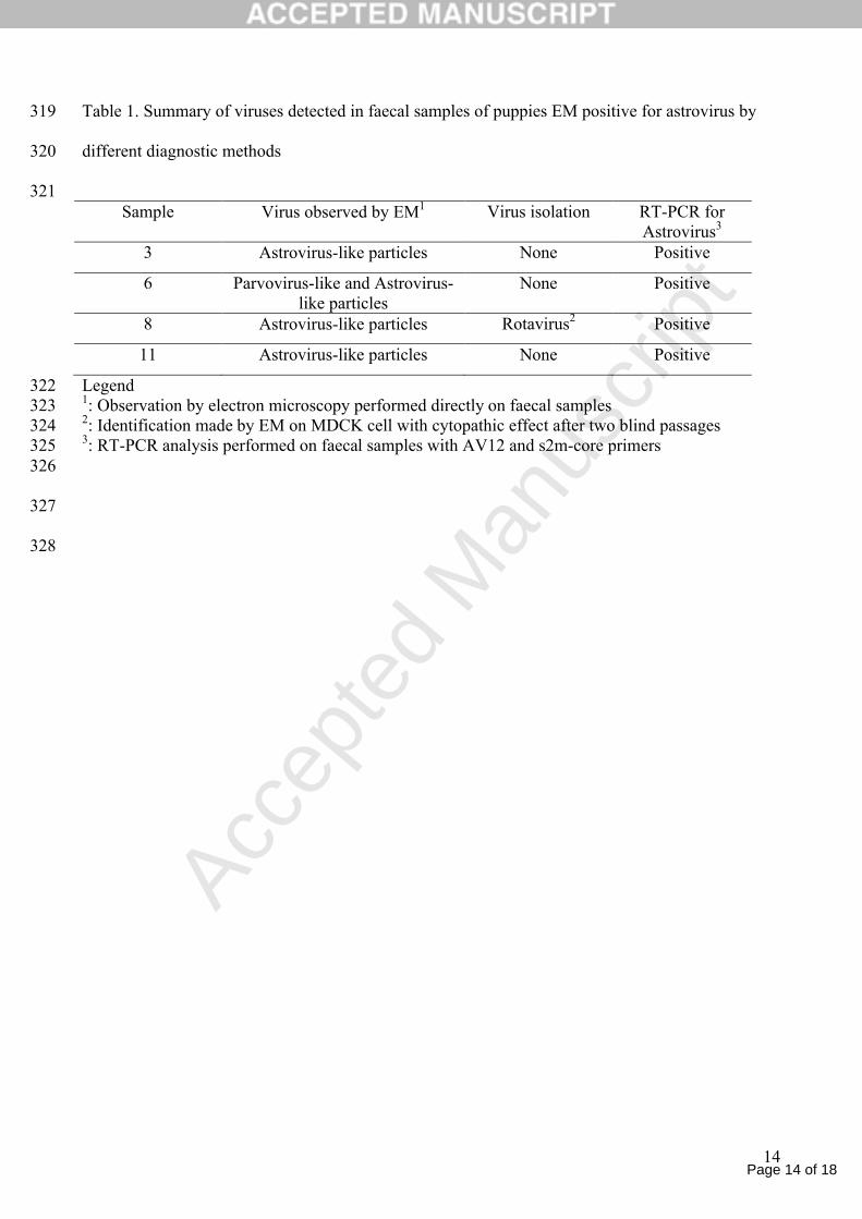
for the capsid precursor protein. None of the possible start codons for the capsid precursor were in 237
an optimal context for translation initiation as proposed by Kozak (1989), as they both lack a purine 238
at position -3, but the most upstream codon did have a context most resembling the consensus 239
vertebrate sequence around start codons GCCRCCAUGG (Kozak 1989). The canine astrovirus 240
genome, however, displayed the very conserved region right upstream of the mamastrovirus-241
homologous start codon, that could be involved in subgenomic RNA transcription (Jonassen et al., 242
2003), and it is therefore not clear if a subgenomic RNA spanning the first initiation codon is 243
synthesised. However, the fact that a functional domain could be predicted for the additional N-244
terminal peptide could suggest that longer transcripts actually are translated. The role of the 245
transmembrane domain and membrane localisation of the resulting protein would still need to be 246
elucidated. 247
The phylogenetic analysis clearly clustered canine astroviruses in the genus Mamastrovirus, in a 248
well defined and distinct branch of the phylogenetic tree. The relatively closest similarities were 249
revealed with a cluster comprising human, porcine and feline astroviruses, based on the ORF2 250
sequences available. 251

Page 11 of 18
Accep
ted
Man
uscr
ipt
11
Based on the species definition for astroviruses (Fauquet et al., 2005) and on the data obtained in 252
this study, we suggest a new species of astrovirus (canine astrovirus, CaAstV) to be included in the 253
genus Mamastrovirus.254
255
Acknowledgment256
The authors wish to thank Alice Fusaro for her technical assistance in sequence and phylogenetic 257
analysis and Paola De Benedictis for virus isolation attempts. Part of this study was financially 258
supported by the Italian Ministry of Health (RC IZSVe 27/2007).259
260
References261
262
1. Bridger, J.C., Hall, G.A., Brown J.F., 1984. Characterization of calici-like virus (Newbury 263
agent) found in association with astrovirus in bovine diarrhoea. Infect. Imun. 43, 133-138.264
2. Capelli, G., Frangipane di Regalbono, A., Iorio, R., Pietrobelli, M., Paoletti, B., 265
Giangaspero, A., 2006. Giardia species and other intestinal parasites in dogs in north-east 266
and central Italy. Vet. Record 159, 422-424.267
3. Chu, D.K., Poon,L.L., Guan, Y., Peiris, J.S., 2008. Novel astroviruses in insectivorous 268
bats. J. Virol. 82, 9107-9114.269
4. Fauquet, C.M., Mayo, M.A., Maniloff, J., Desselberger, U., Ball, L.A., 2005. Virus 270
Taxonomy: VIIIth Report of the International Committee on Taxonomy of Viruses. 271
Elsevier Academic Press, London UK.272
5. Gough, R.E., Collins, M.S., Borland, E., 1984. Astrovirus-like particles associated with 273
hepatitis in ducklings. Vet. Record 114, 279.274

Page 12 of 18
Accep
ted
Man
uscr
ipt
12
6. Guy, J.S., Miles, A.M., Smith, L., Fuller, F.J., Schultz-Cherry, S.L., 2004. Antigenic 275
and genomic characterization of turkey enterovirus–like virus (North Carolina, 1988 276
isolate): identification of the virus as turkey astrovirus 2. Avian Dis. 48, 206-211.277
7. Imada, T., Yamaguchi, S., Mase, M., Tsukamoto, K., Kubo, M., Morooka, A., 2000. 278
Avian nephritis virus (AVN) as a new member of the family Astroviridae, J. Virol. 74, 279
8487-8493.280
8. Jonassen C.M., 2008. Detection and sequence charaterization of the 3’-end of 281
coronaviruses genomes harbouring the highly conserved motif s2m. Methods in Molecular282
Biology, vol 454, SARS- and other coronaviruses, Ed. D. Cavanagh, 27-34.283
9. Jonassen, C.M., Jonassen, T.Ø., Grinde, B., 1998. A common RNA motif in the 3’ end 284
of the genome of astroviruses, avian infectious bronchitis virus and an equine rhinovirus. 285
J. Gen. Virol. 79, 715-718.286
10. Jonassen, C.M., Jonassen, T.Ø., Saif, Y.M., Snodgrass, D.R., Ushijima, H., Shimizu, 287
M., Grinde, B., 2001. Comparison of capsid sequences from human and animal 288
astroviruses. J. Gen. Virol. 82, 1061-1067.289
11. Jonassen, C.M., Jonassen, T.Ø., Sveen, T.M., Grinde, B., 2003. Complete genomic 290
sequence of astroviruses from sheep and turkey: comparison with related viruses. Virus 291
Res. 91, 195-201.292
12. Kozak, M., 1989. The scanning model for translation: an update. The Journal of Cell 293
Biology, 108, 229-241.294
13. Kumar, S., Tamura, K., Nei, M., 2004. MEGA3: Integrated software for molecular 295
evolutionary genetics analysis and sequence alignment. Brief bioinform. 5,150-163.296
14. Lee, T.W., Kurtz, J.B., 1981. Serial propagation of astrovirus in tissue culture with the 297
aid of trypsin. J. Gen. Virol. 57, 421-424.298

Page 13 of 18
Accep
ted
Man
uscr
ipt
13
15. Marshall, J.A., Healey, D.S., Studdert, M.J., Scott, P.C., Kennett, M.L., Ward, B.K., 299
Gust, I.D., 1984. Viruses and virus-like particles in the faeces of dogs with and without 300
diarrhoea. Aust. Vet. J. 61. 33-38.301
16. Marshall, J.D., Kennett, M.L., Rodger, S.M., Studdert, M.J., Thompson, W.L., Gust,302
I.D., 1984. Viruses and virus-like particles in the faeces of cats with and without diarrhoea. 303
Aust. Vet. J. 64, 100-105.304
17. Moser, L.A., Schultz-Cherry, S., 2005. Pathogenesis of astrovirus infection. Review. Viral 305
Immunol. 18, 4-10.306
18. Quinn, P.J., Carter, M.E., Markey, B., Carter, G.R., 1994. Clinical Veterinary 307
Microbiology, Wolfe Publishing, London.308
19. Robertson, M. P., Igel, H., Baertsch, R., Haussler, D., Ares, M., Jr., Scott, W. G., 2005.309
The Structure of a Rigorously Conserved RNA Element Within the SARS Virus Genome. 310
PLoS.Biol. 3, e5.311
20. Vieler, E., Herbst, W., 1995. Electron microscopic demonstration of viruses in feces of 312
dogs with diarrhea. Tierarztl Prax 23, 66-69.313
21. Walter, J. E., Mitchell, K., 2003. Astrovirus infection in children. Curr. Opin. Infect. Dis. 314
16, 247-253.315
22. Williams F.P.JR. 1980. Astrovirus–like, coronavirus-like and parvovirus–like particles 316
detected in diarrheal stools of beagle pups. Arch Virol 66:215-226317
318
Page 14 of 18
Accep
ted
Man
uscr
ipt
14
Table 1. Summary of viruses detected in faecal samples of puppies EM positive for astrovirus by 319
different diagnostic methods320
321Sample Virus observed by EM1 Virus isolation RT-PCR for
Astrovirus3
3 Astrovirus-like particles None Positive
6 Parvovirus-like and Astrovirus-like particles
None Positive
8 Astrovirus-like particles Rotavirus2 Positive
11 Astrovirus-like particles None Positive
Legend3221: Observation by electron microscopy performed directly on faecal samples3232: Identification made by EM on MDCK cell with cytopathic effect after two blind passages 3243: RT-PCR analysis performed on faecal samples with AV12 and s2m-core primers325
326
327
328

Page 15 of 18
Accep
ted
Man
uscr
ipt
15
Figure 1: Electron microscopy picture of astrovirus (larger particles of 18-20 nm) and parvovirus 329
(smaller particles of 27-30 nm) observed directly in faecal samples of puppies n. 6. (36,000 Kx). 330
The characteristic “star-like appearance” of astrovirus particles is evident compared to the round 331
shaped morphology of parvovirus. Negative staining was obtained with 2% sodium 332
phosphotungstate solution. 333
334
Figure 2. Phylogenetic tree constructed by Bayesian analysis of the nucleotide sequences of ORF2. 335
Posterior probabilities of the clades are indicated at the nodes. In this figure, Bat Astrovirus 336
sequence is identified as BAstV; Sheep Astrovirus as SAstV; Mink Astrovirus as MAstV; Feline 337
Astrovirus FAstV, Human Astroviruses as HAstV; Porcine Astrovirus as PAstV; Canine 338
Astroviruses as CaAstV; Turkey Astrovirus type I and II as TAstV1 and TAstV2, respectively. 339
GenBank accession numbers are indicated.340
341
Fig 3. Part of the CaAstV sequence in the junction between the ORF1b and ORF2 genes. Both 342
nucleotide sequences and the amino acid sequences for the C-terminal part of the ORF1b (with grey 343
shade), and for the putative capsid precursor protein (in bold) are shown, displaying the two 344
possible start codons for the putative caspid precursor in bold italics. The start of the ORF2 protein 345
homologous to other mamastroviruses is underlined. The stop codon of the ORF1b is shaded grey.346
347

Page 16 of 18
Accep
ted
Man
uscr
ipt
Figure

Page 17 of 18
Accep
ted
Man
uscr
ipt
Figure

Page 18 of 18
Accep
ted
Man
uscr
ipt
1 - GGCTTTACTGTAGATGAGAATCTTGAACCAATACCTACACAACCTGACAAATTGATGGCC - 60 - G F T V D E N L E P I P T Q P D K L M A - A L L * M R I L N Q Y L H N L T N * W P 61 - TCACTTCTCAAACCAGCATCCAAACTTCCGGATCTTGAATCACTCCATGGGAAACTCCTG - 120 - S L L K P A S K L P D L E S L H G K L L - H F S N Q H P N F R I L N H S M G N S C
121 - TGCTATCAGCTCCTCTCGGCTTTCCTACCTGAGGAACACCCTTTTAAGGTGTATGTCGAG - 180 - C Y Q L L S A F L P E E H P F K V Y V E - - A I S S S R L S Y L R N T L L R C M S R -
181 - AGTTGTCTGGCTGCCACTAGCAGGCAGCTTCGTGATTCTGGCTTACCCACCAGATTCACA - 240 - S C L A A T S R Q L R D S G L P T R F T - - V V W L P L A G S F V I L A Y P P D S Q -
241 - GAAGAGCAAATGCATCGCATATGGAGGGGAGGACCAAAAAATTGCGATGGCTAGCAAGCC - 300 - E E Q M H R I W R G G P K N C D G * Q A - - K S K C I A Y G G E D Q K I A M A S K P -
Figure
Related Documents