Genetic Characterisation of Human ABO Blood Group Variants with a Focus on Subgroups and Hybrid Alleles Hosseini Maaf, Bahram Published: 2007-01-01 Link to publication Citation for published version (APA): Hosseini Maaf, B. (2007). Genetic Characterisation of Human ABO Blood Group Variants with a Focus on Subgroups and Hybrid Alleles Division of Hematology and Transfusion Medicine, Department of Laboratory Medicine, Lund University General rights Copyright and moral rights for the publications made accessible in the public portal are retained by the authors and/or other copyright owners and it is a condition of accessing publications that users recognise and abide by the legal requirements associated with these rights. • Users may download and print one copy of any publication from the public portal for the purpose of private study or research. • You may not further distribute the material or use it for any profit-making activity or commercial gain • You may freely distribute the URL identifying the publication in the public portal

Welcome message from author
This document is posted to help you gain knowledge. Please leave a comment to let me know what you think about it! Share it to your friends and learn new things together.
Transcript

LUND UNIVERSITY
PO Box 117221 00 Lund+46 46-222 00 00
Genetic Characterisation of Human ABO Blood Group Variants with a Focus onSubgroups and Hybrid Alleles
Hosseini Maaf, Bahram
Published: 2007-01-01
Link to publication
Citation for published version (APA):Hosseini Maaf, B. (2007). Genetic Characterisation of Human ABO Blood Group Variants with a Focus onSubgroups and Hybrid Alleles Division of Hematology and Transfusion Medicine, Department of LaboratoryMedicine, Lund University
General rightsCopyright and moral rights for the publications made accessible in the public portal are retained by the authorsand/or other copyright owners and it is a condition of accessing publications that users recognise and abide by thelegal requirements associated with these rights.
• Users may download and print one copy of any publication from the public portal for the purpose of privatestudy or research. • You may not further distribute the material or use it for any profit-making activity or commercial gain • You may freely distribute the URL identifying the publication in the public portal

Take down policyIf you believe that this document breaches copyright please contact us providing details, and we will removeaccess to the work immediately and investigate your claim.
Download date: 22. Jun. 2018
Genetic Characterisation of HumanABO Blood Group Variants with a Focus on
Subgroups and Hybrid Alleles
Doctoral thesis by
Bahram Hosseini-Maaf
Division of Hematology and Transfusion Medicine Department of Laboratory Medicine
Lund University, Sweden
With the approval of the Faculty of Medicine at Lund University, this thesis will be defended on March 16, 2007, at 9:00 in Segerfalksalen,
Wallenberg Neurocentrum, BMC, Sölvegatan 17, Lund
Faculty opponent: Professor Peter Påhlsson
Department of Biomedicine and Surgery Division of Cell Biology
Linköping University Sweden

© Bahram Hosseini-Maaf
ISBN 978-91-85559-16-9 Printed by Media-Tryck, Lund, Sweden 2007

Till min familj
“the chief part of the organisation of every being is simply due to inheritance; and consequently, though each being assuredly is well fitted for its place in nature, many structures now have no direct relation to the habits of life of each species”
The Origin of Species Charles Darwin,1859

CONTENTSORIGINAL PAPERS……………………………………………………………………….. 6
ABSTRACT………………………………………………………………………………… 7
ABBREVIATIONS………………………………………………………………………… 8
PROLOGUE………………………………………………………………………………... 9
BACKGROUND…………………………………………………………………………… 9
History of blood transfusion and basic principles of the ABO blood group system …... 9 Definition of blood group antigens and antibodies……………………………………... 11 Blood group terminology……………………………………………………………….. 12 Biochemistry of the ABO blood groups ………………………………………………… 15 The molecular structure of ABO glycosyltransferase …………………………………. 17 Genetics of the ABO blood group system ……………………………………………… 19
The ABO gene ………………………………………………………………………. 19 Serology versus molecular genetics ……………………………………………………. 21
Relationship between phenotype and genotype for the major alleles……………….. 21 Genetic background of hybrid alleles, rare O alleles and weak subgroups …………. 27
AIMS OF THE THESIS……………………………………………………………………. 33
MATERIAL AND METHODS…………………………………………………………….. 33
Blood and DNA samples……………………………………………………………….. 33 ABO genotyping………………………………………………………………………… 33
Polymerase chain reaction-restriction fragment length polymorphism (PCR-RFLP) 33Allele-specific primer PCR (PCR-ASP)……………………………………………. 34
Direct DNA sequencing………………………………………………………………… 34 Reverse transcriptase PCR (RT-PCR)………………………………………………….. 34 Site-directed mutagenesis and cloning …..…………………………………………….. 35 Protein purification and enzyme kinetics ………………………………………………. 35 Computer modelling and crystallization ……………………………………………….. 35
RESULTS AND DISCUSSION ……………………………………………………………. 36 Single point mutations associated with A or B subgroups (Papers I and V) …………… 36 Hybrid ABO alleles explain some other weak ABO subgroups (Papers I, II and V) …… 41 The genetic basis of the Abantu phenotype is also a hybrid allele (Paper IV) ……………. 45 Novel O alleles involved in unexpected blood group phenotypes (Paper III) ………….. 47 Development of an improved ABO genotyping assay based on PCR-ASP amplification across intron 6 (Paper VI) ………………………………………………………………. 51
CONCLUSION……………………………………………………………………………… 54
FUTURE PERSPECTIVES ………………………………………………………………… 54
SUMMARY IN SWEDISH (sammanfattning på svenska)…………………………………. 56
ACKNOWLEDGEMENTS ………………………………………………………………… 61
REFERENCES ……………………………………………………………………………… 63
APPENDIX: Papers I-VI ……………………………………………………………………. 70
6
ORIGINAL PAPERSThis thesis is based on the following papers, which will be referred to in the text by their Roman numerals (I-VI).
I. Olsson ML, Irshaid NM, Hosseini-Maaf B, Hellberg Å, Moulds MK, Sareneva H, Chester MA. Genomic analysis of clinical samples with serologic ABO blood grouping discrepancies: Identification of 15 novel A and B subgroup alleles.Blood, 2001;98(5):1585-1593.
II. Hosseini-Maaf B, Hellberg Å, Rodrigues MJ, Chester MA, Olsson ML. ABO exon and intron analysis in individuals with the AweakB phenotype reveals a novel O1v-A2 hybrid allele that causes four missense mutations in the A transferase.
BMC Genetics, 2003; 4:17 (11 pages, doi: 10.1186/1471-2156-4-17).
III. Hosseini-Maaf B, Irshaid NM, Hellberg Å, Wagner T, Levene C, Hustinx H, Steffensen R, Chester MA, Olsson ML. New and unusual O alleles at the ABOlocus are implicated in unexpected blood group phenotypes.
Transfusion, 2005;45(1):70-81.
IV. Hosseini-Maaf B, Smart E, Chester MA, Olsson ML. The Abantu phenotype in the ABO blood group system is due to a splice-site mutation in a hybrid between a new O1-like allelic lineage and the A2 allele. Vox Sanguinis, 2005;88(4):256-264.
V. Hosseini-Maaf B, Letts JA, Persson M, Smart E, Le Pennec P-Y, Hustinx H, Zhao Z, Palcic MM, Evans SV, Chester MA, Olsson ML. Structural basis for red cell phenotypic changes in newly-identified, naturally-occurring subgroup mutants of the human blood group B glycosyltransferase. Transfusion, 2007; in press.
VI. Hosseini-Maaf B, Hellberg Å, Chester MA, Olsson ML.An extensive PCR-ASP strategy for clinical ABO blood group genotyping that avoids potential errors caused by null, subgroup and hybrid alleles.
Manuscript.

7
ABSTRACT
ABO is the most important blood group system in transfusion medicine and transplantation
immunology. The ABO blood groups differ by the presence or absence of antigens on RBCs
and antibodies in plasma. Accurate determination of ABO status is critical. Genomic typing
can increase the precision of blood group determination in complicated cases, e.g. when
variant expression of A or B antigen is encountered.
The overall aim of this study was to compare the molecular diversity of ABO alleles with
various phenotypes, and to contribute to our knowledge of the ABO gene and encoded
glycosyltransferases.
Novel alleles (six Aweak, eleven Bweak, seven O) were identified containing single-point
mutations. Structure/function studies explained the weakening of some B subgroup
glycosyltransferases. Two new hybrid Ax alleles were characterised. Analysis of introns 2-5
revealed 44 previously unknown, allele-related polymorphisms that proved valuable allelic
markers. These findings enabled localisation of cross-over regions in two other new hybrids:
1) an O1v allele fused with an A2 allele, 2) the novel O1bantu-A2 combination that explained the
Abantu phenotype. Phylogenetic and population analyses indicated that O1bantu is a unique and
distinct evolutionary lineage so far only found among individuals of African descent.
Of clinical importance, a new approach to ABO genotyping was developed that identifies all
common alleles, most null and weak A/B subgroups as well as hybrid alleles resulting from
recombinational crossing-over events.
In summary, 30 novel alleles were identified and characterized, representing 30% of all alleles
reported since the start of this study in 2001.

8
ABBREVIATIONSa.a. Amino acid
bp Base pair
kb kilobase pairs
DNA Deoxyribonucleic acid
EC Enzyme commision
GTA or A-enzyme 3- -N-acetylgalactosaminyltransferase
(EC 2.4.1.40) encoded by the blood group A gene
GTB or B-enzyme 3- -galactosyltransferase (EC 2.4.1.37)
encoded by the blood group B gene
nt. Nucleotide
ISBT International Society of Blood Transfusion
MAb Monoclonal antibody
PCR Polymerase chain reaction
RBC Red blood cell
RNA Ribonucleic acid
RFLP Restriction fragment length polymorphism
ASP Allele-specific primer
SNP Single nucleotide polymorphism
VNTR Variable number of tandem repeats

9
PROLOGUE
Elucidation of the human genome has now been completed through “the Human Genome
Project” and the genetic makeup of man is known. The biologically interpretation of the
information contained in the genetic sequence is an extremely complex process, resulting in
the production of proteins that act as structural elements and metabolic mediators, e.g.
enzymes in each cell and organ of an individual. With few exceptions, genetic variation
makes every individual exceptional.
Many aspects of this universal uniqueness between individuals are central to transfusion
medicine but blood group variation is probably the most important and a prerequisite for
modern health care.
The need for donor-recipient matching when performing blood transfusion became apparent
at early stages in the history of transfusion medicine, due to the interaction of substances on
transfused red blood cells (RBCs), antigens, with other substances, antibodies, in the
recipient’s plasma.
The advances in scientific knowledge have allowed characterisation of many of these antigens
and antibodies. The structures of the antigens have been elucidated in many cases, some of
the biochemical pathways for their biosynthesis have been determined and currently the
primary sequences of the genes encoding these antigens are being revealed. The main subject
of this thesis will be limited to the ABO blood groups and the following background describes
chronologically the development of our knowledge about this important system from the
empirical observations of transfusion reactions to the structural variations in the ABO gene
itself and its products.
BACKGROUND
History of blood transfusion and basic principles of the ABO blood group systemBlood as a medical term is often related to words including hemo- or hemato- from the Greek
stem "haima" for blood. Blood is a circulating tissue composed of fluid plasma and cells
(RBCs, white blood cells and platelets).

10
Blood has been used symbolically in both spoken language as “blood money, stir up bad
blood, run in the blood, blood brother, bad blood, blood feud, bloodthirsty” and in the
literature in different contexts, e.g. “We few, we happy few, we band of brothers; For he
today that sheds his blood with me, Shall be my brother” (Shakespeare: Henry V, act 4).
“The old concept of the movement of blood in the body was elaborated by Galen (129-200
AD). He incorrectly believed that food from the stomach was digested and taken to the liver,
where it was transformed into blood, passed to heart, through arteries and veins to the tissues
where it was burned up, as wood is consumed by fire” (Farr, 1980).
Different cultures put varying significance on blood. Some believed that it could be the
treatment for many maladies and others were convinced that blood contained the soul of man.
Ancient Egyptians used it for baths to resuscitate the dead. The first human-to-human blood
transfusion recorded took place in 1492. Pope Innocent VIII sank in a coma, and the blood of
three ten-year old boys was infused into the dying Pope’s veins at the suggestion of a
physician. Unfortunately, both the recipient and all the donors died (Barsoum & Kleeman,
2002). There is no documentation of the fate of the physician, however.
In 1628, British physician W Harvey described the circulation of the blood in humans after
having pumped water through the blood vessels of a corpse (Harvey, 1628). This observation
opened a new era and some decades later, in 1667, J-B Denis (Denis, 1667) in France and R
Lower (Lower, 1666) in England separately reported transfusions from animal to animal and
animal to humans. However, Denis was accused of murder when a young boy died after he
was transfused the blood from a lamb, and transfusion of animal blood to man was prohibited
by law after the death of further patients (Farr, 1979). Nothing of importance was reported in
this field until December 22 in 1818, nearly 150 years later, when J Blundell performed his
first successful blood transfusion of man to man (Blundell, 1829).The tendency of blood to
clot was the principal barrier to transfusions, and to storage of blood. Furthermore, serious
reactions to transfusion (e.g. fever, hypotension, allergic shock, death) continued to occur
until 1900 when Landsteiner published his preliminary results and the year after described the
first system of blood groups, the so-called ABO system (Landsteiner, 1900, 1901).
Incompatibility between donor and recipient in these blood groups could explain most of
these serious transfusion reactions. Landsteiner based his observation on an unexpected
interaction between serum and RBCs from himself and a few of his colleagues at the Institute

11
of Pathological Anatomy in the University of Vienna. He later received the Nobel Prize for
his discovery (Landsteiner, 1930). This elucidation laid the basis of a new field in biology and
the foundation of transfusion medicine.
This principal finding led to what has become Landsteiner´s rule, i.e. serological behaviour of
the A, B and O (O was initially named type “C”) blood groups (Table 1). Subsequently,
another Austrian group, Decastello and Sturli, identified the fourth group, i.e. AB, in 1902
(von Decastello & Sturli, 1902). Several other investigators independently duplicated
Landsteiner´s findings. Jansky in Poland named the four blood groups I, II, III and IV based
on their frequency in European populations in 1907, and Moss described in 1910 the four
blood groups in the reverse order of Jansky (reviewed in Garratty et al., 2000; Farr, 1979).
Table 1. Basic principles of the ABO system. Relation between antigens on RBCs and antibodies in serum.
ABO blood group Red cell antigen Antibodies in serum
O None Anti-A, anti-B, anti-A,B A A Anti-B B B Anti-A AB A and B None
In clinical practice, the ABO blood group system is one of the most important since the A and
B epitopes may provoke a strong immune reaction. With the introduction of blood typing and
crossmatching techniques, blood transfusion became not only a simple but also a much safer
procedure. However, blood transfusion has continued to be associated with risks, notably the
transmission of blood-borne infectious diseases such as syphilis, viral hepatitis and human
immunodeficiency virus. Furthermore, although ABO typing reduced the occurrence of
transfusion reactions, they still occurred, indicating the presence of other genetic differences
in blood groups of importance in transfusion medicine, as well as in the later emerging field
of organ transplantation.
Definition of blood group antigens and antibodiesThe term blood group is generally based on the presence or absence of certain antigens on the
RBC membrane. These are identified by characteristic agglutination reactions with specific
antibodies and this field is referred to as blood group serology (Daniels, 2002).

12
An antigen is in fact any substance that is able to stimulate the production of an antibody
under certain conditions. Some blood group antigens, e.g. in blood group systems ABO, P1,
H, LE and I, are due to differences in carbohydrate structures and they are all found on
glycolipids or glycoproteins on the RBC membrane. In the case of the ABO blood groups, the
antigens are found on the surface of RBCs and other cells and is recoverable from body fluids
and secretions. Most other blood group systems result from polymorphisms on different RBC
membrane-associated proteins (Daniels, 2002).
Blood group antibodies can be divided into induced and naturally-occurring antibodies.
Induced antibodies are raised by the immune system as a response to specific antigens, e.g.
following transfusion, pregnancy or transplantation. Natural antibodies are also of immune
origin, with the difference that the antigen eliciting their production is unknown. In the ABO
system, these antibodies are sometimes referred to as isoagglutinins because they can cause
blood cells from certain individuals to clump together. The two isoagglutinins, anti-A and
anti-B, occur naturally in humans, contrary to most other blood groups antibodies (Erskine &
Socha, 1978; Mollison et al., 1993).
Naturally-occurring antibodies can be detected in serum of persons who have never been
transfused or women who never have been pregnant (Mollison et al., 1993). Natural
antibodies may occur because of unidentified infections during early life or, in the case of
blood group antibodies, introduction of substances that are antigenically similar to blood
group substances. Interestingly, Springer et al., as early as 1959, demonstrated by an
experiment with chickens bred under sterile conditions, that some of these antibodies appear
following stimulation by certain bacteria, such as Escherichia coli, while the appearance of
others was related to exposure to various kinds of food. The origin of natural antibodies is still
a matter of debate, though.
Naturally-occurring antibodies are often IgM and sometimes also consist of an IgG
component. With exception for antibodies against ABO, Pk and P, the majority of naturally-
occurring antibodies are only cold reactive and of limited clinical importance.
Blood group terminology A rather incoherent terminology has evolved as more and more blood group antigens were
discovered during several decades. Providing a common acceptable and reliable terminology,

13
which would cover both the serology and genetics, has been difficult. The International
Society of Blood Transfusion (ISBT) Working Committee on Terminology for Red Cell
Surface Antigens (http://www.blood.co.uk/ibgrl/) was set up in 1980 to establish and define a
meaningful nomenclature for different blood groups as shown in Table 2 (Daniels et al.,
2003; 2004; Storry & Olsson, 2004). Every valid blood group antigen is given a six digit
identification number, according to the following categorization:
- Blood group systems; one or more antigens governed either by a single gene locus or by a
complex of two or more very closely linked homologous genes with virtually no
recombination occurring between them. There are 29 different systems to date, and the first
three digits represent the systems (001-029).
- Blood group collections; genetically-, biochemically-, or serologically-related sets of
antigens, which do not, at this time, merit system status because of insufficient data (205-
211).
- A series of low frequency-antigens (the 700 series)
- A series of high frequency-antigens (the 901 series)
The symbol for a gene or cluster of genes controlling a blood group system is often the
italicized symbol for the system. The ABO genotypes should consequently be written in
italicised capital letters (with superscripts indicating the subgroup alleles).
However, ABO allele nomenclature poses significant problems that are still under
consideration by the ISBT. In the absence of an officially agreed terminology, in this thesis,
alleles are referred to by their serological activity and an alternative allele name is given in
square brackets.
ISBT has recently set up a subgroup from the Terminology Working Committee mentioned
above. Its task is to create a unified allele nomenclature for blood groups, (ML Olsson
personal communication).

14
Table 2. Human blood group systems recognised by the ISBT (http://www.blood.co.uk/ibgrl/).
System System no. Symbol Antigens Gene Locus Epitope / carrier, name ISBT ISBT no. or functions
ABO 001 ABO 4* ABO 9q34.1-q34.2 Carbohydrate
MNS 002 MNS 43 GYPA 4q28.2-q31.1 Glycophorin A&B GYPB (CD235a-b)‡
GYPE
P 003 P1 1 P1 22q11.2-qter Carbohydrate
Rh 004 RH 56 RHD 1q34-36.2 Protein, CD240D RHCE CD240CE Lutheran 005 LU 19 LU 19q13.2 Glycoproteinprotein (IgSF†), adhesion molecule, CD239
Kell 006 KEL 23 KEL 7q33 Glycoprotein, CD238
Lewis 007 LE 6 FUT3 19p13.3 Carbohydrate
Duffy 008 FY 6 FY 1q22-q23 Glycoprotein, receptor, CD234
Kidd 009 JK 3 JK 18q11-q12 Glycoprotein, urea transporter
Diego 010 DI 19 AE1 17q12-q21 Glycoprotein (band 3, AE1), CD233
Yt 011 YT 2 ACHE 7q22 Enzyme, (Cartwright) acetylcholinesterase
Xg 012 XG 1 XG Xp22.32 Glycoprotein, CD99
Scianna 013 SC 3 ERMAP 1q36.2-p22.1 Glycoprotein
Dombrock 014 DO 5 DO 12q13.2-p12.1 Glycoprotein, CD297
Colton 015 CO 3 AQP1 7p14 Aquaporin 1 (channel)
Landsteiner/ 016 LW 3 LW 19p13.3 LW glycoprotein (IgSF†)Weiner CD242
Chido-Rogers 017 CH/RG 9 C4A,C4B 6p21.3 Complement protein 4
Hh 018 H 1 FUT1 19q13 Carbohydrate, CD173
Kx 019 XK 1 XK Xp21.1 Glycoprotein
Gerbich 020 GE 7 GYPC 2q14-q21 Glycophorins C and D, CD236
Cromer 021 CROM 10 DAF 1q32 Glycoprotein, CD55
Knops 022 KN 5 CR1 1q32 Glycoprotein, CR1 or CD35
Indian 023 IN 2 CD44 11p13 Glycoprotein, CD44
Ok 024 OK 1 BSG 19p13.3 Glycoprotein, CD147
Raph 025 RAPH 1 MER2 11p15.5 Transmembrane glycoprotein, CD151
John Milton 026 JMH SEMA7A 15q23-q24 Glycoprotein, CD108 -Hagen
I 027 I GCNT2 6p24 Carbohydrate
Globoside 028 GLOB B3GALNT1 3q25 Glycolipide
GIL 029 GIL AQP3 9p13 Aquaporin 3 (channel)
* Four ABO antigens are recognised by the ISBT and noted as 001 (A), 002 (B), 003 (AB) and 004 (A1)‡ Cluster of Differentiation (antigens) † The immunoglobulin superfamily, IgSF, http://imgt.cines.fr/textes/IMGTindex/superfamily.html Complement component (3b/4b) receptor 1 (Knops blood group)

15
In 1999 a database, The Blood Group Antigen Gene Mutation Database (dbRBC), was also
set up under support of Human Genom Variation Society (HGVS) lead by O Blumenfeld
(Blumenfeld & Patnaik, 2004). This database contains information about variations in genes
that affect blood group expression (http://www.ncbi.nlm.nih.gov/projects/mhc/xslcgi.fcgi?cm
d=bgmut/home).
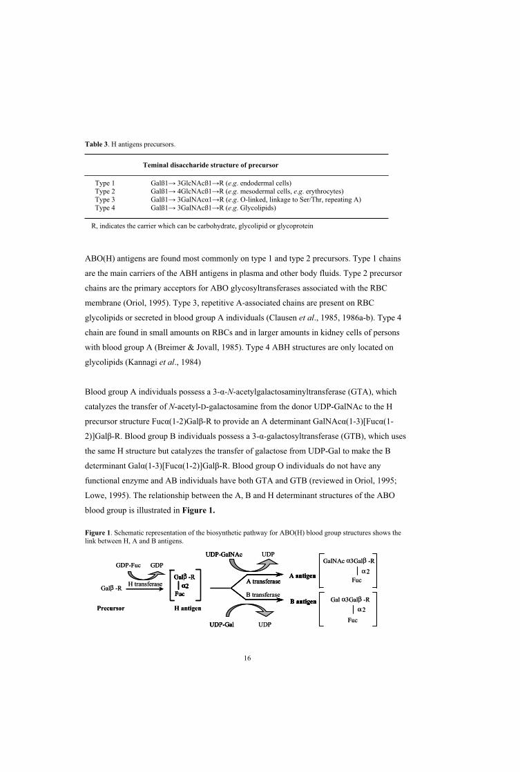
Biochemistry of the ABO blood groups The biochemical basis of the ABO and H antigens is well understood due to intensive studies
during the 1950s and 1960s by the pioneering work on ovarian cyst fluids (which contain
large amounts of water-soluble blood-group-active glycoproteins) by Morgan & Watkins and
Kabat. Subsequently, this led to the explanation of biosynthesis pathways for ABH antigens,
Figure 1 (Kabat, 1956; references and review in Watkins 1980; Lowe, 1995).
The ABO antigens are not limited to erythroid tissues, but are also found in different tissues
and on some epithelial cells (Oriol et al., 1992). Therefore, they can sometimes be noted as
histo-blood group antigens (Clausen & Hakomori, 1989). On the other hand, they have not
been observed in connective tissues, muscles, and the nervous system (Schenkel-Brunner,
2000). However, in this thesis, as in the field of transfusion medicine, these antigens will be
referred as blood groups and not histo-blood groups.
The H antigen is the immediate precursor for creation of blood-group-A-and B-active
structures. Two genes, FUT1 (responsible for synthesis of H antigen on type 2 precursor, e.g.
on RBC) or FUT2 (responsible for synthesis of H antigen on type 1 precursor, e.g. in
secretions) on chromosome 19 encode fucosyltransferases (Oriol, 1995; Lowe, 1995). These
2- -L-fucosyltransferases catalyse the transfer of L-fucose from the
guanosinediphosphofucose (GDP-L-fucose) donor to the terminal galactose of at least one of
the precursor types. Six precursor types have been identified but only four types (Table 3) are
known to carry ABO activity (Clausen & Hakomori, 1989; Clausen et al., 1994).
These substances subsequently act as acceptor substrate for the enzyme product of the ABO
gene. The ABO gene product, i.e. glycosyltransferases A and B (GTA and GTB) use UDP-
GalNAc and UDP-Gal, respectively, as substrates.
16
Table 3. H antigens precursors.
Teminal disaccharide structure of precursor
Type 1 Galß1 3GlcNAcß1 R (e.g. endodermal cells) Type 2 Galß1 4GlcNAcß1 R (e.g. mesodermal cells, e.g. erythrocytes) Type 3 Galß1 3GalNAc 1 R (e.g. O-linked, linkage to Ser/Thr, repeating A) Type 4 Galß1 3GalNAcß1 R (e.g. Glycolipids)
R, indicates the carrier which can be carbohydrate, glycolipid or glycoprotein
ABO(H) antigens are found most commonly on type 1 and type 2 precursors. Type 1 chains
are the main carriers of the ABH antigens in plasma and other body fluids. Type 2 precursor
chains are the primary acceptors for ABO glycosyltransferases associated with the RBC
membrane (Oriol, 1995). Type 3, repetitive A-associated chains are present on RBC
glycolipids or secreted in blood group A individuals (Clausen et al., 1985, 1986a-b). Type 4
chain are found in small amounts on RBCs and in larger amounts in kidney cells of persons
with blood group A (Breimer & Jovall, 1985). Type 4 ABH structures are only located on
glycolipids (Kannagi et al., 1984)
Blood group A individuals possess a 3- -N-acetylgalactosaminyltransferase (GTA), which
catalyzes the transfer of N-acetyl-D-galactosamine from the donor UDP-GalNAc to the H
precursor structure Fuc (1-2)Gal -R to provide an A determinant GalNAc (1-3)[Fuc (1-
2)]Gal -R. Blood group B individuals possess a 3- -galactosyltransferase (GTB), which uses
the same H structure but catalyzes the transfer of galactose from UDP-Gal to make the B
determinant Gal (1-3)[Fuc (1-2)]Gal -R. Blood group O individuals do not have any
functional enzyme and AB individuals have both GTA and GTB (reviewed in Oriol, 1995;
Lowe, 1995). The relationship between the A, B and H determinant structures of the ABO
blood group is illustrated in Figure 1.
Figure 1. Schematic representation of the biosynthetic pathway for ABO(H) blood group structures shows the link between H, A and B antigens.
A transferaseA antigen
B antigenH antigen
UDP-GalNAc
Galβ -R
Fucα2
GalNAc α3Galβ -R
2Fuc
Gal α3Galβ -R
Fuc
α2
UDP
UDP
B transferase
H transferase
GDP-Fuc GDP
Galβ -R
Precursor
A transferaseA antigen
B antigen
UDP-GalNAc
UDP-GalUDP-Gal
Galβ -R
Fucα2
Galβ -R
Fucα2
αA transferase
A antigen
B antigenH antigen
UDP-GalNAc
Galβ -R
Fucα2
GalNAc α3Galβ -R
2Fuc
Gal α3Galβ -R
Fuc
α2
UDP
UDP
B transferase
H transferase
GDP-Fuc GDP
Galβ -R
Precursor
A transferaseA antigen
B antigen
UDP-GalNAc
UDP-GalUDP-Gal
Galβ -R
Fucα2
Galβ -R
Fucα2
α

17
The existence of subgroups of A, i.e. A1 and A2, was first recognized by von Dungern and
Hirszfield in 1911 (von Dungern & Hirszfeld , 1911). A1 RBCs have precursor types 1, 2, 3
and 4, while A2 erythrocytes appear to have mainly type 1 and type 2, and even small
amounts of type 3 (Clausen et al., 1985, 1986b).
The A1 and A2 transferases are qualitatively different with pH optima at 5.6 for A1 and 7-8 for
A2. The activity of A1 is higher, it has a lower Km for acceptors, and A1 has a higher
isoelectric point than the A2 enzyme (Watkins, 1980). There is also a quantitative difference
between A1 and A2 cells that has been illustrated by various techniques, among others,
radioimmunoassay with labelled antibodies and lectins, electron microscopy. These studies
showed that the number of antigen sites per RBC for A1 is 8-12x105 and for A2 is 1-4x10⁵
(Economidou et al., 1967; Cartron et al., 1974).
Expression of ABO(H) antigens may be affected by different factors, e.g. the amount and type
of precursor, place of expression and disease. The development of ABO(H) antigens is a
complex process and many factors are involved and may affect the final antigen. No attempts
will be made to go deeply into the biosynthesis of ABO blood groups in this thesis because of
the complexity of the topic and taking into account that the scope of the study is mainly
genetic.
The molecular structure of ABO glycosyltransferaseGlycosyltransferases (GTs), [Enzyme Commission, (EC) 2.4], make up a large family of
enzymes that are involved in the biosynthesis of oligosaccharides, polysaccharides, and
glycoconjugates. These enzymes show an enormous diversity and are present both in
prokaryotes and eukaryotes (Breton et al., 2006). Glycosyltransferases are classified into 87
different families based on substrate/product stereochemistry according to the CAZy database
(http://afmb.cnrs-mrs.fr/~pedro/CAZY/db.html) (Henrissat, 1998). Knowledge of the
sequences of the ABO genes (Yamamoto et al., 1990a-b) have established that mammalian
GTA (EC 2.4.1.40) and GTB (EC 2.4.1.37) are type II integral membrane proteins containing
354 amino acids (a.a.) and are localised in the lumen of the Golgi apparatus. Type II integral
membrane enzymes typically have a short amino-terminal cytoplasmic tail, a hydrophobic
membrane domain, a short protease-sensitive stem region and a large catalytic domain that
includes the carboxy terminus (Paulson & Colley, 1989), as shown in Figure 2a. GTA and

18
GTB are very similar in the coding regions and the soluble enzyme can be found in serum
(Schenkel-Brunner et al., 1972), urine (Chester, 1974) and milk (Ginsburg, 1972).
The high-resolution X-ray-crystal structures of the catalytic domains (a.a. residues 63-354) in
the GTA and GTB enzymes were resolved by Patenaude and co-workers (Patenaude et al.,
2002). Figure 2b shows a three-dimensional surface model that predicts the appearance of the
active domain in humans. The glycosyltransferase structure has a central cleft where the
enzymatic action takes place (Figure 2b and 2c). The enzyme contains an acceptor-
recognition domain that binds H antigen and a donor recognition domain that binds
UDP-GalNAc or UDP-Gal. GTA and GTB are retaining, metal-dependent family
glycosyltransferases (Boix et al., 2001; Pak et al., 2006).
GTA and GTB require the metal ion Mn2+ (manganese) for activity and have a characteristic 211DVD213 motif that coordinates attachment of the phosphate in the donor via the metal ion
(Breton et al. 2006). In vitro conversion of O cells to A by GTA as mentioned above, requires
the presence of Mn2+ ions but if Mn2+ is changed to Mg2+, A1 glycosyltransferase remains
active, but A2 transferase does not (Schachter et al., 1973).
Figure 2. ABO glycosyltransferase. The topology of the transferase is shown in (a) as Golgi-localised membrane-bound enzyme with a cytoplasmic N-terminus and catalytic C-teminal domain (modified from Paulsen & Colley, 1989). The 3D-surface model (b) was created with the Deep View Swiss Pdb Viewer version 3.7 and the 3D-ribbon structure (c) was generated using SETOR and SetoRibbon (unpublished).
N
Globularenzymaticallyactive domain
Stem region
Transmembranedomain
Cytoplasmic tail
Proteolyticcleavage
a.
b.
c.
N
cc
Corresponding to synthetic andnaturally solubleglycosyltransferase
cleft
C-terminalrecognizes
acceptor
N-terminalrecognizes
donor
cleftNN
Globularenzymaticallyactive domain
Stem region
Transmembranedomain
Cytoplasmic tail
Proteolyticcleavage
a.
b.
c.
N
cc
Corresponding to synthetic andnaturally solubleglycosyltransferase
cleft
C-terminalrecognizes
acceptor
N-terminalrecognizes
donor
cleft

19
Genetics of the ABO blood group system The genetic background for a protein polymorphism is straightforward. The occurrence of
most such variations is due to a single nucleotide polymorphism (SNP) that results in a single
a.a. change, which can directly affect the protein’s antigenicity if it is a blood group carrier.
The genetics governing carbohydrate structures (e.g. ABO antigens) are more complex
because the sequential action of a number of gene products, i.e. enzymes, is needed to
generate antigens of carbohydrate nature. Any polymorphism within these genes may create
extra sources of complexity.
In addition, many different genetic backgrounds may underlie one phenotype and different
phenotypes appears to be possible result from the same genetic variant. This makes phenotype
prediction from an ABO genotype difficult (but not impossible).
The ABO gene
The ABO blood group locus is located on the terminal portion of the long arm of chromosome
9 and has been determined through quantitative red cell adenylate kinase (AK-1) assay
(Ferguson-Smith et al., 1976). This was later confirmed by linkage analysis (Allderdice et al.,
1986) to position 9q34.1-q34.2.
The first successful isolation and purification of GTA, followed by cloning of cDNA
representing the blood group A transferase mRNA, were completed in 1990 from human lung
tissue (Clausen et al., 1990; Yamamoto et al., 1990a-b).
The coding region of the ABO gene consists of seven exons (exon 1 exists in two variants),
spanning about 19.5 kb and ranging in size from 28 to 688 bp (Figure 3). The 1062 bp
sequence encodes 354 a.a. corresponding to a 41 kDa protein. The ABO gene contains six
introns with sizes ranging from 554 to 12982 bp.
Figure 3. Organisation of the ABO gene. The seven exons and six introns are not drawn to scale. The numerals above boxes represent the first and last nucleotides of the coding region in each exon and those below boxes show the corresponding a.a. numbers (a). The size of each intron is indicated with a thin oblique bar (a). The ABO gene drawn to scale; exons are black and introns grey (b).
nt. 1-28 29-98 99-155 156-203 204-239 240 374 375 1062
a.a. 1-9 10-33 34-52 53-68 69-80 81-125 126-354
1 2 3 4 5 6 7
Intron: 12982 724 1451 1686 554 1052
Exon
a
b
nt. 1-28 29-98 99-155 156-203 204-239 240 374 375 1062
a.a. 1-9 10-33 34-52 53-68 69-80 81-125 126-354
1 2 3 4 5 6 7
Intron: 12982 724 1451 1686 554 1052
Exon
a
b

20
The two longest exons (6 and 7) contain 823 of 1062 bp in the transcribed A1-mRNA. This
covers 77% of the whole encoded protein or 91% of the catalytic domain of the ABO
glycosyltransferase. Exons 1-5 encode the amino-terminal cytosolic domain, a transmembrane
domain (a.a.17-37), the stem region, and the remaining 9% of the catalytic domain
(Figure 2a) (Yamamoto et al., 1990a-b; Bennett et al., 1995; Paulson & Colley, 1989). An
alternative exon 1a has been identified, located 682 bp upstream of the original exon 1. This
exon was found in AC133-CD34+ cultured cells obtained from peripheral blood and
represents approximately 2% of all transcripts (Kominato et al., 2002).
Our knowledge of transcriptional regulation of the ABO gene is still limited. The presumed
promotor of ABO was identified by sequencing and functional testing of the 5´-upstream
region and it is located between -117 and +31, relative to the upstream translation codon
(Kominato et al., 2002; Hata et al., 2002). The promoter region contains CpG islands that can
start transcription but neither CAAT or TATA (TATAAAA) boxes were found close to these
sites (Yamamoto et al., 1995; Kominato et al., 1997). An enhancer-active minisatellite motif
(CBF/NF-Y), located approximately 3.8 kbp upstream of exon 1 in the ABO gene, contains
either one (alleles A1 [A101] and O2 [O03]) or four (A2 [A201], O1 [O01], O1v [O02] and B
[B101]) 43-bp repetitive units (also recognized as a variable number of tandem repeats,
VNTR) and appears to play a role in expression (Kominato et al., 1997; Irshaid et al., 1999;
Yu et al., 2000), Figure 4. A nucleotide substitution G>A at nt. 41 of 43 was identified in all
alleles with only one repeat (Irshaid et al., 1999). This region contains a CBF/NF-Y binding
site and mutations in this site appeared to decrease expression in an experimental gastric cell
line system (Kominato et al., 1997) but a recent report found transcript levels to be
independent of VNTR status (Thuresson et al., 2006). A zinc finger transcription factor, Sp1
or Sp1-like protein, may also play a role since there is a binding site in the proximal promoter
of ABO (Hata et al., 2002, 2003), Figure 4.
Figure 4. Schematic depiction of the upstream region in different alleles of the ABO gene. The figure indicates the relative locations of exon 1, 1a and the repetitive 43 bp units with binding motifs for CBF/NF-Y in the enhancer region and for Sp1in the proximal promoter. The figure is modified from an original kindly provided by B Thuresson.
Exon 1a Exon 1A1, O2
A2, B, O1, O1v
CBF/NF-Y Sp1
Exon 1a Exon 1A1, O2
A2, B, O1, O1v
CBF/NF-Y Sp1
21
The ABO polymorphism is a relatively recent evolutionary event. It probably developed 13
million years ago from a old ancestral gene (Martinko et al., 1993). It has been proposed that
A and B antigens were established long before humans and primates diverged (Saitou et al.,
1997). A high homology in nt. and a.a. sequences was detected in the ABO genes of primates
and furthermore human ABO shares significant sequence homology with primates (Kominato
et al., 1992). Other investigations showed, e.g. the murine gene to consist of at least six exons
and encoding a cisAB-like gene with both GTA and GTB activities but unlike the separate
human GTA and GTB (Yamamoto et al., 2001). The genetics of other species with special
relevance for human research have been described, e.g. the porcine ABO gene (Yamamoto &
Yamamoto, 2001). The ABO equivalents in other animals like cats (Griot-Wenk et al., 1993)
and dogs (Yamamoto et al., 2001) have also been studied but will not be discussed further
here. Although the genetic basis of many subgroups has been mapped at the molecular level,
samples with unresolved structure are still encountered in a reference laboratory. Numerous
point mutations resulting in a number of a.a. changes abolish or weaken the enzymatic
activity of ABO glycosyltransferases.
Serology versus molecular genetics
Relationship between phenotype and genotype for the major alleles Prior to DNA analysis of the ABO gene, serological analyses have been the only practical way
to determine blood groups. In general the ABO typing process has two steps which determine
either the presence or absence of A and B antigens on the RBCs and also the presence or
absence of IgM anti-A and anti-B in the serum or plasma. These two parts of the ABO
grouping are sometimes referred to as forward and reverse typing. Human or monoclonal
murine anti-A and anti-B reagents are mixed with the RBCs in the first step. The second step
involves mixing the serum with suspensions of RBCs known to be group A or B. For the
blood group to be interpretable as one of the four major ABO groups A, B, O or AB, the
forward and reverse typings must be concordant. Discrepancies can be due to inherited or
acquired (e.g. transfusion, chimerism, disease etc) phenotype variants. Other ABO subgroups
with different A and B characteristics have reduced amounts of A and B antigens on the RBC
membrane.
Before this study, there were approximately 70 ABO alleles reported to the Blood Group
Antigen Gene Mutation Database, “dbRBC” (Chester & Olsson, 2001). The majority of these
22
alleles are very uncommon; some of them have arisen by simple SNPs, whilst others were
formed by recombination between two ABO alleles. Certain alleles are more common
depending on which populations are involved. The frequency of ABO phenotypes varies
notably between different geographic areas and ethnic groups. E.g populations with a high
frequency of A phenotype are found mainly in Northern and central Europe. The A2
phenotype is most common among the Sami people in Northern Scandinavia and the general
frequency of A2 is relatively high in Europe, Southwestern Asia and Africa. On the contrary,
A2 is very rare in native Americans and Australian Aborigines but is also rare in the Eastern
Asia (Mourant & Kopec, 1976). On the other hand, the B phenotype is more common in the
Far East but almost absent in Amerindians (Mourant & Kopec, 1976). Blood group O is very
frequent among native American Indians. Parts of Australia and Africa also show high
frequencies of blood group O (Mourant & Kopec, 1976).
This thesis will henceforth consider the A1, A2, B, O1, O1v and O2 alleles as the six major ABO
alleles. A1 and A2 subgroups can be distinguished serologically as shown in Table 4. RBCs
from A1 and A2 persons both react strongly with monoclonal or human anti-A reagents in
direct agglutination tests.
Distinction between RBCs of these two subgroups can be made with preparations of the
lectins from Dolichos biflorus seeds or from albumin glands of vineyard snails, Helix
pomatia. These lectins react both with A1 and A2 RBCs, but at the right dilution, the lectin
reagents will readily differentiate A1 and A1B from A2 and A2B. In addition, the plasma from
A1 individuals may contain Anti-H while A2 person can make anti-A1.
Table 4. Typical serological reactions in blood samples from persons with A1 and A2 phenotype.
Blood Group Forward typing Reverse typing
Phenotype Anti-A Anti-B Anti-H Anti-A1 A1 A2 B
A1 4+ - - 4+ - -* 4+
A2 4+ - 4+ - -** - 4+
* or 1+ to 4+ representing anti-H ** or 1+ to 4+ representing anti-A1
A1 alleles
The A1 allele represents the "wild-type" A1 phenotype. The sequence of the A1 [A101] allele is
often denoted as the “consensus sequence” in an ABO genotype context. There are variants of
A1 alleles, one of which is very common in Asian populations with a 467C>T [A102]

23
polymorphism resulting in the substitution Pro156Leu (Yamamoto et al., 1990a). Two minor
A1 alleles [A103 and A104] have been described and differ as follows. The first one also has
the 467C>T point mutation but an additional silent mutation 567C>T, the second allele
contains a silent polymorphism (also found in B and O2 alleles) in nucleotide 297A>G
(Ogasawara et al., 1996b).
A105 is like A102 with the same mutation in exon 7, 467C>T but analysis of intron 6 showed
additional SNPs compared to A102 (Ogasawara et al., 2001). Another A1 allele [A106]
contains both 297A>G and 467C>T (Ogasawara et al., 2001).
A2 alleles
The A2 subgroup is the most common A phenotype after the A1 subgroup and the serology is
discussed above. The main genetic difference between A1 [A101] and A2 alleles [A201] is one
point mutation in exon 7, 467C>T (Pro156Leu), and a deletion of one of the three cytosines at
nt. 1059-1061 (CCC to CC). The latter mutation results in an extension of the reading frame
by 64 nucleotides. This deletion occurs in the codon before the translation stop codon (TGA),
resulting in a gene product with an extra 21a.a. at its C-terminus (Yamamoto et al., 1992).
The glycosyltransferases encoded by the A2 allele have lower efficiency, leading to a weaker
A phenotype. The enzyme activity has been suggested to be decreased by 30-50 times
compared to A1, at least according to immunostaining of HeLa cells transfected with A201
constructs (Yamamoto et al., 1992). Some other variants of A2 alleles have subsequently been
elucidated (A202-206). Three A2-like alleles were shown to have three different single
mutations near the 3´ end of exon 7 by Ogasawara, A2-2 [A202] contains 1054C>T
(Arg352Trp), A2-3 [A203] with 1054C>G (Arg352Gly) (Ogasawara et al., 1996a) and A2-4
[A205] with both 467C>T and an additional new mutation 1009A>G (Arg337Gly)
(Ogasawara et al., 1998). A204 with four common B-related base substitutions (297A>G,
526C>G, 657C>T and 703G>A) seems to be a hybrid with two extra substitutions 771C>T
(silent mutation) and 829G>A (Val277Met) (Ogasawara et al., 1996a). One other rare A2
subgroup, A2-5, [A206] carried only the single deletion (1061delC) without the usual SNP
467C>T (Olsson & Chester, 1996b; Yip, 2000).
B alleles
Serologically, blood group B shows strong reaction with anti-B in forward typing.
Accordingly, in reverse typing the appearance of agglutination confirms the presence of A

24
antibodies in the B plasma. High frequencies of group B are found in Central and East Asia
and differ significantly compared to Europe, i.e. the frequency decreases from east to west
(Mourant & Kopec, 1976). When cloning cDNA sequences from cell lines, B-1 [B101] was
found in the clone FY-59-5 and showed seven single nucleotide substitutions, 297 (silent
mutation), 526, 657 (silent mutation), 703, 796, 803 and 930 (silent mutation) throughout
exon 6 and 7 and later one extra mutation outside the coding region at the 3´ end, at nt. 1096
(Olsson & Chester, 1995). The four a.a. substitutions governed by nt. 526C>G (Arg176Gly),
703G>A (Gly235Ser), 796C>A (Leu266Met) and 803G>C (Gly268Ala) discriminate GTA
from GTB (Yamamoto & Hakomori, 1990; Yamamoto et al., 1990a). ABO
glycosyltransferases can accordingly be described by using the letters A and B to illustrate the
derivation of the a.a. at these four residues. GTA would be represented by AAAA indicating
the presence of Arg/Gly/Leu/Gly and similarly BBBB would describe the GTB with
Gly/Ser/Met/Ala at residues 176/235/266 and 268. The substitutions at positions 266 and 268
were shown to be responsible for the nucleotide/donor specificity of the transferases
(Yamamoto & Hakomori, 1990) and the other residues may have a role in acceptor binding
and turnover (Patenaude et al., 2002). Some other variants have been reported afterwards that
differ from B-1 [B101] by lacking the 930 substitution for B-2 [B102], the 657 substitution for
B-3 [B103] (Ogasawara et al., 1996b), the 526 substitution for B-4 [B107] (Ogasawara et al.,
1998) and 297 for B108 (Ogasawara et al., 2001).
O alleles
The serologically-determined pattern of blood group O demonstrates the absence of A and B
antigens on the RBC surface in forward blood typing. Reverse blood typing indicates the
presence of both anti-A and -B in the plasma.
The first O allele [O1-1, O01] cloned was shown to be identical to the consensus A allele
[A101] except for a nucleotide deletion, 261delG, in exon 6. This results in a shift in the
reading frame, giving rise to a truncated protein that alters the protein sequence after a.a. 88.
A stop codon halts translation after a.a. 117 and the resulting protein is enzymically inactive
(Yamamoto et al., 1990a). Some other O1-1-like alleles that are characterized by the presence
of the 261delG and at least one additional point mutation are presented in Table 5.
A second kind of O allele has the same inactivating deletion (261delG) as the original O allele
(O1-1 [O01]), but in addition has nine point mutations spread throughout exons 3 to 7
25
(Yamamoto et al., 1990a; Olsson & Chester, 1996a) and a further 13 mutations have been
found amongst the intron 6 (Suzuki et al., 1997; Olsson & Chester, 1998a). This allele is
referred to as O1variant (O1v-1) [O02] since the inactivating mutation is the same as in the
original O1 allele. Table 5 shows some more variants of this allele.
Other O alleles not due to 261delG also exist that are caused by other inactivating mutations
along the reading frame. The first allele described of this type, O2-1 [O03], has a critical
mutation (802G>A) which causes an a.a. change (Gly268Arg) that prevents the enzyme from
utilising the nucleotide sugar donor (Yamamoto et al., 1993, 1996). Additionally, two
polymorphic sites in exon 2 and 5, 53G>T and 220 C>T, respectively (Amado et al., 2000)
and 297A>G in exon 6, 526 C>G in exon 7 (Yamamoto et al., 1993d) and 1096G>A (Olsson
& Chester, 1995) have been found. This O allele comprises approximately 2-5% of O alleles
in Caucasians but seems to be absent, or at least very rare, in other populations (Grunnet et
al., 1994;Chester & Olsson, 2001).
It has been debated whether O alleles are translated into protein at all. Even if definitive proof
is lacking, the presence of O2 protein has been shown by MAb staining of tissue sections
(Amado et al., 2000) and a hint towards alloreactivity against polymorphic peptides derived
from the N-terminal parts of O1- and O1v-translated sequences suggested in mixed lymphocyte
cultures (Eiz-Vesper et al., 2005).
Tab
le 5
. Sch
emat
ic c
ompa
rison
of k
now
n O
alle
le m
utat
ions
and
ded
uced
a.a
. seq
uenc
e, [O
1 -1, O
01],
O1v
aria
nt (O
1v-1
)[O
02] a
nd O
2 -1 [O
03].
Exo
n3
56
7R
efer
ence
nt. p
ositi
on10
618
818
919
022
026
129
731
845
446
752
654
257
959
564
665
768
172
177
180
282
993
010
5410
96C
onse
nsus
GG
CG
CG
AC
TC
CG
TC
TC
GC
CG
GG
CG
O1
alle
les
O1
-1 [O
01]
-Y
amam
oto
et a
l., 1
993d
[1]
O1
-2 [O
04]
-C
Oga
saw
ara
et a
l., 1
996a
[2]
O1
-3 [O
05]
-G
[2]
O1
-4A
-O
lsso
n et
al.
, 199
7
O1
-5 -
TO
lsso
n et
al.
, 199
7
O1
-6 [O
09]
-T
TY
ip, 2
000
O1
-7 [O
10]
-T
Yip
, 200
0
O1
-8 -
CO
gasa
war
a et
al.
, 199
8O1v
alle
les
O1v
-1 [O
02]
TA
TT
-G
AA
TA
[1] &
Ols
son&
Che
ster
, 199
6
O1v
-2 [O
06]
-A
AT
A[2
]&
Yip
, 200
0
O1v
-3 [O
07]
-G
AA
TT
AO
gasa
war
a et
al.
, 199
6a
O1v
-4 [O
11]
TA
TT
-G
AA
AT
AO
lsso
n et
al.
, 199
8a
O1v
-5 [O
12]
-G
TA
AT
AY
ip, 2
000
O1v
-6T
AT
T -
GA
TA
Yip
, 200
0
O1v
-7T
AT
T -
GA
TA
TO
lsso
n et
al.
, 199
7O
ther
O a
llele
s
O2
[O03
] *G
GA
A
[2
]
Am
ino
acid
3663
6364
7487
9910
615
215
617
618
119
319
921
621
922
724
125
726
827
731
035
2
Con
sens
usV
alA
rgA
rgV
alPr
oV
alTh
rA
snPh
ePr
oA
rgTr
pSe
rA
rgPh
eH
isPr
oA
rgPr
oG
lyV
alLe
uA
rg
Cha
nge
Phe
His
Ser
Leu
Gly
Arg
* A
dditi
onal
ly, t
wo
mor
e po
lym
orph
ic si
tes w
ere
show
n in
exo
n 2
(53G
>T) a
nd e
xon
5 (2
20 C
>T).
26
Tab
le 5
. O
O1
O1v
aria
ntO
1vO
2
Exo
n3
56
7nt
. pos
itio
nC
onse
nsus
O1
alle
les
O1
-et
al
O1
-et
al
O1
-
O1
A -
et a
l
O1
-et
al
O1
-
O1
-
O1
-et
al
O1v
alle
les
O1v
TA
T -
O1v
-
O1v
-et
al
O1v
TA
T -
et a
l
O1v
-
O1v
TA
T -
O1v
TA
T -
et a
l
Oth
er O
alle
les
O2
GA

Genetic background of hybrid alleles, rare O alleles and weak subgroups
O
A2 B O1 O1v
ABO
O Table 6
Table 6
Exon 2 4 5 6 7 Referencent. position
ConsensusHybrid allelesO 1v -B
O R O 1 -O 1v
O R O 1 -O 1v
O R O 1 -O 1v
O R -5 O 1 -O 1v -O 1
O R- 6 O 1 -A 2
O 1v -O 1
Amino acid
ConsensusChange
O
O3
O O3
A2
Ael
27
Genetic background of hybrid alleles, rare O alleles and weak subgroups
Hybrid alleles
From 1996 and onwards, it became increasingly clear that O alleles with the 261delG mutation could
also include A2-or B- related SNPs. Mixtures of O1 and O1v sequences were also observed. The
implications and consequences for ABO genotyping were serious (Olsson & Chester 2001). Some
important O hybrids have been summarized in Table 6.
Table 6. Hybrid alleles reported before the started of this study. Nucleotide changes are compared with the consensus allele.
Exon 2 4 5 6 7 Referencent. position 1
1 1 1 2 2 2 4 5 6 6 6 7 7 7 8 8 9 00 8 8 2 6 9 6 2 4 5 8 0 7 9 0 2 3 66 8 9 0 1 7 7 6 6 7 1 3 1 6 2 9 0 1
Consensus G G C C G A C C T C G G C C G G G CHybrid allelesO 1v -B [O24] T A T T delG G G T A A C A Olsson et al., 1997 [1]O R -2 [O 1 -O 1v ](1) delG G A A T A [1]O R -3 [O 1 -O 1v ](2) T delG G A A T A [1]O R -4 [O 1 -O 1v ] delG A A T A Suzuki et al., 1997O R -5 [O 1 -O 1v -O 1 ] A T delG [1]O R- 6 [O 1 -A 2 ], [Ο22] delG T delC [1] & Gassner et al., 1996O202 [Ο18] delG A A T A Ogasawara et al., 2001 [2]OR-7 [O17], [O 1v -O 1 ] delG G [2] & Ogasawara et al., 1996
Amino acid 1 1 2 2 2 2 2 2 2 2 3 33 6 6 7 8 9 5 7 1 1 2 3 5 6 6 7 1 56 3 3 4 7 9 6 6 6 9 7 5 7 6 8 7 0 4
Consensus V R R P V T P R F H P G P L G V L PChange F H S L G I S M A M
Rare O alleles
O3 allele
A few other rare O alleles have subsequently been found in single samples, such as O3 [O08] that
does not have 261delG but instead contains both the common A2 allele polymorphisms 467C>T and
C-deletion at nt. 1059-1061 and an insertion of an extra guanosine in the 7-guanosine sequence at nt.
798-804 (Olsson & Chester, 1996c). This insertion is also responsible for Ael-1 [Ael01, A109] (see
later).

28
Other rare O alleles
Two other rare O alleles lacking 261delG were reported in the Japanese population by Ogasawara
O301 [O14] has the missense mutation 893C>T (Ala268Val) on an A102 background whereas O302
[O15] has the nonsense mutation 927C>A (Tyr309Stop) on an A101 background (Ogasawara et al.,
2001).
Weak subgroups
In addition to the major phenotypes characterised by either strong or absent
haemagglutination with anti-A/-A1 and -B reagents, the ABO blood group system also
includes phenotypes in which erythrocytes react weakly with the anti-A and -B reagents, for
example A3, Ax, Afinn, Ael, B3, Bx, Bv, Bel, cis-AB and B(A) (Daniels, 2002).
In some cases of weak phenotypes, it is difficult to determine clearly their specific ABO
subgroup by conventional serological methods. Indeed this is sometimes a question of
interpretation between laboratories and even between individuals in the same laboratory.
The weak subgroups are important in more than one way. First, they risk complicating patient
and donor blood group determination. At worst, the wrong group can be assigned if e.g. a
weak antigen is missed. Second, they allow us to characterize and understand
glycosyltransferase mechanisms by studying the results of mutations in the underlying alleles.
Any defect in the ABO gene complicates ABO biosynthesis and the transcription and
translation of the gene products (e.g. GTA and GTB) can fail. In addition, any factor
including substrate or acceptor availability or localisation of the GTA/GTB could be the
limiting factor, at least in theory. In general, SNPs in exon 7 have turned out to be significant
in generating weaker phenotypes.
A3 alleles
A typical serological pattern illustrates that RBCs from individuals with the A3 phenotype
agglutinate strongly with anti-A and anti-A,B but show a large number of free cells.
Investigation of enzyme activity in sera of A3 individuals have shown two different results. In
one study, normal A1 enzyme activity was reported and in another much lower than A1
(Cartron et al., 1978; Nakamura et al., 1989). One A3B individual had a novel point mutation,
871G>A (Asp291Asn), on the A1 [A101] background (Yamamoto et al., 1993a) and this allele
was named A301. Two research groups (Olsson & Chester, 1996b; Barjas-Castro et al., 1997)

29
reported separately the absence of this missense mutation in seven Swedish and 11 Brazilian
serologically defined A3 samples, thus finding only consensus A1 sequences in exons 6 and 7.
The latter research group instead presented A302 which has two single point mutations, the
substitution 829G>A (Val277Met) and 1061delC. In fact, A3 appears to be more complex
compared to other subgroups and the true allelic background is not yet solved.
Ax alleles
These alleles are responsible for the rare subgroup Ax (Fischer & Hahn, 1935) and the RBCs
typically show a positive reaction with anti-A,B and anti-H but notably weaker (if any)
reactions with anti-A. There are four base substitutions involved in these alleles; 646T>A
(Phe216Ile), 681G>A (silent), 771C>T (silent), 829G>A (Val277Met). Ax-1 [Ax01, A108]
has the missense mutation 646T>A on an A1 background (Yamamoto 1993b, Olsson &
Chester 1998a). Combination of 646T>A and 681G>A on the A1 background was
demonstrated and designated as Ax04 [A113] (Ogasawara et al., 2001). Olsson & Chester
presented additional alleles associated with the Ax phenotype, namely Ax-2 [Ax03] and Ax-03
[Ax02] which contained all four defining substitutions but Ax-03 had a 297A>G mutation
(silent) in addition. These authors concluded that Ax-2 and -3 alleles are in fact hybrid alleles
between common ABO alleles (A and O1v or B/O2 and O1v) differing by their crossing-over
regions in intron 6 (Olsson & Chester, 1998a).
Ael alleles
Forward typing in this subgroup does not show any agglutination by anti-A or anti-A,B but
RBCs react strongly with anti-H. The reverse typing shows that plasma lacks antibodies
against A1 and A2 RBCs. The key analysis is a positive eluate after adsorption with human
polyclonal anti-A or anti-A,B. Ael-1 [Ael01] has a single G insertion in the seven-guanosine
sequence at nt. 798 to 804. This novel insertion results in a frame-shift that alters the a.a.
sequences after the glycine at position 268 which is located immediately at the nucleotide-
sugar binding site of the enzyme (Olsson et al., 1995a). The encoded transferase is expected
to be 37 a.a. longer than the normal consensus allele, and 16 a.a. longer than A2-encoded
transferase. It is surprising but very consistent in the literature that this anomalous transferase
has the ability to synthesize A antigens at all (Ogasawara et al., 1996a). Experiments with
gold-labelled monoclonal anti-A showed that approximately 1% of all Ael RBC express A
antigen strongly whereas the rest of the cells remain almost unlabelled (Hansen et al., 1998).

30
Heier´s group had hypothesized that similar findings in other subgroups could be based on
recombination tendency and that eventually somatic cell clones in or outside the
haematopoietic cells may result in small amounts of cells with another phenotype in the same
individual, thus causing a pseudo-chimeric pattern (Heier et al. 1994).
One other allelic member in this subgroup is Ael-2 [Ael02] that contains two SNPs, 646T>A
and 681G>A on the A102 background with two a.a. substitutions, Pro156Leu and Phe216Ile
compared to consensus (Ogasawara et al., 1996a). This is actually very similar to an Ax allele.
It is probably the a.a. substitution Phe216Ile in combination with the otherwise neutral
Pro156Leu that dramatically decreases the enzymatic activity of the GTA.
Other Aweak alleles
The serological characteristics of some other minor phenotypes included among A subgroups,
e.g. Aend, Am, Ay, Afinn, Abantu and the collective description Aw are more unclear. Guiding
principles exist (Table 7), but any pattern of agglutination is due to several factors, e.g. the
condition of the RBCs, the antibodies used to test them and the experience of the analyst.
Thus, it can be discussed if all samples categorized and reported as Aend e.g. can be
differentiated from all other A subgroups.
Table 7. Serological reaction patterns adopted from current text books. A negative reaction is noted by 0 and positive reactions are denoted from + (very weak agglutination) to 4+ (maximal agglutination).
Subgroup RBC reactions with ABO substances Anti-A1 in
of A in saliva serum Anti-A Anti-A,B Anti-A1 Anti-H
A1 4+ 4+ 4+ 0 A, H No
A2 4+ 4+ 0 4+ A, H Sometimes
Aint 4+ 4+ 2+/3+ 2+/3+ A, H No
A3 2+/+mf 2+/+ mf 0 4+ A, H No
Ax 0/+ 2+/+ 0 4+ H Often
Ael 0§ 0§ 0 4+ H Sometimes
Aend + + 0 4+ H Sometimes
Afinn + + 0 4+ H Yes
Abantu +(+) +(+) 0 4+ H Yes
Am 0/+ 0/+ 0 4+ A, H No
Ay 0§ 0§ 0 4+ A, H No § anti-A can adsorbed and eluted from these cells despite absence of agglutination.mf mixed field agglutination Bweak alleles

31
The above uncertainty is particularly true about Bweak phenotypes which are especially
difficult to classify. B variants are much more uncommon than A variants, while this may just
reflect the relatively low frequency of the B blood group in many populations. Accordingly,
most B subgroups so far have been characterized in Asian populations.
The molecular basis underlying many of these B subgroups has been studied and some Bweak
alleles elucidated so far. Guidelines exist and are summarized in Table 8 (Daniels, 2002).
Typically, these phenotypes often appear to result from missense mutations at the ABO locus
causing single a.a. changes in the GTB.
Table 8. Characteristics of some more frequent, Bweak phenotypes
Subgrou p RBC reactions with ABO substance Anti-B GTB of B in saliva in serum in serum Anti-A Anti-B Anti-A,B Anti-H
B 0 ++++ ++++ ++ B, H None Yes B3 0 mf mf +++ B, H None Yes Bx 0 w w +++ (Bx), H Yes None Bm 0 */w 0/w +++ B, H None Yes Bel 0 0 0 +++ H Sometimes None
mf, mixed field; w, very weak agglutination *Anti-B may be adsorbed onto and eluted from these cells (Bx), may require inhibition of agglutination of Bx cells for detection
The B3 phenotype was proposed to depend on the missense mutation 1054C>T (Arg352Trp)
on a B-1 [ B101] background and named B301 (Yamamoto et al., 1993a). A Bx allele
[Bx01 or B104] responsible for the Bx phenotype in a Japanese sample had a point mutation at
nt. 871G>A (Asp291Asn) (Ogasawara et al., 1996a) which was also found in an A3 sample
(Yamamoto et al., 1993a).
The Bel phenotype was divided into two suballeles, Bel-1 and Bel-2 [Bel01 and Bel02], which
had substitutions at 641T>G (Met214Arg) and 669G>T (Glu223Asp), respectively
(Ogasawara et al., 1996a). Presumably, these missense mutations reduce the enzymatic
activities of the GTB. Ogasawara et al. also found another B3 allele, B302, which differs from
B consensus, by two nucleotide substitutions, 646T>A (Phe216Ile) and 657T>C (same
nucleotide as A consensus). Thus, again the same mutation found in Ax also appears to be
responsible for a B3 phenotype. It can be speculated if this allele was really a hybrid, B-O1v-B

32
CisAB alleles
H Seyfried described this rare phenotype first in 1964 (Seyfried et al., 1964). It represents a
very interesting phenomenon that proved that it is possible for a child with blood group O to
have a parent with blood group AB. Seyfried hypothesized that both A and B determinants of
the AB blood group could be located on the same chromosome. Later on, this idea confirmed
in that the existence of an exceptional ABO allele encoding a glycosyltransferase is indeed
able to produce both A and B enzymes at the same time. This rare blood group is
characterized by the presence of A at levels comparable to the A2 phenotype, weakened B and
prominent H antigen expression on RBCs. An anti-B reactive only at room temperature and
lower is often present on reverse typing of cis-AB individuals and most also has a cold-
reacting anti-A1 (Pacuszka et al., 1975; Daniels, 2002). Cis-AB01 was sequenced and showed
the substitution 803G>C (Gly268Ala) on the A1-2 [A102] background and thus can be
described as AAAB (Yamamoto et al., 1993c). Another allele named cis-AB02 was
discovered when a Vietnamese man who was to undergo organ transplantation showed
irregular blood grouping results. The sequencing showed that his ABO genes was nearly
identical to the normal B allele except for a 796A>C (Met266Leu) substitution. This enzyme
can also be designated BBAB (Mifsud et al., 2000).
B(A) alleles
As the name suggests, this phenomenon was found in individuals who express normal B
antigens on RBCs with anti-A in serum but whose RBCs also react with some potent
monoclonal anti-A reagents (Goldstein et al., 1989). The first reported B(A) allele (Yamamoto
et al., 1993b) showed that the nucleotide constellation in exon 7 was BABB, [noted in bold
face: 526C>G (Arg176Gly), 703G>A (Gly235Ser), 796C>A (Leu266Met), 803G>C
(Gly268Ala)]. This allele was designated B(A)01. Another B(A) allele named B(A)02, had a
composition of BBBB and further showed a novel nonsynonymous substitution at 700C>G
which produces an a.a. substitution, Pro234Ala. It is important to note that this substitution
occurs next to the second (residue 235) of the four A- vs. B-characteristic a.a. residues just
mentioned above. Accordingly, this reduced the GTB activity and enhanced the normally
small amounts of GTA activity that are present in serum (Yu et al., 1999).

33
AIMS OF THE THESIS
The aim of this thesis was to continue earlier work at this department on the elucidation and
description of the diversity of ABO alleles. Furthermore, the purpose was also to contribute to
a deeper knowledge about the relationship between the structure and function of ABO
subgroups.
More specifically the aims were:
• To elucidate new alleles in the ABO blood group gene in samples either with
suspected A and B subgroups or other blood grouping discrepancies
• To assess the impact of polymorphisms in the ABO gene in some weak subgroup
phenotypes by 3D-modelling, enzyme kinetics and crystal structure resolution of the
mutant glycosyltransferase
• To develop and evaluate a novel ABO genotyping method for safer blood grouping in
clinical practice
MATERIAL AND METHODS
Blood and DNA samples Blood samples with common ABO phenotypes, undefined or rare phenotypes in Papers I-VI
were obtained either from apparently healthy blood donors or in some cases patients whose
samples had been referred to the Nordic Reference Laboratory for Genetic Blood Group
Typing, and were from different geographic/ethnic backgrounds. This included (but was not
limited to) Swedish, Swiss, South African, Brazilian, Chinese, Israeli, English, Turkish and
Jordanian samples. The blood was obtained in tubes containing ethylenediaminetetraacetic
acid (EDTA) or acid-citrate dextrose (ACD) as anticoagulants. DNA was isolated using a
salting-out procedure described by Miller et al., 1988.
ABO genotyping
Polymerase chain reaction-restriction fragment length polymorphism (PCR-RFLP)
A PCR-RFLP method was used for the initial ABO genotype screening. The principle for this
method is based on using the PCR technique to amplify exons 6 and 7. The amplified
fragments cover the two major exons and constitute 91% of the catalytic domain. Subsequent
digestion of the fragments simultaneously using the restriction enzymes HpaII and KpnI

34
results in a specific fragment pattern for the common ABO alleles and thus interpretation of at
least 15 different ABO genotype can be obtained (Olsson & Chester, 1995). The limitation in
this method is that it can only detect mutations at these specific points (restriction enzyme
sites) in exon 6 and 7. Accordingly, the major ABO alleles (A1, A2, B, O1/O1v and O2) can be
identified as well as a few subgroup alleles. As with other ABO genotyping methods, other
rare or novel mutations would be missed.
Allele-specific primer PCR (PCR-ASP)
PCR was used throughout (Papers I-VI) both to amplify gene-specific fragments (regardless
of allele) and allele-specific fragments (PCR-ASP). A method linking mutations in exons 6
and 7 by PCR with allele-specific primers (ASP) across intron 6 was used when exclusion of
recombinant hybrid alleles was required (first mentioned in Paper I but mainly used in Paper
VI). Oligonucleotide primers used in this thesis were synthesized by DNA Technology APS.
The primers and conditions used for PCR are described in the respective articles and more
extensively in Paper VI.
Direct DNA sequencing In all studies PCR was used to obtain sufficient material for analysis either by gene-specific or
allele-specific primers (ASP). The former primers allow amplification of all known variants
of the particular gene being examined. The latter approach only allows amplification of
segments having a polymorphism specific for the primer. After electrophoretic separation of
the reaction products on agarose gels these were visualised by staining with ethidium
bromide, excised from the gel and purified using the Qiaquick gel extraction kit (for details
see Papers I-VI). The Big Dye Terminator Cycle Sequencing kit and an ABI PRISM 310 or
3130 Genetic Analyser were used for direct DNA sequencing by capillary electrophoresis and
automated fluorescence-based detection according to the manufacturer’s instructions.
Sequence analysis was performed with SeqEd software 1.03.
Reverse transcriptase PCR (RT-PCR)In Paper IV, RNA was prepared from peripheral blood samples using both TRIzol® LS
Reagent and RNeasy kit. Complementary DNA (cDNA) was synthesized using random
hexamers, random octamers or oligo-d(T) primers from the GeneAmp RNA PCR kit
according to the manufacturer’s instructions. Two rounds of PCR amplification, i.e. nested

35
PCR, were performed improving both the specificity and the amount of required fragments
for further analysis.
Site-directed mutagenesis and cloningA Quick Change II XL Site-directed Mutagenesis Kit (Stratagene) was used to generate the
required mutant enzymes. The plasmid pCW lac harbouring the -10GTB gene was used as a
template. This sequence for -10GTB lacks the N-terminal transmembrane domain-encoding
region and encodes a GTB of residues 63-354 containing the catalytic domain. The PCR
products generated (Paper V) were treated with restriction enzyme DpnI to digest template
methylated, non-mutated plasmid DNA. The mutated DpnI-treated fragments were cloned and
transformed into E. coli XL10-Gold cells. Conformation of mutagenesis was done by
sequencing as described above. Positive mutants were transformed into E. coli BL21 Gold
cells for expression. These experiments were performed at the Carlsberg Laboratory,
Copenhagen, Denmark.
Protein purification and enzyme kinetics The recombinant enzyme was purified using a two-step protocol comprising ion-exchange
chromatography followed by UDP-hexanolamine affinity column chromatography as has
been described elsewhere (Seto et al., 1999). Enzyme kinetic studies were performed using a
radiochemical assay (Palcic et al., 1988).
In this assay a Sep-Pak reverse-phase cartridge is used to isolate radiolabelled reaction
products that are produced when label is transferred from a radioactive donor to a hydro-
phobic acceptor (Palcic et al., 1988). The kinetic parameters kcat and Km were obtained by
non-linear regression analysis of the Michaelis Menten equation with the Graph Pad PRISM
3.0 program (GraphPad Software). A unit of enzyme is the amount that converts one
micromole of substrate to product in one minute. Protein concentrations were estimated using
the Bradford method using bovine gamma globulin as a protein standard.
Computer modelling and crystallization The GTB mutant structures were crystallized by collaborators in Canada using conditions
similar to the native GTB enzymes as described previously (Patenaude et al., 2002; Breton et
al., 2006). Data were collected on a detector (Pflugrath, 1999) and X-rays were produced and
subsequently the crystals were frozen. All structures were solved by using molecular

36
replacement techniques with wildtype GTB (Protein Data Bank accession code 1LZ7) as a
starting model and were refined using the CCP4 program suite (The CCP4 program suite,
1994).
RESULTS AND DISCUSSION
The molecular genetic background of the common and a few of the variant phenotypes at the
ABO blood group locus was described by Yamamoto and his colleagues in the 1990s.
Until 2001, this studies was started, several research groups had reported new findings, and
the number of ABO alleles registered in dbRBC* had increased to more than 70 (Chester &
Olsson, 2001). However, the molecular basis for a number of variant phenotypes in the ABO
blood group system had not been yet defined.
Single point mutations associated with A or B subgroups (Papers I and V)To date, we have investigated the ABO gene in numerous samples of clinical or research
purposes. For the studies presented in Papers I and V approximately 400 samples were
included. These collected data constitute one of the most extensive sets of clinical samples of
its sort and have been referred to our laboratory during the last decade. In most cases,
serological discrepancies were the reasons for the referral to the Nordic Reference Laboratory
for Genetic Blood Group Typing. On the basis of medical history, phenotype condition and
even initial genotyping results (for some more recent samples from the referring centres), all
324 samples investigated in Paper I were categorized in different subdivisions, e.g. ABO
discrepancies due to weak A or B expression in pregnancy or haemological disorders,
acquired B syndrome or acquired A-like antigen, suspected chimeras or weak A/B expression
without known reason. DNA was prepared and genotype determined according to published
methods (Olsson & Chester, 1995;Olsson & Chester, 1996b;Hosseini-Maaf et al, 1998).
Even if Paper I was a rather clinically oriented article, the main focus in this thesis is on
alleles and mutations associated with ABO subgroups. The 1st and 2nd authors are both MDs
who were responsible for the clinical interpretations and consequences of our findings.
All samples were found in the initial ABO genotype screening by PCR-RFLP and PCR-ASP
to be heterozygous for at least one A- or B-like allele. Similarly, all samples were examined at
* http://www.ncbi.nlm.nih.gov/projects/mhc/xslcgi.fcgi?cmd=bgmut/home (Blumenfeld & Patnaik, 2004)

37
the CBF-NF/Y enhancer regions approximately 4 kbp upstream from the translation start
codon, and the PCR results were as expected without any deviation (Irshaid et al., 1999).
In total, the phenotype and genotype mis-correlation led us to sequence 42 samples (20 with
suspected A subgroups and 22 with suspected B subgroups) for exons 1-7 of the ABO gene
and two proposed regulatory motifs (Kominato et al, 1997). All samples were homozygous
for the consensus sequence in exons 1-5, except those containing an O1v allele or Aw-3 (see
below). As expected, O1v alleles were polymorphic at nt. 106 (exon 3), 188/189 (exon 4) and
220 (exon 5) (Olsson & Chester, 1996a) and the O2 alleles contained polymorphic sites at nt.
53 (exon 2) and 220 (exon 5) (Amado et al., 2000).
The summarized sequencing results of seventeen novel A and B subgroup alleles with single
point mutations are shown in Table 9.
Table 9. The seventeen novel A and B subgroup alleles reported in Papers I and V. Nucleotide and a.a. changes are given in comparison to the common A or B alleles.Allele Nucleotide Amino acid Pos/ Invariant‡ Conserved‡ Geographic Accession no.
description change(s) change(s) Neg* origin
Aw-1 407C>T Thr136Met X United Kingdom AF321010
Aw-2 350G>C Gly117Ala X Various** AF324009
Aw-3 203G>C Arg68Thr Pos Scandinavia AF324008
Aw-4 721C>T Arg241Trp Pos Belgium/ Germany AF324011
Aw-5 965A>G Glu322Gly Neg X Finland AF324012
Ax-6 996G>A Trp332stop X New Zealand AF324013
Bw-2 873C>G Asp291Glu Neg X France AF324018
Bw-3 721C>T Arg241Trp Pos Sweden AF324014
Bw-4 548A>G Asp183Gly Sweden AF324016
Bw-5 539G>A Arg180His Pos X USA AF324015
Bw-6 1036A>G Lys346Glu Pos X Finland AF324019
Bw-7 1055G>A Arg352Gln Pos X USA AF324020
Bw-8 863T>G Met288Arg Pos X Turkey AF324017
Bw-15 565A>G Met189Val X Turkey DQ646577
Bw-16 575T>C Ile192Thr X Pakistan DQ646578
Bw-17 784G>A Asp262Asn Neg X South Africa DQ646580
Bw-18 802G>A Ala268Thr X France DQ646581
* Charged residues involved in a.a. changes, Pos = positive and Neg = negative ** New Zealand, England, Australia and USA ‡ According to published data on nine homologous genes (Gastinel et al., 2001).

38
All these novel alleles carried single missense mutation exept the Ax-6 allele which carried a
nonsense mutation. All mutations except one were located in exon 7 and affect the
enzymatically active domain.
The mutation 203G>C provides the first example of a mutation outside exon 6-7 that is
associated with phenotype change. It has been later shown by transfections to HeLa cells that
this mutation appears to weaken enzyme activity (Seltsam & Blasczyk, 2005). Screening for
subgroup mutations found in this study (Paper I) by PCR-ASP in samples of normal ABO
phenotypes was negative except for one sample with the additional presence of the 203G>C
(Arg68Thr) mutation in an A2 allele. The screening undertaken in Paper V was negative.
How can the effects of these mutations be explored?
The obvious question raised was how these mutations affected the enzyme structure and/or
activity and furthermore the quality/quantity of antigens on RBC. There are different methods
to examine this, e.g. transfection to cell lines, measuring the enzyme activity and kinetics, and
using flow cytometry. An early attempt to use molecular modelling was done but not shown
by Yamamoto but this was presumably limited by the available software (Yamamoto et al.,
1993a). In collaboration with colleagues in Canada, a computed molecular model was used to
show the presumed effect of the mutations in the 3D structure of the B glycosyltransferase.
Together with Monica Palcic´s group in Denmark, we purified the enzyme and measured the
enzyme kinetics for some of the Bweak subgroups (Paper V). Computed modelling to show the
location of all these SNPs is illustrated in Figure 5. We investigated two new missense
mutations found in Bw-17 (Asp262Asn) and Bw-19 (Phe216Ile) in Paper V. The calculations
of computed modelling and kinetic constants supported each other in these cases and the
predictions were good enough to understand the potential effects of SNPs in these weak B
blood groups.
We also showed computed modelling of one recently published Bw-11 (Leu232Pro) (Deng et
al., 2005) in Paper V.
Of special interest was the missense mutation 721C>T (Arg241Trp), a mutation that can lead
to both a weak A and B phenotype. This is the first example of the same point mutation in two
different alleles affecting the expression of either antigen: another example is the 502C>G
mutation which results in both weak A and B antigens (Seltsam et al., 2002; Lin et al. 2003).
Later, the 539G>A mutation found here was shown also to cause both weak B and A
phenotypes (Svensson et al., 2005).

39
In one case, we succeeded in determining the crystal structure of a subgroup
glycosyltransferase, the Arg268Thr mutant enzyme in Bw-18 with the 802G>A mutation.
Unfortunately, the structure resolution was not clear enough to determine precisely the effect
of this mutation but enzyme kinetic data indicated a dramatically decreasing kcat in
comparison with the native GTB. This mutation is interesting, however, because the effect on
GTA (Gly268Arg) is an O allele (O2) whilst the same mutation in B (Gly268Thr in GTB)
caused only moderate weakening of the phenotype.
Figure 5. Ribbon diagram of the general B enzyme structure showing the sites of the naturally-occurring mutations in this study. Asterisks indicate those alleles which have been described recently and shown here for the second time. Main chain atoms are shown as a thin white bar with β-pleated sheets as grey ribbons. The connectivity of the main chain across the 21- and 10-residue-disordered internal or C-terminal loop, respectively, are represented as a black dashed line. UDP-Gal modelled in the active site is shown in the enzyme cleft. The figure was modified from an original kindly provided by JA Letts.
Thr175Pro156
Thr136
Gly117
Ala268
Met288
Glu322
Leu232
Asp262
Arg241
Asp291
Arg68
Arg180
Asp183
Met189
Ile192
Arg352
Lys346
Thr175*Pro156
Thr136
Gly117
Ala268
Met288
Glu322
Leu232*
Asp262*
Arg241
Asp291
Arg68
Arg180
Asp183
Met189
Ile192
Arg352
Lys346
Thr175Pro156
Thr136
Gly117
Ala268
Met288
Glu322
Leu232
Asp262
Arg241
Asp291
Arg68
Arg180
Asp183
Met189
Ile192
Arg352
Lys346
Thr175*Pro156
Thr136
Gly117
Ala268
Met288
Glu322
Leu232*
Asp262*
Arg241
Asp291
Arg68
Arg180
Asp183
Met189
Ile192
Arg352
Lys346

40
The genetic background for the Bw phenotype appears to be very heterogeneous and usually
arises through seemingly random missense mutations throughout the last two exons of the
ABO gene. However, the targeted a.a. residues are well conserved during evolution and based
on modelling of the resulting structural changes in the glycosyltransferase. The mutations are
likely to disrupt molecular bonds of importance for enzymatic function.
Six of the novel mutations, i.e. 548A>G, 539G>A, 565A>G, 575T>C, 1036A>G and
1055G>A were situated on disordered loops. These six mutations are either within the internal
disordered loop (a.a. residues 175-195) or the C-terminus (residues 344-355) which may cros
the active site and are thought to be involved in catalysis, although it is unknown which a.a.
residues in the loops may contact the donor substrate or be involved in proper loop
functioning. However, the effect of a.a. mutations in the disordered loops of other species has
been studied and it is known that corresponding disordered loops are important for catalysis in
other glycosyltransferases of other species (Boix et al., 2001; Unligil et al., 2000; Morera et
al., 1999 and Persson et al., 2001). Furthermore, a mutation in this region can directly impact
catalytic activity. Other glycosyltransferases from both related and unrelated species also
demonstrate the presence of such a disordered loop of amino acids (Yazer & Palcic, 2005).
An Ax sample from New Zealand was the only sample with a nonsense mutation, 996G>A,
predicting loss of 23 a.a. due to a premature stop codon (Ax-6). Whether this sample is really a
classical Ax phenotype or differs compared to regular Ax samples due to 646T>A (Phe216Ile)
is unclear, but it was noted by the referring centre as a weak Ax.
This study also showed ABO discrepancies associated with weakened A antigen expression in
leukaemia and pregnancy, or acquired B antigens (Table 4 in Paper I). The relationship
between leukaemia and weak expression of ABO antigens was first described in 1957 (van
Loghem JJ et al, 1957), while the acquired B phenomenon has been described in association
with bacterial infections of the gastrointestinal tract (Cameron et al, 19590).
ABO genotyping is an important complement to serology for correct determination of donor
and patient ABO blood group status. As an independent tool in the clinical laboratory it can
be used to confirm a weakly expressed A or B antigen in inherited subgroups (Olsson &
Chester, 1996b) but also e.g. to exclude B allele markers in the acquired B antigen phenotype
(Yip et al, 1996). The findings of these studies may help in improving ABO genotyping
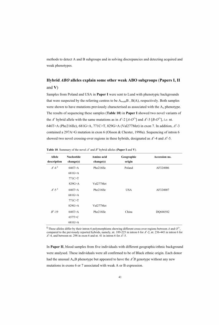
41
methods to detect A and B subgroups and in solving discrepancies and detecting acquired and
weak phenotypes.
Hybrid ABO alleles explain some other weak ABO subgroups (Papers I, II
and V)Samples from Poland and USA in Paper I were sent to Lund with phenotypic backgrounds
that were suspected by the referring centres to be AweakB , B(A), respectively. Both samples
were shown to have mutations previously characterised as associated with the Ax phenotype.
The results of sequencing these samples (Table 10) in Paper I showed two novel variants of
the Ax hybrid allele with the same mutations as in Ax-2 [A-O1v] and Ax-3 [B-O1v], i.e. nt.
646T>A (Phe216Ile), 681G>A, 771C>T, 829G>A (Val277Met) in exon 7. In addition, Ax-3
contained a 297A>G mutation in exon 6 (Olsson & Chester, 1998a). Sequencing of intron 6
showed two novel crossing-over regions in these hybrids, designated as Ax-4 and Ax-5.
Table 10. Summary of the novel Ax and Bw hybrid alleles (Paper I and V).
Allele Nucleotide Amino acid Geographic Accession no. description change(s) change(s) origin
Ax-4 § 646T>A
681G>A
771C>T
829G>A
Phe216Ile
Val277Met
Poland AF324006
Ax-5 § 646T>A
681G>A
771C>T
829G>A
Phe216Ile
Val277Met
USA AF324007
Bw-19 646T>A
657T>C
681G>A
Phe216Ile China DQ646582
§ These alleles differ by their intron 6 polymorphisms showing different cross-over regions between A and O1v,compared to the previously reported hybrids, namely, nt. 189-225 in intron 6 for Ax-2, nt. 236-445 in intron 6 for Ax-4, and between nt. 298 in exon 6 and nt. 41 in intron 6 for Ax-5.
In Paper II, blood samples from five individuals with different geographic/ethnic background
were analysed. These individuals were all confirmed to be of Black ethnic origin. Each donor
had the unusual AwB phenotype but appeared to have the A2B genotype without any new
mutations in exons 6 or 7 associated with weak A or B expression.
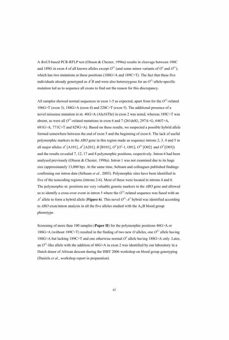
42
A BstUI-based PCR-RFLP test (Olsson & Chester, 1996a) results in cleavage between 188C
and 189G in exon 4 of all known alleles except O1v (and some minor variants of O1 and O1v),
which has two mutations at these positions (188G>A and 189C>T). The fact that these five
individuals already genotyped as A2B and were also heterozygous for an O1v-allele-specific
mutation led us to sequence all exons to find out the reason for this discrepancy.
All samples showed normal sequences in exon 1-5 as expected, apart from for the O1v-related
106G>T (exon 3), 188G>A (exon 4) and 220C>T (exon 5). The additional presence of a
novel missense mutation in nt. 46G>A (Ala16Thr) in exon 2 was noted, whereas 189C>T was
absent, as were all O1v-related mutations in exon 6 and 7 (261delG, 297A>G, 646T>A,
681G>A, 771C>T and 829G>A). Based on these results, we suspected a possible hybrid allele
formed somewhere between the end of exon 5 and the beginning of exon 6. The lack of useful
polymorphic markers in the ABO gene in this region made us sequence introns 2, 3, 4 and 5 in
all major alleles A1 [A101], A2 [A201], B [B101], O1 [O1-1, O01], O1v [O02] and O2 [O03])
and the results revealed 7, 12, 17 and 8 polymorphic positions, respectively. Intron 6 had been
analysed previously (Olsson & Chester, 1998a). Intron 1 was not examined due to its huge
size (approximately 13,000 bp). At the same time, Seltsam and colleagues published findings
confirming our intron data (Seltsam et al., 2003). Polymorphic sites have been identified in
five of the noncoding regions (introns 2-6). Most of these were located in introns 4 and 6.
The polymorphic nt. positions are very valuable genetic markers in the ABO gene and allowed
us to identify a cross-over event in intron 5 where the O1v-related sequence was fused with an
A2 allele to form a hybrid allele (Figure 6). This novel O1v-A2 hybrid was identified according
to ABO exon/intron analysis in all the five alleles studied with the AwB blood group
phenotype.
Screening of more than 100 samples (Paper II) for the polymorphic positions 46G>A or
188G>A (without 189C>T) resulted in the finding of two new O alleles, one O1v allele having
188G>A but lacking 189C>T and one otherwise normal O1 allele having 188G>A only. Later,
an O1v-like allele with the addition of 46G>A in exon 2 was identified by our laboratory in a
Dutch donor of African descent during the ISBT 2006 workshop on blood group genotyping
(Daniels et al., workshop report in preparation).

43
Figure 6. Representation of exons 2-7 and intervening introns in A2 and O1v alleles and the new O1v-A2 hybrid allele in Paper II. Exons are represented as boxes and introns as black lines. Black boxes indicate A2 and grey boxes O1v sequences. The dashed X indicates the cross-over region. White lines represent exonic SNPs used for allele identification.
In Paper V, samples from a Chinese family submitted to us were subjected to an extensive
serological and genetic investigation. In this family, the propositus was phenotyped as A1B3
and his mother was initially typed as group O but was later found to have very weak
expression of B antigen. We found in the initial genotyping that the samples were
heterozygous for a B-like allele and for either an A1 allele [A101] in the son or an O allele
[O01] in the mother. Sequencing of these samples (shown in Figure 7a) revealed that the B-
like allele contained a segment of O1v [O02]-related polymorphisms in the B-specific DNA
fragments, nts. 646, 657 and 681, only the first of which would lead to an a.a. change,
Phe216Ile. The new hybrid allele was named Bw-19 and is effectively a new hybrid allele with
B and O1v characteristics (Table 3 in Paper V). It is produced by an apparent double cross-
over in exon 7 (Figure 7a).
The structural consequences of the Phe216Ile change due to the 646T>A mutation in this
hybrid allele has been demonstrated by molecular modelling. The result showed an empty
space in the core of the glycosyltransferase that resulting from the substitution of an a.a. with
a smaller side chain (Figure 4a in Paper V). This mutation occurs in a part of the enzyme that
is responsible for donor binding, and a mutation that generates space in the hydrophobic core
of the protein would likely destabilize the protein fold and result in lower activity. The kinetic
data for the Phe216Ile mutant support the molecular modelling and phenotyping results. The
crystallization was unsuccessful for this subgroup GTB, which may be due to the
destabilizing effect of the mutation.
A2
O1v
46 G
>A
10
6 C
>T
18
8 G
>A
18
9 C
>T
220
C>
T
29
7 A
>G
26
1 G
>d
el
Exon
46
7 C
>T
10
61
C>
de
l
Novel
O1v-A2
hybrid 2 3 54 6 7
Hybrid
formation
A2
O1v
46 G
>A
10
6 C
>T
18
8 G
>A
18
9 C
>T
220
C>
T
29
7 A
>G
26
1 G
>d
el
Exon
46
7 C
>T
10
61
C>
de
l
Novel
O1v-A2
hybrid 2 3 54 6 7
Hybrid
formation

44
Figure 7. A schematic representation showing how the B-O1v-B hybrid may have been formed (a). DNA direct sequencing chromatogram showing exon 7 in the ABO gene of a normal B allele compared to the new hybrid allele (b).
b.
70
3
64
6
65
7
68
1
Exon 7
Co
nsen
su
s B
alle
leH
yb
rid
alle
le B
- O1v
-B
nt.
CA A A
AGTTCCGCGACCATGTGGGCGTGGAGATCCTGACTCCGCTGTTC GGCACCC TGCACCCCAGC
70
3
64
6
65
7
68
1
Exon 7
Co
nsen
su
s B
alle
leH
yb
rid
alle
le B
- O1v
-B
nt.
CA A A
AGTTCCGCGACCATGTGGGCGTGGAGATCCTGACTCCGCTGTTC GGCACCC TGCACCCCAGC
O1vO1v
Hybrid
formation
a.
X
New
B-O1v-Bhybrid
BAllele
Hybrid
formation
New
B-O1v-Bhybrid
O1vO1vO1vO1v
Hybrid
formation
a.
X
New
B-O1v-Bhybrid
BAllele
Hybrid
formation
New
B-O1v-Bhybrid

45
The genetic basis of the Abantu phenotype is also a hybrid allele (Paper IV)About 50 years ago, Shapiro observed the existence of a large proportion of blood group A
individuals whose cells did not react with anti-A1 and only weakly with anti-A (Shapiro,
1951). This A subgroup, designated Abantu, has its highest frequency in a restricted population
but globally seems to be a rare allele. In about 1000 B.C., a bi-directional migration of Bantu
people began that created two main language families, Eastern and Western Bantu. The
former migrated to Zimbabwe, Mozambique and down into South Africa and the latter
migrated into Angola and Namibia. The Abantu blood group has been identified in almost 4%
of Black Africans of blood group A and in up to 8% of San and Khoikhoi, the ethnic groups
from which the Abantu phenotype may have originated (Jenkins, 1974). In Paper IV, we
investigated blood samples from a total of seven individuals serologically characterized as
Abantu: six from KwaZulu-Natal in South Africa and one from a person of African descent
living in England. Genomic ABO typing indicated that all samples were heterozygous for
what appeared to be the common A2-1 [A201] allele and for an O allele with the 261delG
mutation.
Direct sequencing of exons 1-7 was performed. All samples lacked any of the previously
reported mutations associated with weak A and B phenotypes except for the silent 297A>G
SNP in exon 6. A deviation found compared to the usual A2 sequence was a single nt. deletion
(AGGT>AGT) at the exon 4 and intron 4 junction, i.e. a presumed splice-site mutation.
However, since it is not clear what the consequences of this mutation would be, different
hypotheses concerning the potential effects on the GTA were formed. These are illustrated in
Figure 8. As concluded from identical splice-site mutations in other genes, skipping of the
preceding exon in the mRNA is predicted. RT-PCR analysis based on synthesis of ABO-
cDNA from RNA extracts failed in spite of repeated attempts. We could detect only O-mRNA
but no Abantu-mRNA.
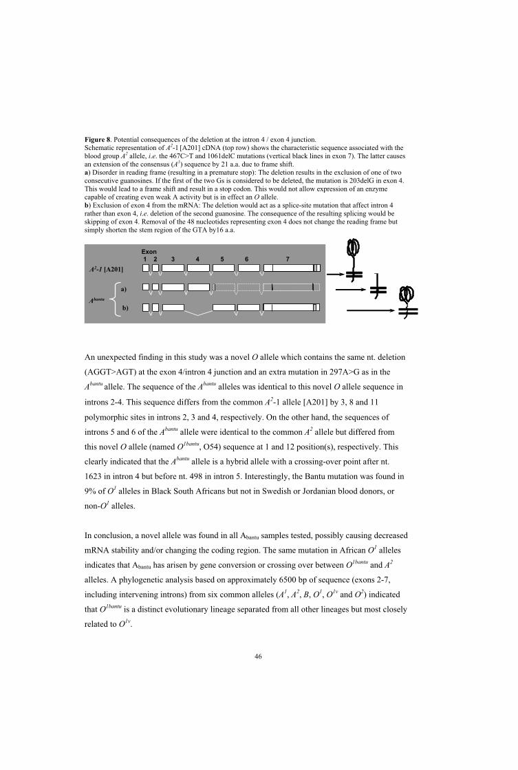
46
Figure 8. Potential consequences of the deletion at the intron 4 / exon 4 junction. Schematic representation of A2-1 [A201] cDNA (top row) shows the characteristic sequence associated with the blood group A2 allele, i.e. the 467C>T and 1061delC mutations (vertical black lines in exon 7). The latter causes an extension of the consensus (A1) sequence by 21 a.a. due to frame shift. a) Disorder in reading frame (resulting in a premature stop): The deletion results in the exclusion of one of two consecutive guanosines. If the first of the two Gs is considered to be deleted, the mutation is 203delG in exon 4. This would lead to a frame shift and result in a stop codon. This would not allow expression of an enzyme capable of creating even weak A activity but is in effect an O allele.b) Exclusion of exon 4 from the mRNA: The deletion would act as a splice-site mutation that affect intron 4 rather than exon 4, i.e. deletion of the second guanosine. The consequence of the resulting splicing would be skipping of exon 4. Removal of the 48 nucleotides representing exon 4 does not change the reading frame but simply shorten the stem region of the GTA by16 a.a.
An unexpected finding in this study was a novel O allele which contains the same nt. deletion
(AGGT>AGT) at the exon 4/intron 4 junction and an extra mutation in 297A>G as in the
Abantu allele. The sequence of the Abantu alleles was identical to this novel O allele sequence in
introns 2-4. This sequence differs from the common A2-1 allele [A201] by 3, 8 and 11
polymorphic sites in introns 2, 3 and 4, respectively. On the other hand, the sequences of
introns 5 and 6 of the Abantu allele were identical to the common A2 allele but differed from
this novel O allele (named O1bantu, O54) sequence at 1 and 12 position(s), respectively. This
clearly indicated that the Abantu allele is a hybrid allele with a crossing-over point after nt.
1623 in intron 4 but before nt. 498 in intron 5. Interestingly, the Bantu mutation was found in
9% of O1 alleles in Black South Africans but not in Swedish or Jordanian blood donors, or
non-O1 alleles.
In conclusion, a novel allele was found in all Abantu samples tested, possibly causing decreased
mRNA stability and/or changing the coding region. The same mutation in African O1 alleles
indicates that Abantu has arisen by gene conversion or crossing over between O1bantu and A2
alleles. A phylogenetic analysis based on approximately 6500 bp of sequence (exons 2-7,
including intervening introns) from six common alleles (A1, A2, B, O1, O1v and O2) indicated
that O1bantu is a distinct evolutionary lineage separated from all other lineages but most closely
related to O1v.
Exon
3 4 5 6 721
A2-1 [A201]
Abantu
a)
b)
Exon
3 4 5 6 721
A2-1 [A201]
Abantu
a)
b)

47
Novel O alleles involved in unexpected blood group phenotypes (Paper III)
The sequence of the most common allele [O1-1, O01] is identical to the A1 sequence, apart
from one nonsense mutation in exon 6, i.e. 261delG which leads to a truncated protein lacking
enzymatic activity (Yamamoto & Hakomori, 1990: Yamamoto et al., 1990a). Various O
alleles including hybrid O1v-B and O1-A2 alleles coding for non-functional
glycosyltransferases have been described by several groups but the vast majority show the
261delG mutation (for a review, see Chester & Olsson, 2001) only very few non-deletional O
alleles had been described by the start of this study: O3 (Olsson & Chester, 1996c) and four
Japanese alleles (Ogasawara et al., 2001), two of which are identical to the Ax hybrids
discussed above. All these alleles are based on the consensus A sequence. Fundamentally,
however any change which leads to the inactivation of the ABO glycosyltransferase can be
considered as a lethal null mutation. In this study, we included the investigation of samples
referred to our laboratory in Lund because of suspected A subgroups following discrepant
serologic blood grouping results but where ABO genotyping or sequencing surprisingly found
only O alleles. A correlation between these weak A phenotypes and genotypes by ABO
genotyping analysis (PCR-RFLP and PCR-ASP) showed an overrepresentation of the
otherwise infrequent O2 allele (Yamamoto et al., 1993d; Grunnet et al., 1994). Thus, we
examined 57 existing O2 alleles in detail both from regular group O donors and from the
above-mentioned samples with suspected A subgroup phenotypes. A flow chart summarizes
the samples that were included in this study (Figure 1, Paper III). All 57 O2 alleles differed
from the consensus allele [A101] in exons 2, 5, 6 and 7 at nt. 53, 220, 297, 526, 802 and nt.
1096 located in the 3´-untranslated region as expected (Yamamoto et al., 1993d; Olsson &
Chester, 1995; Amado et al., 2000). We confirmed in this study that all O2 alleles contain the
mutations 53G>T and 220C>T. This had only been described in few samples in a single paper
before (Amado et al., 2000).
Two new O2 alleles were identified, having the 689G>A mutations resulting in Gly229Asp
with or without 649C>T (Arg217Cys). These O2 variants were called O2-2 [O48] and O2-3
[O49], respectively. There was no difference in the coding sequences of O2 alleles found in
normal group O donors or in samples referred to us for ABO discrepancies and suspected A
subgroups. Furthermore, the same O2 allele could produce different phenotypes (O or weak
A) in different members of the same family.

48
Yet three novel O alleles surfaced in six samples with suspected A subgroups. Further
analysis including sequencing of coding regions showed that these were A1-like alleles having
nonsense mutations, 88dupG [O4], 322C>T [O5] or 542G>A [O6 ] causing premature
truncation at codons 56, 107 or 181, respectively. All of these alleles would be expected to be
totally incapable of producing an active enzyme. Consequently, when present with another
inactive allele with 261delG they should produce a normal O phenotype. This is clearly not
the case (Table 2 in Paper III). Kominato et al. suggested that perhaps A antigen expression
could be the result of the use of an alternative translational start codon (Kominato et al., 2002)
or that alternative splicing might explain the effect of early mutations.
In another unusual case in Paper III, we investigated an Australian blood donor who by DNA
sequencing, showed homozygosity for a silent mutation (768C>A) and denoted as O26. The
same mutation was described in the Basque population and proposed to be a specific Basque
marker (Roubinet et al., 2001). Surprisingly our screening study showed that this mutation
was quite frequent among Swedish donors and has a frequency of about 9% of all O alleles.
We also examined blood samples from two other populations (Jordanian and Swiss) and the
results indicate a widespread distribution of this mutation, but at lower frequencies than in
Sweden. Thus, this appears to be the third most common O allele among Swedes, following
O1 and O1v whilst clearly more frequent than O2.
A recently-described O2 variant, 488C>T (Thr163Met), denoted Aw08 (Seltsam et al., 2003)
was also found in our study and classified as O2-4. Surprisingly, the original and variant O2
alleles could all be associated with either O or Aweak phenotypes. Our data (Paper III)
disagree with the idea that 488C>T would result in an a.a. substitution with the ability to
restore some enzymatic function as suggested by Seltsam and co-workers (Seltsam et al.,
2003). Recent work from Yazer et al. indicated that they could not find any reason (based on
sensitive adsorbtion-elution studies and flow cytometry) not to use blood from group O
donors with an O2 allele to recipients with anti-A (Yazer et al., 2006). Structural studies have
also supported that the crucial mutation 802G>A (Gly268Arg), the most significant mutation
in the O2 allele, completely blocks the donor sugar recognition site (Lee et al., 2005),
although extremely low GTA-like enzyme activities could be measured.
The presence of the Ael-characteristic insertion at nt. 804 and the A2-characteristic
polymorphisms at nt. 467 and 1061 indicated the presence of an O3 allele in a Swedish blood

49
donor (Olsson & Chester, 1996c). In this study we observed yet another O3 allele but resulting
in a different phenotype (suspected Aweak) than the original one (clear-cut group O). No other
polymorphisms were detected after sequence analysis of exon 1-7.
How can non-deletional O alleles result in small amounts of A antigen?
Thus, we found that apparent O alleles lacking 261delG (non-deletional O alleles) may cause
weak A expression on erythrocytes and/or inhibit anti-A production. The Chi sequence
(5’-GCTGGCGG-3’) motif has been described in bacteria and been shown to influence the
recombination frequency in Escherichia coli and has also been implicated as a hot spot for
recombination in human genes (Smith, 1994; Stahl & Meyers, 1995). Several investigators
have published numerous examples of inheritable, recombinant ABO alleles generated during
meiosis (Suzuki et al., 1997; Olsson & Chester, 1998b; Ogasawara et al., 1996a, 2001).
According to the data in this study, O alleles without 261delG may cause weak A expression
on RBCs in the presence of an another O allele with 261delG, which in turn may result in
weak anti-A in plasma. The challenge is that it does not always happen when different
individuals with the same genotype are compared, even within the same family. To explain
this interesting phenomenon, we hypothesized that exchange of genetic material between
alleles during mitosis may result in so-called ”autologous chimerism” that restores
glycosyltransferase activity in some cells. The duplicated Chi sequences in the 3’ end of
intron 6 in the ABO gene may also increase the probability of recombination in somatic cells
(during mitosis). Gene conversion/recombination events in somatic cell clones in or outside
the bone marrow can then result in ”autologous chimerism” which produces low levels of A
antigen and inhibits the natural production of anti-A (Figure 9). However, we have been
unable to provide experimental evidence to support this hypothesis and alternative
explanations may well exist.

50
Figure 9. Recombination or gene conversion events that may explain how O alleles can result in weak A phenotypes. Probable creation of enzymically active gene products from inactive alleles by recombination (a), gene conversion (b).
”Autologous chimerism”
26
1O1 allele1 2 3 4 5 6 7
O2 allele
80
2
A1-like allele
80
2
O1-like allele
26
1Recombination
Exon
a
”Autologous chimerism”
26
1O1 allele1 2 3 4 5 6 7
O2 allele
80
2
A1-like allele
80
2
O1-like allele
26
1Recombination
Exon
a
26
1
O1 allele1 2 3 4 5 6 7
O2 allele
80
2
O1-like allele
80
2
26
1
A1 allele
”Autologous chimerism”
Recombination
Exon
b
26
1
O1 allele1 2 3 4 5 6 7
O2 allele
80
2
O1-like allele
80
2
26
1
A1 allele
”Autologous chimerism”
Recombination
Exon
b

51
Development of an improved ABO genotyping assay based on PCR-ASP
amplification across intron 6 (Paper VI)The majority of individuals possess the standard blood group A, B, and O phenotypes. In
addition, a number of A and B subgroups, B(A) and cis-AB exist. Generally, these subgroups
are characterised phenotypically by the occurrence and strength of the corresponding A and B
antigens on the RBCs and in secretions such as saliva.
The invention of the PCR technique and the elucidation of the sequence (Yamamoto &
Kominato,1990; Yamamoto et al., 1990a-b) and organisation (Yamamoto et al., 1995;
Bennett et al., 1995) and the increasing number of alleles (now almost 170 alleles reported to
dbRBC) of the ABO gene has opened another dimension that complements serological
analysis.
Many alleles responsible for the rarer subgroups have been examined and an increasing
number of polymorphisms have been found (Chester & Olsson, 2001; Yamamoto, 2004; Yip,
2002). Knowledge and improvement of ABO genotyping methods are very important for
practical implications in transfusion medicine. A number of different screening methods have
been developed and used with varying degrees of success (reviewed in Olsson & Chester,
2001). Improvement of these ABO genotyping methods for clinical use is much needed for
several reasons.
Recombination and hybrid formation between common alleles in the ABO gene are recent
findings that threaten to complicate ABO genotype interpretation. These hybrid alleles may
lead to unexpected ABO phenotypes, including rare subgroups. Hybrids are formed by
interaction between two alleles, e.g. O1 or O1v alleles with A2 or B alleles. These phenomena
are interesting and complex because they can give rise to O alleles if the nt. 261-containing
exon 6 is donated by the O1 or O1v allele. However, if nt. 261 is consensus (A, B or O2) and
combined with an O allele, then there are various consequences depending on the cross-over
point. Hybrid alleles obviously exhibit a whole range of serological activities. Different
investigators have published genotyping methods without any attention to addressing the
problem that can arise due to hybrid alleles (for a detailed description see review in Olsson &
Chester, 2001). All ABO genotyping methods to date can fail in the prediction of ABO
phenotype in the presence of hybrid alleles or the newly found O alleles and are potentially
dangerous and difficult to use in the clinical routine laboratory because of interpretation

52
problems (see example in Figure 10). In addition, all ABO genotyping methods published to
date will fail in predicting the correct phenotype when A or B subgroup alleles including
cisAB and B(A) are present.
We tested two versions of a commercially available ABO genotyping kit and confirmed its
limitations in detecting hybrid genes and many subgroup alleles. This kit (2nd version) which
is actually CE-marked predicts the common alleles and a few subgroups, but fails to define
hybrid alleles and most non-deletional O alleles except O2 and O3, the latter being one of the
improvements from the previous version.
Figure 10. Two samples with A or B phenotypes would illustrate an identical genotyping pattern in most, if not all current ABO genotyping methods used routinely today. Triangles illustrate the intron 6 with black circles where the Chi sequence may act as a hotspot. The white vertical line shows the 261delG mutation. Filled rectangles show consensus sequence shared between A1 and O1. Open rectangles represent B sequences. Thus, heterozygosity for both 261delG in exon 6 and B-SNPs in exon 7 are present in both samples, a fact that renders genotyping results potentially identical.
Over the past ten years, we have developed and expanded a PCR-ASP method that is flexible
and capable of detecting an increasing number of known alleles. This method gives flexibility
to different users and can be adjusted according to their requirements for detection of the vast
array of ABO alleles (see Paper VI). For example, the suggested approach is flexible so that
each user can add or remove primers according to the population tested or as new alleles
continue to be described. In analogy with many HLA typing systems, it is also possible to
choose the relevant level of resolution (standard or high) depending on the purpose of the
analysis.
The goal of this part of the study was to develop and establish an uncomplicated method
which can detect different alleles including hybrid, non-deletional null and subgroup alleles.
The proposed method is able to detect all major and a large number of rare alleles due to its
ability to cis-link the mutations in exons 6 and 7 and thus predict the ABO genotype with high
A
B
A1
O1-B
B
O1
Ph
en
oty
pe
Ge
no
typ
e
A
B
A1
O1-B
B
O1
Ph
en
oty
pe
Ge
no
typ
e

53
accuracy, even in the presence of hybrids or subgroup alleles with SNPs in common with the
major alleles (e.g. Ax-1 to Ax-5 and O1v sharing 646T>A). Most combinations of the following
alleles can be discriminated: A1, A2, Ax, Ael, other Aweak, B, a number of Bweak alleles and
various null alleles including O1, O1v, O1-B, O1-A2, O2, O3, O4, O5, O6 and also several cisAB
and B(A) variants.
In a first step, all the major alleles are defined and at the same time multiplex PCR-ASP
screening for most of the known but unusual or rare hybrid, non-deletional null and A or B
subgroup alleles is undertaken in four separate reactions. In most cases, this screening will
turn out negative and the genotype can be defined immediately. If one of the four screening
mixes are positive, the user has the choice to report the genotype at this level of resolution
(e.g. AwO1, A1Bw, B Ohybrid, A2Onon-del). If, for some reason, a better definition of the genotype
is desirable, a second round of high-resolution mixes is performed in which all the
multiplexed primers pairs in each screening mix have been separated into a number of
reactions. The result will further detail the genotype (e.g. the above-mentioned genotype
examples may now instead be expressed as AbantuO1, A1Bw-18, B O1v-B, A2O5). We believe that
this technique is the most cost-efficient, flexible and informative ABO genotyping method
reported to date. Compared to the commercial method which is based on a published PCR-
ASP method originally utilizing eight PCR tubes (Gassner et al., 1996) but now upgraded to
16 tubes, the number of genotypes resolved correctly by the method presented here is
superior. In principle, no ABO genotyping method should be used unless hybrid alleles and
known non-deletional O alleles can be detected properly, especially if the method is used in a
clinical reference laboratory and the results are used to predict the blood group of donors or
patients.
54
CONCLUSIONS The results of the studies presented as part of this Ph.D. thesis have led to a number of
conclusions, some of which can be summarized as follows.
The molecular genetic basis for ABO subgroups is very heterogeneous but single missense
mutations targeting evolutionarily conserved a.a. residues is a common theme.
Hybrids based on common alleles constitute another principal background for both A and
B subgroup phenotypes.
Some Aweak phenotypes which appear to be characteristic among people of African descent
are due to recombinant hybrid formation involving previously unknown O alleles, so far
only found in Black individuals.
Surprisingly, non-deletional O alleles can result in either common O or suspected Aweak
phenotypes but the reason for this has not yet been elucidated.
Clarification of the relationship between mutation, structural change in the
glycosyltransferase and enzymatic function can be achieved and is helpful when trying to
understand ABO subgroups as well as the importance of different domains in the enzyme.
Hybrids, non-deletional null and A or B subgroup alleles constitute major challenges for
ABO genotyping.
Improvement of the current standard of ABO genotyping was made possible by
development of a new approach for allele-specific amplification across intron 6 and
incorporation of multiplexed primers sets screening for rare or previously difficult-to-
detect alleles.
FUTURE PERSPECTIVESThe number of SNPs known at the ABO locus has dramatically increased during the last
decade. This has raised a number of new questions about how these allelic changes affect the
quantity, structure, function and localisation of GTA and GTB, and subsequently the A and B
antigens on RBCs. Future studies will also have to address the mechanisms by which the ABO
transcription is regulated, especially in the most relevant compartment, namely the bone
marrow. Until now, most work has been carried out in cell lines and most of them of gastric
or colonic origin.
New imaging technologies like cellular computer tomography may help us close in on what is
actually going on, in a cell biological and biochemical sense, in the Golgi apparatus where the
55
glycosyltransferases reside. In addition to computer-based 3D predictions and x-ray
crystallography as used in this study, further improvements in software and resolution may
yield further insight into the molecular enzymology of ABO. Another attractive method could
be using electron microscopy, which may facilitate the understanding of what is happening
with ABO antigens in different genotypes or in single erythroid progenitors and their
successors including the mature RBCs.
Current and future studies will focus on applying these techniques for the characterisation of
Aweak and Bweak samples in which the mutations are located in other, principally interesting
areas of the enzyme, e.g. close to the DXD motif (Persson et al., 2007) or in the disordered
loops. Also, there are still samples sent to reference laboratories for which the atypical ABO
phenotype has not yet been explained at the genetic level. One of the most challenging
examples is the most common weak A subgroup, A3. Despite the finding of a complete A1
consensus sequence, this inheritable variant is completely different from the standard A1
phenotype. One possibility in this case would be to look at mRNA stability and quantities in
combination with analysis of regulatory regions outside the coding region.
Another potential aim in future studies could be to address the hypothesis in Paper III
regarding gene conversion or recombination events in somatic cell clones in or outside the
bone marrow. Our suggestion about a phenomenon of so-called ”autologous chimerism” as
well as other theories about how apparent O alleles predicted to result in severely truncated
proteins without enzymatic activity, can sometimes give rise to weak A expression require
further work. The mechanisms responsible for these apparent anomalies in correlation
between serological and genetic blood group typing, is very interesting to us. As yet, we have
not provided enough experimental evidence to confirm our theory, so it would be interesting
to explore this hypothesis. It may be that other principally different methods are required for
this goal. Preliminary work using flow cytometry or erythropoietic culture models have not
yet been successful. Cloning of ABO transcripts from cell populations or even single cells
may be used to identify low levels recombinant transcripts that can translate into functional
GTA.
In general, the ABO blood group system continues to serve as one of the most useful models
to study both human genetics, carbohydrate synthesis and glycosyltransferase functional
anatomy.

56
SUMMARY IN SWEDISH (sammanfattning på svenska)
De första blodtransfusionerna kan härledas till 1600-talet då man försökte transfundera blod från
djur till djur, djur till människa och människa till människa, vilket ofta resulterade i
komplikationer och i många fall patientens död.
I början av 1900-talet upptäckte Karl Landsteiner (1868-1943) blodgruppssystemet ABO (läses
A, B, noll) efter empiriska försök där han blandade blod från sig själv och sina kollegor (utanför
kroppen).
Transfusionsmedicin är därmed en ung vetenskap inom den medicinska läran. Däremot har ordet
blod länge använts i olika sammanhang, inte bara mediciniska, utan även i talspråk såsom t.ex.
blodtörstig, blodsbröder, blodsband och blodshämnd etc.
Landsteiner beskrev principen för den antigen-antikroppsreaktion vilken numera benämns
hemagglutination och som används rutinmässigt idag vid de transfusionsmedicinska
laboratorierna, blodcentralerna.
Antigenen utgörs av kolhydratstrukturer
på cellerna medan antikroppar är immun-
försvarets verktyg som kan attackera
främmande kroppar som t.ex. bakterier
eller blodgivarens blodceller.
Bilden här intill visar de möjliga ABO-
blodgruppskombinationerna vid transfusion av
röda blodkroppar (erytrocyter) och plasma.
Fenotypning innebär att man fastställer vilka antigen som förekommer hos individen i fråga, och
tolkas oftast som ”finns” (positivt) eller ”finns inte” (negativt). Förekomsten av de vanliga ABO-
fenotyperna (A1, A2, B, A1B, A2B och O) varierar liksom andra blodgruppsfenotyper i olika
populationer. Denna variation benämns polymorfism. ABO-systemets antikroppar är oftast av
IgM-typ. Dessa bildas normalt (kallas även naturliga/spontana antikroppar eller isoagglutininer)
mot de ABO-antigen som saknas hos individen i fråga. ABO-systemet är på grund av dessa
naturligt förekommande antikroppar det kliniskt viktigaste blodgruppssystemen. För att utföra en
säker blodtransfusion krävs det att ett laboratorium kontrollerar vilken ABO-blodgrupp individen
tillhör och om denna har bildat antikroppar mot något annat blodgruppssystem. Därefter kan
passande givarblod transfunderas. Även vid transplantation av organ som njure, lever, hjärta och
lungor är ABO-systemet bland det viktigaste att ta hänsyn till.
O
A B
AB
AB
A B
O
plasma
röda
blodkroppar
O
A B
AB
AB
A B
O
O
A B
AB
O
A B
AB
AB
A B
O
plasma
röda
blodkroppar

57
Det finns enligt International Society of Blood Transfusion (ISBT) ca. 270 erkända
blodgruppsantigener på erytrocyternas yta. Blodgruppsantigenen delas in i olika
blodgruppssystem, hittills finns 29 beskrivna. Gemensamt för alla blodgruppsmarkörer är att de
sitter på utsidan av de röda blodkropparnas membran. ABO-blodgruppsantigen finns förutom på
dessa celler även på många andra celltyper. Exempel på andra viktiga system är Rh, Duffy, Kell,
Kidd och P.
I vissa situationer, bland annat vid bestämning av ärftligt svagare ABO-uttryck, s.k. subgrupper
som Ax och Ael, förvärvade blodgruppsegenskaper (t.ex. förvärvat B-antigen, där en individ med
blodgrupp A kan framstå som AB) eller vid olika sjukdomar, kan dock resultaten vara
svårtolkade och framstår som svagt positiva (”finns men svagare än normalt”). Detta kan bero på
någon ärftlig variant av ABO-genen eller förändrat uttryck i olika typer av tumörceller och vid
vissa svåra infektioner som sannolikt beror på nedsatt aktivitet i enzymet som tillverkas med
ABO-genen som mall (se nedan).
Intensiv forskning på 1950-60-talet klargjorde de biokemiska grunderna för ABO-systemet och
resulterade i en bättre förståelse av detsamma. ABO-genen kartlades till den långa armen av
kromosom 9. Yamamoto och medarbetare visade 1990 att ABO-genen kodar för ett
glykosyltransferas. Detta innebär att ABO-genen ger upphov till ett protein av enzymtyp som i
sin tur bidrar till att bygga enkla socker i en viss ordning så att en kolhydratkedja bildas. ABO-
systemets antigener består av olika specifika kombinationer av dessa kolhydrater.
Olika mutationer, d.v.s. ärftliga och bestående förändringar i cellens genetiska material (DNA),
har upptäckts i ABO-genen vilket i sin tur kan leda till minskning av enzymaktiviteten och
följaktligen till en minskning av antalet antigen på erytrocyter. Detta kan göra blodgruppering
vansklig att tolka och risken finns att fel blodrekommendation utfärdas.
Enstaka punktmutationer i ABO-genen ansvarar för många kända fenotyper. A- och B-generna
ger upphov till enzymen A- respektive B-transferas. Blodgrupp O beror däremot på dubbel
uppsättning av O-gener innehållande någon typ av defekt (t.ex. punktdeletion), som orsakar ett
inaktivt (ofunktionellt) enzym eller t.o.m. avsaknad därav. Det finns även ett stort antal
undergrupper inom A/B-blodgrupperna, t.ex. A3, Ax, Ael, Aweak, B3, Bel och Bweak som uppvisar
svagare reaktioner vid försök till blodgruppering. Studier avseende A- och B-specificiteten har
förfinats på senare tid genom de nya DNA-baserade teknikerna, bl.a. PCR-teknik (en
”kopieringsapparat för DNA”), kloning etc. Mina studier i denna avhandling har kunnat utföras
58
tack vare alla dessa framsteg inom molekylärbiologin tillsammans med upptäckten av ABO-
genen.
I stort handlar avhandlingen om en fördjupad studie av bakomliggande orsaker till och
konsekvenser av den överraskande mångfald som ses i ABO-genen. Länge trodde man att det
fanns fyra genetiska varianter, s.k. ABO-alleler (A1, A2, B och O), men redan 1993 stod det klart
att situationen var betydligt mer komplex än så. Syftet i de fem första vetenskapliga artiklarna
har varit att klarlägga den molekylärgenetiska bakgrunden och blodgruppsolikheter inom den
svenska populationen och i andra befolkningsgrupper med fokus på svaga ABO-blodgrupper. I
den sista artikeln försökte vi att utveckla en säker och kostnadseffektiv metod för att detektera
alla dessa vanliga och sällsynta alleler.
En omfattande insamling av kliniska fall, remitterade till det Nordiska Referenslaboratoriet för
Genomisk Blodgruppstypning vid Blodcentralen Skåne på Universitetssjukhuset i Lund,
undersöktes i de olika delprojekten. I delarbete I och V identifierades olika nya punktmutationer,
både s.k. missense- (som ger upphov till ett aminosyrabyte) och nonsense-mutationer (som
förstör genens möjlighet att ge upphov till ett funktionellt enzym). Därmed karakteriserades den
genetiska bakgrunden hos 17 nya undergrupper inom ABO-systemet.
Efter detta mödosamma arbete, började jag/vi fundera på om hur man skulle kunna bevisa att
dessa mutationer verkligen ligger bakom det avvikande uttrycket av antigenerna på erytrocyter.
Det fanns ett antal olika analyser som kunde användas i detta syfte men jag valde att genomföra
mina undersökningar i Köpenhamn där professor Monica Palcics grupp forskar inom området
som handlar om enzymers aktivitet och specificitet samt hur enzymerna kan visualiseras med s.k.
datormodellering eller röntgenkristallografi. Jag fick lära mig att uttrycka olika typer av ABO-
undergrupper genom att införa konstgjorda mutationer (motsvarande våra nyupptäckta ABO-
subgrupper) i en syntetisk ABO-gen i E.coli-bakterier. Stora mängder enzym/protein uttrycktes
och egenskaperna hos dessa glykosyltransferas-mutanterna kunde fastställas. Slutligen skickades
proteinet iväg för röntgenkristallografi (en sorts visualisering av den tredimensionella strukturen)
detta skedde hos en kanadensisk grupp, ledd av professor Stephen Evans, som samarbetar med
Köpenhamnsgruppen. Kollegorna i Kanada lyckades att kristallisera ett av våra enzymer och
analyserade dessutom flera andra nya mutationer via datorsimulering. Analyserna visade att de
nya mutationerna med stor sannolikheten är orsaken till de svaga reaktioner som man funnit på
blodcentralernas laboratorier.

59
I delarbete II beskrivs en hybridallel (en blandning mellan två alleler) hos personer av afrikanskt
ursprung med en variant av blodgruppen AB varvid svagt uttryck av A-antigenet på
blodkropparna gjorde blodgruppering svårtolkad. Resultatet av en lång rad av undersökningar på
ABO-genen visade att en fusion av en vanlig O-allel (O1v) och en vanlig A2-allel hade bildats.
Denna nya allel verkade orsaka ett mindre effektivt enzym. För att med säkerhet kunna definiera
allelen som en hybrid, undersöktes och kartlades intronerna (de icke-kodande regioner som finns
mellan genens exoner, d.v.s. de kodande regionerna) och deras genetiska variation
(polymorfism) hos personer med olika blodgrupper. Med denna kartläggning kunde vi bevisa var
sammanblandningen (hybridformation) av dessa alleler hade skett.
I delarbete III undersöktes ett stort antal prover, och 5 nya O-alleler i ABO-genen detekterades,
något som kan få stor betydelse för säkerheten vid genetisk blodgruppering. Vi upptäckte bl.a.
vad alla hittills trott var en sällsynt O-variant i den baskiska populationen, i själva verket var den
3:e vanligaste O-allelen överhuvudtaget, åtminstone bland kaukasier (t.ex. svenskar). I detta
arbete lades även en hypotes fram för att förklara varför vissa O-alleler (d.v.s. förstörda gener
utan förmåga att ge upphov till funktionella enzymer) faktiskt kunde ge upphov till svagt A-
uttryck på röda blodkroppar trots allt. Dessvärre har denna hypotes ännu inte gått att bevisa men
utgör en möjlighet för ev. framtida projekt.
I delarbete IV, rapporterade vi att den sedan flera decennier välkända ABO-undergruppen
Abantu överraskande nog, även den, beror på en hybridbildning: denna gång mellan den vanliga
A2-allelen och en aldrig tidigare beskriven O-allel som utgör en helt egen evolutionär (historisk)
utvecklingslinje inom ABO-allelfamiljen. Allelen förekommer huvudsakligen bland afrikanska
folkgrupper inkl. de s.k. Bantu-folken, precis som varit känt länge i fallet Abantu och därför
kallades allelen O1bantu. Även här spelade intron-polymorfismerna en avgörande roll för att kunna
bestämma var hybridformationen skett. Medan andra svaga ABO-undergrupper är sällsynta, är
Abantu den A-undergrupp som har högst frekvens i någon befolkning, upp till 8% av vissa
folkgrupper i södra Afrika.
Genetiska metoder finner allt fler applikationer inom det transfusionsmedicinska fältet.
Det sista delarbetet (VI), är en sammanfattning av nästan tio års arbete (från mitt 20-
poängsarbete 1997 för M.Sc. fram till idag). Under många år har Martin L Olsson (min
handledare), Åsa Hellberg (min käresta) och jag ständigt förbättrat och justerat en metod för att
kunna detektera de allt fler vanliga och sällsynta ABO-alleler som bör kunna upptäckas (idag

60
minst 168 st att välja bland). Denna analys har använts parallellt med andra metoder som
publicerades från Lund 1995-6, vilket gett en god validering under kliniska betingelser. En
jämförelse med en kommersiellt tillgänglig analys visade också att vår nya metod är överlägsen,
framför allt vid s.k. hybrid-, ovanliga O och subgruppsalleler.
Att detektera dessa komplexa alleler är viktigt när blodgivare och/eller -mottagares prover ger
svårtolkade resultat vid blodgruppering. Då kan DNA-baserad analys användas som ett
komplement för att minska risken för falska provsvar, som skulle kunna leda till felaktiga
konklusioner och transfusionsrekommendationer.
Sammanfattningsvis har studierna i denna avhandling lett till en bättre förståelse kring de
funktionella och genetiska sambanden inom ABO-systemet. Det långsiktiga målet är att med
någon typ av automatiserade plattformar kunna fastställa blodgruppen helt genetiskt, så som
redan görs för vävnadstypen (HLA) inför transplantationer.

61
ACKNOWLEDGEMENTSAlthough I spent many years of hard work to complete this thesis, it all depended on the
fortune of having many generous friends and colleagues around me who supported me during
this study. I am especially grateful to the following persons, knowing that there are many
others who are not mentioned here.
First and foremost, my ever-enthusiastic supervisor Professor Martin L Olsson, for giving me
the opportunity to work with such exciting projects, for guidance through scientific thinking
and sharing his knowledge.
Britt Thuresson, PCR expert, for guidance and teaching me scientific methodology, especially
in the field of molecular biology, and not least for taking me down to earth at occasions when
too many “good ideas” were occupying my mind.
My colleagues at the Department of whom I particularly will mention Alan Chester, Jill
Storry and Åsa Hellberg who always helped me with language revision and adopted a critical
attitude towards the thesis.
My friend and colleague Elisabet SW, who shared with us her nice house many times during
our kick-off meetings (Landet). The most enjoyable times have been those everyday-moments
in the corner (dum-forum) of the lab where we could allow us to be dumb and discuss freely
about different things.
Nidal and all other people in the research group who inspired me and generously provided
both time for discussions (genlunch) and good advice.
I would especially like to thank my roommate and good colleague, Gunilla Hochberg for all
support and patience throughout these years, which made it possible to write this thesis. Li
and Alicja are thanked, for all help during this long time in the same office.
I would like to express my appreciation towards all the colleagues at the Blood Centre who
helped me in all weather when I needed to be cheered up, especially Kerstin x 3 (B, H & T)
and of course Elisabeth, Ingela Å, and Eva Marie.

62
Professor Tor Olofsson, Louise, May-Louise and Bodil in the laboratory at the Department of
Hematology and Transfusion Medicine, BMC, for support and for introducing me to RNA
preparation.
Professor Monica Palcic, Mattias Persson and James Letts for valuable discussions and help
with enzyme purification and computer modelling at the Carlsberg laboratory in Copenhagen.
Charlie, Hannah and Gerd for helping me with sequencing at the Department of Clinical
Chemistry.
All my co-authors in different parts of the world for fruitful collaboration.
The friends for reminding me about life outside the lab, Ramtin, Per, Mohammad, Mikael,
Stefan (lektorn), Anna, Percy, Sören, Marika, Anica (?-alliansen), Leif, Åsa, Erik, Jytte, Ewa,
Magda, Ann-sofie, Orlando, Pasha, Ola, Birger, Örjan, Hamid, Nori, Mahmood and Mattias.
My dear Family, my beloved Parents, Bahareh, Azita and specially Göran and Ingrid, for
always believing in me and for being there when Åsa & I needed them.
And most of all, my friend, roommate and my love for her support and good cooperation both
at work and at home. Finally the youngest one, my sunshine Arvin (Seed-Nisse) who gave me
new strength every day.
This project was supported by the Medical Faculty at Lund University, the Swedish Research
Council, governmental ALF research grants to Lund University Hospital, and Region Skåne,
Sweden, the Claes Högman SAGMAN-stipendium, the Inga and John Hain Foundation for
Medical Research, Georg Danielssons Fund for haematological disease, Tore Nilsons
Foundation for Medical Research.
63
REFERENCESAmado M, Bennett EP, Carneiro F, Clausen H. Characterization of the histo-blood group O(2) gene and its protein product. Vox Sang 79:219-226, 2000.
Allderdice PW, Kaita H, Lewis M, Mcalpine PJ, Wong P, Anderson J and Giblett ER. Segregation of marker loci in families with an inherited paracentric insertion of chromosome 9. Am J Hum Genet 39:612-617, 1986.
Barjas-Castro ML, Saad ST. Absence of the G871A mutation in A3 blood donors from Brazil. Transfusion37:564, 1997.
Barsoum N, Kleeman C. Now and then, the history of parenteral fluid administration. Am J of Nephrol 22: 284-289, 2002.
Bennett EP, Steffensen R, Clausen H, Weghuis DO, Geurts van Kessel A. Genomic cloning of the human histo-blood group ABO locus. Biochem Biophys Res Commun 206:318-325, 1995.
Blumenfeld OO, Patnaik SK. Allelic genes of blood group antigens: a source of human mutations and cSNPs documented in the Blood Group Antigen Gene Mutation Database. Hum Mutat 23:8-16, 2004.
Blundell J. A successful case of transfusion. Lancet. 431, 1829.
Boix E, Swaminathan GJ, Zhang Y, Natesh R, Brew K, Acharya KR. Structure of UDP complex of UDP-galactose:beta-galactoside-alpha -1,3-galactosyltransferase at 1.53-A resolution reveals a conformational change in the catalytically important C terminus. J Biol Chem 276:48608-48614, 2001.
Breimer ME, Jovall PA. Structural characterization of a blood group A heptaglycosylceramide with globo-series structure. The major glycolipid based blood group A antigen of human kidney. FEBS Lett 1;179:165-172, 1985.
Breton C, Snajdrova L, Jeanneau C, Koca J, Imberty A. Structures and mechanisms of glycosyltransferases Glycobiology 16:29R-37R, 2006.
Cameron C, Graham F, Dunsford I. Acquisition of a B-like antigen by red blood cells. Br. Med. J. 32-34, 1959.
Cartron JP, Gerbal A, Hughes-Jones NC, Salmon C. 'Weak A' phenotypes. Relationship between red cell agglutinability and antigen site density. Immunology 27:723-727, 1974.
Cartron JP, Badet J, Mulet C, Salmon C. Study of the alpha-N-acetylgalactosaminyltransferase in sera and red cell membranes of human A subgroups. J Immunogenet 5:107-116, 1978.
Chester MA. The occurrence of a beta-galactosyltransferase in normal human urine. FEBS Lett 46:59-62, 1974.
Chester MA, Olsson ML. The ABO blood group gene - A locus of considerable genetic diversity. Transfus Med Rev 11: 295-313, 2001.
Clausen H, Watanabe K, Kannagi R, Levery SB, Nudelman E, Arao-Tomono Y, Hakomori S. Blood group A glycolipid (Ax) with globo-series structure which is specific for blood group A1 erythrocytes: one of the chemical bases for A1 and A2 distinction. Biochem Biophys Res Commun 124:523-529, 1984.
Clausen H, Levery SB, Nudelman E, Tsuchiya S, Hakomori S. Repetitive A epitope (type 3 chain A) defined by blood group A1-specific monoclonal antibody TH-1: chemical basis of qualitative A1 and A2distinction. Proc Natl Acad Sci U S A. 82:1199-1203, 1985.
Clausen H, Levery SB, Kannagi R, Hakomori S. Novel blood group H glycolipid antigens exclusively expressed in blood group A and AB erythrocytes (type 3 chain H). I. Isolation and chemical characterization. J Biol Chem 261:1380-1387, 1986a.
Clausen H, Holmes E, Hakomori S. Novel blood group H glycolipid antigens exclusively expressed in blood group A and AB erythrocytes (type 3 chain H). II. Differential conversion of different H substrates by A1and A2 enzymes, and type 3 chain H expression in relation to secretor status. J Biol Chem 261:1388-1392, 1986b.
Clausen H, Hakomori S. ABH and related histo-blood group antigens; immunochemical differences in carrier isotypes and their distribution. Vox Sang 56:1-20, 1989.

64
Clausen H, White T, Takio K, Titani K, Stroud M, Holmes E, Karkov J, Thim L, Hakomori S. Isolation to homogeneity and partial characterization of a histo-blood group A defined Fuc-alpha1-2Gal alpha1-3-N-acetylgalactosaminyltransferase from human lung tissue. J Biol Chem 265:1139-1145, 1990.
Clausen H, Bennett EP, Grunnet N. Molecular genetics of ABO histo-blood groups. Transfus Clin Biol1:79-89, 1994.
Daniels G. Human blood groups. Blackwell Scientific, Oxford, UK, 2002.
Daniels GL, Cartron JP, Fletcher A, Garratty G, Henry S, Jorgensen J, Judd WJ, Levene C, Lin M, Lomas-Francis C, Moulds JJ, Moulds JM, Moulds M, Overbeeke M, Reid ME, Rouger P, Scott M, Sistonen P, Smart E, Tani Y, Wendel S, Zelinski T. International Society of Blood Transfusion Committee on terminology for red cell surface antigens. Vancouver Report. Vox Sang 84:244-247, 2003.
Daniels GL, Fletcher A, Garratty G, Henry S, Jorgensen J, Judd WJ, Levene C, Lomas-Francis C, Moulds JJ, Moulds JM, Moulds M, Overbeeke M, Reid ME, Rouger P, Scott M, Sistonen P, Smart E, Tani Y, Wendel S, Zelinski T. International Society of Blood Transfusion. Blood group terminology 2004, from the International Society of Blood Transfusion committee on terminology for red cell surface antigens. Vox Sang 87:304-316, 2004.
Decastello A von & Sturli A. Überdie Isoagglutinie im Serum gesunder und kranker menschen. München Med Wchnschr 26:1090-1095, 1902. (Translation in Camp & Ellis,1966)
Deng ZH, Yu Q, Lian YL, Wu GG, Su YQ, Zhang X. Identification of a novel B variant allele at the ABO locus in Chinese Han individuals with B subgroup. Ann Clin Lab Sci 35:265-269, 2005.
Denis J-B. A letter concerning a new way of curing sundry diseases by transfusion of blood. Philos Trans. R. Soc 2:489-504, 1667.
Dungern E von, Hirszfeld L. Über Vererbung gruppenspezifischer Strukturen des Blutes III. Z Immunforsch 8:526-562, 1911. (Translation in Camp & Ellis, 1966)
Economidou J, Hughes-Jones NC, Gardner B. The reactivity of subunits of IgM anti-B. Immunology13:235-240, 1967.
Eiz-Vesper B, Seltsam A, Blasczyk R. ABO glycosyltransferases as potential source of minor histocompatibility antigens in allogeneic peripheral blood progenitor cell transplantation. Transfusion.45:960-968, 2005.
Erskine AG, Socha WW. The principles and practice of blood grouping. The C.V. Mosby Company, Missouri, USA, 1978.
Farr A.D. Blood group serology the first four decades (1900-1939). Medical History 23:215-226, 1979.
Farr A.D. The first human blood transfusion. Medical History 24:143-162, 1980.
Ferguson-Smith MA, Aitken DA, Turleau C, de Grouchy J. Localisation of the human ABO: Np-1: AK-1 linkage group by regional assignment of AK-1 to 9q34. Hum Genet 34:35-43, 1976.
Fischer W, Hahn F. Uber auffallende Schwäche der gruppenspezifischen Reaktionsfähigkeit bei einem Erwachsensen. Z Immunforsch 84:177-188, 1935.
Garratty G, Dzik W, Issitt PD, Lublin DM, Reid ME, Zelinski T. Terminology for blood group antigens and genes-historical origins and guidelines in the new millennium. Transfusion 40:477-489, 2000.
Gassner C, Schmarda A, Nussbaumer W, Schonitzer D. ABO glycosyltransferase genotyping by polymerase chain reaction using sequence-specific primers. Blood 88: 1852-1856, 1996.
Gastinel LN, Bignon C, Misra AK, Hindsgaul O, Shaper JH, Joziasse DH. ine alpha1,3-galactosyltransferase catalytic domain structure and its relationship with ABO histo-blood group and glycosphingolipid glycosyltransferases. EMBO J. 15;20:638-649, 2001.
Ginsburg V. Enzymatic basis for blood groups in man. Adv Enzymol Relat Areas Mol Biol 36:131-149, 1972.
Goldstein J, Lenny L, Davies D, Voak D. Further evidence for the presence of A antigen on group B erythrocytes through the use of specific exoglycosidases. Vox Sang 57:142-146, 1989.
Griot-Wenk M, Pahlsson P, Chisholm-Chait A, Spitalnik PF, Spitalnik SL, Giger U. Biochemical characterization of the feline AB blood group system. Anim Genet 24:401-407, 1993.

65
Grunnet N, Steffensen R, Bennett EP, Clausen H. Evaluation of histo-blood group ABO genotyping in a Danish population: frequency of a novel O allele defined as O2. Vox Sang 67:210-215, 1994.
Hansen T, Namork E, Olsson ML, Chester MA, Heier HE. Different genotypes causing indiscernible patterns of A expression on A(el) red blood cells as visualized by scanning immunogold electron microscopy. Vox Sang 75:47-51, 1998.
Harvey W. Exercitatio anatomica de moto cordis et sanguinis in animalibus. Frankfurt, 1628.
Hata Y, Kominato Y, Yamamoto FI, Takizawa H. Characterization of the human ABO gene promoter in erythroid cell lineage. Vox Sang 82: 39-46, 2002.
Hata Y, Kominato Y, Takizawa H, Tabata S, Michino J, Nishino K, Yasumura S, Yamamoto F. Transcription starting from an alternative promoter leads to the expression of the human ABO histo-blood group antigen. Transfusion 43:656-662, 2003.
Heier HE, Namork E, Calkovska Z, Sandin R, Kornstad L. Expression of A antigens on erythrocytes of weak blood group A subgroups. Vox Sang 66:231-236, 1994.
Henissat B. Glycosidase families. Biochem Soc Trans 26:153-156, 1998.
Irshaid NM, Chester MA, Olsson ML. Allele-related variation in minisatellite repeats involved in the transcription of the blood group ABO gene. Transfusion Med 9:219-226, 1999.
Jenkins T. Blood group Abantu population and family studies. Vox Sang 26:537-550, 1974.
Kabat EA. Blood group substances. Their chemistry and immunochemistry. Academic Press, New York, 1956.
Kannagi R, Levery SB, Hakomori S. Blood group H antigen with globo-series structure. Isolation and characterization from human blood group O erythrocytes. FEBS Lett 175:397-401, 1984.
Kominato Y, McNeill PD, Yamamoto M, Russell M, Hakomori S, Yamamoto F. Animal histo-blood group ABO genes. Biochem Biophys Res Commun 189:154-164, 1992.
Kominato Y, Tsuchiya T, Hata N, Takizawa H, Yamamoto F. Transcription of human ABO histo-blood group genes is dependent upon binding of transcription factor CBF/NF-Y to minisatellite sequence. J Biol Chem 272:25890-25898, 1997.
Kominato Y, Hata Y, Takizawa H, Matsumoto K, Yasui K, Tsukada J. Alternative promoter identified between a hypermethylated upstream region of repetitive elements and a CpG island in human ABO histo-blood group genes. J Biol Chem 277: 37936-37948, 2002.
Landsteiner K. Zur kenntnis der antifermativen, lytischen und agglutinierenden Wirkungen des Blusterums und der Lymphe.Zbl Bakt 27:357-366, 1900. (Translation in Camp & Ellis, 1966)
Landsteiner K. Über Agglutinationserscheinungen normalen menschlichen Blutes. Klein.Wschr. 14,1132-1134, 1901. (Translation in Camp & Ellis, 1966)
Landsteiner K. On Individual Differences in Human Blood. Physiology or Medicine 1922-1941:234-245, Nobel Lectures 1930.
Lee HJ, Barry CH, Borisova SN, Seto NO, Zheng RB, Blancher A, Evans SV, Palcic MM. Structural basis for the inactivity of human blood group O2 glycosyltransferase. J Biol Chem 280:525-529, 2005.
Lin PH, Li L, Lin-Tsai SJ, Lin KT, Chen JM, Chu DC. A unique 502C>T mutation in exon 7 of ABO gene associated with the Bel phenotype in Taiwan. Transfusion 43:1254-1259, 2003.
Loghem JJ van, Dorfmeier H, Hart M van der. Two antigens with abnormal serologic properties. Vox Sang 2:16-24, 1957.
Lowe JB. Biochemistry and biosynthesis of ABH and Lewis antigens. In: Cartron JP, Rouger P, eds. Blood cell biochemistry: Molecular basis of major human blood group antigens. Plenum Press, New York, 75-115, 1995.
Lower R. The success of the experiment of transfusing the blood of one animal into another. Philos Trans R Soc 1, 352, 1666.
Martinko JM, Vincek V, Klein D, Klein J. Primate ABO glycosyltransferases: evidence for trans-species evolution. Immunogenetics 37:274-278, 1993.

66
Mifsud NA, Watt JM, Condon JA. A novel cis-AB variant allele arising from a nucleotide substitution A796C in the B transferase gene. Transfusion 40:1276-1277, 2000.
Miller SA, Dykes DD, Polesky HF. A simple salting out procedure for extracting DNA from human nucleated cells. Nucleic Acids Res 16:1215, 1988.
Mollison PL, Engelfriet CP, Contreras M. Blood transfusion in clinical medicine. Blackwell Scientific, Oxford, 1993.
Morera S, Imberty A, Aschke-Sonnenborn U. T4 phage beta-glucosyltransferase: substrate binding and proposed catalytic mechanism. J Mol Biol 292:717-730, 1999.
Mourant AE, Kopec K, Domaniewska-sobczak K. The distribution of the human blood groups and other polymorphisms. Oxford university press, 1976.
Nakamura I, Takizawa H, Nishino K. A3 phenotype with A1 gene-specified enzyme character in serum. Exp Clin Immunogenet 6:143-149, 1989.
Ogasawara K, Yabe R, Uchikawa M, Saitou N, Bannai M, Nakata K, Takenaka M, Fujisawa K, Ishikawa Y, Juji T, Tokunaga K. Molecular genetic analysis of variant phenotypes of the ABO blood group system. Blood 88:2732-2737, 1996a.
Ogasawara K, Bannai M, Saitou N. Extensive polymorphism of ABO blood group gene: three major lineages of the alleles for the common ABO phenotypes. Hum Genet 97:777-783, 1996b.
Ogasawara K, Yabe R, Uchikawa M, Bannai M, Nakata K, Takenaka M, Takahashi Y, Juji T, Tokunaga K. Different alleles cause an imbalance in A2 and A2B phenotypes of the ABO blood group. Vox Sang 74:242-247, 1998.
Ogasawara K, Yabe R, Uchikawa M, Nakata K, Watanabe J, Takahashi Y. Recombination and gene conversion-like events may contribute to ABO gene diversity causing various phenotypes. Immunogenetics53: 190-199, 2001.
Olsson ML, Chester MA. A rapid and simple ABO genotype screening method using a novel B/O2 versus A/O1 discriminating nucleotide substitution at the ABO locus. Vox Sang 69:242-247, 1995.
Olsson ML, Thuresson B, Chester MA. An Ael allele-specific nucleotide insertion at the blood group ABO locus and its detection using a sequence-specific polymerase chain reaction. Biochem Biophys Res Commun.216:642-647, 1995.
Olsson ML, Chester MA. Frequent occurrence of a variant O1 gene at the blood group ABO locus. Vox Sang70:26-30, 1996a.
Olsson ML, Chester MA. Polymorphisms at the ABO locus in subgroup A individuals. Transfusion 36:309-313, 1996b.
Olsson ML, Chester MA. Evidence for a new type of O allele at the ABO locus, due to a combination of the A2 nucleotide deletion and the Ael nucleotide insertion. Vox Sang 71:113-117, 1996c.
Olsson ML, Guerreiro JF, Zago MA, Chester MA. Molecular analysis of the O alleles at the blood group ABO locus in populations of different ethnic origin reveals novel crossing-over events and point mutations. Biochem Biophys Res Commun 234:779-782, 1997.
Olsson ML, Chester MA. Heterogeneity of the blood group Ax allele: genetic recombination of common alleles can result in the Ax phenotype. Transfus Med 8:231-238, 1998a.
Olsson ML, Santos SE, Guerreiro JF, Zago MA, Chester MA. Heterogeneity of the O alleles at the blood group ABO locus in Amerindians. Vox Sang 74:46-50, 1998b.
Olsson ML, Hosseini-Maaf B, Hellberg Å, Chester MA. Allele-specific primer PCR across intron 6 resolves potential genotyping errors caused by recombinant hybrid alleles at the ABO locus (plenary poster).Transfusion 38(10S):3S, 1998c.
Olsson ML, Chester MA. Polymorphism and recombination events at the ABO locus: a major challenge for genomic ABO blood grouping strategies. Transfus Med 11:295-313, 2001.
Oriol R, Mollicone R, Coullin P, Dalix AM, Candelier JJ. Genetic regulation of the expression of ABH and Lewis antigens in tissues. APMIS 100:28-38, 1992.

67
Oriol, R. 1995. ABO, Hh, Lewis and secretion. Serology, genetics and tissue distribution. In J. P. Cartron and P. Rouger, eds. blood Cell Biochemistry. Vol. 6. New York: Plenum Press 75-115, 1995.
Pacuszka T, Koscielak J, Seyfried H, Walewska I. Biochemical, serological and family studies in individuals with Cis AB phenotypes. Vox Sang 29:292-300, 1975.
Pak JE, Arnoux P, Zhou S, Sivarajah P, Satkunarajah M, Xing X, Rini JM. X-ray crystal structure of leukocyte type core 2 beta1,6-N-acetylglucosaminyltransferase. Evidence for a convergence of metal ion-independent glycosyltransferase mechanism. J Biol Chem 281:26693-26701, 2006.
Palcic MM, Heerze LD, Pierce M, Hindsgaul O. The use of hydrophobic synthetic glycoside as acceptors in glycosyltransferase assays. Glycoconj J 5:49-63, 1988.
Patenaude SI, Seto NO, Borisova SN, Szpacenko A, Marcus SL, Palcic MM. The structural basis for specificity in human ABO(H) blood group biosynthesis. Nat Struct Biol 9:685-690, 2002.
Paulson JC, Colley KJ. Glycosyltransferases. Structure, localization, and control of cell type- specific glycosylation. J Biol Chem 264:17615-17618, 1989.
Persson K, Ly HD, Dieckelmann M. Crystal structure of the retaining galactosyltransferase LgtC from Neisseria meningitidis in complex with donor and acceptor sugar analogs. Nat Struct Biol 8:166-175, 2001.
Persson M, Letts JA, Hosseini-Maaf B, Borisova SN, Evans SV, Palcic MM, Olsson ML. Structural Effects of Naturally-Occurring Human Blood Group B Galactosyltransferase Mutations Adjacent to the DXD Motif. J Biol Chem 2007, in press.
Pflugrath JW. The finer things in X-ray diffraction data collection. Acta Crystallogr D Biol Crystallogr55:1718-1725, 1999.
Roubinet F, Kermarrec N, Despiau S, Apoil PA, Dugoujon JM, Blancher A. Molecular polymorphism of O alleles in five populations of different ethnic origins. Immunogenetics 53:95-104, 2001. Erratum in: Immunogenetics 53:718, 2001.
Saitou N, Yamamoto F. Evolution of primate ABO blood group genes and their homologous genes. Mol Biol Evol 14:399-411, 1997.
Schachter H, Michaels MA, Tilley CA, Crookston MC, Crookston JH. Qualitative differences in the N-acetyl-D-galactosaminyltransferases produced by human A1 and A2 genes. Proc Natl Acad Sci U S A. 70:220-224, 1973.
Schachter H, Michaels MA, Tilley CA, Crookston MC, Crookston JH. Qualitative differences in the N-acetyl-D-galactosaminyltransferases produced by human A1 and A2 genes. Proc Natl Acad Sci U S A.70:220-224, 1973.
Schenkel-Brunner H, Chester MA, Watkins WM. Alpha-L-fucosyltransferases in human serum from donors of different ABO, secretor and Lewis blood-group phenotypes. Eur J Biochem 30:269-277, 1972.
Schenkel-Brunner H. In human blood groups. Springer-Verlag, New York, 2000
Seltsam A, Hallensleben M, Eiz-Vesper B, Lenhard V, Heymann G, Blasczyk R. A weak blood group A phenotype caused by a new mutation at the ABO locus. Transfusion 42:294-301, 2002.
Seltsam A, Hallensleben M, Kollmann A, Blasczyk R. The nature of diversity and diversification at the ABO locus. Blood 102:3035-3042, 2003.
Seltsam A, Blasczyk R. Missense mutations outside the catalytic domain of the ABO glycosyltransferase can cause weak blood group A and B phenotypes. Transfusion 45:1663-1669, 2005.
Seto NO, Compston CA, Evans SV, Bundle DR, Narang SA, Palcic MM. Donor substrate specificity of recombinant human blood group A, B and hybrid A/B glycosyltransferases expressed in Escherichia coli.Eur J Biochem 259:770-775, 1999.
Seyfried H, Walewska I, Werbinska B. Unusual inheritance of ABO group in family with weak B antigens. Vox Sang 9:268-277, 1964.
Shapiro M. The ABO, MN, P and Rh blood group systems in the South African Bantu, a genetic study. S Afr Med J 25:165-170, 1951.
Smith GR. Hotspots of homologous recombination. Experientia 50:234-241, 1994.
Spriger GF, Horton RS and Forbes M. Origin of anti-human blood group B agglutinins in white Leghorn chicks. J Exp Med. 110:221-244, 1959.
68
Stahl F, Myers R. Old and new concepts for the role of chi in bacterial recombination. J Hered 86:327-329, 1995.
Storry JR, Olsson ML. Genetic basis of blood group diversity. Br J Haematol 126:759-771, 2004.
Suzuki K, Iwata M, Tsuji H, Takagi T, Tamura A, Ishimoto G, Ito S, Matsui K, Miyazaki T. A de novo recombination in the ABO blood group gene and evidence for the occurence of recombination products. Hum Genet 99:454-461, 1997.
Svensson L, Rydberg L, Hellberg A, Gilliver LG, Olsson ML, Henry SM. Novel glycolipid variations revealed by monoclonal antibody immunochemical analysis of weak ABO subgroups of A. Vox Sang 89:27-38, 2005.
The CCP4 suite: programs for protein crystallography. Acta Crystallogr D Biol Crystallogr 50:760-763, 1994.
Thuresson B, Dykes J, Olofsson T, Olsson ML The number of repeats in the CBF/NF-Y enhancer VNTR region does not influence the level of ABO transcripts in blood and erythropoietic culture. Vox Sang 91:135, 2006.
Unligil UM, Zhou S, Yuwaraj S, Sarkar M, Schachter H, Rini JM. X-ray crystal structure of rabbit N-acetylglucosaminyltransferase I: catalytic mechanism and a new protein superfamily. EMBO J 19:5269-5280, 2000.
Watkins WM. Biochemistry and genetics of the ABO, Lewis and P blood group systems. In: Harris H, Hirschhorn K, (eds.) Advances in Human Genetics. Plenum Press, New York, USA. Vol. 10:1-136, 1980.
Watkins WM. Blood-Group Substances. Science 152:172-181, 1966.
Yamamoto F. Cloning the ABH genes. Transfusion 30:671-672, 1990.
Yamamoto F, Hakomori S. Sugar-nucleotide donor specificity of histo-blood group A and B transferases is based on amino acid substitutions. J Biol Chem 265:19257-19262, 1990.
Yamamoto F, Clausen H, White T, Marken J, Hakomori S. Molecular genetic basis of the histo-blood group ABO system. Nature 345:229-233, 1990a.
Yamamoto F, Marken J, Tsuji T, White T, Clausen H, Hakomori S. Cloning and characterization of DNA complementary to human UDP-GalNAc:Fuc-alpha1-2Gal alpha1-3GalNAc transferase (histo-blood group A transferase) mRNA. J Biol Chem 265:1146-1151, 1990b.
Yamamoto F, McNeill PD, Hakomori S. Human histo-blood group A2 transferase coded by A2 allele, one of the A subtypes, is characterized by a single base deletion in the coding sequence, which results in an additional domain at the carboxyl terminal. Biochem Biophys Res Commun 187:366-374, 1992.
Yamamoto F, McNeill PD, Yamamoto M, Hakomori S, Harris T, Judd WJ, Davenport RD. Molecular genetic analysis of the ABO blood group system: 1. Weak subgroups: A3 and B3 alleles. Vox Sang 64:116-119, 1993a.
Yamamoto F, McNeill PD, Yamamoto M, Hakomori S, Harris T. Molecular genetic analysis of the ABO blood group system: 3. Ax and B(A) alleles. Vox Sang 64:171-174, 1993b.
Yamamoto F, McNeill PD, Kominato Y, Yamamoto M, Hakomori S, Ishimoto S, Nishida S, Shima M, Fujimura Y. Molecular genetic analysis of the ABO blood group system: 2. cis-AB alleles. Vox Sang 64:120-123, 1993c.
Yamamoto F, McNeill PD, Yamamoto M, Hakomori S, Bromilow IM, Duguid JK. Molecular genetic analysis of the ABO blood group system: 4. Another type of O allele. Vox Sang 64:175-178, 1993d.
Yamamoto F, McNeill PD, Hakomori S. Genomic organization of human histo-blood group ABO genes. Glycobiology 5:51-58, 1995.
Yamamoto F, McNeill PD. Amino acid residue at codon 268 determines both activity and nucleotide-sugar donor substrate specificity of human histo-blood group A and B transferases. In vitro mutagenesis study. J Biol Chem 271:10515-10520, 1996.
Yamamoto F & Yamamoto M. Molecular genetic basis of porcine histo-blood group AO system. Blood 15;97:3308-3310, 2001.

69
Yamamoto M, Lin XH, Kominato Y, Hata Y, Noda R, Saitou N, Yamamoto F. Murine equivalent of the human histo-blood group ABO gene is a cis-AB gene and encodes a glycosyltransferase with both A and B transferase activity. J Biol Chem 276:13701-13708, 2001.
Yamamoto F. ABO blood group system-ABH oligosaccharide antigens, anti-A and anti-B, A and B glycosyltransferases, and ABO genes. Immunohematol 20:3-22, 2004.
Yazer MH, Palcic MM. The importance of disordered loops in ABO glycosyltransferases. Transfus Med Rev 19:210-216, 2005.
Yazer MH, McGuirt J, Hellberg Å, Hosseini-Maaf B, Hult A, Cortese-Hassett A, Triulzi DJ, Olsson ML. Heterozygosity for the non-deletional O2 allele does not cause discrepancies in automated blood donor ABO grouping (abstract). Blood 108:957, 2006.
Yip SP, Choy WL, Chan CW, Choi CH. The absence of a B allele in acquired B blood group phenotype confirmed by a DNA based genotyping method. J Clin Pathol 49:180-181, 1996.
Yip SP. Single-tube multiplex PCR-SSCP analysis distinguishes 7 common ABO alleles and readily identifies new alleles. Blood 95:1487-1492, 2000.
Yip SP. Sequence variation at the human ABO locus. Ann Hum Genet 66:1-27, 2002.
Yu LC, Lee HL, Chan YS, Lin M. The molecular basis for the B(A) allele: an amino acid alteration in the human histoblood group B alpha-(1,3)-galactosyltransferase increases its intrinsic alpha-(1,3)-N-acetylgalactosaminyltransferase activity. Biochem Biophys Res Commun. 262:487-493, 1999.
Yu LC, Chang CY, Twu YC, Lin M. Human histo-blood group ABO glycosyltransferase genes: different enhancer structures with different transcriptional activities. Biochem Biophys Res Commun 273:459-66, 2000.
Related Documents