REVIEW Hereditary causes of kidney stones and chronic kidney disease Vidar O. Edvardsson & David S. Goldfarb & John C. Lieske & Lada Beara-Lasic & Franca Anglani & Dawn S. Milliner & Runolfur Palsson Received: 9 January 2012 / Revised: 13 September 2012 / Accepted: 17 September 2012 # IPNA 2013 Abstract Adenine phosphoribosyltransferase (APRT) defi- ciency, cystinuria, Dent disease, familial hypomagnesemia with hypercalciuria and nephrocalcinosis (FHHNC), and primary hyperoxaluria (PH) are rare but important causes of severe kidney stone disease and/or chronic kidney disease in children. Recurrent kidney stone disease and nephrocal- cinosis, particularly in pre-pubertal children, should alert the physician to the possibility of an inborn error of metabolism as the underlying cause. Unfortunately, the lack of recogni- tion and knowledge of the five disorders has frequently resulted in an unacceptable delay in diagnosis and treatment, sometimes with grave consequences. A high index of sus- picion coupled with early diagnosis may reduce or even prevent the serious long-term complications of these dis- eases. In this paper, we review the epidemiology, clinical features, diagnosis, treatment, and outcome of patients with APRT deficiency, cystinuria, Dent disease, FHHNC, and PH, with an emphasis on childhood manifestations. Keywords Nephrolithiasis . Nephrocalcinosis . Kidney failure . Crystalline nephropathy . Hereditary disorders . Adenine phosphoribosyltransferase deficiency . 2,8-dihydroxyadeninuria . Cystinuria . Dent disease . Familial hypomagnesemia with hypercalciuria and nephrocalcinosis . Primary hyperoxaluria Introduction Recent epidemiological studies have suggested an increased frequency of kidney stone disease in all age groups during the last decades [1, 2]. Although this increase is mostly V. O. Edvardsson : D. S. Goldfarb : J. C. Lieske : L. Beara-Lasic : F. Anglani : D. S. Milliner : R. Palsson The Rare Kidney Stone Consortium, Mayo Clinic, Rochester, MN, USA V. O. Edvardsson (*) Children’ s Medical Center, Landspitali - The National University Hospital of Iceland, Reykjavik, Iceland e-mail: [email protected] V. O. Edvardsson : R. Palsson Faculty of Medicine, School of Health Sciences, University of Iceland, Reykjavik, Iceland D. S. Goldfarb : L. Beara-Lasic Nephrology Section, NY Harbor VA Medical Center, and Division of Nephrology, NYU School of Medicine, New York, NY, USA F. Anglani Division of Nephrology, Department of Medicine, University of Padua, Padova, Italy J. C. Lieske Mayo Clinic, Division of Nephrology and Hypertension, Department of Internal Medicine and Renal Function Laboratory, Rochester, MN, USA J. C. Lieske Mayo Clinic, Department of Laboratory Medicine and Pathology, Rochester, MN, USA D. S. Milliner Mayo Clinic Division of Nephrology, Departments of Pediatrics and Internal Medicine, Mayo Clinic Hyperoxaluria Center, Rochester, MN, USA R. Palsson Division of Nephrology, Internal Medicine Services, Landspitali – The National University Hospital of Iceland, Reykjavik, Iceland Pediatr Nephrol DOI 10.1007/s00467-012-2329-z

Welcome message from author
This document is posted to help you gain knowledge. Please leave a comment to let me know what you think about it! Share it to your friends and learn new things together.
Transcript
REVIEW
Hereditary causes of kidney stones and chronickidney disease
Vidar O. Edvardsson & David S. Goldfarb &
John C. Lieske & Lada Beara-Lasic & Franca Anglani &Dawn S. Milliner & Runolfur Palsson
Received: 9 January 2012 /Revised: 13 September 2012 /Accepted: 17 September 2012# IPNA 2013
Abstract Adenine phosphoribosyltransferase (APRT) defi-ciency, cystinuria, Dent disease, familial hypomagnesemiawith hypercalciuria and nephrocalcinosis (FHHNC), andprimary hyperoxaluria (PH) are rare but important causesof severe kidney stone disease and/or chronic kidney diseasein children. Recurrent kidney stone disease and nephrocal-cinosis, particularly in pre-pubertal children, should alert thephysician to the possibility of an inborn error of metabolismas the underlying cause. Unfortunately, the lack of recogni-tion and knowledge of the five disorders has frequentlyresulted in an unacceptable delay in diagnosis and treatment,sometimes with grave consequences. A high index of sus-picion coupled with early diagnosis may reduce or evenprevent the serious long-term complications of these dis-eases. In this paper, we review the epidemiology, clinicalfeatures, diagnosis, treatment, and outcome of patients with
APRT deficiency, cystinuria, Dent disease, FHHNC, andPH, with an emphasis on childhood manifestations.
Keywords Nephrolithiasis . Nephrocalcinosis . Kidneyfailure . Crystalline nephropathy . Hereditary disorders .
Adenine phosphoribosyltransferase deficiency .
2,8-dihydroxyadeninuria . Cystinuria . Dent disease .
Familial hypomagnesemia with hypercalciuria andnephrocalcinosis . Primary hyperoxaluria
Introduction
Recent epidemiological studies have suggested an increasedfrequency of kidney stone disease in all age groups duringthe last decades [1, 2]. Although this increase is mostly
V. O. Edvardsson :D. S. Goldfarb : J. C. Lieske : L. Beara-Lasic :F. Anglani :D. S. Milliner :R. PalssonThe Rare Kidney Stone Consortium,Mayo Clinic, Rochester, MN, USA
V. O. Edvardsson (*)Children’s Medical Center,Landspitali - The National University Hospital of Iceland,Reykjavik, Icelande-mail: [email protected]
V. O. Edvardsson : R. PalssonFaculty of Medicine, School of Health Sciences,University of Iceland,Reykjavik, Iceland
D. S. Goldfarb : L. Beara-LasicNephrology Section, NY Harbor VA Medical Center,and Division of Nephrology, NYU School of Medicine,New York, NY, USA
F. AnglaniDivision of Nephrology, Department of Medicine,University of Padua,Padova, Italy
J. C. LieskeMayo Clinic, Division of Nephrology and Hypertension,Department of Internal Medicine and Renal Function Laboratory,Rochester, MN, USA
J. C. LieskeMayo Clinic, Department of Laboratory Medicine and Pathology,Rochester, MN, USA
D. S. MillinerMayo Clinic Division of Nephrology, Departments of Pediatricsand Internal Medicine, Mayo Clinic Hyperoxaluria Center,Rochester, MN, USA
R. PalssonDivision of Nephrology, Internal Medicine Services,Landspitali – The National University Hospital of Iceland,Reykjavik, Iceland
Pediatr NephrolDOI 10.1007/s00467-012-2329-z
driven bymore frequent diagnosis of idiopathic calcium kidneystone disease, inherited metabolic disorders such as adeninephosphoribosyltransferase (APRT) deficiency, cystinuria, Dentdisease, familial hypomagnesemia with hypercalciuria andnephrocalcinosis (FHHNC), and primary hyperoxaluria (PH),should always be considered in the differential diagnosis ofpediatric stone disease. All of the above hereditary disorderscause urinary hyperexcretion of insoluble mineral salts, whichcan lead to recurrent kidney stones or nephrocalcinosis. All ofthese disorders except cystinuria frequently cause chronic kid-ney disease (CKD) and can progress to end-stage renal disease(ESRD) [3]. Unfortunately, the lack of recognition and knowl-edge of these diseases has frequently resulted in unacceptabledelay in diagnosis and treatment, sometimes with graveconsequences.
Patients with each of the five aforementioned disordersoften have their initial kidney stone episode in the first decadeof life. Recurrent stones, particularly in pre-pubertal children,should alert the physician to the possibility of an inborn errorof metabolism as the underlying cause. Nephrocalcinosis,with or without a history of stone disease, is a common featureof Dent disease, FHHNC, and PH, but not APRT deficiency orcystinuria. Children with APRT deficiency [4], FHHNC [5],and PH [6] may develop crystalline nephropathy and CKD,while patients with Dent disease and cystinuria usually do notsuffer renal impairment until adulthood [7]. All children with
reduced renal function and/or stone disease should undergo athorough metabolic evaluation, which includes screening forAPRT deficiency, cystinuria, Dent disease, FHHNC, and PH.
Kidney stones are common in the general population,affecting approximately one in ten individuals in their life-time. However, it is important to remember that stone dis-ease is different in children compared to adults. In particular,it has been suggested that a metabolic workup is not neces-sary in all adults after a single stone event. In contrast, allchildren that present with stones deserve a complete evalu-ation, since these rare forms of stone disease are associatedwith a distinct risk of renal failure. A key part of thepediatric kidney stone workup is the stone analysis. Cystineand 2,8-dihydroxyadenine (DHA) will be easily recognizedby infrared spectroscopy. Stone composition is not defini-tive for PH (calcium oxalate) and Dent disease (calciumoxalate or calcium phosphate), but still narrows the differ-ential diagnosis. Urine chemistries are the next importanttest. Twenty-four hour collections are most helpful, thoughrandom urine specimens can be employed in younger chil-dren. Interpretation of random values is difficult, especiallyfor children under 1 year of age, and often several collec-tions will be needed to confirm a normal or abnormal result.Hypomagnesemia and hypercalciuria in patients with kidneystones or nephrocalcinosis is strongly suggestive of FHHNC.In the absence of fat malabsorption and enteric hyperoxaluria,
*
c d e
a b
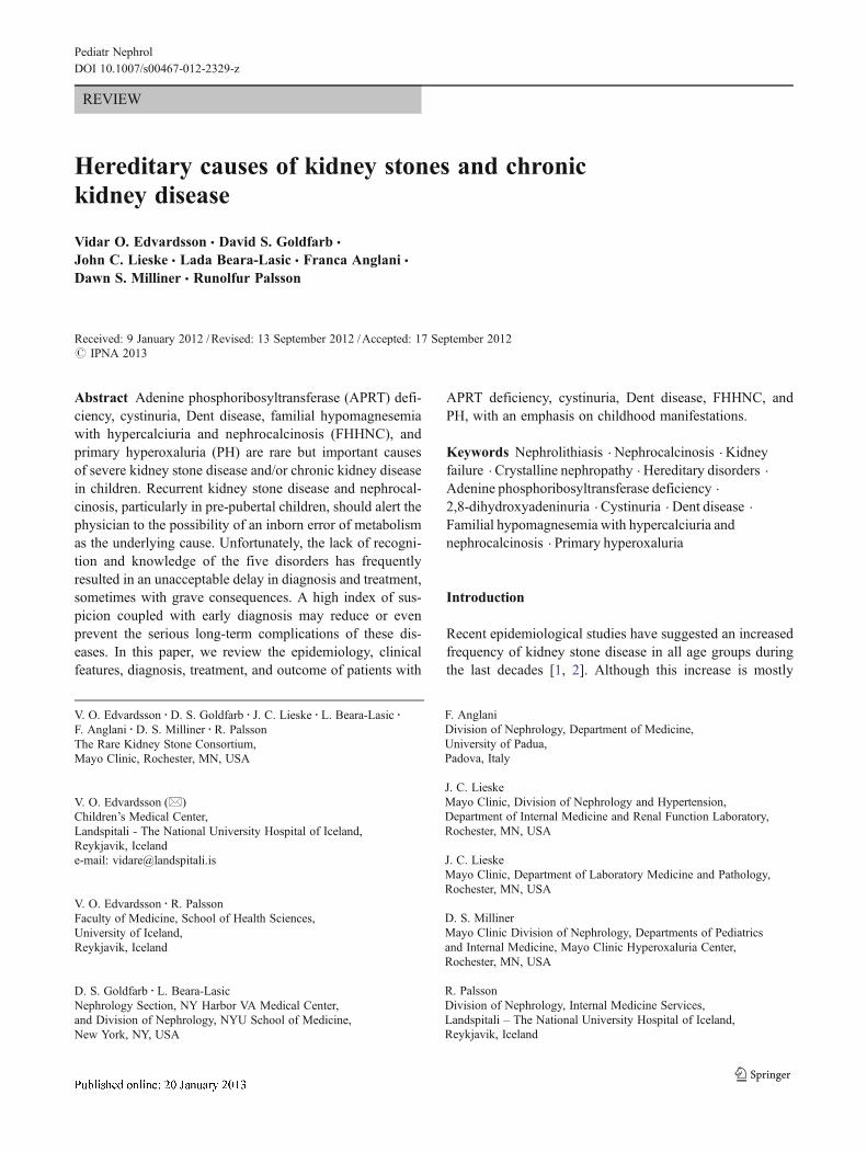
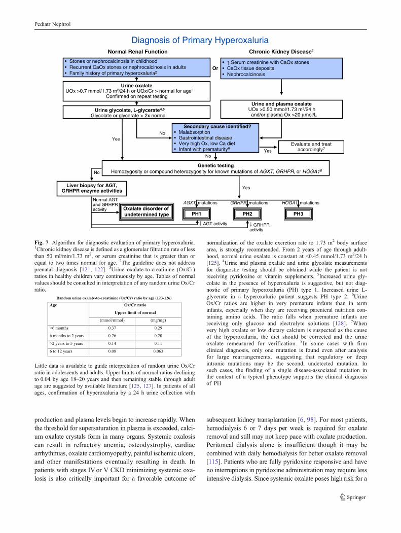
Fig. 1 Urinary crystals. a Typical small- and medium-sized 2,8-dihy-droxyadenine crystals. The medium-sized crystals are brown with darkoutline and central spicules. b The same field viewed with polarizedlight microscopy. The small- and medium-sized crystals appear yellowin color and produce a central Maltese cross pattern. c Urinary cystinecrystals. The typical 6-sided crystal is diagnostic of cystinuria. A goodexample can be seen on the left side of the Figure (arrow). d Urinary
calcium oxalate crystals. The typical bipyramidal calcium oxalatedihydrate crystals (arrows) and a large dumbbell calcium oxalatemonohydrate crystal (asterisk) are both seen. e Amorphous calciumphosphate crystals. Although their appearance is not as distinctive,amorphous calcium phosphate crystals should be suspected if the urineis alkaline (pH>6.0)
Pediatr Nephrol
high urine oxalate levels (>0.7 mmol/1.73 m2 per 24 h) arehighly suggestive of PH and should lead to further testing.Cystinuria should be excluded with a qualitative (nitroprus-side test) or quantitative urinary cystine study in a patient withradiopaque stones in whom stone analysis has not been per-formed, particularly if other urine chemistries are unremark-able. Round, brown DHA crystals (Fig. 1a) detected bymicroscopic examination of the urine are pathognomonic ofdihydroxyadeninuria and he typical hexagonal cystine crystals(Fig. 1c) are diagnostic of cystinuria. Patients with PH oftenhave calcium oxalate crystals (Fig. 1d) in their urine, whileurinary calcium oxalate or calcium phosphate crystals(Fig. 1e) may be detected in patients with Dent disease.Finally, all boys with unexplained stones, nephrocalcinosis,and/or proteinuria should have a quantitative test for lowmolecular weight (LMW) proteinuria to exclude the possibil-ity of Dent disease. A high index of suspicion coupled withearly diagnosis and treatment may reduce or even prevent theserious long-term complications of these well-defined hered-itary causes of severe stone disease and/or CKD.
In this paper, we review the epidemiology and genetics,clinical features, diagnosis, treatment and outcome of APRTdeficiency, cystinuria, Dent disease, FHHNC and PH.
Emphasis will be placed on childhood manifestations andfeatures suggestive of these rare causes of kidney stonedisease, nephrocalcinosis and CKD (Table 1). Diagnosticalgorithms will be presented. Other rare hereditary causes ofkidney stones or nephrocalcinosis, including xanthinuriaand hypoxanthine-guanine phosphoribosyltransferase defi-ciency (Lesch–Nyhan syndrome), will not be discussed inthis review.
Adenine phosphoribosyltransferase deficiencyand 2,8-dihydroxyadeninuria
Background
APRT deficiency (OMIM 102600) is a rare autosomal reces-sive inborn error of adenine metabolism resulting in the gen-eration of large amounts of the poorly soluble DHA, whichleads to stone formation in the urinary tract and crystallinenephropathy [8, 9]. APRT deficiency was first described in1968 by Kelley and coworkers [10] who reported partial defi-ciency of the enzyme in four asymptomatic subjects. Thedisorder has since been described in all ethnic groups, althoughthe majority of reported cases have come from Japan, France,and Iceland [8].
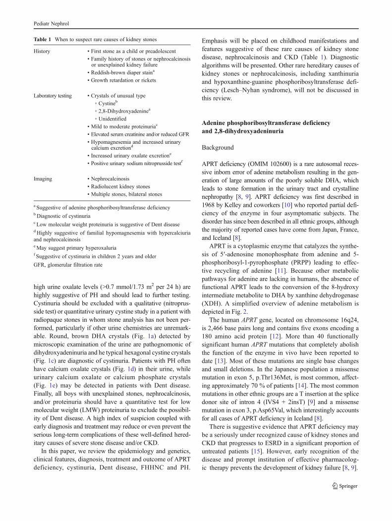
APRT is a cytoplasmic enzyme that catalyzes the synthe-sis of 5′-adenosine monophosphate from adenine and 5-phosphoribosyl-1-pyrophosphate (PRPP) leading to effec-tive recycling of adenine [11]. Because other metabolicpathways for adenine are lacking in humans, the absence offunctional APRT leads to the conversion of the 8-hydroxyintermediate metabolite to DHA by xanthine dehydrogenase(XDH). A simplified overview of adenine metabolism isdepicted in Fig. 2.
The human APRT gene, located on chromosome 16q24,is 2,466 base pairs long and contains five exons encoding a180 amino acid protein [12]. More than 40 functionallysignificant human APRT mutations that completely abolishthe function of the enzyme in vivo have been reported todate [13]. Most of these mutations are single base changesand small deletions. In the Japanese population a missensemutation in exon 5, p.Thr136Met, is most common, affect-ing approximately 70 % of patients [14]. The most commonmutations in other ethnic groups are a T insertion at the splicedonor site of intron 4 (IVS4 + 2insT) [9] and a missensemutation in exon 3, p.Asp65Val, which interestingly accountsfor all cases of APRT deficiency in Iceland [8].
There is suggestive evidence that APRT deficiency maybe a seriously under recognized cause of kidney stones andCKD that progresses to ESRD in a significant proportion ofuntreated patients [15]. However, early recognition of thedisease and prompt institution of effective pharmacolog-ic therapy prevents the development of kidney failure [8, 9].
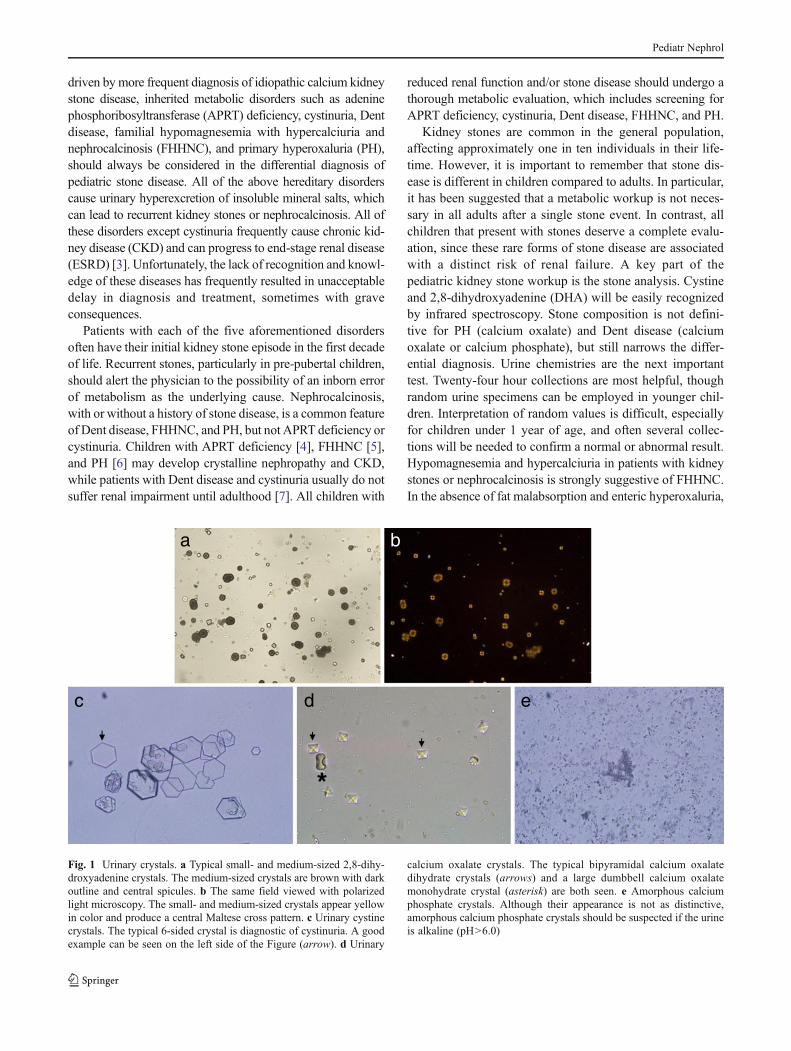
Table 1 When to suspect rare causes of kidney stones
History • First stone as a child or preadolescent
• Family history of stones or nephrocalcinosisor unexplained kidney failure
• Reddish-brown diaper staina
• Growth retardation or rickets
Laboratory testing • Crystals of unusual type
◦ Cystineb
◦ 2,8-Dihydroxyadeninea
◦ Unidentified• Mild to moderate proteinuriac
• Elevated serum creatinine and/or reduced GFR
• Hypomagnesemia and increased urinarycalcium excretiond
• Increased urinary oxalate excretione
• Positive urinary sodium nitroprusside testf
Imaging • Nephrocalcinosis
• Radiolucent kidney stones
• Multiple stones, bilateral stones
a Suggestive of adenine phosphoribosyltransferase deficiencyb Diagnostic of cystinuriac Low molecular weight proteinuria is suggestive of Dent diseased Highly suggestive of familial hypomagnesemia with hypercalciuriaand nephrocalcinosiseMay suggest primary hyperoxaluriaf Suggestive of cystinuria in children 2 years and older
GFR, glomerular filtration rate
Pediatr Nephrol
The estimated heterozygote frequency in different popu-lations ranges from 0.4 to 1.2 % [16, 17], suggesting that theprevalence of a homozygous state is at least 1:50,000–100,000. If this holds true, there should be at least 70–80,000 cases worldwide of which 40,000 would be expectedto be in Asia, 9,000 in Europe, 8,000 in the Americas,including at least 3,000 cases in the U.S. alone. The diseaseis currently unrecognized in the majority of these patientswho, therefore, are not receiving the benefits of medicaltherapy. Lack of familiarity with dihydroxyadeninuriaamong clinicians and pathologists is a major concern andundoubtedly contributes to the low number of reported casesworldwide. Other plausible explanations include inadequateevaluation of patients with kidney stones and confusion ofDHA calculi with uric acid and/or xanthine stones.
Clinical features
Radiolucent kidney stones are by far the most commonclinical manifestation of APRT deficiency in the pediatricpopulation [8, 18]. An initial presentation of acute kidneyinjury due to bilateral DHA calculi causing urinary tractobstruction has also been well documented in children [4,8, 19, 20]. Other common clinical features include recurrenturinary tract infections, hematuria, and reddish-brown dia-per stains [8, 9, 20]. However, some children remain asymp-tomatic throughout childhood and, not uncommonly, thedisease is diagnosed by the detection of DHA crystals onroutine urine microscopy or through screening of siblings ofindex cases [8, 18].
Although CKD with reduced glomerular filtration rate(GFR) has been reported in a number of children with APRT
deficiency [18], ESRD secondary to crystalline nephropathyis a much more common presenting feature of APRT defi-ciency in adults [8, 9], which in a number of cases has notbeen recognized until after kidney transplantation has beenperformed [15, 21, 22]. This point is well demonstrated in arecent report from the Mayo Clinic of 3 adult patients withESRD due to dihydroxyadeninuria [15]. While all thesepatients were found to have crystalline nephropathy, twodid not have a past history of kidney stones and the thirdone had only experienced a single stone episode 36 yearsprior to diagnosis. Early DHA crystalline nephropathy in achild was reported by Greenwood and colleagues [4] whoperformed a kidney biopsy in a 4-year-old girl with bilateralstone disease and acute kidney injury.
Fifteen of 32 Icelandic patient with data in the APRTDeficiency Registry of the Rare Kidney Stone Consor-tium (RKSC) presented before the age of 18 years. Themajor presenting features included kidney stones in twopatients, reddish-brown diaper stain in ten, renal colicin six, and one was asymptomatic (unpublishedobservation).
The kidney and urinary tract appear to be the onlyorgan system affected in APRT deficiency. However, wehave encountered occasional adults with this disorderwho have complained of eye discomfort, though it isunclear if this is related to DHA crystal deposition inocular tissues.
Diagnosis
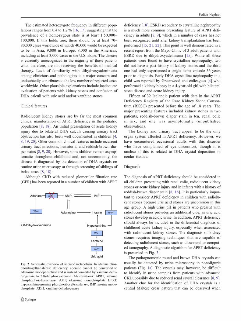
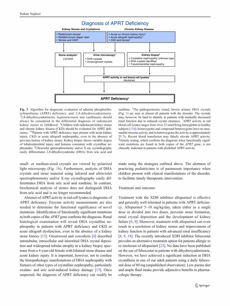
The diagnosis of APRT deficiency should be considered inall children presenting with renal colic, radiolucent kidneystones or acute kidney injury and in infants with a history ofreddish-brown diaper stain [8, 18]. It is particularly impor-tant to consider APRT deficiency in children with radiolu-cent stones because uric acid stones are uncommon in thisage group. A high urine pH in patients who present withradiolucent stones provides an additional clue, as uric acidstones develop in acidic urine. In addition, APRT deficiencyshould always be included in the differential diagnosis ofchildhood acute kidney injury, especially when associatedwith radiolucent kidney stones. The diagnosis of kidneystones requires imaging techniques that are capable ofdetecting radiolucent stones, such as ultrasound or comput-ed tomography. A diagnostic algorithm for APRT deficiencyis presented in Fig. 3.
The pathognomonic round and brown DHA crystals canusually be detected by urine microscopy in nonoliguricpatients (Fig. 1a). The crystals may, however, be difficultto identify in urine samples from patients with advancedCKD, possibly due to reduced renal crystal clearance [8, 9].Another clue for the identification of DHA crystals is acentral Maltese cross pattern that can be observed when
Adenine AMP IMP
Adenosine Inosine
Hypoxanthine
Xanthine
Uric acid
XDH HPRT
XDH
XDH
2,8-Dihydroxyadenine
APRT
Fig. 2 Schematic overview of adenine metabolism. In adenine phos-phoribosyltransferase deficiency, adenine cannot be converted toadenosine monophosphate and is instead converted by xanthine dehy-drogenase to 2,8-dihydroxyadenine. Abbreviations: APRT, adeninephosphoribosyltransferase; AMP, adenosine monophosphate; HPRT,hypoxanthine-guanine phosphoribosyltransferase; IMP, inosine mono-phosphate; XDH, xanthine dehydrogenase
Pediatr Nephrol
small- or medium-sized crystals are viewed by polarizedlight microscopy (Fig. 1b). Furthermore, analysis of DHAcrystals and stone material using infrared and ultravioletspectrophotometry and/or X-ray crystallography easily dif-ferentiates DHA from uric acid and xanthine. In contrast,biochemical analysis of stones does not distinguish DHAfrom uric acid and is no longer recommended.
Absence of APRTactivity in red cell lysates is diagnostic ofAPRT deficiency. Enzyme activity measurements are alsoneeded to determine the functional significance of novelmutations. Identification of functionally significant mutationsin both copies of the APRT gene confirms the diagnosis. Renalhistological examination will reveal DHA crystalline ne-phropathy in patients with APRT deficiency and CKD oracute allograft dysfunction, even in the absence of a kidneystone history [15]. Greenwood and coworkers [4] describedintratubular, intracellular and interstitial DHA crystal deposi-tion and widespread tubular atrophy in a kidney biopsy spec-imen from a 4-year-old female with bilateral stone disease andacute kidney injury. It is important, however, not to confusethe histopathologic manifestations of DHA nephropathy withfeatures of other types of crystalline nephropathy, particularlyoxalate- and uric acid-induced kidney damage [15]. Oncesuspected, the diagnosis of APRT deficiency can readily be
made using the strategies outlined above. The alertness ofpracticing pediatricians is of paramount importance whenchildren present with clinical manifestations of the disorder,to facilitate timely therapeutic intervention.
Treatment and outcome
Treatment with the XDH inhibitor allopurinol is effectiveand generally well tolerated in patients with APRT deficien-cy. Allopurinol 5–10 mg/kg/day, taken either as a singledose or divided into two doses, prevents stone formation,renal crystal deposition and the development of kidneyfailure [8, 9]. Moreover, treatment with allopurinol can evenresult in a resolution of kidney stones and improvement ofkidney function in patients with advanced renal insufficiency[8, 9, 18]. The recently introduced XDH inhibitor febuxostatprovides an alternative treatment option for patients allergic toor intolerant of allopurinol [23]. No data have been publishedon the use of febuxostat in patients with dihydroxyadeninuria.However, we have achieved a significant reduction in DHAcrystalluria in one of our adult patients using a daily febuxo-stat dose of 80 mg (unpublished observation). Low purine dietand ample fluid intake provide adjunctive benefits to pharma-cologic therapy.
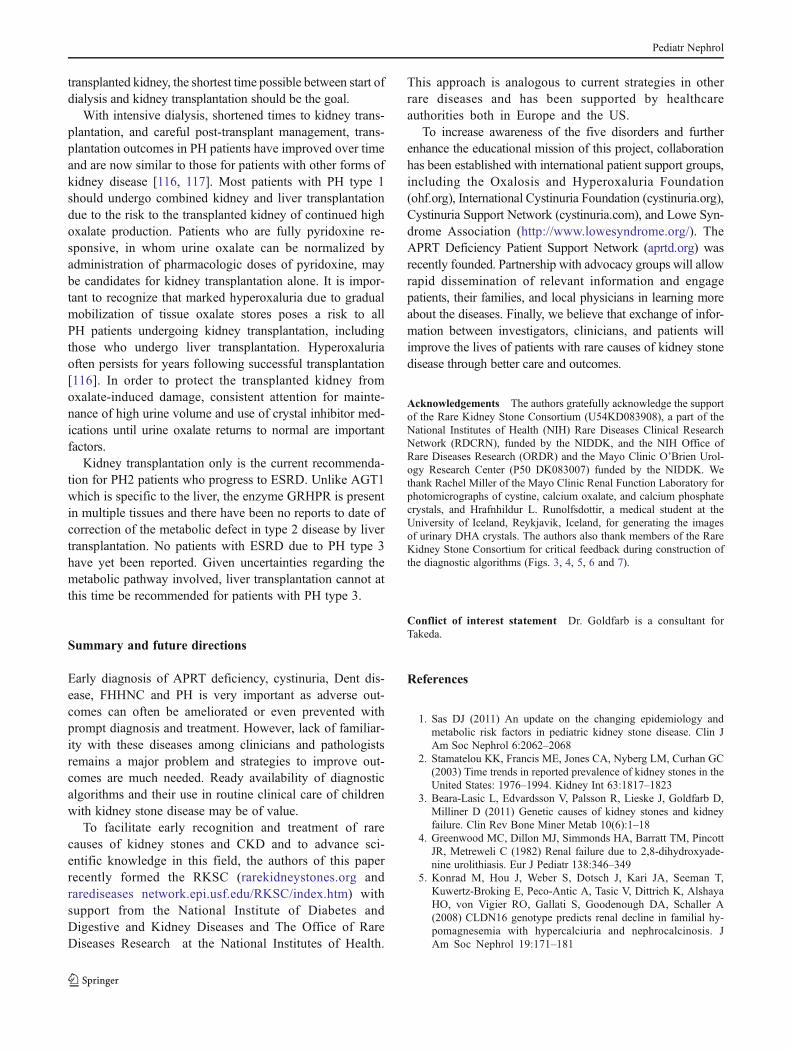
Diagnosis of APRT Deficiency
• Radiolucent stones1
• Reddish-brown diaper stain • Stones and CKD2
• Acute on chronic kidney injury3
• Acute allograft nephropathy3
• CKD and stones2
Kidney biopsy6
• Crystalline nephropathy unknown type• DHA crystals identified • Tubulointerstitial nephropathy
Urine microscopy5
• DHA crystals• Unrecognized crystals
Stone analysis4
2,8-DHA
APRT activity in red blood cell lysates7
Abolished
APRT Deficiency8
Kidney Stones and Crystalluria Chronic Kidney Disease
Fig. 3 Algorithm for diagnostic evaluation of adenine phosphoribo-syltransferase (APRT) deficiency and 2,8-dihydroxyadeninuria.12,8-dihydroxyadeninuria, hyperuricosuria and xanthinuria shouldalways be considered in the differential diagnosis of radiolucentkidney stones in childhood. 2Children with radiolucent kidney stonesand chronic kidney disease (CKD) should be evaluated for APRT defi-ciency. 3,6Patients with APRT deficiency may present with acute kidneyinjury, CKD or acute allograft nephropathy, even in the absence ofprevious history of kidney stones. Kidney biopsy shows variable degreeof tubulointerstitial injury and features consistent with crystalline ne-phropathy. 4Ultraviolet spectrophotometry and/or X-ray crystallographyeasily differentiates 2,8-dihydroxyadenine (DHA) from uric acid and
xanthine. 5The pathognomonic round, brown urinary DHA crystals(Fig. 1) are seen in almost all patients with the disorder. The crystalsmay, however, be hard to identify in patients with markedly decreasedrenal function due to reduced crystal clearance. 7APRT activity in redblood cell lysates ranges from 16 to 32 nmol/h/mg hemoglobin in healthysubjects [118]; homozygotes and compound heterozygotes have no mea-surable enzyme activity and in heterozygotes the activity is approximately25 %. Recent blood transfusion may falsely elevate APRT activity.8Genetic testing, which confirms the diagnosis when functionally signif-icant mutations are found in both copies of the APRT gene, is notclinically indicated in patients with abolished APRT activity
Pediatr Nephrol
Cystinuria
Background
Though it meets the definition of a rare disease, cystinuria(OMIM 220100) is the most common cause of inherited kid-ney stones addressed in this article. It is said to account forabout 1 % of all kidney stones and up to 25 % of pediatricstones [24]. The worldwide prevalence of cystinuria is estimat-ed at 1:7,000, ranging from 1:2,500 among a population ofLibyan Jews to 1:100,000 persons in Sweden. In the UnitedStates, one estimate is that cystinuria occurs in approximately1:15,000 adults. However, the true prevalence may be higheras some affected individuals never make a stone or make theminfrequently and never have the diagnosis made.
Cystinuria is the result of a defect in proximal tubularreabsorption of filtered cystine, a homodimer of the aminoacid cysteine [25]. Normally, 100 % of filtered cystine isreabsorbed so that normal urine cystine excretion is 0–100 μmoles of cystine per gram of creatinine. The disorder isthe result of autosomal recessive inheritance of a genetic defectin one of two genes whose protein products are required fortubular cystine reabsorption [26]. The transport protein isencoded by SLC7A9 and is called b0,+AT (amino acid trans-porter of positively charged or neutral amino acids). It forms aheterodimer in the apical membrane of proximal tubular epi-thelial cells with the protein product of SLC3A1, called rBAT(related to b0,+ATamino acid transporter). The rBAT protein isrequired to correctly direct b0,+AT to the apical membrane.Mutation of both alleles of SLC3A1 (type A genotype) orSLC7A9 (type B genotype) will lead to failure to reabsorbcystine filtered at the glomerulus and excretion of abnormalamounts of the relatively insoluble cystine. The result is for-mation of recurrent renal stones. Defective cystine transportphysiology is also associated with concomitant loss of thedibasic amino acids ornithine, arginine, and lysine, but theirloss is not clinically significant. People heterozygous forSLC3A1mutations will generally not have cystine in the urine,while heterozygotes for abnormal SLC7A9 sequences willhave abnormal amounts that are very rarely sufficient to causestones, perhaps if other factors such as low urine volume orlow urine pH are also present. SLC7A9mutations can thereforebe considered as incompletely dominant mutations. The samecystine transporter is expressed in the small intestine, respon-sible for absorption of cystine from the intestinal lumen, and isdefective in cystinuria. However, cystine can also be absorbedas a component of oligopeptides and, therefore, the intestinaldefect does not lead to any known clinical consequence.
Clinical features
Recurrent urinary tract stone disease is the only clinicalmanifestation of cystinuria in childhood. The hypotonia-
cystinuria syndrome (OMIM 606407) arises when muta-tions occur both in SLC3A1 and the contiguous gene PREPL(prolyl endopeptidase-like protein). Besides cystinuria, af-fected individuals have severe hypotonia at birth, delayedgrowth and hyperphagia and excessive weight gain later inchildhood [27]. Cystinuria can also result in CKD due torecurrent stones, obstructive uropathy and repeated urologicinterventions. Most patients with cystinuria present in child-hood with stone formation. Average age of detection of afirst renal stone is about 12–13 years [28], with 50 % form-ing a first stone in the first decade of life and another 25 %in teenage years. Males and females have a similar age ofonset but more male patients than female patients present inthe first 3 years of life and males tend to have new stonesmore frequently than females. On the other hand, somepatients surprisingly have their first stone between 40 and80 years of age [29]. Type A and B genotypes apparently havesimilar phenotypes with similar rates of stone recurrence.
A significant proportion of adults with cystinuria developCKD and the renal pathologic findings correlate with thereduction in GFR. One study demonstrated that 52 adultpatients with cystinuria had a creatinine clearance of63.2 ml/min compared with 3,215 patients without cystin-uria whose creatinine clearance was 111.1 ml/min [30]. How-ever, more open surgical stone removing procedures andnephrectomy are variables correlated with an increased serumcreatinine, with 14.1 % of cystinuric patients compared to2.9 % of calcium oxalate stone formers in one cohort havingundergone nephrectomy [31].
Diagnosis
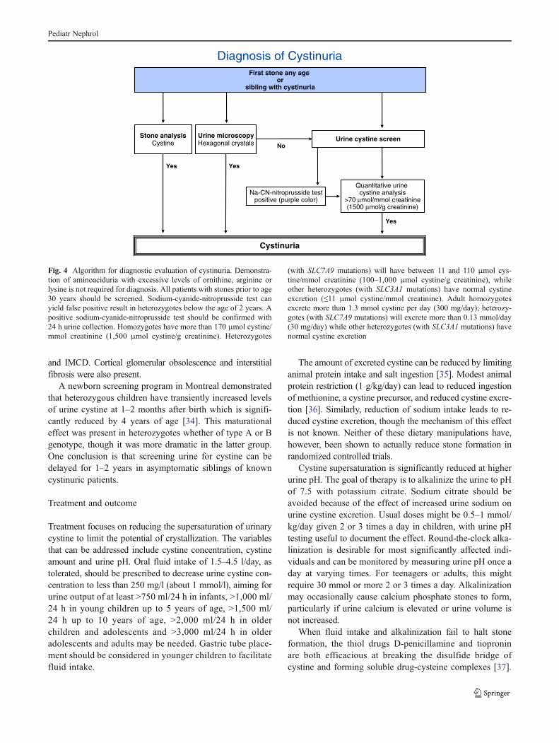
Diagnosis of cystinuria is outlined in the diagnostic algo-rithm shown in Fig. 4. Demonstration of hexagonal crys-tals on urinalysis is pathognomonic of cystinuria (Fig. 1c).Some clinical laboratories screen all 24 h urine collectionsfrom new clients with the sensitive and specific nitroprus-side test [32]. Demonstrating increased urine content oflysine, ornithine and arginine is merely confirmatory andnot necessary if cystine stones or crystals are identified. Atpresent, genotyping patients with cystinuria is not requiredfor diagnostic purposes and yields no clinically usefulinformation.
Although data regarding the renal histopathology of cys-tinuria are relatively sparse, affected patients may developcrystalline nephropathy. One paper stands out in this regard,presenting the results of renal papillary biopsy of sevenpatients who underwent percutaneous nephrolithotomy[33]. The ducts of Bellini and inner medullary collectingducts (IMCD) were frequently plugged with cystine crystals,had injured or absent lining cells with nearby inflammation orfibrosis of the interstitium. In addition, hydroxyapatite (calciumphosphate) crystals were noted in the lumens of loops of Henle
Pediatr Nephrol
and IMCD. Cortical glomerular obsolescence and interstitialfibrosis were also present.
A newborn screening program in Montreal demonstratedthat heterozygous children have transiently increased levelsof urine cystine at 1–2 months after birth which is signifi-cantly reduced by 4 years of age [34]. This maturationaleffect was present in heterozygotes whether of type A or Bgenotype, though it was more dramatic in the latter group.One conclusion is that screening urine for cystine can bedelayed for 1–2 years in asymptomatic siblings of knowncystinuric patients.
Treatment and outcome
Treatment focuses on reducing the supersaturation of urinarycystine to limit the potential of crystallization. The variablesthat can be addressed include cystine concentration, cystineamount and urine pH. Oral fluid intake of 1.5–4.5 l/day, astolerated, should be prescribed to decrease urine cystine con-centration to less than 250 mg/l (about 1 mmol/l), aiming forurine output of at least >750 ml/24 h in infants, >1,000 ml/24 h in young children up to 5 years of age, >1,500 ml/24 h up to 10 years of age, >2,000 ml/24 h in olderchildren and adolescents and >3,000 ml/24 h in olderadolescents and adults may be needed. Gastric tube place-ment should be considered in younger children to facilitatefluid intake.
The amount of excreted cystine can be reduced by limitinganimal protein intake and salt ingestion [35]. Modest animalprotein restriction (1 g/kg/day) can lead to reduced ingestionof methionine, a cystine precursor, and reduced cystine excre-tion [36]. Similarly, reduction of sodium intake leads to re-duced cystine excretion, though the mechanism of this effectis not known. Neither of these dietary manipulations have,however, been shown to actually reduce stone formation inrandomized controlled trials.
Cystine supersaturation is significantly reduced at higherurine pH. The goal of therapy is to alkalinize the urine to pHof 7.5 with potassium citrate. Sodium citrate should beavoided because of the effect of increased urine sodium onurine cystine excretion. Usual doses might be 0.5–1 mmol/kg/day given 2 or 3 times a day in children, with urine pHtesting useful to document the effect. Round-the-clock alka-linization is desirable for most significantly affected indi-viduals and can be monitored by measuring urine pH once aday at varying times. For teenagers or adults, this mightrequire 30 mmol or more 2 or 3 times a day. Alkalinizationmay occasionally cause calcium phosphate stones to form,particularly if urine calcium is elevated or urine volume isnot increased.
When fluid intake and alkalinization fail to halt stoneformation, the thiol drugs D-penicillamine and tioproninare both efficacious at breaking the disulfide bridge ofcystine and forming soluble drug-cysteine complexes [37].
Diagnosis of CystinuriaFirst stone any age
orsibling with cystinuria
Na-CN-nitroprusside testpositive (purple color)
Cystinuria
Yes
NoUrine cystine screen
Quantitative urine cystine analysis
>70 μmol/mmol creatinine (1500 μmol/g creatinine)
Urine microscopyHexagonal crystals
Stone analysisCystine
Yes
Yes
Fig. 4 Algorithm for diagnostic evaluation of cystinuria. Demonstra-tion of aminoaciduria with excessive levels of ornithine, arginine orlysine is not required for diagnosis. All patients with stones prior to age30 years should be screened. Sodium-cyanide-nitroprusside test canyield false positive result in heterozygotes below the age of 2 years. Apositive sodium-cyanide-nitroprusside test should be confirmed with24 h urine collection. Homozygotes have more than 170 μmol cystine/mmol creatinine (1,500 μmol cystine/g creatinine). Heterozygotes
(with SLC7A9 mutations) will have between 11 and 110 μmol cys-tine/mmol creatinine (100–1,000 μmol cystine/g creatinine), whileother heterozygotes (with SLC3A1 mutations) have normal cystineexcretion (≤11 μmol cystine/mmol creatinine). Adult homozygotesexcrete more than 1.3 mmol cystine per day (300 mg/day); heterozy-gotes (with SLC7A9 mutations) will excrete more than 0.13 mmol/day(30 mg/day) while other heterozygotes (with SLC3A1 mutations) havenormal cystine excretion
Pediatr Nephrol
Both drugs reduce stone formation but have significant ratesof discontinuation due to a variety of side effects, includingrash, allergy, hematologic and liver function abnormalities andrarely nephrotic syndrome due to membranous nephropathy.
Collection of urine for 24 h can be useful in monitoringtherapy. Measurement of urine pH, sodium and surrogatesof protein ingestion such as urea can help monitor adherenceto dietary therapy and judge the adequacy of citrate supple-mentation. Measurement of cystine is useful but can beartifactually lowered by precipitation of the amino acid aftervoiding; alkalinization of the collected urine ensures thatcystine is in a soluble and measurable state [38]. Thioltherapy may also interfere with cystine measurement andcan be overcome by use of a solid phase assay to directlymeasure urinary cystine capacity or supersaturation [35, 39].Such an assay may eventually prove to be useful in pre-scribing thiol drugs and judging metabolic stone-formingactivity.
Cystinuria has a better prognosis today in the era of effec-tive medical therapy and modern endourology. Although norandomized controlled trials have been performed in cystin-uria, some small, retrospective cohorts suggest that the out-come of prophylactic regimens can reduce stone recurrence[40]. Cystine stones are often less amenable to being fracturedby shockwave lithotripsy, but can be effectively turned to dustby ureteroscopy and the holmium laser [41]. The result is thevirtual elimination of open surgery and nephrectomy.The expectation then is that less significant reductionsin glomerular filtration rate should be seen in the com-ing years, though the impact of recurrent stones, crys-talline nephropathy, and nephronal obstruction maypersist.
Dent disease
Background
Dent disease (OMIM 300009) is a rare renal tubular disor-der, with about 250 affected families having been reportedaround the world to date. The term Dent disease was firstintroduced in 1990 [42] and identifies a group of X-linkedrenal disorders characterized by LMW proteinuria, and var-iable presence of hypercalciuria, nephrocalcinosis and/ornephrolithiasis. This triad of symptoms has been variablynamed X-linked recessive nephrolithiasis with renal failure(XRN; OMIM 310468), X-linked recessive hypophosphate-mic rickets (XLRH; OMIM 300554) or idiopathic LMWproteinuria of Japanese children (JILMWP; OMIM 308990)[43, 44]. The disease usually presents in childhood or earlyadult life with males much more severely affected thanfemales [42, 45]. Progression to CKD occurs between the3rd and 5th decades of life in 30–80 % of affected males
[42, 43]. It is hoped that early diagnosis can delay or preventCKD. However, effective treatments have not yet beenclearly established. LMW proteinuria is universal andhypercalciuria is common among affected Dent diseasepatients, but both conditions are rarely screened for. There-fore, many cases go undetected until more significant se-quelae, such as progressive renal failure, develop.
Most diagnosed cases of Dent disease have been causedby mutations in the CLCN5 gene, located on the X chromo-some (Xp11.22). This gene encodes a 746-amino-acid ClC-5 chloride channel that belongs to a voltage-gated chloridechannel family [46–48]. In the human kidney, ClC-5 isprimarily expressed in proximal tubular cells, alpha- andbeta-intercalated cells of the cortical collecting duct, and inthe thick ascending limb (TAL) of Henle’s loop [49]. Inproximal tubular cells it is predominantly located in intra-cellular subapical endosomes, which are involved in theendocytic reabsorption of LMW proteins that have passedthe glomerular filter. Specifically, ClC-5 has been proposedto function by providing a shunt conductance in early endo-somes, thus permitting their efficient intraluminal acidifica-tion by a V-type H+-ATPase [49, 50]. Recently, however,two independent groups demonstrated that ClC-5 functionsas a 2Cl-/H+ antiporter when activated by positive voltages[51, 52]. Interestingly, it appears that endosomal chlorideconcentration is at least as important as pH for normalendosomal function [53]. A small portion of ClC-5 channelsare also located on the cell surface, where they are thoughtto mediate plasma membrane chloride currents [54], or asrecently demonstrated, join macromolecular complexes atthe plasma membrane believed to mediate LMW proteinand albumin endocytosis [55].
ClC-5 knock-out mice demonstrate severe impairment ofreceptor-mediated endocytosis [56], including the endocyticretrieval of the plasma membrane multiligand megalin andcubilin receptors [57, 58]. Like humans, knock-out mousemodels have LMW proteinuria, but not all of them develophypercalciuria and nephrocalcinosis [48, 59]. Indeed, therelationship between impaired endocytosis and renal cal-cium handling remains unclear. It has been suggestedthat the hypercalciuria seen in many affected patientsstems from secondary changes in the regulation of calciotropichormone caused by urinary loss of key hormone-bindingproteins [59, 60].
To date, more than 100 different nonsense or missensemutations, insertions or deletions and splicing mutations inthe CLCN5 gene have been reported [61, 62], meaning thatthe spectrum of CLCN5 mutations is highly varied and denovo mutations are frequent. Genotype-phenotype correla-tions have yet to be established. CLCN5 mutations are scat-tered throughout the gene’s coding sequence and generatetruncated or absent ClC-5 channels in approximately 70 % ofcases. In approximately 40% of patients with classic symptoms
Pediatr Nephrol

of Dent disease, no CLCN5 mutations were detected, suggest-ing locus heterogeneity [63, 64]. Subsequently, Hoopes andcolleagues [64] demonstrated that OCRL1 mutations, initiallyassociated with Lowe syndrome, account for 15 % of patientswith Dent disease, which is now termedDent 2 disease (OMIM300555). OCRL1 encodes a phosphatidylinositol 4,5-biphos-phate 5-phosphatase localized at the Golgi apparatus. Themechanism by which loss of OCRL1 protein function leadsto disease has not yet been elucidated. Very recently, however,the OCRL1 protein was localized to early endosomes and thetrans-Golgi network (TGN), and clathrin coated transport inter-mediates [65, 66]. Depletion of OCRL1 perturbs trafficking atthe TGN/endosome interface, suggesting a role in regulatingtransport between these compartments. Renal tubulopathy inLowe syndrome is quite similar to that of Dent disease, and ismainly characterized by altered protein reabsorption. It is inter-esting to note, however, thatOCRL1mutations associated withDent 2 disease do not overlap those causing Lowe syndrome[67, 68].
Clinical features
The presentation of patients with Dent disease appears quitevariable. Most patients are recognized due to the presence ofnephrocalcinosis or unexplained CKD. Hypophosphatemiaand bone disease can also be observed, but appear to be a lessprominent part of the phenotype than first believed. A univer-sal feature is proteinuria, which can be detected in typicalurinary total protein screens, including dipstick (Fig. 5). Alarge proportion of this is LMW proteins which must bespecifically screened for. More recently, patients have beenrecognized with nephrotic-range proteinuria and glomerularchanges of focal segmental glomerulosclerosis (FSGS) onrenal biopsy [69, 70]. It is not clear what percentage of FSGSpatients might have unrecognized Dent disease, or what roleglomerular dysfunctionmight play in the loss of renal functionin the majority of Dent patients. Other presenting features inchildren and adolescents often include nephrocalcinosis andhypercalciuria, with or without kidney stones, although theprevalence of stones versus nephrocalcinosis remains unclear.Hematuria may also be seen, presumably due to the stones andnephrocalcinosis. It must be stressed that LMW proteinuria isthe pathognomonic feature of Dent disease, and can occur inisolation [69, 70]. The prevalence of other manifestations suchas nephrocalcinosis or stones among those with CLCN5 and/or OCRL1 mutations is currently unknown.
The initial patients described with Dent 2 disease caused byOCRL1mutations exhibited none of the classic extrarenal man-ifestations of Lowe syndrome such as mental retardation, bonedisease, growth retardation, congenital cataracts, or delayedmotor milestones [64]. This milder phenotype could not beattributed to lesser protein expression or enzyme activity. Inter-estingly, patients with classic Lowe syndrome have a renal
phenotype very similar to those with Dent disease, with somesubtle differences. Renal tubular acidosis is one of the cardinalsigns of Lowe syndrome, whereas in Dent disease it is rare.Features of Fanconi syndrome are also observed more frequent-ly in patients with Lowe syndrome than in patients with Dentdisease, whereas hypercalciuria, nephrocalcinosis and nephroli-thiasis are common for Dent disease but rare in Lowe syndrome.However, more recently it has been suggested that a continuumexists since patients with OCRL1 mutations may present withclinical phenotypes ranging from a severe Lowe syndrome withthe typical ocular, neurological and renal features to Dent 2disease with only renal impairment [71]. To date, around 25patients with Dent 2 disease have been reported.
Many affected males (30–80 %) will develop ESRD be-tween the ages of 30–50 years, although somemay be delayeduntil later in life [42, 43]. Deterioration of renal function canbe observed even in the absence of nephrocalcinosis [69, 70].
Diagnosis
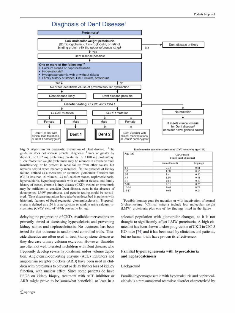
Diagnostic features of Dent disease include LMW proteinuria,hypercalciuria, nephrolithiasis and/or nephrocalcinosis and re-nal insufficiency. Since proteinuria is often screened for, espe-cially in patients with CKD, it is stressed as an entry point in thesuggested diagnostic algorithm (Fig. 5). The important clue fora diagnosis of Dent disease is markedly increased levels ofLMW proteins in the urine (5–10 times above normal orgreater), in the absence of other causes of proximal tubulardysfunction. Commonly screened LMW proteins are retinol-binding protein and α1 microglobulin. β2-microglobulin isalso often measured to screen for LMW proteinuria but we donot recommend it since it is not very stable in even minimallyacidic urine [72]. It has been reported that proximal tubularuptake of 99Tc-DMSA is abnormally low in patients withproximal tubular dysfunction, including those with CLCN5mutations [73]. Therefore, abnormal 99Tc-DMSA in the settingof normal GFR can be another clue to the diagnosis of Dentdisease [70]. Although genetically-confirmed Dent diseasecases have been reported with LMW proteinuria alone, one ormore of the following additional features is sufficient to make aclinical diagnosis of Dent disease when LMW proteinuria ispresent: stones or nephrocalcinosis, hypercalciuria, hypophos-phatemia with or without rickets, or a family history of Dentdisease (Fig. 5). Genetic screening of the two implicated genes(CLCN5 and OCRL1) can be confirmatory. Findings on renalbiopsy consistent with Dent disease include nephrocalcinosis,interstitial fibrosis, and FSGS [69, 70]. Stones, when present,are composed of calcium oxalate and/or calcium phosphate.
Treatment and outcome
There is no treatment that targets the specific defect causingDent disease. The primary goal of treatment, therefore, is
Pediatr Nephrol
delaying the progression of CKD. Available interventions areprimarily aimed at decreasing hypercalciuria and preventingkidney stones and nephrocalcinosis. No treatment has beentested for that outcome in randomized controlled trials. Thia-zide diuretics are often used to treat kidney stone disease asthey decrease urinary calcium excretion. However, thiazidesare often not well tolerated in children with Dent disease, whofrequently develop severe hypokalemia and/or volume deple-tion. Angiotensin-converting enzyme (ACE) inhibitors andangiotensin receptor blockers (ARB) have been used in chil-dren with proteinuria to prevent or delay further loss of kidneyfunction, with unclear effect. Since some patients do haveFSGS on kidney biopsy, treatment with ACE inhibitor orARB might prove to be somewhat beneficial, at least in a
selected population with glomerular changes, as it is notthought to significantly affect LMW proteinuria. A high cit-rate diet has been shown to slow progression of CKD in ClC-5KO mice [74] and it has been used by clinicians and patients,but no human trials have proven its effectiveness.
Familial hypomagnesemia with hypercalciuriaand nephrocalcinosis
Background
Familial hypomagnesemia with hypercalciuria and nephrocal-cinosis is a rare autosomal recessive disorder characterized by
Diagnosis of Dent Disease1
Proteinuria2
Low molecular weight proteinuriaβ2 microglobulin, α1 microglobulin, or retinol
binding protein >5x the upper reference range3
Dent disease possible
Genetic testing, CLCN5 and OCRL1
Dent disease possible
One or more of the following: 4,5
• Calcium stones or nephrocalcinosis• Hypercalciuria6
• Hypophosphatemia with or without rickets• Family history of stones, CKD, rickets, proteinuria
No other identifiable cause of proximal tubular dysfunction
No mutation
If meets clinical criteriafor Dent disease8
consider novel genetic cause
Dent disease unlikelyNo
Yes
NoYes
Dent 1 Dent 2 Dent 2 carrier withclinical manifestations,or Dent 2 homozygote7
Dent 1 carrier withclinical manifestations,or Dent 1 homozygote7
OCRL1 mutationCLCN5 mutation
FemaleMaleMaleFemale
Dent disease likely
Fig. 5 Algorithm for diagnostic evaluation of Dent disease. 1Theguideline does not address prenatal diagnosis. 2Trace or greater bydipstick; or >0.2 mg protein/mg creatinine; or >100 mg protein/day.3Low molecular weight proteinuria may be reduced in advanced renalinsufficiency, or be present in renal failure from other causes, butremains helpful when markedly increased. 4In the presence of kidneyfailure, defined as a measured or estimated glomerular filtration rate(GFR) less than 15 ml/min/1.73 m2, calcium stones, nephrocalcinosis,hypercalciuria, hypophosphatemia with or without rickets, and familyhistory of stones, chronic kidney disease (CKD), rickets or proteinuriamay be sufficient to consider Dent disease, even in the absence ofdocumented LMW proteinuria, and genetic testing could be consid-ered. 5Dent disease mutations have also been described in patients withhistologic features of focal segmental glomerulosclerosis. 6Hypercal-ciuria is defined as a 24 h urine calcium or random urine calcium-to-creatinine (Ca/Cr) ratio of >95th percentile for age.
Random urine calcium-to-creatinine (Ca/Cr) ratio by age (119)
Ca/Cr ratioUpper limit of normal
7Possibly homozygous for mutation or with inactivation of normalX-chromosome. 8Clinical criteria include low molecular weight(LMW) proteinuria plus one of the findings listed in the figure
Pediatr Nephrol
renal magnesium wasting, hypercalciuria, nephrocalcinosisand kidney failure. The disease was first described in 1972[75] and approximately 110 cases have since been reported inthe literature [5, 76–78]. Hypomagnesemia is invariably pres-ent and is a characteristic feature of the disease which usuallypresents in early childhood [76]. Progressive CKD commonlyoccurs in children with most patients developing ESRD inadolescence or early adult life. Clearance studies in patientswith FHHNC reported 25 years ago suggested impairment inthe reabsorption of magnesium and calcium in the TAL of theloop of Henle as the primary tubular abnormality [79]. Themolecular defect has been characterized and was found to becaused by mutations in two genes, CLDN16 located on thelong arm of chromosome 3 (3q27) [80] and CLDN19 on theshort arm of chromosome 1 (1p34.2) [80, 81]. The proteinsencoded by these two genes, claudin-16 (previously known asparacellin 1) and claudin-19, belong to the claudin family ofintegral membrane proteins that are important components oftight junctions in many tissues [82]. Both proteins areexpressed in the TAL where they are involved in paracellularreabsorption of calcium and magnesium, a process driven bylumen-positive transepithelial potential [83, 84]. Claudin-16 isexclusively expressed in the TAL [85], whereas claudin-19 isalso expressed in tight junctions of the retina [81]. Mutationalanalysis has identified a locus of negatively charged aminoacids in the first extracellular loop of claudin-16 as critical forits cation selectivity [86]. Moreover, in vitro studies haveshown that claudin-16 interacts with claudin-19 to form het-eromultimers causing a dramatic upregulation of PNa anddown-regulation of PCl, and thereby generating a cation-selective paracellular channel in the tight junctions of theTAL that regulates magnesium and calcium permeability[87]. The interaction between these two proteins is requiredfor their stable integration into tight junctions [88]. Confirma-tion that FHHNC is caused by loss-of-function mutations inCLDN16 and CLDN19 was provided by siRNA-mediatedknockdown of the two genes in mice, yielding a phenotypecharacterized by severe renal wasting of magnesium andcalcium [88, 89].
In FHHNC, hypercalciuria leads to nephrocalcinosis andstone formation. Renal failure is caused by a chronic tubu-lointerstitial nephropathy, the pathogenesis of whichremains poorly understood. The deposition of calcium crys-tals in the renal tissue seems unlikely to be the sole causesince other disorders associated with nephrocalcinosis donot generally result in progressive deterioration of renalfunction [5]. Inactivating mutations of CLDN16 or CLDN19result in urinary loss of calcium and magnesium. The renalphenotype is identical, while severe ocular involvementoccurs in patients with the CLDN19 mutation [5, 78, 81].The majority of reported FHHNC cases harbor mutations inCLDN16, which predominantly are missense mutationsfound in the sequence encoding the first extracellular loop
of the protein. More than 40 mutations in CLDN16 havebeen reported, the most common of which is p.Leu151Phe,detected in patients from Germany and Eastern Europe [76].An analysis of microsatellite markers has provided supportfor a common founder effect. Another missense mutation,p.Ala139Val, has been observed in North African families,also with an apparent common founder effect [78]. Lessthan 10 mutations in CLDN19 have been characterized.The most commonly observed is a missense mutation,p.Gly20Asp, located in the first transmembrane domain,which has been detected in patients of Spanish, Hispanicor French origin with haplotype analysis suggesting a com-mon ancestor [77, 78, 81]. Functional characterization ofmutant claudin-16 by heterologous expression in MDCKand LLC-PK1 cells showed significant residual function inseveral missense mutants [86]. While loss-of-function muta-tions, caused by a stop codon, frameshift, or splice-sitemutations, have been shown to interfere with intracellulartrafficking of claudin-16, mutants with residual claudin-16function were correctly targeted to tight junctions [5, 90].Substantial interfamilial variability and intrafamilial concor-dance has been noted in FHHNC, suggesting a genotype-phenotype correlation. In patients with a CLDN16 mutation,age at onset and severity of the renal disease appears to bepredicted by the genotype [5]. In a recently published studyfrom France [78], progression of CKD was more frequentlyobserved in patients harboring CLDN19 mutations, with61 % of patients having developed ESRD at last follow-upcompared with 33 % of patients with CLDN16 mutations.The genetic heterogeneity in FHHNC is further highlightedby the phenotypic difference caused by mutations inCLDN19, which is characterized by severe ocular involve-ment [78, 81]. Finally, the phenotype of children with com-plete loss of claudin-16 function appears to be more severe,with the disease presenting earlier and often progressing tokidney failure at a significantly younger age than in thosewith residual claudin-16 function [5, 91].
Clinical features
The reported median age at onset of symptoms ranges from3.5 to 7 years [76, 78]. The most common presenting fea-tures are recurrent urinary tract infection, polyuria and poly-dipsia, hematuria and pyuria [76]. Additional manifestationsat onset include kidney stones, failure to thrive, seizures,abdominal pain and muscular tetany due to symptomatichypocalcemia or hypomagnesemia. All affected patientshave hypomagnesemia, hypercalciuria and nephrocalcinosisat the time of diagnosis. Other notable biochemical abnor-malities observed during the course of the disease are hy-pocalcemia, incomplete distal renal tubular acidosis,hypocitraturia, increased parathyroid hormone levels inde-pendent of GFR and hyperuricemia. Approximately 30 % of
Pediatr Nephrol
patients experience episodes of kidney stones [76, 78, 81].CKD is frequently present in childhood with one-third ofpatients reaching ESRD during adolescence [5, 78]. Signif-icant proteinuria has not been reported in patients withFHHNC. Ocular findings in patients with CLDN19 muta-tions include myopia, pigmentary retinitis, macular colo-boma, strabismus, astigmatism and nystagmus [78].
Diagnosis
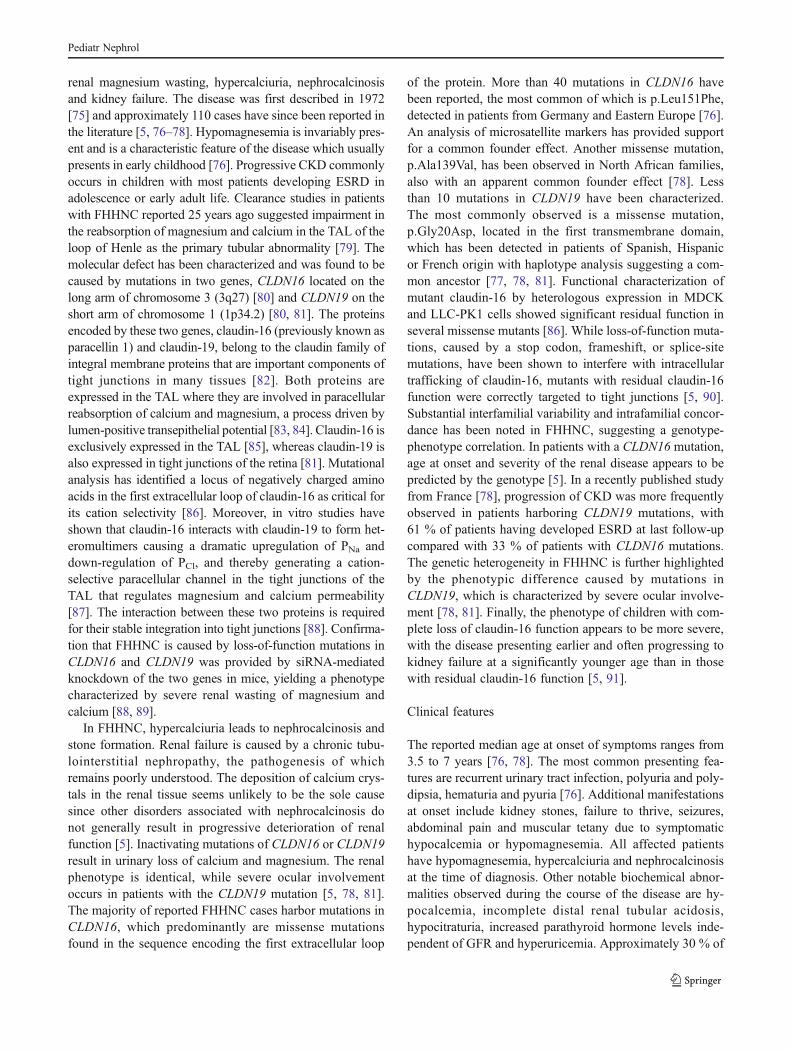
The diagnosis of FHHNC should be considered in all childrenwith nephrocalcinosis, history of kidney stones, and/or re-duced kidney function. The diagnosis is based on the triad ofhypomagnesemia, hypercalciuria and nephrocalcinosis (Fig.6). Hypocalcemia, incomplete distal renal tubular acidosis andhypocitraturia are supportive features [5, 92]. Unfortunately, adelay in the diagnosis is common, reflecting lack of awarenessamong physicians [78]. Levels of serum magnesium and
urinary calcium should be measured in all patients with neph-rocalcinosis and/or CKD. The average serum magnesiumconcentration was 0.4 and 0.5 mmol/l, respectively, in twodifferent reports [76, 78]. High fractional urinary excretion ofmagnesium is found while serum magnesium is inappropri-ately low [93]. Urinary calcium-to-creatinine ratio can be usedto estimate the urinary calcium excretion which is generally inthe range of 0.8–2.5 mmol/mmol (see annotations to Fig. 5)[5, 78]. Renal histologic findings are not specific and includecalcium deposits, glomerulosclerosis, tubular atrophy and in-terstitial fibrosis, consistent with a chronic tubulointerstitialnephropathy. In the differential diagnosis, the presence ofCKD distinguishes FHHNC from other magnesium wastingdisorders. Excluding hypokalemic metabolic alkalosis is im-portant in the diagnostic process to avoid confusion withBartter syndrome. The diagnosis of FHHNC can be confirmedby demonstrating mutations in both copies of CLDN16 orCLDN19.
Diagnosis of Familial Hypomagnesemia with Hypercalciuriaand Nephrocalcinosis1
Chronic kidney disease3
Hypomagnesemia4
FHHNC8
Nephrocalcinosis and/or history of kidney stones 2
Hyperalciuria5
HypocalcemiaIncomplete distal renal tubular acidosisHypermagnesuria6
Hypocitraturia7
Fig. 6 Algorithm for diagnostic evaluation of familial hypomagne-semia with hypercalciuria and nephrocalcinosis (FHHNC). 1Thediagnosis of familial hypomagnesemia with hypercalciuria andnephrocalcinosis is based on the triad of hypomagnesemia, hyper-calciuria and nephrocalcinosis [5]. Tables of normal values shouldbe consulted in the interpretation of any random urine solute-to-creatinine ratios. 2All patients have nephrocalcinosis early in thecourse of the disease and 30 % of patients eventually developkidney stones. 3Progressive chronic kidney disease is apparentduring childhood and adolescence, with half of patients reachingend-stage renal disease at 20 years of age [5]. Renal histologicalfindings are not specific and include calcium deposits, glomerularsclerosis, tubular atrophy and interstitial fibrosis consistent with atubulointerstitial nephropathy. 4The serum magnesium concentrationat presentation has been shown to range from 0.23 to 0.61 mmol/l with a median concentration of 0.40 mmol/l [76]. 5Normal limitsfor urinary calcium excretion are presented above in the annotationsto Fig. 5. 6Hypermagnesuria is defined as the random urinemagnesium-to-creatinine (Mg/Cr) ratio of >95th percentile for age.
Random urine magnesium-to-creatinine (Mg/Cr) ratio by age (119)
Mg/Cr ratioUpper limit of normal
7Lower limits for the random citrate-to-creatinine ratio are presented below.Random urine citrate-to-creatinine (Cit/Cr) ratio by age (120)
Cit/Cr ratioLower limit of normal
8Familial hypomagnesemia with hypercalciuria and nephrocalcinosis iscaused by mutations in the CLDN16 or CLDN19 genes. Genetic testingmay be available at research institutions. Ocular abnormalities are indica-tive of a CLDN19 defect
Pediatr Nephrol
Treatment and outcome
No effective therapeutic interventions for FHHNC are avail-able.Medical management has included administration of mag-nesium supplements in high doses and thiazide diuretics toreduce urinary calcium excretion and the progression of neph-rocalcinosis. Unfortunately, thiazides neither yield ameaningfulreduction in urinary calcium excretion nor delay the progres-sion of CKD [76, 78, 93]. Moreover, thiazides can aggravaterenal magnesiumwasting, often leading to their discontinuation[5, 92, 94]. The serum magnesium level tends to remain lowdespite high-dose magnesium supplementation [78]. Indo-methacin has been used for treatment of FHHNC as it decreasessodium reabsorption in the proximal tubule and, thus, may theo-retically increase the paracellular reabsorption of calcium. Limitedexperience suggests that indomethacin does not affect the serummagnesium concentration or urinary calcium excretion [78].
Therapies aimed at delaying the progression of CKDshould be provided as well as conventional managementstrategies for kidney stones. Renal transplantation is the opti-mal treatment for ESRD in most cases and corrects the defectin renal handling of magnesium and calcium [76, 78, 93].
Primary hyperoxaluria
Background
The primary hyperoxalurias are inherited disorders of metab-olism in which hepatic enzyme deficiencies result in overpro-duction of oxalate. The excess oxalate cannot be degraded inhumans and is excreted largely by the kidneys, resulting inhigh urinary concentrations. Calcium oxalate crystals form inrenal tubules acting as a nidus for crystal aggregation andgrowth, thus initiating stone formation. Crystals attach to renaltubular epithelial cells causing degenerative change and necro-sis and they also can undergo transcytosis into the interstitiumof the kidney where they incite an inflammatory reaction withresulting renal injury and scarring [95, 96]. Health consequen-ces include frequent formation of calcium oxalate kidneystones as well as renal injury that leads to kidney failure in asignificant proportion of affected patients [6].
Primary hyperoxaluria is worldwide in its distribution.The incidence and prevalence are unknown, though surveysof physicians in central Europe suggest an incidence of1:120,000 live births and prevalence of 1–3 per millionpopulation [97, 98]. The prevalence of PH appears to behigher in certain regions including the Canary Islands, Tuni-sia, and parts of the Middle East [99].
Three types of PH have been described to date, eachinvolving a different enzyme of the oxalate metabolic path-ways. Type 1 PH (PH1, OMIM 259900), due to mutations inAGXT is found in approximately 80 % of PH patients. When
the activity of alanine-glyoxylate aminotransferase (AGT) isreduced or absent, glyoxylate accumulates and is shunted tooxalate and glycolate. For effective disposition of glyoxy-late, AGT must be present in the peroxisome of the hepato-cyte. Mistargeting of AGT to mitochondria, as results fromcertain mutations in AGXT, also results in hyperoxaluriaeven though enzyme activity is measurable [100].
Type 2 PH (PH2, OMIM 260000) which accounts forapproximately 10 % of patients with known types of PH, iscaused by mutations in GRHPR, and results from deficiencyor absence of the enzyme glyoxylate reductase/hydroxy pyru-vate reductase (GRHPR). Increased glycerate in the urinealong with hyperoxaluria is indicative of type 2 disease[101]. A third type of PH (PH3, OMIM 613616) was recentlyshown to be due to mutations of the HOGA1 (formerlyDHDPSL) [102]. A founder mutation was detected amongAshkenazi-Jewish patients [102], though PH type 3 has beenconfirmed in other populations [103]. Based on what is knownof the gene product, PH type 3 is thought to be due to abnor-malities of the enzyme 4-hydroxy-2-oxaloglutarate aldolasethat is found in hepatic mitochondria [102, 104]. Data fromthe RKSC PHRegistry suggests that PH3 accounts for approx-imately 10 % of patients with PH of known type. In addition,there are patients who have marked hyperoxaluria and clinicalmanifestations indistinguishable from those of PH, but whohave no demonstrable abnormalities of the enzymes or thegenes implicated in types 1, 2, and 3 PH. It is expected thatadditional PH types will be identified.
PH1 is an autosomal recessive disease. To date, morethan 140 mutations of AGXT have been implicated(www.hgmd.cf.ac.uk). Since patients with mistargeting ofenzyme to mitochondria have been shown to respond totreatment with a pharmacologic dose of pyridoxine (vitaminB6), identification of the genotype in individual patients hasimportant implications for clinical management. Urine oxa-late excretion rates in patients homozygous for mutationscausing mistargeted AGT can correct into the normal rangeduring pyridoxine treatment, while those heterozygous forthese mutations demonstrate partial correction [105]. One ofthe mutations responsible for mistargeting, p.Gly170Arg, isthe most common mutation in North American and North-ern European patients with PH1 [106].
PH2 is autosomal recessive. Though in PH3 autosomalrecessive inheritance is suggested by family studies, therealso appear to be variable and intermittent elevations ofurine oxalate in some heterozygotes [103].
Clinical features
Most patients present with signs or symptoms related to kidneystones. Among 300 patients in the RKSC PH Registry medianage at onset of symptoms was 5 years. Symptoms were presentin 65 % of patients with PH before 10 years of age and in 85 %
Pediatr Nephrol
before 20 years of age. A small minority of patients initiallypresent with failure to thrive, nausea, pallor or fatigue of ESRDwith no history of stones. Such patients may have diffuse calci-fication of kidney tissue visible by ultrasound or other imagingstudies (nephrocalcinosis) without discrete stones. Extensivecalcium oxalate crystal deposition is observed on renal biopsy.This mode of presentation is seen especially in infants. Irrevers-ible kidney failure can be evident as early as 4 months of age.
Recurring stones throughout childhood, adolescence, andadulthood are characteristic. Nephrocalcinosis may also beobserved. Growth and development are normal unless com-promised by CKD or other unrelated medical problems. Overtime, however, progressive renal damage leads to reducedkidney function. Effects on kidney function vary by type, withPH1 the most severe. Data from the RKSC PH Registry inpatients with PH1 showed that median age at progression tokidney failure was 33 years [107]. Recent analysis of a largerRKSC cohort of 222 PH1 patients by the samemethod showeda renal survival of 89% at 10 years of age and 75% at 20 yearsof age (unpublished data). There was no change in the medianage at kidney failure in the larger cohort. Data from theOxalEurope registry [108] show similar findings. Whetherthese findings hold true in other populations remains to beestablished. Observations in Israeli patients with PH type 1suggest early onset of symptoms with progression to kidneyfailure within the first decade of life as a prevalent phenotype[109]. Patients with PH2 and PH3 appear to have betterkidney outcomes. In the RKSC PH Registry, preservation ofrenal function has been found in 100 % of PH2 and PH3patients at 20 years of age. However, renal outcome in PH2and PH3 must be interpreted with caution due to the smallnumber of patients studied.
Diagnosis
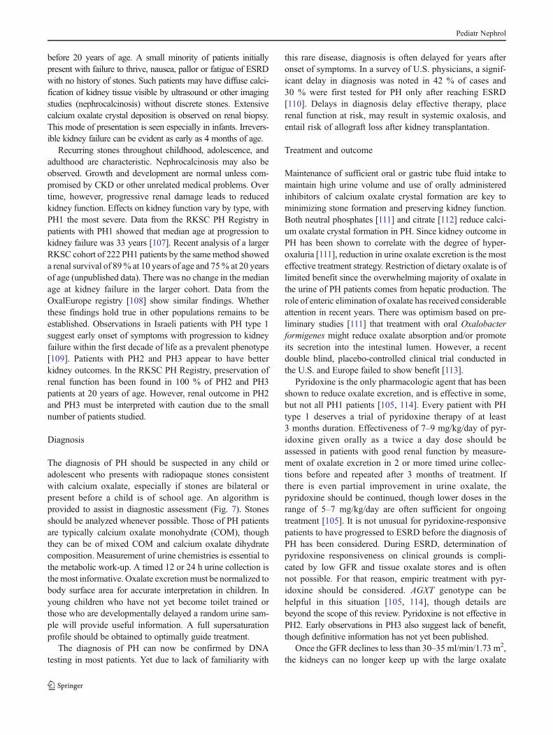
The diagnosis of PH should be suspected in any child oradolescent who presents with radiopaque stones consistentwith calcium oxalate, especially if stones are bilateral orpresent before a child is of school age. An algorithm isprovided to assist in diagnostic assessment (Fig. 7). Stonesshould be analyzed whenever possible. Those of PH patientsare typically calcium oxalate monohydrate (COM), thoughthey can be of mixed COM and calcium oxalate dihydratecomposition. Measurement of urine chemistries is essential tothe metabolic work-up. A timed 12 or 24 h urine collection isthe most informative. Oxalate excretionmust be normalized tobody surface area for accurate interpretation in children. Inyoung children who have not yet become toilet trained orthose who are developmentally delayed a random urine sam-ple will provide useful information. A full supersaturationprofile should be obtained to optimally guide treatment.
The diagnosis of PH can now be confirmed by DNAtesting in most patients. Yet due to lack of familiarity with
this rare disease, diagnosis is often delayed for years afteronset of symptoms. In a survey of U.S. physicians, a signif-icant delay in diagnosis was noted in 42 % of cases and30 % were first tested for PH only after reaching ESRD[110]. Delays in diagnosis delay effective therapy, placerenal function at risk, may result in systemic oxalosis, andentail risk of allograft loss after kidney transplantation.
Treatment and outcome
Maintenance of sufficient oral or gastric tube fluid intake tomaintain high urine volume and use of orally administeredinhibitors of calcium oxalate crystal formation are key tominimizing stone formation and preserving kidney function.Both neutral phosphates [111] and citrate [112] reduce calci-um oxalate crystal formation in PH. Since kidney outcome inPH has been shown to correlate with the degree of hyper-oxaluria [111], reduction in urine oxalate excretion is the mosteffective treatment strategy. Restriction of dietary oxalate is oflimited benefit since the overwhelming majority of oxalate inthe urine of PH patients comes from hepatic production. Therole of enteric elimination of oxalate has received considerableattention in recent years. There was optimism based on pre-liminary studies [111] that treatment with oral Oxalobacterformigenes might reduce oxalate absorption and/or promoteits secretion into the intestinal lumen. However, a recentdouble blind, placebo-controlled clinical trial conducted inthe U.S. and Europe failed to show benefit [113].
Pyridoxine is the only pharmacologic agent that has beenshown to reduce oxalate excretion, and is effective in some,but not all PH1 patients [105, 114]. Every patient with PHtype 1 deserves a trial of pyridoxine therapy of at least3 months duration. Effectiveness of 7–9 mg/kg/day of pyr-idoxine given orally as a twice a day dose should beassessed in patients with good renal function by measure-ment of oxalate excretion in 2 or more timed urine collec-tions before and repeated after 3 months of treatment. Ifthere is even partial improvement in urine oxalate, thepyridoxine should be continued, though lower doses in therange of 5–7 mg/kg/day are often sufficient for ongoingtreatment [105]. It is not unusual for pyridoxine-responsivepatients to have progressed to ESRD before the diagnosis ofPH has been considered. During ESRD, determination ofpyridoxine responsiveness on clinical grounds is compli-cated by low GFR and tissue oxalate stores and is oftennot possible. For that reason, empiric treatment with pyr-idoxine should be considered. AGXT genotype can behelpful in this situation [105, 114], though details arebeyond the scope of this review. Pyridoxine is not effective inPH2. Early observations in PH3 also suggest lack of benefit,though definitive information has not yet been published.
Once the GFR declines to less than 30–35 ml/min/1.73 m2,the kidneys can no longer keep up with the large oxalate
Pediatr Nephrol
production and plasma levels begin to increase rapidly. Whenthe threshold for supersaturation in plasma is exceeded, calci-um oxalate crystals form in many organs. Systemic oxalosiscan result in refractory anemia, osteodystrophy, cardiacarrhythmias, oxalate cardiomyopathy, painful ischemic ulcers,and other manifestations eventually resulting in death. Inpatients with stages IV or V CKD minimizing systemic oxa-losis is also critically important for a favorable outcome of
subsequent kidney transplantation [6, 98]. For most patients,hemodialysis 6 or 7 days per week is required for oxalateremoval and still may not keep pace with oxalate production.Peritoneal dialysis alone is insufficient though it may becombined with daily hemodialysis for better oxalate removal[115]. Patients who are fully pyridoxine responsive and haveno interruptions in pyridoxine administration may require lessintensive dialysis. Since systemic oxalate poses high risk for a
Diagnosis of Primary Hyperoxaluria
• Stones or nephrocalcinosis in childhood• Recurrent CaOx stones or nephrocalcinosis in adults• Family history of primary hyperoxaluria2
•• CaOx tissue deposits• Nephrocalcinosis
Or
Normal Renal Function Chronic Kidney Disease1
Urine oxalateUOx >0.7 mmol/1.73 m2/24 h or UOx/Cr > normal for age3
Confirmed on repeat testing
Urine and plasma oxalateUOx >0.50 mmol/1.73 m2/24 h
and/or plasma Ox >20 μmol/LUrine glycolate, L-glycerate4,5
Glycolate or glycerate > 2x normal
Secondary cause identified?• Malabsorption• Gastrointestinal disease• Very high Ox, low Ca diet• Infant with prematurity6
Genetic testingHomozygosity or compound heterozygosity for known mutations of AGXT, GRHPR, or HOGA18
Liver biopsy for AGT, GRHPR enzyme activities
PH1 PH2 PH3Oxalate disorder of undetermined type
Normal AGTand GRHPR activity
YesNo
Yes
AGT activity GRHPRactivity
AGXT mutations GRHPR mutations HOGA1 mutations
Evaluate and treataccordingly7
NoYes
No
Serum creatinine with CaOx stones
Fig. 7 Algorithm for diagnostic evaluation of primary hyperoxaluria.1Chronic kidney disease is defined as a glomerular filtration rate of lessthan 50 ml/min/1.73 m2, or serum creatinine that is greater than orequal to two times normal for age. 2The guideline does not addressprenatal diagnosis [121, 122]. 3Urine oxalate-to-creatinine (Ox/Cr)ratios in healthy children vary continuously by age. Tables of normalvalues should be consulted in interpretation of any random urine Ox/Crratio.
Random urine oxalate-to-creatinine (Ox/Cr) ratio by age (123-126)
Little data is available to guide interpretation of random urine Ox/Crratio in adolescents and adults. Upper limits of normal ratios decliningto 0.04 by age 18–20 years and then remaining stable through adultage are suggested by available literature [125, 127]. In patients of allages, confirmation of hyperoxaluria by a 24 h urine collection with
normalization of the oxalate excretion rate to 1.73 m2 body surfacearea, is strongly recommended. From 2 years of age through adult-hood, normal urine oxalate is constant at <0.45 mmol/1.73 m2/24 h[125]. 4Urine and plasma oxalate and urine glycolate measurementsfor diagnostic testing should be obtained while the patient is notreceiving pyridoxine or vitamin supplements. 5Increased urine gly-colate in the presence of hyperoxaluria is suggestive, but not diag-nostic of primary hyperoxaluria (PH) type 1. Increased urine L-glycerate in a hyperoxaluric patient suggests PH type 2. 6UrineOx/Cr ratios are higher in very premature infants than in terminfants, especially when they are receiving parenteral nutrition con-taining amino acids. The ratio falls when premature infants arereceiving only glucose and electrolyte solutions [128]. 7Whenvery high oxalate or low dietary calcium is suspected as the causeof the hyperoxaluria, the diet should be corrected and the urineoxalate remeasured for verification. 8In some cases with firmclinical diagnosis, only one mutation is found even after analysisfor large rearrangements, suggesting that regulatory or deepintronic mutations may be the second, undetected mutation. Insuch cases, the finding of a single disease-associated mutation inthe context of a typical phenotype supports the clinical diagnosisof PH
Pediatr Nephrol
transplanted kidney, the shortest time possible between start ofdialysis and kidney transplantation should be the goal.
With intensive dialysis, shortened times to kidney trans-plantation, and careful post-transplant management, trans-plantation outcomes in PH patients have improved over timeand are now similar to those for patients with other forms ofkidney disease [116, 117]. Most patients with PH type 1should undergo combined kidney and liver transplantationdue to the risk to the transplanted kidney of continued highoxalate production. Patients who are fully pyridoxine re-sponsive, in whom urine oxalate can be normalized byadministration of pharmacologic doses of pyridoxine, maybe candidates for kidney transplantation alone. It is impor-tant to recognize that marked hyperoxaluria due to gradualmobilization of tissue oxalate stores poses a risk to allPH patients undergoing kidney transplantation, includingthose who undergo liver transplantation. Hyperoxaluriaoften persists for years following successful transplantation[116]. In order to protect the transplanted kidney fromoxalate-induced damage, consistent attention for mainte-nance of high urine volume and use of crystal inhibitor med-ications until urine oxalate returns to normal are importantfactors.
Kidney transplantation only is the current recommenda-tion for PH2 patients who progress to ESRD. Unlike AGT1which is specific to the liver, the enzyme GRHPR is presentin multiple tissues and there have been no reports to date ofcorrection of the metabolic defect in type 2 disease by livertransplantation. No patients with ESRD due to PH type 3have yet been reported. Given uncertainties regarding themetabolic pathway involved, liver transplantation cannot atthis time be recommended for patients with PH type 3.
Summary and future directions
Early diagnosis of APRT deficiency, cystinuria, Dent dis-ease, FHHNC and PH is very important as adverse out-comes can often be ameliorated or even prevented withprompt diagnosis and treatment. However, lack of familiar-ity with these diseases among clinicians and pathologistsremains a major problem and strategies to improve out-comes are much needed. Ready availability of diagnosticalgorithms and their use in routine clinical care of childrenwith kidney stone disease may be of value.
To facilitate early recognition and treatment of rarecauses of kidney stones and CKD and to advance sci-entific knowledge in this field, the authors of this paperrecently formed the RKSC (rarekidneystones.org andrarediseases network.epi.usf.edu/RKSC/index.htm) withsupport from the National Institute of Diabetes andDigestive and Kidney Diseases and The Office of RareDiseases Research at the National Institutes of Health.
This approach is analogous to current strategies in otherrare diseases and has been supported by healthcareauthorities both in Europe and the US.
To increase awareness of the five disorders and furtherenhance the educational mission of this project, collaborationhas been established with international patient support groups,including the Oxalosis and Hyperoxaluria Foundation(ohf.org), International Cystinuria Foundation (cystinuria.org),Cystinuria Support Network (cystinuria.com), and Lowe Syn-drome Association (http://www.lowesyndrome.org/). TheAPRT Deficiency Patient Support Network (aprtd.org) wasrecently founded. Partnership with advocacy groups will allowrapid dissemination of relevant information and engagepatients, their families, and local physicians in learning moreabout the diseases. Finally, we believe that exchange of infor-mation between investigators, clinicians, and patients willimprove the lives of patients with rare causes of kidney stonedisease through better care and outcomes.
Acknowledgements The authors gratefully acknowledge the supportof the Rare Kidney Stone Consortium (U54KD083908), a part of theNational Institutes of Health (NIH) Rare Diseases Clinical ResearchNetwork (RDCRN), funded by the NIDDK, and the NIH Office ofRare Diseases Research (ORDR) and the Mayo Clinic O’Brien Urol-ogy Research Center (P50 DK083007) funded by the NIDDK. Wethank Rachel Miller of the Mayo Clinic Renal Function Laboratory forphotomicrographs of cystine, calcium oxalate, and calcium phosphatecrystals, and Hrafnhildur L. Runolfsdottir, a medical student at theUniversity of Iceland, Reykjavik, Iceland, for generating the imagesof urinary DHA crystals. The authors also thank members of the RareKidney Stone Consortium for critical feedback during construction ofthe diagnostic algorithms (Figs. 3, 4, 5, 6 and 7).
Conflict of interest statement Dr. Goldfarb is a consultant forTakeda.
References
1. Sas DJ (2011) An update on the changing epidemiology andmetabolic risk factors in pediatric kidney stone disease. Clin JAm Soc Nephrol 6:2062–2068
2. Stamatelou KK, Francis ME, Jones CA, Nyberg LM, Curhan GC(2003) Time trends in reported prevalence of kidney stones in theUnited States: 1976–1994. Kidney Int 63:1817–1823
3. Beara-Lasic L, Edvardsson V, Palsson R, Lieske J, Goldfarb D,Milliner D (2011) Genetic causes of kidney stones and kidneyfailure. Clin Rev Bone Miner Metab 10(6):1–18
4. Greenwood MC, Dillon MJ, Simmonds HA, Barratt TM, PincottJR, Metreweli C (1982) Renal failure due to 2,8-dihydroxyade-nine urolithiasis. Eur J Pediatr 138:346–349
5. Konrad M, Hou J, Weber S, Dotsch J, Kari JA, Seeman T,Kuwertz-Broking E, Peco-Antic A, Tasic V, Dittrich K, AlshayaHO, von Vigier RO, Gallati S, Goodenough DA, Schaller A(2008) CLDN16 genotype predicts renal decline in familial hy-pomagnesemia with hypercalciuria and nephrocalcinosis. JAm Soc Nephrol 19:171–181
Pediatr Nephrol
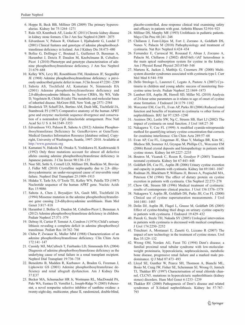
6. Hoppe B, Beck BB, Milliner DS (2009) The primary hyperox-alurias. Kidney Int 75:1264–1271
7. Rule AD, Krambeck AE, Lieske JC (2011) Chronic kidney diseasein kidney stone formers. Clin J Am Soc Nephrol 6:2069– 2075
8. Edvardsson V, Palsson R, Olafsson I, Hjaltadottir G, Laxdal T(2001) Clinical features and genotype of adenine phosphoribosyl-transferase deficiency in Iceland. Am J Kidney Dis 38:473–480
9. Bollee G, Dollinger C, Boutaud L, Guillemot D, Bensman A,Harambat J, Deteix P, Daudon M, Knebelmann B, Ceballos-Picot I (2010) Phenotype and genotype characterization of ade-nine phosphoribosyltransferase deficiency. J Am Soc Nephrol21:679–688
10. Kelley WN, Levy RI, Rosenbloom FM, Henderson JF, SeegmillerJE (1968) Adenine phosphoribosyltransferase deficiency: a previ-ously undescribed genetic defect in man. J Clin Invest 47:2281–2289
11. Sahota AS, Tischfield AJ, Kamatani N, Simmonds HA(2001) Adenine phosphoribosyltransferase deficiency and2,8-dihydroxyadenine lithiasis. In: Scriver CRBA, Sly WS, ValleD, Vogelstein B, Childs B (eds) The metabolic and molecular basesof inherited disease. McGraw-Hill, New York, pp 2571–2584
12. Broderick TP, Schaff DA, Bertino AM, Dush MK, Tischfield JA,Stambrook PJ (1987) Comparative anatomy of the human APRTgene and enzyme: nucleotide sequence divergence and conserva-tion of a nonrandom CpG dinucleotide arrangement. Proc NatlAcad Sci U S A 84:3349–3353
13. Edvardsson VO, Palsson R, Sahota A (2012) Adenine Phosphor-ibosyltransferase Deficiency In: GeneReviews at GeneTests:Medical Genetics Information Resource [database online]. Copy-right, University of Washington, Seattle, 1997–2010. Available athttp://www.genetests.org
14. Kamatani N, Hakoda M, Otsuka S, Yoshikawa H, Kashiwazaki S(1992) Only three mutations account for almost all defectivealleles causing adenine phosphoribosyltransferase deficiency inJapanese patients. J Clin Invest 90:130–135
15. Nasr SH, Sethi S, Cornell LD, Milliner DS, Boelkins M, BroviacJ, Fidler ME (2010) Crystalline nephropathy due to 2,8- dihy-droxyadeninuria: an under-recognized cause of irreversible renalfailure. Nephrol Dial Transplant 25:1909–1915
16. Hidaka Y, Tarle SA, O’Toole TE, Kelley WN, Palella TD (1987)Nucleotide sequence of the human APRT gene. Nucleic AcidsRes 15:9086
17. Sahota A, Chen J, Boyadjiev SA, Gault MH, Tischfield JA(1994) Missense mutation in the adenine phosphoribosyltransfer-ase gene causing 2,8-dihydroxyadenine urolithiasis. Hum MolGenet 3:817–818
18. Harambat J, Bollee G, Daudon M, Ceballos-Picot I, Bensman A(2012) Adenine phosphoribosyltransferase deficiency in children.Pediatr Nephrol 27:571–579
19. Debray H, Cartier P, Temstet A, Cendron J (1976) Child’s urinarylithiasis revealing a complete deficit in adenine phosphoribosyltransferase. Pediatr Res 10:762–766
20. Chiba P, Zwiauer K, Muller MM (1988) Characterization of anadenine phosphoribosyltransferase deficiency. Clin Chim Acta172:141–147
21. Cassidy MJ, McCulloch T, Fairbanks LD, Simmonds HA (2004)Diagnosis of adenine phosphoribosyltransferase deficiency as theunderlying cause of renal failure in a renal transplant recipient.Nephrol Dial Transplant 19:736–738
22. Benedetto B, Madden R, Kurbanov A, Braden G, Freeman J,Lipkowitz GS (2001) Adenine phosphoribosyltransferase de-ficiency and renal allograft dysfunction. Am J Kidney Dis37:E37
23. Becker MA, Schumacher HR Jr, Wortmann RL, MacDonald PA,Palo WA, Eustace D, Vernillet L, Joseph-Ridge N (2005) Febuxo-stat, a novel nonpurine selective inhibitor of xanthine oxidase: atwenty-eight-day, multicenter, phase II, randomized, double-blind,
placebo-controlled, dose–response clinical trial examining safetyand efficacy in patients with gout. Arthritis Rheum 52:916–923
24. Milliner DS, Murphy ME (1993) Urolithiasis in pediatric patients.Mayo Clin Proc 68:241–248
25. Chillaron J, Font-Llitjos M, Fort J, Zorzano A, Goldfarb DS,Nunes V, Palacin M (2010) Pathophysiology and treatment ofcystinuria. Nat Rev Nephrol 6:424–434
26. Fernandez E, Carrascal M, Rousaud F, Abian J, Zorzano A,Palacin M, Chillaron J (2002) rBAT-b(0,+)AT heterodimer isthe main apical reabsorption system for cystine in the kidney.Am J Physiol Renal Physiol 283:F540–548
27. Martens K, Jaeken J, Matthijs G, Creemers JW (2008) Multi-system disorder syndromes associated with cystinuria type I. CurrMol Med 8:544–550
28. Dello Strologo L, Laurenzi C, Legato A, Pastore A (2007) Cys-tinuria in children and young adults: success of monitoring free-cystine urine levels. Pediatr Nephrol 22:1869–1873
29. Lambert EH, Asplin JR, Herrell SD, Miller NL (2010) Analysisof 24-hour urine parameters as it relates to age of onset of cystinestone formation. J Endourol 24:1179–1182
30. Worcester EM, Coe FL, Evan AP, Parks JH (2006) Reduced renalfunction and benefits of treatment in cystinuria vs other forms ofnephrolithiasis. BJU Int 97:1285–1290
31. Assimos DG, Leslie SW, Ng C, Streem SB, Hart LJ (2002) Theimpact of cystinuria on renal function. J Urol 168:27–30
32. Nakagawa Y, Coe FL (1999) A modified cyanide-nitroprussidemethod for quantifying urinary cystine concentration that correctsfor creatinine interference. Clin Chim Acta 289:57–68
33. Evan AP, Coe FL, Lingeman JE, Shao Y, Matlaga BR, Kim SC,Bledsoe SB, Sommer AJ, Grynpas M, Phillips CL, Worcester EM(2006) Renal crystal deposits and histopathology in patients withcystine stones. Kidney Int 69:2227–2235
34. Boutros M, Vicanek C, Rozen R, Goodyer P (2005) Transientneonatal cystinuria. Kidney Int 67:443–448
35. Goldfarb DS, Coe FL, Asplin JR (2006) Urinary cystine excretionand capacity in patients with cystinuria. Kidney Int 69:1041–1047
36. Rodman JS, Blackburn P, Williams JJ, Brown A, Pospischil MA,Peterson CM (1984) The effect of dietary protein on cystineexcretion in patients with cystinuria. Clin Nephrol 22:273–278
37. Chow GK, Streem SB (1996) Medical treatment of cystinuria:results of contemporary clinical practice. J Urol 156:1576–1578
38. Nakagawa Y, Asplin JR, Goldfarb DS, Parks JH, Coe FL (2000)Clinical use of cystine supersaturation measurements. J Urol164:1481–1485
39. Dolin DJ, Asplin JR, Flagel L, Grasso M, Goldfarb DS (2005)Effect of cystine-binding thiol drugs on urinary cystine capacityin patients with cystinuria. J Endourol 19:429–432
40. Pareek G, Steele TH, Nakada SY (2005) Urological interventionin patients with cystinuria is decreased with medical compliance.J Urol 174:2250–2252
41. Trinchieri A, Montanari E, Zanetti G, Lizzano R (2007) Theimpact of new technology in the treatment of cystine stones. UrolRes 35:129–132
42. Wrong OM, Norden AG, Feest TG (1994) Dent’s disease; afamilial proximal renal tubular syndrome with low-molecular-weight proteinuria, hypercalciuria, nephrocalcinosis, metabolicbone disease, progressive renal failure and a marked male pre-dominance. Q J Med 87:473–493
43. Lloyd SE, Gunther W, Pearce SH, Thomson A, Bianchi ML,Bosio M, Craig IW, Fisher SE, Scheinman SJ, Wrong O, JentschTJ, Thakker RV (1997) Characterisation of renal chloride chan-nel, CLCN5, mutations in hypercalciuric nephrolithiasis (kidneystones) disorders. Hum Mol Genet 6:1233–1239
44. Thakker RV (2000) Pathogenesis of Dent’s disease and relatedsyndromes of X-linked nephrolithiasis. Kidney Int 57:787–793
Pediatr Nephrol
45. Reinhart SC, Norden AG, Lapsley M, Thakker RV, Pang J,Moses AM, Frymoyer PA, Favus MJ, Hoepner JA, ScheinmanSJ (1995) Characterization of carrier females and affected maleswith X-linked recessive nephrolithiasis. J Am Soc Nephrol5:1451–1461
46. Waldegger S, Jentsch TJ (2000) From tonus to tonicity:physiology of CLC chloride channels. J Am Soc Nephrol11:1331–1339
47. Dutzler R (2004) Structural basis for ion conduction and gating inClC chloride channels. FEBS Lett 564:229–233
48. Gunther W, Luchow A, Cluzeaud F, Vandewalle A, Jentsch TJ(1998) ClC-5, the chloride channel mutated in Dent’s disease,colocalizes with the proton pump in endocytotically active kidneycells. Proc Natl Acad Sci U S A 95:8075–8080
49. Devuyst O, Christie PT, Courtoy PJ, Beauwens R, Thakker RV(1999) Intra-renal and subcellular distribution of the human chlo-ride channel, CLC-5, reveals a pathophysiological basis forDent’s disease. Hum Mol Genet 8:247–257
50. Hara-Chikuma M, Wang Y, Guggino SE, Guggino WB, VerkmanAS (2005) Impaired acidification in early endosomes of ClC-5deficient proximal tubule. Biochem Biophys Res Commun329:941–946
51. Picollo A, Pusch M (2005) Chloride/proton antiporter activity ofmammalian CLC proteins ClC-4 and ClC-5. Nature 436:420–423
52. Scheel O, Zdebik AA, Lourdel S, Jentsch TJ (2005) Voltage-dependent electrogenic chloride/proton exchange by endosomalCLC proteins. Nature 436:424–427
53. Novarino G, Weinert S, Rickheit G, Jentsch TJ (2010) Endosomalchloride-proton exchange rather than chloride conductance iscrucial for renal endocytosis. Science 328:1398–1401
54. Friedrich T, Breiderhoff T, Jentsch TJ (1999) Mutational analysisdemonstrates that ClC-4 and ClC-5 directly mediate plasmamembrane currents. J Biol Chem 274:896–902
55. Hryciw DH, Ekberg J, Pollock CA, Poronnik P (2006) ClC-5: achloride channel with multiple roles in renal tubular albuminuptake. Int J Biochem Cell Biol 38:1036–1042
56. Piwon N, Gunther W, Schwake M, Bosl MR, Jentsch TJ (2000)ClC-5 Cl--channel disruption impairs endocytosis in a mousemodel for Dent’s disease. Nature 408:369–373
57. Wang SS, Devuyst O, Courtoy PJ, Wang XT, Wang H, Wang Y,Thakker RV, Guggino S, Guggino WB (2000) Mice lacking renalchloride channel, CLC-5, are a model for Dent’s disease, a neph-rolithiasis disorder associated with defective receptor-mediated en-docytosis. Hum Mol Genet 9:2937–2945
58. Christensen EI, Devuyst O, Dom G, Nielsen R, Van der SmissenP, Verroust P, Leruth M, Guggino WB, Courtoy PJ (2003) Loss ofchloride channel ClC-5 impairs endocytosis by defective traffick-ing of megalin and cubilin in kidney proximal tubules. Proc NatlAcad Sci U S A 100:8472–8477
59. Silva IV, Cebotaru V,WangH,WangXT,Wang SS, GuoG, DevuystO, Thakker RV, Guggino WB, Guggino SE (2003) The ClC-5knockout mouse model of Dent’s disease has renal hypercalciuriaand increased bone turnover. J Bone Miner Res 18:615–623
60. Leheste JR, Melsen F, Wellner M, Jansen P, Schlichting U,Renner-Muller I, Andreassen TT, Wolf E, Bachmann S, NykjaerA, Willnow TE (2003) Hypocalcemia and osteopathy in micewith kidney-specific megalin gene defect. FASEB J 17:247–249
61. Devuyst O, Thakker RV (2010) Dent’s disease. Orphanet J RareDis 5:28
62. Claverie-Martin F, Ramos-Trujillo E, Garcia-Nieto V (2011)Dent’s disease: clinical features and molecular basis. PediatrNephrol 26:693–704
63. Hoopes RR Jr, Raja KM, Koich A, Hueber P, Reid R, Knohl SJ,Scheinman SJ (2004) Evidence for genetic heterogeneity inDent’s disease. Kidney Int 65:1615–1620
64. Hoopes RR Jr, Shrimpton AE, Knohl SJ, Hueber P, Hoppe B,Matyus J, Simckes A, Tasic V, Toenshoff B, Suchy SF, NussbaumRL, Scheinman SJ (2005) Dent Disease with mutations inOCRL1. Am J Hum Genet 76:260–267
65. Ghanekar Y, Lowe M (2005) Signalling for secretion. Nat Cell Biol7:851–853
66. Choudhury R, Diao A, Zhang F, Eisenberg E, Saint-Pol A,Williams C, Konstantakopoulos A, Lucocq J, Johannes L,Rabouille C, Greene LE, Lowe M (2005) Lowe syndrome proteinOCRL1 interacts with clathrin and regulates protein traffickingbetween endosomes and the trans-Golgi network. Mol Biol Cell16:3467–3479
67. Tosetto E, Addis M, Caridi G, Meloni C, Emma F, Vergine G,Stringini G, Papalia T, Barbano G, Ghiggeri GM, Ruggeri L,Miglietti N, D Angelo A, Melis MA, Anglani F (2009) Locusheterogeneity of Dent’s disease: OCRL1 and TMEM27 genesin patients with no CLCN5 mutations. Pediatr Nephrol24:1967–1973
68. Hichri H, Rendu J, Monnier N, Coutton C, Dorseuil O, PoussouRV, Baujat G, Blanchard A, Nobili F, Ranchin B, Remesy M,Salomon R, Satre V, Lunardi J (2011) From Lowe syndrome toDent disease: correlations between mutations of the OCRL1 geneand clinical and biochemical phenotypes. Hum Mutat 32:379–388
69. Copelovitch L, Nash MA, Kaplan BS (2007) Hypothesis: Dentdisease is an underrecognized cause of focal glomerulosclerosis.Clin J Am Soc Nephrol 2:914–918
70. Frishberg Y, Dinour D, Belostotsky R, Becker-Cohen R, Rinat C,Feinstein S, Navon-Elkan P, Ben-Shalom E (2009) Dent’s diseasemanifesting as focal glomerulosclerosis: is it the tip of the ice-berg? Pediatr Nephrol 24:2369–2373
71. Bokenkamp A, Bockenhauer D, Cheong HI, Hoppe B, Tasic V,Unwin R, Ludwig M (2009) Dent-2 disease: a mild variant ofLowe syndrome. J Pediatr 155:94–99
72. Davey PG, Gosling P (1982) Beta 2-microglobulin instability inpathological urine. Clin Chem 28:1330–1333
73. Suzuki S, Suzuki J, Kume K, Yoshida K, Suyama H, Kawasaki Y,Nozawa R, Suzuki H, Fujiki T, Kamiyama S, Suzuki A (1999)Poor renal accumulation of 99mTc-DMSA in idiopathic tubularproteinuria. Nephron 81:49–54
74. Cebotaru V, Kaul S, Devuyst O, Cai H, Racusen L, Guggino WB,Guggino SE (2005) High citrate diet delays progression of renalinsufficiency in the ClC-5 knockout mouse model of Dent’sdisease. Kidney Int 68:642–652
75. Michelis MF, Drash AL, Linarelli LG, De Rubertis FR, Davis BB(1972) Decreased bicarbonate threshold and renal magnesiumwasting in a sibship with distal renal tubular acidosis. (Evaluationof the pathophysiological role of parathyroid hormone). Metabo-lism 21:905–920
76. Weber S, Schneider L, Peters M, Misselwitz J, RonnefarthG, Boswald M, Bonzel KE, Seeman T, Sulakova T, Kuwertz-Broking E, Gregoric A, Palcoux JB, Tasic V, Manz F,Scharer K, Seyberth HW, Konrad M (2001) Novelparacellin-1 mutations in 25 families with familial hypomag-nesemia with hypercalciuria and nephrocalcinosis. J Am SocNephrol 12:1872–1881
77. Faguer S, Chauveau D, Cintas P, Tack I, Cointault O, Rostaing L,Vargas-Poussou R, Ribes D (2011) Renal, ocular, and neuromus-cular involvements in patients with CLDN19 mutations. Clin JAm Soc Nephrol 6:355–360
78. Godron A, Harambat J, Boccio V, Mensire A, May A, Rigothier C,Couzi L, Barrou B, Godin M, Chauveau D, Faguer S, Vallet M,Cochat P, Eckart P, Guest G, Guigonis V, Houillier P, Blanchard A,Jeunemaitre X, Vargas-Poussou R (2012) Familial hypomagnesemiawith hypercalciuria and nephrocalcinosis: phenotype-genotype
Pediatr Nephrol
correlation and outcome in 32 patients with CLDN16 or CLDN19mutations. Clin J Am Soc Nephrol 7:801–809
79. Rodriguez-Soriano J, Vallo A, Garcia-Fuentes M (1987) Hypo-magnesaemia of hereditary renal origin. Pediatr Nephrol 1: 465–472
80. Simon DB, Lu Y, Choate KA, Velazquez H, Al-Sabban E,Praga M, Casari G, Bettinelli A, Colussi G, Rodriguez-Soriano J, McCredie D, Milford D, Sanjad S, Lifton RP(1999) Paracellin-1, a renal tight junction protein requiredfor paracellular Mg2+ resorption. Science 285:103–106
81. Konrad M, Schaller A, Seelow D, Pandey AV, Waldegger S,Lesslauer A, VitzthumH, Suzuki Y, Luk JM, Becker C, SchlingmannKP, SchmidM, Rodriguez-Soriano J, Ariceta G, Cano F, Enriquez R,Juppner H, Bakkaloglu SA, Hediger MA, Gallati S, Neuhauss SC,Nurnberg P, Weber S (2006) Mutations in the tight-junction geneclaudin 19 (CLDN19) are associated with renal magnesium wasting,renal failure, and severe ocular involvement. Am J Hum Genet79:949–957
82. Li J, Ananthapanyasut W, Yu AS (2011) Claudins in renal phys-iology and disease. Pediatr Nephrol 26:2133–2142
83. Blanchard A, Jeunemaitre X, Coudol P, Dechaux M, FroissartM, May A, Demontis R, Fournier A, Paillard M, Houillier P(2001) Paracellin-1 is critical for magnesium and calcium reab-sorption in the human thick ascending limb of Henle. KidneyInt 59:2206–2215
84. Haisch L, Almeida JR, Abreu da Silva PR, Schlingmann KP,Konrad M (2011) The role of tight junctions in paracellular iontransport in the renal tubule: lessons learned from a rare inheritedtubular disorder. Am J Kidney Dis 57:320–330
85. Kiuchi-Saishin Y, Gotoh S, Furuse M, Takasuga A, Tano Y,Tsukita S (2002) Differential expression patterns of claudins,tight junction membrane proteins, in mouse nephron segments.J Am Soc Nephrol 13:875–886
86. Hou J, Paul DL, Goodenough DA (2005) Paracellin-1 and themodulation of ion selectivity of tight junctions. J Cell Sci118:5109–5118
87. Hou J, Renigunta A, Konrad M, Gomes AS, Schneeberger EE,Paul DL, Waldegger S, Goodenough DA (2008) Claudin-16 andclaudin-19 interact and form a cation-selective tight junctioncomplex. J Clin Invest 118:619–628
88. Hou J, Renigunta A, Gomes AS, Hou M, Paul DL, Waldegger S,Goodenough DA (2009) Claudin-16 and claudin-19 interaction isrequired for their assembly into tight junctions and for renalreabsorption of magnesium. Proc Natl Acad Sci U S A106:15350–15355
89. Hou J, Shan Q, Wang T, Gomes AS, Yan Q, Paul DL, Bleich M,Goodenough DA (2007) Transgenic RNAi depletion of claudin-16 and the renal handling of magnesium. J Biol Chem282:17114–17122
90. Kausalya PJ, Amasheh S, Gunzel D, Wurps H, Muller D, FrommM, Hunziker W (2006) Disease-associated mutations affect intra-cellular traffic and paracellular Mg2+ transport function ofClaudin-16. J Clin Invest 116:878–891
91. Hampson G, Konrad MA, Scoble J (2008) Familial hypomagne-saemia with hypercalciuria and nephrocalcinosis (FHHNC): com-pound heterozygous mutation in the claudin 16 (CLDN16) gene.BMC Nephrol 9:12
92. Stechman MJ, Loh NY, Thakker RV (2009) Genetic causes ofhypercalciuric nephrolithiasis. Pediatr Nephrol 24:2321–2332
93. Praga M, Vara J, Gonzalez-Parra E, Andres A, Alamo C, AraqueA, Ortiz A, Rodicio JL (1995) Familial hypomagnesemia withhypercalciuria and nephrocalcinosis. Kidney Int 47:1419–1425
94. Zimmermann B, Plank C, Konrad M, Stohr W, Gravou-Apostolatou C, Rascher W, Dotsch J (2006) Hydrochlorothiazidein CLDN16 mutation. Nephrol Dial Transplant 21:2127–2132
95. Lieske JC, Spargo BH, Toback FG (1992) Endocytosis of calciumoxalate crystals and proliferation of renal tubular epithelial cells in apatient with type 1 primary hyperoxaluria. J Urol 148:1517–1519
96. Muda AO, Barsotti P, Rinaldi S, Rizzoni G, Faraggiana T (1997)Renal pathology in hyperoxaluria. In: Sessa A, Conte F, MeroniM, Battini G (eds) Hereditary kidney diseases. Karger, Basel, pp160–166
97. van Woerden CS, Groothoff JW, Wanders RJ, Davin JC, WijburgFA (2003) Primary hyperoxaluria type 1 in The Netherlands:prevalence and outcome. Nephrol Dial Transplant 18:273–279
98. Cochat P, Liutkus A, Fargue S, Basmaison O, Ranchin B, RollandMO (2006) Primary hyperoxaluria type 1: still challenging!Pediatr Nephrol 21:1075–1081
99. Coulter-Mackie MB (2005) Preliminary evidence for ethnic dif-ferences in primary hyperoxaluria type 1 genotype. Am J Nephrol25:264–268
100. Danpure CJ (2005) Molecular etiology of primary hyperoxaluriatype 1: new directions for treatment. Am J Nephrol 25:303–310
101. Milliner DS (2005) The primary hyperoxalurias: an algorithm fordiagnosis. Am J Nephrol 25:154–160
102. Belostotsky R, Seboun E, Idelson GH, Milliner DS, Becker-CohenR, Rinat C, Monico CG, Feinstein S, Ben-Shalom E, Magen D,Weissman I, Charon C, Frishberg Y (2010) Mutations in DHDPSLare responsible for primary hyperoxaluria type III. Am JHumGenet87:392–399
103. Monico CG, Rossetti S, Belostotsky R, Cogal AG, Herges RM,Seide BM, Olson JB, Bergstrahl EJ, Williams HJ, Haley WE,Frishberg Y, Milliner DS (2011) Primary hyperoxaluria type IIIgene HOGA1 (formerly DHDPSL) as a possible risk factor foridiopathic calcium oxalate urolithiasis. Clin J Am Soc Nephrol6:2289–2295
104. Riedel TJ, Johnson LC, Knight J, Hantgan RR, Holmes RP,Lowther WT (2011) Structural and biochemical studies of human4-hydroxy-2-oxoglutarate aldolase: implications for hydroxypro-line metabolism in primary hyperoxaluria. PLoS One 6:e26021
105. Monico CG, Rossetti S, Olson JB, Milliner DS (2005) Pyridoxineeffect in type I primary hyperoxaluria is associated with the mostcommon mutant allele. Kidney Int 67:1704–1709
106. Monico CG, Rossetti S, Schwanz HA, Olson JB, Lundquist PA,Dawson DB, Harris PC, Milliner DS (2007) Comprehensivemutation screening in 55 probands with type 1 primary hyper-oxaluria shows feasibility of a gene-based diagnosis. J Amer SocNephrol 18:1905–1914
107. Lieske JC, Monico CG, Holmes WS, Bergstralh EJ, Slezak JM,Rohlinger AL, Olson JB, Milliner DS (2005) International regis-try for primary hyperoxaluria. Am J Nephrol 25:290–296
108. Harambat J, Fargue S, Acquaviva C, Gagnadoux MF, Janssen F,Liutkus A, Mourani C, Macher MA, Abramowicz D, Legendre C,Durrbach A, Tsimaratos M, Nivet H, Girardin E, Schott AM,Rolland MO, Cochat P (2010) Genotype-phenotype correlationin primary hyperoxaluria type 1: the p.Gly170Arg AGXT muta-tion is associated with a better outcome. Kidney Int 77:443–449
109. Frishberg Y, Rinat C, Shalata A, Khatib I, Feinstein S, Becker-Cohen R, Weismann I, Wanders RJ, Rumsby G, Roels F, MandelH (2005) Intra-familial clinical heterogeneity: absence ofgenotype-phenotype correlation in primary hyperoxaluria type 1in Israel. Am J Nephrol 25:269–275
110. Hoppe B, Langman CB (2003) A United States survey on diag-nosis, treatment, and outcome of primary hyperoxaluria. PediatrNephrol 18:986–991
111. Milliner D (2006) Treatment of the primary hyperoxalurias: a newchapter. Kidney Int 70:1198–1200
112. Leumann E, Hoppe B, Neuhaus T (1993) Management of prima-ry hyperoxaluria: efficacy of oral citrate administration. PediatrNephrol 7:207–211
Pediatr Nephrol
113. Hoppe B, Groothoff JW, Hulton SA, Cochat P, Niaudet P, KemperMJ, Deschenes G, Unwin R, Milliner D (2011) Efficacy and safetyof Oxalobacter formigenes to reduce urinary oxalate in primaryhyperoxaluria. Nephrol Dial Transplant 26:3609–3615
114. Monico CG, Olson JB, Milliner DS (2005) Implications of geno-type and enzyme phenotype in pyridoxine response of patientswith type I primary hyperoxaluria. Am J Nephrol 25:183– 188
115. Illies F, Bonzel KE, Wingen AM, Latta K, Hoyer PF (2006)Clearance and removal of oxalate in children on intensified dial-ysis for primary hyperoxaluria type 1. Kidney Int 70:1642–1648
116. Bergstralh EJ, Monico CG, Lieske JC, Herges RM, Langman CB,Hoppe B, Milliner DS (2010) Transplantation outcomes in pri-mary hyperoxaluria. Am J Transplant 10:2493–2501
117. Millan MT, Berquist WE, So SK, Sarwal MM, Wayman KI, CoxKL, Filler G, Salvatierra O Jr, Esquivel CO (2003) One hundredpercent patient and kidney allograft survival with simultaneousliver and kidney transplantation in infants with primary hyper-oxaluria: a single-center experience. Transplantation 76:1458–1463
118. SilvaM, Silva CH, Iulek J, ThiemannOH (2004) Three-dimensionalstructure of human adenine phosphoribosyltransferase and itsrelation to DHA-urolithiasis. Biochemistry 43:7663–7671
119. Matos V, van Melle G, Boulat O, Markert M, Bachmann C,Guignard JP (1997) Urinary phosphate/creatinine, calcium/creat-inine, and magnesium/creatinine ratios in a healthy pediatricpopulation. J Pediatr 131:252–257
120. Hoppe B, Kemper MJ (2010) Diagnostic examination of the childwith urolithiasis or nephrocalcinosis. Pediatr Nephrol 25:403–413
121. Rumsby G (1998) Experience in prenatal diagnosis of primaryhyperoxaluria type 1. J Nephrol 11(Suppl 1):13–14
122. Barratt TM, Kasidas GP, Murdoch I, Rose GA (1991) Urinaryoxalate and glycolate excretion and plasma oxalate concentration.Arch Dis Child 66:501–503
123. von Schnakenburg C, Byrd DJ, Latta K, Reusz GS, Graf D,Brodehl J (1994) Determination of oxalate excretion in spoturines of healthy children by ion chromatography. Eur J ClinChem Clin Biochem 32:27–29
124. Gibbs DA, Watts RW (1969) The variation of urinary oxalateexcretion with age. J Lab Clin Med 73:901–908
125. Morgenstern BZ, Milliner DS, Murphy ME, Simmons PS, MoyerTP, Wilson DM, Smith LH (1993) Urinary oxalate and glycolateexcretion patterns in the first year of life: a longitudinal study. JPediatr 123:248–251
126. Campfield T, Braden G (1989) Urinary oxalate excretion by verylow birth weight infants receiving parenteral nutrition. Pediatrics84:860–863
127. Milliner DS, Eickholt JT, Bergstralh EJ,Wilson DM, Smith LH (1994)Results of long-term treatment with orthophosphate and pyridoxine inpatients with primary hyperoxaluria. N Engl J Med 331:1553–1558
128. Morgan SH, Purkiss P, Watts RW, Mansell MA (1987) Oxalatedynamics in chronic renal failure. Comparison with normal subjectsand patients with primary hyperoxaluria. Nephron 46:253–257
Pediatr Nephrol
Related Documents


