2006;66:2000-2011. Cancer Res Rosamonde E. Banks, Prasanna Tirukonda, Claire Taylor, et al. Sporadic Renal Cancer Gene Alterations and Relationship with Clinical Variables in ) VHL Genetic and Epigenetic Analysis of von Hippel-Lindau ( Updated version http://cancerres.aacrjournals.org/content/66/4/2000 Access the most recent version of this article at: Material Supplementary http://cancerres.aacrjournals.org/content/suppl/2006/02/15/66.4.2000.DC1.html Access the most recent supplemental material at: Cited Articles http://cancerres.aacrjournals.org/content/66/4/2000.full.html#ref-list-1 This article cites by 50 articles, 14 of which you can access for free at: Citing articles http://cancerres.aacrjournals.org/content/66/4/2000.full.html#related-urls This article has been cited by 27 HighWire-hosted articles. Access the articles at: E-mail alerts related to this article or journal. Sign up to receive free email-alerts Subscriptions Reprints and . [email protected] Department at To order reprints of this article or to subscribe to the journal, contact the AACR Publications Permissions . [email protected] Department at To request permission to re-use all or part of this article, contact the AACR Publications Cancer Research. on September 16, 2014. © 2006 American Association for cancerres.aacrjournals.org Downloaded from Cancer Research. on September 16, 2014. © 2006 American Association for cancerres.aacrjournals.org Downloaded from

Welcome message from author
This document is posted to help you gain knowledge. Please leave a comment to let me know what you think about it! Share it to your friends and learn new things together.
Transcript
2006;66:2000-2011. Cancer Res Rosamonde E. Banks, Prasanna Tirukonda, Claire Taylor, et al. Sporadic Renal CancerGene Alterations and Relationship with Clinical Variables in
)VHLGenetic and Epigenetic Analysis of von Hippel-Lindau (
Updated version
http://cancerres.aacrjournals.org/content/66/4/2000
Access the most recent version of this article at:
Material
Supplementary
http://cancerres.aacrjournals.org/content/suppl/2006/02/15/66.4.2000.DC1.html
Access the most recent supplemental material at:
Cited Articles
http://cancerres.aacrjournals.org/content/66/4/2000.full.html#ref-list-1
This article cites by 50 articles, 14 of which you can access for free at:
Citing articles
http://cancerres.aacrjournals.org/content/66/4/2000.full.html#related-urls
This article has been cited by 27 HighWire-hosted articles. Access the articles at:
E-mail alerts related to this article or journal.Sign up to receive free email-alerts
Subscriptions
Reprints and
To order reprints of this article or to subscribe to the journal, contact the AACR Publications
Permissions
To request permission to re-use all or part of this article, contact the AACR Publications
Cancer Research. on September 16, 2014. © 2006 American Association forcancerres.aacrjournals.org Downloaded from
Cancer Research. on September 16, 2014. © 2006 American Association forcancerres.aacrjournals.org Downloaded from
Genetic and Epigenetic Analysis of von Hippel-Lindau (VHL)
Gene Alterations and Relationship with Clinical Variables
in Sporadic Renal Cancer
Rosamonde E. Banks,1Prasanna Tirukonda,
2Claire Taylor,
3Nick Hornigold,
1Dewi Astuti,
5
Dena Cohen,4Eamonn R. Maher,
5Anthea J. Stanley,
1Patricia Harnden,
1Adrian Joyce,
2
Margaret Knowles,1and Peter J. Selby
1
1Cancer Research UK Clinical Centre, 2Department of Urology, and 3Cancer Research UK Mutation Detection Facility, St. James’s UniversityHospital; 4Clinical Trials Research Unit, University of Leeds, Springfield Mount, Leeds; and 5Cancer Research UK Renal MolecularOncology Group, University of Birmingham, Edgbaston, United Kingdom
Abstract
Genetic and epigenetic changes in the von Hippel-Lindau(VHL) tumor suppressor gene are common in sporadicconventional renal cell carcinoma (cRCC). Further insightinto the clinical significance of these changes may lead toincreased biological understanding and identification ofsubgroups of patients differing prognostically or who maybenefit from specific targeted treatments. We have compre-hensively examined the VHL status in tissue samples from 115patients undergoing nephrectomy, including 96 with sporadiccRCC. In patients with cRCC, loss of heterozygosity was foundin 78.4%, mutation in 71%, and promoter methylation in 20.4%of samples. Multiplex ligation–dependent probe amplificationidentified intragenic copy number changes in several samplesincluding two which were otherwise thought to be VHL-noninvolved. Overall, evidence of biallelic inactivation wasfound in 74.2% of patients with cRCC. Many of the mutationswere novel and approximately two-thirds were potentiallytruncating. Examination of these and other published findingsconfirmed mutation hotspots affecting codons 117 and 164,and revealed a common region of mutation in codons 60 to 78.Gender-specific differences in methylation and mutation wereseen, although not quite achieving statistical significance(P = 0.068 and 0.11), and a possible association betweenmethylation and polymorphism was identified. No significantdifferences were seen between VHL subgroups with regard toclinicopathologic features including stage, grade, tumor size,cancer-free and overall survival, with the exception of asignificant association between loss of heterozygosity andgrade, although a possible trend for survival differencesbased on mutation location was apparent. (Cancer Res 2006;66(4): 2000-11)
Introduction
Almost 210,000 cases of renal cancer are diagnosed annuallyworldwide (GLOBOCAN 2002).6 The most common subtype ofrenal parenchymal carcinoma is the conventional (clear cell) type(cRCC) accounting for f75% of cases. The most frequent
karyotypic change in cRCC is loss of 3p with several candidategenes implicated, in particular, the von Hippel-Lindau (VHL) tumorsuppressor gene at 3p25. The VHL gene was identified in 1993 fromfamilial studies of VHL disease which predisposes to tumors of thecentral nervous system, adrenal gland, kidney, and eye (1), and isnow known to be involved in 50% to 75% of sporadic cRCC cases(2, 3). In the familial cases, germ line mutations are followed bymutation, methylation, or loss of the remaining wild-type VHLallele in the tumor, and in sporadic cases, the biallelic loss offunction occurs through a combination of somatic allele loss,mutation, and/or methylation (2, 3).The gene consists of three exons with two translation initiation
sites resulting in two protein isoforms of 160 and 213 amino acids,pVHL19 and pVHL30, both seeming to have tumor suppressoractivity. Consisting of a h-sheet domain and a smaller a-helicaldomain held together by two linkers and a polar interface, VHLnormally functions as the substrate recognition subunit of amultiprotein E3 ubiquitin ligase complex involving elongins B andC, Cul-2, and Rbx1 with members of the hypoxia-inducible factor(HIF-a) family of transcription factors being principal targets forubiquitination and subsequent proteasomal degradation. Inhypoxic conditions or in the absence of functional VHL, HIFaccumulates, up-regulating many genes involved in angiogenesis,cellular metabolism, and cell growth (reviewed in refs. 2, 3).However, VHL has also been implicated in a variety of othercellular processes including cell cycle regulation, extracellularmatrix assembly, and cytoskeleton stability and there is evidencethat some VHL activities are HIF-independent.In familial disease, genotype-phenotype correlations exist (2, 3).
Type 1 disease has a low risk of phaeochromocytoma and germ linemutations are often large deletions or truncating mutations. Type 2has a high risk of phaeochromocytoma and is subdivided into highrisk (2B), low risk (2A), or absence (2C) of cRCC, with germ linemissense mutations more commonly seen. Defective regulation ofHIF is found in all types except type 2C. The mechanism underlyingthe tissue-specific influences of the germ line mutations may reflectunknown tissue-specific functions of VHL driven by differentregions of the protein. Recently, increased risk of renal involvementhas been associated with germ line mutations leading totruncations (4) and specific missense mutations affecting proteinstructural integrity (4, 5).In sporadic cRCC, studies have analyzed VHL mutation and
methylation (6–34) but few have examined the clinical relevance of
Note: Supplementary data for this article are available at Cancer Research Online(http://cancerres.aacrjournals.org/).
Requests for reprints: Rosamonde E. Banks, Cancer Research UK Clinical Centre,St. James’s University Hospital, Beckett Street, Leeds LS9 7TF, United Kingdom. Phone:44-113-206-4927; Fax: 44-113-242-9886; E-mail: [email protected].
I2006 American Association for Cancer Research.doi:10.1158/0008-5472.CAN-05-3074 6 http://www-dep.iarc.fr/.
Cancer Res 2006; 66: (4). February 15, 2006 2000 www.aacrjournals.org
Research Article
Cancer Research. on September 16, 2014. © 2006 American Association forcancerres.aacrjournals.org Downloaded from
these alterations. However, such information may be of criticalimportance in terms of prognosis and for developing therapiestargeting specific downstream pathways in patient subgroups (2).Several studies have found no differences in mutation frequency inrelation to tumor grade, stage or tumor size (12, 24, 25, 34), ormicrovessel density and tumor cell proliferation (25), although anincreased rate of mutation or methylation in pT3 tumors has beendescribed (16). Poorer overall and progression-free survival havebeen reported in patients with ‘‘loss of function’’ (LOF) mutations(25, 32), although a much larger study found VHL mutation ormethylation to be strongly associated with better prognosis instage I to III patients but not stage IV patients (35).The need for further large comprehensive clinical studies of
sporadic RCC and VHL status is clear. We have analyzed renaltissue samples from 115 patients including 107 with sporadic RCC,with mutation analysis, loss of heterozygosity (LOH) analysis,promoter-specific methylation, and multiplex ligation–dependentprobe amplification (MLPA) to detect intragenic deletions.Findings have been related to clinicopathologic features such astumor grade, size, stage, as well as survival. The results have alsobeen analyzed in the context of a comprehensive review of existingpublished7 results to highlight potential areas of interest.
Materials and Methods
Patient samples. A total of 117 renal samples from 115 previously
untreated patients undergoing nephrectomy (two bilateral) were analyzedfollowing informed consent. The samples were selected from a total of 155
samples banked during October 1998 to November 2001, to include all the
cRCC cases (n = 96; Table 1) and examples of other subgroups [RCC: seven
papillary, one chromophobe, one collecting duct, two unclassified (mixed),and two familial VHL, three TCC renal pelvis, one metanephric nephroma,
one oncocytoma, and one retention cyst]. For the cRCC cases, follow-up
time ranged from 36 to 70 months with a median relapse-free survival of
40.3 months and cancer-specific survival of 40.4 months. Samples of tumorfrom each patient were snap-frozen and stored in liquid nitrogen. For
genomic DNA, the buffy coats from venous blood samples (EDTA) were
stored at �80jC. Frozen tissue sections (50 � 20 Am thickness) or theequivalent of 200 AL of frozen buffy coat were placed in microcentrifuge
tubes and DNA extracted using QIAamp DNA Mini kit (Qiagen, Crawley,
United Kingdom) and quantified using the PicoGreen dsDNA kit (Molecular
Probes, Leiden, Netherlands).Mutation detection using denaturing high-pressure liquid chroma-
tography and DNA sequencing. Primers were designed using the Primer3
software (Whitehead Research Institute, Cambridge, MA) with two
overlapping primer pairs (1a and 1b) used to amplify the promoter regionand exon 1 with a further pair (1a/b) used for some sequencing, and one
pair for each of exons 2 and 3 (Supplementary Table S1).
For denaturing high-pressure liquid chromatography (DHPLC) screen-
ing, PCR products (3-10 AL) from the renal tissue samples were denaturedat 95jC for 5 minutes and allowed to cool to 65jC for the formation of
homo- and heteroduplexes. DHPLC was carried out using a Transgenomic
WAVE HPLC and DNasep column with (A) 0.1 mol/L triethylammoniumacetate/0.1 mmol/L EDTA and (B) 0.1 mol/L triethylammonium acetate/
0.1 mmol/L EDTA/25% v/v acetonitrile (Transgenomic, Elancourt, France).
Analysis was carried out at a flow rate of 0.9 mL/min and gradient
increase of buffer B of 2%/min for 4 minutes with start and endconcentrations of buffer B being determined empirically for each
fragment. Elution of DNA from the column was determined by absorbance
at 260 nm. The optimum temperature for mutation detection for each
fragment is f1jC below Tm and was determined empirically for eachfragment. Samples were analyzed both alone and with the addition of 50%
wild-type reference DNA to ensure the detection of homozygous and
heterozygous mutations.
DNA sequencing of tissue samples found to be positive by DHPLC, andcorresponding buffy coat genomic DNA samples was carried out following
treatment of the PCR products (5 AL) for 30 minutes at 37jC with shrimp
alkaline phosphatase (2 AL of 1 unit/AL; USB-GE Healthcare-Biosciences,
Amersham, United Kingdom), and exonuclease I (1 AL of 10 units/AL; USB),with subsequent inactivation at 80jC for 15 minutes. Sequencing was carried
out in 10 AL reactions using 3 AL of the purified PCR products, 1.6 pmol of
the appropriate forward or reverse primer and the BigDye (v1.1) Terminatorkit (Applied Biosystems, Warrington, United Kingdom) according to the
manufacturer’s protocol. Reactions were carried out for 25 cycles using a
GeneAmp 9700 Thermal and sequencing was carried out by fluorescence
capillary electrophoresis using an ABI PRISM 3100 Genetic Analyzer withdenaturing POP-6 polymer/Tris-TAPS-EDTA buffer (Applied Biosystems).
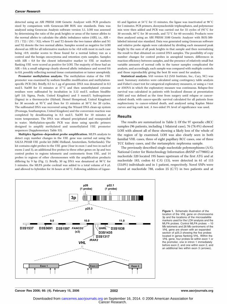
LOH analysis of 3p. Six highly polymorphic microsatellite markers
flanking the VHL gene (Fig. 1) were selected.8 Initially D3S1317 and D3S1597
were used for all samples and those found to be noninformative orequivocal were then also analyzed using further markers as necessary. The
marker, primer, and PCR conditions are provided in Supplementary Table
S2. Fluorescent PCR products were electrophoretically separated and
7 A. Harris, some unpublished data.
Table 1. Summary of the clinical characteristics of the 96patients with sporadic conventional RCC
Variables Number of patients
Sex 39 female/57 male
Age range, 38-86; median, 63 (y)Grade
1 6
2 323 34
4 24
Tumor
1a 191b 20
2 7
3a 23
3b 253c 0
4 2
Node
X 80 79
1 7
2 2Metastasis
0 74
1 22
StageI 38
II 5
III 31 (4 at least stage III)
IV 22
NOTE: Pathologic diagnosis is according to Fuhrman’s grading systemand UICC tumor-node-metastasis staging system. For the six tumors
in which accurate assessment of tumor stage was not possible, the
minimum T value is indicated.
8 http://www.gdb.org/ and http://www.ensembl.org.
VHL and Sporadic Renal Cancer
www.aacrjournals.org 2001 Cancer Res 2006; 66: (4). February 15, 2006
Cancer Research. on September 16, 2014. © 2006 American Association forcancerres.aacrjournals.org Downloaded from
detected using an ABI PRISM 3100 Genetic Analyzer with PCR productssized by comparison with Genescan-500 ROX size standards. Data was
analyzed using Genescan Analysis software version 3.1. LOH was assessed
by determining the ratio of the peak heights or areas of the tumor alleles to
the normal alleles to calculate the allelic imbalance ratios (AIR), i.e., AIR =(T1 / T2) / (N1 / N2), where T1 and T2 denote the two tumor alleles and N1
and N2 denote the two normal alleles. Samples scored as negative for LOH
showed an AIR for all informative markers to be >0.8 with most in each case
being >0.9, similar scores to those found for a normal kidney run as acontrol and the benign oncocytoma and retention cysts samples. Samples
with AIR < 0.8 for the closest informative marker to VHL or markers
flanking VHL were scored as positive for LOH. The majority of these had an
AIR < 0.6; a small subgroup clearly showed allelic imbalance and scored 0.6to 0.8, possibly reflecting normal tissue contamination or tumor aneuploidy.
Promoter methylation analysis. The methylation status of the VHL
promoter was examined by sodium bisulfite modification and methylation-specific PCR (36). Briefly, 0.5 to 1 Ag of genomic DNA was denatured in 0.3
mol/L NaOH for 15 minutes at 37jC and then unmethylated cytosine
residues were sulfonated by incubation in 3.12 mol/L sodium bisulfite
(pH 5.0; Sigma, Poole, United Kingdom) and 5 mmol/L hydroquinone(Sigma) in a thermocycler (Hybaid, Hemel Hempstead, United Kingdom)
for 30 seconds at 95jC and then for 15 minutes at 50jC for 20 cycles.
The sulfonated DNA was recovered using the Wizard DNA clean-up system
(Promega, Southampton, United Kingdom) and the conversion reaction wascompleted by desulfonating in 0.3 mol/L NaOH for 10 minutes at
room temperature. The DNA was ethanol precipitated and resuspended
in water. Methylation-specific PCR was done using specific primersdesigned to amplify methylated and unmethylated VHL promoter
sequences (Supplementary Table S3).
Multiplex ligation–dependent probe amplification. MLPA analysis to
detect copy number changes in the VHL gene was carried out using theSALSA P016B VHL probe kit (MRC-Holland, Amsterdam, Netherlands). The
kit contains eight probes to the VHL gene ( four in exon 1 and two in each of
exons 2 and 3), an additional five probes to three other genes on 3p and two
control probes to regions telomeric and centromeric from VHL , and 14probes to regions of other chromosomes with the amplification products
differing by 9 bp (Fig. 1). Briefly, 50 ng DNA was denatured at 98jC for
5 minutes, the MLPA probe cocktail was added to a total volume of 8 ALand allowed to hybridize for 16 hours at 60jC. Following addition of Ligase-
65 and ligation at 54jC for 15 minutes, the ligase was inactivated at 98jCfor 5 minutes. PCR primers, deoxynucleotide triphosphates, and polymerase
mix were then added and PCR was carried out for 33 cycles of (95jC for
30 seconds, 60jC for 30 seconds, and 72jC for 60 seconds). Products were
then analyzed using an ABI PRISM 3100 Genetic Analyzer with ROX-500-labeled internal size standard. Data was generated using Genescan software
and relative probe signals were calculated by dividing each measured peak
height by the sum of all peak heights in that sample and then normalizing
the result to that obtained on control DNA samples. The possibility of copynumber changes for control probes in aneuploid tumors, differences in
reaction efficiency between samples, and the presence of relatively small but
variable amounts of normal cells in the tumor samples complicated the
analysis, and accordingly, each sample was compared with multiple controlsand those reproducibly giving the best fit were used for analysis.
Statistical analysis. SAS version 8.2 (SAS Institute, Inc., Cary, NC) was
used. Summary statistics were calculated using contingency table analysisand Fisher’s exact test for categorical exploratory measures, or using a t test
or ANOVA in which the exploratory measure was continuous. Relapse-free
survival was calculated in patients with localized disease at presentation
(M0) and was defined as the time from surgery until relapse or cancer-related death, with cancer-specific survival calculated for all patients from
nephrectomy to cancer-related death, and analyzed using Kaplan Meier
curves and log-rank test. A two-sided 5% level of significance was used.
Results
The results are summarized in Table 2. Of the 97 sporadic cRCCsamples (96 patients, including 1 bilateral case), 76 (78.4%) showedLOH with almost all of these showing a likely loss of the whole ofthe region of 3p examined. LOH was also clearly seen in bothfamilial VHL cases, three of eight papillary RCC cases, one of threeTCC kidney cases, and the metanephric nephroma sample.The previously described single nucleotide polymorphisms (A/G;
National Center for Biotechnology Information dbSNP rs779805) atnucleotide 520 located 195 bases upstream of the first ATG and atnucleotide 183, codon 61 C/G (13), were detected in 61 of 115(53.0%) individuals and in 1 patient, respectively. Novel SNPs werefound at nucleotide 788, codon 25 (C/T) in two patients and at
Figure 1. Schematic illustration of thelocation of the VHL gene on chromosome3p and the locations of the microsatellitemarkers used for the LOH analysis and theMLPA probes. Control MLPA probes f10Mb telomeric and 26 Mb centromeric of theVHL gene are shown with an expandedsection of p25.3 showing the five probeslocated in genes flanking VHL. Within theVHL gene, four probes lie within exon 1 orthe promoter, one in intron 1 immediatelybefore exon 2, and one within exon 2, andan additional two within exon 3 (arrows ).
Cancer Research
Cancer Res 2006; 66: (4). February 15, 2006 2002 www.aacrjournals.org
Cancer Research. on September 16, 2014. © 2006 American Association forcancerres.aacrjournals.org Downloaded from
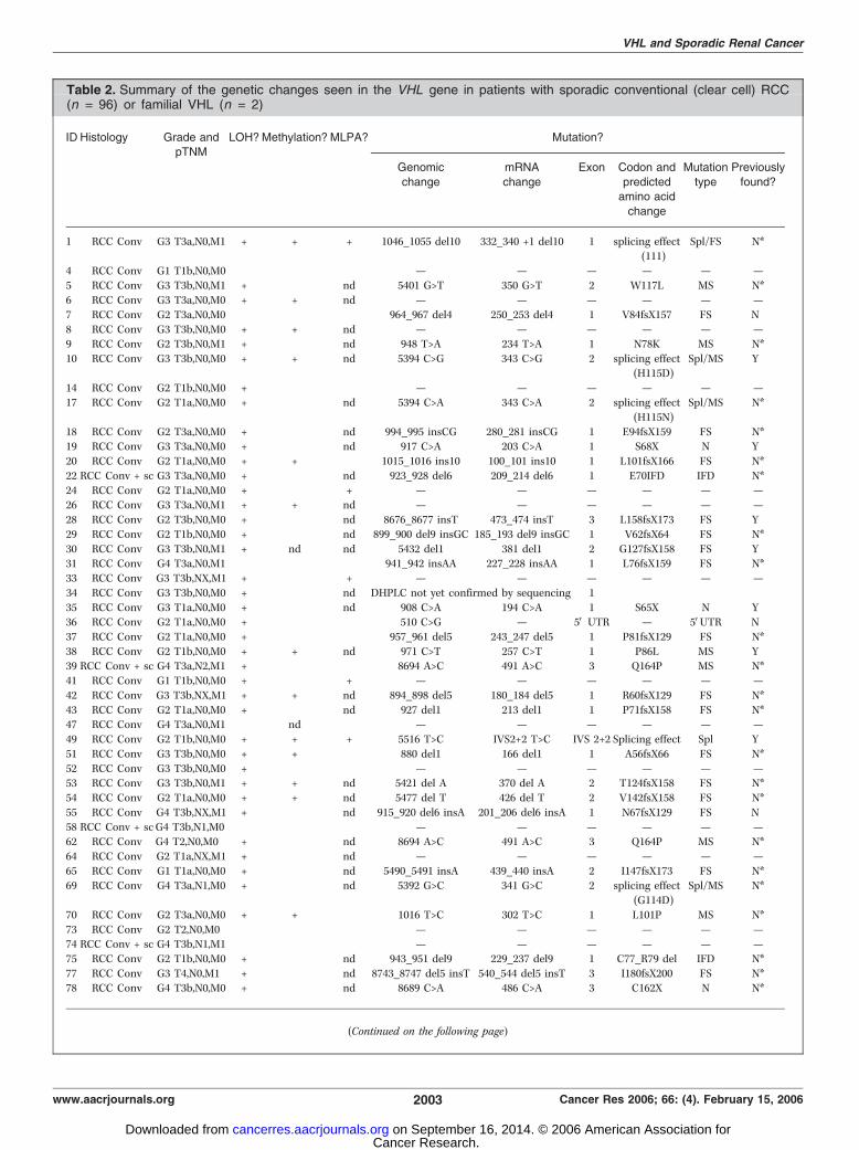
Table 2. Summary of the genetic changes seen in the VHL gene in patients with sporadic conventional (clear cell) RCC(n = 96) or familial VHL (n = 2)
ID Histology Grade andpTNM
LOH?Methylation? MLPA? Mutation?
Genomic
change
mRNA
change
Exon Codon and
predictedamino acid
change
Mutation
type
Previously
found?
1 RCC Conv G3 T3a,N0,M1 + + + 1046_1055 del10 332_340 +1 del10 1 splicing effect
(111)
Spl/FS N*
4 RCC Conv G1 T1b,N0,M0 � � � — — — — — —5 RCC Conv G3 T3b,N0,M1 + � nd 5401 G>T 350 G>T 2 W117L MS N*
6 RCC Conv G3 T3a,N0,M0 + + nd — — — — — —
7 RCC Conv G2 T3a,N0,M0 � � � 964_967 del4 250_253 del4 1 V84fsX157 FS N
8 RCC Conv G3 T3b,N0,M0 + + nd — — — — — —9 RCC Conv G2 T3b,N0,M1 + � nd 948 T>A 234 T>A 1 N78K MS N*
10 RCC Conv G3 T3b,N0,M0 + + nd 5394 C>G 343 C>G 2 splicing effect
(H115D)
Spl/MS Y
14 RCC Conv G2 T1b,N0,M0 + � � — — — — — —17 RCC Conv G2 T1a,N0,M0 + � nd 5394 C>A 343 C>A 2 splicing effect
(H115N)
Spl/MS N*
18 RCC Conv G2 T3a,N0,M0 + � nd 994_995 insCG 280_281 insCG 1 E94fsX159 FS N*19 RCC Conv G3 T3a,N0,M0 + � nd 917 C>A 203 C>A 1 S68X N Y
20 RCC Conv G2 T1a,N0,M0 + + � 1015_1016 ins10 100_101 ins10 1 L101fsX166 FS N*
22 RCC Conv + sc G3 T3a,N0,M0 + � nd 923_928 del6 209_214 del6 1 E70IFD IFD N*
24 RCC Conv G2 T1a,N0,M0 + � + — — — — — —26 RCC Conv G3 T3a,N0,M1 + + nd — — — — — —
28 RCC Conv G2 T3b,N0,M0 + � nd 8676_8677 insT 473_474 insT 3 L158fsX173 FS Y
29 RCC Conv G2 T1b,N0,M0 + � nd 899_900 del9 insGC 185_193 del9 insGC 1 V62fsX64 FS N*
30 RCC Conv G3 T3b,N0,M1 + nd nd 5432 del1 381 del1 2 G127fsX158 FS Y31 RCC Conv G4 T3a,N0,M1 � � � 941_942 insAA 227_228 insAA 1 L76fsX159 FS N*
33 RCC Conv G3 T3b,NX,M1 + � + — — — — — —
34 RCC Conv G3 T3b,N0,M0 + � nd DHPLC not yet confirmed by sequencing 1
35 RCC Conv G3 T1a,N0,M0 + � nd 908 C>A 194 C>A 1 S65X N Y36 RCC Conv G2 T1a,N0,M0 + � � 510 C>G — 5V UTR — 5VUTR N
37 RCC Conv G2 T1a,N0,M0 + � � 957_961 del5 243_247 del5 1 P81fsX129 FS N*
38 RCC Conv G2 T1b,N0,M0 + + nd 971 C>T 257 C>T 1 P86L MS Y39 RCC Conv + sc G4 T3a,N2,M1 + � � 8694 A>C 491 A>C 3 Q164P MS N*
41 RCC Conv G1 T1b,N0,M0 + � + — — — — — —
42 RCC Conv G3 T3b,NX,M1 + + nd 894_898 del5 180_184 del5 1 R60fsX129 FS N*
43 RCC Conv G2 T1a,N0,M0 + � nd 927 del1 213 del1 1 P71fsX158 FS N*47 RCC Conv G4 T3a,N0,M1 � nd � — — — — — —
49 RCC Conv G2 T1b,N0,M0 + + + 5516 T>C IVS2+2 T>C IVS 2+2 Splicing effect Spl Y
51 RCC Conv G3 T3b,N0,M0 + + � 880 del1 166 del1 1 A56fsX66 FS N*
52 RCC Conv G3 T3b,N0,M0 + � � — — — — — —53 RCC Conv G3 T3b,N0,M1 + + nd 5421 del A 370 del A 2 T124fsX158 FS N*
54 RCC Conv G2 T1a,N0,M0 + + nd 5477 del T 426 del T 2 V142fsX158 FS N*
55 RCC Conv G4 T3b,NX,M1 + � nd 915_920 del6 insA 201_206 del6 insA 1 N67fsX129 FS N58 RCC Conv + scG4 T3b,N1,M0 � � � — — — — — —
62 RCC Conv G4 T2,N0,M0 + � nd 8694 A>C 491 A>C 3 Q164P MS N*
64 RCC Conv G2 T1a,NX,M1 + � nd — — — — — —
65 RCC Conv G1 T1a,N0,M0 + � nd 5490_5491 insA 439_440 insA 2 I147fsX173 FS N*69 RCC Conv G4 T3a,N1,M0 + � nd 5392 G>C 341 G>C 2 splicing effect
(G114D)
Spl/MS N*
70 RCC Conv G2 T3a,N0,M0 + + � 1016 T>C 302 T>C 1 L101P MS N*
73 RCC Conv G2 T2,N0,M0 � � � — — — — — —74 RCC Conv + sc G4 T3b,N1,M1 � � � — — — — — —
75 RCC Conv G2 T1b,N0,M0 + � nd 943_951 del9 229_237 del9 1 C77_R79 del IFD N*
77 RCC Conv G3 T4,N0,M1 + � nd 8743_8747 del5 insT 540_544 del5 insT 3 I180fsX200 FS N*78 RCC Conv G4 T3b,N0,M0 + � nd 8689 C>A 486 C>A 3 C162X N N*
(Continued on the following page)
VHL and Sporadic Renal Cancer
www.aacrjournals.org 2003 Cancer Res 2006; 66: (4). February 15, 2006
Cancer Research. on September 16, 2014. © 2006 American Association forcancerres.aacrjournals.org Downloaded from
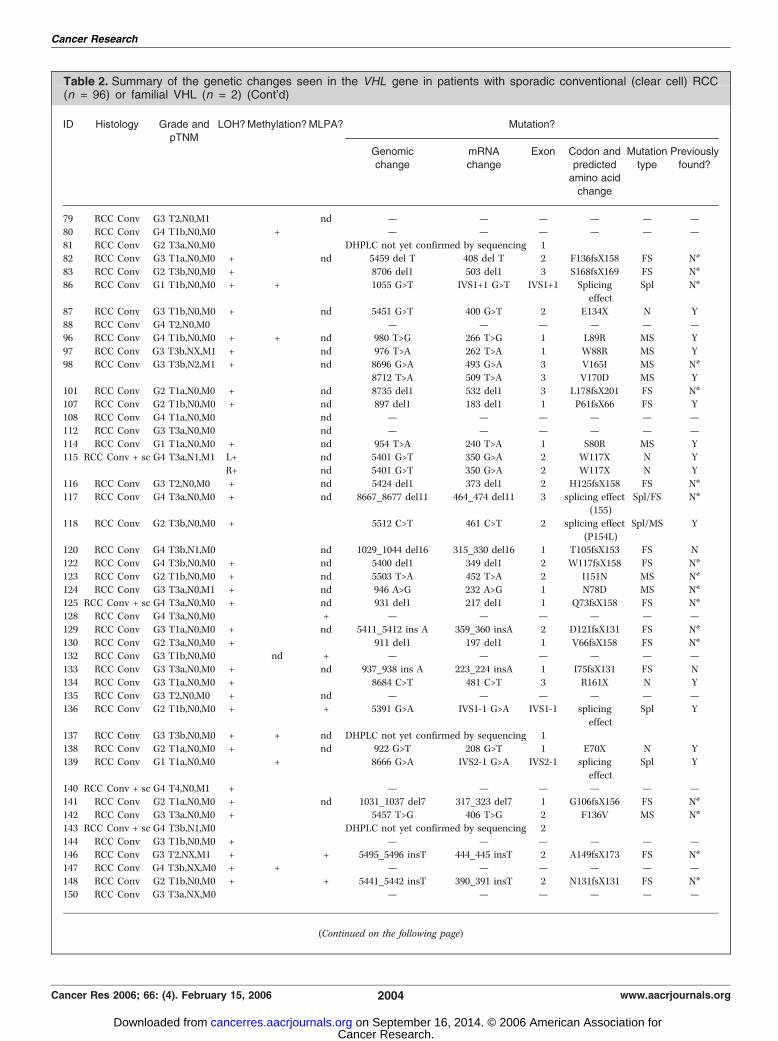
Table 2. Summary of the genetic changes seen in the VHL gene in patients with sporadic conventional (clear cell) RCC(n = 96) or familial VHL (n = 2) (Cont’d)
ID Histology Grade andpTNM
LOH?Methylation?MLPA? Mutation?
Genomic
change
mRNA
change
Exon Codon and
predictedamino acid
change
Mutation
type
Previously
found?
79 RCC Conv G3 T2,N0,M1 � � nd — — — — — —
80 RCC Conv G4 T1b,N0,M0 � + � — — — — — —
81 RCC Conv G2 T3a,N0,M0 � � � DHPLC not yet confirmed by sequencing 182 RCC Conv G3 T1a,N0,M0 + � nd 5459 del T 408 del T 2 F136fsX158 FS N*
83 RCC Conv G2 T3b,N0,M0 + � � 8706 del1 503 del1 3 S168fsX169 FS N*
86 RCC Conv G1 T1b,N0,M0 + + � 1055 G>T IVS1+1 G>T IVS1+1 Splicing
effect
Spl N*
87 RCC Conv G3 T1b,N0,M0 + � nd 5451 G>T 400 G>T 2 E134X N Y
88 RCC Conv G4 T2,N0,M0 � � � — — — — — —
96 RCC Conv G4 T1b,N0,M0 + + nd 980 T>G 266 T>G 1 L89R MS Y
97 RCC Conv G3 T3b,NX,M1 + � nd 976 T>A 262 T>A 1 W88R MS Y98 RCC Conv G3 T3b,N2,M1 + � nd 8696 G>A 493 G>A 3 V165I MS N*
8712 T>A 509 T>A 3 V170D MS Y
101 RCC Conv G2 T1a,N0,M0 + � nd 8735 del1 532 del1 3 L178fsX201 FS N*107 RCC Conv G2 T1b,N0,M0 + � nd 897 del1 183 del1 1 P61fsX66 FS Y
108 RCC Conv G4 T1a,N0,M0 � � nd — — — — — —
112 RCC Conv G3 T3a,N0,M0 � � nd — — — — — —
114 RCC Conv G1 T1a,N0,M0 + � nd 954 T>A 240 T>A 1 S80R MS Y115 RCC Conv + sc G4 T3a,N1,M1 L+ � nd 5401 G>T 350 G>A 2 W117X N Y
R+ � nd 5401 G>T 350 G>A 2 W117X N Y
116 RCC Conv G3 T2,N0,M0 + � nd 5424 del1 373 del1 2 H125fsX158 FS N*
117 RCC Conv G4 T3a,N0,M0 + � nd 8667_8677 del11 464_474 del11 3 splicing effect(155)
Spl/FS N*
118 RCC Conv G2 T3b,N0,M0 + � � 5512 C>T 461 C>T 2 splicing effect
(P154L)
Spl/MS Y
120 RCC Conv G4 T3b,N1,M0 � � nd 1029_1044 del16 315_330 del16 1 T105fsX153 FS N
122 RCC Conv G4 T3b,N0,M0 + � nd 5400 del1 349 del1 2 W117fsX158 FS N*
123 RCC Conv G2 T1b,N0,M0 + � nd 5503 T>A 452 T>A 2 I151N MS N*
124 RCC Conv G3 T3a,N0,M1 + � nd 946 A>G 232 A>G 1 N78D MS N*125 RCC Conv + sc G4 T3a,N0,M0 + � nd 931 del1 217 del1 1 Q73fsX158 FS N*
128 RCC Conv G4 T3a,N0,M0 � � + — — — — — —
129 RCC Conv G3 T1a,N0,M0 + � nd 5411_5412 ins A 359_360 insA 2 D121fsX131 FS N*
130 RCC Conv G2 T3a,N0,M0 + � � 911 del1 197 del1 1 V66fsX158 FS N*132 RCC Conv G3 T1b,N0,M0 � nd + — — — — — —
133 RCC Conv G3 T3a,N0,M0 + � nd 937_938 ins A 223_224 insA 1 I75fsX131 FS N
134 RCC Conv G3 T1a,N0,M0 + � � 8684 C>T 481 C>T 3 R161X N Y135 RCC Conv G3 T2,N0,M0 + � nd — — — — — —
136 RCC Conv G2 T1b,N0,M0 + � + 5391 G>A IVS1-1 G>A IVS1-1 splicing
effect
Spl Y
137 RCC Conv G3 T3b,N0,M0 + + nd DHPLC not yet confirmed by sequencing 1138 RCC Conv G2 T1a,N0,M0 + � nd 922 G>T 208 G>T 1 E70X N Y
139 RCC Conv G1 T1a,N0,M0 � + � 8666 G>A IVS2-1 G>A IVS2-1 splicing
effect
Spl Y
140 RCC Conv + sc G4 T4,N0,M1 + � � — — — — — —141 RCC Conv G2 T1a,N0,M0 + � nd 1031_1037 del7 317_323 del7 1 G106fsX156 FS N*
142 RCC Conv G3 T3a,N0,M0 + � � 5457 T>G 406 T>G 2 F136V MS N*
143 RCC Conv + sc G4 T3b,N1,M0 � � � DHPLC not yet confirmed by sequencing 2
144 RCC Conv G3 T1b,N0,M0 + � � — — — — — —146 RCC Conv G3 T2,NX,M1 + � + 5495_5496 insT 444_445 insT 2 A149fsX173 FS N*
147 RCC Conv G4 T3b,NX,M0 + + � — — — — — —
148 RCC Conv G2 T1b,N0,M0 + � + 5441_5442 insT 390_391 insT 2 N131fsX131 FS N*150 RCC Conv G3 T3a,NX,M0 � � � — — — — — —
(Continued on the following page)
Cancer Research
Cancer Res 2006; 66: (4). February 15, 2006 2004 www.aacrjournals.org
Cancer Research. on September 16, 2014. © 2006 American Association forcancerres.aacrjournals.org Downloaded from
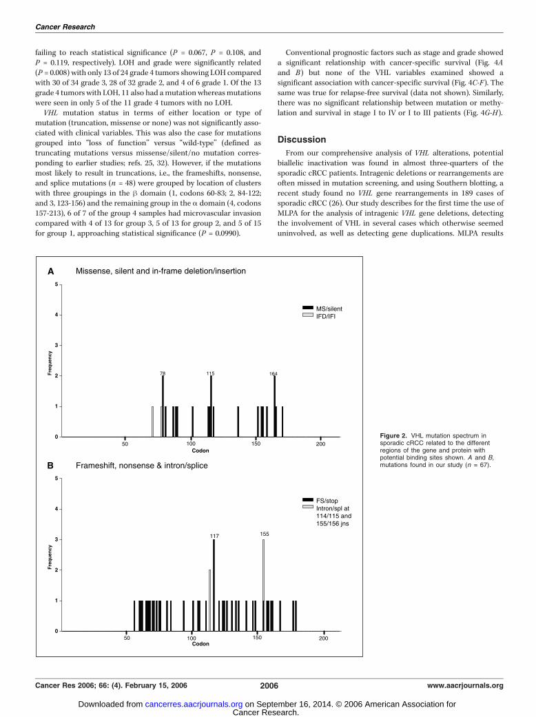
nucleotide 5557 (A/G) in the intron 43 bp 3V to the end of exon2 in 8 of 115 (7.0%) individuals, confirmed as polymorphisms bycomparison with constitutional DNA. VHL mutations were onlyseen in the sporadic and familial cRCC cases. Unequivocalmutation status was determined in 93 sporadic cRCC tumors,66 (71%) of which had mutations with 65 of these being singlemutations. An additional four tumors positive for DHPLC were notconfirmed by sequencing. The distribution of mutations is shownin Fig. 2A and B , illustrating the absence of mutations in the firsthalf of exon 1, 50% of the exonic mutations in the second half ofexon 1, 31% in exon 2, and only 19% in exon 3.Frameshifts accounted for 48.5% of mutations with 30.9% being
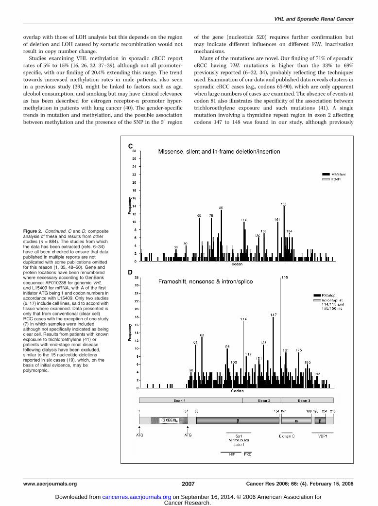
missense, 11.8% nonsense, 5.9% intronic, and 2.9% in-framedeletions. Ten mutations potentially affect exon splicing. Insertionsor deletions were more common in exon 1 and nucleotidesubstitutions were more frequent in exon 3. Approximately two-thirds of the mutations would be predicted to result in truncated orabsent proteins due either to frameshift or substitutions resultingin stop codons. Many of these mutations are novel, with noprevious descriptions either in the literature or VHL databases.Codons affected in >1 tumor (78, 115, 164, and 117) and mutationsaffecting splicing were also among those previously reportedin sporadic cRCC and featured as major or moderate ‘‘hotspots’’(Fig. 2A-D). Other than these, particular clusters of frameshift/nonsense mutations were apparent in codons 60 to 62, 65 to 68,and 70 to 76, which also match the pattern of mutations seen inprevious studies (n = 886) when collated (Fig. 2A-D).MLPA analysis was carried out on 41 sporadic cRCC cases, 11 of
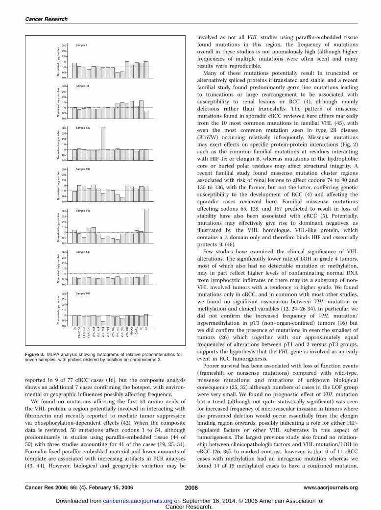
which had intragenic deletions/duplications (Fig. 3; Table 2).Deletion of all or part of 3p in a diploid background was found infive cases (samples 24, 41, 49, 146, and 148). In sample 149, thedetected mutation falls on the ligation point of one of the MLPAprobes resulting in clear underrepresentation. Sample 128 showedrelative 3p loss by MPLA without corresponding LOH, which maybe due to the masking effects of normal DNA or may represent twocopies of 3p in a triploid cellular background. Sample 33 is
subtriploid and showed probable loss of two identical regions in 3p,suggesting that chromosome duplication followed the deletionevent. In contrast, sample 1 contained three copies of 3p with twononidentical deletions, so that in this case, at least one deletionevent followed duplication. Sample 136 with three copies of partsof 3p, two copies of other parts, and for some probes, just one copy,may be explained by a deletion and nonreciprocal translocationwith a breakpoint within exon 1 of VHL. Sample 132 shows a >3-fold duplication of a small region of exon 1 which can’t beexplained by simple translocations and at least two breakpoints arelikely to be involved. Importantly, the changes detected by MLPA insamples 128 and 132 were found in samples which otherwise wouldhave been assessed as having no disruption of the VHL gene.Promoter methylation was shown in 19 of 93 (20.4%) sporadic
cRCC samples examined and in one of three TCC kidney, fourof eight papillary RCC, and one of two unclassified RCC samples.LOH or mutation were present in all but one of the methylatedcRCC cases. Of the 75 cRCC tumors with LOH, 61 also had aconfirmed mutation of which 11 were also positive for methylation.Therefore, of the 93 samples for which sufficient information wasavailable, 69 (74.2%) showed evidence of biallelic inactivation ofVHL . A total of 10 samples (11.5%) of the 87 for whom LOH,methylation analysis, and confirmed mutation status was availableshowed no evidence of any alteration of the VHL gene, although notall of these were examined by MLPA to rule out intragenic deletions.No significant differences were seen between the presence/
absence of methylation, LOH, mutation, or potential biallelicinactivation when examined with regard to age, grade, stage (pTNMindividual categories or groupings), maximum tumor diameter, orthe presence of microvascular invasion. Possible associationsbetween methylation and gender (15 of 19 methylated samplesoccurred in male patients, i.e., 27.3% of male patients comparedwith10.5% of female patients), mutation and gender (31 of 39 femalepatients had a mutation compared with 33 of 53 males), andmethylation and the A>G SNP at nucleotide 520 were seen (14 of 19methylated cases being associated with the SNP), although just
Table 2. Summary of the genetic changes seen in the VHL gene in patients with sporadic conventional (clear cell) RCC(n = 96) or familial VHL (n = 2) (Cont’d)
ID Histology Grade andpTNM
LOH?Methylation?MLPA? Mutation?
Genomic
change
mRNA
change
ExonCodon and
predictedamino acid
change
Mutation
type
Previously
found?
151 RCC Conv + sc G4 T1b,N0,M0 � � � 8676 T>A 473 T>A 3 L158Q MS Y
153 RCC Conv G2 T1b,N0,M0 � � nd — — — — — —
155 RCC Conv G2 T1b,N0,M0 + � nd 8678_8683 del6 insT 475_480 del6 insT 3 K159fsX171 FS N*21 RCC familial VHL G1 T1a,N0,M0 + nd � 942_943 insC 228_229 insC 1 C77fsX131 FS N*
149 RCC familial VHL G2 T2,N0,M0 + � + 943_944 insC 228_229 insC 1 C77fsX131 FS N*
NOTE: Mutation nomenclature is in accordance with recent recommendations (47). All nucleotide numbering is in accord with the GenBank sequence
AF010238 for genomic VHL and L15409 for mRNA with A of the first initiator ATG being 1. Codon numbers are in accordance with L15409. Previous
description of the mutation in HGMD or Necker VHL mutations database was ascertained using the web sites http://www.hgmd.org and http://www.umd.be:2020 (N* indicates that the precise mutation wasn’t seen but that a mutation of the same type involving the same codon or intronic
nucleotide was reported) and potential effects of mutations on splicing were checked using http://www.fruitfly.org/seq_tools/splice.html. Conv,
conventional; sc, plus sarcomatoid change; nt, nucleotides; del, deletion; ins, insertion; MS, missense; FS, frameshift; N, nonsense; Spl, splice error;
S, silent error; IFD, in-frame deletion; nd, not determined.
VHL and Sporadic Renal Cancer
www.aacrjournals.org 2005 Cancer Res 2006; 66: (4). February 15, 2006
Cancer Research. on September 16, 2014. © 2006 American Association forcancerres.aacrjournals.org Downloaded from
failing to reach statistical significance (P = 0.067, P = 0.108, andP = 0.119, respectively). LOH and grade were significantly related(P = 0.008) with only 13 of 24 grade 4 tumors showing LOH comparedwith 30 of 34 grade 3, 28 of 32 grade 2, and 4 of 6 grade 1. Of the 13grade 4 tumors with LOH, 11 also had amutation whereasmutationswere seen in only 5 of the 11 grade 4 tumors with no LOH.VHL mutation status in terms of either location or type of
mutation (truncation, missense or none) was not significantly asso-ciated with clinical variables. This was also the case for mutationsgrouped into ‘‘loss of function’’ versus ‘‘wild-type’’ (defined astruncating mutations versus missense/silent/no mutation corres-ponding to earlier studies; refs. 25, 32). However, if the mutationsmost likely to result in truncations, i.e., the frameshifts, nonsense,and splice mutations (n = 48) were grouped by location of clusterswith three groupings in the h domain (1, codons 60-83; 2, 84-122;and 3, 123-156) and the remaining group in the a domain (4, codons157-213), 6 of 7 of the group 4 samples had microvascular invasioncompared with 4 of 13 for group 3, 5 of 13 for group 2, and 5 of 15for group 1, approaching statistical significance (P = 0.0990).
Conventional prognostic factors such as stage and grade showeda significant relationship with cancer-specific survival (Fig. 4Aand B) but none of the VHL variables examined showed asignificant association with cancer-specific survival (Fig. 4C-F). Thesame was true for relapse-free survival (data not shown). Similarly,there was no significant relationship between mutation or methy-lation and survival in stage I to IV or I to III patients (Fig. 4G-H).
Discussion
From our comprehensive analysis of VHL alterations, potentialbiallelic inactivation was found in almost three-quarters of thesporadic cRCC patients. Intragenic deletions or rearrangements areoften missed in mutation screening, and using Southern blotting, arecent study found no VHL gene rearrangements in 189 cases ofsporadic cRCC (26). Our study describes for the first time the use ofMLPA for the analysis of intragenic VHL gene deletions, detectingthe involvement of VHL in several cases which otherwise seemeduninvolved, as well as detecting gene duplications. MLPA results
Figure 2. VHL mutation spectrum insporadic cRCC related to the differentregions of the gene and protein withpotential binding sites shown. A and B,mutations found in our study (n = 67).
Cancer Research
Cancer Res 2006; 66: (4). February 15, 2006 2006 www.aacrjournals.org
Cancer Research. on September 16, 2014. © 2006 American Association forcancerres.aacrjournals.org Downloaded from
overlap with those of LOH analysis but this depends on the regionof deletion and LOH caused by somatic recombination would notresult in copy number change.Studies examining VHL methylation in sporadic cRCC report
rates of 5% to 15% (16, 26, 32, 37–39), although not all promoter-specific, with our finding of 20.4% extending this range. The trendtowards increased methylation rates in male patients, also seenin a previous study (39), might be linked to factors such as age,alcohol consumption, and smoking but may have clinical relevanceas has been described for estrogen receptor-a promoter hyper-methylation in patients with lung cancer (40). The gender-specifictrends in mutation and methylation, and the possible associationbetween methylation and the presence of the SNP in the 5V region
of the gene (nucleotide 520) requires further confirmation butmay indicate different influences on different VHL inactivationmechanisms.Many of the mutations are novel. Our finding of 71% of sporadic
cRCC having VHL mutations is higher than the 33% to 69%previously reported (6–32, 34), probably reflecting the techniquesused. Examination of our data and published data reveals clusters insporadic cRCC cases (e.g., codons 65-90), which are only apparentwhen large numbers of cases are examined. The absence of events atcodon 81 also illustrates the specificity of the association betweentrichloroethylene exposure and such mutations (41). A singlemutation involving a thymidine repeat region in exon 2 affectingcodons 147 to 148 was found in our study, although previously
Figure 2. Continued. C and D, compositeanalysis of these and results from otherstudies (n = 884). The studies from whichthe data has been extracted (refs. 6–34)have all been checked to ensure that datapublished in multiple reports are notduplicated with some publications omittedfor this reason (1, 35, 48–50). Gene andprotein locations have been renumberedwhere necessary according to GenBanksequence: AF010238 for genomic VHLand L15409 for mRNA, with A of the firstinitiator ATG being 1 and codon numbers inaccordance with L15409. Only two studies(6, 17) include cell lines, said to accord withtissue where examined. Data presented isonly that from conventional (clear cell)RCC cases with the exception of one study(7) in which samples were includedalthough not specifically indicated as beingclear cell. Results from patients with knownexposure to trichloroethylene (41) orpatients with end-stage renal diseasefollowing dialysis have been excluded,similar to the 15 nucleotide deletionsreported in six cases (19), which, on thebasis of initial evidence, may bepolymorphic.
VHL and Sporadic Renal Cancer
www.aacrjournals.org 2007 Cancer Res 2006; 66: (4). February 15, 2006
Cancer Research. on September 16, 2014. © 2006 American Association forcancerres.aacrjournals.org Downloaded from
reported in 9 of 77 cRCC cases (16), but the composite analysisshows an additional 7 cases confirming the hotspot, with environ-mental or geographic influences possibly affecting frequency.We found no mutations affecting the first 53 amino acids of
the VHL protein, a region potentially involved in interacting withfibronectin and recently reported to mediate tumor suppressionvia phosphorylation-dependent effects (42). When the compositedata is reviewed, 50 mutations affect codons 1 to 54, althoughpredominantly in studies using paraffin-embedded tissue (44 of50) with three studies accounting for 41 of the cases (19, 25, 34).Formalin-fixed paraffin-embedded material and lower amounts oftemplate are associated with increasing artifacts in PCR analyses(43, 44). However, biological and geographic variation may be
involved as not all VHL studies using paraffin-embedded tissuefound mutations in this region, the frequency of mutationsoverall in these studies is not anomalously high (although higherfrequencies of multiple mutations were often seen) and manyresults were reproducible.Many of these mutations potentially result in truncated or
alternatively spliced proteins if translated and stable, and a recentfamilial study found predominantly germ line mutations leadingto truncations or large rearrangement to be associated withsusceptibility to renal lesions or RCC (4), although mainlydeletions rather than frameshifts. The pattern of missensemutations found in sporadic cRCC reviewed here differs markedlyfrom the 10 most common mutations in familial VHL (45), witheven the most common mutation seen in type 2B disease(R167W) occurring relatively infrequently. Missense mutationsmay exert effects on specific protein-protein interactions (Fig. 2)such as the common familial mutations at residues interactingwith HIF-1a or elongin B, whereas mutations in the hydrophobiccore or buried polar residues may affect structural integrity. Arecent familial study found missense mutation cluster regionsassociated with risk of renal lesions to affect codons 74 to 90 and130 to 136, with the former, but not the latter, conferring geneticsusceptibility to the development of RCC (4) and affecting thesporadic cases reviewed here. Familial missense mutationsaffecting codons 65, 128, and 167 predicted to result in loss ofstability have also been associated with cRCC (5). Potentially,mutations may effectively give rise to dominant negatives, asillustrated by the VHL homologue, VHL-like protein, whichcontains a h domain only and therefore binds HIF and essentiallyprotects it (46).Few studies have examined the clinical significance of VHL
alterations. The significantly lower rate of LOH in grade 4 tumors,most of which also had no detectable mutation or methylation,may in part reflect higher levels of contaminating normal DNAfrom lymphocytic infiltrates or there may be a subgroup of non-VHL involved tumors with a tendency to higher grade. We foundmutations only in cRCC, and in common with most other studies,we found no significant association between VHL mutation ormethylation and clinical variables (12, 24–26 34). In particular, wedid not confirm the increased frequency of VHL mutation/hypermethylation in pT3 (non–organ-confined) tumors (16) butwe did confirm the presence of mutations in even the smallest oftumors (26) which together with our approximately equalfrequencies of alterations between pT1 and 2 versus pT3 groups,supports the hypothesis that the VHL gene is involved as an earlyevent in RCC tumorigenesis.Poorer survival has been associated with loss of function events
( frameshift or nonsense mutations) compared with wild-type,missense mutations, and mutations of unknown biologicalconsequence (25, 32) although numbers of cases in the LOF groupwere very small. We found no prognostic effect of VHL mutationbut a trend (although not quite statistically significant) was seenfor increased frequency of microvascular invasion in tumors wherethe presumed deletion would occur essentially from the elonginbinding region onwards, possibly indicating a role for either HIF-regulated factors or other VHL substrates in this aspect oftumorigenesis. The largest previous study also found no relation-ship between clinicopathologic factors and VHL mutation/LOH incRCC (26, 35). In marked contrast, however, is that 0 of 11 cRCCcases with methylation had an intragenic mutation whereas wefound 14 of 19 methylated cases to have a confirmed mutation,
Figure 3. MLPA analysis showing histograms of relative probe intensities forseven samples, with probes ordered by position on chromosome 3.
Cancer Research
Cancer Res 2006; 66: (4). February 15, 2006 2008 www.aacrjournals.org
Cancer Research. on September 16, 2014. © 2006 American Association forcancerres.aacrjournals.org Downloaded from
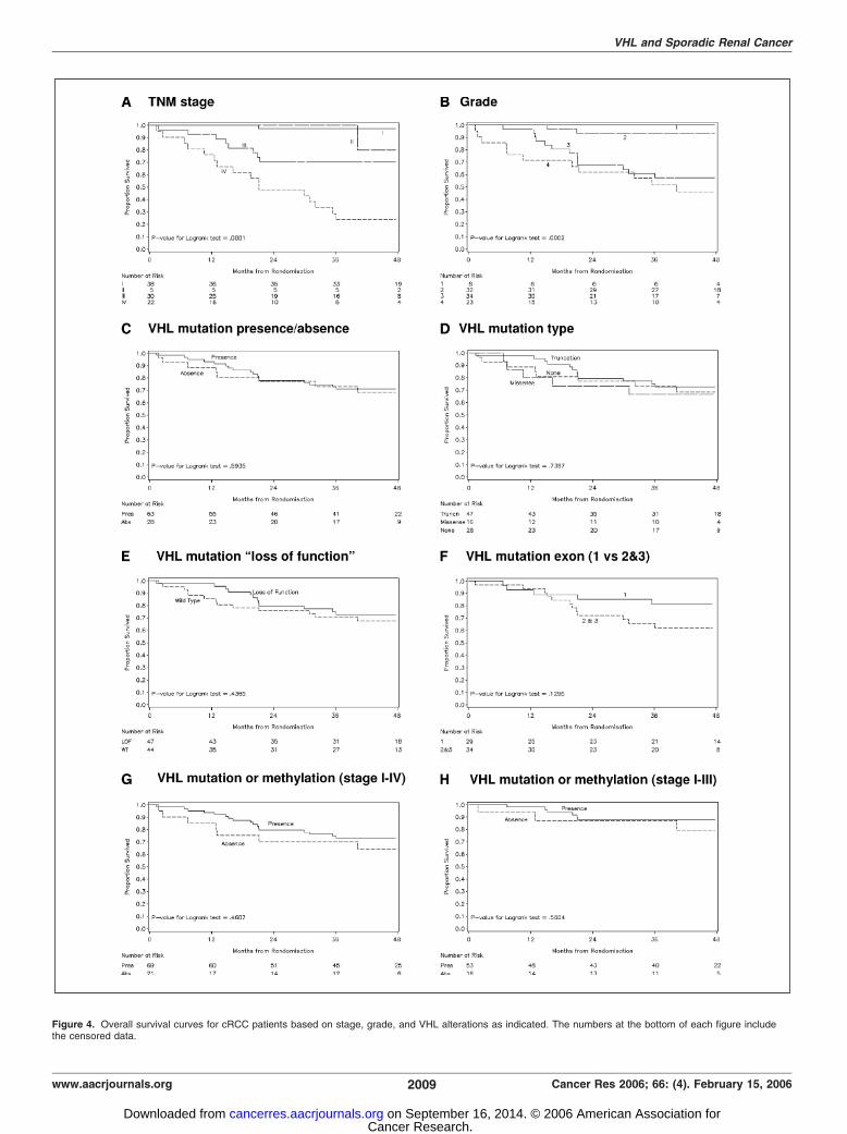
Figure 4. Overall survival curves for cRCC patients based on stage, grade, and VHL alterations as indicated. The numbers at the bottom of each figure includethe censored data.
VHL and Sporadic Renal Cancer
www.aacrjournals.org 2009 Cancer Res 2006; 66: (4). February 15, 2006
Cancer Research. on September 16, 2014. © 2006 American Association forcancerres.aacrjournals.org Downloaded from
although their mutation and methylation frequencies overall weremuch lower (51% and 5.4%, respectively). However, a significantly
better cancer-free and overall survival was seen in patients with
stage I to III cancer with VHL alteration (mutation or methylation)but wasn’t seen in stage IV disease (35). Our data fails to confirm
this although when stage IV patients were included a similar trend
was seen. Differences between studies might reflect samples sizes,
length of follow-up, and postoperative treatment.The involvement of VHL in cRCC is complex and warrants
further large studies to clarify the potential clinical implications.This may aid prognosis, define subgroups of patients for specific
targeted therapies, and patients where other genetic pathways may
be involved. Given the uncertainty of the effects of mutations or
methylation and whether single mutation events alone could resultin haploinsufficiency, functional insight should be sought fromlinking such results with analysis of the resultant form(s), level andsubcellular location of VHL protein and downstream pathways.
Acknowledgments
Received 8/29/2005; revised 11/14/2005; accepted 12/14/2005.Grant support: Cancer Research UK is gratefully acknowledged.The costs of publication of this article were defrayed in part by the payment of page
charges. This article must therefore be hereby marked advertisement in accordancewith 18 U.S.C. Section 1734 solely to indicate this fact.
We are also grateful to Jo Robinson for help in tissue banking, Elizabeth Butler forassistance with DHPLC, the members of staff of the Departments of Urology, MedicalOncology, and Pathology at St. James’s University Hospital and the patients whoparticipated in the study.
References1. Latif F, Tory K, Gnarra J, et al. Identification of the vonHippel-Lindau disease tumor suppressor gene. Science1993;260:1317–20.
2. Kim WY, Kaelin WG. Role of VHL gene mutation inhuman cancer. J Clin Oncol 2004;22:4991–5004.
3. Maher ER. von Hippel-Lindau disease. Curr Mol Med2004;4:833–42.
4. Gallou C, Chauveau D, Richard S, et al. Genotype-phenotype correlation in von Hippel-Lindau familieswith renal lesions. Hum Mutat 2004;24:215–24.
5. Ruiz-Llorente S, Bravo J, Cebrian A, et al. Geneticcharacterization and structural analysis of VHL Spanishfamilies to define genotype-phenotype correlations.Hum Mutat 2004;23:160–9.
6. Gnarra JR, Tory K, Weng Y, et al. Mutations of the VHLtumour suppressor gene in renal carcinoma. Nat Genet1994;7:85–90.
7. Whaley JM, Naglich J, Gelbert L, et al. Germ-linemutations in the von Hippel-Lindau tumor-suppressorgene are similar to somatic von Hippel-Lindau aberra-tions in sporadic renal cell carcinoma. Am J Hum Genet1994;55:1092–102.
8. Foster K, Prowse A, van den BA, et al. Somaticmutations of the von Hippel-Lindau disease tumoursuppressor gene in non-familial clear cell renal carci-noma. Hum Mol Genet 1994;3:2169–73.
9. Shuin T, Kondo K, Torigoe S, et al. Frequent somaticmutations and loss of heterozygosity of the von Hippel-Lindau tumor suppressor gene in primary human renalcell carcinomas. Cancer Res 1994;54:2852–5.
10. Bailly M, Bain C, Favrot MC, Ozturk M. Somaticmutations of von Hippel-Lindau (VHL) tumor-suppressorgene in European kidney cancers. Int J Cancer 1995;63:660–4.
11. Kenck C, Wilhelm M, Bugert P, Staehler G, Kovacs G.Mutation of the VHL gene is associated exclusively withthe development of non-papillary renal cell carcinomas.J Pathol 1996;179:157–61.
12. Suzuki H, Ueda T, Komiya A, et al. Mutationalstate of von Hippel-Lindau and adenomatous poly-posis coli genes in renal tumors. Oncology 1997;54:252–7.
13. Gallou C, Joly D, Mejean A, et al. Mutations of theVHL gene in sporadic renal cell carcinoma: definition ofa risk factor for VHL patients to develop an RCC. HumMutat 1999;13:464–75.
14. Lemm I, Lingott A, Strandmann E, et al. Loss ofHNF1a function in human renal cell carcinoma:frequent mutations in the VHL gene but not the HNF1agene. Mol Carcinog 1999;24:305–14.
15. Ashida S, Okuda H, Chikazawa M, et al. Detection ofcirculating cancer cells with von Hippel-Lindau genemutation in peripheral blood of patients with renal cellcarcinoma. Clin Cancer Res 2000;6:3817–22.
16. Brauch H, Weirich G, Brieger J, et al. VHL alterationsin human clear cell renal cell carcinoma: associationwith advanced tumor stage and a novel hot spotmutation. Cancer Res 2000;60:1942–8.
17. Meyer AJ, Hernandez A, Florl AR, et al. Novelmutations of the von Hippel-Lindau tumor-suppressorgene and rare DNA hypermethylation in renal-cellcarcinoma cell lines of the clear-cell type. Int J Cancer2000;87:650–3.
18. Gallou C, Longuemaux S, Delomenie C, et al.Association of GSTT1 non-null and NAT1 slow/rapidgenotypes with von Hippel-Lindau tumour suppressorgene transversions in sporadic renal cell carcinoma.Pharmacogenetics 2001;11:521–35.
19. Ma X, Yang K, Lindblad P, Egevad L, Hemminki K.VHL gene alterations in renal cell carcinoma patients:novel hotspot or founder mutations and linkagedisequilibrium. Oncogene 2001;20:5393–400.
20. Wiesener MS, Seyfarth M, Warnecke C, et al.Paraneoplastic erythrocytosis associated with aninactivating point mutation of the von Hippel-Lindaugene in a renal cell carcinoma. Blood 2002;99:3562–5.
21. Oh RR, Park JY, Lee JH, et al. Expression of HGF/SFandMet protein is associated with genetic alterations of VHLgene in primary renal cell carcinomas. APMIS 2002;110:229–38.
22. Igarashi H, Esumi M, Ishida H, Okada K. Vascularendothelial growth factor overexpression is correlatedwith von Hippel-Lindau tumor suppressor gene inacti-vation in patients with sporadic renal cell carcinoma.Cancer 2002;95:47–53.
23. Barnabas N, Amin MB, Pindolia K, Nanavati R,Amin MB, Worsham MJ. Mutations in the von Hippel-Lindau (VHL) gene refine differential diagnosticcriteria in renal cell carcinoma. J Surg Oncol 2002;80:52–60.
24. Hamano K, Esumi M, Igarashi H, et al. Biallelicinactivation of the von Hippel-Lindau tumor suppressorgene in sporadic renal cell carcinoma. J Urol 2002;167:713–7.
25. Schraml P, Struckmann K, Hatz F, et al. VHLmutations and their correlation with tumour cellproliferation, microvessel density, and patient progno-sis in clear cell renal cell carcinoma. J Pathol 2002;196:186–93.
26. Kondo K, Yao M, Yoshida M, et al. Comprehensivemutational analysis of the VHL gene in sporadicrenal cell carcinoma: relationship to clinicopatholog-ical parameters. Genes Chromosomes Cancer 2002;34:58–68.
27. Ashida S, Furihata M, Tanimura M, et al. Moleculardetection of von Hippel-Lindau gene mutations inurine and lymph node samples in patients with renalcell carcinoma: potential biomarkers for early diagno-sis and postoperative metastatic status. J Urol 2003;169:2089–93.
28. Hughson MD, He Z, Liu S, Coleman J, Shingleton WB.Expression of HIF-1 and ubiquitin in conventional renalcell carcinoma: relationship to mutations of the vonHippel-Lindau tumor suppressor gene. Cancer GenetCytogenet 2003;143:145–53.
29. Na X, Wu G, Ryan CK, Schoen SR, di’Santagnese PA,Messing EM. Overproduction of vascular endothelial
growth factor related to von Hippel-Lindau tumorsuppressor gene mutations and hypoxia-induciblefactor-1a expression in renal cell carcinomas. J Urol2003;170:588–92.
30. He Z, Liu S, Guo M, Mao J, Hughson MD.Expression of fibronectin and HIF-1a in renal cellcarcinomas: relationship to von Hippel-Lindau geneinactivation. Cancer Genet Cytogenet 2004;152:89–94.
31. Brauch H, Weirich G, Klein B, Rabstein S, BoltHM, Bruning T. VHL mutations in renal cellcancer: does occupational exposure to trichloro-ethylene make a difference? Toxicol Lett 2004;151:301–10.
32. Kim JH, Jung CW, Cho YH, et al. Somatic VHLalteration and its impact on prognosis in patientswith clear cell renal cell carcinoma. Oncol Rep 2005;13:859–64.
33. Brieger J, Weidt EJ, Gansen K, Decker HJ. Detection ofa novel germline mutation in the von Hippel-Lindautumour-suppressor gene by fluorescence-labelled baseexcision sequence scanning (F-BESS). Clin Genet1999;56:210–5.
34. van Houwelingen KP, Van Dijk BA, Hulsbergen-vande Kaa CA, et al. Prevalence of von Hippel-Lindau genemutations in sporadic renal cell carcinoma: resultsfrom the Netherlands cohort study. BMC Cancer 2005;5:57.
35. Yao M, Yoshida M, Kishida T, et al. VHL tumorsuppressor gene alterations associated with goodprognosis in sporadic clear-cell renal carcinoma. J NatlCancer Inst 2002;94:1569–75.
36. Herman JG, Graff JR, Myohanen S, Nelkin BD, BaylinSB. Methylation-specific PCR: a novel PCR assay formethylation status of CpG islands. Proc Natl Acad SciU S A 1996;93:9821–6.
37. Clifford SC, Prowse AH, Affara NA, Buys CH, MaherER. Inactivation of the von Hippel-Lindau (VHL)tumour suppressor gene and allelic losses at chromo-some arm 3p in primary renal cell carcinoma: evidencefor a VHL-independent pathway in clear cell renaltumourigenesis. Genes Chromosomes Cancer 1998;22:200–9.
38. Herman JG, Latif F, Weng Y, et al. Silencing of theVHL tumor-suppressor gene by DNA methylation inrenal carcinoma. Proc Natl Acad Sci U S A 1994;91:9700–4.
39. Dulaimi E, De Caceres II, Uzzo RG, et al. Promoterhypermethylation profile of kidney cancer. Clin CancerRes 2004;10:3972–9.
40. Lai JC, Cheng YW, Chiou HL, Wu MF, Chen CY,Lee H. Gender difference in estrogen receptor a
promoter hypermethylation and its prognostic valuein non-small cell lung cancer. Int J Cancer 2005;117:974–80.
41. Brauch H, Weirich G, Hornauer MA, Storkel S, WohlT, Bruning T. Trichloroethylene exposure and specificsomatic mutations in patients with renal cell carcinoma.J Natl Cancer Inst 1999;91:854–61.
42. Lolkema MP, Gervais ML, Snijckers CM, et al. Tumor
Cancer Research
Cancer Res 2006; 66: (4). February 15, 2006 2010 www.aacrjournals.org
Cancer Research. on September 16, 2014. © 2006 American Association forcancerres.aacrjournals.org Downloaded from
suppression by the von Hippel-Lindau protein requiresphosphorylation of the acidic domain. J Biol Chem2005;280:22205–11.
43. Williams C, Ponten F, Moberg C, et al. A highfrequency of sequence alterations is due to formalinfixation of archival specimens. Am J Pathol 1999;155:1467–71.
44. Akbari M, Hansen MD, Halgunset J, Skorpen F,Krokan HE. Low copy number DNA template canrender polymerase chain reaction error prone in asequence-dependent manner. J Mol Diagn 2005;7:36–9.
45. Barry RE, Krek W. The von Hippel-Lindau tumoursuppressor: a multi-faceted inhibitor of tumourigenesis.Trends Mol Med 2004;10:466–72.
46. Qi H, Gervais ML, Li W, DeCaprio JA, Challis JR,Ohh M. Molecular cloning and characterization of thevon Hippel-Lindau-like protein. Mol Cancer Res 2004;2:43–52.
47. den Dunnen JT, Antonarakis SE. Nomenclature forthe description of human sequence variations. HumGenet 2001;109:121–4.
48. Zhuang Z, Gnarra JR, Dudley CF, Zbar B, LinehanWM, Lubensky IA. Detection of von Hippel-Lindau
disease gene mutations in paraffin-embedded sporadicrenal cell carcinoma specimens. Mod Pathol 1996;9:838–42.
49. Yang K, Lindblad P, Egevad L, Hemminki K. Novelsomatic mutations in the VHL gene in Swedisharchived sporadic renal cell carcinomas. Cancer Lett1999;141:1–8.
50. Ashida S, Nishimori I, Tanimura M, Onishi S, Shuin T.Effects of von Hippel-Lindau gene mutation andmethylation status on expression of transmembranecarbonic anhydrases in renal cell carcinoma. J CancerRes Clin Oncol 2002;128:561–8.
VHL and Sporadic Renal Cancer
www.aacrjournals.org 2011 Cancer Res 2006; 66: (4). February 15, 2006
Cancer Research. on September 16, 2014. © 2006 American Association forcancerres.aacrjournals.org Downloaded from
Related Documents


![Von Hippel Lindau Disease [VHL]: Magnetic Resonance ...inaactamedica.org/archives/2012/23314974.pdf · Von Hippel Lindau Disease [VHL]: Magnetic Resonance Imaging Spectrum ... Artikel](https://static.cupdf.com/doc/110x72/5c79e5d509d3f2a9708bea51/von-hippel-lindau-disease-vhl-magnetic-resonance-von-hippel-lindau-disease.jpg)